Embed Size (px)
Citation preview

nutrients
Article
Curcumin Anti-Apoptotic Action in a Model ofIntestinal Epithelial Inflammatory Damage
Claudia Loganes 1, Sara Lega 2, Matteo Bramuzzo 1, Liza Vecchi Brumatti 1, Elisa Piscianz 2,Erica Valencic 1, Alberto Tommasini 1 and Annalisa Marcuzzi 2,*
1 Department of Paediatrics, Institute for Maternal and Child Health, IRCCS Burlo Garofolo,Via dell’Istria 65/1, Trieste 34137, Italy; [email protected] (C.L.);[email protected] (M.B.); [email protected] (L.V.B.);[email protected] (E.V.); [email protected] (A.T.)
2 Department of Medicine, Surgery, and Health Sciences, University of Trieste, Strada di Fiume, 447,Trieste 34100, Italy; [email protected] (S.L.); [email protected] (E.P.)
* Correspondence: [email protected]; Tel.: +39-040-3785422
Received: 24 April 2017; Accepted: 1 June 2017; Published: 6 June 2017
Abstract: The purpose of this study is to determine if a preventive treatment with curcumin canprotect intestinal epithelial cells from inflammatory damage induced by IFNγ. To achieve this goalwe have used a human intestinal epithelial cell line (HT29) treated with IFNγ to undergo apoptoticchanges that can reproduce the damage of intestinal epithelia exposed to inflammatory cytokines.In this model, we measured the effect of curcumin (curcuminoid from Curcuma Longa) added as apre-treatment at different time intervals before stimulation with IFNγ. Curcumin administrationto HT29 culture before the inflammatory stimulus IFNγ reduced the cell apoptosis rate. This effectgradually declined with the reduction of the curcumin pre-incubation time. This anti-apoptoticaction by curcumin pre-treatment was paralleled by a reduction of secreted IL7 in the HT29 culturemedia, while there was no relevant change in the other cytokine levels. Even though curcuminpre-administration did not impact the activation of the NF-κB pathway, a slight effect on thephosphorylation of proteins in this inflammatory signaling pathway was observed. In conclusion,curcumin pre-treatment can protect intestinal cells from inflammatory damage. These results can bethe basis for studying the preventive role of curcumin in inflammatory bowel diseases.
Keywords: curcumin; epithelial cell line; intestinal inflammation; apoptosis; cytokines
1. Introduction
Chronic inflammation in inflammatory bowel disease (IBD) results from an inappropriate responseto intestinal microbes in a genetically-susceptible host [1].
The incidence of IBD is increasing worldwide, and this increase seems to be more rapid in areas ofthe world that are experiencing a rapid socioeconomic transformation, particularly from low-income,rural to high-income, urbanized countries [2]. This epidemiological data, together with the observationthat children of immigrants moving from countries with a low incidence of IBD to countries withhigh incidence have the same risk of developing IBD as the population of the country of immigration,indicate that environmental factors may play a major role in determining the risk of the disease [3].
Among the environmental factors, the role of diet gained a large amount of attention, and varioussubstances in food have been found to influence the composition of gut microbiota, as well as themucosal permeability and immune function within the gut [4]. Restrictive diets have a therapeutic rolein pediatric Crohn’s disease (CD). Exclusive enteral nutrition (EEN) with elemental and semi-elementalformulas are the most widely studied restrictive diet which, in clinical trials in children with CD,proved to reduce clinical disease activity and improve mucosal inflammation [5].
Nutrients 2017, 9, 578; doi:10.3390/nu9060578 www.mdpi.com/journal/nutrients

Nutrients 2017, 9, 578 2 of 12
The role of dietary supplements known for their anti-inflammatory properties such as vitamin D,soluble fibers, and long chain fatty acid have also been investigated both in animal models and clinicalstudies, and some evidence exists to suggest a potential protective role against the development orprogression of gut inflammation [6].
Preventive dietary intervention may be critical, particularly in children, considered that there is atrend toward an earlier onset of IBD in Western countries.
Curcuma Longa, also known as turmeric, is a perennial plant of the ginger family widely cultivatedin Southern Asia. The root powder of Curcuma Longa is widely used for culinary purposes and intraditional medicine, mostly in Asia. The best-studied component of this plant is curcumin, whichusually represents about 3% of turmeric powder. Curcumin has several pharmacological properties,including anti-inflammatory, anti-microbial, and anti-tumorigenic activities which seem all to be relatedto the ability of curcumin to modulate the expression of molecules involved in the inflammatory cascadeand programmed cell death [7,8]. The role of curcumin in IBD has been investigated in two small,randomized, placebo-controlled trials where curcumin was given in association with a 5-aminosalicylicacid (5-ASA) compound to induce and maintain remission in patients with mild to moderate ulcerativecolitis (UC). In both studies, the association of curcumin to 5-ASA was superior to the 5-ASA alone ininducing and maintaining clinical and endoscopic remission [9,10].
The effects of curcumin in gut inflammation has been studied both in mouse and human modelsproviding a rationale for the possible beneficial role of curcumin use in IBD.
Preclinical studies in cellular and animal models showed that curcumin can modulate theinflammatory cytokine cascade and the vascular adhesion molecule expression and inhibit the nuclearfactor-kappa B (NF-κB) [11]. Recently Midura-Kiela et al. found that, in human and mouse colonocytes,curcumin reduces the expression of IFNγ, one of the most prominent proinflammatory cytokineswhich drives a cascade of events that are thought to contribute to the pathogenesis of IBD [12].
In mouse models of colitis induced by chemicals, curcumin administration before the chemicalinsult exerted a protective effect limiting the histopathological damage and the activation of theinflammatory cascade [13–15].
In this study, we investigated the effect of curcumin pre-administration on human intestinal cellsexposed to IFNγ as a proinflammatory insult.
We used the human colorectal adenocarcinoma cell line HT29. HT29 is a model of intestinalepithelium susceptible to the damage induced by inflammatory cytokines, such as IFNγ andTNFα which represent a suitable in vitro model to study the effect of inflammation on intestinalepithelia [16,17].
Increased apoptosis of epithelial cells is a well-known pathological feature in IBD. In particular,colonic biopsies of subjects with UC typically show increased apoptosis in crypt epithelial cells [18,19];an increased enterocyte apoptosis is a common finding also in CD [20]. Of note, anti-inflammatorytreatments also downregulate epithelial apoptosis [21]. This is particularly important if we consider thatepithelial cell apoptosis can lead to barrier dysfunction and amplification of disease. Taken together,these data highlight the role of epithelial cells apoptosis in IBD.
It is commonly assumed that curcumin exerts its action through antioxidant and anti-apoptoticeffects [22–24]. However, other authors recently claimed that chemical properties of curcumin couldhave interfered with in vitro experiments in most studies, raising doubts on the beneficial effect of thissubstance [25]. To obtain more robust data, we analyzed the anti-apoptotic properties of curcumin byexploiting different methods including label-free measures.
The primary aim of this study was to investigate whether pre-treatment with curcumin canprevent the development of apoptosis in HT29 cells treated with IFNγ. Apoptosis and cell deathwere measured by classical methods, such as flow cytometry, MTT assay, but also by more innovativelabel-free instrumentation measuring the change in impedance due to the adherent cells on the surfaceof the plate well.

Nutrients 2017, 9, 578 3 of 12
In addition, we assessed if the action of curcumin on IFNγ-induced apoptosis is associated with amodulation of NF-κB signaling and inflammatory cytokine production [8,26,27].
We showed that pretreatment with curcumin 24 h before the administration of IFNγ candownregulate apoptosis in intestinal epithelial cells. Further studies will address whether curcuminanti-apoptotic properties may have a possible role in IBD prevention and treatment.
2. Materials and Methods
2.1. Cell Culture and Experimental Design
HT29 cells (human colorectal adenocarcinoma cell line), obtained from Sigma Aldrich (SigmaAldrich, St. Louis, MO, USA) were cultured in Roswell Park Memorial Institute medium (RPMI)supplemented with 10% fetal bovine serum (FBS), 2 mM L-glutamine, 100 U/mL penicillin, and0.1 mg/mL streptomycin (all from EuroClone, Milano, Italy). HT29 cells were seeded in different wellplates, depending on the experimental needs, at a density of about 80,000–90,000/cm2.
Forty-eight hours after seeding, cells were stimulated with 100 U/mL IFNγ (Interferon Gamma 1b,Imukin, Boehringer Ingelheim, Milano, Italy) as inflammatory stimulus. To investigate the protectiveeffect of curcumin, HT29 cells were pre-incubated with 1 µM curcumin (derived from Curcuma Longa,Sigma Aldrich) at different time intervals: 24, 12, 6, 4, 2, or 0 h before the IFNγ treatment.Curcumin alone, without any inflammatory stimulus, was also added to the cells to evaluate theeffect on cell status. At 96 h of culture, after 48 h from the addition of IFNγ, cells were harvested foranalysis. The experimental design is reproduced graphically in Figure 1. Curcumin was dissolvedand solubilized in 100% ethanol (Sigma Aldrich). The final concentration of the vehicle in the culturedmedia did not exceed 0.01% (v/v).
Nutrients 2017, 9, 578 3 of 12
label-free instrumentation measuring the change in impedance due to the adherent cells on the surface of the plate well.
In addition, we assessed if the action of curcumin on IFNγ-induced apoptosis is associated with a modulation of NF-κB signaling and inflammatory cytokine production [8,26,27].
We showed that pretreatment with curcumin 24 h before the administration of IFNγ can downregulate apoptosis in intestinal epithelial cells. Further studies will address whether curcumin anti-apoptotic properties may have a possible role in IBD prevention and treatment.
2. Materials and Methods
2.1. Cell Culture and Experimental Design
HT29 cells (human colorectal adenocarcinoma cell line), obtained from Sigma Aldrich (Sigma Aldrich, St. Louis, MO, USA) were cultured in Roswell Park Memorial Institute medium (RPMI) supplemented with 10% fetal bovine serum (FBS), 2 mM L-glutamine, 100 U/mL penicillin, and 0.1 mg/mL streptomycin (all from EuroClone, Milano, Italy). HT29 cells were seeded in different well plates, depending on the experimental needs, at a density of about 80,000–90,000/cm2.
Forty-eight hours after seeding, cells were stimulated with 100 U/mL IFNγ (Interferon Gamma 1b, Imukin, Boehringer Ingelheim, Milano, Italy) as inflammatory stimulus. To investigate the protective effect of curcumin, HT29 cells were pre-incubated with 1 µM curcumin (derived from Curcuma Longa, Sigma Aldrich) at different time intervals: 24, 12, 6, 4, 2, or 0 h before the IFNγ treatment. Curcumin alone, without any inflammatory stimulus, was also added to the cells to evaluate the effect on cell status. At 96 h of culture, after 48 h from the addition of IFNγ, cells were harvested for analysis. The experimental design is reproduced graphically in Figure 1. Curcumin was dissolved and solubilized in 100% ethanol (Sigma Aldrich). The final concentration of the vehicle in the cultured media did not exceed 0.01% (v/v).
Figure 1. Representative illustration of the experimental design. Forty-eight hours after seeding, HT29 cells were stimulated with 100 U/mL IFNγ as inflammatory stimulus. To investigate the protective effect of curcumin, cells were pre-incubated with 1 µM curcumin (derived from Curcuma Longa) at different time points: 24, 12, 6, 4, 2, or 0 h before the IFNγ treatment. At 96 h of culture (48 h after the addition of IFNγ) cells were harvested for analysis.
2.2. Programmed Cell Death Assay
The apoptosis rate of HT29 was analyzed by flow cytometry using FITC-conjugated Annexin V (An) and propidium iodide staining (Annexin V-FITC Apoptosis Detection Kit, Immunostep, Salamanca, Spain), according to the producer’s instructions. HT29 cells were seeded in a 24-well plate at an initial concentration of 1.8 × 10⁵ per well. Forty-eight hours after IFNγ treatment, cells were harvested from the culture plate using 0.05% Trypsin-EDTA solution (EuroClone), washed with phosphate-buffered saline, and resuspended in the provided staining buffer for the incubation with Annexin V and propidium iodide. Flow cytometry analysis was performed on a BD FACSCalibur cytometer (Becton Dickinson, NJ, USA), and data were then analyzed with FlowJo software (version 7.6, Treestar, Inc., Ashland, OR, USA). The apoptotic cells (Annexin V positive, An+) were characterized based on the fluorescence emitted. Results were obtained by three independent experiments, performed in duplicate, and expressed as the fold increase after normalization on untreated controls.
Figure 1. Representative illustration of the experimental design. Forty-eight hours after seeding,HT29 cells were stimulated with 100 U/mL IFNγ as inflammatory stimulus. To investigate the protectiveeffect of curcumin, cells were pre-incubated with 1 µM curcumin (derived from Curcuma Longa) atdifferent time points: 24, 12, 6, 4, 2, or 0 h before the IFNγ treatment. At 96 h of culture (48 h after theaddition of IFNγ) cells were harvested for analysis.
2.2. Programmed Cell Death Assay
The apoptosis rate of HT29 was analyzed by flow cytometry using FITC-conjugated AnnexinV (An) and propidium iodide staining (Annexin V-FITC Apoptosis Detection Kit, Immunostep,Salamanca, Spain), according to the producer’s instructions. HT29 cells were seeded in a 24-wellplate at an initial concentration of 1.8 × 10⁵ per well. Forty-eight hours after IFNγ treatment, cellswere harvested from the culture plate using 0.05% Trypsin-EDTA solution (EuroClone), washed withphosphate-buffered saline, and resuspended in the provided staining buffer for the incubation withAnnexin V and propidium iodide. Flow cytometry analysis was performed on a BD FACSCaliburcytometer (Becton Dickinson, NJ, USA), and data were then analyzed with FlowJo software (version 7.6,Treestar, Inc., Ashland, OR, USA). The apoptotic cells (Annexin V positive, An+) were characterizedbased on the fluorescence emitted. Results were obtained by three independent experiments, performedin duplicate, and expressed as the fold increase after normalization on untreated controls.

Nutrients 2017, 9, 578 4 of 12
2.3. Cell Viability Assay
Cytotoxicity was evaluated with MTT (3-(4,5-dimethylthiazol-2-yl)-2,5-diphenyltetrazoliumbromide; Sigma Aldrich) colorimetric assay. Briefly, 2.5 × 10⁴ HT29 cells per well were seededin a 96-well plate, pre-treated or not with curcumin for 24 h and stimulated with IFNγ at 48 h ofculture. Ninety-six hours after seeding the MTT solution (5 mg/mL) was added to cell culture.The tetrazolium dye was reduced by metabolically-viable cells in purple formazan. After four hoursof incubation at 37 ◦C, dimethyl sulfoxide was used to dissolve the insoluble formazan in a coloredsolution, quantified by a spectrophotometer at 595 nm (GloMax®-Multi+ Microplate MultimodeReader, Promega Corporation, Madison, WI, USA). The absorbance values are directly proportional tothe number of viable cells and normalized to the untreated condition.
2.4. Analysis of Impedance with xCELLigence System
The xCELLigence RTCA DP Instrument (Roche, Penzberg, Germany) was used to assess number,viability, and morphology of the cultured cells. The xCELLigence RTCA system is a label-free approachthat uses the electrical impedance of the adherent cells, expressed as a cell index (CI) value, to monitorthe biological status of cells. With this instrument, any change in cellular morphology or growth ratethat leads to detachment or death of cultured cells (for example the addition of toxic compounds)determines a reduction of the CI.
For this analysis, 2.5 × 10⁴ HT29 cells per well were seeded in a 16-well plate (E-plate; Roche)in 200 µL of complete medium, and cultured at 37 ◦C in 5% CO2. The cells were stimulated withIFNγ, with a curcumin 24-h pre-incubation. The impedance values were automatically measuredevery 15 min for 96 h. This assay was repeated twice in triplicate. The CI curves were normalized atthe addition of IFNγ to compare more precisely the effect of the tested treatment. Ninety-six hoursafter seeding, CI was also normalized to the untreated control.
2.5. NF-kB Signaling Assay
The MILLIPLEX® MAP 6-plex magnetic bead signaling kit (EMD Millipore Corporation, Billerica,MA, USA), was used to detect changes in phosphorylated NF-κB (Ser536), FADD (Ser194), IKKα/β(Ser177/Ser181), IκBα (Ser32) and in total protein levels of TNFR1 and c-Myc, in HT29 cell lysates.Briefly, cells were seeded in a 12-well plate at a cell density of 3.3 × 10⁵ per well, pre-incubated or notwith curcumin, and then stimulated with IFNγ. Forty-eight hours after stimulation cell were harvested,washed with phosphate-buffered saline and lysed in the provided lysis buffer supplemented withprotease inhibitors. To remove particulate matter, lysates were centrifuged at 14,000× g for 20 min at4 ◦C. Cell lysates were analyzed following manufacturer’s instructions using the Luminex technology.Samples were acquired with the Bio-Plex® 200 reader (BioRad, Hemel Hempstead, UK) and data wereanalyzed by Bio-Plex Manager® software, which returned data as median fluorescence intensities(MFI). Results were represented as means and standard deviations of four replicate wells.
2.6. Cytokine Quantifications
Cytokines and chemokines released in the culture medium were quantified using the Bio-PlexPro® Human Cytokine 17-plex Assay, according to the manufacturer’s instructions (BioRad). Briefly,for the cytokine analysis, cells were seeded in a 12-well plate at a cell density of 3.3 × 10⁵ per well.Forty-eight hours after seeding, cells were pre-incubated or not with curcumin and then stimulatedwith 100 U/mL IFNγ. The cell supernatants were collected forty-eight hours after IFNγ treatment.Analytes examined by this method were Interleukin (IL)1β, IL2, IL4, IL5, IL6, IL7, IL8, IL10, IL12(p70), IL13, IL17, granulocyte-colony stimulating factor (G-CSF), granulocyte/macrophage-colonystimulating factor (GM-CSF), interferon (IFN)γ, monocyte chemotactic and activating factor (MCP1;MCAF), macrophage inflammatory protein (MIP)1β, and Tumor necrosis factor (TNF)α. Data were

Nutrients 2017, 9, 578 5 of 12
acquired with a Bio-Plex® 200 reader (BioRad), using Bio-Plex Manager® software, which returneddata as median fluorescence intensities (MFI) and concentrations (pg/mL).
2.7. Data Analysis
All results are expressed as means ± standard deviation (SD). Statistical significance was calculatedusing one-way analysis of variance (ANOVA) and Bonferroni post-test. Statistical significance was setat p < 0.05 (a), p < 0.01 (b), and p < 0.001 (c). Analyses were carried out using GraphPad Prism software(version 5.0, GraphPad Software Inc., La Jolla, CA, USA).
3. Results
3.1. Time-Dependent Modulation of Apoptosis by Curcumin Pre-Treatment
Apoptosis rate of HT29 was assessed by the Annexin V-FITC (An) Apoptosis Detection Kit.As expected, treatment with IFNγ alone led to a significantly increased cell apoptosis (1.749-fold moreAn-positive cells compared with the unstimulated control, p < 0.001; Figure 2). Cultures pre-incubatedwith curcumin had a lower fraction of apoptotic cells compared to non-pre-treated cell cultures, witha trend of higher preventive effect at longer pre-incubation times. Indeed, the preventive effect wasparticularly evident, albeit not complete, when curcumin pretreatment was started 24 h before theaddition of IFNγ (IFNγ: 1.749 vs. Cur-24h + IFNγ: 1.348; p < 0.05). This beneficial effect graduallydeclined with the reduction of the pre-incubation period. Curcumin, alone (administered 24 h beforethe inflammatory stimulus), did not perturb the cell viability (Figures 2 and 3).
Nutrients 2017, 9, 578 5 of 12
2.7. Data Analysis
All results are expressed as means ± standard deviation (SD). Statistical significance was calculated using one-way analysis of variance (ANOVA) and Bonferroni post-test. Statistical significance was set at p < 0.05 (a), p < 0.01 (b), and p < 0.001 (c). Analyses were carried out using GraphPad Prism software (version 5.0, GraphPad Software Inc., La Jolla, CA, USA).
3. Results
3.1. Time-Dependent Modulation of Apoptosis by Curcumin Pre-Treatment
Apoptosis rate of HT29 was assessed by the Annexin V-FITC (An) Apoptosis Detection Kit. As expected, treatment with IFNγ alone led to a significantly increased cell apoptosis (1.749-fold more An-positive cells compared with the unstimulated control, p < 0.001; Figure 2). Cultures pre-incubated with curcumin had a lower fraction of apoptotic cells compared to non-pre-treated cell cultures, with a trend of higher preventive effect at longer pre-incubation times. Indeed, the preventive effect was particularly evident, albeit not complete, when curcumin pretreatment was started 24 h before the addition of IFNγ (IFNγ: 1.749 vs. Cur-24h + IFNγ: 1.348; p < 0.05). This beneficial effect gradually declined with the reduction of the pre-incubation period. Curcumin, alone (administered 24 h before the inflammatory stimulus), did not perturb the cell viability (Figures 2 and 3).
Figure 2. Representative flow cytograms of Annexin V-propidium iodide staining, for each experimental condition. Figure 2. Representative flow cytograms of Annexin V-propidium iodide staining, for each
experimental condition.

Nutrients 2017, 9, 578 6 of 12Nutrients 2017, 9, 578 6 of 12
Figure 3. The effect of curcumin pre-treatment on HT29 apoptosis. The HT29 cell line was pre-incubated with curcumin at different time intervals and then stimulated with IFNγ for an additional 48 h. Programmed cell death is represented by Annexin V-positive cells (An+) expressed as a fold increase compared to untreated cells. Results are expressed as means ± SD of three independent experiments performed in duplicate. Data were analyzed with one-way ANOVA using the Bonferroni post-test (a: p < 0.05; c: p < 0.001).
3.2. Curcumin Effect on Cell Viability
The HT29 viability was evaluated with the MTT reagent, which allows quantifying the colorimetric reaction of the tetrazolium reduction to formazan in live cells. As expected, in the presence of IFNγ, the HT29 were metabolically less vital when compared to the untreated condition (Untreated: 100 ± 7.21; IFNγ: 44 ± 5.29; p < 0.01; Figure 4). Curcumin pre-incubation led to a significant, albeit partial, restoration of the metabolic activity, coherently with results from An V staining (Cur-
24h + IFNγ: 73.67 ± 9.81; p < 0.05).
Figure 4. Viability analysis on HT29 pre-stimulated with curcumin. HT29 cell line was pre-incubated with curcumin (Cur) and after 24 h stimulated with IFNγ for an additional 48 h, to evaluate cell viability through MTT assay. Results are expressed as means ± SD, in percentage (%), normalized to the control condition (Untr). Data were analyzed with one-way ANOVA using the Bonferroni post-test (a: p < 0.05; b: p < 0.01).
3.3. Curcumin Effect on the Impedance Profile
The impedance measurement has confirmed the toxic effect of the IFNγ on cell status and the protective action of the curcumin administered 24 h before the addition of IFNγ. The representative
Figure 3. The effect of curcumin pre-treatment on HT29 apoptosis. The HT29 cell line was pre-incubatedwith curcumin at different time intervals and then stimulated with IFNγ for an additional 48 h.Programmed cell death is represented by Annexin V-positive cells (An+) expressed as a fold increasecompared to untreated cells. Results are expressed as means ± SD of three independent experimentsperformed in duplicate. Data were analyzed with one-way ANOVA using the Bonferroni post-test(a: p < 0.05; c: p < 0.001).
3.2. Curcumin Effect on Cell Viability
The HT29 viability was evaluated with the MTT reagent, which allows quantifying the colorimetricreaction of the tetrazolium reduction to formazan in live cells. As expected, in the presence of IFNγ, theHT29 were metabolically less vital when compared to the untreated condition (Untreated: 100 ± 7.21;IFNγ: 44 ± 5.29; p < 0.01; Figure 4). Curcumin pre-incubation led to a significant, albeit partial,restoration of the metabolic activity, coherently with results from An V staining (Cur-24h + IFNγ:73.67 ± 9.81; p < 0.05).
Nutrients 2017, 9, 578 6 of 12
Figure 3. The effect of curcumin pre-treatment on HT29 apoptosis. The HT29 cell line was pre-incubated with curcumin at different time intervals and then stimulated with IFNγ for an additional 48 h. Programmed cell death is represented by Annexin V-positive cells (An+) expressed as a fold increase compared to untreated cells. Results are expressed as means ± SD of three independent experiments performed in duplicate. Data were analyzed with one-way ANOVA using the Bonferroni post-test (a: p < 0.05; c: p < 0.001).
3.2. Curcumin Effect on Cell Viability
The HT29 viability was evaluated with the MTT reagent, which allows quantifying the colorimetric reaction of the tetrazolium reduction to formazan in live cells. As expected, in the presence of IFNγ, the HT29 were metabolically less vital when compared to the untreated condition (Untreated: 100 ± 7.21; IFNγ: 44 ± 5.29; p < 0.01; Figure 4). Curcumin pre-incubation led to a significant, albeit partial, restoration of the metabolic activity, coherently with results from An V staining (Cur-
24h + IFNγ: 73.67 ± 9.81; p < 0.05).
Figure 4. Viability analysis on HT29 pre-stimulated with curcumin. HT29 cell line was pre-incubated with curcumin (Cur) and after 24 h stimulated with IFNγ for an additional 48 h, to evaluate cell viability through MTT assay. Results are expressed as means ± SD, in percentage (%), normalized to the control condition (Untr). Data were analyzed with one-way ANOVA using the Bonferroni post-test (a: p < 0.05; b: p < 0.01).
3.3. Curcumin Effect on the Impedance Profile
The impedance measurement has confirmed the toxic effect of the IFNγ on cell status and the protective action of the curcumin administered 24 h before the addition of IFNγ. The representative
Figure 4. Viability analysis on HT29 pre-stimulated with curcumin. HT29 cell line was pre-incubatedwith curcumin (Cur) and after 24 h stimulated with IFNγ for an additional 48 h, to evaluate cell viabilitythrough MTT assay. Results are expressed as means ± SD, in percentage (%), normalized to the controlcondition (Untr). Data were analyzed with one-way ANOVA using the Bonferroni post-test (a: p < 0.05;b: p < 0.01).
Nutrients 2017, 9, 578 7 of 12
3.3. Curcumin Effect on the Impedance Profile
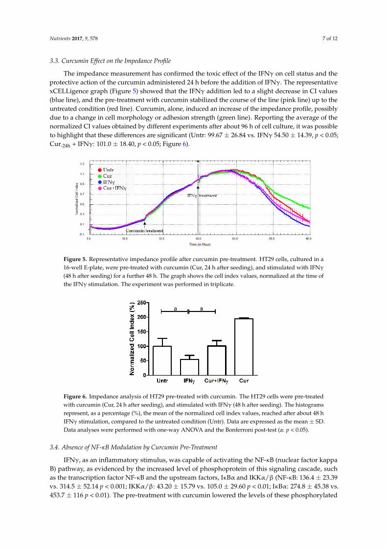
The impedance measurement has confirmed the toxic effect of the IFNγ on cell status and theprotective action of the curcumin administered 24 h before the addition of IFNγ. The representativexCELLigence graph (Figure 5) showed that the IFNγ addition led to a slight decrease in CI values(blue line), and the pre-treatment with curcumin stabilized the course of the line (pink line) up to theuntreated condition (red line). Curcumin, alone, induced an increase of the impedance profile, possiblydue to a change in cell morphology or adhesion strength (green line). Reporting the average of thenormalized CI values obtained by different experiments after about 96 h of cell culture, it was possibleto highlight that these differences are significant (Untr: 99.67 ± 26.84 vs. IFNγ 54.50 ± 14.39, p < 0.05;Cur-24h + IFNγ: 101.0 ± 18.40, p < 0.05; Figure 6).
Nutrients 2017, 9, 578 7 of 12
xCELLigence graph (Figure 5) showed that the IFNγ addition led to a slight decrease in CI values (blue line), and the pre-treatment with curcumin stabilized the course of the line (pink line) up to the untreated condition (red line). Curcumin, alone, induced an increase of the impedance profile, possibly due to a change in cell morphology or adhesion strength (green line). Reporting the average of the normalized CI values obtained by different experiments after about 96 h of cell culture, it was possible to highlight that these differences are significant (Untr: 99.67 ± 26.84 vs. IFNγ 54.50 ± 14.39, p < 0.05; Cur-24h + IFNγ: 101.0 ± 18.40, p < 0.05; Figure 6).
Figure 5. Representative impedance profile after curcumin pre-treatment. HT29 cells, cultured in a 16-well E-plate, were pre-treated with curcumin (Cur, 24 h after seeding), and stimulated with IFNγ (48 h after seeding) for a further 48 h. The graph shows the cell index values, normalized at the time of the IFNγ stimulation. The experiment was performed in triplicate.
Figure 6. Impedance analysis of HT29 pre-treated with curcumin. The HT29 cells were pre-treated with curcumin (Cur, 24 h after seeding), and stimulated with IFNγ (48 h after seeding). The histograms represent, as a percentage (%), the mean of the normalized cell index values, reached after about 48 h IFNγ stimulation, compared to the untreated condition (Untr). Data are expressed as the mean ± SD. Data analyses were performed with one-way ANOVA and the Bonferroni post-test (a: p < 0.05).
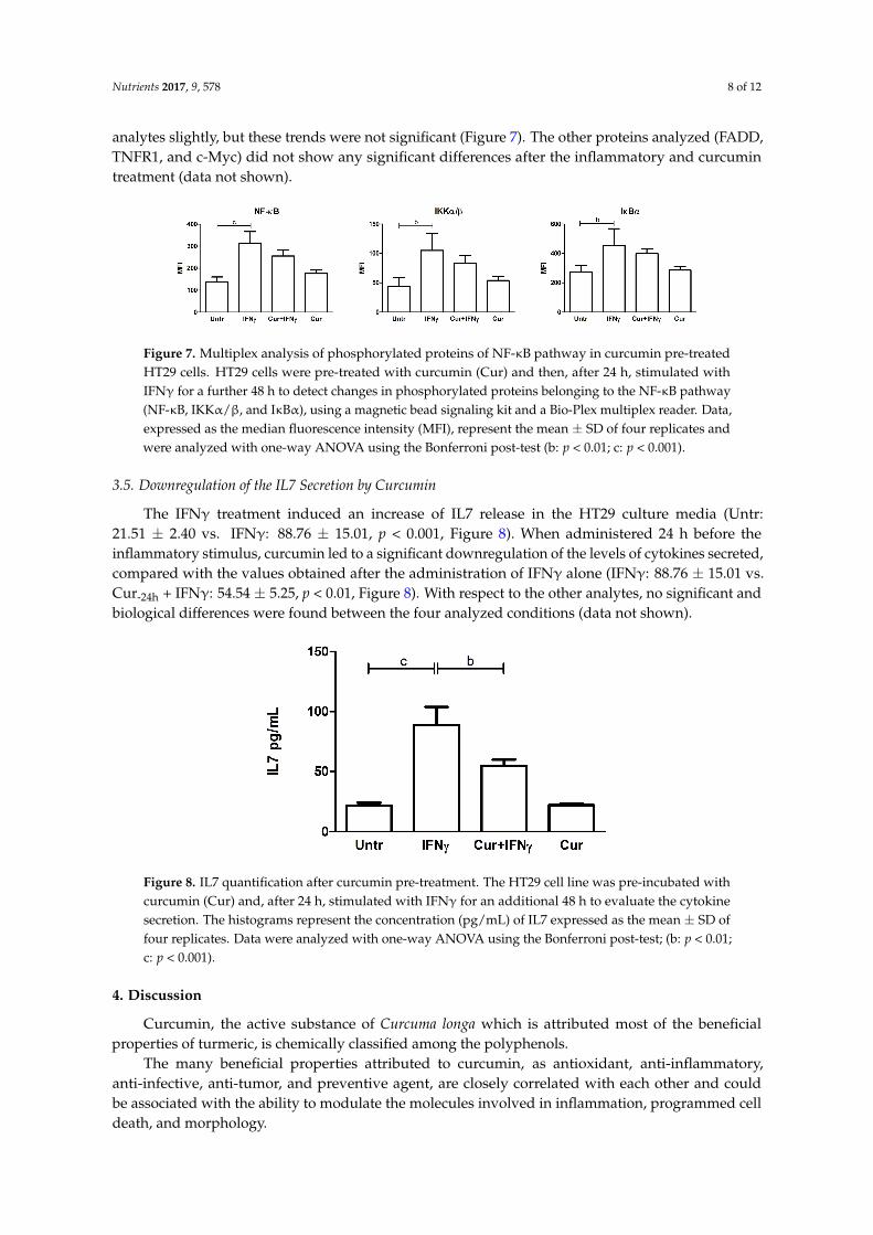
3.4. Absence of NF-κB Modulation by Curcumin Pre-Treatment
IFNγ, as an inflammatory stimulus, was capable of activating the NF-κB (nuclear factor kappa B) pathway, as evidenced by the increased level of phosphoprotein of this signaling cascade, such as the transcription factor NF-κB and the upstream factors, IκBα and IKKα/β (NF-κB: 136.4 ± 23.39 vs. 314.5 ± 52.14 p < 0.001; IKKα/β: 43.20 ± 15.79 vs. 105.0 ± 29.60 p < 0.01; IκBα: 274.8 ± 45.38 vs. 453.7 ± 116 p < 0.01). The pre-treatment with curcumin lowered the levels of these phosphorylated analytes slightly, but these trends were not significant (Figure 7). The other proteins analyzed (FADD, TNFR1, and c-Myc) did not show any significant differences after the inflammatory and curcumin treatment (data not shown).
Figure 5. Representative impedance profile after curcumin pre-treatment. HT29 cells, cultured in a16-well E-plate, were pre-treated with curcumin (Cur, 24 h after seeding), and stimulated with IFNγ
(48 h after seeding) for a further 48 h. The graph shows the cell index values, normalized at the time ofthe IFNγ stimulation. The experiment was performed in triplicate.
Nutrients 2017, 9, 578 7 of 12
xCELLigence graph (Figure 5) showed that the IFNγ addition led to a slight decrease in CI values (blue line), and the pre-treatment with curcumin stabilized the course of the line (pink line) up to the untreated condition (red line). Curcumin, alone, induced an increase of the impedance profile, possibly due to a change in cell morphology or adhesion strength (green line). Reporting the average of the normalized CI values obtained by different experiments after about 96 h of cell culture, it was possible to highlight that these differences are significant (Untr: 99.67 ± 26.84 vs. IFNγ 54.50 ± 14.39, p < 0.05; Cur-24h + IFNγ: 101.0 ± 18.40, p < 0.05; Figure 6).
Figure 5. Representative impedance profile after curcumin pre-treatment. HT29 cells, cultured in a 16-well E-plate, were pre-treated with curcumin (Cur, 24 h after seeding), and stimulated with IFNγ (48 h after seeding) for a further 48 h. The graph shows the cell index values, normalized at the time of the IFNγ stimulation. The experiment was performed in triplicate.
Figure 6. Impedance analysis of HT29 pre-treated with curcumin. The HT29 cells were pre-treated with curcumin (Cur, 24 h after seeding), and stimulated with IFNγ (48 h after seeding). The histograms represent, as a percentage (%), the mean of the normalized cell index values, reached after about 48 h IFNγ stimulation, compared to the untreated condition (Untr). Data are expressed as the mean ± SD. Data analyses were performed with one-way ANOVA and the Bonferroni post-test (a: p < 0.05).
3.4. Absence of NF-κB Modulation by Curcumin Pre-Treatment
IFNγ, as an inflammatory stimulus, was capable of activating the NF-κB (nuclear factor kappa B) pathway, as evidenced by the increased level of phosphoprotein of this signaling cascade, such as the transcription factor NF-κB and the upstream factors, IκBα and IKKα/β (NF-κB: 136.4 ± 23.39 vs. 314.5 ± 52.14 p < 0.001; IKKα/β: 43.20 ± 15.79 vs. 105.0 ± 29.60 p < 0.01; IκBα: 274.8 ± 45.38 vs. 453.7 ± 116 p < 0.01). The pre-treatment with curcumin lowered the levels of these phosphorylated analytes slightly, but these trends were not significant (Figure 7). The other proteins analyzed (FADD, TNFR1, and c-Myc) did not show any significant differences after the inflammatory and curcumin treatment (data not shown).
Figure 6. Impedance analysis of HT29 pre-treated with curcumin. The HT29 cells were pre-treatedwith curcumin (Cur, 24 h after seeding), and stimulated with IFNγ (48 h after seeding). The histogramsrepresent, as a percentage (%), the mean of the normalized cell index values, reached after about 48 hIFNγ stimulation, compared to the untreated condition (Untr). Data are expressed as the mean ± SD.Data analyses were performed with one-way ANOVA and the Bonferroni post-test (a: p < 0.05).
3.4. Absence of NF-κB Modulation by Curcumin Pre-Treatment
IFNγ, as an inflammatory stimulus, was capable of activating the NF-κB (nuclear factor kappaB) pathway, as evidenced by the increased level of phosphoprotein of this signaling cascade, suchas the transcription factor NF-κB and the upstream factors, IκBα and IKKα/β (NF-κB: 136.4 ± 23.39vs. 314.5 ± 52.14 p < 0.001; IKKα/β: 43.20 ± 15.79 vs. 105.0 ± 29.60 p < 0.01; IκBα: 274.8 ± 45.38 vs.453.7 ± 116 p < 0.01). The pre-treatment with curcumin lowered the levels of these phosphorylated
Nutrients 2017, 9, 578 8 of 12
analytes slightly, but these trends were not significant (Figure 7). The other proteins analyzed (FADD,TNFR1, and c-Myc) did not show any significant differences after the inflammatory and curcumintreatment (data not shown).
Nutrients 2017, 9, 578 8 of 12
Figure 7. Multiplex analysis of phosphorylated proteins of NF-κB pathway in curcumin pre-treated HT29 cells. HT29 cells were pre-treated with curcumin (Cur) and then, after 24 h, stimulated with IFNγ for a further 48 h to detect changes in phosphorylated proteins belonging to the NF-κB pathway (NF-κB, IKKα/β, and IκBα), using a magnetic bead signaling kit and a Bio-Plex multiplex reader. Data, expressed as the median fluorescence intensity (MFI), represent the mean ± SD of four replicates and were analyzed with one-way ANOVA using the Bonferroni post-test (b: p < 0.01; c: p < 0.001).
3.5. Downregulation of the IL7 Secretion by Curcumin
The IFNγ treatment induced an increase of IL7 release in the HT29 culture media (Untr: 21.51 ± 2.40 vs. IFNγ: 88.76 ± 15.01, p < 0.001, Figure 8). When administered 24 h before the inflammatory stimulus, curcumin led to a significant downregulation of the levels of cytokines secreted, compared with the values obtained after the administration of IFNγ alone (IFNγ: 88.76 ± 15.01 vs. Cur-24h + IFNγ: 54.54 ± 5.25, p < 0.01, Figure 8). With respect to the other analytes, no significant and biological differences were found between the four analyzed conditions (data not shown).
Figure 8. IL7 quantification after curcumin pre-treatment. The HT29 cell line was pre-incubated with curcumin (Cur) and, after 24 h, stimulated with IFNγ for an additional 48 h to evaluate the cytokine secretion. The histograms represent the concentration (pg/mL) of IL7 expressed as the mean ± SD of four replicates. Data were analyzed with one-way ANOVA using the Bonferroni post-test; (b: p < 0.01; c: p < 0.001).
4. Discussion
Curcumin, the active substance of Curcuma longa which is attributed most of the beneficial properties of turmeric, is chemically classified among the polyphenols.
The many beneficial properties attributed to curcumin, as antioxidant, anti-inflammatory, anti-infective, anti-tumor, and preventive agent, are closely correlated with each other and could be associated with the ability to modulate the molecules involved in inflammation, programmed cell death, and morphology.
In the present work, we demonstrated that pre-treatment with curcumin is able to reduce the levels of apoptosis in intestinal epithelial cells exposed to IFNγ. The fact that concordant data were
Figure 7. Multiplex analysis of phosphorylated proteins of NF-κB pathway in curcumin pre-treatedHT29 cells. HT29 cells were pre-treated with curcumin (Cur) and then, after 24 h, stimulated withIFNγ for a further 48 h to detect changes in phosphorylated proteins belonging to the NF-κB pathway(NF-κB, IKKα/β, and IκBα), using a magnetic bead signaling kit and a Bio-Plex multiplex reader. Data,expressed as the median fluorescence intensity (MFI), represent the mean ± SD of four replicates andwere analyzed with one-way ANOVA using the Bonferroni post-test (b: p < 0.01; c: p < 0.001).
3.5. Downregulation of the IL7 Secretion by Curcumin
The IFNγ treatment induced an increase of IL7 release in the HT29 culture media (Untr:21.51 ± 2.40 vs. IFNγ: 88.76 ± 15.01, p < 0.001, Figure 8). When administered 24 h before theinflammatory stimulus, curcumin led to a significant downregulation of the levels of cytokines secreted,compared with the values obtained after the administration of IFNγ alone (IFNγ: 88.76 ± 15.01 vs.Cur-24h + IFNγ: 54.54 ± 5.25, p < 0.01, Figure 8). With respect to the other analytes, no significant andbiological differences were found between the four analyzed conditions (data not shown).
Nutrients 2017, 9, 578 8 of 12
Figure 7. Multiplex analysis of phosphorylated proteins of NF-κB pathway in curcumin pre-treated HT29 cells. HT29 cells were pre-treated with curcumin (Cur) and then, after 24 h, stimulated with IFNγ for a further 48 h to detect changes in phosphorylated proteins belonging to the NF-κB pathway (NF-κB, IKKα/β, and IκBα), using a magnetic bead signaling kit and a Bio-Plex multiplex reader. Data, expressed as the median fluorescence intensity (MFI), represent the mean ± SD of four replicates and were analyzed with one-way ANOVA using the Bonferroni post-test (b: p < 0.01; c: p < 0.001).
3.5. Downregulation of the IL7 Secretion by Curcumin
The IFNγ treatment induced an increase of IL7 release in the HT29 culture media (Untr: 21.51 ± 2.40 vs. IFNγ: 88.76 ± 15.01, p < 0.001, Figure 8). When administered 24 h before the inflammatory stimulus, curcumin led to a significant downregulation of the levels of cytokines secreted, compared with the values obtained after the administration of IFNγ alone (IFNγ: 88.76 ± 15.01 vs. Cur-24h + IFNγ: 54.54 ± 5.25, p < 0.01, Figure 8). With respect to the other analytes, no significant and biological differences were found between the four analyzed conditions (data not shown).
Figure 8. IL7 quantification after curcumin pre-treatment. The HT29 cell line was pre-incubated with curcumin (Cur) and, after 24 h, stimulated with IFNγ for an additional 48 h to evaluate the cytokine secretion. The histograms represent the concentration (pg/mL) of IL7 expressed as the mean ± SD of four replicates. Data were analyzed with one-way ANOVA using the Bonferroni post-test; (b: p < 0.01; c: p < 0.001).
4. Discussion
Curcumin, the active substance of Curcuma longa which is attributed most of the beneficial properties of turmeric, is chemically classified among the polyphenols.
The many beneficial properties attributed to curcumin, as antioxidant, anti-inflammatory, anti-infective, anti-tumor, and preventive agent, are closely correlated with each other and could be associated with the ability to modulate the molecules involved in inflammation, programmed cell death, and morphology.
In the present work, we demonstrated that pre-treatment with curcumin is able to reduce the levels of apoptosis in intestinal epithelial cells exposed to IFNγ. The fact that concordant data were
Figure 8. IL7 quantification after curcumin pre-treatment. The HT29 cell line was pre-incubated withcurcumin (Cur) and, after 24 h, stimulated with IFNγ for an additional 48 h to evaluate the cytokinesecretion. The histograms represent the concentration (pg/mL) of IL7 expressed as the mean ± SD offour replicates. Data were analyzed with one-way ANOVA using the Bonferroni post-test; (b: p < 0.01;c: p < 0.001).
4. Discussion
Curcumin, the active substance of Curcuma longa which is attributed most of the beneficialproperties of turmeric, is chemically classified among the polyphenols.
The many beneficial properties attributed to curcumin, as antioxidant, anti-inflammatory,anti-infective, anti-tumor, and preventive agent, are closely correlated with each other and couldbe associated with the ability to modulate the molecules involved in inflammation, programmed celldeath, and morphology.

Nutrients 2017, 9, 578 9 of 12
In the present work, we demonstrated that pre-treatment with curcumin is able to reduce thelevels of apoptosis in intestinal epithelial cells exposed to IFNγ. The fact that concordant data wereobtained with different methods strengthens the interpretation of the results. In particular, there was arecent warning about the possibility that much of the research data on curcumin may be weakened, oreven invalidated, when natural properties of fluorescence and protein binding of the compound aretaken into account [25]. The inclusion in our research plan of a label-free assay based on cell impedanceanalysis allows ruling out such interferences.
When we further investigated the protective mechanisms of action of curcumin on HT29 cells,we observed that pre-treatment with this compound led to a significant reduction in the amount ofIL7 secreted in response to IFNγ. IL7 is a crucial cytokine, usually secreted by intestinal epithelialcells in response to bacterial infections, which contributes to the organization of secondary lymphoidstructures [28,29]. While basal secretion of this cytokine in response to the intestinal microbiota maybe required for an optimal homeostasis of the mucosal immunity [30,31], higher levels may play a rolein the pathogenesis of IBD and inhibition of IL7 has been proposed as a complementary approachto target inflammation in these disorders [32–35]. Since mucosal production of IL7 in vivo has beenimplicated in the activation of pathogenic CD4 T lymphocytes, the action of curcumin on this cytokinemay be independently useful to contrast intestinal inflammation [36,37].
Although a cell line cultured in the laboratory can hardly reflect the complex physiology ofintestinal mucosa, HT29 cells are considered a good model to study cytokine-induced apoptosis inepithelial cells. The model is coherent with in vivo data showing that epithelial apoptosis is increasedin intestinal mucosa in subjects with the active disease while it is reversed by anti-inflammatorytreatment with anti-TNFα antibodies. Increased apoptosis in epithelial cells is thought to contribute toincreased permeability and amplification of inflammation. Thus, inhibiting inflammation-inducedepithelial cell apoptosis may be relevant to the prevention or the treatment of IBD, as suggested byrecent data [10].
It is not clear how curcumin acts on IFNγ-induced apoptosis and cytokine secretion. One ofthe known actions of curcumin regards iron chelation and reduction [38–41], but it is not clear if theanti-inflammatory and anti-apoptotic effects of the drug may depend on this property. We evaluatedNF-κB activation since this pathway is a known target of IFNγ. Even if there was a trend towardreduction of the levels of phosphorylated components of NF-κB, this did not reach statisticalsignificance. Thus, we could not conclude that the downregulation of the NF-κB pathway is themain mechanism of action of curcumin. A possible alternative explanation is that curcumin exerts itsanti-inflammatory property by a direct modulation of mitochondrial function, as suggested by otherauthors [42].
Although we must put much caution in translating data obtained in vitro on a cell line, we canhypothesize that our results may be relevant to the understanding of the action of curcumin in vivo.In particular, a protective effect of curcumin was described in a mouse model of UC [43]. Moreover,small clinical trials in UC seem to suggest that curcumin may be a promising and safe complementarymedication for maintaining remission in patients with quiescent disease [44].
An issue that should be addressed to assess the real potential of curcumin into the clinic concernsits poor aqueous solubility and low bioavailability, which may hinder in vivo efficacy [45]; lowplasma levels and limited tissue distribution of curcumin appear to be due to poor absorption,high-rate metabolism, and rapid systemic clearance. A possible solution to these issues would bethe development of different types of curcumin formulations, such as nanoparticles, liposomes, orphospholipid complexes, which increase and improve not only its stability, but also its biodistributionand absorption [46]. With these new strategies, an enhanced bioavailability and efficacy have beendocumented in different experimental models. Therefore, further exploration to increase the medicalvalue for human applications are needed.
Our study was carried out on a single cell-type model. Even if preliminary studies in animalsand patients are promising and coherent with our data, we should consider the possibility that similar

Nutrients 2017, 9, 578 10 of 12
results cannot be obtained in vivo. Moreover, since we used ultra-pure curcumin, we should warnabout the risk of translating our results to commercially-available preparations of curcuma, which maycontain several other compounds with contrasting effects on mucosal immunity [47].
5. Conclusions
Despite being encouraging, our results need further and deeper investigations aimed atestablishing whether curcumin could be a potential therapeutic molecule for IBD and, in particular,for UC.
In conclusion, even though our results are preliminary and need to be verified in order to betranslated into the model of IBD patients, they should be considered as a first step to better characterizeturmeric anti-inflammatory properties.
Acknowledgments: This work was supported by a grant from the Institute for Maternal and Child Health IRCCS“Burlo Garofolo” (RC 03/2009).
Author Contributions: A.M., A.T., M.B. and S.L. designed the research; C.L. carried out the experiments andperformed the statistical analysis; E.P., L.V.B. and E.V. analyzed and interpreted the data; A.M., C.L., S.L. and A.T.wrote the paper; and E.P., L.V.B., E.V. and S.L. revised the manuscript critically for important intellectual content.All authors read and approved the final version of this manuscript.
Conflicts of Interest: The authors declare no conflict of interest.
References
1. Abraham, C.; Cho, J.H. Inflammatory bowel disease. N. Engl. J. Med. 2009, 361, 2066–2078. [CrossRef] [PubMed]2. Yang, Y.; Owyang, C.; Wu, G.D. East Meets West: The Increasing Incidence of Inflammatory Bowel
Disease in Asia as a Paradigm for Environmental Effects on the Pathogenesis of Immune-Mediated Disease.Gastroenterology 2016, 151, e1–e5. [CrossRef] [PubMed]
3. Benchimol, E.I.; Mack, D.R.; Guttmann, A.; Nguyen, G.C.; To, T.; Mojaverian, N.; Quach, P.; Manuel, D.G.Inflammatory bowel disease in immigrants to Canada and their children: A population-based cohort study.Am. J. Gastroenterol. 2015, 110, 553–563. [CrossRef] [PubMed]
4. Kim, H. Natural products to improve quality of life targeting for colon drug delivery. Curr. Drug Deliv. 2012,9, 132–147. [CrossRef] [PubMed]
5. Zachos, M.; Tondeur, M.; Griffiths, A.M. Enteral nutritional therapy for induction of remission in Crohn’sdisease. Cochrane Database Syst. Rev. 2007, 1, CD000542. [CrossRef]
6. Lewis, J.D.; Abreu, M.T. Diet as a Trigger or Therapy for Inflammatory Bowel Diseases. Gastroenterology 2016,152, 398–414. [CrossRef] [PubMed]
7. Vecchi Brumatti, L.; Marcuzzi, A.; Tricarico, P.M.; Zanin, V.; Girardelli, M.; Bianco, A.M. Curcumin andinflammatory bowel disease: Potential and limits of innovative treatments. Molecules 2014, 19, 21127–21153.[CrossRef] [PubMed]
8. Shishodia, S. Molecular mechanisms of curcumin action: Gene expression. Biofactors 2013, 39, 37–55.[CrossRef] [PubMed]
9. Taylor, R.A.; Leonard, M.C. Curcumin for inflammatory bowel disease: A review of human studies.Altern. Med. Rev. 2011, 16, 152–156. [PubMed]
10. Lang, A.; Salomon, N.; Wu, J.C.; Kopylov, U.; Lahat, A.; Har-Noy, O.; Ching, J.Y.; Cheong, P.K.; Avidan, B.;Gamus, D.; et al. Curcumin in Combination With Mesalamine Induces Remission in Patients WithMild-to-Moderate Ulcerative Colitis in a Randomized Controlled Trial. Clin. Gastroenterol. Hepatol. 2015,13, 1444–1449.e1. [CrossRef] [PubMed]
11. Panahi, Y.; Darvishi, B.; Ghanei, M.; Jowzi, N.; Beiraghdar, F.; Varnamkhasti, B.S. Molecular mechanisms ofcurcumins suppressing effects on tumorigenesis, angiogenesis and metastasis, focusing on NF-κB pathway.Cytokine Growth Factor Rev. 2016, 28, 21–29. [CrossRef] [PubMed]
12. Midura-Kiela, M.T.; Radhakrishnan, V.M.; Larmonier, C.B.; Laubitz, D.; Ghishan, F.K.; Kiela, P.R. Curcumininhibits interferon-γ signaling in colonic epithelial cells. Am. J. Physiol. Gastrointest. Liver Physiol. 2012,302, G85–G96. [CrossRef] [PubMed]

Nutrients 2017, 9, 578 11 of 12
13. Jian, Y.T.; Mai, G.F.; Wang, J.D.; Zhang, Y.L.; Luo, R.C.; Fang, Y.X. Preventive and therapeutic effects ofNF-kappaB inhibitor curcumin in rats colitis induced by trinitrobenzene sulfonic acid. World J. Gastroenterol.2005, 11, 1747–1752. [CrossRef] [PubMed]
14. Deguchi, Y.; Andoh, A.; Inatomi, O.; Yagi, Y.; Bamba, S.; Araki, Y.; Hata, K.; Tsujikawa, T.; Fujiyama, Y.Curcumin prevents the development of dextran sulfate Sodium (DSS)-induced experimental colitis.Dig. Dis. Sci. 2007, 52, 2993–2998. [CrossRef] [PubMed]
15. Arafa, H.M.; Hemeida, R.A.; El-Bahrawy, A.I.; Hamada, F.M. Prophylactic role of curcumin in dextran sulfatesodium (DSS)-induced ulcerative colitis murine model. Food Chem. Toxicol. 2009, 47, 1311–1317. [CrossRef][PubMed]
16. Mastropietro, G.; Tiscornia, I.; Perelmuter, K.; Astrada, S.; Bollati-Fogolín, M. HT-29 and Caco-2 reportercell lines for functional studies of nuclear factor kappa B activation. Mediat. Inflamm. 2015, 2015. [CrossRef][PubMed]
17. Guo, Y.; Shu, L.; Zhang, C.; Su, Z.Y.; Kong, A.N. Curcumin inhibits anchorage-independent growth ofHT29 human colon cancer cells by targeting epigenetic restoration of the tumor suppressor gene DLEC1.Biochem. Pharmacol. 2015, 94, 69–78. [CrossRef] [PubMed]
18. Iwamoto, M.; Koji, T.; Makiyama, K.; Kobayashi, N.; Nakane, P.K. Apoptosis of crypt epithelial cells inulcerative colitis. J. Pathol. 1996, 180, 152–159. [CrossRef]
19. Hagiwara, C.; Tanaka, M.; Kudo, H. Increase in colorectal epithelial apoptotic cells in patients with ulcerativecolitis ultimately requiring surgery. J. Gastroenterol. Hepatol. 2002, 17, 758–764. [CrossRef] [PubMed]
20. Di Sabatino, A.; Ciccocioppo, R.; Luinetti, O.; Ricevuti, L.; Morera, R.; Cifone, M.G.; Solcia, E.; Corazza, G.R.Increased enterocyte apoptosis in inflamed areas of Crohn’s disease. Dis. Colon. Rectum. 2003, 46, 1498–1507.[CrossRef] [PubMed]
21. Zeissig, S.; Bojarski, C.; Buergel, N.; Mankertz, J.; Zeitz, M.; Fromm, M.; Schulzke, J.D. Downregulation ofepithelial apoptosis and barrier repair in active Crohn’ s disease by tumour necrosis factor alpha antibodytreatment. Gut 2004, 53, 1295–1302. [CrossRef] [PubMed]
22. Banerjee, S.; Singh, S.K.; Chowdhury, I.; Lillard, J.W., Jr.; Singh, R. Combinatorial effect of curcumin withdocetaxel modulates apoptotic and cell survival molecules in prostate cancer. Front. Biosci. (Elite Ed.) 2017,1, 235–245.
23. Park, S.I.; Lee, E.H.; Kim, S.R.; Jang, Y.P. Anti-apoptotic effects of Curcuma longa L. extract and itscurcuminoids against blue light-induced cytotoxicity in A2E-laden human retinal pigment epithelial cells.J. Pharm. Pharmacol. 2017, 69, 334–340. [CrossRef] [PubMed]
24. Zheng, L.; Li, Y.; Li, X.; Kou, J.; Zhong, Z.; Jiang, Y.; Liu, Z.; Tian, Y.; Yang, L. Combination of HydroxylAcetylated Curcumin and Ultrasound Induces Macrophage Autophagy with Anti-Apoptotic and Anti-LipidAggregation Effects. Cell Physiol. Biochem. 2016, 39, 1746–1760. [CrossRef] [PubMed]
25. Baker, M. Deceptive curcumin offers cautionary tale for chemists. Nature 2017, 541, 144–145. [CrossRef][PubMed]
26. Hackler, L., Jr.; Ózsvári, B.; Gyuris, M.; Sipos, P.; Fábián, G.; Molnár, E.; Marton, A.; Faragó, N.; Mihály, J.;Nagy, L.I.; et al. The Curcumin Analog C-150, Influencing NF-κB, UPR and Akt/Notch Pathways Has PotentAnticancer Activity in Vitro and in Vivo. PLoS ONE 2016, 11, e0149832. [CrossRef] [PubMed]
27. Li, W.; Wang, H.; Kuang, C.Y.; Zhu, J.K.; Yu, Y.; Qin, Z.X.; Liu, J.; Huang, L. An essential role for theId1/PI3K/Akt/NFkB/survivin signalling pathway in promoting the proliferation of endothelial progenitorcells in vitro. Mol. Cell Biochem. 2012, 363, 135–145. [CrossRef] [PubMed]
28. Cimbro, R.; Vassena, L.; Arthos, J.; Cicala, C.; Kehrl, J.H.; Park, C.; Sereti, I.; Lederman, M.M.; Fauci, A.S.;Lusso, P. IL-7 induces expression and activation of integrin α4β7 promoting naive T-cell homing to theintestinal mucosa. Blood 2012, 120, 2610–2619. [CrossRef] [PubMed]
29. Zhang, W.; Du, J.Y.; Yu, Q.; Jin, J.O. Interleukin-7 produced by intestinal epithelial cells in response toCitrobacter rodentium infection plays a major role in innate immunity against this pathogen. Infect. Immun.2015, 83, 3213–3223. [CrossRef] [PubMed]
30. Shalapour, S.; Deiser, K.; Sercan, O.; Tuckermann, J.; Minnich, K.; Willimsky, G.; Blankenstein, T.;Hämmerling, G.J.; Arnold, B.; Schüler, T. Commensal microflora and interferon-gamma promote steady-stateinterleukin-7 production in vivo. Eur. J. Immunol. 2010, 40, 2391–2400. [CrossRef] [PubMed]

Nutrients 2017, 9, 578 12 of 12
31. Shalapour, S.; Deiser, K.; Kühl, A.A.; Glauben, R.; Krug, S.M.; Fischer, A.; Sercan, O.; Chappaz, S.; Bereswill, S.;Heimesaat, M.M.; et al. Interleukin-7 links T lymphocyte and intestinal epithelial cell homeostasis. PLoS ONE2012, 7, e31939. [CrossRef] [PubMed]
32. Kanai, T.; Nemoto, Y.; Kamada, N.; Totsuka, T.; Hisamatsu, T.; Watanabe, M.; Hibi, T. Homeostatic (IL-7)and effector (IL-17) cytokines as distinct but complementary target for an optimal therapeutic strategy ininflammatory bowel disease. Curr. Opin. Gastroenterol. 2009, 25, 306–313. [CrossRef] [PubMed]
33. Watanabe, M.; Ueno, Y.; Yajima, T.; Iwao, Y.; Tsuchiya, M.; Ishikawa, H.; Aiso, S.; Hibi, T.; Ishii, H.Interleukin 7 is produced by human intestinal epithelial cells and regulates the proliferation of intestinalmucosal lymphocytes. J. Clin. Investig. 1995, 95, 2945–2953. [CrossRef] [PubMed]
34. Andreu-Ballester, J.C.; Pérez-Griera, J.; Garcia-Ballesteros, C.; Amigo, V.; Catalán-Serra, I.;Monforte-Albalat, A.; Bixquert-Jiménez, M.; Ballester, F. Deficit of interleukin-7 in serum of patients withCrohn’s disease. Inflamm. Bowel Dis. 2013, 19, E30-1. [CrossRef] [PubMed]
35. Totsuka, T.; Kanai, T.; Nemoto, Y.; Makita, S.; Okamoto, R.; Tsuchiya, K.; Watanabe, M. IL-7 is essential for thedevelopment and the persistence of chronic colitis. J. Immunol. 2007, 178, 4737–4748. [CrossRef] [PubMed]
36. Nemoto, Y.; Kanai, T.; Takahara, M.; Oshima, S.; Nakamura, T.; Okamoto, R.; Tsuchiya, K.; Watanabe, M.Bone marrow-mesenchymal stem cells are a major source of interleukin-7 and sustain colitis by forming theniche for colitogenic CD4 memory T cells. Gut 2013, 62, 1142–1152. [CrossRef] [PubMed]
37. Willis, C.R.; Seamons, A.; Maxwell, J.; Treuting, P.M.; Nelson, L.; Chen, G.; Phelps, S.; Smith, C.L.; Brabb, T.;Iritani, B.M.; et al. Interleukin-7 receptor blockade suppresses adaptive and innate inflammatory responsesin experimental colitis. J. Inflamm. (Lond.) 2012, 9, 39. [CrossRef] [PubMed]
38. Yang, C.; Ma, X.; Wang, Z.; Zeng, X.; Hu, Z.; Ye, Z.; Shen, G. Curcumin induces apoptosis and protectiveautophagy in castration-resistant prostate cancer cells through iron chelation. Drug Des. Dev. Ther. 2017,11, 431–439. [CrossRef] [PubMed]
39. Du, X.X.; Xu, H.M.; Jiang, H.; Song, N.; Wang, J.; Xie, J.X. Curcumin protects nigral dopaminergic neurons byiron-chelation in the 6-hydroxydopamine rat model of Parkinson’ s disease. Neurosci. Bull. 2012, 28, 253–258.[CrossRef] [PubMed]
40. Dairam, A.; Fogel, R.; Daya, S.; Limson, J.L. Antioxidant and iron-binding properties of curcumin, capsaicin,and S-allylcysteine reduce oxidative stress in rat brain homogenate. J. Agric. Food Chem. 2008, 56, 3350–3356.[CrossRef] [PubMed]
41. Galleggiante, V.; De Santis, S.; Cavalcanti, E.; Scarano, A.; De Benedictis, M.; Serino, G.; Caruso, M.L.;Mastronardi, M.; Pinto, A.; Campiglia, P.; et al. Dendritic Cells Modulate Iron Homeostasis and InflammatoryAbilities Following Quercetin Exposure. Curr. Pharm. Des. 2017. [CrossRef]
42. Jiang, A.J.; Jiang, G.; Li, L.T.; Zheng, J.N. Curcumin induces apoptosis through mitochondrial pathway andcaspases activation in human melanoma cells. Mol. Biol. Rep. 2015, 42, 267–275. [CrossRef] [PubMed]
43. Sugimoto, K.; Hanai, H.; Tozawa, K.; Aoshi, T.; Uchijima, M.; Nagata, T.; Koide, Y. Curcumin preventsand ameliorates trinitrobenzene sulfonic acid-induced colitis in mice. Gastroenterology 2002, 123, 1912–1922.[CrossRef] [PubMed]
44. Hanai, H.; Iida, T.; Takeuchi, K.; Watanabe, F.; Maruyama, Y.; Andoh, A.; Tsujikawa, T.; Fujiyama, Y.;Mitsuyama, K.; Sata, M.; et al. Curcumin maintenance therapy for ulcerative colitis: Randomized, multicenter,double-blind, placebo-controlled trial. Clin. Gastroenterol. Hepatol. 2006, 4, 1502–1506. [CrossRef] [PubMed]
45. Anand, P.; Kunnumakkara, A.B.; Newman, R.A.; Aggarwal, B.B. Bioavailability of curcumin: Problems andpromises. Mol. Pharm. 2007, 4, 807–818. [CrossRef] [PubMed]
46. Prasad, S.; Tyagi, A.K.; Aggarwal, B.B. Recent developments in delivery, bioavailability, absorption andmetabolism of curcumin: The golden pigment from golden spice. Cancer Res. Treat. 2014, 46, 2–18. [CrossRef][PubMed]
47. Holt, P.R. Curcumin for Inflammatory Bowel Disease: A Caution. Clin. Gastroenterol. Hepatol. 2016, 14, 168.[CrossRef] [PubMed]
© 2017 by the authors. Licensee MDPI, Basel, Switzerland. This article is an open accessarticle distributed under the terms and conditions of the Creative Commons Attribution(CC BY) license (http://creativecommons.org/licenses/by/4.0/).