Embed Size (px)
Citation preview

lable at ScienceDirect
Seminars in Fetal & Neonatal Medicine 16 (2011) 164e172
Contents lists avai
Seminars in Fetal & Neonatal Medicine
journal homepage: www.elsevier .com/locate/s iny
Neonatal abdominal wall defects
Emily R. Christison-Lagay a, Cassandra M. Kelleher b, Jacob C. Langer a,*aDivision of General and Thoracic Surgery, Hospital for Sick Children, 555 University Ave, Toronto, Ontario M5G 1X8, CanadabDepartment of Pediatric Surgery, Massachusetts General Hospital for Children, Boston, MA, USA
Keywords:Fetal diagnosisGastroschisisGenetic counsellingOmphalocele
* Corresponding author. Tel.: þ1 416 813 7340; faxE-mail address: [email protected] (J.C. Lang
1744-165X/$ e see front matter � 2011 Published bydoi:10.1016/j.siny.2011.02.003
s u m m a r y
Gastroschisis and omphalocele are the two most common congenital abdominal wall defects. Both arefrequently detected prenatally due to routine maternal serum screening and fetal ultrasound. Prenataldiagnosis may influence timing, mode and location of delivery. Prognosis for gastroschisis is primarilydetermined by the degree of bowel injury, whereas prognosis for omphalocele is related to the numberand severity of associated anomalies. The surgical management of both conditions consists of closure ofthe abdominal wall defect, while minimizing the risk of injury to the abdominal viscera either throughdirect trauma or due to increased intra-abdominal pressure. Options include primary closure or a varietyof staged approaches. Long-term outcome is favorable in most cases; however, significant associatedanomalies (in the case of omphalocele) or intestinal dysfunction (in the case of gastroschisis) may resultin morbidity and mortality.
� 2011 Published by Elsevier Ltd.
1. Introduction
Gastroschisis and omphalocele are the two most commoncongenital abdominal wall defects (Fig. 1). Described in the litera-ture as early as the first century AD, today these anomalies arefrequently detected prenatally due to routine maternal serumscreening and fetal ultrasound. Principal differences between gas-troschisis and omphalocele are summarized in Table 1. Prognosti-cally, however, the most important distinguishing feature comesnot from the defect itself but from the differential rate of associatedanomalies; the risk of an associated structural or chromosomalabnormality in an infant with omphalocele exceeds 50%, whereasinfants with gastroschisis rarely have associated abnormalities,except for an increased incidence of intestinal atresia. Therefore,the long term outcome for neonates with omphalocele is oftendetermined by its associated anomalies, whereas infants withgastroschisis tend to achieve normal growth and developmentalmilestones as they progress through childhood.
Gastroschisis occurs in 1 in 4000 live births.1 The majority ofpregnancies complicated by gastroschisis are diagnosed prena-tally.2 An elevated maternal serum a-fetoprotein level may be theearliest indicator of the presence of gastroschisis. Subsequentsonographic visualization of freely floating loops of bowel withinthe amniotic fluid with an abdominal wall defect to the right of the
: þ1 416 813 7477.er).
Elsevier Ltd.
insertion of the umbilical cord at any point after the normalembryonic return of the intestine to the abdominal cavity at 10weeks of gestation confirms the diagnosis. Adolescent mothershave an increased incidence of gastroschisis compared to oldermothers but recent epidemiological surveillance data have showna 10e20-fold increase in the overall incidence of gastroschisis in allage groups over the past two decades.3e6 Preterm delivery is morefrequent in infants with gastroschisis, with an incidence of 28%compared with 6% of normal deliveries.7
Bowel atresia is the most common associated anomaly inpatients with gastroschisis. Recent studies report concomitantatresia and gastroschisis in 6.9e28% of patients.8,9 Other moreunusual associations of gastroschisis include the limbebody walldefect syndrome (amniotic band syndrome). In this rare syndrome,thoracic wall malformations or gastroschisis are found associatedwith limb abnormalities, meningocele, abnormal genitalia, intes-tinal atresias, and umbilical cord abnormalities.10,11
Omphalocele can also be a cause of elevation of maternal seruma-fetoprotein, though less commonly than gastroschisis. Prenataldiagnosis may be made as early as the first trimester if three-dimensional ultrasonography is available but is more commonlymade on routine 18-week two-dimensional ultrasound.2 The inci-dence of omphalocele seen on ultrasonography at 14e18 weeks isas high as 1 in 1100, but due to both spontaneous intrauterine fetaldeath and pregnancy termination, the incidence in live births isw1in 4000.1,12
A prenatal diagnosis of omphalocele should be followed bya comprehensive fetal ultrasound, including fetal echocardiography

Fig. 1. Typical appearance of gastroschisis and omphalocele. (A) Gastroschisis with relatively normal bowel. (B) Gastroschisis with significantly damaged bowel with evidence ofmatting, foreshortening, and peel. (C) Small omphalocele. (D) Giant omphalocele with liver out.
E.R. Christison-Lagay et al. / Seminars in Fetal & Neonatal Medicine 16 (2011) 164e172 165
as omphalocele is accompanied by an 18e24% incidence of cardiacanomalies.1 Pulmonary hypoplasia is also commonly associatedwith giant omphalocele and may result in early respiratory distressrequiring intubation and ventilatory support at the time ofdelivery.13 Associated syndromes such as cloacal exstrophy, Don-naieBarrow syndrome and pentalogy of Cantrell can also be sug-gested by fetal ultrasound. Chromosomal abnormalities, mostcommonly trisomies 13, 18 and 21, occur in up to 49% of fetusesdiagnosed with omphalocele.14 The risk of a chromosomal abnor-mality appears to be more common in fetuses with a centralomphalocele than those with epigastric omphaloceles.14 Of fetuseswith normal karyotypes, nearly 80% have multiple other anoma-lies.14 Interestingly, multiple associated anomalies appear to bemore common with minor omphalocele (�4 cm) than giantomphalocele (55% vs 36%).15 Cardiac defects are the most commonanomalies in this group, but such lethal entities as hol-oprosencephaly and anencephalus, as well as the complete spec-trum of VACTERL defects, may be seen.16
Table 1Characteristics of omphalocele and gastroschisis.
Omphalocele Gastroschisis
Sac Present AbsentAssociated anomalies Common UncommonLocation of defect Umbilicus Right of umbilicusMaternal age Average YoungerMode of delivery Cesarean/vaginal VaginalSurgical management Not urgent UrgentPrognostic factors Associated anomalies Condition of bowel
2. Embryology
An omphalocele occurs when the intestines fail to return to theabdominal cavity after normal embryonic herniation into theumbilical cord during weeks 6e10 of development. This is typicallyattributed to a folding defect in the abdominalwall rather than to thegenes involved in gut elongation and rotation.17,18 Varying amountsof bowel may be containedwithin the omphalocele sac. Other intra-abdominal viscera including liver, bladder, stomach, ovary, and testiscan also be found within the sac. The sac consists of the coveringlayers of the umbilical cord, which include amnion, Wharton’s jelly,and peritoneum. The umbilical cord is attached to the sac itself.
The etiology of gastroschisis is subject to some debate. It iscommonly held that the pathogenesis involves an in-utero vascularaccident and, along these lines, two theories have been advanced.One theory suggests that involution of the right umbilical veincauses necrosis in the abdominal wall leading to a right-sideddefect; a second theory posits that the right omphalomesenteric(vitelline) artery prematurely involutes causing a weakening in theabdominal wall through which the intestinal contents subse-quently rupture.19,20 These theories are supported by the observa-tion that gastroschisis is associated with intestinal atresia,a condition that is also thought to be associated with an ischemicetiology.21 Additionally, retrospective data have suggested anincreased risk of gastroschisis and intestinal atresia with maternaluse of vasoconstrictive drugs (ephedrine, pseudoephedrine, orcocaine) as well as with smoking.22 More recent epidemiologicaland scientific data suggest that these explanations may be insuffi-cient. Feldkamp et al. note that both umbilical veins degenerate,which does not explain the predominant right-sided occurrence ofgastroschisis.23 Moreover, the body wall derives arterial supply

E.R. Christison-Lagay et al. / Seminars in Fetal & Neonatal Medicine 16 (2011) 164e172166
from a rich arcading network of vessels arising from the dorsalaorta that is neither dependent upon nor intersects with theumbilical or vitelline vessels. A recent large scale epidemiologicalstudy measuring the associations between maternal vasoactiveexposures, as part of the National Birth Defects Prevention Study,found that vasoactive risk factors play a minor role, if any, in theetiology of gastroschisis in young mothers, but may play a largerrole in mothers aged>25 years.24 Nonvascular explanations for theorigin of gastroschisis include failure of incorporation of the vitel-line duct into the umbilical cord and abnormal development of theventral abdominal wall resulting in the failure of midline fusion ofthe lateral folds.23,25 In-utero rupture of an omphalocele has alsobeen proposed as a mechanism of gastroschisis formation.26
3. Gastroschisis
3.1. Perinatal care
The outcome for infants with gastroschisis is primarily deter-mined by the amount of intestinal damage that occurs during fetallife. The etiology of this injury is likely a combination of exposure toamniotic fluid and constriction of the bowel at the abdominal walldefect and much of the damage seems to occur toward the end ofpregnancy.27,28 Intestinal damage results in impaired motility andmucosal absorptive functionwhich, in turn, lead to prolonged needfor total parenteral nutrition and, in some cases, severe irreversibleintestinal failure.29 Prenatal diagnosis provides a potential oppor-tunity to modulate mode, location, and timing of delivery in orderto minimize these complications.
The optimal mode of delivery for fetuses with gastroschisis isdebated. Proponents of routine cesarean delivery argue that theprocess of vaginal birth may injure the exposed bowel. However,this philosophy is not supported by published data which havefailed to demonstrate a difference in outcomes between cesareansection and vaginal delivery.30,31 Therefore, the delivery method ofa neonate with gastroschisis should be at the discretion of theobstetrician and the mother.
Timing of delivery is also controversial. Some centers advocateearly delivery of the fetus with gastroschisis in an attempt to reducethe inflammatory peel on the surface of the bowel. Evidencesupports a role of amniotic fluid cytokines and proinflammatorymediators (including interleukin-6 and interleukin-8) in damagingthe myenteric nerve plexus and interstitial cells of Cajal in gastro-schisis.30,32e35 Because bowel edema and peel formation increaseas pregnancy progresses, early delivery is thought by some tomitigate these effects. However, the literature is mixed in terms ofthe benefit of preterm delivery. Labor can be successfully induced ina high percentage of cases at 36e37 weeks of gestation in gastro-schisis pregnancies, probably because of the inherent tendencytoward preterm labor.8 The argument against early delivery is thatlow birth weight negatively influences outcome, with neonatesweighing <2 kg having increased time to full enteral feeding,ventilated days, and duration of parenteral nutrition comparedwith those weighing >2 kg.36 Some authors advocate selectivepreterm delivery based on the appearance of bowel distention andthickening on prenatal ultrasonography. The presence of dilatedfetal bowel has been shown to correlate with poor outcome,including fetal distress and demise in some e but not all e
series.37,38 Many of these data are confounded by non-standardizedparadigms for measuring bowel and lack of consensus upon thedefinition of ‘dilated’ at any given gestational age.
Most authors advocate delivery at a tertiary perinatal center so asto provide immediate access to neonatal and pediatric surgicalexpertise. A recentoutcomesanalysis found that deliveryat aperinatalcenter with immediate access to level 3 neonatal ICU and pediatric
surgeon was associated with an overall reduction in risk adjustedmorbidity when compared to delivery at a community hospital.39
3.2. Neonatal resuscitation and management
Gastroschisis causes significant evaporative water losses fromthe exposed bowel. Following delivery, appropriate intravenousaccess should be obtained and fluid resuscitation initiated. Gastricdecompression is important to prevent intestinal distension. Theherniated bowel should be wrapped in warm saline-soaked gauze,placed in a central position on the abdominal wall with the babypositioned on the right side to prevent kinking of the mesentery,and wrapped with plastic wrap to reduce evaporative losses andtemperature instability. A thorough examination of the neonateshould be performed to exclude the coexistence of other anomalieswith specific attention to the bowel for evidence of intestinalatresia, necrosis or perforation.
3.3. Surgical management
Surgical management of gastroschisis varies from center tocenter and has evolved over the past several decades, particularlywith the introduction of the spring-loaded silo. The primary goal ofevery surgical repair is to return the viscera to the abdominal cavitywhile minimizing the risk of damage to the viscera due to directtrauma or increased intra-abdominal pressure. Options include:(i) primary reduction with operative closure of the fascia; (ii) siloplacement, serial reductions, and delayed fascial closure;(iii) primary ordelayed reductionwithout fascial closure. In addition,the timing and location of surgical intervention is controversial,ranging from immediate repair in the delivery room, to reductionand closure in the neonatal intensive care unit, to surgical closure inthe operating room.40,41 In all cases, inspection of the bowel forobstructing bands, perforation, or atresia should be undertaken.Bands crossing the bowel loops should be divided before siloplacement or primary abdominal closure to avoid subsequent bowelobstruction. Consideration should be given to the early establish-ment of central venous access, as intestinal hypomotility is invari-ably present.
3.3.1. Primary closureHistorically, urgent primary closure of gastroschisis was advo-
cated in all cases. This approach was supported by multiple retro-spective reviews of primary versus staged closure documentingimproved outcomes in those patients undergoing primary closure.Upon closer analysis, however, these results were likely skewed bya significant selection bias because only unstable patients, or thosewith the greatest intestinal damage or largest defects, were likely toundergo staged repair. Nonetheless, many centers still practiceprimary closure in neonates considered to possess sufficient intra-abdominal domain to permit full reduction of the herniated viscera.Recent data from the Canadian Pediatric Surgeons Network (CAP-SNet) database suggests that infants who are able to undergoimmediate primary reduction and closure have a shorter length ofparenteral nutrition use and total length of stay when comparedwith those who require staged reduction and delayed repair.42
3.3.2. Staged closureOriginally, staged closure consisted of placing the bowel into
a silo constructed of Silastic sheets sewn together and sutured tothe abdominal wall. In recent years, the use of a hand-fashioned silohas been eclipsed by the introduction of a prefabricated silo witha circular spring that can be placed into the abdominal defect at thebedside, without the need for sutures or general anesthesia (Fig. 2).In either case, the bowel is reduced once or twice daily into the

E.R. Christison-Lagay et al. / Seminars in Fetal & Neonatal Medicine 16 (2011) 164e172 167
abdominal cavity as the silo is shortened by sequential ligation.When the eviscerated contents are entirely reduced, the definitiveclosure can be performed. This process usually takes between oneand 14 days, depending on the condition of the bowel and theinfant. The preformed silo can also be used at the bedside to reducethe bowel, with abdominal closure being done immediatelywithout sequential ligation of the sac.
Advocates of delayed closure argue that avoidance of high intra-abdominal pressure reduces the risk of ischemic injury to theviscera and may permit earlier extubation.43,44 In a prospective
Fig. 2. Preformed silo for the management of gastroschisis. (A) Initial placement of thesilo. (B) Complete reduction prior to definitive fascial closure.
study using historical controls, our group demonstrated that theroutine use of a spring-loaded silo was associated with a shortertime on the ventilator, lower postoperative airway pressures,shorter hospital stay, lower cost, and lower risk of complications,although other centers have had more variable success.45e48 Thediscrepancy of these results in otherwise similarly designed studiessuggests that institutional practice protocols, more than the routineuse of spring-loaded silo alone, may affect endpoint outcome. In allstudies, routine silo management was found to be safe and effec-tive, without an increase in morbidity or mortality over attemptedprimary closure techniques.
3.3.3. Closure techniquesDefinitive closure in the operating room consists of raising skin
flaps around the fascial defect to facilitate fascial closure followedby subsequent skin closure. Closure of the skin in a linear fashioncreates a ‘keyhole’ appearance with a horizontal scar to the right ofthe umbilicus. Some surgeons advocate a purse-string suture of theskin around the umbilicus to create a circular scar with improvedcosmesis. Recently, the ‘plastic closure’ method has been intro-duced in which the umbilical cord, if not too macerated or dry, istailored to fill the gastroschisis defect and is then covered with anadhesive dressing.49 This technique allows for a centralized umbi-licus without any linear scar component and is best utilized withsmall defects. If the umbilical cord is not salvageable, the bowel canbe directly covered with a non-permeable dressing. This dressing ischanged every five to seven days and the wound inspected. In-growth of granulation tissue and epithelialization occurs over time.With this technique, an operation can be avoided entirely in manyinfants. Residual ventral hernia rates are reported to be 60e84%, themajority of which close spontaneously.50,51
Multiple methods of closure have been described for children inwhom primary fascial closure cannot be achieved. The simplestinvolves using the umbilicus as an allograft.52 Prosthetic optionsinclude both non-absorbable mesh and biosynthetic absorbablepatches such as dura or porcine small intestinal submucosa(Surgisis�, Cook Inc., Bloomington, IN, USA). Another option inselected cases is to reduce the bowel and place a piece of Silasticsheeting under the abdominal wall to prevent evisceration. Thistechnique is useful when the surgeon is concerned about increasedintra-abdominal pressure leading to a deterioration in pulmonaryfunctionwith fascial and skin closure. The Silastic sheet is removedin 4e5 days, and the abdominal wall and skin are closed.
Intra-abdominal pressure, measured either as intravesical orintragastric pressure, can be used to guide the surgeon duringreduction. Pressures >20 mmHg are correlated with decreasedperfusion to the kidneys and bowel.53 Similarly, an increase incentral venous pressure >4 mmHg has been correlated with theneed for silo placement or patch closure during attempted primaryrepair.54 Following reduction, careful attention should be paid tophysical examination, urine output, and lower limb perfusion witha low threshold to reopen a closed abdomen for signs of abdominalcompartment syndrome.
3.3.4. Management of associated intestinal atresia or perforationUp to 10% of neonates with gastroschisis will have an associated
intestinal atresia, most commonly jejunal or ileal. These atresias canbe treated at the time of abdominal wall closure with resection andprimary anastomosis if bowel inflammation is minimal. If thecondition of the bowel makes primary anastomosis inadvisable, thebowel can be reduced with the atresia intact and repair can beundertaken four to sixweeks after the initial abdominalwall closure.Stoma creation is another alternative and is particularly helpful inthe case of distal atresia.55 Intestinal atresia should be differentiatedfrom ‘vanishing gastroschisis’. This condition is usually associated

E.R. Christison-Lagay et al. / Seminars in Fetal & Neonatal Medicine 16 (2011) 164e172168
with a very small abdominal wall defect that strangulates the extra-abdominal viscera and is characterized by necrosis and disappear-ance of some or all of the intestine (Fig. 3). Although this happensrarely, it usually results in short bowel syndrome.
Perforation may be managed through many of the same tech-niques as atresia. If bowel inflammation is minimal, the perforatedsegment can be resected and continuity established with a primaryanastomosis. Other options include ostomy creation and primaryfascial closure. If primary closure is impossible, exteriorization ofthe perforation through a hole in the silo is another option. Oncethe bowel has been reduced, a formal stoma can be created at thetime of abdominal wall closure.
3.3.5. Postoperative courseGastroschisis is associatedwith abnormal intestinal motility and
nutrient absorption, both of which gradually improve over time inmost patients. Introduction of enteral feeding is often delayed forweeks while awaiting return of bowel function. During the periodof dysmotility, nasogastric decompression and parenteral nutritionis required. Secure central venous access through a percutaneouslyinserted central catheter (PICC) or tunneled cuffed central line is ofutmost importance. Often there is initial feeding intolerance withthe need for slow progression of feeds. It is also important toinclude early oral stimulation, because the suckingeswallowingreflex can be lost while awaiting bowel function and a significantoral aversion may develop.
Gastrointestinal dysmotility is often treated with prokineticagents in these infants. However, there is little documentation in
Fig. 3. (A) Gastroschisis with associated intestinal atresia. (B) Gastroschisis with‘vanishing bowel’ due to a very small abdominal wall defect, which is usually associ-ated with short bowel syndrome.
the literature to support their use. Commonly utilized prokineticsinclude erythromycin, metoclopramide, domperidone and cis-apride. A randomized controlled trial of erythromycin versusplacebo showed that enterally administered erythromycin did notimprove time to achieve full enteral feedings over placebo.56
However, a similar randomized trial examining the use of cis-apride in postoperative neonates, most of whom had gastroschisis,did show a beneficial effect.57 Cisapride is available only ona compassionate basis in North America due to its relatively highrisk of cardiovascular side effects, and it therefore can only be usedin infants with severe dysmotility who have not responded to otheragents and who have undergone cardiac evaluation and clearance.
Necrotizing enterocolitis has been encountered in full-terminfants with gastroschisis in higher than expected frequencies (upto 18.5%).58 While significant bowel loss from necrotizing entero-colitis can predispose patients to short bowel syndrome and itsassociated hepatic and septic complications, at least one groupfound that most gastroschisis patients with necrotizing enteroco-litis exhibited a subsequently uncomplicated clinical course.59
Attempts to stratify patients with gastroschisis according to riskhave found that intestinal damage or complex anomalies (thosewith atresia, volvulus, necrosis, or perforation) predict a more pro-longed hospital course and increased morbidity and mortality.60,61
Infants with gastroschisis complicated by intestinal atresia, necro-tizing enterocolitis, cardiac disease, or pulmonary hypoplasia/bronchopulmonary dysplasia are at a 2e14-fold increased risk ofdeath compared to those with an isolated defect.62 The ability torisk-stratify gastroschisis patients with respect to increased mor-bidity and mortality has utility in counseling families, predictinghospital utilization, and identifying a group of patients who wouldbenefit from further strategies to improve outcomes.
4. Omphalocele
4.1. Perinatal care
The delivery of patients with omphalocele, like those withgastroschisis, should be dictated by obstetric considerations,because neither vaginal delivery nor cesarean section has beenshown to be superior. Nonetheless, most practitioners choose todeliver neonates with large omphaloceles by cesarean sectionbecause of the fear of liver injury or sac rupture during vaginaldelivery.16 In addition, most authors advocate delivery at a tertiaryperinatal center to allow immediate access to neonatal and pedi-atric surgical expertise. There is no advantage to preterm deliveryfor fetuses with omphalocele.
4.2. Neonatal resuscitation and management
Initial management in the delivery room for an infant withomphalocele involves careful attention to cardiopulmonary status,since these children may have unsuspected pulmonary hypoplasiathat requires immediate intubation and ventilation.63 A thoroughsearch for associated anomalies should then be undertaken. Thehigh risk of associated cardiac defects mandates a directed cardiacevaluation, including auscultation, four-limb blood pressures, andperipheral pulse examination.64 Once stabilized, a more detailedevaluation can be provided by echocardiography. Likewise, anabdominal ultrasound should be obtained to evaluate the possi-bility of associated renal anomalies. Neonatal hypoglycemia shouldalert the practitioner to the possibility of BeckwitheWiedemannsyndrome.
In preparing infants with omphalocele for transport, risksarising from associated anomalies should be specifically addressed.Adequate intravenous access should be obtained and fluid

E.R. Christison-Lagay et al. / Seminars in Fetal & Neonatal Medicine 16 (2011) 164e172 169
resuscitation begun. Infants with omphalocele and an intact sac donot have fluid and temperature losses as significant as those withgastroschisis, but the losses are higher than those with an intactabdominal wall. The omphalocele itself can be dressed with saline-soaked gauze and an impervious dressing to minimize these losses.A nasogastric or orogastric tube should be inserted and placed tosuction or gravity drainage. In cases of a ruptured omphalocele, theinitial management of the viscera should be the same as describedfor the infant with gastroschisis.
4.3. Surgical management
4.3.1. Primary closureTreatment options in infants with omphalocele depend on the
size of the defect, gestational age, and the presence of associatedanomalies. In infants with small defects, primary closure e con-sisting of excision or inversion of the sac, with closure of the fasciaand skin emay be easily accomplished. This option is predicated onthe ability of the patient to withstand an operation if associatedcardiac disease is present. Care should be takenwhen attempting toexcise the portion of the sac covering the liver as it is typicallydensely adherent and excision can tear Glissen’s capsule leading tohemorrhage. There is also significant anatomic distortion of the liverin patients with omphalocele and the hepatic veins may be locatedjust under the epithelial/sac interface in the midline, making themsusceptible to inadvertent injury. Thus, most pediatric surgeonsadvocate leaving part of the sac on the liver. Additionally, the infe-rior portion of the sac covering the bladder can be quite thin, andexcision of the sac in this area can lead to inadvertent bladder injury.It is not unusual for an omphalomesenteric duct remnant to beassociated with a small omphalocele, and this should be closedduring primary repair. As with gastroschisis, intra-abdominalpressure can be measured during reduction in order to avoidabdominal compartment syndrome. In a review of omphaloceletreatment at one institution, the authors reported a 12% incidence ofcomplications of increased intra-abdominal pressure after closure,including acute hepatic congestion requiring reoperation, renalfailure requiring dialysis and bowel infarction.65 A high index ofsuspicion and careful attention to ventilatory pressures, intra-abdominal pressure, urine output, and distal perfusion can mini-mize these types of complications. Specific to omphalocele, whenreducing the contents, attentionmust be given to the position of thehepatic veins because kinking may result in acute obstruction.
In many cases, the defect is large and the loss of domain in theperitoneal cavity prevents primary closure due to an unacceptableincrease in intra-abdominal pressure. Multiple methods have beenproposed to achieve primary fascial closure of the abdominal wallin this setting. Flaps that mobilize the muscle, fascia, and skin of theabdominal wall toward the midline and allow midline fascialclosure have been used successfully.66,67 Component separation atthe level of the external oblique has also been described.68 A morerecent approach is the use of tissue expanders inside the abdominalcavity to reduce abdominovisceral disproportion.69 Volume isadded to the tissue expander over time until a primary fascialclosure can be performed. Some surgeons prefer to place a patch inthe abdominal wall and close the skin over the patch. Infection ofnonabsorbable patchmaterials (Marlex, Gortex, Prolene), leading tomesh removal, has prompted investigation into the use of bio-absorbable materials (small intestinal submucosa, dura, or acellulardermis).70e72
4.3.2. Staged closureIn 1967, Schuster described the use of a silicone plastic ‘silo’ to
provide staged reduction for children with omphalocele.73 The sacwas excised, and the silo was sewn to the rectus fascia and over the
top of the viscera. Some surgeons created a short circumferentialskin flap so that the silo was sewn to the fascia only, and someattached the Silastic to the full thickness of the abdominal wall.Some surgeons have recently recommended the use of preformedspring-loaded silos in this setting, but this is usually unsuccessfulowing to the relatively large size of the defect that prevents the silofrom remaining in place.
Another option for moderate-sized omphaloceles with a rela-tively thick sac is to use sequential ligation of the sac itself forgradual reduction of the viscera.74 Serial reductions, similar to thatfor gastroschisis, are performed on a once- to twice-daily scheduleuntil definitive closure can be obtained. At this time, the infant isreturned to the operating theater for definitive closure of thedefect. If the fascial edges cannot be approximated at this time,prosthetic closure can be utilized.
‘Escharotic therapy’, which results in gradual epithelialization ofthe omphalocele sac, is another form of staged closure that can beused for neonates who cannot tolerate operation due to prematu-rity, pulmonary hypoplasia, congenital heart disease, or otheranomalies (Fig. 4). Historically, mercurochrome was used as botha scarificant and a disinfectant; however, reports of deaths due tomercury poisoning led to abandonment of this treatment option.75
Povidone-iodine has also been used; systemic absorption of theiodine component during the initial therapy has been associatedwith transient hypothyroidism. Absorption is negligible afterescharification, but infants treated with povidone-iodine as a scar-ifactant should undergo monitoring of thyroid function.76 Silversulfadiazine is the most common topical applicant currently in use.Once initial cicatrization has begun, silver sulfadiazine may beexchanged for an absorbant synthetic fiber such as Aquacel�(ConvaTec, Québec, Canada) to keep the scarred sac dry whileepithelialization gradually occurs. Escharotic therapy usually takesmany months for the sac to granulate and epithelialize. Onceepithelialization has occurred and the infant is stable enough toundergo anesthesia and surgery, the remaining ventral hernia canbe repaired by one of the previously mentioned methods, usuallyrequiring use of prosthetic mesh with skin flap coverage, especiallyat the upper end of the defect. Tissue expanders have been used atthis stage as well as in the neonatal period to create an abdominalcavity big enough to house the viscera.77
4.4. Postoperative course
If primary closure has been accomplished, the majority ofpatients will require mechanical ventilation for a few days post-operatively. During this time, the abdominal wall and bowel walledemawill resolve and the intra-abdominal pressure will decrease.A nasogastric tube should be utilized for gastric decompression.Feeding can beginwhen the nasogastric output is no longer bilious,the volume is minimal and bowel activity has occurred.
The method of closure (primary, staged with delayed primaryclosure or prosthetic mesh) has not been shown to affect length ofhospital stay. The time to resumption of enteral feeding, however,may be shorter with primary closure although this finding may bebiased by omphalocele size and comorbidities.72
5. Long-term outcomes
5.1. Gastroschisis
Long-term outcomes for patients born with gastroschisis aregenerally excellent. Although historically the presence of bowelatresia was felt to be the most important prognostic determinantfor a poor outcome, many patients with atresia do very well as longas the bowel is not irreversibly damaged during fetal life.78 Patients
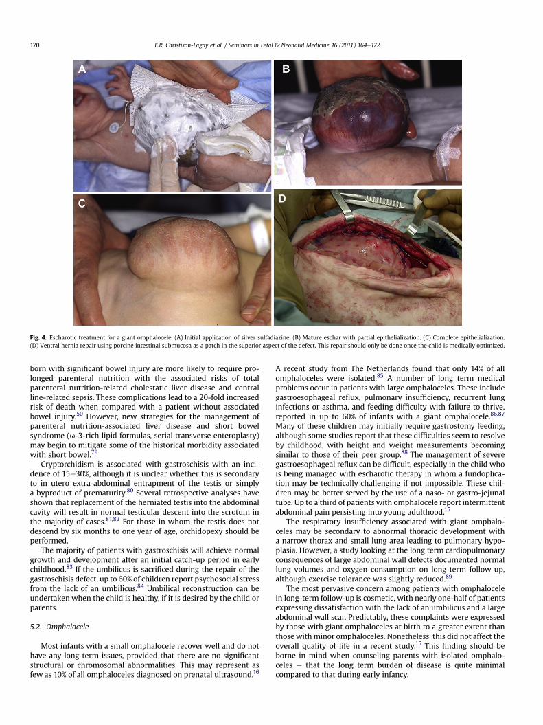
Fig. 4. Escharotic treatment for a giant omphalocele. (A) Initial application of silver sulfadiazine. (B) Mature eschar with partial epithelialization. (C) Complete epithelialization.(D) Ventral hernia repair using porcine intestinal submucosa as a patch in the superior aspect of the defect. This repair should only be done once the child is medically optimized.
E.R. Christison-Lagay et al. / Seminars in Fetal & Neonatal Medicine 16 (2011) 164e172170
born with significant bowel injury are more likely to require pro-longed parenteral nutrition with the associated risks of totalparenteral nutrition-related cholestatic liver disease and centralline-related sepsis. These complications lead to a 20-fold increasedrisk of death when compared with a patient without associatedbowel injury.50 However, new strategies for the management ofparenteral nutrition-associated liver disease and short bowelsyndrome (u-3-rich lipid formulas, serial transverse enteroplasty)may begin to mitigate some of the historical morbidity associatedwith short bowel.79
Cryptorchidism is associated with gastroschisis with an inci-dence of 15e30%, although it is unclear whether this is secondaryto in utero extra-abdominal entrapment of the testis or simplya byproduct of prematurity.80 Several retrospective analyses haveshown that replacement of the herniated testis into the abdominalcavity will result in normal testicular descent into the scrotum inthe majority of cases.81,82 For those in whom the testis does notdescend by six months to one year of age, orchidopexy should beperformed.
The majority of patients with gastroschisis will achieve normalgrowth and development after an initial catch-up period in earlychildhood.83 If the umbilicus is sacrificed during the repair of thegastroschisis defect, up to 60% of children report psychosocial stressfrom the lack of an umbilicus.84 Umbilical reconstruction can beundertaken when the child is healthy, if it is desired by the child orparents.
5.2. Omphalocele
Most infants with a small omphalocele recover well and do nothave any long term issues, provided that there are no significantstructural or chromosomal abnormalities. This may represent asfew as 10% of all omphaloceles diagnosed on prenatal ultrasound.16
A recent study from The Netherlands found that only 14% of allomphaloceles were isolated.85 A number of long term medicalproblems occur in patients with large omphaloceles. These includegastroesophageal reflux, pulmonary insufficiency, recurrent lunginfections or asthma, and feeding difficulty with failure to thrive,reported in up to 60% of infants with a giant omphalocele.86,87
Many of these children may initially require gastrostomy feeding,although some studies report that these difficulties seem to resolveby childhood, with height and weight measurements becomingsimilar to those of their peer group.88 The management of severegastroesophageal reflux can be difficult, especially in the child whois being managed with escharotic therapy in whom a fundoplica-tion may be technically challenging if not impossible. These chil-dren may be better served by the use of a naso- or gastro-jejunaltube. Up to a third of patients with omphalocele report intermittentabdominal pain persisting into young adulthood.15
The respiratory insufficiency associated with giant omphalo-celes may be secondary to abnormal thoracic development witha narrow thorax and small lung area leading to pulmonary hypo-plasia. However, a study looking at the long term cardiopulmonaryconsequences of large abdominal wall defects documented normallung volumes and oxygen consumption on long-term follow-up,although exercise tolerance was slightly reduced.89
The most pervasive concern among patients with omphalocelein long-term follow-up is cosmetic, with nearly one-half of patientsexpressing dissatisfaction with the lack of an umbilicus and a largeabdominal wall scar. Predictably, these complaints were expressedby those with giant omphaloceles at birth to a greater extent thanthosewithminor omphaloceles. Nonetheless, this did not affect theoverall quality of life in a recent study.15 This finding should beborne in mind when counseling parents with isolated omphalo-celes e that the long term burden of disease is quite minimalcompared to that during early infancy.

Practice points
� Prenatal diagnosis of omphalocele and gastroschisis
may influence timing, mode and location of delivery.
� Neonatal resuscitation should focus on temperature
stability, fluid resuscitation, placement of a nasogastric
tube, and careful physical examination looking for
associated anomalies and the condition of the herni-
ated viscera.
� The goal of surgical management is to close the
abdominal wall without injury to the viscera either
directly or due to increased intra-abdominal pressure.
The choice between primary and staged closure should
be based on this balance.
� Measurement of intra-abdominal pressure can be
helpful in guiding the decision to use primary versus
staged closure.
� The main cause of morbidity and mortality in infants
with omphalocele is associated anomalies, and the
main cause in infants with gastroschisis is the degree of
bowel injury.
Research directions
� Prediction of intestinal damage in fetuses with
gastroschisis.
� Determination of the role of cesarean section and/or
preterm delivery in fetuses with gastroschisis.
� Prediction of pulmonary hypoplasia in fetuses with
omphalocele.
� Selection of best closure technique for infants with
omphalocele and gastroschisis based on neonatal
factors.
� Development of pharmacological therapy for amelio-
rating hypomotility in children with gastroschisis.
E.R. Christison-Lagay et al. / Seminars in Fetal & Neonatal Medicine 16 (2011) 164e172 171
Conflict of interest statementNone declared.
Funding sourcesNone.
References
1. Baird PA, MacDonald EC. An epidemiologic study of congenital malformationsof the anterior abdominal wall in more than half a million consecutive livebirths. Am J Hum Genet 1981;33:470e8.
2. Barisic I, Clementi M, Husler M, et al. Evaluation of prenatal ultrasound diag-nosis of fetal abdominal wall defects by 19 European registries. UltrasoundObstet Gynecol 2001;18:309e16.
3. Rasmussen SA, Frias JL. Non-genetic risk factors for gastroschisis. Am J MedGenet 2008;148C:199e212.
4. Castilla EE, Mastroiacovo P, Orioli IM. Gastroschisis: international epidemiologyand public health perspective. Am J Med Genet 2008;148C:162e79.
5. Loane M, Dolk H, Bradbury I. Increasing prevalence of gastroschisis in Europe1980e2002: a phenomenon restricted to younger mothers? Paediatr PerinatEpidemiol 2007;21:363e9.
6. Alvarez SM, Burd RS. Increasing prevalence of gastroschisis repairs in theUnited States: 1996e2003. J Pediatr Surg 2007;42:943e6.
7. Lausman AY, Langer JC, Tai M, et al. Gastroschisis: what is the average gesta-tional age of spontaneous delivery? J Pediatr Surg 2007;42:1816e21.
8. Kronfli R, Bradnock TJ, Sabharwal A. Intestinal atresia in association with gas-troschisis: a 26 year review. J Pediatr Surg 2010;26:891e4.
9. Arnold MA, Chang DC, Nabaweesi R, et al. Risk stratification of 4344 patientswith gastroschisis into simple and complex categories. J Pediatr Surg2007;42:1520e5.
10. Russo R, D’Armiento M, Angrisani P, et al. Limb body wall complex: a criticalreview and a nosological proposal. Am J Med Genet 1993;47:893e900.
11. Craven CM, Carey JC, Ward K. Umbilical cord agenesis in limb body wall defect.Am J Med Genet 1997;71:97e105.
12. Blazer S, Zimmer EZ, Gover A, et al. Fetal omphalocele detected early inpregnancy: associated anomalies and outcomes. Radiology 2004;232:191e5.
13. Kamata SN, Usui N, Sawai T, Nose K, Fukuzawa M. Prenatal detection ofpulmonary hypoplasia in giant omphalocele. Pediatr Surg Int 2008;24:107e11.
14. Brantberg A, Blaas HG, Haugen SE, et al. Characteristics and outcome of 90cases of fetal omphalocele. Ultrasound Obstet Gynecol 2005;26:527e37.
15. van Eijck FC, Hoggeveen YL, van Weel C, Rieu PNMA, Wijnen RMH. Minor andgiant omphalocele: long-term outcomes and quality of life. J Pediatr Surg2009;44:1355e9.
16. Heider AL, Strauss RA, Kuller JA. Omphalocele: clinical outcomes in cases withnormal karyotypes. Am J Obstet Gynecol 2004;190:135e41.
17. Davis NM, Kurpios NA, Sun X, Gros J, Martin JF, Tabin CJ. The chirality of gutrotation derives from lefteright asymmetric changes in the architecture of thedorsal mesentery. Dev Cell 2008;15:134e45.
18. Thieme G. Developmental malformations of the fetal ventral body wall.Ultrasound Q 1992;10:225e65.
19. deVries PA. The pathogenesis of gastroschisis and omphalocele. J Pediatr Surg1980;15:245e51.
20. Hoyme HE, Higginbottom MC, Jones KL. The vascular pathogenesis of gastro-schisis: intrauterine interruption of the omphalomesenteric artery. J Pediatr1981;98:228e31.
21. Louw JH, Barnard CN. Congenital intestinal atresia; observations on its origin.Lancet 1955;269:1065e7.
22. Werler MM, Sheehan JE, Mitchell AA. Association of vasoconstrictive exposureswith risks of gastroschisis and small intestinal atresia. Epidemiology2003;14:349e54.
23. Feldkamp ML, Carey JC, Sadler TW. Development of gastroschisis: review ofhypotheses, a novel hypothesis, and implications for research. Am J Med GenetA 2007;143:639e52.
24. Werler MM, Mitchell AA, Moore CA, Honein MA. Is there epidemiologicevidence to support vascular disruption as a pathogenesis of gastroschisis? AmJ Med Genet 2009;149A:1399e406.
25. Stevenson RE, Rogers RC, Chandler JC, et al. Escape of the yolk sac: a hypothesisto explain the embryogenesis of gastroschisis. Clin Genet 2009;75:326e33.
26. Shaw A. The myth of gastroschisis. J Pediatr Surg 1975;10:235e44.27. Langer JC, Longaker MT, Crombleholme TM, et al. Etiology of intestinal damage
in gastroschisis. I: effects of amniotic fluid exposure and bowel constriction ina fetal lamb model. J Pediatr Surg 1989;24:992e7.
28. Langer JC, Bell JG, Castillo RO, et al. Etiology of intestinal damage in gastro-schisis. II. Timing and reversibility of histologic changes, mucosal function, andcontractility. J Pediatr Surg 1990;25:1122e6.
29. Wales PW, Christison-Lagay ER. Short bowel syndrome: epidemiology andetiology. Semin Pediatr Surg 2010;19:3e9.
30. Salihu HM, Emusu D, Aliyu ZY, et al. Mode of delivery and neonatal survival ofinfants with isolated gastroschisis. Obstet Gynecol 2004;104:678e83.
31. Segel SY, Marder SJ, Parry S, et al. Fetal abdominal wall defects and mode ofdelivery: a systematic review. Obstet Gynecol 2001;98:867e73.
32. Srinathan SK, Langer JC, Blennerhassett MG, et al. Etiology of intestinal damagein gastroschisis: III. Morphometric analysis of the smooth muscle andsubmucosa. J Pediatr Surg 1995;30:379e83.
33. Guibourdenche J, Berrebi D, Vuillard E, et al. Biochemical investigations ofbowel inflammation in gastroschisis. Pediatr Res 2006;60:565e8.
34. Vargun R, Aktug T, Heper A, et al. Effects of intrauterine treatment on inter-stitial cells of Cajal in gastroschisis. J Pediatr Surg 2007;42:783e7.
35. Luton D, de Lagausie P, Guibourdenche J, et al. Effect of amnioinfusion on theoutcome of prenatally diagnosed gastroschisis. Fetal Diagn Ther1999;14:152e5.
36. Charlesworth P, Njere I, Allotey J, et al. Postnatal outcome in gastroschisis:effect of birth weight and gestational age. J Pediatr Surg 2007;42:815e8.
37. Piper HG, Jaksic T. The impact of prenatal bowel dilation on clinical outcomesin neonates with gastroschisis. J Pediatr Surg 2006;41:897e900.
38. Langer JC, Khanna J, Caco C, et al. Prenatal diagnosis of gastroschisis: devel-opment of objective sonographic criteria for predicting outcome. ObstetGynecol 1993;81:53e6.
39. Nasr A, Langer JC. Influence of location of delivery on outcome in neonates withgastroschisis. APSA 42nd Annual Meeting, Palm Desert, CA, USA, 22e25 May2011 (abstract).
40. Coughlin JP, Drucker DE, Jewell MR, et al. Delivery room repair of gastroschisis.Surgery 1993;114:822e6.
41. Bianchi A, Dickson AP, Alizai NK. Elective delayed midgut reduction: noanesthesia for gastroschisis: selection and conversion criteria. J Pediatr Surg2002;37:1334e6.
42. Skarsgard ED, Claydon J, Bouchard S, et al. Canadian Pediatric SurgicalNetwork: a population-based pediatric surgery network and database foranalyzing surgical birth defects. The first 100 cases of gastrochisis. J Pediatr Surg2008;43:30e4.
43. Owen A, Marven S, Jackson L, et al. Experience of bedside preformed silo stagedreduction and closure for gastroschisis. J Pediatr Surg 2006;41:1830e5.
44. Allotey J, Davenport M, Njere I, et al. Benefit of preformed silos in themanagement of gastroschisis. Pediatr Surg Int 2007;23:1065e9.
45. Minkes RK, Langer JC, Mazziotti MV, et al. Routine insertion of a silastic spring-loaded silo for infants with gastroschisis. J Pediatr Surg 2000;35:843e6.
46. Pastor A, Phillips J, Fenton S, Meyers R, et al. Routine use of a Silastic spring-loaded silo for infants with gastroschisis: a multicenter randomized controlledtrial. J Pediatr Surg 2008;43:1807e12.

E.R. Christison-Lagay et al. / Seminars in Fetal & Neonatal Medicine 16 (2011) 164e172172
47. Banyard D, Ramones T, Phillips SE, Leys CM, Rauth T, Yang EY. Method to ourmadness: an 18-yr retrospective analysis on gastroschisis closure. J Pediatr Surg2010;45:579e84.
48. Jenson AR, Waldhausen JHT, Kim S. The use of a spring-loaded silo for gas-troschisis: impact on practice patterns and outcomes. Arch Surg 2009;144:1e4.
49. Sandler A, Lawrence J, Meehan J, et al. A “plastic” sutureless abdominal wallclosure in gastroschisis. J Pediatr Surg 2004;39:738e41.
50. Bonnard A, Zamakhshary M, de Silva N, et al. Nonoperative management ofgastroschisis: a case-matched study. Pediatr Surg Int 2008;24:767e71.
51. Riboh J, Abrajano CT, Garber K, et al. Outcomes of sutureless gastroschisisclosure. J Pediatr Surg 2009;44:1947e51.
52. Zivkovic SM. Repair of gastroschisis using umbilical cord as a patch. J PediatrSurg 1991;26:1179e80.
53. Lacey SR, Bruce J, Brooks SP, et al. The relative merits of various methods ofindirect measurement of intraabdominal pressure as a guide to closure ofabdominal wall defects. J Pediatr Surg 1987;22:1207e11.
54. Yaster M, Scherer TL, Stone MM, et al. Prediction of successful primary closureof congenital abdominal wall defects using intraoperative measurements.J Pediatr Surg 1989;24:1217e20.
55. Fleet MS, de la Hunt MN. Intestinal atresia with gastroschisis: a selectiveapproach to management. J Pediatr Surg 2000;35:1323e5.
56. Curry JI, Lander AD, Stringer MD. A multicenter, randomized, double-blind,placebo-controlled trial of the prokinetic agent erythromycin in the post-operative recovery of infants with gastroschisis. J Pediatr Surg 2004;39:565e9.
57. Lander A, Redkar R, Nicholls G, et al. Cisapride reduces neonatal postoperativeileus: randomised placebo controlled trial. Arch Dis Child Fetal Neonatal Ed1997;77:F119e22.
58. Jayanthi S, Seymour P, Puntis JW, et al. Necrotizing enterocolitis after gastro-schisis repair: a preventable complication? J Pediatr Surg 1998;33:705e7.
59. Oldham KT, Coran AG, Drongowski RA, et al. The development of necrotizingenterocolitis following repair of gastroschisis: a surprisingly high incidence.J Pediatr Surg 1988;23:945e9.
60. Molik KA, Gingalewski CA, West KW, et al. Gastroschisis: a plea for risk cate-gorization. J Pediatr Surg 2001;36:51e5.
61. Abdullah F, Arnold MA, Nabaweesi R, et al. Gastroschisis in the United States,1988e2003: analysis and risk categorization of 4344 patients. J Perinatol2007;27:50e5.
62. Arnold MA, Chang DC, Nabaweesi R, et al. Development and validation of a riskstratification index to predict death in gastroschisis. J Pediatr Surg2007;42:950e5.
63. Vacharajani AJ, Rao R, Keswani S, Mathur AM. Outcomes of exomphalos: aninstitutional experience. Pediatr Surg Int 2009;25:139e44.
64. Nasr A, McNamara PJ, Mertens L, et al. Is routine preoperative 2-dimenstionalechocardiography necessary for infants with esophageal atresia, omphalocele,or anorectal malformations? J Pediatr Surg 2010;45:876e9.
65. Dunn JC, Fonkalsrud EW. Improved survival of infants with omphalocele. Am JSurg 1997;173:284e7.
66. Zama M, Gallo S, Santecchia L, et al. Early reconstruction of the abdominal wallin giant omphalocele. Br J Plast Surg 2004;57:749e53.
67. Zaccara A, Zama M, Trucchi A, et al. Bipedicled skin flaps for reconstruction ofthe abdominal wall in newborn omphalocele. J Pediatr Surg 2003;38:613e5.
68. van Eijck FC, de Blaauw I, Bleichrodt RP, et al. Closure of giant omphaloceles bythe abdominal wall component separation technique in infants. J Pediatr Surg2008;43:246e50.
69. Tenenbaum MJ, Foglia RP, Becker DB, et al. Treatment of giant omphalocelewith intraabdominal tissue expansion. Plast Reconstr Surg 2007;120:1564e7.
70. Gabriel A, Gollin G. Management of complicated gastroschisis with porcinesmall intestinal submucosa and negative pressure wound therapy. J PediatrSurg 2006;41:1836e40.
71. Rahn S, Bahr M, Schalaman J, Saxena AK. Single center experience in themanagement of anterior abdominal wall defects. Hernia 2008;12:345e50.
72. Maksoud-Filho JG, Tannuri U, daSilva MM, Maksoud JG. The outcome ofnewborns with abdominal wall defects according to the method of abdominalclosure: the experience of a single center. Pediatr Surg Int 2006;22:503e7.
73. Schuster SR. A new method for the staged repair of large omphaloceles. SurgGynecol Obstet 1967;125:837e50.
74. Hong AR, Sigalet DL, Guttman FM, et al. Sequential sac ligation for giantomphalocele. J Pediatr Surg 1994;29:413e5.
75. Yeh TF, Pildes RS, Firor HV. Mercury poisoning from mercurochrome therapy ofan infected omphalocele. Clin Toxicol 1978;13:463e7.
76. Whitehouse JS, Gourlay DM, Masonbrink AR, et al. Conservative managementof giant omphalocele with topical povidone-iodine and its effect on thyroidfunction. J Pediatr Surg 2010;45:1192e7.
77. Foglia R, Kane A, Becker D, et al. Management of giant omphalocele with rapidcreation of abdominal domain. J Pediatr Surg 2006;41:704e9.
78. Cusick E, Spicer RD, Beck JM. Small-bowel continuity: a crucial factor indetermining survival in gastroschisis. Pediatr Surg Int 1997;12:34e7.
79. Le HD, Fallon EM, deMeijer VE, et al. Innovative parenteral and enteral nutri-tion for intestinal failure. Semin Pediatr Surg 2010;19:27e34.
80. Berger AP, Hager J. Management of neonates with large abdominal wall defectsand undescended testis. Urology 2006;68:175e8.
81. Lawson A, de la Hunt MN. Gastroschisis and undescended testis. J Pediatr Surg2001;36:366e7.
82. Chowdhary SK, Lander AD, Buick RG, et al. The primary management oftesticular maldescent in gastroschisis. Pediatr Surg Int 2001;17:359e60.
83. Henrich K, Huemmer HP, Reingruber B, et al. Gastroschisis and omphalocele:treatments and long-term outcomes. Pediatr Surg Int 2008;24:167e73.
84. Davies BW, Stringer MD. The survivors of gastroschisis. Arch Dis Child1997;77:158e60.
85. Cohen-Overbeek TE, Tong WH, Hatzmann TR, et al. Omphalocele: comparisonof outcome following prenatal or postnatal diagnosis. Ultrasound ObstetGynecol 2010;36:687e92.
86. Biard JM, Wilson RD, Johnson MP, et al. Prenatally diagnosed giant omphalo-celes: short- and long-term outcomes. Prenat Diagn 2004;24:434e9.
87. Koivusalo A, Rintala R, Lindahl H. Gastroesophageal reflux in children witha congenital abdominal wall defect. J Pediatr Surg 1999;34:1127e9.
88. Berseth CL, Malachowski N, Cohn RB, et al. Longitudinal growth and latemorbidity of survivors of gastroschisis and omphalocele. J Pediatr GastroenterolNutr 1982;1:375e9.
89. Zaccara A, Iacobelli BD, Calzolari A, et al. Cardiopulmonary performances inyoung children and adolescents born with large abdominal wall defects.J Pediatr Surg 2003;38:478e81.

![Cloacal exstrophy associated with gastroschisis: Case ...gastroschisis, omphalocele, bladder exstrophy, and cloacal exs-trophy [1,2]. Gastroschisis is a defect of the anterior abdominal](https://img.dokumen.tips/doc/110x75/5f82b6822991d932fc2027c1/cloacal-exstrophy-associated-with-gastroschisis-case-gastroschisis-omphalocele.jpg)