Embed Size (px)
Citation preview
Modificarea suprafetelor implantabile, in vederea cresterii
bioperformantelor.Efectul nanodimensiunii si
dimensiunea critica in bioaplicatii.I. Demetrescu
Curs POSDRU
University Politehnica of Bucharest, University Politehnica of Bucharest, ROMANIAROMANIA
Faculty of Applied Chemistry and Materials ScienceFaculty of Applied Chemistry and Materials Science
Motivatie si suport
• Motivatie : necesitatea de a imbunatati biomaterialele implantabile in contextul dezvoltarii acoperirilor de suprafata
• Suport : Proiecte si colaborari • C.N.C.S.I.S.TIPA. „Obtinerea si caracterizarea de noi micro si nanostructuri
compozite cu utilizare in ingineria tisulara”• Elaborarea si testarea in vitro si in vivo a unor elemente de protezare pentru
ortopedie, realizate din noi biomateriale romanesti • Bilaterala Franta Brincusi «Couches minces d'oxyde d'aluminium et d'oxyde
de titane pour différentes applications technologiques et biomédicales• Proiect CEEX Micro si nanostructuri obtinute prin bioactivare chimica si
electrochimica cu aplicatii in medicina regenerativa• Proiect PN2 IDEI Studii exploratorii asupra mecanismului de formare si
inducere de noi proprietati unor electrozi modificati cu forme structurale TiO2nanotuburi / nanoparticule si compozite polimerice
• Proiect PN2 IDEI complexe PCCE Noi concepte si strategii pentru
dezvoltarea cunoasterii unor noi structuri biocompatibile in bioinginerie
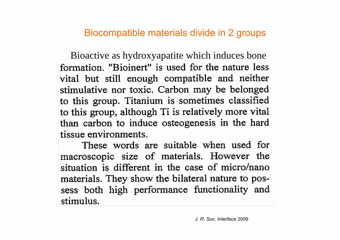
Biocompatible materials divide in 2 groups
Bioactive as hydroxyapatite which induces bone
J. R. Soc. Interface 2009
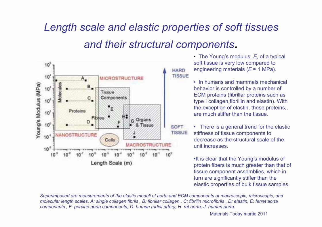
Length scale and elastic properties of soft tissues and their structural components.
Superimposed are measurements of the elastic moduli of aorta and ECM components at macroscopic, microscopic, and molecular length scales. A: single collagen fibrils , B: fibrillar collagen , C: fibrilin microfibrils , D: elastin, E: ferret aorta components , F: porcine aorta components, G: human radial artery, H: rat aorta, J: human aorta.
• The Young’s modulus, E, of a typical soft tissue is very low compared to engineering materials (E ≈ 1 MPa).
• In humans and mammals mechanical behavior is controlled by a number of ECM proteins (fibrillar proteins such as type I collagen,fibrillin and elastin). With the exception of elastin, these proteins,, are much stiffer than the tissue.
• There is a general trend for the elastic stiffness of tissue components to decrease as the structural scale of the unit increases.
•It is clear that the Young’s modulus of protein fibers is much greater than that of tissue component assemblies, which inturn are significantly stiffer than the elastic properties of bulk tissue samples.
Materials Today martie 2011
Biocompatible materials show different
particle size dependence.• Non-soluble, biocompatible materials show different particle size dependence. The transition of bioreaction occurs at the critical sizes of approximately 100 mm,10 mm and 200 nm.
• This physical size effect is nonspecific,which is independent of material type, and especially pronounced for a cell size of less than approximately 10 mm. •It causes stimulus through the biological process of phagocytosis in cell and inflammation in tissue.•When the particle size is 200 nm or less, they become less stimulative and the recognition by body defence systems becomes weaker. They mayinvade directly into the internal body through the respiratory or digestive system and diffuse inside the whole body.
•Thus, the physical micro/nanosizing of materials makes them biointeractive with cells and tissue.
Watari, F.: Bioreactive nature of nanobiomaterials, Nanobiomedicine, 1, 2-8 (2010).
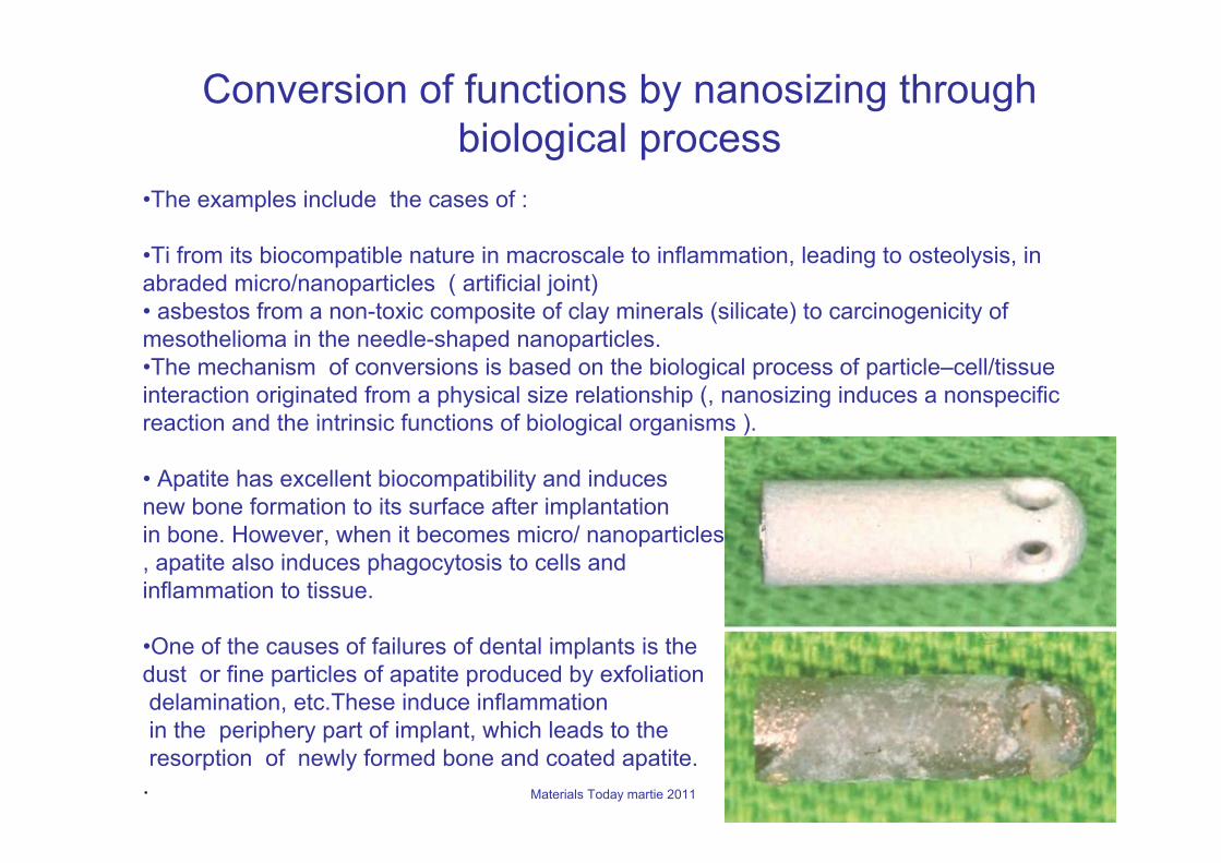
Conversion of functions by nanosizing through biological process
•The examples include the cases of :
•Ti from its biocompatible nature in macroscale to inflammation, leading to osteolysis, in abraded micro/nanoparticles ( artificial joint)• asbestos from a non-toxic composite of clay minerals (silicate) to carcinogenicity of mesothelioma in the needle-shaped nanoparticles.•The mechanism of conversions is based on the biological process of particle–cell/tissue interaction originated from a physical size relationship (, nanosizing induces a nonspecificreaction and the intrinsic functions of biological organisms ).
• Apatite has excellent biocompatibility and inducesnew bone formation to its surface after implantation in bone. However, when it becomes micro/ nanoparticles, apatite also induces phagocytosis to cells and inflammation to tissue.
•One of the causes of failures of dental implants is the dust or fine particles of apatite produced by exfoliationdelamination, etc.These induce inflammationin the periphery part of implant, which leads to theresorption of newly formed bone and coated apatite.
. Materials Today martie 2011
Improved bone-forming functionality on diameter-controlled TiO2
nanotube surface (30–100 nm diameter)
TiO2 nanotube surface enables accelerated osteoblast adhesion and exhibits strong bonding with bone. Various sizes of TiO2 nanotubes on titanium substrates were prepared and the osteoblast cellular behavior was investigated.
The result is that a change in osteoblast behavior is obtained in a relatively narrow range of nanotube dimensions, with small diameter (30 nm) promoting the highest degree of osteoblast adhesion, while larger diameter (70–100 nm) nanotubes elicit a lower population of cells with extremely elongated cellular morphology and much higher alkaline phosphatase levels. ( merit and demerit aspects as size dependent behavior)
Such nanotubes already being a strongly osseointegrating implant material, offer proofs for the optimization of orthopedics-related treatments
K.S. Brammer, O. Seunghan, C.J. Cobb, L.M. Bjursten, H. van der Heyde, Sungho Jin, Improved bone-forming functionality on diameter-controlled TiO2 nanotube surface, Acta Biomater (5) (2009) 3215–3223.
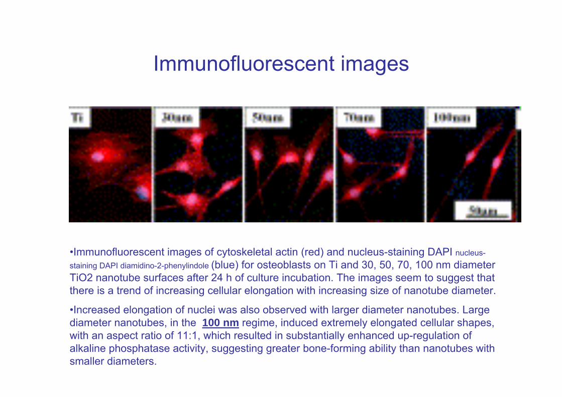
Immunofluorescent images
•Immunofluorescent images of cytoskeletal actin (red) and nucleus-staining DAPI nucleus-staining DAPI diamidino-2-phenylindole (blue) for osteoblasts on Ti and 30, 50, 70, 100 nm diameter TiO2 nanotube surfaces after 24 h of culture incubation. The images seem to suggest that there is a trend of increasing cellular elongation with increasing size of nanotube diameter.
•Increased elongation of nuclei was also observed with larger diameter nanotubes. Large diameter nanotubes, in the 100 nm regime, induced extremely elongated cellular shapes, with an aspect ratio of 11:1, which resulted in substantially enhanced up-regulation of alkaline phosphatase activity, suggesting greater bone-forming ability than nanotubes with smaller diameters.
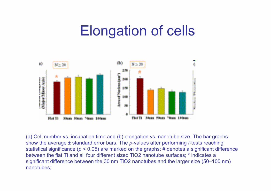
Elongation of cells
(a) Cell number vs. incubation time and (b) elongation vs. nanotube size. The bar graphs show the average ± standard error bars. The p-values after performing t-tests reaching statistical significance (p < 0.05) are marked on the graphs: # denotes a significant difference between the flat Ti and all four different sized TiO2 nanotube surfaces; * indicates a significant difference between the 30 nm TiO2 nanotubes and the larger size (50–100 nm) nanotubes;
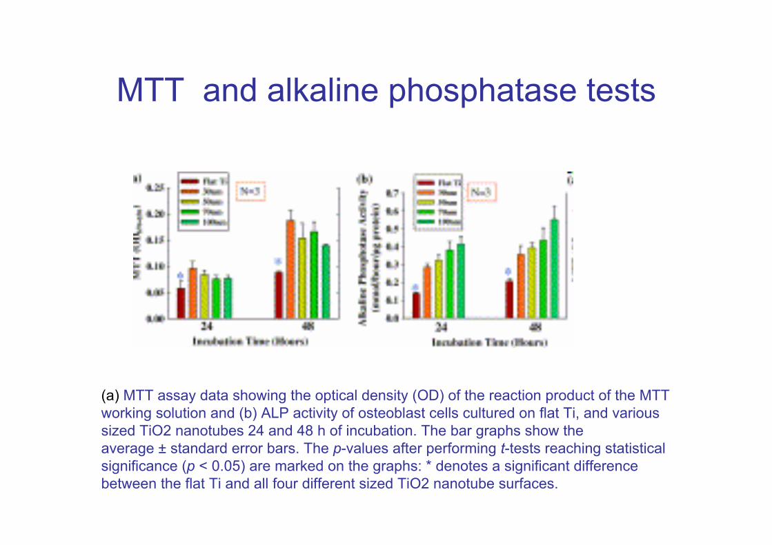
MTT and alkaline phosphatase tests
(a) MTT assay data showing the optical density (OD) of the reaction product of the MTT working solution and (b) ALP activity of osteoblast cells cultured on flat Ti, and various sized TiO2 nanotubes 24 and 48 h of incubation. The bar graphs show the average ± standard error bars. The p-values after performing t-tests reaching statistical significance (p < 0.05) are marked on the graphs: * denotes a significant difference between the flat Ti and all four different sized TiO2 nanotube surfaces.
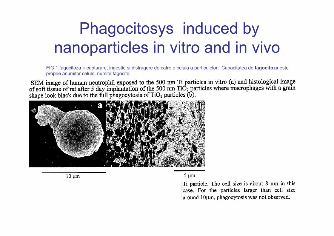
Phagocitosys induced by nanoparticles in vitro and in vivo
FIG 1.fagocitoza = capturare, ingestie si distrugere de catre o celula a particulelor. Capacitatea de fagocitoza este proprie anumitor celule, numite fagocite,
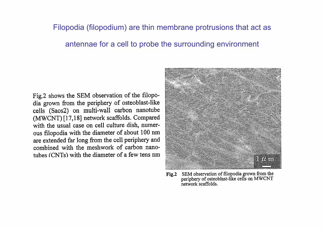
Filopodia (filopodium) are thin membrane protrusions that act as
antennae for a cell to probe the surrounding environment
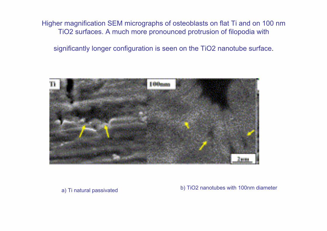
Higher magnification SEM micrographs of osteoblasts on flat Ti and on 100 nm TiO2 surfaces. A much more pronounced protrusion of filopodia with
significantly longer configuration is seen on the TiO2 nanotube surface.
a) Ti natural passivated b) TiO2 nanotubes with 100nm diameter
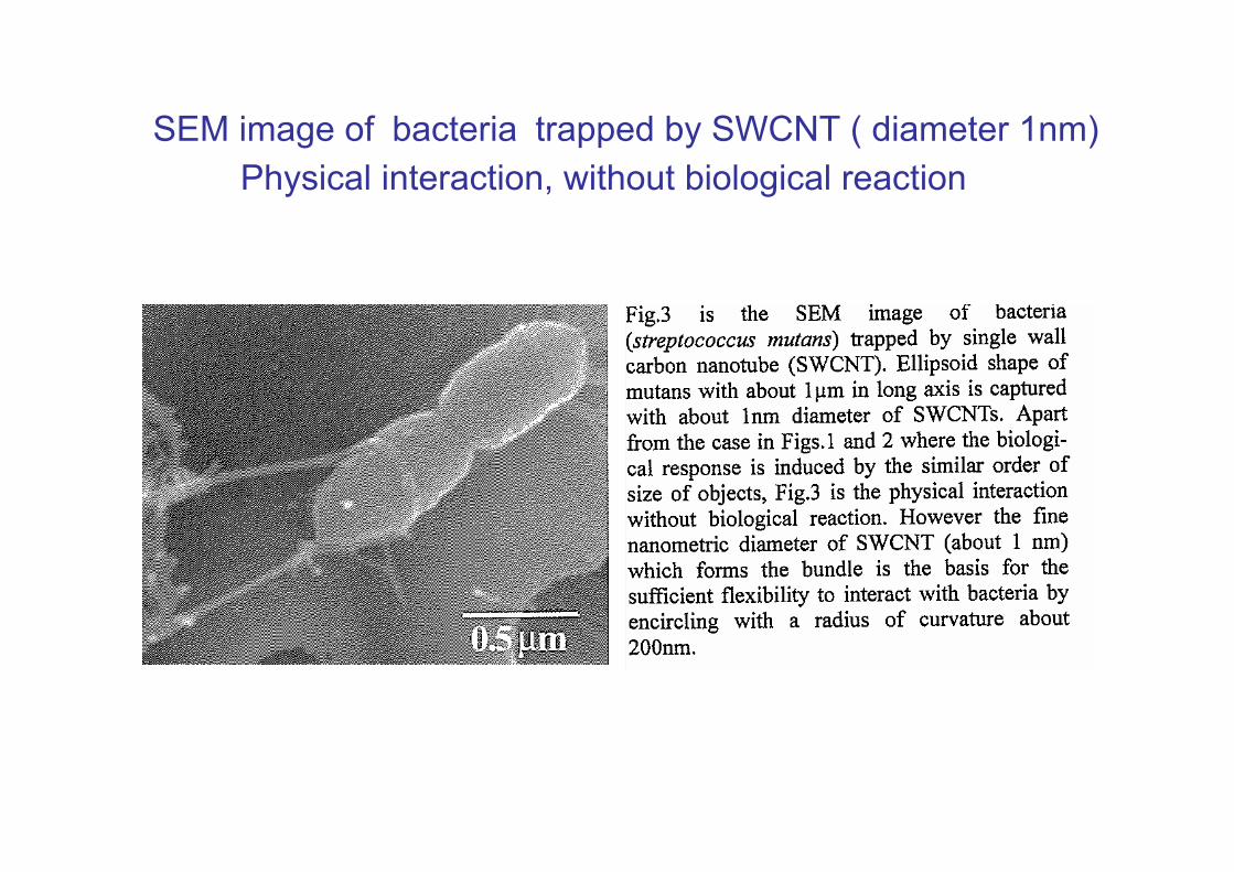
SEM image of bacteria trapped by SWCNT ( diameter 1nm)Physical interaction, without biological reaction
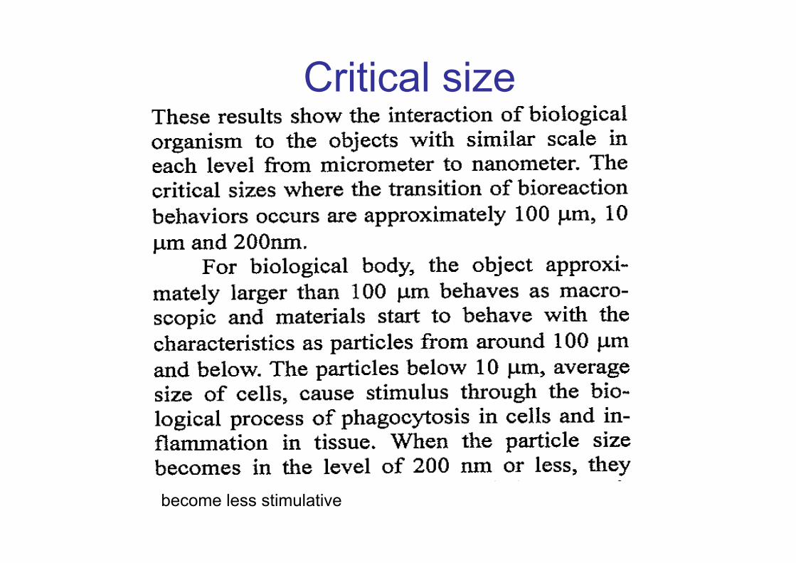
Critical size
become less stimulative
Biointeractive particles which arouse phagocitosys
Nanosizing effect
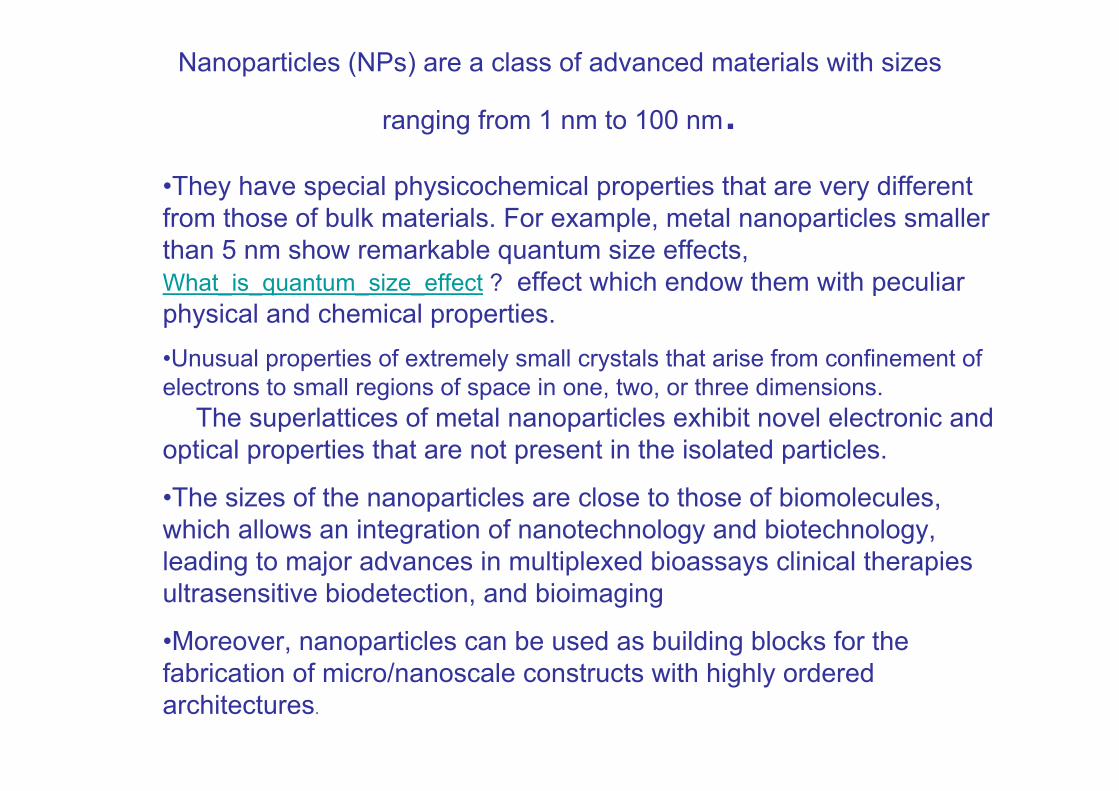
Nanoparticles (NPs) are a class of advanced materials with sizes
ranging from 1 nm to 100 nm.•They have special physicochemical properties that are very different from those of bulk materials. For example, metal nanoparticles smaller than 5 nm show remarkable quantum size effects, What_is_quantum_size_effect ? effect which endow them with peculiar physical and chemical properties. •Unusual properties of extremely small crystals that arise from confinement of electrons to small regions of space in one, two, or three dimensions.
The superlattices of metal nanoparticles exhibit novel electronic and optical properties that are not present in the isolated particles.
•The sizes of the nanoparticles are close to those of biomolecules, which allows an integration of nanotechnology and biotechnology,leading to major advances in multiplexed bioassays clinical therapies ultrasensitive biodetection, and bioimaging
•Moreover, nanoparticles can be used as building blocks for the fabrication of micro/nanoscale constructs with highly ordered architectures.
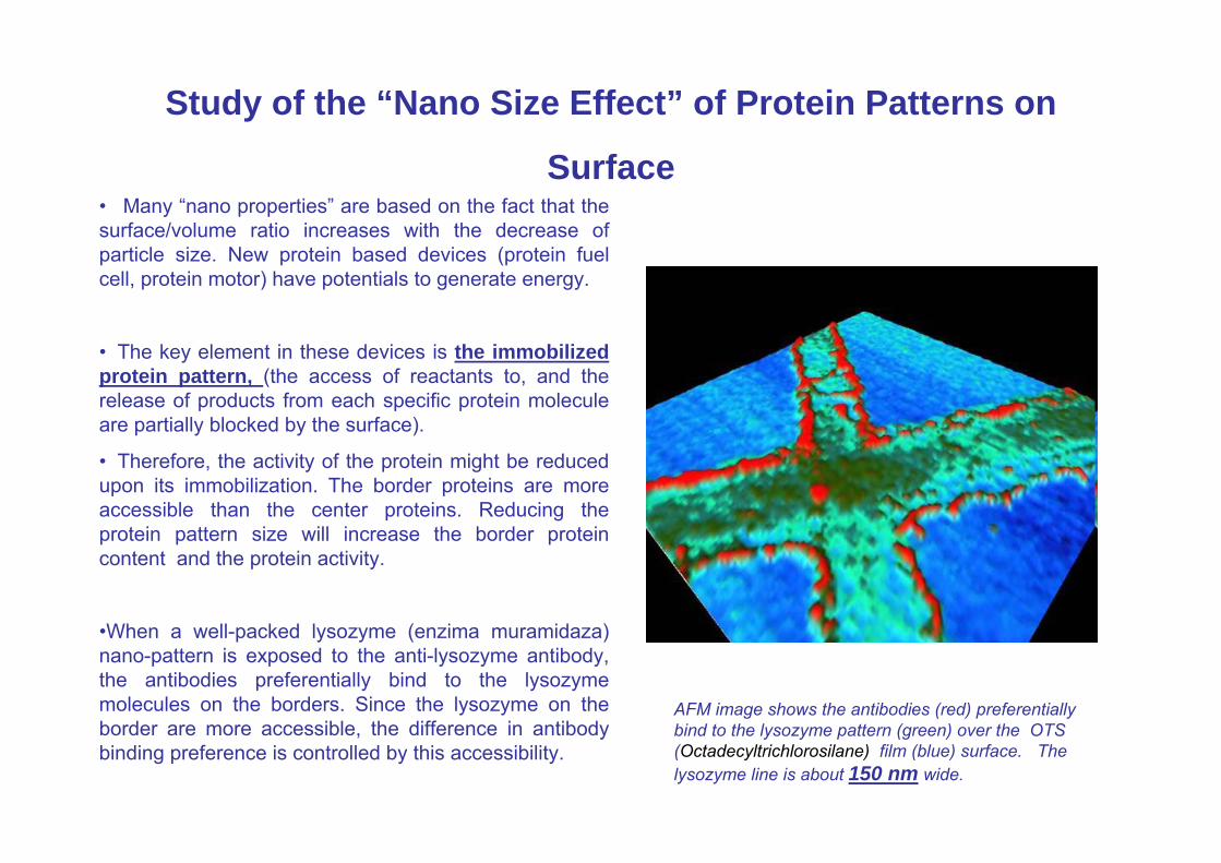
Study of the “Nano Size Effect” of Protein Patterns on
Surface• Many “nano properties” are based on the fact that the surface/volume ratio increases with the decrease of particle size. New protein based devices (protein fuel cell, protein motor) have potentials to generate energy.
• The key element in these devices is the immobilized protein pattern, (the access of reactants to, and the release of products from each specific protein molecule are partially blocked by the surface).
• Therefore, the activity of the protein might be reduced upon its immobilization. The border proteins are more accessible than the center proteins. Reducing the protein pattern size will increase the border protein content and the protein activity.
•When a well-packed lysozyme (enzima muramidaza) nano-pattern is exposed to the anti-lysozyme antibody, the antibodies preferentially bind to the lysozyme molecules on the borders. Since the lysozyme on the border are more accessible, the difference in antibody binding preference is controlled by this accessibility.
AFM image shows the antibodies (red) preferentially bind to the lysozyme pattern (green) over the OTS(Octadecyltrichlorosilane) film (blue) surface. The lysozyme line is about 150 nm wide.
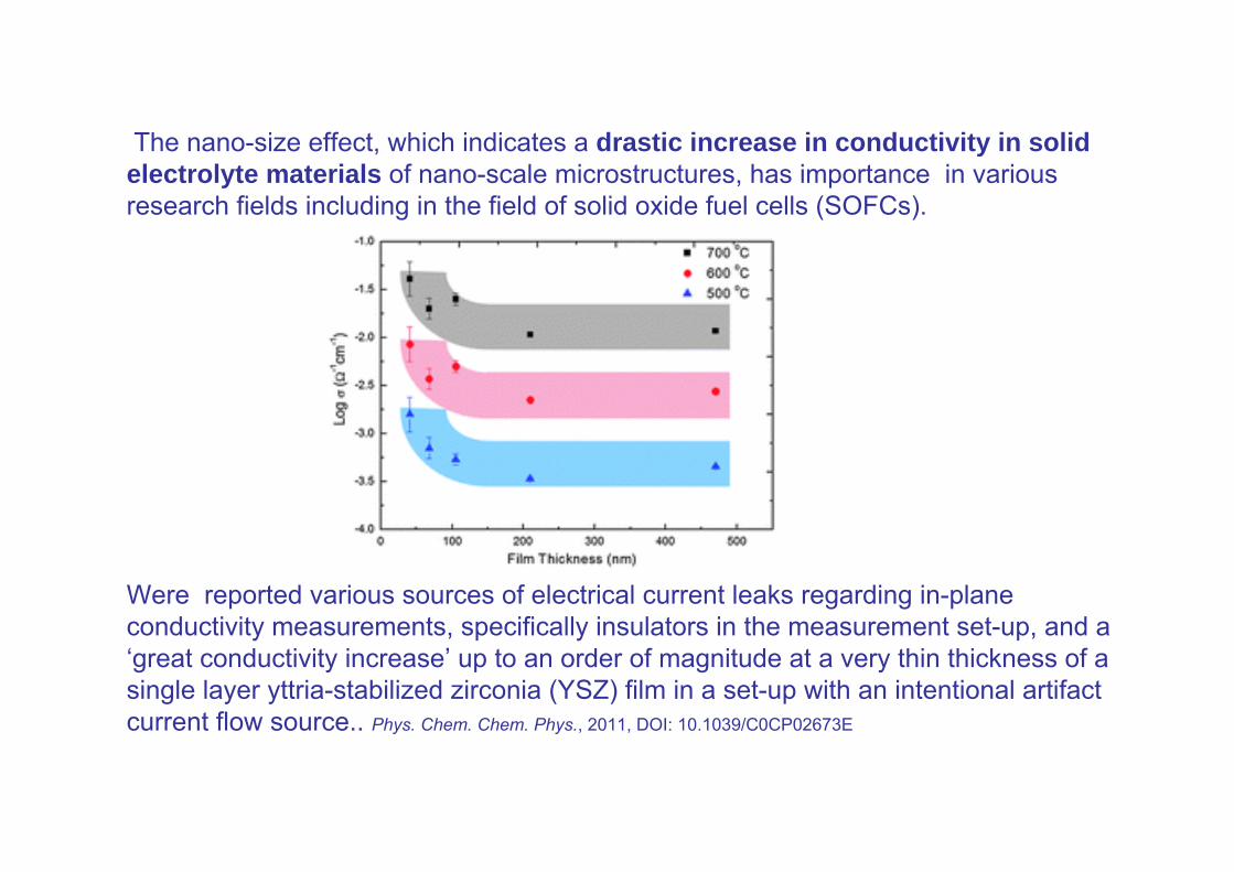
The nano-size effect, which indicates a drastic increase in conductivity in solid electrolyte materials of nano-scale microstructures, has importance in various research fields including in the field of solid oxide fuel cells (SOFCs).
Were reported various sources of electrical current leaks regarding in-plane conductivity measurements, specifically insulators in the measurement set-up, and a ‘great conductivity increase’ up to an order of magnitude at a very thin thickness of a single layer yttria-stabilized zirconia (YSZ) film in a set-up with an intentional artifact current flow source.. Phys. Chem. Chem. Phys., 2011, DOI: 10.1039/C0CP02673E
Nanosize effect on the hygroscopic growth factor of
aerosol particles
The hygroscopic growth factors of NaCl particles having dry mobility diameters of 6 to 60 nm were measured using a tandem nano�Differential Mobility Analyzer.
The growth factors steadily decreased within detection limit for dry sizes below 40 nm. The decrease is quantitatively predicted by a model that includes the Kelvin effect and a size�dependent shape factor. This factor is not tuned to the data but rather is grounded in theoretical predictions from literature.
Agreement in growth factors for particles generated by two independent methods (namely, vaporization�condensation and electrospray), as well as observations of prompt deliquescence, indicates the absence of significant chemical impurities
G. Biskos, L. M. Russell, P. R. Buseck, and S. T. Martin (2006), Nanosize effect on the hygroscopic growth factor of aerosol particles, Geophys. Res. Lett., 33, L07801, doi:10.1029/2005GL025199. .
Nanosize Effect on High-Rate Li-Ion Intercalation in LiCoO2 Electrode
• The main difficulty in high-rate charge−discharge experiments is kinetic problems due to the slow diffusion of Li-ions in electrodes. Nanosizing is a way to achieve a higher surface area and shorter Li-ion diffusion length for fast diffusion.• However, while various nanoelectrodes that provide excellent high-rate capability have been synthesized, a size-controlled synthesis and a systematic study of nanocrystalline LiCoO2 have not been carried out because of the difficulty in controlling the size. • Now, have been established the size-controlled synthesis of nanocrystalline LiCoO2 through hydrothermal reaction and, clarified the structural and electrochemical properties of this intercalation cathode material.• Lattice expansion in nanocrystalline LiCoO2 was found from X-ray diffraction and Raman spectroscopy. Electrochemical measurements and theoretical analyses on nanocrystalline LiCoO2 revealed that extreme size reduction below 15 nm was not favorable for most applications
J. Am. Chem. Soc., 2007, 129 (23), pp 7444–7452DOI: 10.1021/ja0681927
Effects of nanosize stress concentrators (NSCs) Mechanics Research Communications 33 (2006) 352–358
• There is evidence that when at least one spatial dimension of a material component is in the nanometer range, the effects of nanosize stress concentrators (NSCs) such as impurities, inclusions, pores, and cracks are either eliminated orsignificantly reduced. • Using atomistic simulation techniques for a crystalline metal the critical dimensions below which the effects of NSCs are minimal or even nonexistentwas identified . The preliminary results reported herein show that for Cu single crystals subjected to constant external strain rate, such critical dimensions are larger than about 30 nm‘‘• Straightforward’’ reasoning suggests that as size approaches atomic dimensions, the theoretical material strength should be approached. • For Cu subjected to tensile strain at the examined strain rates (109 s-1), the critical dimensions for the effects of NSCs are larger than the examined (up to) 28.8 nm. Multiscale simulations are necessary to identify critical dimensions and also examine slower strain rates.
The nanotube size-dependent melting of TaC and Pb single crystals
•A model for the dependence of the melting temperature of nanocrystals on the size of carbon nanotubes is developed. •The model is applied to investigate the nanotube size-dependent melting of TaC and Pb single crystals encapsulated in carbon nanotubes. It is shown that the melting temperature for these single crystals in carbon nanotubes can be strongly suppressed. The results also imply that nanotubes may provide an effective means for investigating the supercooled state of liquids and the liquid–glass transition.
Nanodimensions and catalysis• Catalysis research is focusing on nanosized catalysts since they often show an improved activity; improvement due to their greatly increased surface / volume ratio.
•Bulk gold is exceptionally inert. Nanosized gold particles are, catalytically very activethe structure of the nano-particles is uncertain, with claims being made for truncated octahedra, cub-octahedra, icosahedra, various kinds of decahedra, and amorphousstructures.
• Gold nanoparticles were synthesized by chemical reduction of cloroauric acid (HAuCl4) inside the pores of a polycarbonate-based membrane followed by dissolving the membrane in dichloromethane and further sonication Int.J.Nano.Dim. 1(4): 275-278, Spring 2011
•Gold nanoparticles dispersed across the surfaces of certain oxides have amazingly active and selective as catalysts for a variety of important reactions. -There is intense interest in these catalysts for carbon monoxide oxidation, because they are active at room temperature. The low-temperature gold catalysts are totally inactive unless the gold is in the form of particles smaller than ~8 nm in diameter Chen and Goodman have produced a highly active model gold catalyst where the gold is incorporated in a crystalline film, spread uniformly over a Ti2O3 surface
Charles T. Campbell (Science 2004 306:234)
Gold nanoparticles closer to a treatment for cancer
Average particle size < 100 nm NanoLett. 2011, 11 (3), 1358
• Small dose of gold nanoparticles can activate or inhibit genes that are involved in angiogenesis (growth of new blood vessels from pre-existing vessels). - a complex process responsible for the supply of oxygen and nutrients to most types of cancer .•"The peptide-functionalised gold nanoparticles are very effective in the deliberate activation or inhibition of angiogenic genes,“
•Endothelial cells construct the interior of blood vessels and play a pivotal role in angiogenesis• control the degree of damage to the endothelial cells using laser illumination.•gold nanoparticle could be used as effective tools in cellular nanosurgery.gold nanoparticles can have a dual role in cellular manipulation.
•Applying laser iradiation the nanoparticles can destroy endothelial cells, as a measure to cut the blood supply to tumours, or to deliberately open up the cellular membrane in order to deliver a drug efficiently."
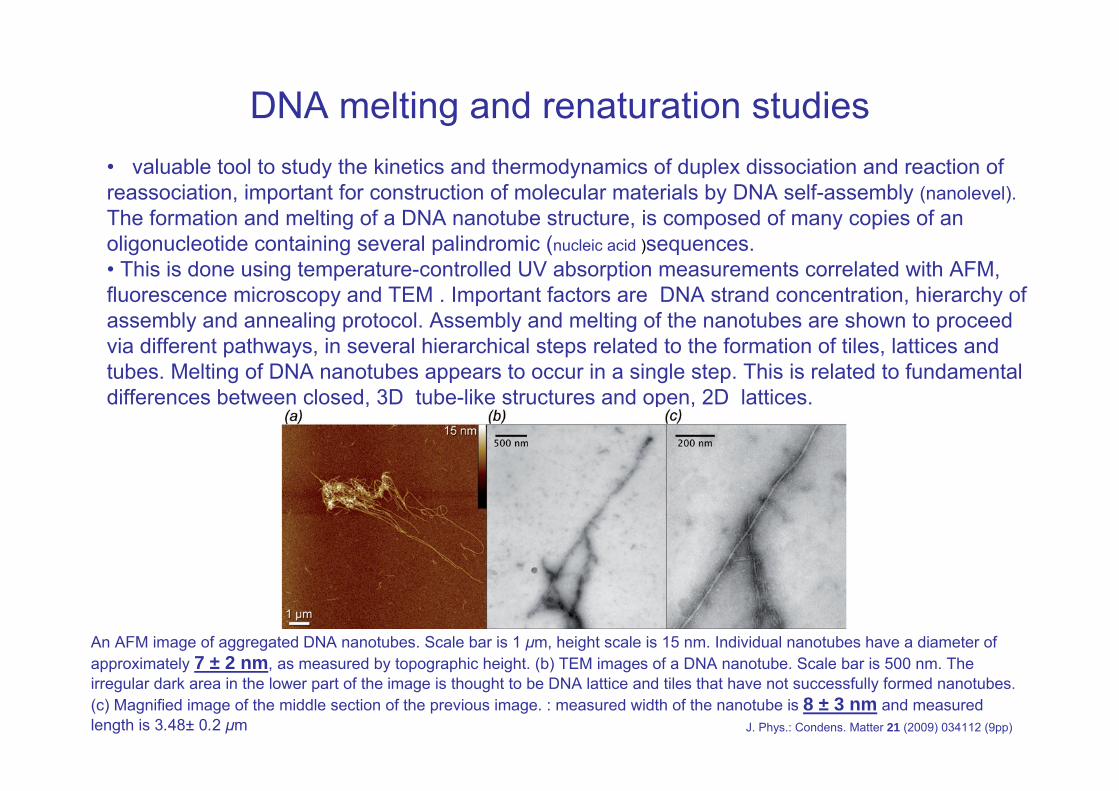
DNA melting and renaturation studies• valuable tool to study the kinetics and thermodynamics of duplex dissociation and reaction of reassociation, important for construction of molecular materials by DNA self-assembly (nanolevel).The formation and melting of a DNA nanotube structure, is composed of many copies of an oligonucleotide containing several palindromic (nucleic acid )sequences.• This is done using temperature-controlled UV absorption measurements correlated with AFM,fluorescence microscopy and TEM . Important factors are DNA strand concentration, hierarchy of assembly and annealing protocol. Assembly and melting of the nanotubes are shown to proceed via different pathways, in several hierarchical steps related to the formation of tiles, lattices and tubes. Melting of DNA nanotubes appears to occur in a single step. This is related to fundamental differences between closed, 3D tube-like structures and open, 2D lattices.
An AFM image of aggregated DNA nanotubes. Scale bar is 1 μm, height scale is 15 nm. Individual nanotubes have a diameter of approximately 7 ± 2 nm, as measured by topographic height. (b) TEM images of a DNA nanotube. Scale bar is 500 nm. The irregular dark area in the lower part of the image is thought to be DNA lattice and tiles that have not successfully formed nanotubes. (c) Magnified image of the middle section of the previous image. : measured width of the nanotube is 8 ± 3 nm and measured length is 3.48± 0.2 μm J. Phys.: Condens. Matter 21 (2009) 034112 (9pp)
Silver medicinal usesHistorically• The Phoenicians have stored water, wine, and vinegar in silver bottles to prevent spoiling.• In the early 1900s, people would put silver coins in milk bottles to prolong the milk's freshness.• Hippocrates had discussed silver antidisease properties.
• In the early 1900s, silver gained approval as an antimicrobial agent.• Prior to the introduction of antibiotics, colloidal silver was used as a germicide and disinfectant. • Although "silver products were infrequently promoted for oral use, benefits have been even more questionable
• with introduction of antibiotics (1940s), silver use as antimicrobial agent diminished , until the introduction on large scale of silver nanoparicles with various size as antibacterial agent
• recently antibacterial treatments with antibiotics are becoming less effective due to their intensive use.
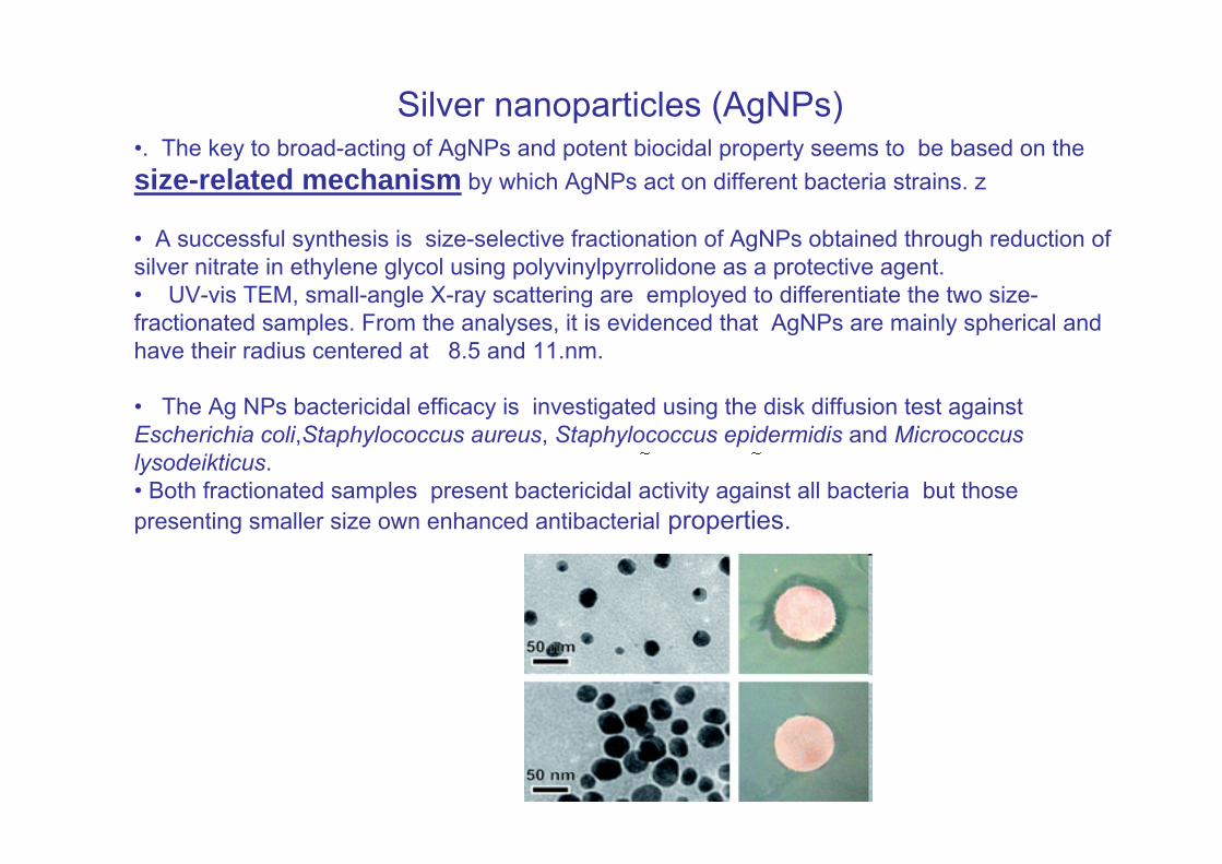
Silver nanoparticles (AgNPs)•. The key to broad-acting of AgNPs and potent biocidal property seems to be based on the size-related mechanism by which AgNPs act on different bacteria strains. z
• A successful synthesis is size-selective fractionation of AgNPs obtained through reduction of silver nitrate in ethylene glycol using polyvinylpyrrolidone as a protective agent. • UV-vis TEM, small-angle X-ray scattering are employed to differentiate the two size-fractionated samples. From the analyses, it is evidenced that AgNPs are mainly spherical and have their radius centered at 8.5 and 11.nm.
• The Ag NPs bactericidal efficacy is investigated using the disk diffusion test against Escherichia coli,Staphylococcus aureus, Staphylococcus epidermidis and Micrococcus lysodeikticus. • Both fractionated samples present bactericidal activity against all bacteria but those presenting smaller size own enhanced antibacterial properties.
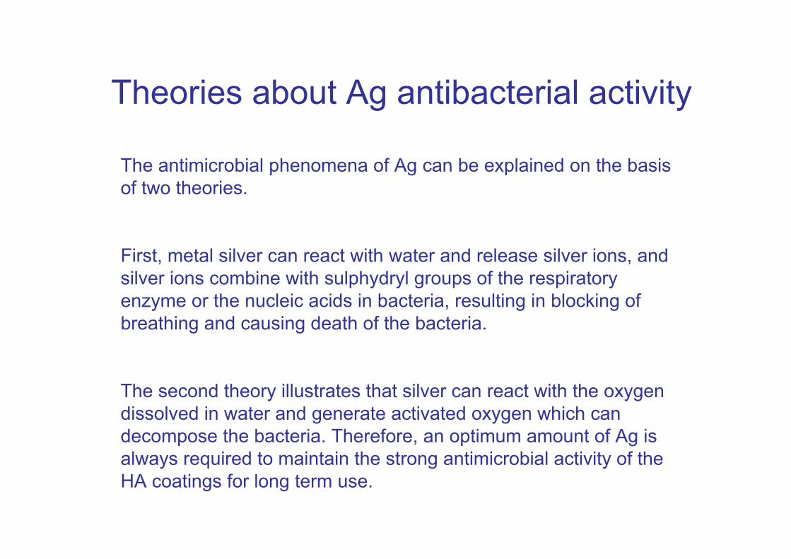
Theories about Ag antibacterial activity
The antimicrobial phenomena of Ag can be explained on the basis of two theories.
First, metal silver can react with water and release silver ions, and silver ions combine with sulphydryl groups of the respiratory enzyme or the nucleic acids in bacteria, resulting in blocking of breathing and causing death of the bacteria.
The second theory illustrates that silver can react with the oxygen dissolved in water and generate activated oxygen which can decompose the bacteria. Therefore, an optimum amount of Ag is always required to maintain the strong antimicrobial activity of the HA coatings for long term use.
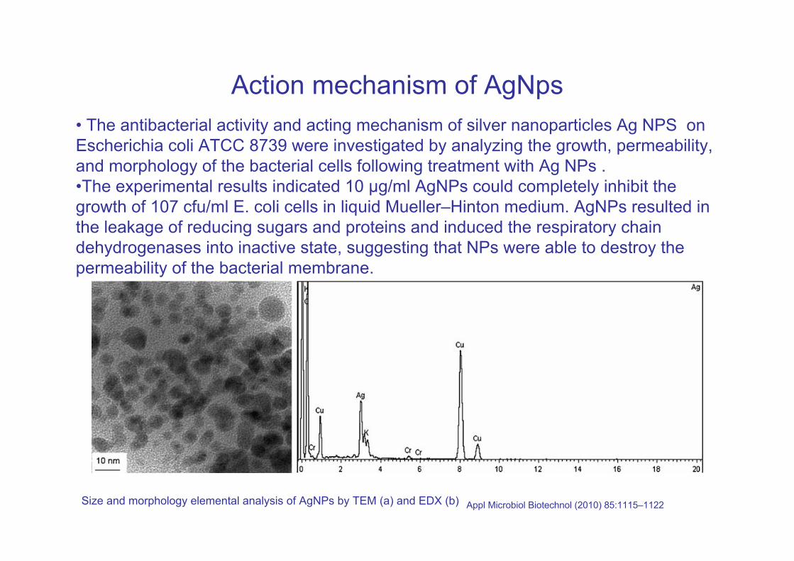
Action mechanism of AgNps• The antibacterial activity and acting mechanism of silver nanoparticles Ag NPS on Escherichia coli ATCC 8739 were investigated by analyzing the growth, permeability, and morphology of the bacterial cells following treatment with Ag NPs . •The experimental results indicated 10 μg/ml AgNPs could completely inhibit thegrowth of 107 cfu/ml E. coli cells in liquid Mueller–Hinton medium. AgNPs resulted in the leakage of reducing sugars and proteins and induced the respiratory chaindehydrogenases into inactive state, suggesting that NPs were able to destroy the permeability of the bacterial membrane.
Size and morphology elemental analysis of AgNPs by TEM (a) and EDX (b) Appl Microbiol Biotechnol (2010) 85:1115–1122
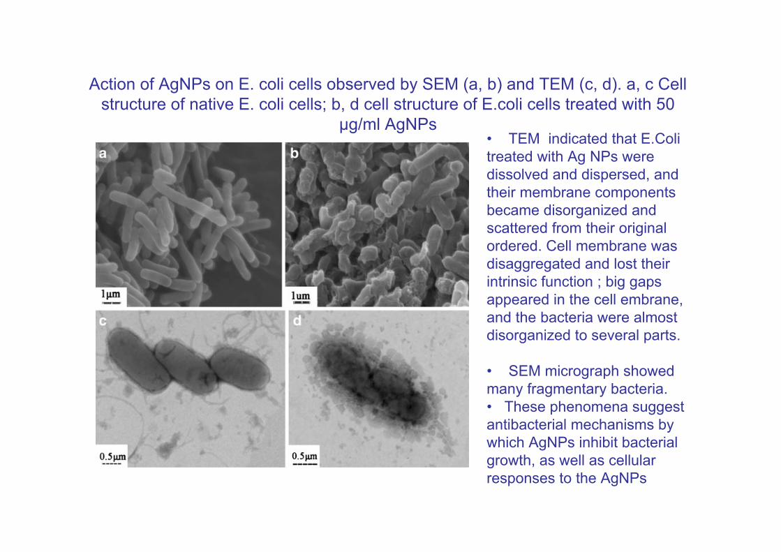
Action of AgNPs on E. coli cells observed by SEM (a, b) and TEM (c, d). a, c Cell structure of native E. coli cells; b, d cell structure of E.coli cells treated with 50
μg/ml AgNPs• TEM indicated that E.Coli treated with Ag NPs were dissolved and dispersed, and their membrane components became disorganized and scattered from their original ordered. Cell membrane was disaggregated and lost their intrinsic function ; big gaps appeared in the cell embrane, and the bacteria were almost disorganized to several parts.
• SEM micrograph showedmany fragmentary bacteria. • These phenomena suggestantibacterial mechanisms by which AgNPs inhibit bacterial growth, as well as cellular responses to the AgNPs
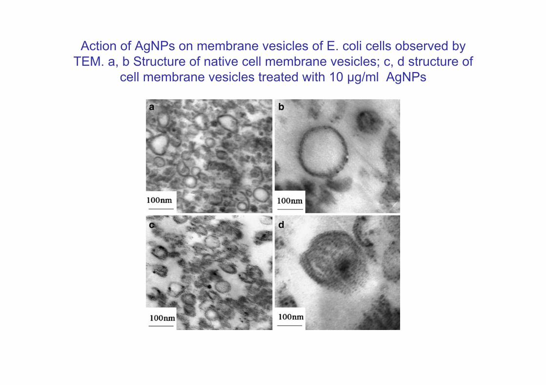
Action of AgNPs on membrane vesicles of E. coli cells observed by TEM. a, b Structure of native cell membrane vesicles; c, d structure of
cell membrane vesicles treated with 10 μg/ml AgNPs
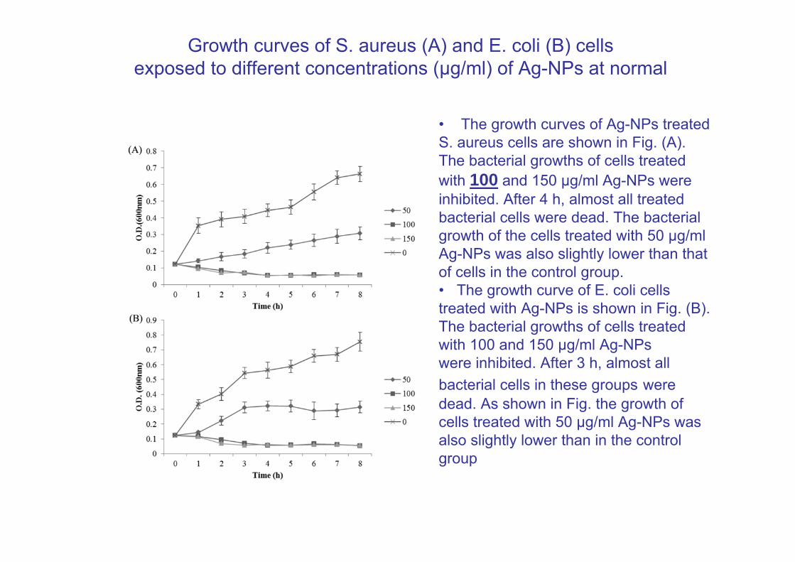
Growth curves of S. aureus (A) and E. coli (B) cellsexposed to different concentrations (μg/ml) of Ag-NPs at normal
• The growth curves of Ag-NPs treated S. aureus cells are shown in Fig. (A). The bacterial growths of cells treated with 100 and 150 μg/ml Ag-NPs were inhibited. After 4 h, almost all treated bacterial cells were dead. The bacterial growth of the cells treated with 50 μg/ml Ag-NPs was also slightly lower than that of cells in the control group. • The growth curve of E. coli cellstreated with Ag-NPs is shown in Fig. (B). The bacterial growths of cells treated with 100 and 150 μg/ml Ag-NPswere inhibited. After 3 h, almost all bacterial cells in these groups were dead. As shown in Fig. the growth of cells treated with 50 μg/ml Ag-NPs was also slightly lower than in the control group
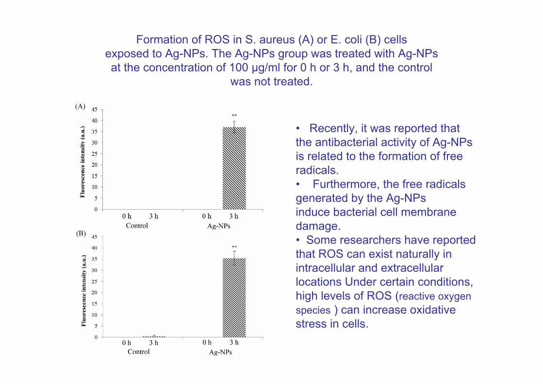
Formation of ROS in S. aureus (A) or E. coli (B) cellsexposed to Ag-NPs. The Ag-NPs group was treated with Ag-NPsat the concentration of 100 μg/ml for 0 h or 3 h, and the control
was not treated.
• Recently, it was reported that the antibacterial activity of Ag-NPs is related to the formation of free radicals.• Furthermore, the free radicals generated by the Ag-NPsinduce bacterial cell membrane damage. • Some researchers have reported that ROS can exist naturally in intracellular and extracellular locations Under certain conditions,high levels of ROS (reactive oxygen species ) can increase oxidative stress in cells.
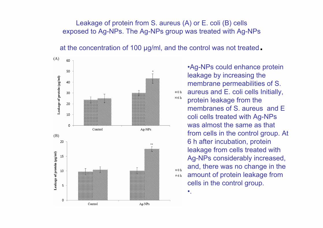
Leakage of protein from S. aureus (A) or E. coli (B) cellsexposed to Ag-NPs. The Ag-NPs group was treated with Ag-NPs
at the concentration of 100 μg/ml, and the control was not treated.•Ag-NPs could enhance protein leakage by increasing the membrane permeabilities of S. aureus and E. coli cells Initially, protein leakage from themembranes of S. aureus and E coli cells treated with Ag-NPs was almost the same as that from cells in the control group. At6 h after incubation, protein leakage from cells treated withAg-NPs considerably increased, and, there was no change in the amount of protein leakage from cells in the control group.•.
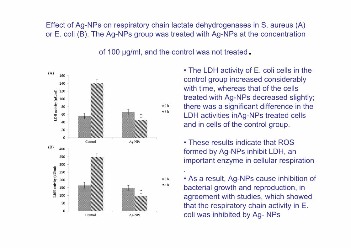
Effect of Ag-NPs on respiratory chain lactate dehydrogenases in S. aureus (A) or E. coli (B). The Ag-NPs group was treated with Ag-NPs at the concentration
of 100 μg/ml, and the control was not treated.• The LDH activity of E. coli cells in thecontrol group increased considerably with time, whereas that of the cells treated with Ag-NPs decreased slightly;there was a significant difference in the LDH activities inAg-NPs treated cells and in cells of the control group.
• These results indicate that ROS formed by Ag-NPs inhibit LDH, an important enzyme in cellular respiration. • As a result, Ag-NPs cause inhibition of bacterial growth and reproduction, in agreement with studies, which showed that the respiratory chain activity in E. coli was inhibited by Ag- NPs
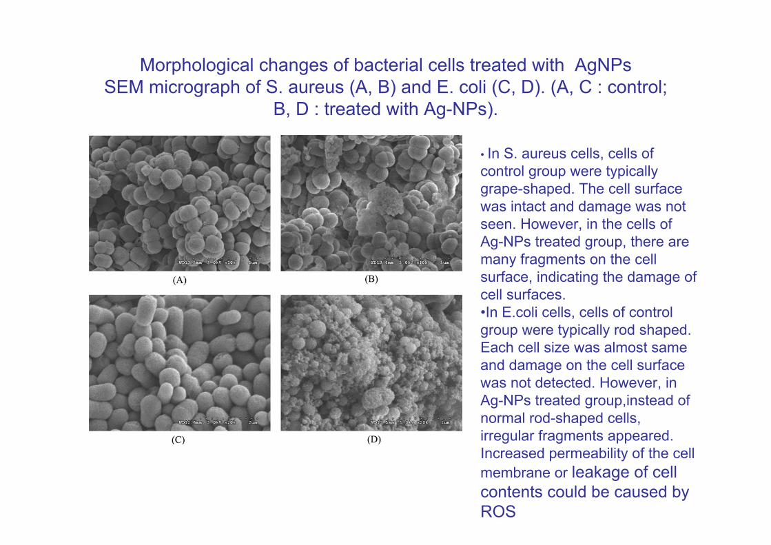
Morphological changes of bacterial cells treated with AgNPsSEM micrograph of S. aureus (A, B) and E. coli (C, D). (A, C : control;
B, D : treated with Ag-NPs).
• In S. aureus cells, cells ofcontrol group were typically grape-shaped. The cell surfacewas intact and damage was not seen. However, in the cells of Ag-NPs treated group, there are many fragments on the cell surface, indicating the damage of cell surfaces. •In E.coli cells, cells of control group were typically rod shaped.Each cell size was almost same and damage on the cell surface was not detected. However, in Ag-NPs treated group,instead of normal rod-shaped cells, irregular fragments appeared. Increased permeability of the cell membrane or leakage of cell contents could be caused by ROS
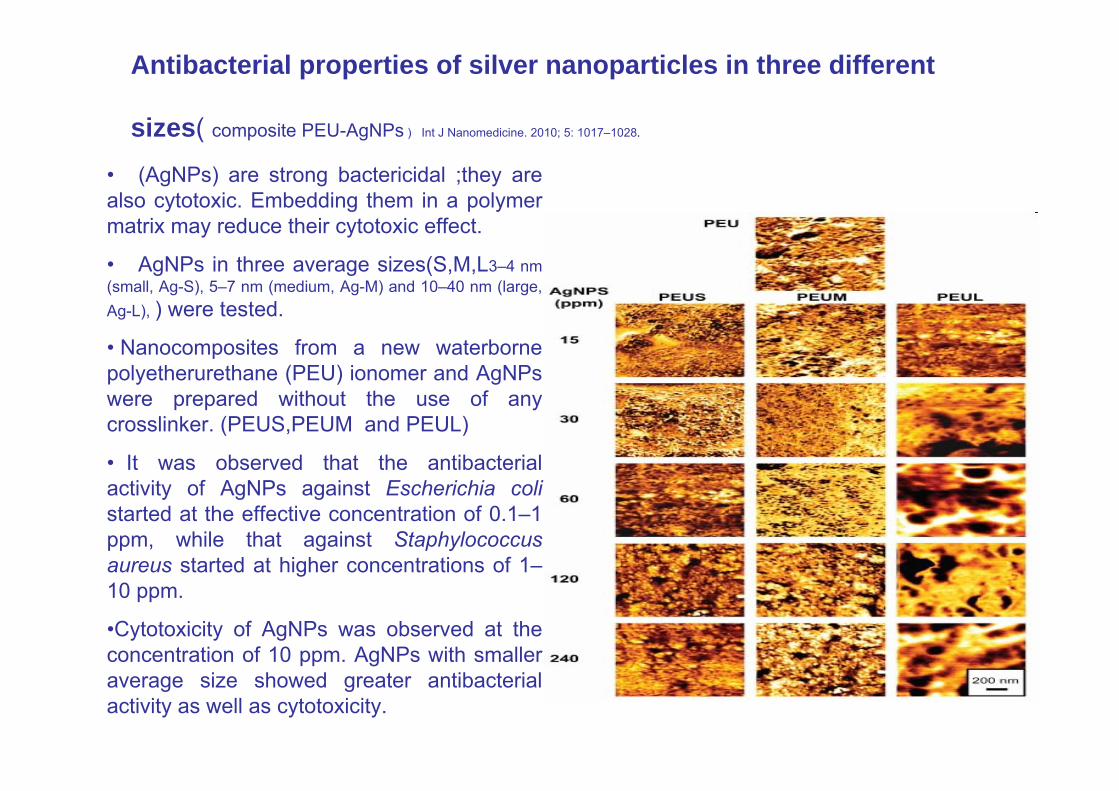
Antibacterial properties of silver nanoparticles in three different
sizes( composite PEU-AgNPs ) Int J Nanomedicine. 2010; 5: 1017–1028.
• (AgNPs) are strong bactericidal ;they are also cytotoxic. Embedding them in a polymer matrix may reduce their cytotoxic effect.
• AgNPs in three average sizes(S,M,L3–4 nm (small, Ag-S), 5–7 nm (medium, Ag-M) and 10–40 nm (large, Ag-L), ) were tested.
• Nanocomposites from a new waterborne polyetherurethane (PEU) ionomer and AgNPs were prepared without the use of any crosslinker. (PEUS,PEUM and PEUL)
• It was observed that the antibacterial activity of AgNPs against Escherichia colistarted at the effective concentration of 0.1–1 ppm, while that against Staphylococcus aureus started at higher concentrations of 1–10 ppm.
•Cytotoxicity of AgNPs was observed at the concentration of 10 ppm. AgNPs with smaller average size showed greater antibacterial activity as well as cytotoxicity.
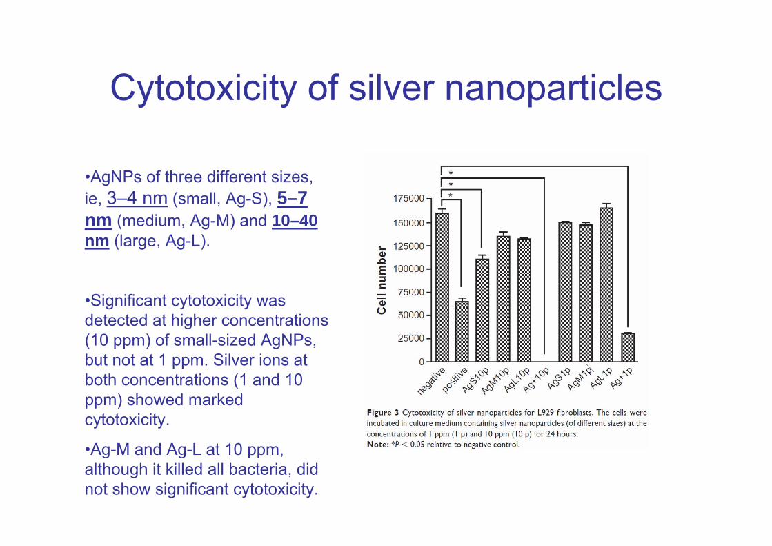
Cytotoxicity of silver nanoparticles
•AgNPs of three different sizes, ie, 3–4 nm (small, Ag-S), 5–7 nm (medium, Ag-M) and 10–40 nm (large, Ag-L).
•Significant cytotoxicity was detected at higher concentrations (10 ppm) of small-sized AgNPs, but not at 1 ppm. Silver ions at both concentrations (1 and 10 ppm) showed marked cytotoxicity.
•Ag-M and Ag-L at 10 ppm, although it killed all bacteria, did not show significant cytotoxicity.
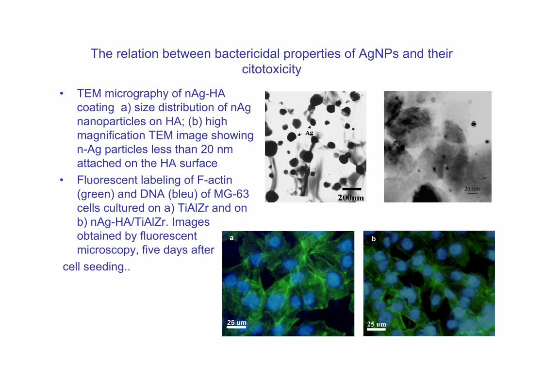
The relation between bactericidal properties of AgNPs and their citotoxicity
• TEM micrography of nAg-HA coating a) size distribution of nAg nanoparticles on HA; (b) high magnification TEM image showing n-Ag particles less than 20 nm attached on the HA surface
• Fluorescent labeling of F-actin (green) and DNA (bleu) of MG-63 cells cultured on a) TiAlZr and on b) nAg-HA/TiAlZr. Images obtained by fluorescent microscopy, five days after
cell seeding..
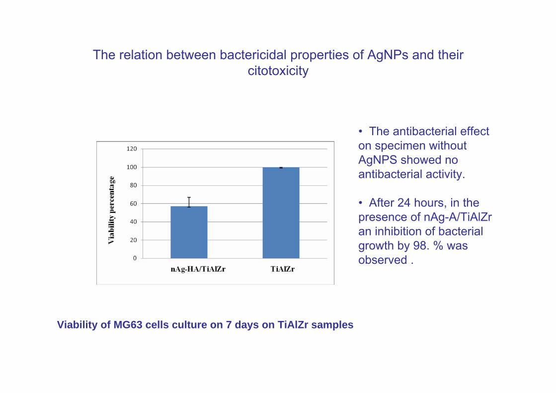
The relation between bactericidal properties of AgNPs and their citotoxicity
Viability of MG63 cells culture on 7 days on TiAlZr samples
• The antibacterial effect on specimen without AgNPS showed no antibacterial activity.
• After 24 hours, in the presence of nAg-A/TiAlZr an inhibition of bacterial growth by 98. % was observed .
Antibacterial Activity of Nanosilver Ions and ParticlesEnviron. Sci. Technol., 2010, 44 (14), pp 5649–5654 dOI: 10.1021/es101072s
The antibacterial activity of nanosilver against Gram negative Escherichia coli bacteria is investigated by immobilizing nanosilver on nanostructured silica particles and closely controlling Ag content and size.
These Ag/SiO2 nanoparticles were characterized by SEM/TEM, EDX spectroscopy, X-ray diffraction the exposed Ag surface area was measured qualitatively by O2 chemisorption. The fraction of dissolved nanosilver was determined by measuring the released (leached) Ag+ ion concentration in aqueous suspensions of such Ag/SiO2 particles.
The antibacterial effect of Ag+ ions was distinguished from that of nanosilver particles by monitoring the growth of E. coli populations in the presence and absence of Ag/SiO2 particles. The antibacterial activity of nanosilver was dominated by Ag+ ions when fine Ag nanoparticles (less than about 10 nm in average diameter) were employed that release high concentrations of Ag+ ions. In contrast, when relatively larger Ag nanoparticles were used, the concentration of the released Ag+ ions was lower..
Increasing AgNPs bactericidal properties
• D.K.Tiwari and J. Behari reported that the AgNPs treated with short time exposure with ultrasound show increased antibacterial effect but this time was not enough to kill the bacteria with ultrasound only .
• It indicated that synergistic effect of ultrasound & silver nanoparticles.
• The ultrasound facilitates the entry of Ag-nanoparticles inside the cells and the antibacterial effect was enhanced with same concentration of nanoparticles in presence of ultrasound waves. The biocidaleffect was more pronounced when compared to the actions of silver nanoparticles alone.
D.K. Tiwari and J. Behari, Advances in Biological Research, 3, 3-4, 2009
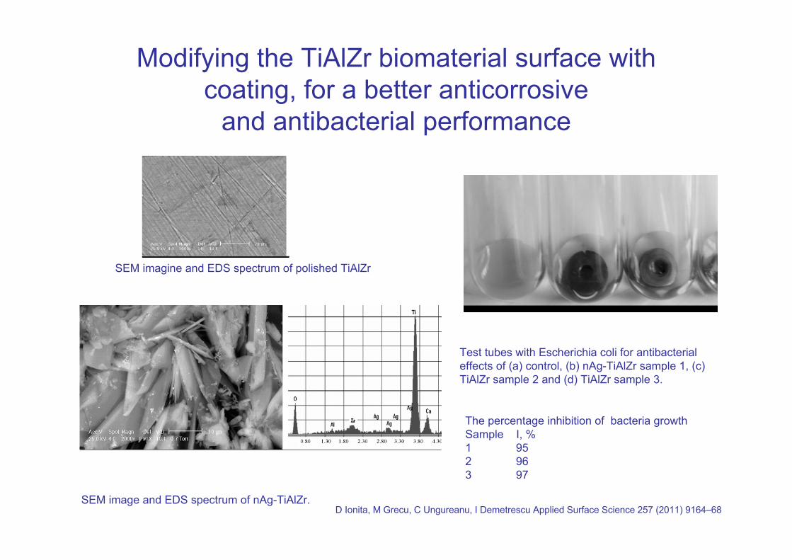
Modifying the TiAlZr biomaterial surface with coating, for a better anticorrosive
and antibacterial performance
SEM image and EDS spectrum of nAg-TiAlZr.
SEM imagine and EDS spectrum of polished TiAlZr
Test tubes with Escherichia coli for antibacterial effects of (a) control, (b) nAg-TiAlZr sample 1, (c) TiAlZr sample 2 and (d) TiAlZr sample 3.
The percentage inhibition of bacteria growthSample I, %1 952 963 97
D Ionita, M Grecu, C Ungureanu, I Demetrescu Applied Surface Science 257 (2011) 9164–68
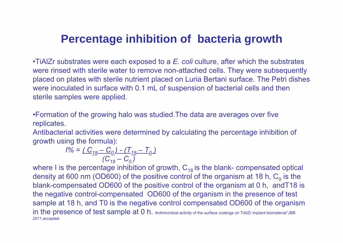
Percentage inhibition of bacteria growth
•TiAlZr substrates were each exposed to a E. coli culture, after which the substrates were rinsed with sterile water to remove non-attached cells. They were subsequently placed on plates with sterile nutrient placed on Luria Bertani surface. The Petri dishes were inoculated in surface with 0.1 mL of suspension of bacterial cells and then sterile samples were applied.
•Formation of the growing halo was studied.The data are averages over five replicates.Antibacterial activities were determined by calculating the percentage inhibition of growth using the formula):
I% = ( C18 – C0 ) - (T18 – T0 )(C18 – C0 )
where I is the percentage inhibition of growth, C18 is the blank- compensated optical density at 600 nm (OD600) of the positive control of the organism at 18 h, C0 is the blank-compensated OD600 of the positive control of the organism at 0 h, andT18 is the negative control-compensated OD600 of the organism in the presence of test sample at 18 h, and T0 is the negative control compensated OD600 of the organism in the presence of test sample at 0 h. Antimicrobial activity of the surface coatings on TiAlZr implant biomaterial”JBB 2011,accepted
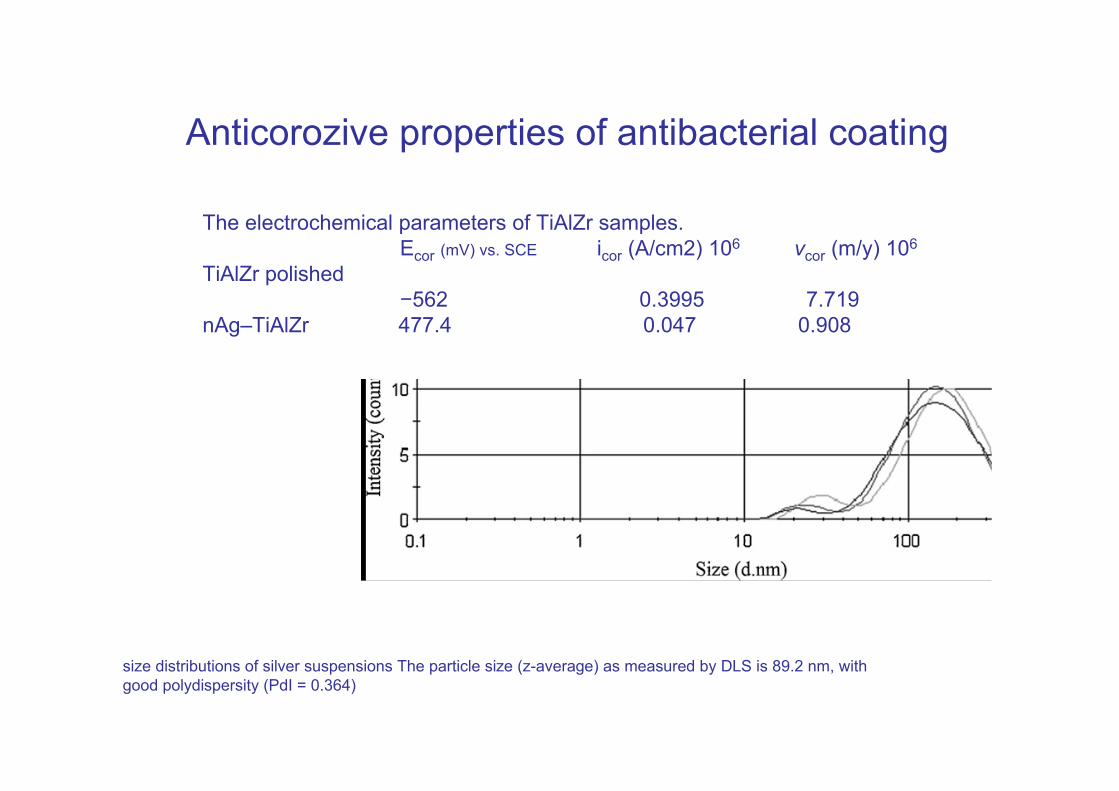
Anticorozive properties of antibacterial coating
The electrochemical parameters of TiAlZr samples.Ecor (mV) vs. SCE icor (A/cm2) 106 vcor (m/y) 106
TiAlZr polished−562 0.3995 7.719
nAg–TiAlZr 477.4 0.047 0.908
size distributions of silver suspensions The particle size (z-average) as measured by DLS is 89.2 nm, withgood polydispersity (PdI = 0.364)
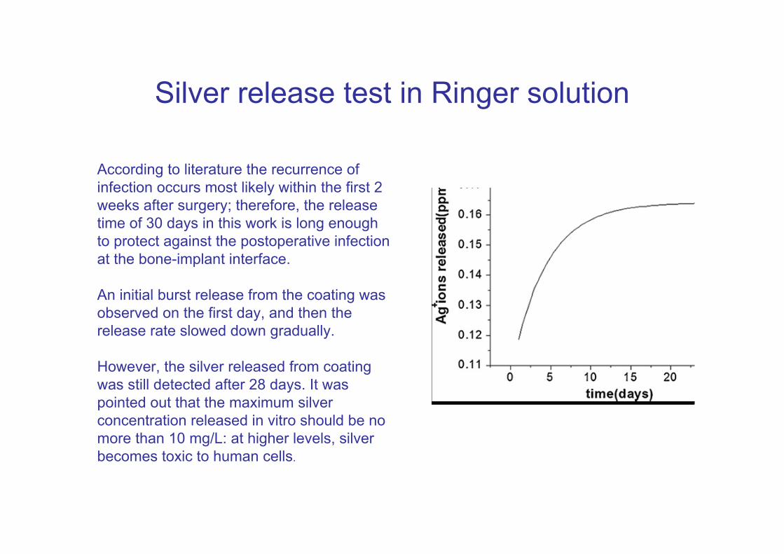
Silver release test in Ringer solution
According to literature the recurrence of infection occurs most likely within the first 2 weeks after surgery; therefore, the release time of 30 days in this work is long enough to protect against the postoperative infection at the bone-implant interface.
An initial burst release from the coating was observed on the first day, and then the release rate slowed down gradually.
However, the silver released from coating was still detected after 28 days. It waspointed out that the maximum silver concentration released in vitro should be no more than 10 mg/L: at higher levels, silverbecomes toxic to human cells.
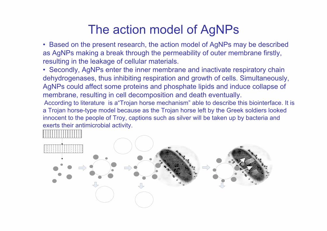
The action model of AgNPs• Based on the present research, the action model of AgNPs may be described as AgNPs making a break through the permeability of outer membrane firstly, resulting in the leakage of cellular materials.• Secondly, AgNPs enter the inner membrane and inactivate respiratory chain dehydrogenases, thus inhibiting respiration and growth of cells. Simultaneously, AgNPs could affect some proteins and phosphate lipids and induce collapse of membrane, resulting in cell decomposition and death eventually.According to literature is a“Trojan horse mechanism” able to describe this biointerface. It is a Trojan horse-type model because as the Trojan horse left by the Greek soldiers looked innocent to the people of Troy, captions such as silver will be taken up by bacteria andexerts their antimicrobial activity.
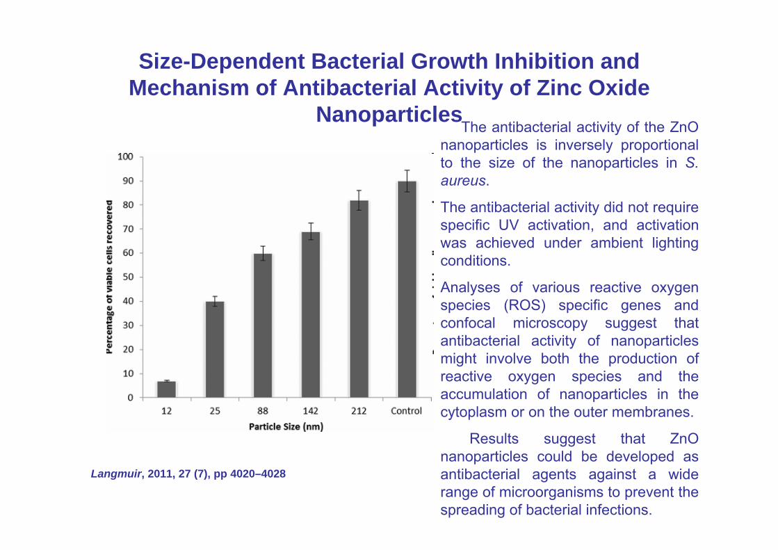
Size-Dependent Bacterial Growth Inhibition and Mechanism of Antibacterial Activity of Zinc Oxide
NanoparticlesThe antibacterial activity of the ZnO
nanoparticles is inversely proportional to the size of the nanoparticles in S. aureus.
The antibacterial activity did not require specific UV activation, and activation was achieved under ambient lighting conditions.
Analyses of various reactive oxygen species (ROS) specific genes and confocal microscopy suggest that antibacterial activity of nanoparticles might involve both the production of reactive oxygen species and the accumulation of nanoparticles in the cytoplasm or on the outer membranes.
Results suggest that ZnO nanoparticles could be developed as antibacterial agents against a wide range of microorganisms to prevent the spreading of bacterial infections.
Langmuir, 2011, 27 (7), pp 4020–4028
ZnO NP with sizes of 70 nm
• ZnO NP with sizes of 70 nm and concentrations of 0, 3, 6 and 12 mmol l(-1) were used in antimicrobial tests against E. coli O157:H7. • ZnO NP increases inhibitory effects on the growth of E. coli O157:H7 as the at its higher concentration. A complete inhibition of microbial growth was achieved at the concentration level of 12 mmol l(-1) or higher. • (SEM), (TEM), and Raman spectroscopy were used to characterize the changes of morphology and cellular compositions of bacterial cells treated with ZnO NP and study the mode of action of ZnO NP against E. coli O157:H7. • The intensity of lipid and protein bands in the Raman spectra of bacterial cells increased after exposure to ZnO NP, while no significant changes in nucleic acid bands were observed
• ZnO NP may distort and damage bacterial cell membrane, resulting in a leakage of intracellular contents and eventually the death of bacterial cells.
SIGNIFICANCE AND IMPACT OF THE STUDY: These results suggest that ZnO NP could potentially be used as an effective antibacterial agent to protect agricultural and food safety.
Carbon nanotubes (CNT)
• Arguments : present toxicity due acicular and fibrous forms ( associated with lung cacer pulmonar induced by azbestos .(Takagi et al. 2008).
•
•
•
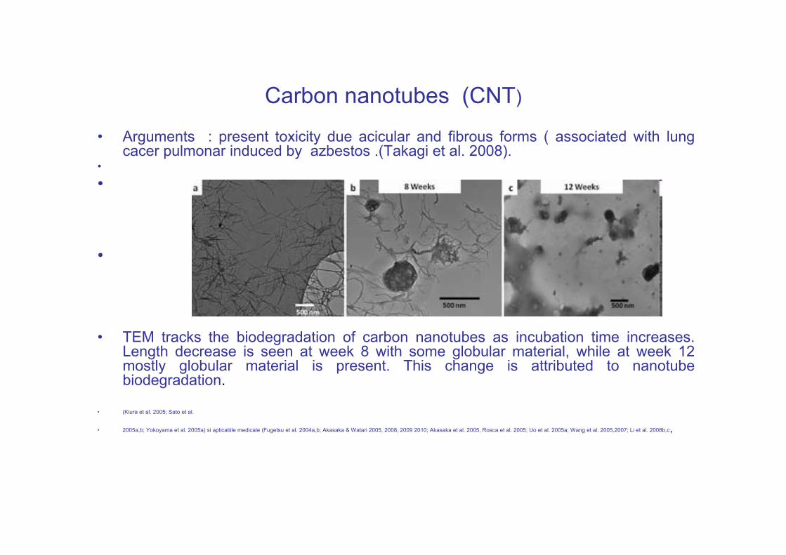
• TEM tracks the biodegradation of carbon nanotubes as incubation time increases. Length decrease is seen at week 8 with some globular material, while at week 12 mostly globular material is present. This change is attributed to nanotube biodegradation.
• (Kiura et al. 2005; Sato et al.
• 2005a,b; Yokoyama et al. 2005a) si aplicatiile medicale (Fugetsu et al. 2004a,b; Akasaka & Watari 2005, 2008, 2009 2010; Akasaka et al. 2005, Rosca et al. 2005; Uo et al. 2005a; Wang et al. 2005,2007; Li et al. 2008b,c,
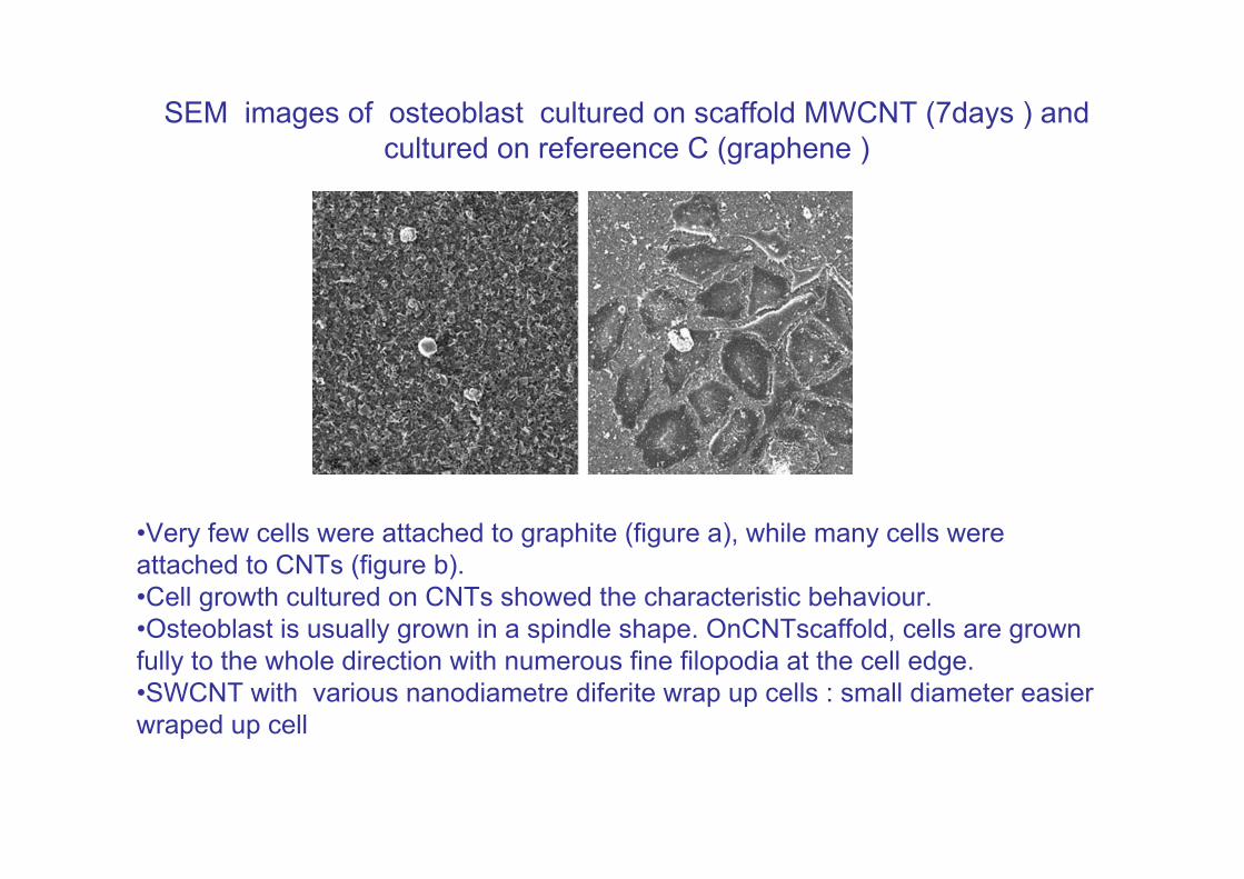
SEM images of osteoblast cultured on scaffold MWCNT (7days ) and cultured on refereence C (graphene )
•Very few cells were attached to graphite (figure a), while many cells were attached to CNTs (figure b).•Cell growth cultured on CNTs showed the characteristic behaviour. •Osteoblast is usually grown in a spindle shape. OnCNTscaffold, cells are grown fully to the whole direction with numerous fine filopodia at the cell edge. •SWCNT with various nanodiametre diferite wrap up cells : small diameter easier wraped up cell
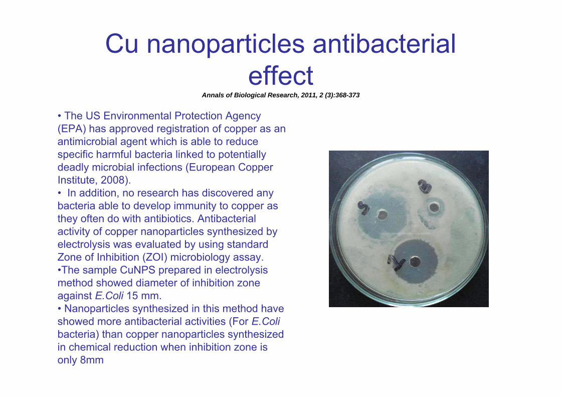
Cu nanoparticles antibacterial effect
Annals of Biological Research, 2011, 2 (3):368-373
• The US Environmental Protection Agency (EPA) has approved registration of copper as an antimicrobial agent which is able to reduce specific harmful bacteria linked to potentially deadly microbial infections (European Copper Institute, 2008).• In addition, no research has discovered any bacteria able to develop immunity to copper as they often do with antibiotics. Antibacterial activity of copper nanoparticles synthesized by electrolysis was evaluated by using standard Zone of Inhibition (ZOI) microbiology assay. •The sample CuNPS prepared in electrolysis method showed diameter of inhibition zone against E.Coli 15 mm.• Nanoparticles synthesized in this method have showed more antibacterial activities (For E.Coli bacteria) than copper nanoparticles synthesized in chemical reduction when inhibition zone is only 8mm
Antibacterial activities of CuNPs (50and 100nm)
• Due to the increasing bacterial resistance to variety of antibiotics, the potential of antibacterial activities of ( CuNPs) became interesting . NPs have the ability to interact with target cell at multiple levels. Despite copper bactericidal properties, Escherichia coli is equipped with multiple (~5) systems to handle copper toxicity under varying environmental conditions (dynamic aquatic system).
• At the initial contact, Cu NPs50 has faster inactivation of E. coli cells resulting in 3.5 times reduction in viable cells during the first hour; whereas, only 1.2 times reduction is recorded during the subsequent 1 hour.
• , over the course of two hours, the impact of both Cu NPs sizes on E. coli cell was statically similar. The pattern of viability loss of E. coli cells after exposure to Cu NPs100 was reverse to the one observed for Cu NPs50. Conclusion Ps size is a factor in their pattern of antibacterial activities.
• The smaller Cu NPs act fast at initial contact and gradually loose toxic effect over time. In contrast, larger Cu NPs showed gradual increase in their toxicity.
( ASM 2011).
Strain specificity in antimicrobial activity of silver and copper nanoparticles
• The antimicrobial properties of Ag and Cu NPs were investigated using Escherichia coli (four strains), Bacillus subtilis and Staphylococcus aureus (three strains).
• The average sizes of the Ag and CU NPs were 3 nm and 9 nm, respectively, as determined throughTEM . Energy-dispersive X-ray spectra of Ag and CU NPs revealed that while Ag was in its pure form, an oxide layer existed on theCuNPs. The bactericidal effect of Ag and CuNPS were compared based on diameter of inhibition zone in disk diffusion tests and minimum inhibitory concentration (MIC) and minimum bactericidal concentration (MBC) of nanoparticles dispersed in batch cultures.
• Bacterial sensitivity to nanoparticles was found to vary depending on the microbial species. Disk diffusion studies with E. coli and S. aureus revealed greater effectiveness of the AgNPs compared to theCuNPs. B. subtilis depicted the highest sensitivity to nanoparticles compared to the other strains and was more adversely affected by theCuNPs .
• Good correlation was observed between MIC and MBC (r2=0.98) measured in liquid cultures. For CuNPs a good negative correlation was observed between the inhibition zone observed in disk diffusion test and MIC/MBC determined based on liquid cultures with the various strains (r2=-0.75). Although strain-specific variation in MIC/MBC was negligible for S. aureus, some strain-specific variation was observed for E. coli.
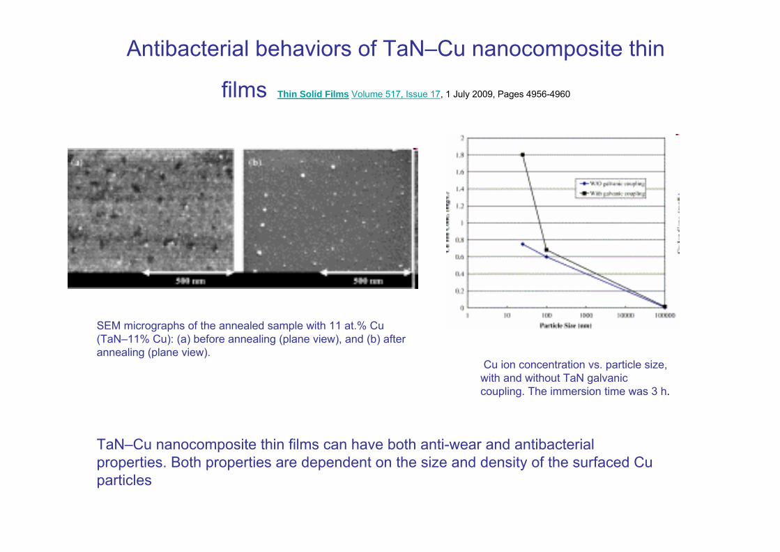
Antibacterial behaviors of TaN–Cu nanocomposite thin
films Thin Solid Films Volume 517, Issue 17, 1 July 2009, Pages 4956-4960
SEM micrographs of the annealed sample with 11 at.% Cu (TaN–11% Cu): (a) before annealing (plane view), and (b) after annealing (plane view).
TaN–Cu nanocomposite thin films can have both anti-wear and antibacterial properties. Both properties are dependent on the size and density of the surfaced Cu particles
Cu ion concentration vs. particle size, with and without TaN galvanic coupling. The immersion time was 3 h.
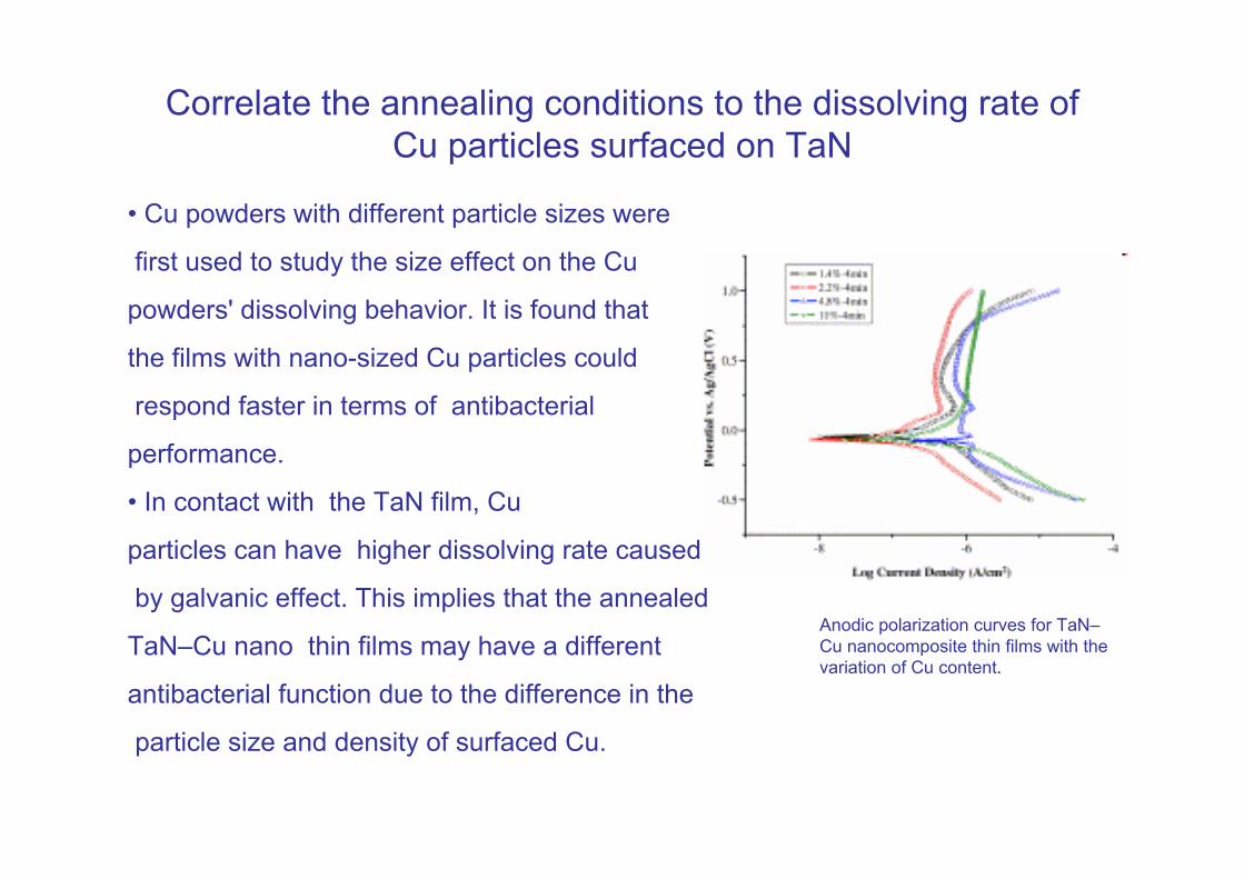
Correlate the annealing conditions to the dissolving rate of Cu particles surfaced on TaN
• Cu powders with different particle sizes were
first used to study the size effect on the Cu
powders' dissolving behavior. It is found that
the films with nano-sized Cu particles could
respond faster in terms of antibacterial
performance.
• In contact with the TaN film, Cu
particles can have higher dissolving rate caused
by galvanic effect. This implies that the annealed
TaN–Cu nano thin films may have a different
antibacterial function due to the difference in the
particle size and density of surfaced Cu.
Anodic polarization curves for TaN–Cu nanocomposite thin films with the variation of Cu content.
Safety and Risk Associated with Nanoparticles
• There is a correlation between a decrease in particle size and an increase in toxicity,because of larger surface area.
• The ability of nanoparticles to penetrate deep into our respiratory system and their better assimilation in the body fluids make them unique.•The nanoparticles are likely to be unsafe for the biological system ( may enter the human body and become toxic at the cellular level in the tissues and organs).•The materials of these particles may or may not be allergic or carcinogenic but even inert nanoparticles show harmful effects due to some absorbed toxic species or formation of toxic products due to reactions with body fluids.
•Lower sized (<10nm) nanoparticles behave more like a gas and can pass through skin and lung tissue to penetrate cell membranes.When we consider environmental exposure for nanoparticles, we may find that nanoparticles are easily airborne and adhere easily to surfaces which are difficult to detect. •Through the environment nanoparticles may enter the food chain, influence the biosphere, influence structural change in liquids like water (biogenic nanoparticles) and chemical/physical transition by recycling.