Embed Size (px)
Citation preview

Intracerebral Hemorrhage

• arterial spontaneous bleeding into brain parenchyma• second most common stroke type • 50% mortality, the majority in the first 30 days• only 20% are independent after 6 months
Intracerebral hemorrhageDefinition

• 5-15% of all stroke• Incidence
Japan 61/100 000Europe 7-15 / 100 000US 32-35 / 100 000
• goes up with age • M:F = 3:2
Intracerebral hemorrhageEpidemiology

• based on etiology• based on localisation
Intracerebral hemorrhageClassification
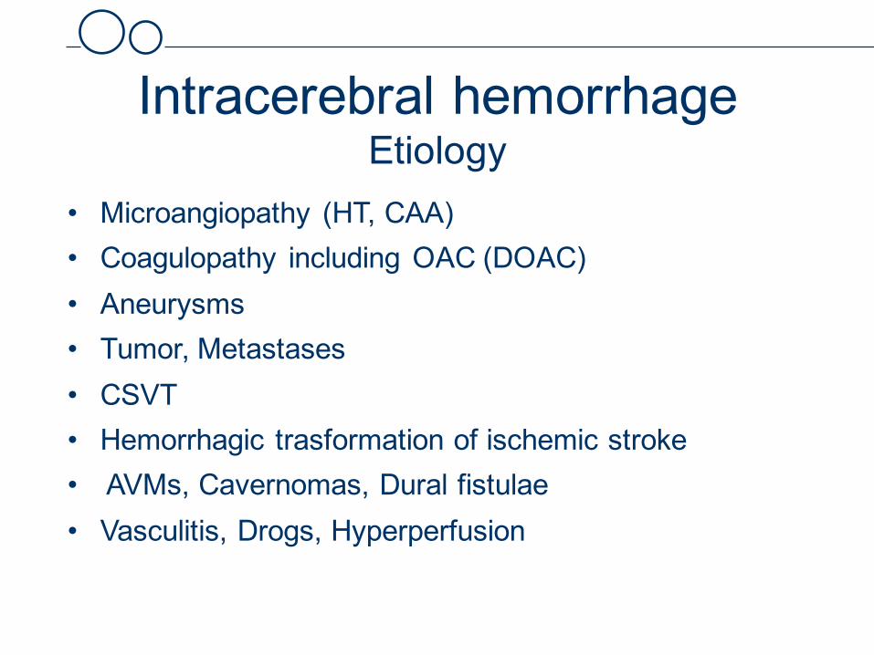
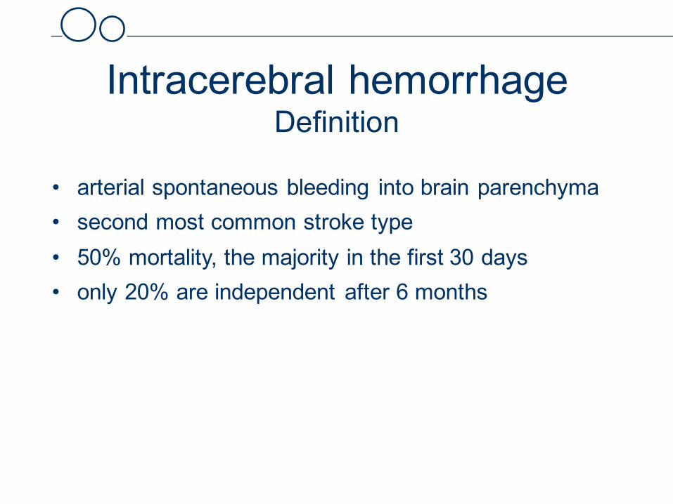
• Microangiopathy (HT, CAA)• Coagulopathy including OAC (DOAC)• Aneurysms• Tumor, Metastases• CSVT• Hemorrhagic trasformation of ischemic stroke• AVMs, Cavernomas, Dural fistulae• Vasculitis, Drogs, Hyperperfusion
Intracerebral hemorrhageEtiology

< 40Y…….AV- Malformations (29-57%)......…..BG40-70Y…..Hypertension (70%)………….......…BG
(loco typico)>70Y….....Amyloid angiopathiy…….…............Lobar hematomas
Intracerebral hemorrhageEtiology

• Lipohyalinosis• Microaneurysms• perforator arteries
• typical localisation –1) BG 2) cerebellum3) pons
Intracerebral hemorrhageEtiology: hypertensive microangiopathy (50-70%)
„Typical localisation“

Intracerebral hemorrhage„Typical localisation“

Intracerebral hemorrhage„Typical localisation“

Intracerebral hemorrhage„Typical localisation“

Intracerebral hemorrhage„Typical localisation“

Intracerebral hemorrhage„Typical localisation“

Intracerebral hemorrhage„Typical localisation“

• Amyloid deposits in cortical vessels• Lobar• Hypertension may also be present• Repetitve or simultaneous bleeding• Incidence goes up with age
Intracerebral hemorrhageEtiology: microangiopathy through CAA (15%)
„Atypical localisation“

Intracerebral hemorrhage„Atypical localisation“

Intracerebral hemorrhage„Atypical localisation“, multiple bleedings

Intracerebral hemorrhageMicrobleeds

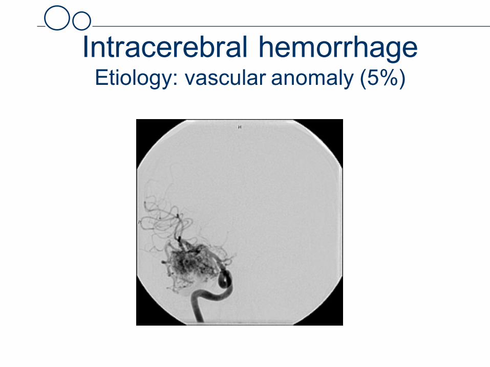
• AVMs, cavernomas, dural fistulae• aneurysmn, teleangiectasiae• young pts, cortical bleeds
Intracerebral hemorrhageEtiology: vascular anomaly (5%)

Intracerebral hemorrhageEtiology: vascular anomaly (5%)

Intracerebral hemorrhageEtiology: vascular anomaly (5%)
Intracerebral hemorrhageEtiology: vascular anomaly (5%)

• OACs ≈1% per year• lobar, with growth, poor prognosis• thrombolysis ≈ 5%• thrombocytopenia, hemophiliae
Intracerebral hemorrhageEtiology: coagulopathy (10-25%)

• identical to „stroke“• ↑ HA, vomitus, conscioussness, seizures• focal symptoms• symptoms of ICH• autonomic symptoms
Intracerebral hemorrhageSymptoms

Intracerebral hemorrhagePathophysiology
• rupture- > blood extravasation into parenchyma• local pressure increase –> rupture of further vessels• generalized ICP increase• bleeding – active mainly in the first 6 hours• growth 18-38% pts – poor prognosis• perihematomal edema• no perihematomal ischemia

• Medical emergency• SU/ ICU• Withholding / Withdrawal of care in large hemorrhages• General ICU therapy• Specific therapy
Intracerebral hemorrhageTherapy

• GCS 3-4………………..…..2• GCS 5-12………………..…1• GCS > 12…………………..0• Volume > 30ml…..………...1• IVH.................….....…….…1• Infratentoriel…….……….…1• Age > 80Y.………………….1
• 5-6 points – Mortality 100%, (Volumen>60ml – 91% )
Intracerebral hemorrhagePrognosis, ICH-Score

Intracerebral hemorrhageGeneral ICU therapy
• Intubation GCS < 8, absent protective reflexes• BP < 140/80• Normoglycemia• Normothermia• Normovolemia• Gastric protection• DVT prevention• Seizures treatment• Infection treatment

Intracerebral hemorrhageSpecific therapy
1) Treatment of hematoma growth2) Surgical management3) EVD und Lysis in IVH4) ICP management

2:00 h from onset 3:50 h from onset
> 25% of all patients in < 4 hours
Intracerebral hemorrhageHematoma growth

CCT7:20 hour
Post-CM-CCT7:22 hour
CTA7:20 hour
Intracerebral hemorrhageHematoma growth, Spot Sign

Intracerebral hemorrhageSpecific therapy
1) Treatment of hematoma growth
- Treat BP to <140/80- Antagonise with PPSB/Vit. K in OAC - Antagonise with Praxbind/PPSB/Adnexanet in NOAC- Antagonise with Protamin in Heparin- Factor VII- not recommanded

Intracerebral hemorrhageSpecific therapy
2) Surgical treatment – NO EVIDENCE FOR EFFICACY
.
Young patientsGCS 9-12Volume 30-60mlHydrocephalusClinical deteriorationCerebellar locationLobar locationAVM as etiology
TechniquesOpen evacuationStereotactic miniinvasive evacuationVetricular drainageDecompressive craniectomy

3) Ventricular drainage and intraventricular Lysis with rtPA- IVH – high mortalitality- no parenchymal damage, but hydrocephalus- impaired conscioussness, autonomic dysfunction
- when blood in 3rd and 4th ventricel OR hydrocephalus –EVD and Lysis
Intracerebral hemorrhageSpecific therapy

Intrazerebrale BlutungSpezifische Massnahmen

4) ICP management
- position, minimal handling, analgesia, sedation, relaxation- EVD- osmotherapy (Mannitol, NaCl)- hypothermia- decompressive craniectomy
Intracerebral hemorrhageSpecific therapy
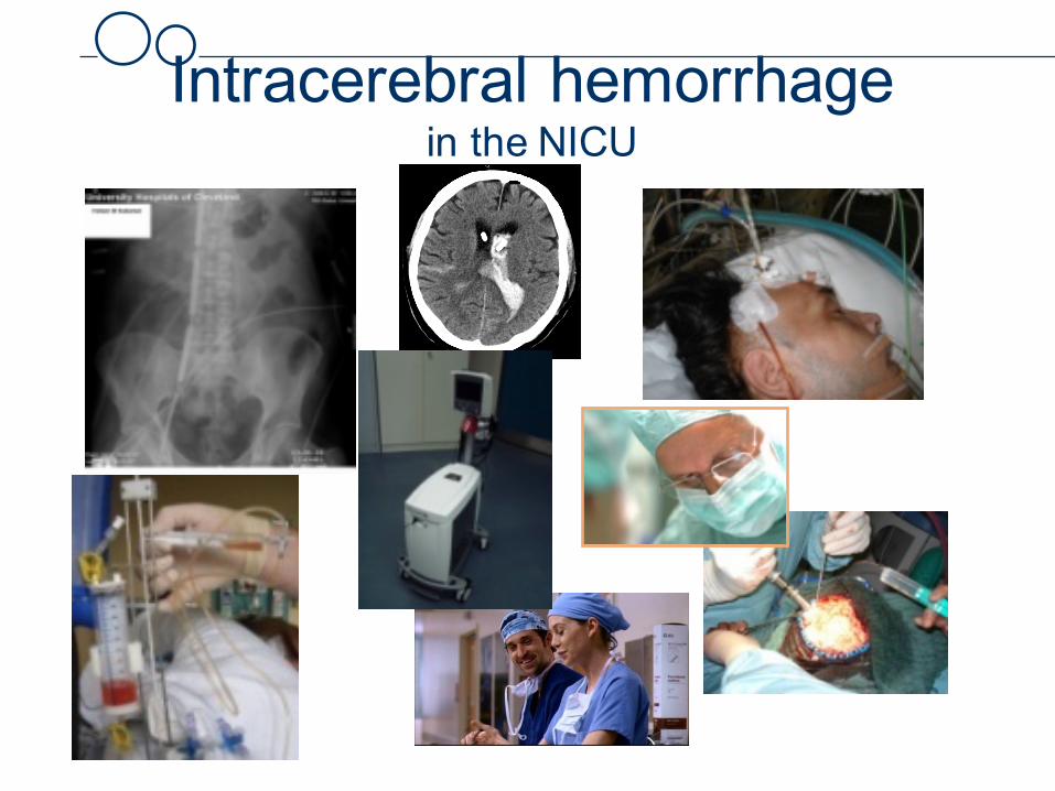
Intracerebral hemorrhagein the NICU

Subarachnoid hemorrhage

• it occurs in young people– 80% in 40-65 year olds– 15% in 20-40 year olds
• it kills quickly– 25% die within 24 hours– 50% will be dead at 6 months
• it causes significant disability– cognitive impairment– neurological disability depending on size of bleed &
complications encountered
Subarachnoid hemorrhageThe problem

• Headache– sudden onset & severe– small leak may cause minor headache & may be
warning sign of rupture– with physical activity
• Reduced consciousness, cranial nerves palsy• Meningism
– Vomiting– Neck stiffness– Photophobia
• Seizures
Subarachnoid hemorrhageSymptoms

Subarachnoid hemorrhageDiagnosis
1. CT 98% sensitive @ 12 hours80% at day 350% at day 7
2. CTA/MRAsensitivite for aneurysm >3mm

2. CSF• Uniformly blood-stained• Xanthochromia: 12 hours to 2-3 weeks• ICP ↑3. DSAetiologic diagnosis, important to surgery
Subarachnoid hemorrhageDiagnosis

1)
2)
3)
Subarachnoid hemorrhage
4)

4/17/20© 2009, American Heart Association. All rights reserved.
Clipping

4/17/20© 2009, American Heart Association. All rights reserved.
Angio Image Courtsey: The University of Texas Health Science Center at San Antonio – Department of Neurosurgery
Clipping

4/17/20© 2009, American Heart Association. All rights reserved.
Coiling

4/17/20© 2009, American Heart Association. All rights reserved.
Coil system embolization: immediate result
Angio showing large ICA aneurysm Same aneurysm - Post GDC Coiling
Angio Image Courtsey: The University of Texas Health Science Center at San Antonio – Department of Neurosurgery

1) Find and secure the aneurysm – NCH/NR (80%)
2) General ICU managementi.Minimal handling, Monitoringii.Sedation and analgesiaiii.Intubation
iv.Aggresive BP treatment < 140/80 (Rebleeding)v.Initial ICP therapyvi.Initial hydrocephalus treatment(EVD) und ICP-measurement (EVD)
vii.Vasospasmus treatment – Nimodipine 6x30mg p.o.
Subarachnoid hemorrhageManagement

Rebleeding Vasospasmus / Delayed ischemic neurological deficitsHydrocephalusEpilepsy
Subarachnoid hemorrhageNeurological complications

Hyponatriemia (SIADH, CSWS)Cardiac ischemia / ArrhytmiaeNeurogenic lung edemaParalitic ileus
Subarachnoid hemorrhageNon-neurological complications

• High risk 2-4% over first 24 hours20% over 10-14 days
• Always treat aneurysm, even if vasospasm
Subarachnoid hemorrhageRebleeding

Subarachnoid hemorrhageVasospasms / Delayed ischemic neurological
Deficits

• Maximum D4 to D14 after onset• Localised around the SAH, however also other parts
• Pathophysiology– Hemoglobin, Oxyhemoglobin , Endothelin– Vasoconstriction– Sterile inflammation
Subarachnoid hemorrhageVasospasms / Delayed ischemic neurological
Deficits

• i.v. / p.o. Nimodipine• HHH (Hypertension, Hypervolemia, Hemodilution)• i.a. Papaverin, Nimodipine, Nitrate• Angioplasty
• Standard TCD-Monitoring• multimodales Neuromonitoring
Subarachnoid hemorrhageVasospasms treatment

• Acute – at admission• Subacute - in the first days• Chronic - after weeks/months
• EVD or VP shunt
Subarachnoid hemorrhageHydrocephalus

• 6% have seizures at admission• Post-operative seizures 1.5%• Late seizures 3%
Subarachnoid hemorrhageEpilepsy
• Some centres start anticonvulsive treatment prophylactically• EEG Monitoring• Phenytoin, Valproate, Carbamazepine• acute BZD (Lorazepam)
Subarachnoid hemorrhageEpilepsy

• SIADH• CSWS
• deleterious for brain edema• Volume restriction/ Natrium Substitution• Mineralocorticosteroids
Subarachnoid hemorrhageHyponatriemia

• Arrhytmiae, ST-changes, high Trop in up to 100%• Associated with poor prognosis• Symptomatic therapie with B-blockers, Clonidine
Subarachnoid hemorrhageCardiac complications

Subarachnoid hemorrhagePeri-mesenzephalic and pre-pontine SAHs

• 5-10% of all SAHs• No aneurysms (95%)• Low hydrocephalus (max. 20% )• No spasms, no rebleeding• Perhaps venous bleed• Good outcome
Subarachnoid hemorrhagePeri-mesenzephalic and pre-pontine SAHs
• Except of peri-mezencephalic/ pre- pontine SAH, all other SAHs have hidden aneurysm
• repeat DSAs• thinks of small/thrombotised aneurysms• AVMs, Sinusthrombosis• Durafistulae• AVMs spinal!!!
Subarachnoid hemorrhageNo aneurysm found
Thank you!