Embed Size (px)
Citation preview
DOI 10.1212/WNL.0b013e318264e3be; Published online before print August 1, 2012;Neurology
Mario Di Napoli, Daniel Agustin Godoy, Veronica Campi, et al.tissue localization, and prognosis
C-reactive protein in intracerebral hemorrhage : Time course,
August 13, 2012This information is current as of
http://www.neurology.org/content/early/2012/08/01/WNL.0b013e318264e3be
located on the World Wide Web at: The online version of this article, along with updated information and services, is
rights reserved. Print ISSN: 0028-3878. Online ISSN: 1526-632X.Allsince 1951, it is now a weekly with 48 issues per year. Copyright © 2012 by AAN Enterprises, Inc.
® is the official journal of the American Academy of Neurology. Published continuouslyNeurology
C-reactive protein inintracerebral hemorrhageTime course, tissue localization, and prognosis
Mario Di Napoli, MDDaniel Agustin Godoy,
MDVeronica Campi, PhDLuca Masotti, MDCraig J. Smith, MD,
MRCPAdrian R. Parry Jones,
PhD, MRCPStephen J. Hopkins, PhDMark Slevin, PhD,
FRCPathFrancesca Papa, MDLaurentiu Mogoanta, MDDaniel Pirici, MD, PhDAurel Popa Wagner, MD,
PhD
ABSTRACT
Objectives: We examined the C-reactive protein (CRP) response after spontaneous intracerebralhemorrhage (sICH) and its relationship to outcome. We additionally characterized early brain lo-calization of CRP.
Methods: In this prospective, multicenter, international, collaborative, longitudinal study withcross-sectional immunohistochemical analysis of brain tissue, 223 patients (M/F: 132/91) wererecruited during the 2010 calendar year. CRP was evaluated at admission (median 93 minutesfrom symptom onset), 24 hours, 48 hours, and 72 hours after sICH. Brains of 5 subjects withsICH were compared to brains of 2 aged controls without evidence of brain pathology and 7patients with ischemic stroke. Plasma CRP was measured over 72 hours following sICH and itsrelationship to 30-day mortality and functional outcome at 30 days (Glasgow Outcome Scale)was determined. CRP immunostaining patterns were analyzed in samples of sICH autopsy brains.
Results: Plasma CRP increased over the 48 hours from admission and was significantly (p �
0.001) related to hematoma volume at later time points. The predictive utility of CRP for morbid-ity and mortality were maintained when adjusted for other risk factors and improved at 48 hoursand 72 hours when compared with admission values. Although an early CRP localization waspresent in both ischemic and hemorrhagic lesions, an intense and diffuse neuropil staining wasonly present in sICH patients and particularly evident proximal to the hemorrhagic areas.
Conclusions: Plasma CRP production increases markedly over the 48 hours to 72 hours periodfollowing sICH and is related to outcome. CRP is also present in large amounts around the hemor-rhagic lesion and within neurons and glia of patients who died within 12 hours of sICH. Neurology®
2012;79:690–699
GLOSSARYAUC � area under receiver operator characteristic curve; CI � confidence interval; CRP � C-reactive protein; GCS � Glas-gow Coma Scale; GOS � Glasgow Outcome Scale; HR � hazard ratio; IL-6 � interleukin-6; IVH � intraventricular extension;LR � likelihood ratio; oICH � Hemphill’s original ICH score; sICH � spontaneous intracerebral hemorrhage.
Experimental and clinical studies indicate that inflammation is involved in the progression ofbrain injury after spontaneous intracerebral hemorrhage (sICH).1,2 These pathologic mecha-nisms include immunologic, endothelial dysfunction, and coagulopathy, contributing to mor-bidity and mortality.3
Increased C-reactive protein (CRP) has been shown previously to relate to poor outcomeafter sICH.4–6 However, direct pathophysiologic evidence that it participates in local inflam-matory response is lacking.7 Potential limitations of previous studies of inflammatory markersin sICH include the possibility that the systemic acute-phase response may have been attribut-able to accompanying infection in some cases, and that the inflammatory and CRP response
From the Neurological Service (M.D.N.), San Camillo de Lellis General Hospital, Rieti; Neurological Section, SMDN—Center for CardiovascularMedicine and Cerebrovascular Disease Prevention (M.D.N., F.P.), Sulmona, L’Aquila, Italy; Neurocritical Care Unit (D.A.G.) and CentralLaboratory (V.C.), Sanatorio Pasteur, Catamarca, Argentina; Internal Medicine (L. Masotti), Cecina Hospital, Cecina, Italy; Brain Injury ResearchGroup (C.J.S., A.R.P.J., S.J.H.), Clinical Sciences Building, Manchester Academic Health Science Centre, Salford Royal NHS Foundation Trust,Manchester; SBCHS (M.S.), John Dalton Building, Manchester Metropolitan University, Manchester, UK; Institut Catala de CienciesCardiovasculars Hospital de la Santa Creu i Sant Pau (M.S.), Pavello del Convent Sant Antoni Maria Claret, Barcelona, Spain; Research Center forMicroscopic Morphology and Immunology & Biochemistry (L. Mogoanta, D.P., A.P.W.), University of Medicine and Pharmacy of Craiova,Romania; and Department of Psychiatry, Molecular Psychiatry (A.P.W.), Rostock Medical School of Medicine, Germany.
Study funding: Funding information is provided at the end of the article.
Go to Neurology.org for full disclosures. Disclosures deemed relevant by the authors, if any, are provided at the end of this article.
Supplemental data atwww.neurology.org
Supplemental Data
Correspondence & reprintrequests to Dr. Di Napoli:[email protected]
690 Copyright © 2012 by AAN Enterprises, Inc.
Published Ahead of Print on August 1, 2012 as 10.1212/WNL.0b013e318264e3be
could still be evolving. Additionally, changesin CRP levels beyond the initial 24 hours aftersICH have not been analyzed in detail, yetthese may be more reflective of the inflamma-tory response to sICH. Therefore, the pri-mary aim of our study was to evaluate thekinetics of plasma CRP concentrations afteracute sICH, up to 72 hours, and therelationship with clinical outcomes. To investi-gate a possible role of CRP in local inflamma-tory responses, we examined CRP localizationby performing immunohistochemical studies onbrain specimens obtained from patients whodied within 12 hours after sICH.
METHODS Participants were included in a prospective,
multicenter international observational collaborative project,
the aims of which are to study the pathophysiology of the
inflammatory response and to determine the prognostic value
of inflammatory biomarkers after sICH. Details of the study
methodology are given in the supplemental Subjects and
Methods on the Neurology® Web site at www.neurology.org.
Briefly, patients admitted with a diagnosis of sICH within
24 hours of symptom onset had baseline clinical data recorded
on an electronic form, including demographic data, medical his-
tory, risk factors, presence of comorbidities, physical examina-
tion findings, Glasgow Coma Scale (GCS)8 after resuscitation,
Hemphill’s original ICH (oICH) score,9 standard routine labo-
ratory panels, and CT scan findings.
Of the participants recruited during 2010 (n � 384), only
patients with serial blood samples within the first 72 hours
and complete clinical and neuroradiologic variables were con-
sidered eligible for the current analysis (figure e-1). To avoid
confounding effects, we excluded patients with a history of
acute or chronic infections in the 4 weeks before sICH or
those with clinical evidence of acute infection at admission, as
well as those with other concurrent inflammatory comorbidities
(n � 62). A further 99 patients were also excluded for other reasons
(figure e-1). Following this screening procedure, 223 patients were
included.
Surgical treatment was performed according to local proto-
cols at participating institutions based on the guidelines of the
Stroke Council of the American Heart Association10 and Euro-
pean Stroke Initiative.11
CRP measurements. In all patients the first plasma CRP con-
centration (CRPAdm) was determined immediately after admis-
sion. The second CRP measurement was done at 24 hours
(CRP24 hours), the third at 48 hours (CRP48 hours), and the fourth
at 72 hours (CRP72 hours) after symptom onset. CRP analysis was
performed locally using high-sensitivity immunoturbidimetric
assays with similar performance characteristics.12
Neuroradiologic analysis. The initial CT brain scan after
admission was reviewed and classified according to site of sICH
(basal ganglia, thalamic, lobar, pontine, cerebellar, or other), vol-
ume of hematoma measured using the ABC/2 method,13 midline
shift (by measuring the displacement of the septum pellucidum
from the midline),14 intraventricular extension (IVH), and
presence of hydrocephalus. Investigators who read CT scanswere blinded to clinical information.
Neuropathology: processing of tissue specimens andimmunohistochemistry. To analyze early CRP brain local-ization after sICH, formalin-fixed, paraffin-embedded archivedbrain tissue blocks containing both lesional and perilesional areaswere selected from 5 sICH patients who died within 12 hours(mean 8; range 4–12 hours). Seven ischemic stroke patients whodied within 12 hours (mean 7; range 4–12 hours) and 2 subjectswithout brain pathology were used as controls. All brain tissuesamples were obtained from patients recruited in the Clinic ofNeurology (University of Medicine and Pharmacy, Craiova,Romania).
Immunohistochemistry was performed using anti-CRPmonoclonal antibody (mouse antihuman, Clone 1, IgG2b,Genetex, 1:100) recognizing human CRP.15 Negative controlsshowed no abnormal crossreactivity. Double immunofluores-cence was performed for anti-CRP and anticollagen IV (rabbitantihuman, Novus Biologicals, 1:1,000).
Outcome measurements. The primary outcome was 30-daymortality. The secondary outcome was 30-day functional out-come assessed using Glasgow Outcome Scale (GOS)16 dichoto-mized into “poor” (GOS scores 2–3, or death) or “good” (GOS4–5). For patients in whom 30-day outcome was not availablefrom medical records (n � 21), follow-up data were obtainedfrom visits using a standardized questionnaire. The cause ofdeath was confirmed by available medical records and nopatients were lost to follow-up. Information regarding inflam-matory markers was not used to modify treatment during in-hospital and follow-up periods.
Standard protocol approvals, registrations, and patientconsent. Informed consent was obtained from all participantsor legal representatives and the protocol was approved by localInstitutional Review Boards.
Statistical analyses. All values are given as mean and SD ormedian and interquartile range, according to manner of distribu-tion. Differences between 2 groups were assessed with indepen-dent t tests, the Fisher exact test, or the Wilcoxon test, asappropriate. The differences between CRPAdm, CRP24 hours,CRP48 hours, and CRP72 hours were analyzed using a 1-way analy-sis of variance with Bonferroni correction if the overall test wassignificant. p Values were corrected for sphericity violation whenappropriate.
For the calculation of Pearson correlation coefficients, welogarithmically transformed positively skewed CRP data to ob-tain a normal distribution. CRP increase was defined as 1 loge
unit/l increase between 2 measurements.CRP concentrations were divided into tertiles to identify
nonlinear effects of the CRP and provide more stable risk esti-mates. We used Kaplan-Meier survival curves to compare event-free survival between groups of patients defined by tertiles ofCRP and compared curves with log-rank trend tests.
We used Cox regression analysis to calculate unadjusted haz-ard ratios (HR) and 95% confidence intervals (95% CI) per loge
unit increase in CRP levels. Logarithmic transformation of theCRP measurement was entered into the Cox regression analysis.To evaluate the impact of study variables and the different time-dependent CRP concentrations on 30-day mortality and pooroutcome, we built 2 multivariate Cox regression models, addingvariables sequentially that were associated in univariable analysis,keeping those variables that significantly improved the fit of themodel (likelihood ratio [LR] test p � 0.05). When the model
Neurology 79 August 14, 2012 691
was complete, we tested the proportional hazards assumptionand its goodness of fit. We looked for first order interactions ofCRP levels with other variables in the final model by addingmultiplicative terms.
C-statistics (area under receiver operator characteristic curves[AUC]) were calculated to estimate predictive discriminatoryability by a nonparametric method for each CRP time point.17
The 95% CIs were constructed using DeLong variance estimate.Sensitivity and specificity to predict the primary and secondaryendpoints were calculated at various cutoff points to identifythe best Youden index (J) of diagnostic test for a comparisonamong the different CRP time points. The CRP time pointperformance was computed using the cutoff values that gen-erated the best J. A calculated difference of p � 0.05 wasconsidered significant.
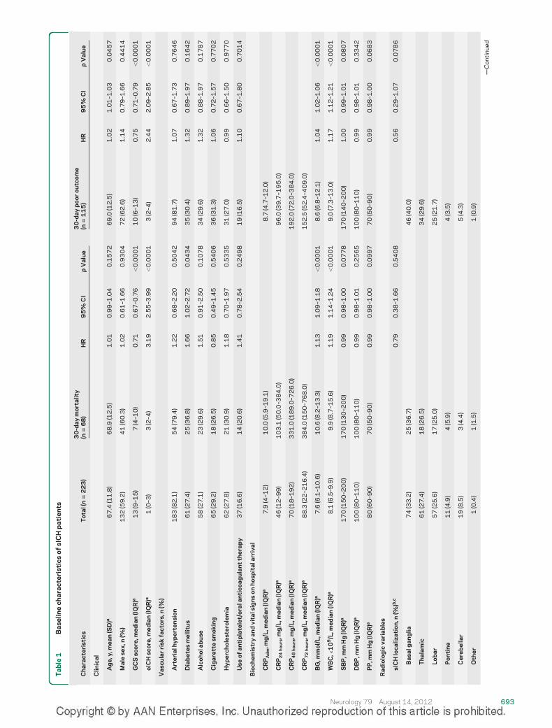
RESULTS Patient characteristics. A total of 132 menand 91 women (M/F ratio: 1.45; mean age, 67.4 �
11.8 years) were included in this study (figure e-1and table 1). Plasma samples were available from all223 patients on admission, but from only 126 at 72hours. Forty-four (19.7%) patients underwent surgicalhematoma evacuation: 22 (50%) of them at admission,a further 16 within 24 hours, and the others within thefirst 52 hours after sICH onset. At 30 days, 68 (30.5%)patients were dead. Deaths were attributable to the ini-tial sICH or rebleeding in 55 cases (80.9%) and sys-temic complications in 13 cases (19.1%). After 30-dayfollow-up, 49% (n � 109) of the patients showed agood functional outcome (GOS 4 and 5).
CRP kinetics. The median time from symptom onsetto the admission CRP measurement was 93 minutes(48–275 minutes). CRP concentration increased sig-nificantly (p � 0.0001, analysis of variance) from themedian value of 7.9 mg/L (4–12 mg/L) at admissionto 88.3 mg/L (22–216.4 mg/L) after 72 hours (figuree-2). Post hoc analysis revealed a significant differ-ence between CRPAdm and CRP24 hours, CRP48 hours,or CRP72 hours (p � 0.0001, for all comparisons).CRP concentration increased from 24 to 48 hours(p � 0.0016) and from 24 to 72 hours (p � 0.0004),but not from 48 to 72 hours (p � 0.424). Relative toCRPAdm, CRP24 hours decreased in 12 patients(5.4%), increased in 190 (85.2%), and remained sta-ble in 13 (5.8%). CRP peak was reached in 52 pa-tients (23.3%) at 24 hours, 60 patients (26.9%) at 48hours, and 46 patients (20.6%) at 72 hours.
Neuroradiologic findings. The median hematomavolume was 18 mL (8–37). The hematoma volumewas larger in patients with an unfavorable outcome(44.5 mL [20.5– 82.5] vs 12 mL [8 –23]; p �
0.0001) and with a worse functional outcome (30mL [15–60] vs 10 mL [6–18]; p � 0.0001). In-creased CRP concentration significantly correlatedwith a larger initial hematoma volume only for thelater measurements. The Pearson correlation coeffi-
cients for the relationship between hematoma vol-ume and the natural logarithm of CRP at thedifferent time points were CRPAdm, r � 0.1 (p �
0.1416); CRP24 hours, r � 0.43 (p � 0.0001);CRP48 hours, r � 0.51 (p � 0.0001); CRP72 hours,r � 0.45 (p � 0.0001).
Neuropathologic findings. General neuropathologicevaluation in sICH patients confirmed petechial orlarge hemorrhages surrounded by softened cerebraldiscolored, edematous brain tissue, ventricular bloodinundation, and asymmetric enlargement. All casespresented fresh hemorrhages, without any gliotic sur-rounding reactions. Ischemic lesions were character-ized by cavitation and surrounding gliosis.
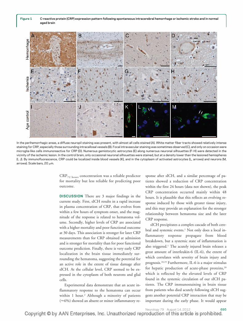
CRP was detected to different extents and pat-terns in sICH and ischemic stroke patients (tablee-1). Thus, for the immediate perihemorrhagic areas,a diffuse neuropil staining was present, together withalmost all cells’ silhouettes taking up the stain (figure1A). Further away from the hemorrhagic core, thediffuse staining pattern diminished, but with someneurons clearly retaining an affinity for the antibody.Although distant from the hemorrhage, white matterfiber tracts showed relatively intense staining forCRP, especially those surrounding intracallosal ves-sels (figure 1B). Focal intravascular staining some-times was observed (figure 1C), and only on occasionwere microglia-like cells immunoreactive for CRP(figure 1D).
In the region immediately surrounding an isch-emic liquefaction area, a high number of gemisto-cytic astrocytes was noted (figure 1E), while furtheraway from the lesion core numerous neuronal silhou-ettes were observed (figure 1, F–H). On occasion,diffuse staining could be noted along the white mat-ter tracts and blood vessels. Vascular walls and some-times plasma inside the vessel were again stained, andthis observation seemed to be constant for the respec-tive area of the lobe.
In the control brain, and in the hemispherescontralateral to the lesion, only occasional neuro-nal silhouettes were stained, with their respectivedensities being clearly lower compared to the le-sioned (ipsilateral) hemispheres (figure 1, I and J).By immunofluorescence, CRP could be clearly lo-calized inside blood vessels (figure 1K), and in thecytoplasm of activated astrocytes (figure 1L) andneurons (figure 1M).
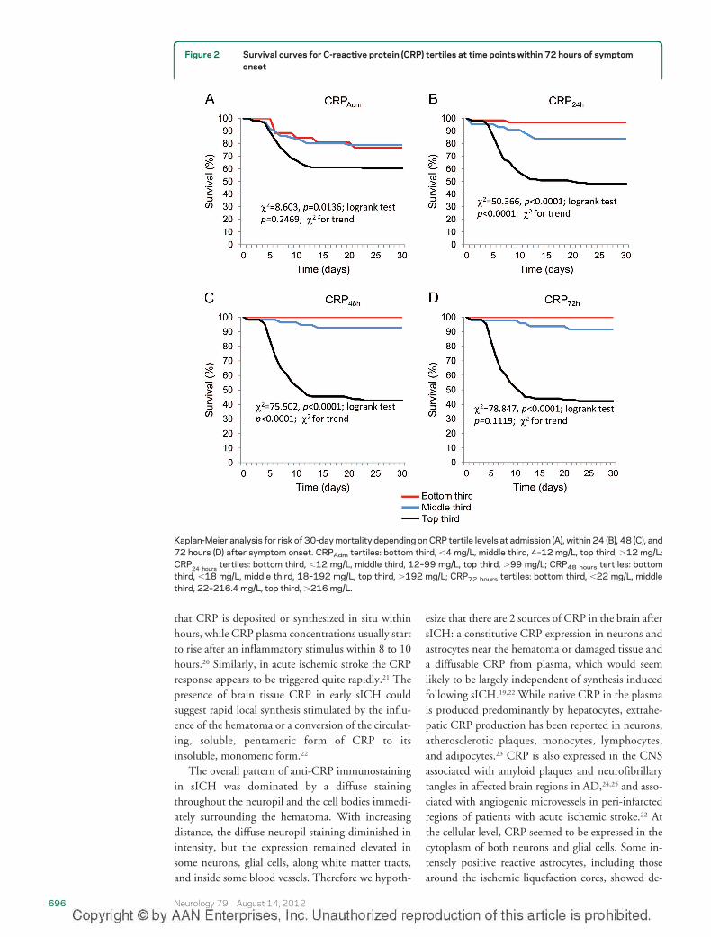
Associations between CRP at different time pointswith death and poor outcome. Survival of patients ac-cording to CRPAdm, CRP24 hours, CRP48 hours, andCRP72 hours tertiles is shown in figure 2. The rela-tionship between CRP and mortality was determinedto be nonlinear by the use of tertile analysis. The
692 Neurology 79 August 14, 2012
Tab
le1
Bas
elin
ech
arac
teri
stic
sof
sIC
Hp
atie
nts
Cha
ract
eris
tics
Tot
al(n
�2
23
)3
0-d
aym
orta
lity
(n�
68
)H
R9
5%
CI
pV
alue
30
-day
poo
rou
tcom
e(n
�1
15
)H
R9
5%
CI
pV
alue
Clin
ical
Ag
e,y,
mea
n(S
D)a
67
.4(1
1.8
)6
8.9
(12
.5)
1.0
10
.99
–1.0
40
.15
72
69
.0(1
2.5
)1
.02
1.0
1–1
.03
0.0
45
7
Mal
ese
x,n
(%)
13
2(5
9.2
)4
1(6
0.3
)1
.02
0.6
1–1
.66
0.9
30
47
2(6
2.6
)1
.14
0.7
9–1
.66
0.4
41
4
GC
Ssc
ore,
med
ian
(IQR
)a1
3(9
–15
)7
(4–1
0)
0.7
10
.67
–0.7
6�
0.0
00
11
0(6
–13
)0
.75
0.7
1–0
.79
�0
.00
01
oIC
Hsc
ore,
med
ian
(IQR
)a1
(0–3
)3
(2–4
)3
.19
2.5
5–3
.99
�0
.00
01
3(2
–4)
2.4
42
.09
–2.8
5�
0.0
00
1
Vas
cula
rri
skfa
ctor
s,n
(%)
Art
eria
lhyp
erte
nsio
n1
83
(82
.1)
54
(79
.4)
1.2
20
.68
–2.2
00
.50
42
94
(81
.7)
1.0
70
.67
–1.7
30
.76
46
Dia
bet
esm
ellit
us6
1(2
7.4
)2
5(3
6.8
)1
.66
1.0
2–2
.72
0.0
43
43
5(3
0.4
)1
.32
0.8
9–1
.97
0.1
64
2
Alc
ohol
abus
e5
8(2
7.1
)2
3(2
9.6
)1
.51
0.9
1–2
.50
0.1
07
83
4(2
9.6
)1
.32
0.8
8–1
.97
0.1
78
7
Cig
aret
tesm
okin
g6
5(2
9.2
)1
8(2
6.5
)0
.85
0.4
9–1
.45
0.5
40
63
6(3
1.3
)1
.06
0.7
2–1
.57
0.7
70
2
Hyp
erch
oles
tero
lem
ia6
2(2
7.8
)2
1(3
0.9
)1
.18
0.7
0–1
.97
0.5
33
53
1(2
7.0
)0
.99
0.6
6–1
.50
0.9
77
0
Use
ofan
tip
late
let/
oral
anti
coag
ulan
tth
erap
y3
7(1
6.6
)1
4(2
0.6
)1
.41
0.7
8–2
.54
0.2
49
81
9(1
6.5
)1
.10
0.6
7–1
.80
0.7
01
4
Bio
chem
istr
yan
dvi
tals
igns
onho
spit
alar
riva
l
CR
PA
dm
,mg
/L,m
edia
n(IQ
R)a
7.9
(4–1
2)
10
.0(5
.9–1
9.1
)8
.7(4
.7–1
2.0
)
CR
P2
4ho
urs,
mg
/L,m
edia
n(IQ
R)a
46
(12
–99
)1
03
.1(5
0.0
–38
4.0
)9
6.0
(39
.7–1
95
.0)
CR
P4
8ho
urs,
mg
/L,m
edia
n(IQ
R)a
70
(18
–19
2)
33
1.0
(18
9.0
–72
6.0
)1
92
.0(7
2.0
–38
4.0
)
CR
P7
2ho
urs,
mg
/L,m
edia
n(IQ
R)a
88
.3(2
2–2
16
.4)
38
4.0
(15
0–7
68
.0)
15
2.5
(52
.4–4
09
.0)
BG
,mm
ol/L
,med
ian
(IQR
)a7
.6(6
.1–1
0.6
)1
0.6
(8.2
–13
.3)
1.1
31
.09
–1.1
8�
0.0
00
18
.6(6
.8–1
2.1
)1
.04
1.0
2–1
.06
�0
.00
01
WB
C,�
10
9/L
,med
ian
(IQR
)a8
.1(6
.5–9
.9)
9.9
(8.7
–15
.6)
1.1
91
.14
–1.2
4�
0.0
00
19
.0(7
.3–1
3.0
)1
.17
1.1
2–1
.21
�0
.00
01
SB
P,m
mH
g(IQ
R)a
17
0(1
50
–20
0)
17
0(1
30
–20
0)
0.9
90
.98
–1.0
00
.07
78
17
0(1
40
–20
0)
1.0
00
.99
–1.0
10
.08
07
DB
P,m
mH
g(IQ
R)a
10
0(8
0–1
10
)1
00
(80
–11
0)
0.9
90
.98
–1.0
10
.25
65
10
0(8
0–1
10
)0
.99
0.9
8–1
.01
0.3
34
2
PP
,mm
Hg
(IQR
)a8
0(6
0–9
0)
70
(50
–90
)0
.99
0.9
8–1
.00
0.0
99
77
0(5
0–9
0)
0.9
90
.98
–1.0
00
.06
83
Rad
iolo
gic
vari
able
s
sIC
Hlo
caliz
atio
n,n
(%)b
,c0
.79
0.3
8–1
.66
0.5
40
80
.56
0.2
9–1
.07
0.0
78
6
Bas
alg
ang
lia7
4(3
3.2
)2
5(3
6.7
)4
6(4
0.0
)
Tha
lam
ic6
1(2
7.4
)1
8(2
6.5
)3
4(2
9.6
)
Lob
ar5
7(2
5.6
)1
7(2
5.0
)2
5(2
1.7
)
Pon
tine
11
(4.9
)4
(5.9
)4
(3.5
)
Cer
ebel
lar
19
(8.5
)3
(4.4
)5
(4.3
)
Oth
er1
(0.4
)1
(1.5
)1
(0.9
)
—C
onti
nued
Neurology 79 August 14, 2012 693
estimated risk ratios for 30-day mortality and 30-daypoor outcome rose sharply between second and thirdtertile of CRP (table 2).
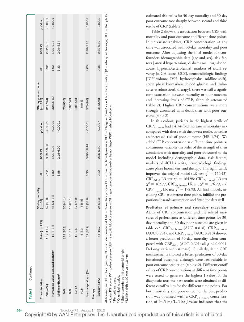
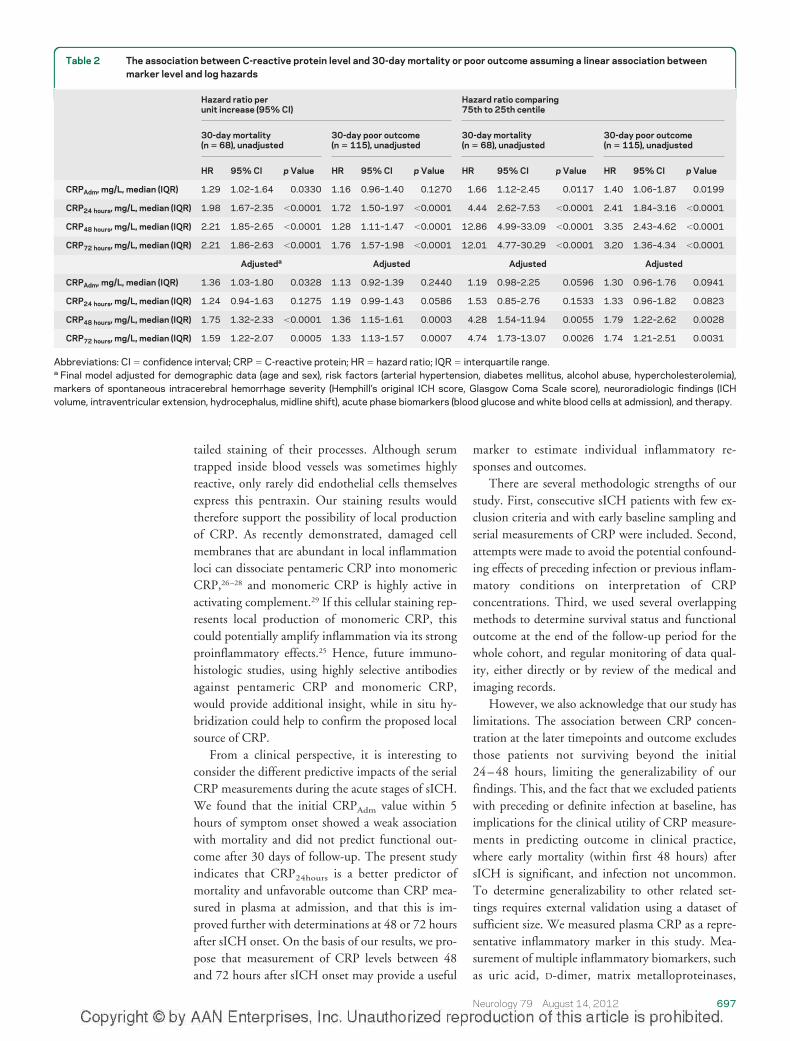
Table 2 shows the association between CRP withmortality and poor outcome at different time points.In univariate analyses, CRP concentration at anytime was associated with 30-day mortality and pooroutcome. After adjusting the final model for con-founders (demographic data [age and sex], risk fac-tors [arterial hypertension, diabetes mellitus, alcoholabuse, hypercholesterolemia], markers of sICH se-verity [oICH score, GCS], neuroradiologic findings[ICH volume, IVH, hydrocephalus, midline shift],acute phase biomarkers [blood glucose and leuko-cytes at admission], therapy), there was still a signifi-cant association between mortality or poor outcomeand increasing levels of CRP, although attenuated(table 2). Higher CRP concentrations were morestrongly associated with death than with poor out-come (table 2).
In this cohort, patients in the highest tertile ofCRP72 hours had a 4.74-fold increase in mortality riskcompared with those with the lowest tertile, as well asan increased risk of poor outcome (HR 1.74). Weadded CRP concentration at different time points ascontinuous variables (in order of the strength of theirassociation with mortality and poor outcome) to themodel including demographic data, risk factors,markers of sICH severity, neuroradiologic findings,acute phase biomarkers, and therapy. This significantlyimproved the original model (LR test �2 � 160.43):CRPAdm, LR test �2 � 164.98; CRP24 hours, LR test�2 � 162.77; CRP48 hours, LR test �2 � 176.29; andCRP
72 hours, LR test �2 � 172.93. All final models, in-
cluding CRP at different time points, fulfilled the pro-portional hazards assumption and fitted the data well.
Prediction of primary and secondary endpoints.AUCs of CRP concentration and the related mea-sures of performance at different time points for 30-day mortality and 30-day poor outcome are given intable e-2. CRP24 hours (AUC 0.818), CRP48 hours
(AUC 0.894), and CRP72 hours (AUC 0.910) showeda better prediction of 30-day mortality when com-pared with CRPAdm (AUC 0.601; all p � 0.0001;DeLong variance estimate). Similarly, later CRPmeasurements showed a better prediction of 30-dayfunctional outcome, although were less reliable inpoor outcome prediction (table e-2). Different cutoffvalues of CRP concentrations at different time pointswere tested to generate the highest J value for thediagnostic test; the best results were obtained at dif-ferent cutoff values for the different time points. Forboth mortality and poor outcome, the best predic-tion was obtained with a CRP72 hours concentra-tion of 94.5 mg/L. The J value indicates that the
Tab
le1
Con
tinu
ed
Cha
ract
eris
tics
Tot
al(n
�2
23
)3
0-d
aym
orta
lity
(n�
68
)H
R9
5%
CI
pV
alue
30
-day
poo
rou
tcom
e(n
�1
15
)H
R9
5%
CI
pV
alue
IVH
,n(%
)1
07
(47
.9)
57
(83
.8)
7.1
23
.73
–13
.60
�0
.00
01
81
(70
.4)
3.9
22
.62
–5.8
6�
0.0
00
1
Hem
atom
avo
lum
e,cc
,med
ian
(IQR
)a1
8(8
–37
)4
5(2
1–8
3)
1.0
21
.01
–1.0
3�
0.0
00
13
0(1
5–6
0)
1.0
21
.01
–1.0
3�
0.0
00
1
Mid
line
shif
t,m
md
3.8
82
.18
–6.9
0�
0.0
00
13
.33
2.0
0–5
.54
0–5
17
9(8
0.3
)3
0(4
4.1
)7
3(6
3.5
)
6–9
20
(9.1
)1
5(2
2.1
)1
8(1
5.6
)
10
–14
18
(7.6
)1
7(2
5.0
)1
8(1
5.6
)
>1
56
(3.3
)6
(8.8
)6
(5.3
)
Hyd
roce
pha
lus,
n(%
)6
9(3
0.9
)2
3(3
3.8
)6
.30
3.8
0–1
0.4
4�
0.0
00
15
7(4
9.6
)4
.05
2.8
0–5
.86
�0
.00
01
The
rap
y
Sur
ger
y,n
(%)
44
(19
.7)
24
(35
.3)
0.4
20
.25
–0.6
90
.00
07
34
(29
.6)
0.4
60
.31
–0.6
90
.00
02
Abb
revi
atio
ns:B
G�
bloo
dgl
ucos
e;C
I�co
nfid
ence
inte
rval
;CR
P�
C-r
eact
ive
prot
ein;
DB
P�
dias
tolic
bloo
dpr
essu
re;G
CS
�G
lasg
owC
oma
Sca
le;H
R�
haza
rdra
tio;
IQR
�in
terq
uart
ilera
nge;
oIC
H�
Hem
phill
’sor
igin
alIC
Hsc
ore;
PP
�pu
lse
pres
sure
;SB
P�
syst
olic
bloo
dpr
essu
re;s
ICH
�sp
onta
neou
sin
trac
ereb
ralh
emor
rhag
e;W
BC
�w
hite
bloo
dce
lls.
aP
erun
itin
crea
se.
bO
nem
issi
ngva
lue.
cS
upra
tent
oria
lvs
infr
aten
tori
alor
igin
.d
Mid
line
shif
t�
10
mm
vs�
10
mm
.
694 Neurology 79 August 14, 2012
CRP72 hours concentration was a reliable predictorfor mortality but less reliable for predicting pooroutcome.
DISCUSSION There are 3 major findings in thecurrent study. First, sICH results in a rapid increasein plasma concentration of CRP, that evolves fromwithin a few hours of symptom onset, and the mag-nitude of the response is related to hematoma vol-ume. Secondly, higher levels of CRP are associatedwith a higher mortality and poor functional outcomeat 30 days. This association is stronger for later CRPmeasurements than for CRP obtained at admissionand is stronger for mortality than for poor functionaloutcome prediction. Finally, there is very early CRPlocalization in the brain tissue immediately sur-rounding the hematoma, suggesting the potential foran active role in the extent of tissue damage aftersICH. At the cellular level, CRP seemed to be ex-pressed in the cytoplasm of both neurons and glialcells.
Experimental data demonstrate that an acute in-flammatory response to the hematoma can occurwithin 1 hour.2 Although a minority of patients(�6%) showed an absent or minor inflammatory re-
sponse after sICH, and a similar percentage of pa-tients showed a reduction of CRP concentrationwithin the first 24 hours (data not shown), the peakCRP concentration occurred mainly within 48hours. It is plausible that this reflects an evolving re-sponse induced by those with greater tissue injury,and this may provide an explanation for the strongerrelationship between hematoma size and the laterCRP response.
sICH precipitates a complex cascade of both cere-bral and systemic events.1 Not only does a local in-flammatory response propagate from bloodbreakdown, but a systemic state of inflammation isalso triggered.1 The acutely injured brain releases agreat amount of interleukin-6 (IL-6), the extent ofwhich correlates with severity of brain injury andprognosis.18,19 Furthermore, IL-6 is a major stimulusfor hepatic production of acute-phase proteins,20
which is reflected by the elevated levels of CRPfound in the systemic circulation of our sICH pa-tients. The CRP immunostaining in brain tissuefrom patients who died acutely following sICH sug-gests another potential CRP interaction that may beimportant during the early phase. It would appear
Figure 1 C-reactive protein (CRP) expression pattern following spontaneous intracerebral hemorrhage or ischemic stroke and in normalaged brain
In the perihemorrhagic areas, a diffuse neuropil staining was present, with almost all cells stained (A). White matter fiber tracts showed relatively intensestaining for CRP, especially those surrounding intracallosal vessels (B). Focal intravascular staining was sometimes observed (C), and only on occasion weremicroglia-like cells immunoreactive for CRP (D). Numerous gemistocytic astrocytes (E) along numerous neuronal silhouettes (F–H) were detected in thevicinity of the ischemic lesion. In the control brain, only occasional neuronal silhouettes were stained, but at a density lower than the lesioned hemispheres(I, J). By immunofluorescence, CRP could be localized inside blood vessels (K), and in the cytoplasm of activated astrocytes (L, arrows) and neurons (M,arrows). Scale bars, 20 �m.
Neurology 79 August 14, 2012 695
that CRP is deposited or synthesized in situ withinhours, while CRP plasma concentrations usually startto rise after an inflammatory stimulus within 8 to 10hours.20 Similarly, in acute ischemic stroke the CRPresponse appears to be triggered quite rapidly.21 Thepresence of brain tissue CRP in early sICH couldsuggest rapid local synthesis stimulated by the influ-ence of the hematoma or a conversion of the circulat-ing, soluble, pentameric form of CRP to itsinsoluble, monomeric form.22
The overall pattern of anti-CRP immunostainingin sICH was dominated by a diffuse stainingthroughout the neuropil and the cell bodies immedi-ately surrounding the hematoma. With increasingdistance, the diffuse neuropil staining diminished inintensity, but the expression remained elevated insome neurons, glial cells, along white matter tracts,and inside some blood vessels. Therefore we hypoth-
esize that there are 2 sources of CRP in the brain aftersICH: a constitutive CRP expression in neurons andastrocytes near the hematoma or damaged tissue anda diffusable CRP from plasma, which would seemlikely to be largely independent of synthesis inducedfollowing sICH.19,22 While native CRP in the plasmais produced predominantly by hepatocytes, extrahe-patic CRP production has been reported in neurons,atherosclerotic plaques, monocytes, lymphocytes,and adipocytes.23 CRP is also expressed in the CNSassociated with amyloid plaques and neurofibrillarytangles in affected brain regions in AD,24,25 and asso-ciated with angiogenic microvessels in peri-infarctedregions of patients with acute ischemic stroke.22 Atthe cellular level, CRP seemed to be expressed in thecytoplasm of both neurons and glial cells. Some in-tensely positive reactive astrocytes, including thosearound the ischemic liquefaction cores, showed de-
Figure 2 Survival curves for C-reactive protein (CRP) tertiles at time points within 72 hours of symptomonset
Kaplan-Meier analysis for risk of 30-day mortality depending on CRP tertile levels at admission (A), within 24 (B), 48 (C), and72 hours (D) after symptom onset. CRPAdm tertiles: bottom third, �4 mg/L, middle third, 4–12 mg/L, top third, �12 mg/L;CRP24 hours tertiles: bottom third, �12 mg/L, middle third, 12–99 mg/L, top third, �99 mg/L; CRP48 hours tertiles: bottomthird, �18 mg/L, middle third, 18–192 mg/L, top third, �192 mg/L; CRP72 hours tertiles: bottom third, �22 mg/L, middlethird, 22–216.4 mg/L, top third, �216 mg/L.
696 Neurology 79 August 14, 2012
tailed staining of their processes. Although serumtrapped inside blood vessels was sometimes highlyreactive, only rarely did endothelial cells themselvesexpress this pentraxin. Our staining results wouldtherefore support the possibility of local productionof CRP. As recently demonstrated, damaged cellmembranes that are abundant in local inflammationloci can dissociate pentameric CRP into monomericCRP,26–28 and monomeric CRP is highly active inactivating complement.29 If this cellular staining rep-resents local production of monomeric CRP, thiscould potentially amplify inflammation via its strongproinflammatory effects.25 Hence, future immuno-histologic studies, using highly selective antibodiesagainst pentameric CRP and monomeric CRP,would provide additional insight, while in situ hy-bridization could help to confirm the proposed localsource of CRP.
From a clinical perspective, it is interesting toconsider the different predictive impacts of the serialCRP measurements during the acute stages of sICH.We found that the initial CRPAdm value within 5hours of symptom onset showed a weak associationwith mortality and did not predict functional out-come after 30 days of follow-up. The present studyindicates that CRP24hours is a better predictor ofmortality and unfavorable outcome than CRP mea-sured in plasma at admission, and that this is im-proved further with determinations at 48 or 72 hoursafter sICH onset. On the basis of our results, we pro-pose that measurement of CRP levels between 48and 72 hours after sICH onset may provide a useful
marker to estimate individual inflammatory re-sponses and outcomes.
There are several methodologic strengths of ourstudy. First, consecutive sICH patients with few ex-clusion criteria and with early baseline sampling andserial measurements of CRP were included. Second,attempts were made to avoid the potential confound-ing effects of preceding infection or previous inflam-matory conditions on interpretation of CRPconcentrations. Third, we used several overlappingmethods to determine survival status and functionaloutcome at the end of the follow-up period for thewhole cohort, and regular monitoring of data qual-ity, either directly or by review of the medical andimaging records.
However, we also acknowledge that our study haslimitations. The association between CRP concen-tration at the later timepoints and outcome excludesthose patients not surviving beyond the initial24 – 48 hours, limiting the generalizability of ourfindings. This, and the fact that we excluded patientswith preceding or definite infection at baseline, hasimplications for the clinical utility of CRP measure-ments in predicting outcome in clinical practice,where early mortality (within first 48 hours) aftersICH is significant, and infection not uncommon.To determine generalizability to other related set-tings requires external validation using a dataset ofsufficient size. We measured plasma CRP as a repre-sentative inflammatory marker in this study. Mea-surement of multiple inflammatory biomarkers, suchas uric acid, D-dimer, matrix metalloproteinases,
Table 2 The association between C-reactive protein level and 30-day mortality or poor outcome assuming a linear association betweenmarker level and log hazards
Hazard ratio perunit increase (95% CI)
Hazard ratio comparing75th to 25th centile
30-day mortality(n � 68), unadjusted
30-day poor outcome(n � 115), unadjusted
30-day mortality(n � 68), unadjusted
30-day poor outcome(n � 115), unadjusted
HR 95% CI p Value HR 95% CI p Value HR 95% CI p Value HR 95% CI p Value
CRPAdm, mg/L, median (IQR) 1.29 1.02–1.64 0.0330 1.16 0.96–1.40 0.1270 1.66 1.12–2.45 0.0117 1.40 1.06–1.87 0.0199
CRP24 hours, mg/L, median (IQR) 1.98 1.67–2.35 �0.0001 1.72 1.50–1.97 �0.0001 4.44 2.62–7.53 �0.0001 2.41 1.84–3.16 �0.0001
CRP48 hours, mg/L, median (IQR) 2.21 1.85–2.65 �0.0001 1.28 1.11–1.47 �0.0001 12.86 4.99–33.09 �0.0001 3.35 2.43–4.62 �0.0001
CRP72 hours, mg/L, median (IQR) 2.21 1.86–2.63 �0.0001 1.76 1.57–1.98 �0.0001 12.01 4.77–30.29 �0.0001 3.20 1.36–4.34 �0.0001
Adjusteda Adjusted Adjusted Adjusted
CRPAdm, mg/L, median (IQR) 1.36 1.03–1.80 0.0328 1.13 0.92–1.39 0.2440 1.19 0.98–2.25 0.0596 1.30 0.96–1.76 0.0941
CRP24 hours, mg/L, median (IQR) 1.24 0.94–1.63 0.1275 1.19 0.99–1.43 0.0586 1.53 0.85–2.76 0.1533 1.33 0.96–1.82 0.0823
CRP48 hours, mg/L, median (IQR) 1.75 1.32–2.33 �0.0001 1.36 1.15–1.61 0.0003 4.28 1.54–11.94 0.0055 1.79 1.22–2.62 0.0028
CRP72 hours, mg/L, median (IQR) 1.59 1.22–2.07 0.0005 1.33 1.13–1.57 0.0007 4.74 1.73–13.07 0.0026 1.74 1.21–2.51 0.0031
Abbreviations: CI � confidence interval; CRP � C-reactive protein; HR � hazard ratio; IQR � interquartile range.a Final model adjusted for demographic data (age and sex), risk factors (arterial hypertension, diabetes mellitus, alcohol abuse, hypercholesterolemia),markers of spontaneous intracerebral hemorrhage severity (Hemphill’s original ICH score, Glasgow Coma Scale score), neuroradiologic findings (ICHvolume, intraventricular extension, hydrocephalus, midline shift), acute phase biomarkers (blood glucose and white blood cells at admission), and therapy.
Neurology 79 August 14, 2012 697

platelets, fibrinogen, and IL-6, may provide a moredetailed measure of the inflammatory response aftersICH. Similarly, inspecting the colocalization ofCRP with complement in brain staining would alsofurnish further data in support of a direct pathophys-iologic role of CRP in sICH and these studies are inprogress. Finally, because of the few available brainautopsies and the few related blood samples, we wereunable to correlate anatomopathologic CRP datawith clinical data in a quantitative way.
We have demonstrated an association betweenhigher levels of CRP mortality and poor outcome inpatients after sICH together with a very early CRPlocalization in the brain tissue, immediately sur-rounding the hematoma. The CRP response evolvesover the initial 72 hours and the association withmortality or poor outcome is stronger using laterCRP measurements.
AUTHOR CONTRIBUTIONSAgreed with the manuscript’s results and conclusions: M.D.N., D.A.G.,
V.C., L. Masotti, C.J.S., A.R.P.-J., S.J.H., M.S., F.P., L. Mogoanta, D.P.,
A.P.-W. Designed the experiments/study: M.D.N., D.A.G., A.P.-W. An-
alyzed the data: M.D.N., D.A.G., M.S., F.P., L. Mogoanta, D.P.,
A.P.-W. Collected data/did experiments for the study: M.D.N., D.A.G.,
V.C., M.S., L. Masotti, F.P., L. Mogoanta, D.P., A.P.-W. Enrolled pa-
tients: M.D.N., D.A.G., V.C., L. Masotti, F.P., L. Mogoanta, D.P.,
A.P.-W. Wrote the first draft of the paper: M.D.N. Contributed to the
writing of the paper: M.D.N., D.A.G., V.C., L. Masotti, C.J.S., A.R.P.-J.,
S.J.H., M.S., F.P., L. Mogoanta, D.P., A.P.-W. Provided statistical ad-
vice: M.D.N. Contributed to the design of the analyses and the interpre-
tation of the data: M.D.N., C.J.S., A.R.P.-J., S.J.H., M.S., A.P.-W.
Contributed to patient characterization: M.D.N., D.A.G., V.C., L. Ma-
sotti, M.S., F.P., L. Mogoanta, D.P., A.P.-W. Principal investigator for
this study: M.D.N.
STUDY FUNDINGSupported in part by the Sectorial Operational Programme Human Re-
sources Development, financed from the European Social Fund and by
the Romanian Government under the contract number POSDRU/89/
1.5/S/64109 to Dr. D. Pirici and grant agreement PN-II-ID-PCE-2011-
3-0848, No 301, to Aurel Popa-Wagner.
DISCLOSUREThe authors report no disclosures relevant to the manuscript. Go to
Neurology.org for full disclosures.
Received December 24, 2011. Accepted in final form March 28, 2012.
REFERENCES1. Aronowski J, Zhao X. Molecular pathophysiology of cere-
bral hemorrhage: secondary brain injury. Stroke 2011;42:1781–1786.
2. Wang J. Preclinical and clinical research on inflammationafter intracerebral hemorrhage. Prog Neurobiol 2010;92:463–477.
3. Malham GM, Souter MJ. Systemic inflammatory responsesyndrome and acute neurological disease. Br J Neurosurg2001;15:381–387.
4. Castellanos M, Leira R, Tejada J, Gil-Peralta A, Davalos A,Castillo J. Predictors of good outcome in medium to largespontaneous supratentorial intracerebral haemorrhages.J Neurol Neurosurg Psychiatry 2005;76:691–695.
5. Di Napoli M, Godoy DA, Campi V, et al. C-reactive pro-
tein level measurement improves mortality prediction
when added to the spontaneous intracerebral hemorrhage
score. Stroke 2011;42:1230–1236.
6. Diedler J, Sykora M, Hahn P, et al. C-reactive-protein
levels associated with infection predict short- and long-
term outcome after supratentorial intracerebral hemor-
rhage. Cerebrovasc Dis 2009;27:272–279.
7. Di Napoli M, Elkind MS, Godoy DA, Singh P, Papa F,
Popa-Wagner A. Role of C-reactive protein in cerebrovas-
cular disease: a critical review. Expert Rev Cardiovasc Ther
2011;9:1565–1584.
8. Teasdale G, Jennett B. Assessment of coma and impaired
consciousness: a practical scale. Lancet 1974;2:81–84.
9. Hemphill JC 3rd, Bonovich DC, Besmertis L, Manley
GT, Johnston SC. The ICH score: a simple, reliable grad-
ing scale for intracerebral hemorrhage. Stroke 2001;32:
891–897.
10. Broderick J, Connolly S, Feldmann E, et al. Guidelines for
the management of spontaneous intracerebral hemorrhage
in adults: 2007 update: a guideline from the American
Heart Association/American Stroke Association Stroke
Council, High Blood Pressure Research Council, and the
Quality of Care and Outcomes in Research Interdisciplin-
ary Working Group. Circulation 2007;116:e391–e413.
11. Steiner T, Kaste M, Forsting M, et al. Recommendations
for the management of intracranial haemorrhage: part I:
spontaneous intracerebral haemorrhage: The European
Stroke Initiative Writing Committee and the Writing
Committee for the EUSI Executive Committee. Cerebro-
vasc Dis 2006;22:294–316.
12. Roberts WL, Moulton L, Law TC, et al. Evaluation of
nine automated high-sensitivity C-reactive protein meth-
ods: implications for clinical and epidemiological applica-
tions. Part 2 Clin Chem 2001;47:418–425.
13. Kothari RU, Brott T, Broderick JP, et al. The ABCs of
measuring intracerebral hemorrhage volumes. Stroke
1996;27:1304–1305.
14. Broderick JP, Brott TG, Duldner JE, Tomsick T, Huster
G. Volume of intracerebral hemorrhage: a powerful and
easy-to-use predictor of 30-day mortality. Stroke 1993;24:
987–993.
15. Ying SC, Gewurz H, Kinoshita CM, Potempa LA, Siegel
JN. Identification and partial characterization of multiple
native and neoantigenic epitopes of human C-reactive pro-
tein by using monoclonal antibodies. J Immunol 1989;
143:221–228.
16. Jennett B, Bond M. Assessment of outcome after severe
brain damage. Lancet 1975;1:480–484.
17. Hanley JA, McNeil BJ. The meaning and use of the area
under a receiver operating characteristic (ROC) curve. Ra-
diology 1982;143:29–36.
18. Silva Y, Leira R, Tejada J, Lainez JM, Castillo J, Davalos A.
Molecular signatures of vascular injury are associated with
early growth of intracerebral hemorrhage. Stroke 2005;36:
86–91.
19. Kuhlmann CR, Librizzi L, Closhen D, et al. Mechanisms
of C-reactive protein-induced blood-brain barrier disrup-
tion. Stroke 2009;40:1458–1466.
20. Pepys MB, Hirschfield GM. C-reactive protein: a critical
update. J Clin Invest 2003;111:1805–1812.
21. Emsley HC, Smith CJ, Gavin CM, et al. An early and
sustained peripheral inflammatory response in acute isch-
698 Neurology 79 August 14, 2012
aemic stroke: relationships with infection and atherosclero-sis. J Neuroimmunol 2003;139:93–101.
22. Slevin M, Matou-Nasri S, Turu M, et al. ModifiedC-reactive protein is expressed by stroke neovessels and is apotent activator of angiogenesis in vitro. Brain Pathol2010;20:151–165.
23. Eisenhardt SU, Thiele JR, Bannasch H, Stark GB, PeterK. C-reactive protein: how conformational changes in-fluence inflammatory properties. Cell Cycle 2009;8:3885–3892.
24. McGeer EG, Yasojima K, Schwab C, McGeer PL. The pen-traxins: possible role in Alzheimer’s disease and other innateinflammatory diseases. Neurobiol Aging 2001;22:843–848.
25. Strang F, Scheichl A, Chen YC, et al. Amyloid plaques disso-ciate pentameric to monomeric C-reactive protein: a novelpathomechanism driving cortical inflammation in Alzhei-mer’s disease? Brain Pathol 2012;22:337–346.
26. Ji SR, Wu Y, Zhu L, et al. Cell membranes and liposomesdissociate C-reactive protein (CRP) to form a new, biolog-ically active structural intermediate: mCRP(m). FASEB J2007;21:284–294.
27. Eisenhardt SU, Habersberger J, Murphy A, et al. Dissocia-tion of pentameric to monomeric C-reactive protein onactivated platelets localizes inflammation to atheroscleroticplaques. Circ Res 2009;105:128–137.
28. Lauer N, Mihlan M, Hartmann A, et al. Complement reg-ulation at necrotic cell lesions is impaired by the age-related macular degeneration-associated factor-H His402risk variant. J Immunol 2011;187:4374–4383.
29. Ji SR, Wu Y, Potempa LA, Liang YH, Zhao J. Effect ofmodified C-reactive protein on complement activation:a possible complement regulatory role of modified ormonomeric C-reactive protein in atherosclerotic lesions.Arterioscler Thromb Vasc Biol 2006;26:935–941.
AAN Publishes Guideline Update on Infantile SpasmsThe AAN has published evidence-based recommendations for the treatment of infantile spasms thatupdate a 2004 guideline. “Evidence-based Guideline Update: Medical Treatment of InfantileSpasms,” published in the June 12, 2012, issue of Neurology®, suggests that the therapy adrenocor-ticotropic hormone, also known as ACTH, and the antiepileptic drug vigabatrin (VGB) may beeffective in the treatment of infantile spasms in children.
To read the guideline and access PDF summaries for clinicians and patients, a slide presentation,and a clinical example, visit www.aan.com/go/practice/guidelines. For more information, contact JulieCox at [email protected] or (612) 928-6069.
It All Starts with an Idea.What Do You Want to Change?
If you see an opportunity for positive change in the delivery of neurologic health care, the AANwants you to apply for the Donald M. Palatucci Advocacy Leadership Forum to be held January17–20, 2013, at the Rancho Bernardo Inn in San Diego, CA.
Only 30 AAN members will be accepted into this exclusive program that empowers neurologists toadvocate for themselves, their patients, and their profession. Participants will be trained in criticalskills to communicate effectively with the media and legislators, and gain an understanding ofgrassroots advocacy. Upon completion of the training, members will have developed an effectiveaction plan, created a clear message to promote community awareness of their issue, and understoodthe dynamics of the legislative process.
Applications for this award-winning program are due by September 16, 2012. For more informa-tion, visit www.aan.com/view/2013PALF or contact Melissa Showers at [email protected] or (612)928-6056.
Neurology 79 August 14, 2012 699
DOI 10.1212/WNL.0b013e318264e3be; Published online before print August 1, 2012;Neurology
Mario Di Napoli, Daniel Agustin Godoy, Veronica Campi, et al.and prognosis
C-reactive protein in intracerebral hemorrhage : Time course, tissue localization,
August 13, 2012This information is current as of
ServicesUpdated Information &
3e318264e3behttp://www.neurology.org/content/early/2012/08/01/WNL.0b01including high resolution figures, can be found at:
Supplementary Material
3e318264e3be.DC1.htmlhttp://www.neurology.org/content/suppl/2012/08/01/WNL.0b01Supplementary material can be found at:
Subspecialty Collections
_prognosishttp://www.neurology.org/cgi/collection/natural_history_studiesNatural history studies (prognosis)
agehttp://www.neurology.org/cgi/collection/intracerebral_hemorrhIntracerebral hemorrhagefollowing collection(s):This article, along with others on similar topics, appears in the
Permissions & Licensing
http://www.neurology.org/misc/about.xhtml#permissionstables) or in its entirety can be found online at: Information about reproducing this article in parts (figures,
Reprints http://www.neurology.org/misc/addir.xhtml#reprintsus
Information about ordering reprints can be found online: