Embed Size (px)
Citation preview

SteroidiEffetti collaterali:
- sanguinamenti gastrointestinali
- infezioni
- iperglicemia difficilmente controllabile
- ipertensione arteriosa
Complicanze neurologiche della fase acuta
Edema cerebrale

malignant MCA syndrome

Malignant MCA stroke
Inclusion criteria
• Age 18–60 years
• Clinical deficits suggestive of
infarction in the territory of the MCA
• NIHSS >15
• Decrease in the level of consciousness
to a score of 1 or greater on item 1a of
the NIHSS
• Signs on CT of an infarct of at least
50% of the MCA territory or infarct
volume >145 cm³ as shown on
diffusion-weighted MRI
• Surgery within 48 h after onset of
symptoms
Exclusion criteria
• Prestroke score on the mRS ≥2
• Two fixed dilated pupils
• Contralateral ischaemia or other brain lesion that could affect outcome
• Space-occupying haemorrhagic transformation of the infarct (≥parenchymal haemorrhage grade 2)
• Life expectancy <3 years
• Other serious illness that could aff ect outcome
• Known coagulopathy or systemic bleeding disorder
• Contraindication for anaesthesia
• Pregnancy


At risk situations
• MCA stroke malignant MCA syndrome
• Cerebellar stroke acute hydrocefalus
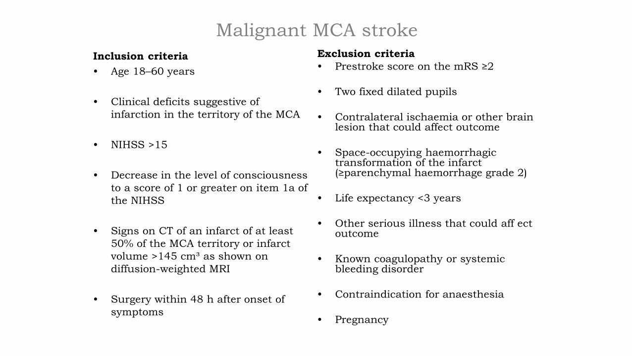
• Posterior stroke basilar occlusion
• Severe (tight) carotid stenosis carotid occlusion
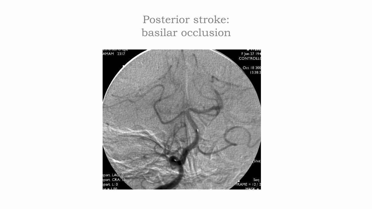
• Lacunar stroke progression of the deficit

Cerebellar stroke


Posterior stroke:
basilar occlusion
Posterior stroke:
basilar occlusion

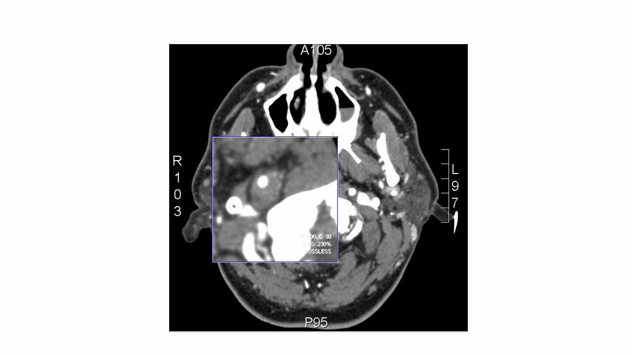
Ecodoppler vasi epiaortici

Posterior stroke:
basilar occlusion

Stenosi carotide interna destra >70%

Severe (tight) carotid stenosis
Benefit Of Carotid Endarterectomy In Patients
With Symptomatic Severe Stenosis
Barnett et al, NEJM 1991

Endarterectomy for symptomatic carotid stenosis in
relation to clinical subgroups and timing of surgery
Lacunar stroke

Lacunar stroke
Group A: with progressive motor deficit
Group B: without progressive motor deficit
Stroke 2002; 33: 1510-1516

In all other cases without a sure underlying
mechanism?
Heparin
• Urgent anticoagulation with the goal of preventing early
recurrent stroke, halting neurological worsening, or
improving outcomes after acute ischemic stroke is not
recommended for treatment of patients with acute ischemic
stroke

Other dangerous conditions
• Hemorrhagic transformation
• Epileptic activity
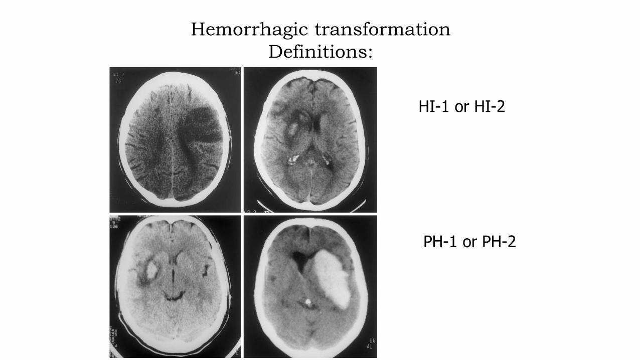
HI-1 or HI-2
PH-1 or PH-2
Hemorrhagic transformation
Definitions:
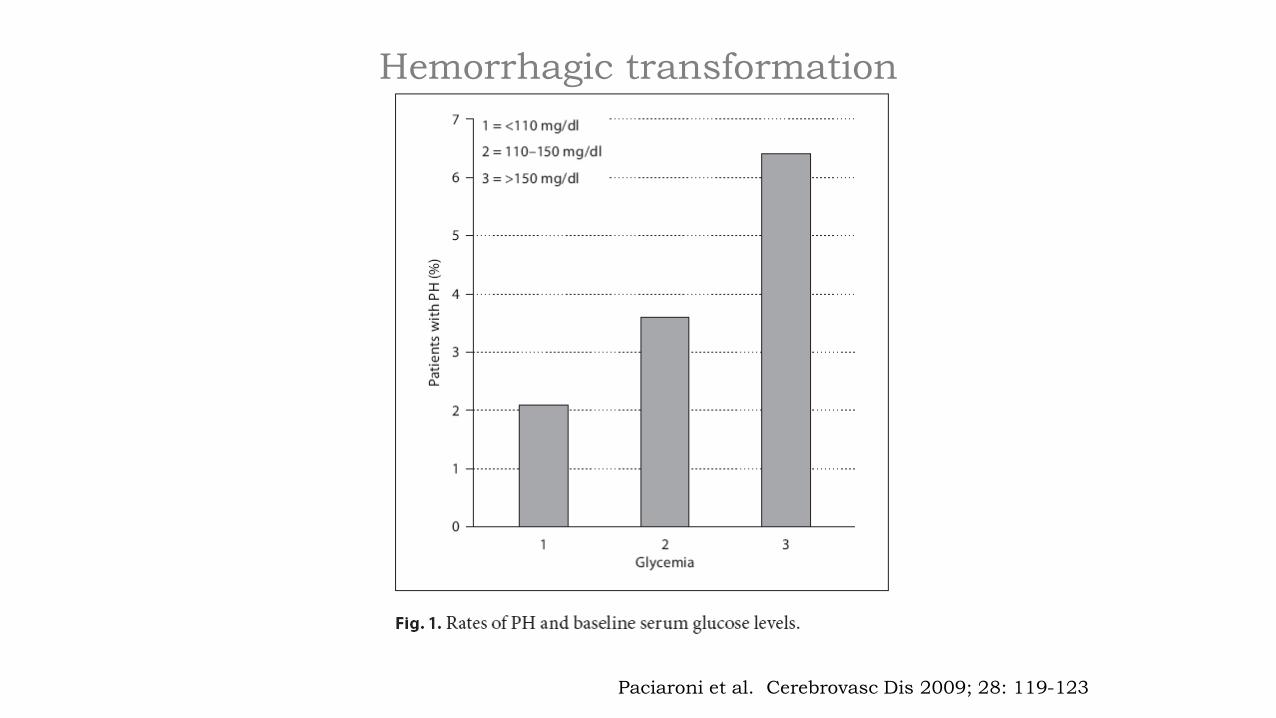
Paciaroni et al. Cerebrovasc Dis 2009; 28: 119-123
Hemorrhagic transformation


Complicanze tardive

Prevenzione e trattamento del tromboembolismo venoso

Epidemiology
Factors increasing the risk of VTE after stroke.
Neurological factors:
- Any reduction in mobility
- Paretic lower limb
- Increasing stroke severity
- Hemorrhagic stroke
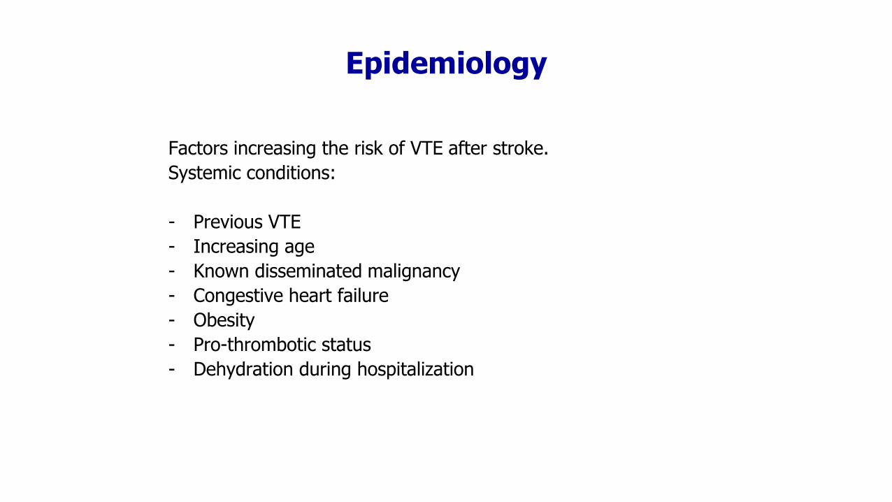
Epidemiology
Factors increasing the risk of VTE after stroke.
Systemic conditions:
- Previous VTE
- Increasing age
- Known disseminated malignancy
- Congestive heart failure
- Obesity
- Pro-thrombotic status
- Dehydration during hospitalization
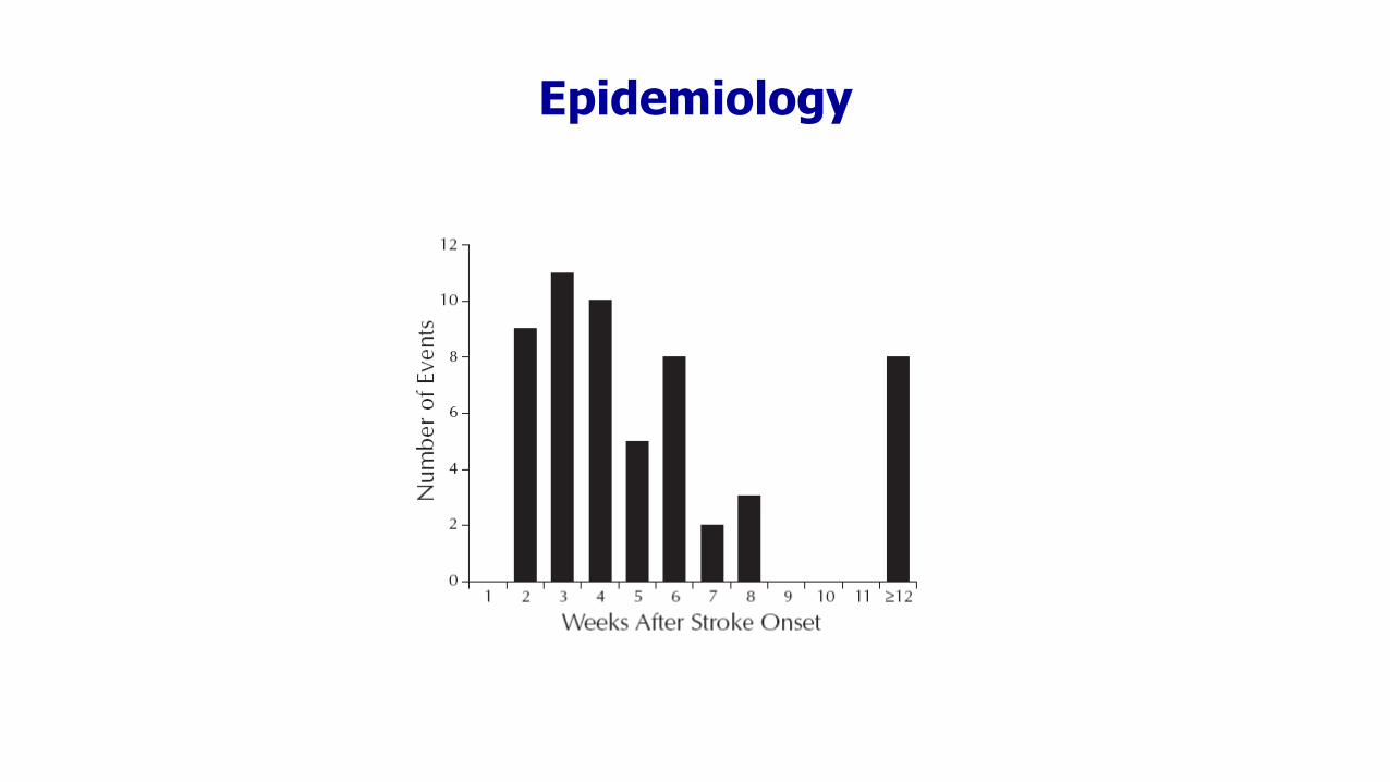
Epidemiology

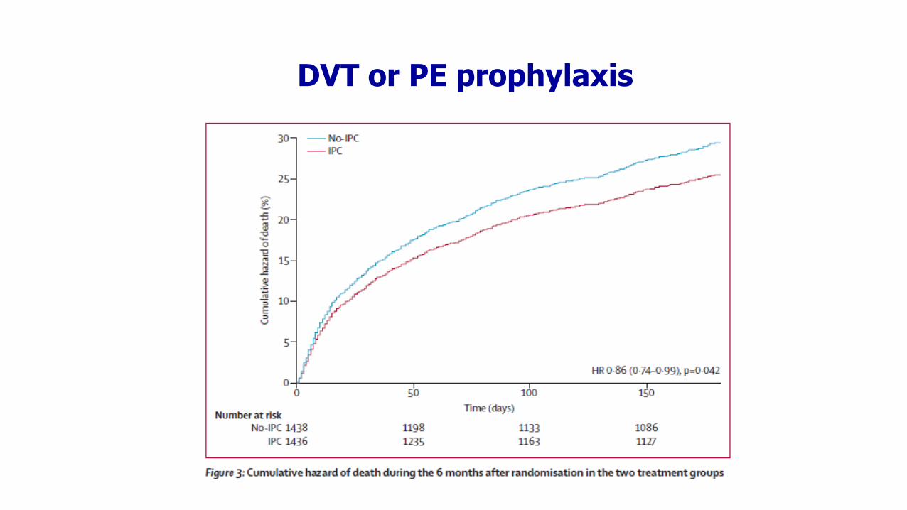
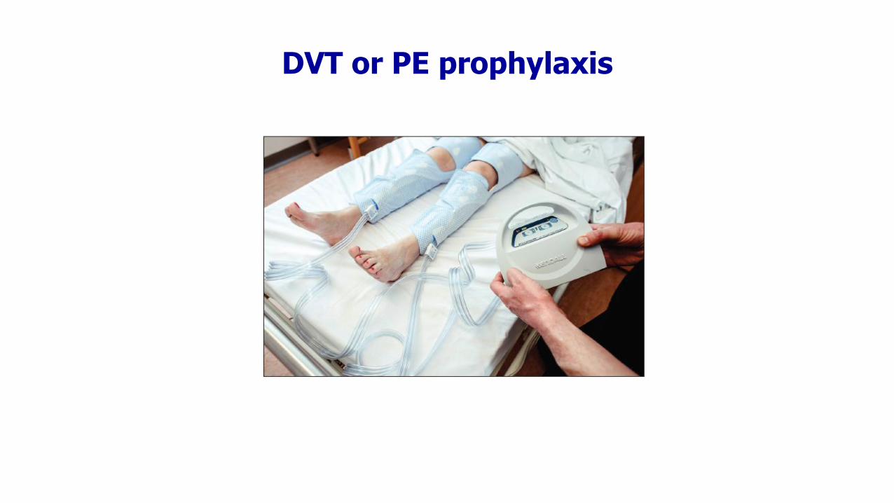
DVT or PE prophylaxis
• Graduated compression stockings• Intermittent pneumatic compression • Anticoagulants:
- UFH- LMWH- Vit. K antagonists- Direct Thrombin inhibitors- Direct Factor Xa Inhibitors
DVT or PE prophylaxis
DVT or PE prophylaxis
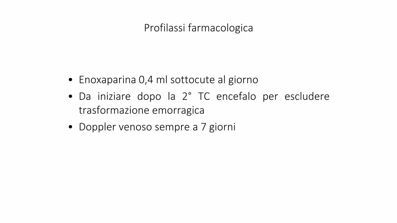
Profilassi farmacologica
• Enoxaparina 0,4 ml sottocute al giorno
• Da iniziare dopo la 2° TC encefalo per escluderetrasformazione emorragica
• Doppler venoso sempre a 7 giorni
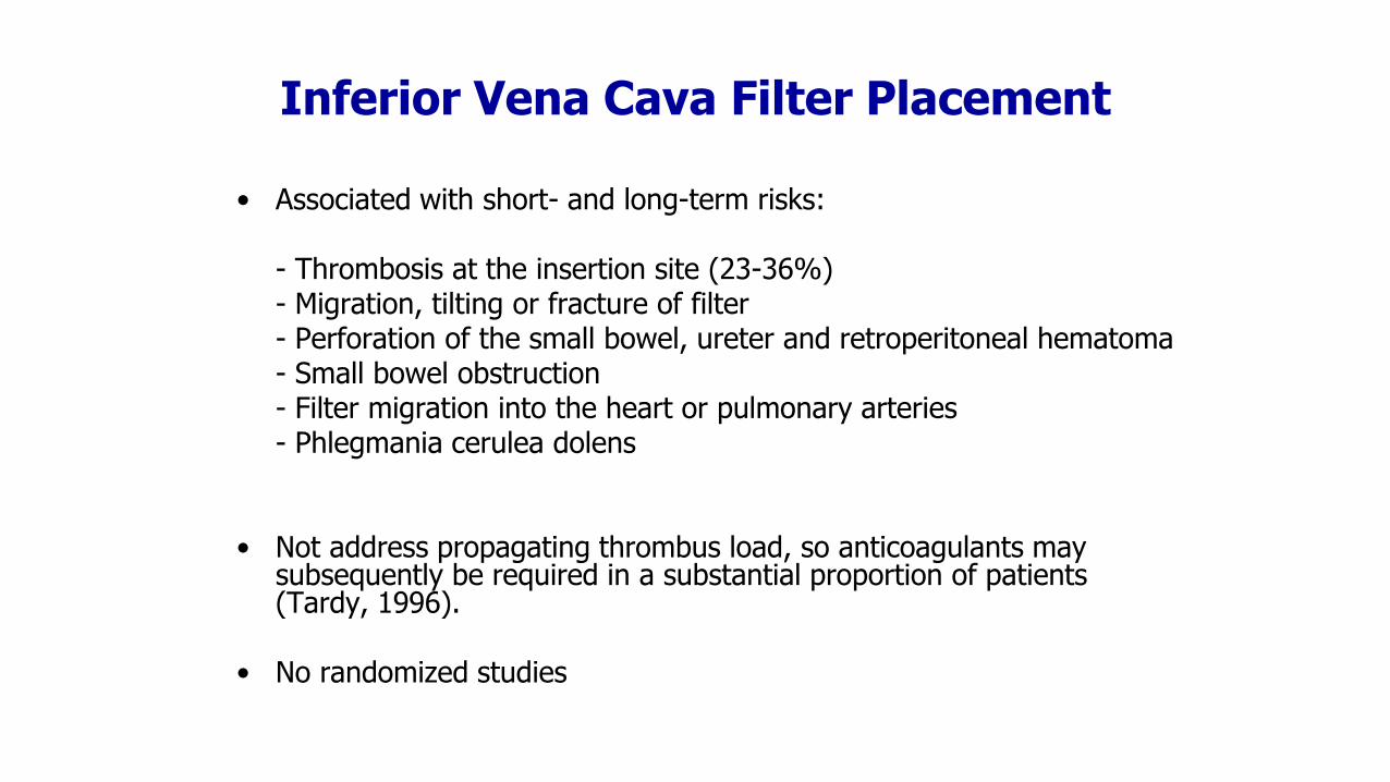
Inferior Vena Cava Filter Placement
• Associated with short- and long-term risks:
- Thrombosis at the insertion site (23-36%)- Migration, tilting or fracture of filter- Perforation of the small bowel, ureter and retroperitoneal hematoma- Small bowel obstruction- Filter migration into the heart or pulmonary arteries- Phlegmania cerulea dolens
• Not address propagating thrombus load, so anticoagulants may subsequently be required in a substantial proportion of patients (Tardy, 1996).
• No randomized studies


Perugia Stroke Registry
• Tot. 2194 pazienti• 340 emorragie (15,5%)
- 547 Cardioembolia 25.0%- AFib 503 (23%)
- 490 Aterosclerosi 22,3%- 421 Malattia dei piccoli vasi 19,2%- 350 Causa indeterminata 15.9%- 202 Più possibili cause 9,2%- 184 Altre cause 8,4%
Paciaroni et al 2014, unpublished data
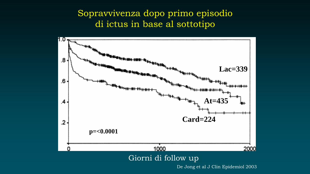
Sopravvivenza dopo primo episodio
di ictus in base al sottotipo
Giorni di follow up
Lac=339
At=435
Card=224
p=<0.0001
De Jong et al J Clin Epidemiol 2003

30 giorni 1 anno 5 anni
Aterotrombotico (%) 8.1 10.8 32.2
Cardioembolico (%) 30.3 53.0 80.4
Lacunare (%) 1.4 6.9 35.1
Criptogenico (%) 14.0 25.6 48.6
p 0.0001 0.0001 0.0001
Sottotipi di ictus ischemico e mortalità
Petty et al, Stroke 2000

Paziente classico
• Maschio
• 72 anni
• Ipertensione arteriosa
• Fumatore
• Emiparesi sinistra (FR 4/5 sia all’arto superiore che all’arto inferiore)
con disturbi sensitivi
• NIHSS=9

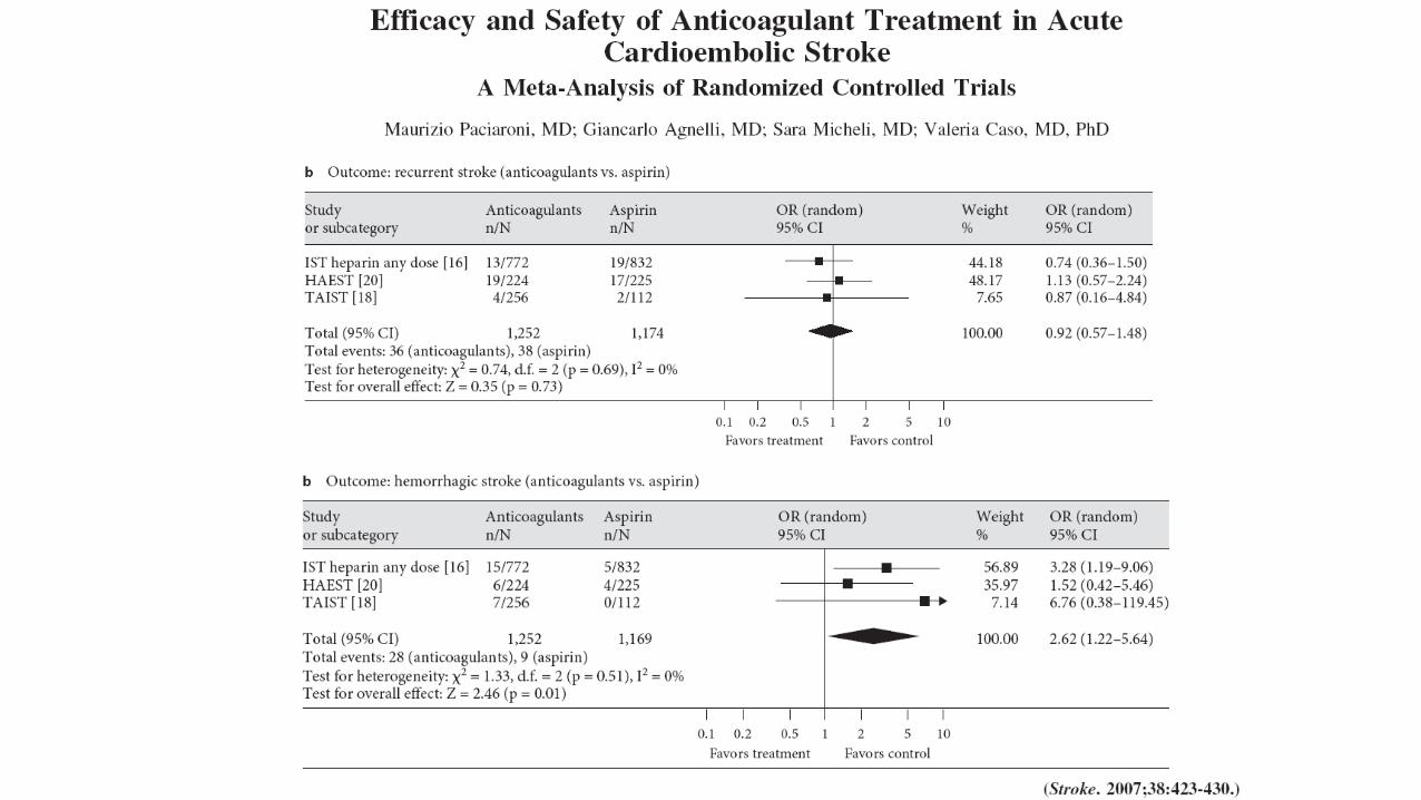
Profilassi secondaria precoce: Aspirina
Morte/ictus non fatale
MorteEmorragia intracranica
Ictus ischemico ricorrente
0
10
2
4
6
8
Effetto per 1000 pazienti trattati
7(2) -2(1) 5(2) 9(3)
ASA (n=20207)Controlli (n=20190)
The Lancet, 1997
Aspirin
Placebo
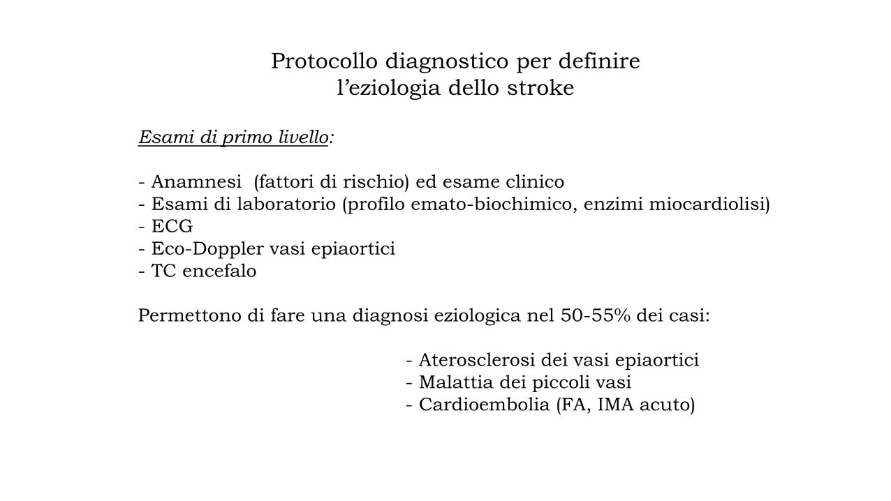
Protocollo diagnostico per definire
l’eziologia dello stroke
Esami di primo livello:
- Anamnesi (fattori di rischio) ed esame clinico
- Esami di laboratorio (profilo emato-biochimico, enzimi miocardiolisi)
- ECG
- Eco-Doppler vasi epiaortici
- TC encefalo
Permettono di fare una diagnosi eziologica nel 50-55% dei casi:
- Aterosclerosi dei vasi epiaortici
- Malattia dei piccoli vasi
- Cardioembolia (FA, IMA acuto)

Aterosclerosi (22%)


1° scenario
Stenosi carotide interna destra >70%

CEA vs. Stenting
Ederle et al. Stroke 2009

Quando operare?
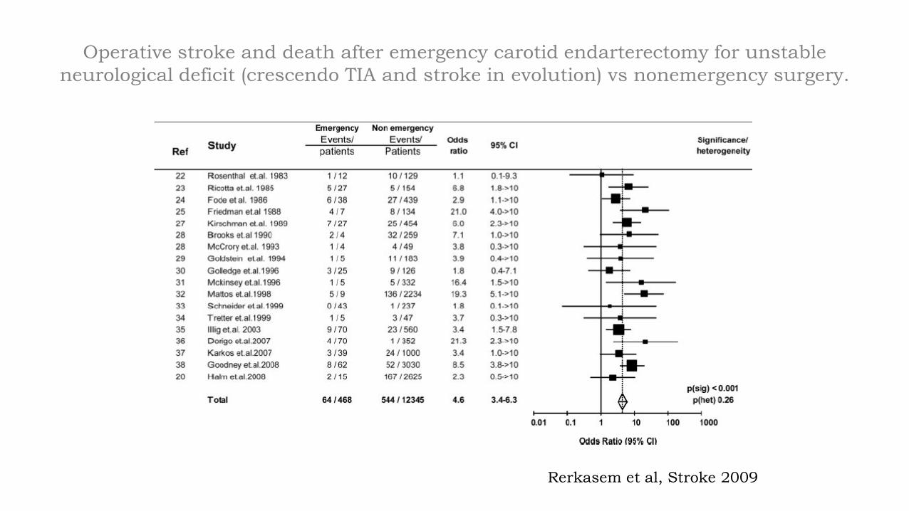
Operative stroke and death after emergency carotid endarterectomy for unstable
neurological deficit (crescendo TIA and stroke in evolution) vs nonemergency surgery.
Rerkasem et al, Stroke 2009

Cardioembolia (25%)
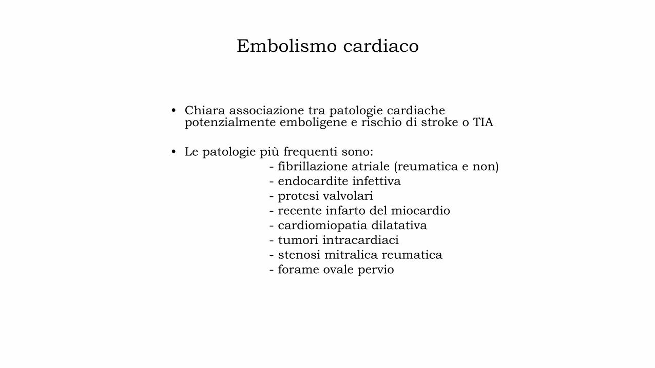
Embolismo cardiaco
• Chiara associazione tra patologie cardiache potenzialmente emboligene e rischio di stroke o TIA
• Le patologie più frequenti sono:
- fibrillazione atriale (reumatica e non)
- endocardite infettiva
- protesi valvolari
- recente infarto del miocardio
- cardiomiopatia dilatativa
- tumori intracardiaci
- stenosi mitralica reumatica
- forame ovale pervio
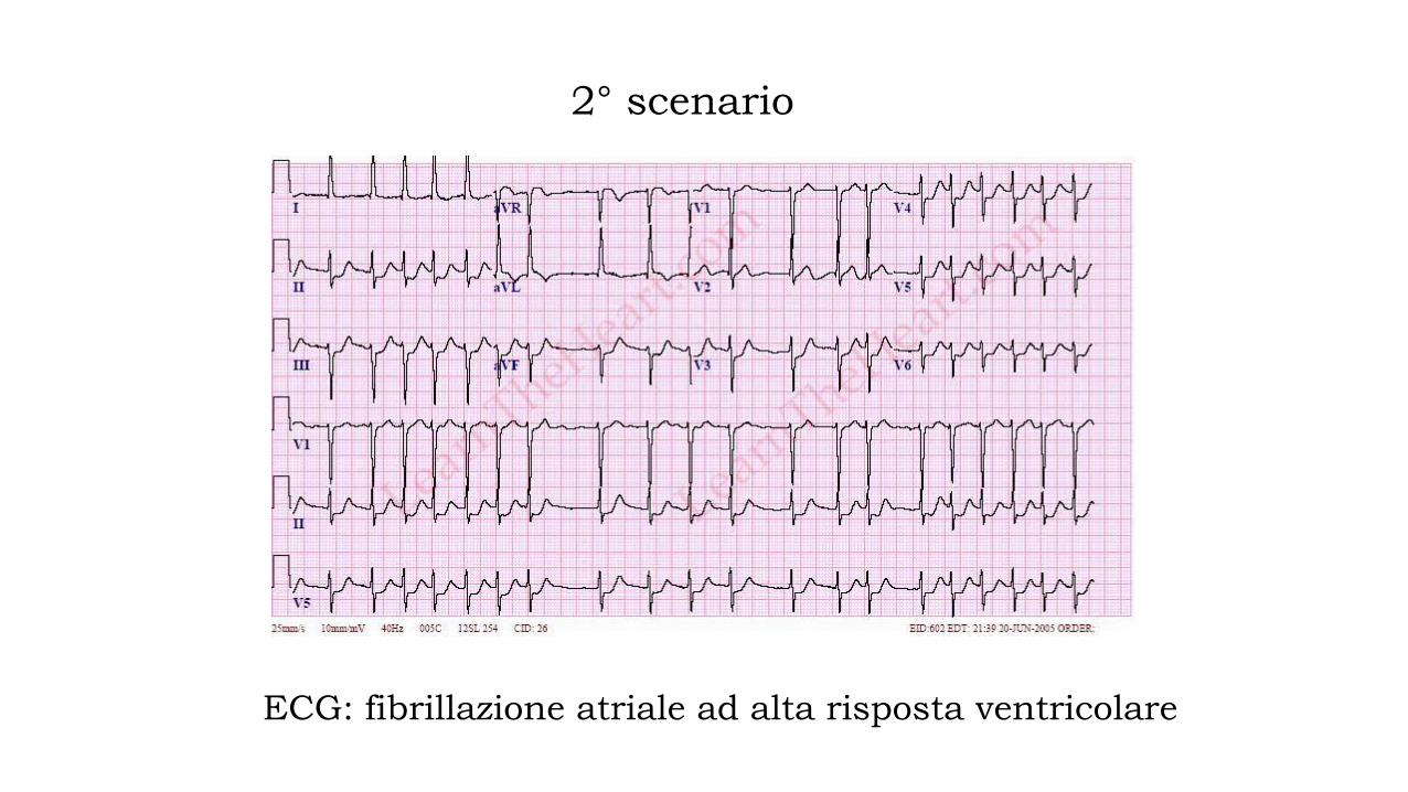
2° scenario
ECG: fibrillazione atriale ad alta risposta ventricolare

Profilassi secondaria nello stroke
dovuto a cardioembolismo
Fibrillazione atriale
RRR per recidiva di stroke:
• Warfarin vs placebo 68%
• Aspirina vs placebo 22%
• Warfarin vs aspirina (325mg) 36-47%
INR: 2.0-3.0
Emorragie maggiori per anno:
• Warfarin 1.3%
• Placebo 1.0%

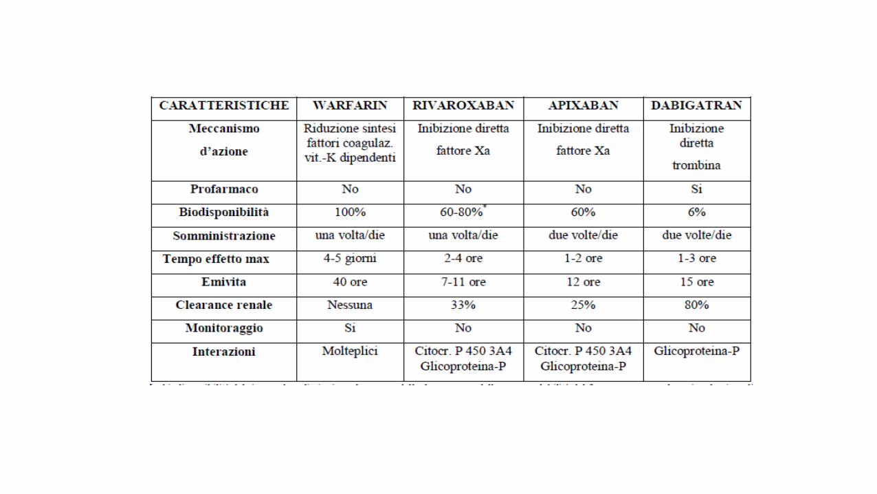
Nuovi anticoagulanti orali
Indicazione: fibrillazione atriale non valvolare
• Apixaban
• Dabigatran
• Edoxaban
• Rivaroxaban

Overview of the New Anticoagulants for Atrial
Fibrillation Comparison with Warfarin
Stroke/Emb ICH Mortality Major
Bleed
Dabigatran 150bid Superior Superior HR 0.88 (0.051) Equivalent
Dabigatran 110 bid Non-Inferior Superior HR 0.91 (NS) Superior
Rivaroxaban 20 qd Non-Inferior Superior HR 0.92 (NS) Equivalent
Apixiban 5 bid Superior Superior HR 0.89 (0.047) Superior

Prevenzione secondaria.
Endpoint: Stroke e embolismo sistemico
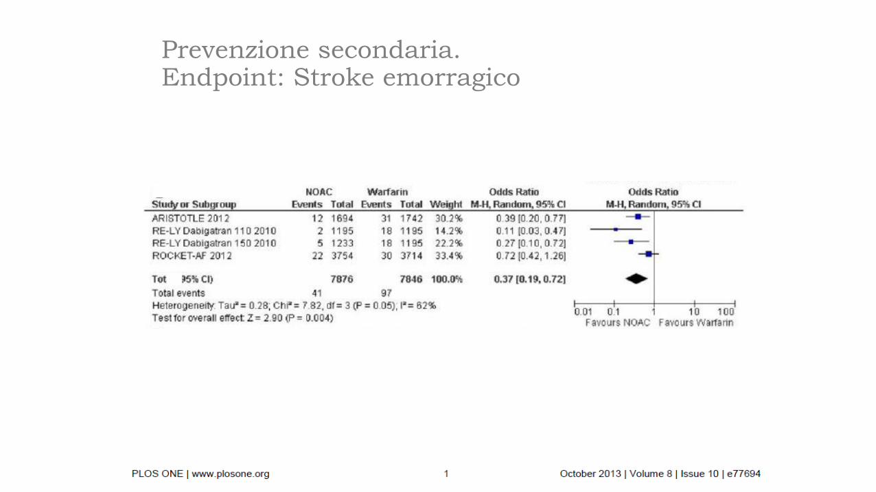
Prevenzione secondaria.Endpoint: Stroke emorragico


Apixaban versus aspirin
Connolly et al, N Engl J Med 2011

Quando iniziare?

Quando iniziare?• Non dati disponibili sull’inizio della terapia (dagli
studi esclusi pazienti con ictus recente)
• Valutare l’estensione della lesione, il quadroclinico ed il rischio ischemico/emorragico globale

*Mild = NIHSS score <8; moderate = NIHSS score 8–16; severe = NIHSS score >16
NIHSS, National Institutes of Health Stroke Scale
Huisman et al. Thromb Haemost 2012; Personal communication, Hans-Christoph Diener, 2015
Initiation or resumption of
anticoagulation depends on severity
of stroke*
TIA Mild
stroke
Moderate
stroke
Severe
stroke
As soon as imaging
has excluded a
cerebral haemorrhage
3–5 days after
symptom onset
5–7 days after
stroke onset
2 weeks after
stroke onset
1 3 6 12Day
Time to re-initiation depends on infarct size:
1 – 3 – 6 – 12 day rule (Diener’s Law)
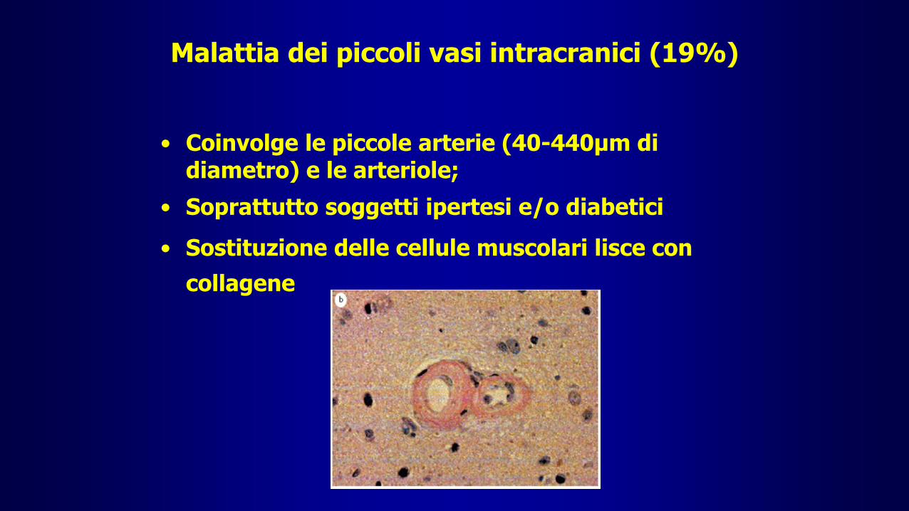
Malattia dei piccoli vasi intracranici (19%)
• Coinvolge le piccole arterie (40-440µm di diametro) e le arteriole;
• Soprattutto soggetti ipertesi e/o diabetici
• Sostituzione delle cellule muscolari lisce con
collagene

3° scenario
• ECG: ritmo sinusale
• Ecodoppler vasi epiartici: non stenosi significative
• EON: classica sindrome lacunare• Emiparesi motoria pura
• Emisindrome sensitiva pura
• Emisindrome sensitivo-motoria
• Emiparesi atassica
• Sindrome disartria-mano goffa

Profilassi secondaria precoce
- Aspirina 160-325 mg al giorno da subito
- Dopo 14 giorni passare a 100 mg al giorno
- Alternative:
- Clopidogrel 75 mg/die
- Aspirina + dipiridamolo
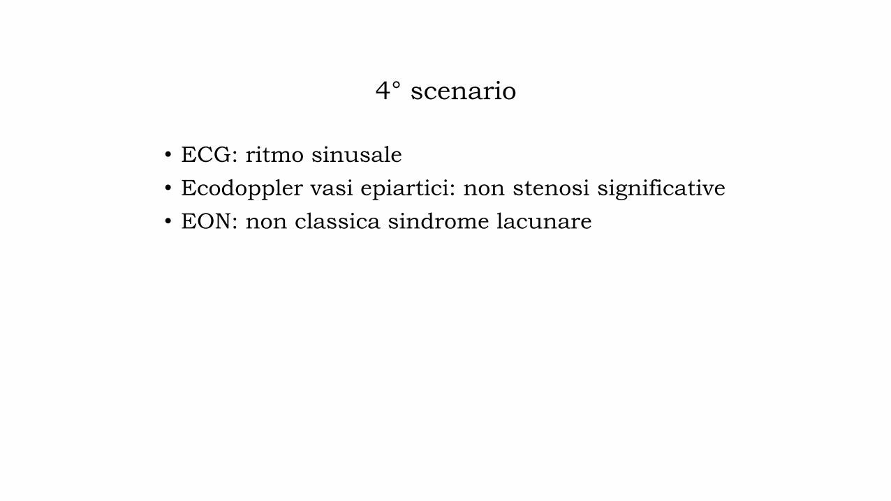
4° scenario
• ECG: ritmo sinusale
• Ecodoppler vasi epiartici: non stenosi significative
• EON: non classica sindrome lacunare
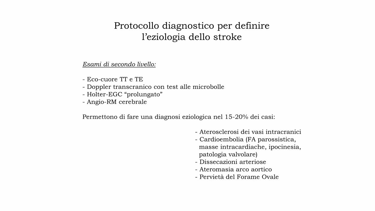
Protocollo diagnostico
Esami di secondo livello:
- Eco-cuore TT e TE
- Doppler transcranico con test alle microbolle
- Holter-EGC “prolungato”
- Angio-RM cerebrale
Permettono di fare una diagnosi eziologica nel 15-20% dei casi:
- Aterosclerosi dei vasi intracranici
- Cardioembolia (FA parossistica,
masse intracardiache, ipocinesia,
patologia valvolare)
- Dissecazioni arteriose
- Ateromasia arco aortico
- Pervietà del Forame Ovale
Protocollo diagnostico per definire
l’eziologia dello stroke

Esami di terzo livello:
- VES, ANA, trombofilia, LAC, HIV, TPHA, elettroforesi Hb;
- Eco-Doppler dei vasi venosi degli arti inferiori
- Dosaggio lattato prima e dopo sforzo muscolare
- Dosaggio alfa-galattosidasi A nel plasma
- Rx torace
- Rachicentesi
- Angiografia cerebrale
- Biopsia cutanea e muscolare
- Indagine genetica
Permettono di fare una diagnosi eziologica nel 3-4% dei casi:
- Connettiviti, vasculiti
- Vasculiti post-infettive
- Displasia fibromuscolare e anomalie vasali
- Disordini emocoagulativi ed ematologici
- Embolia paradossa (fistola
polmonare, Rendu-Osler)
- Malattia di Moyamoya
- CADASIL, MELAS, Fabry e altre patologie
genetiche
Protocollo diagnostico per definire
l’eziologia dello stroke


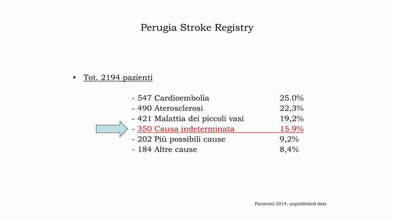
Perugia Stroke Registry
• Tot. 2194 pazienti
- 547 Cardioembolia 25.0%
- 490 Aterosclerosi 22,3%
- 421 Malattia dei piccoli vasi 19,2%
- 350 Causa indeterminata 15.9%
- 202 Più possibili cause 9,2%
- 184 Altre cause 8,4%
Paciaroni 2014, unpublished data
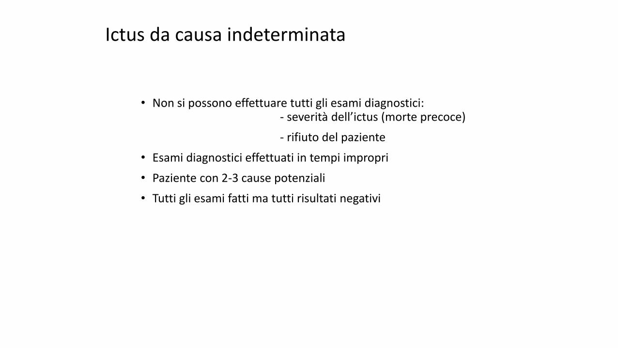
Ictus da causa indeterminata
• Non si possono effettuare tutti gli esami diagnostici:- severità dell’ictus (morte precoce)
- rifiuto del paziente
• Esami diagnostici effettuati in tempi impropri
• Paziente con 2-3 cause potenziali
• Tutti gli esami fatti ma tutti risultati negativi

• 114 pazienti con stroke criptogenetico
ECG secondo Holter 96 ore
• 20 FA nelle prime 24 ore
• 9 FA dopo 24 ore
• Tot. 29 FA (24,3%)Intern Emerg Med 2012

Embolic Stroke of Undetermined Source (ESUS)
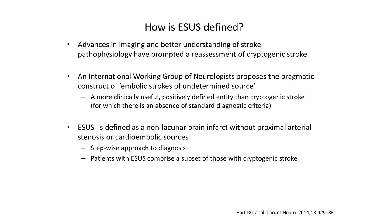
How is ESUS defined?
• Advances in imaging and better understanding of stroke pathophysiology have prompted a reassessment of cryptogenic stroke
• An International Working Group of Neurologists proposes the pragmatic construct of ‘embolic strokes of undetermined source’
– A more clinically useful, positively defined entity than cryptogenic stroke (for which there is an absence of standard diagnostic criteria)
• ESUS is defined as a non-lacunar brain infarct without proximal arterial stenosis or cardioembolic sources
– Step-wise approach to diagnosis
– Patients with ESUS comprise a subset of those with cryptogenic stroke
Hart RG et al. Lancet Neurol 2014;13:429–38

Profilassi secondaria
nello stroke non dovuto
a cardioembolismo, non dovuto a
stenosi carotidea severa

Profilassi secondaria
- Aspirina 100 mg/die
- Clopidogrel 75 mg/die
- Aspirina + dipiridamolo

Correzione dei fattori di rischio
• Correzione dello stile di vita
• Terapia anti-ipertensiva
• Attento controllo del diabete
• Se colesterolo LDL superiore a 100 mg/dL:
atorvastatina 80 mg/die (ictus non cardioembolico)

HOPE: Heart Outcomes Prevention Evaluation
RRR outcome combinato: 22%RRR stroke: 32%

5° scenario
• Maschio
• 72 anni
• Ipertensione arteriosa
• Fumatore
• Emiparesi sinistra (FR 4/5 sia all’arto superiore
che all’arto inferiore) con disturbi sensitivi
• NIHSS=9
• Durata della sintomatologia: 2 ore
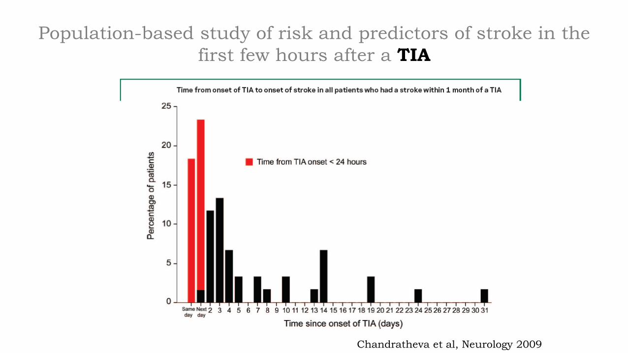
Population-based study of risk and predictors of stroke in the
first few hours after a TIA
Chandratheva et al, Neurology 2009

TIA: emergenza!
Cosa fare subito?
• TC encefalo
• Esami emato-biochimici
• ECG
• Ecodoppler vasi epiaortici
• Calcolare ABCD2 score
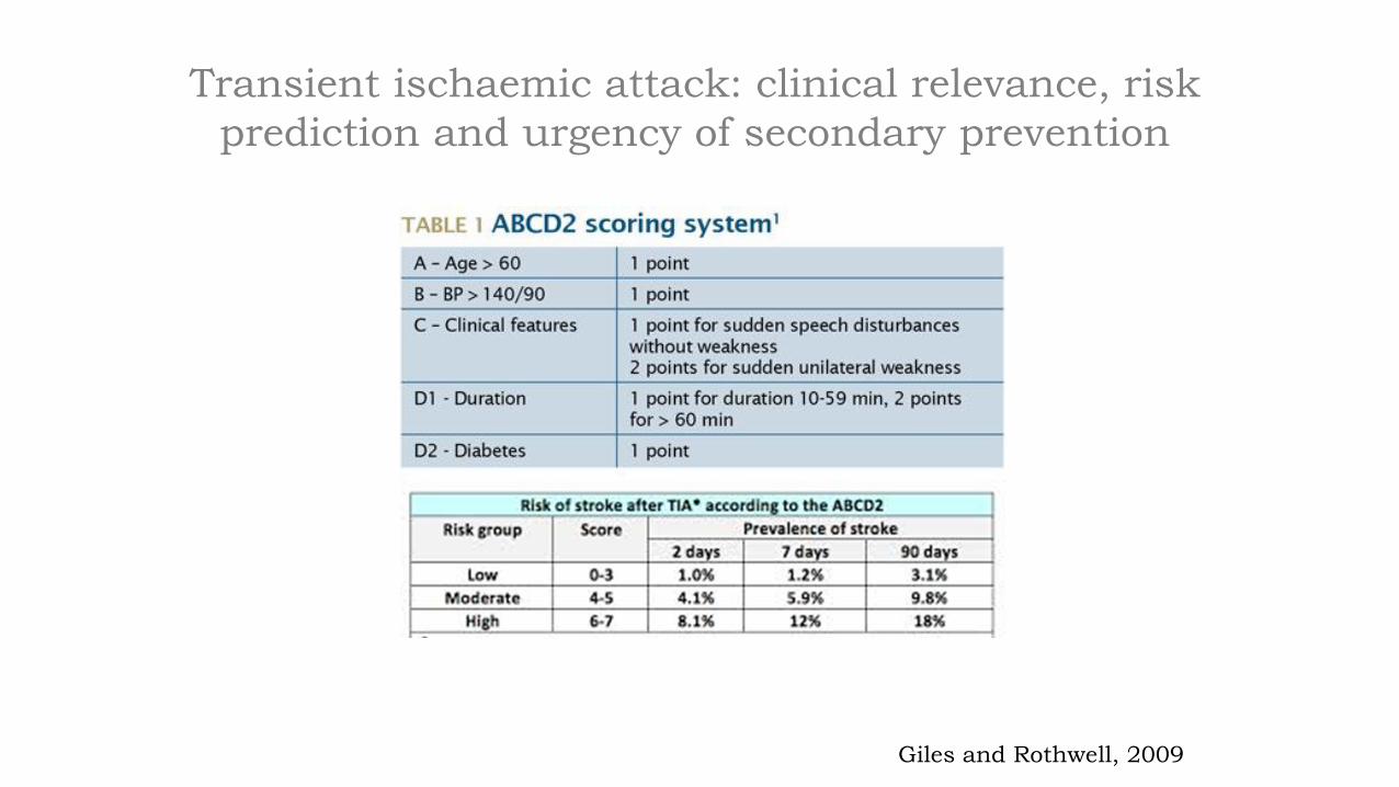
Transient ischaemic attack: clinical relevance, risk
prediction and urgency of secondary prevention
Giles and Rothwell, 2009

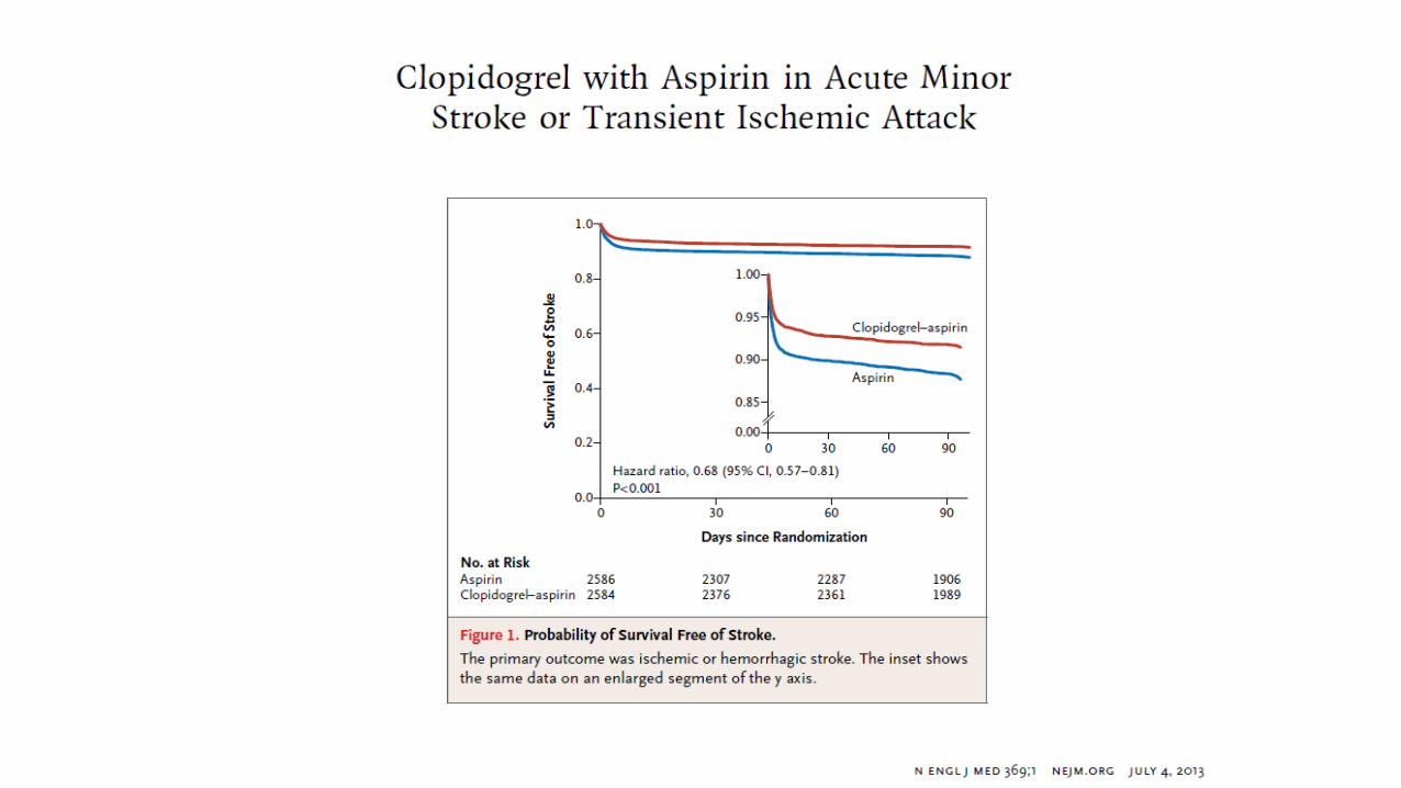
Studio Express
Rothwell et al, Lancet Neurology 2009
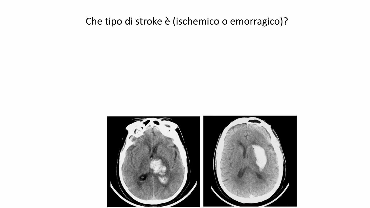
Che tipo di stroke è (ischemico o emorragico)?
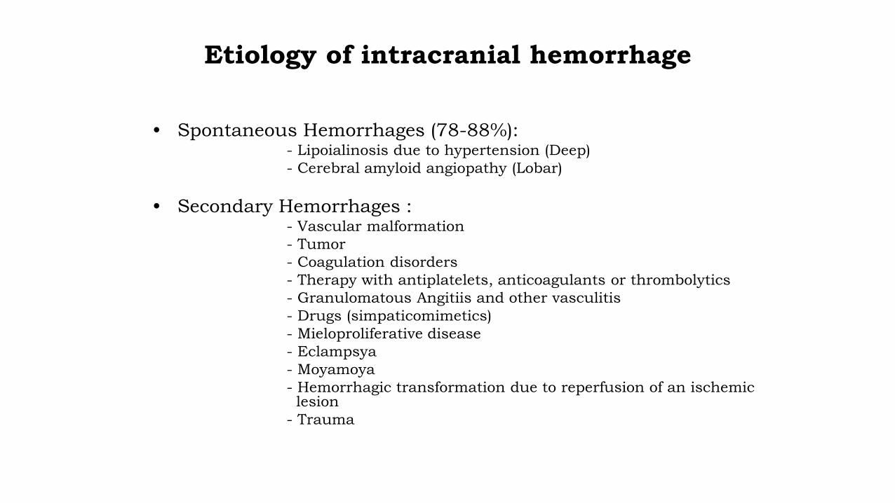
• Spontaneous Hemorrhages (78-88%):- Lipoialinosis due to hypertension (Deep)
- Cerebral amyloid angiopathy (Lobar)
• Secondary Hemorrhages :- Vascular malformation
- Tumor
- Coagulation disorders
- Therapy with antiplatelets, anticoagulants or thrombolytics
- Granulomatous Angitiis and other vasculitis
- Drugs (simpaticomimetics)
- Mieloproliferative disease
- Eclampsya
- Moyamoya
- Hemorrhagic transformation due to reperfusion of an ischemiclesion
- Trauma
Etiology of intracranial hemorrhage

Deep and lobar hemorrhages
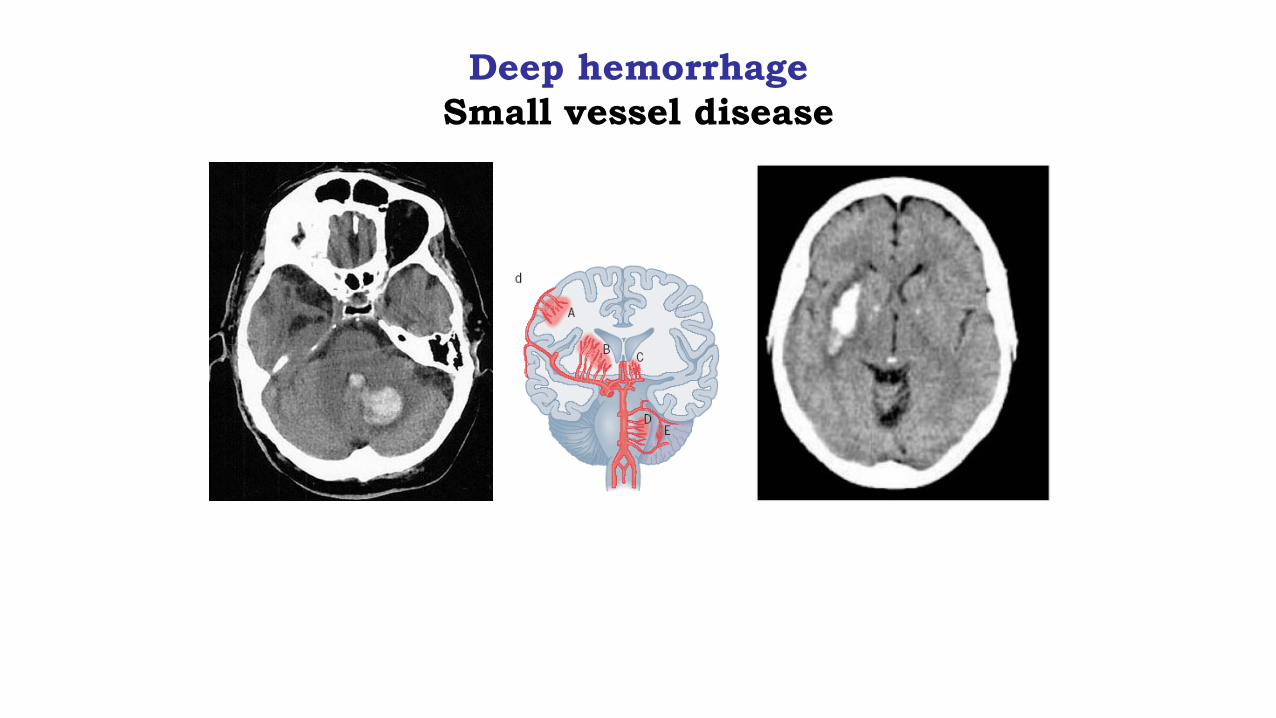
Deep hemorrhage
Small vessel disease

Lobar hemorrhage
Cerebral amyloid angiopathy
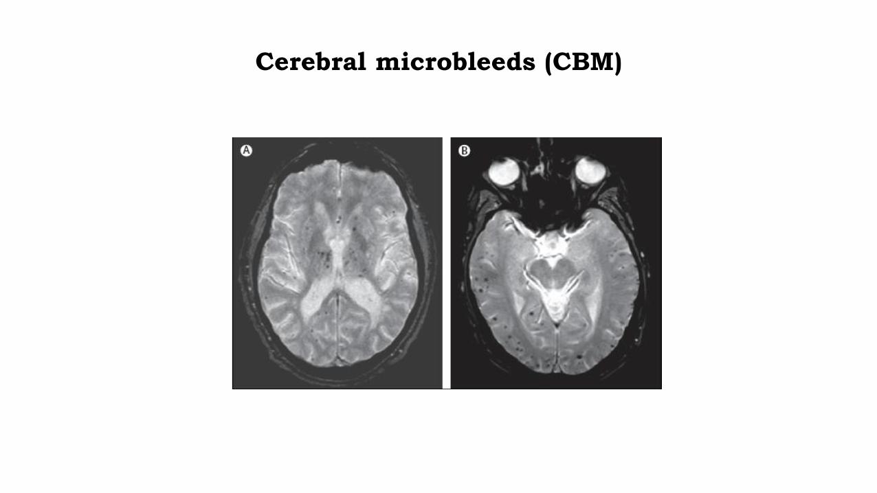
Cerebral microbleeds (CBM)

Cerebral amyloid angiopathy
and focal cisternal microbleeds
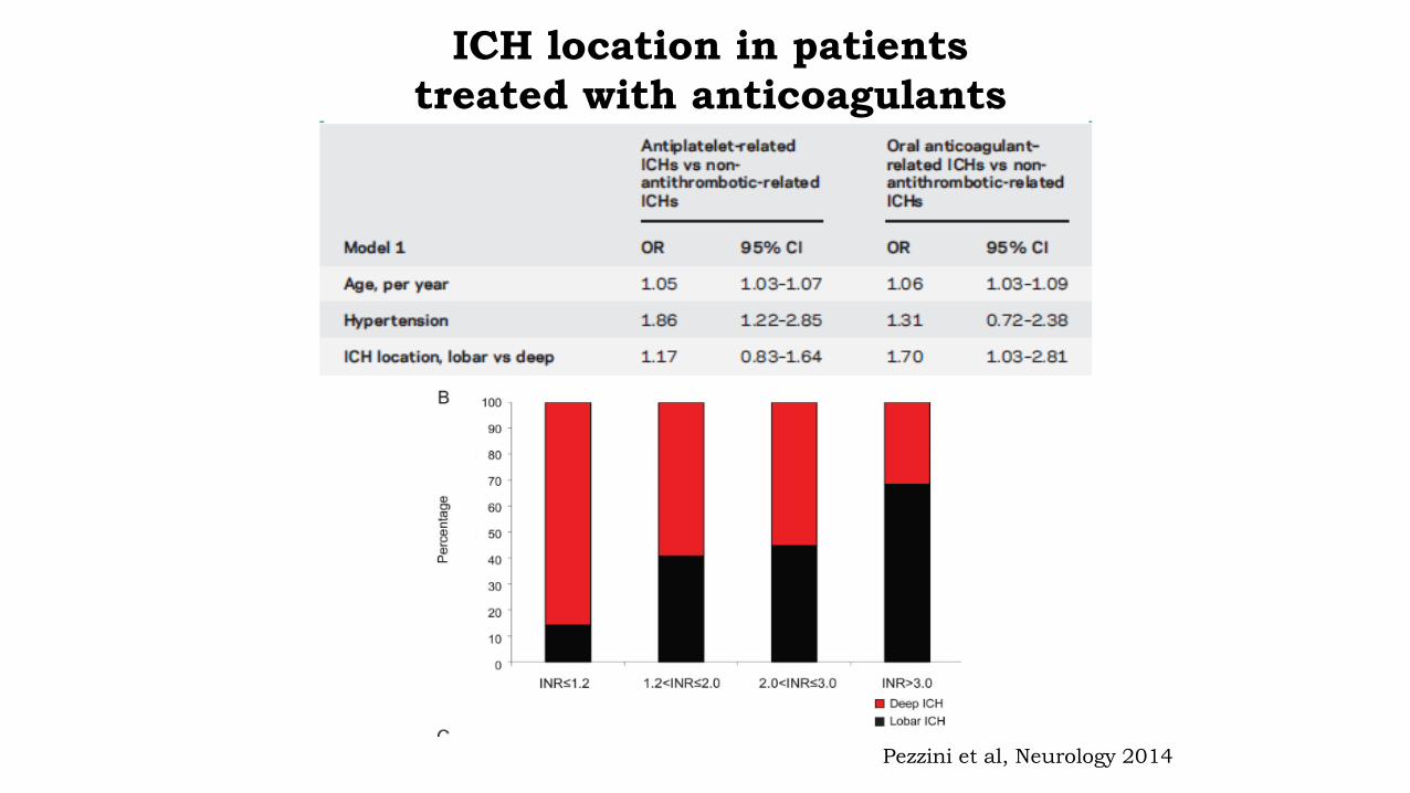
ICH location in patients
treated with anticoagulants
Pezzini et al, Neurology 2014


Distribution of intracranial hemorrhage

Mortality of intracerebral hemorrhage
Stroke, 2009
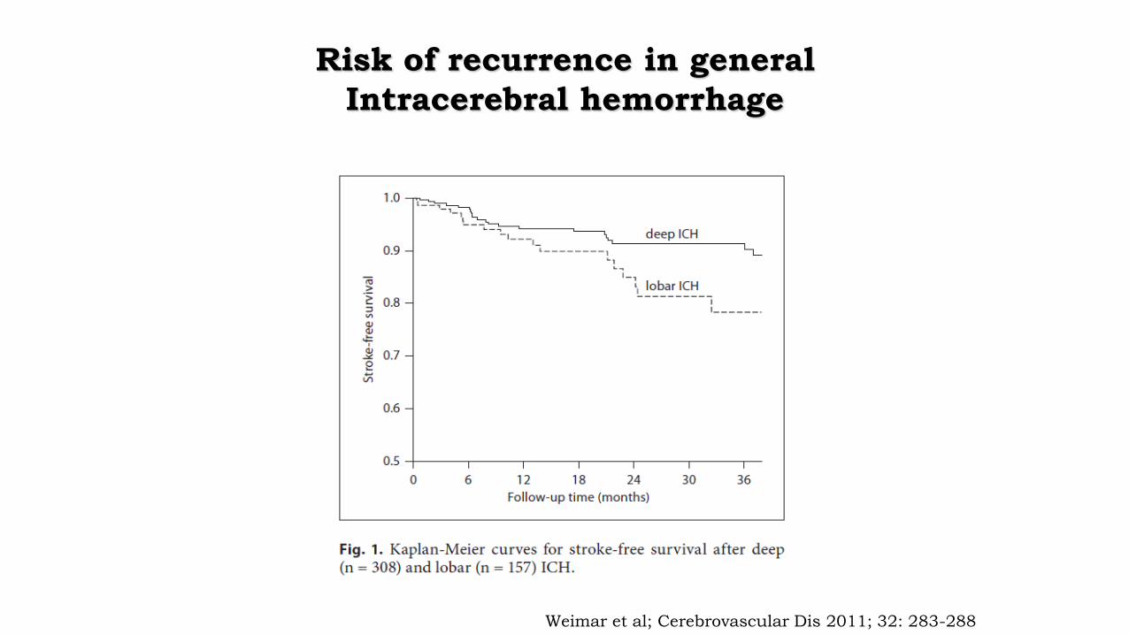
Risk of recurrence in general
Intracerebral hemorrhage
Weimar et al; Cerebrovascular Dis 2011; 32: 283-288

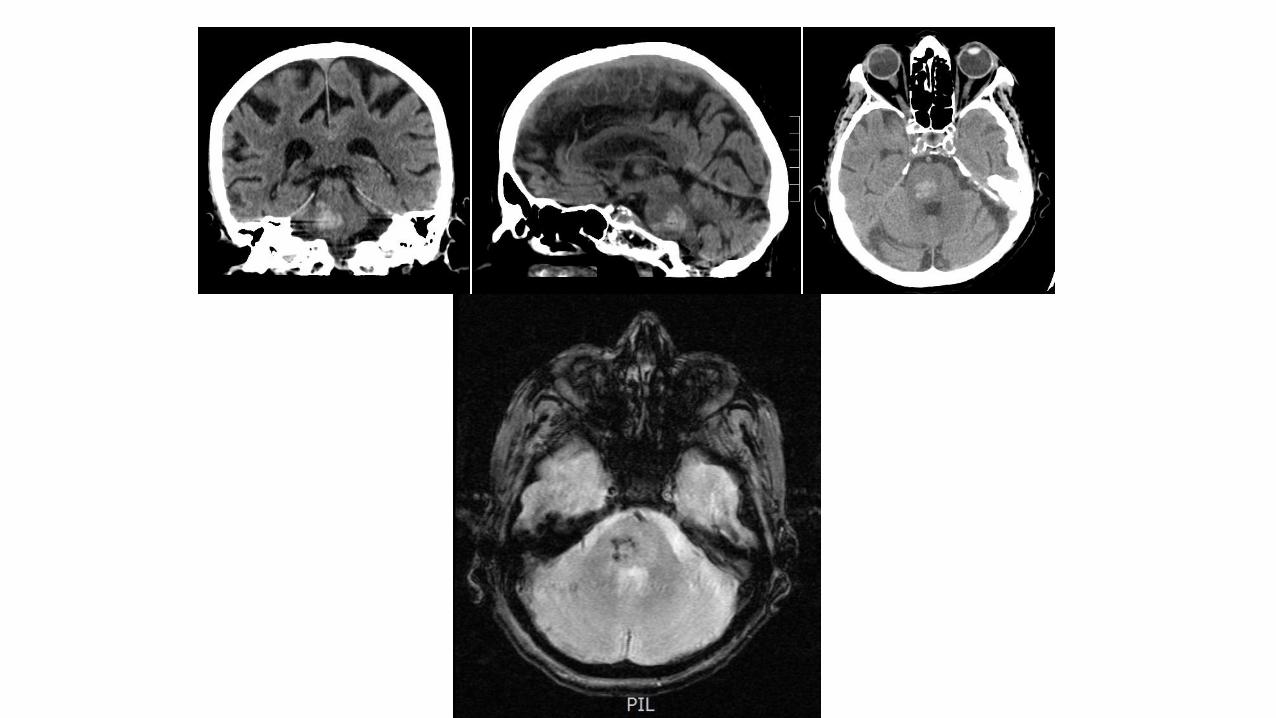
45 year old woman with a history of migraine,
no vascular risk-factors

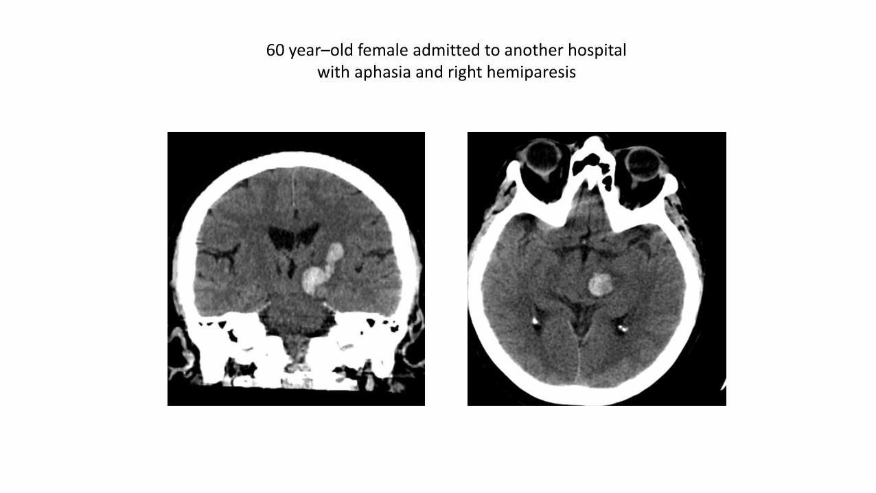
60 year–old female admitted to another hospital with aphasia and right hemiparesis
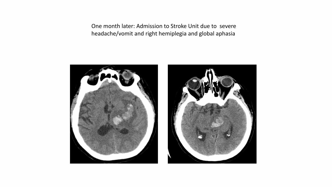
One month later: Admission to Stroke Unit due to severe headache/vomit and right hemiplegia and global aphasia