Embed Size (px)
Citation preview

452 lACC Vol 2. No 3September 1983.452-9
Detection of Deterioration or Infection of Homograft and PorcineXenograft Bioprosthetic Valves in Mitral and Aortic Positions byTwo-Dimensional Echocardiographic Examination
EHUD GRENADIER, MD, DAVID J. SAHN, MD, FACC,* ANTONY H. G. ROCHE, MD,
LILLIAM M. VALDES-CRUZ, MD, FACC, JACK G. COPELAND, MD, FACC,
STANLEY J. GOLDBERG, MD, FACC, HUGH D. ALLEN, MD, FACC
Tucson, Arizona and Auckland, New Zealand
Results of two-dimensional echocardiographic examinations were compared with angiographic, hemodynamic and surgical results in 44 patients with bioprosthetic valves in mitral and aortic positions who wereundergoing elective or urgent reinvestigation 24 to 87months (mean 34) after implantation. In these patients,there were 18 homograft aortic valves in the aortic position, 9 stent-mounted homograft aortic valves in themitral position, 13 porcine xenograft valves in the mitralposition and 12 in the aortic position.
Poor cusp support, gross fluttering and prolapse ofcusps behind or below the anulus identified aortic insufficiency by two-dimensional echocardiography in sixpatients with an aortic homograft and four patients were
Long-term follow-up results for valve replacement with bioprosthetic valves have suggested a significant incidence ofvalve degeneration, especially in younger patients (1--4).Two-dimensional echocardiography has made it possible tononinvasively obtain high quality images of bioprostheticvalves in postoperative patients that are much better thanthe images obtained of nonbiologic valves (5,6). The purpose of this study was to evaluate the utility of two-dimensional echocardiographic examination for identifying patients with deterioration or infection of two kinds ofbioprosthetic valves .
From the Department of Pediatrics , University of Arizona Health SCIences Center , Tucson . Arizona and Green Lane Hospital, Auckland, NewZealand. Manuscript received January 18, 1983; revised manuscript received April 18, 1983, accepted April 27, 1983.
*Present address and address for reprints: David J. Sahn, MD, Department of Pediatric Cardiology. University of California , San Diego,University Hospital , CTF B102, 225 West Dickinson Street, San Diego,California 92103.
© 1983 by the Amencan College of Cardiology
identified with insufficiency of a stent-mounted aortichomograft in the mitral position. Two-dimensional echocardiographic examination revealed mitral stenosis inthree patients with a porcine xenograft valve in the mitral position and suggested mitral insufficiency in twoothers. Bacterial endocarditis on homograft or porcinexenograft valves was associated with easily imaged vegetations by two-dimensional echocardiography in 10patients.
Despite difficulties in imaging valve cusps, and theskill required to obtain good echocardiographic imagesof bioprosthetic valves, significant valve deterioration orinfected prostheses were quite effectively imaged by twodimensional echocardiography in this study.
MethodsStudy patients. All patients who had valve replacement
with porcine aortic xenograft or human homograft aorticvalves and who underwent cardiac catheterization (or operation for valve problems without prior catheterization) atGreen Lane Hospital , Auckland, New Zealand or at theUniversity of Arizona Health Sciences Center, Tucson , Arizona over a period of 24 months were included in thisprospective study . Specifically , the study was designed toevaluate the utility of two-dimensional echocardiographyfor providing noninvasive diagnosis of valve dysfunction .There were 48 patients , of whom 22 were male and 26 werefemale ; their ages ranged between 12 and 42 years (mean31). The study group was unselected and contained a consecutive series of patients studied (electively or urgently)24 to 87 months (mean 34) after valve replacement surgery.Twenty patients underwent follow-up catheterization studies5 years after valve implantation and the others underwentmore urgent evaluation for suspected valve infection or deterioration. The echocardiographic interpreters, however,
0735-1097 /83/$3 .00

lACC Vol 2, No 3September 1983.452-9
Table 1. Degenerative Complications of Bioprosthetic Valves
GRENADIER ET ALECHOCARDIOGRAPHY OF BIOPROSTHETIC VALVES
453
Type of Valve
Free hand-mounted homograft
Stent-mounted homograft
Porcine xenograft
Porcine xenograft
Two-DimensionalEchocardiographic Angio/Cath/Surgical
Position Observations Observations Comments
Aortic (n = 18) 6 gross flutter 10 aortic insuf (4 4 false neg for
prolapse triv, 6 mod) triv msuf
Mitral (n = 9) 4 gross cusp 5 mitral msuf I false neg
prolapse + flutter
Aortic (n = 12) None "abnormal" I mod aorttc msuf I false neg
+ cusp tearMitral (n = 13) 2 unsupported, 2 mitral msuf', 4 2 false neg for
3 thickened, mitral sten (>15 stenosis, I
stenotic mm Hg gradient) false posfor stenosis(see text)
Insuf = insufficiency; mod = moderate; Neg = negatIve; Pos = positive; sten = stenotic, triv = trivial.
were unaware of the clinical status of the patients when thestudies were reviewed in a coded and blinded fashion,
Thirty of the 44 patients with successful imaging had abioprosthetic valve in the aortic position (18 free handmounted aortic homografts, 12 porcine xenografts). Twentytwo patients had a bioprosthetic valve in the mitral position(9 stent-mounted aortic homografts, 13 porcine xenografts).Thus, the 44 patients who had successful imaging and areincluded in the Results section had a total of 52 bioprostheticvalves, 8 patients having 2 bioprosthetic valves. Thirty-twopatients underwent hemodynamic investigation and 15 patients underwent surgery and, therefore, had surgical inspection of their valves for validation of the echocardiographic examination.
Echocardiographic studies. Two-dimensional echocardiographic studies were performed using a commerciallyavailable Toshiba SSH lOA, 2.4 MHz range focused, phased
array sector scanner or an Electronics for Medicine/Honeywell, 3.5 MHz mechanical sector scanner with quantitative pulsed Doppler capabilities (six patients). Completeechocardiographic examinations in all standard views wereobtained emphasizing the imaging of prosthetic valve cusps(7). The two-dimensional echocardiographic illustrations usedin this paper were made from stop action, single frame videoimages with a Polaroid camera system.
ResultsA technically adequate two-dimensional echocardiogram
with images of the valve in question was obtained in 44 ofthe 48 patients. Adequate imaging consisted of visualizationof the ring and the strut for stented valves and the valvecusps in systole and diastole for all valves.
Figure 1. Echocardiogram in long-axis view in systoleshows the thin cusps of free hand-mounted aortic homograft (HO) in the aortic (AO) position in a patient whoalso has a porcine xenograft (px) mitral valve prosthesis.There is a difference between the thickness of the cuspsas imaged on the two types of valves. Note that theanterior leftward cusp of the porcine valve (arrowhead)is thicker than either of the cusps of the homograft valve.Ant = anterior; lnf = inferior; LV = left ventricle;Post = posterior; Sup = superior.

454 GRENADIER ET AL.ECHOCARDIOGRAPHY OF BIOPROSTHETIC VALVES
lACC Vol. 2. No 3September 1983:452-9
Figure 2. A long-axis image is shown of the thin symmetric cusps of a stent-mounted aortic homograft (mV)in the mitral position. LA = left atrium; other abbreviations as in Figure I.
Findings in Normal Bioprosthetic Valves
Eighteen normal bioprosthetic valves were imaged in the44 study patients (Table 1). The leaflets of the normalbioprostheses were imaged in mitral and aortic positions.The sewing ring and all three stents of the normal valveswere visualized and had a smooth contour without extraneous adjacent echoes (Fig. 1 to 3). The valves were wellanchored to the valve ring and moved symmetrically withit. Homograft valve cusps were thin and opened widely. Incontrast, the anterior leftward cusps of the porcine heterografts often did not appear to have opened quite as widely
Figure 3. Short-axis view shows symmetric complete opening ofa stent-mounted homograft valve (H) in the mitral position. L =left; LVOT = left ventricular outflow tract; R = right; RV =right ventricle; S = septum.
as the other cusps in normal functioning valves. Thickerand slightly brighter echoes in the normal porcine heterograft were usually seen in oblique views of the left cuspand relate to the known asymmetry of the porcine aorticvalve (Fig. 4) (4).
Abnormal Valves (Table 1)
Angiography or surgery showed that 22 implanted bioprosthetic valves were stenotic or had some degree ofinsufficiency.
Of the 18 aortic homograft valves in the aortic position,6 showed gross fluttering and inferior prolapse on twodimensional echocardiographic examination (Fig. 5). Tenof the 18 valves were regurgitant by angiography; in fourpatients, regurgitation was trivial. Of the nine stent-mountedaortic homografts in the mitral position, four showed grosscusp prolapse and fluttering (Fig. 6). Five of the nine weremoderately regurgitant on angiography (one false negativefinding by echocardiography occurred in a valve that wasregurgitant by virtue of a perivalvu1ar leak not associatedwith valve dehiscence).
Of the 12 porcine xenograft valves in the aortic position,none showed abnormal support on echocardiography but 1showed moderate aortic regurgitation on angiography inassociation with a cusp tear not detected on the echocardiographic examination (Table 1).
Of the 13 porcine xenograft valves in the mitral position,2 valves had unsupported prolapsing cusps and 3 appearedthickened and stenotic (Fig. 7) with poor cusp motion inexcess of the minimal asymmetry and thickening in normalvalves. Two of the 13 were regurgitant on angiography, and4 of 15 patients with a porcine valve in the mitral positionhad a mean pressure gradient greater than 15 mm Hg atcatheterization. Nonetheless, one valve considered to be

JACC Vol 2, No.3September 1983.452-9
GRENADIER ET AL.ECHOCARDIOGRAPHY OF BIOPROSTHETIC VALVES
455
Figure 4. Short-axis view shows slightly more exaggerated asymmetry of a porcine valve (px) that hadbeen in place only 6 months and was functioningnormally, Again, the left commissure appears prominent and slightly thickened, The patient had beenstudied because of insufficiency of an aortic homograft in the aortic position, LVOT = subaortic leftventricular outflow tract; other abbreviations as before,
possibly stenotic on echocardiography had only a 5 mm Hggradient at catheterization, Hemodynamically significantstenosis was not detected by echocardiography for two othervalves (Table 1), Therefore, there were two false negativeand one false positive diagnoses of mitral stenosis of aporcine xenograft in the mitral position,
Valve Infections
Twelve patients went to surgery with a diagnosis of bacterial endocarditis on a bioprosthetic valve, There were eightaortic valves (five porcine, three free hand-mounted homograft) and five mitral valves (two porcine and three homograft) implanted in these patients (Fig, 8 and 9), Pros-
Figure 5. Long-axis view shows inferior prolapsing of an aortic(Ao) homograft valve (arrow) associated with aortic insufficiencyand cusp degeneration, Abbreviations as before.
thetic valve endocarditis was correctly diagnosed andvegetations were imaged by echocardiography in 10 of these12 patients, There were two false negative interpretationsfor detection of infectious changes in this series. In the firstcase, a porcine mitral valve was infected in a patient whoalso had subacute bacterial endocarditis on a native aorticvalve. The vegetations were imaged on the native aorticvalve but were missed on the mitral valve prosthesis, In thesecond case, vegetations on a porcine xenograft valve inthe aortic position were also missed on echocardiography,and the valve was seen at surgery to be infected with a smallvegetation and a central perforation, The patient had been
Figure 6. Long-axis view shows prolapse superiorly and posteriorly of one of the cusps of a stent-mounted aortic homograft inthe mitral position (arrow) associated with mitral insufficiency,Abbreviations as before.

456 GRENADIER ET ALECHOCARDIOGRAPHY OF BIOPROSTHETIC VALVES
lACC Vol 2. No 3September 1983 452-9
Figure 7. Obliqueshort-axisview showstwo bulbous thickeningson the leaflets of a porcine heterograft in the mitral position (arrow). This valve was not only thickened but moved poorly andwas stenotic at catheterization. Abbreviations as before.
referred for surgery because of persistently positive bloodcultures and angiographic evidence of aortic regurgitation.One patient was correctly diagnosed by echocardiographyas having two infected porcine valves (Fig. 9).
Perivalvular abscess and aneurysm. Two-dimensionalechocardiographic examination was also capable of diagnosing perivalvular abscesses and aneurysms. In this series,
Figure 8. Short-axis view of the supravalvular aorta shows twopockets of a perivalvular abscess (Absc) around an aortic homograft. The thin aortic homograftcusps are seen in the circle medialto the two pockets of the abscess. RA = right atrium; RVOT =right ventricular outflow tract; other abbreviations as before.
we observed one aortic homograft that had a perivalvularaneurysm (Fig. 8), and two dehisced valves that had anabnormal rocking motion and were also infected. One valvewas a porcine xenograft in the aortic position; the othervalve was a dehiscent porcine xenograft in the mitral position. One diagnosis of an abscess around a porcine xenograft valve in the aortic position was missed in a patientwho had an echocardiographically correct diagnosis of bacterial endocarditis. This valve appeared well anchored atsurgery.
DiscussionBioprosthetic valves have a significant advantage over
mechanical valves. There is decreased risk of thromboembolism without the use of anticoagulant therapy and, therefore, decreased risk of anticoagulant-related complications(8,9). The central flow orifice in bioprosthetic valves is morephysiologic (8), and valve failure, when it occurs, is usuallyslowly progressive rather than sudden and potentially fatal(8).
Incidence of bioprosthetic valve failure. The long-termcourse of patients with bioprosthetic valves is, however,not free of complications, most commonly, infection(l ,2,8, 10) or spontaneous sterile valve degeneration(1,3,4,8,10), or both, causing restenosis or cusp degeneration and valve incompetence. This study did not examinethe risk of any specific patient for valve failure. Patientsadmitted to the study prospectively were being reevaluatedfor a suspicion that they were not doing well or were at
Figure 9. Vegetations (V) are shown on an infected porcine xenograft (upper Px) in the aortic (Ao) position. A small vegetationwas also present on the porcine xenograft in the mitral position(lower Px) in this patient. Other abbreviations as before.
JACC Vol. 2, No 3September 1983.452- 9
GRENADIER ET AL.ECHOCARDIOGRAPHY OF BIOPROSTHET1C VALVES
457
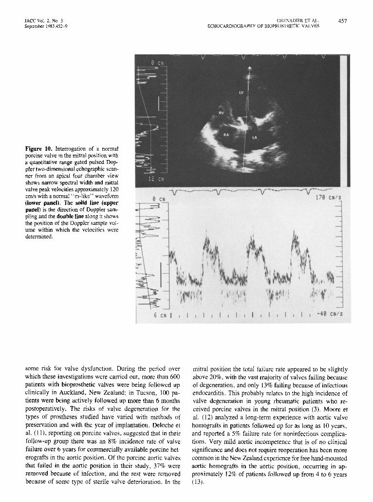
Figure 10. Interrogation of a normalporcine valve in the mitral position witha quantitative range gated pulsed Doppler two-dimensional echographic scanner from an apical four chamber viewshows narrow spectral width and mitralvalve peak velocities approximately 120cm/s with a normal " m-like" waveform(lower panel). The solid line (upperpanel) is the direction of Doppler sampling and the double line along it showsthe position of the Doppler sample volume within which the velocities weredetermined .
, I I I
•
6 CI I
some risk for valve dysfunct ion. During the period overwhich these investigation s were carried out , more than 600patients with bioprosthetic valves were being followed upclinically in Auckland, New Zealand; in Tucson, 100 patients were being actively followed up more than 6 monthspostoperatively. The risks of valve degeneration for thetypes of prostheses studied have varied with methods ofpreservation and with the year of implantation . Deloche etal. (I I) , report ing on porcine valves, sugges ted that in theirfollow-up group there was an 8% incidence rate of valvefailure over 6 years for commercially available porcine heterografts in the aortic position. Of the porcine aortic valvesthat failed in the aortic position in their study, 37% wereremoved because of infection, and the rest were removedbecause of some type of sterile valve deterioration. In the
-40 C I S
mitral position the total failure rate appeared to be slightlyabove 20%, with the vast majority of valves failing becauseof degeneration , and only 13% failing becau se of infectiousendocarditis. This probably relates to the high incidence ofvalve degeneration in young rheumatic patients who received porcine valves in the mitral position (3). Moore etal. ( 2) analyzed a long-term experience with aortic valvehomografts in patients followed up for as long as 10 years,and reported a 5% failure rate for nonin fectious complications. Very mild aortic incompet ence that is of no clinicalsignificance and does not require reoper ation has been morecommon in the New Zealand experience for free hand-mountedaortic homografts in the aortic position, occurring in approximately 12% of patient s followed up from 4 to 6 years(13).

458
CI
GRENADIER ET AL.ECHOCARDIOGRAPHY OF BIOPROSTHETIC VALVES
1{;;1/!
lACC Vol. 2, No 3September 1983.452-9
Figure 11. Long-axis view showsDoppler interrogation of a thickenedporcine xenograft (Px) in the mitralposition. The Doppler trace (lowerpanel) shows a broadened spectrumandveryhighvelocities (above thehighvelocity detection limit for the scanner) associated with severe stenosisofthis xenograft valve. Abbreviations asbefore.
CI
••
Diagnostic accuracy of echocardiography. Two-dimensional echocardiography has proven itself previously ina small series of patients as an accurate noninvasive technique for the diagnosis of porcine prosthetic valve malfunction (5,6). The present study reports greater accuracyfor achieving prospective diagnosis by two-dimensionalechocardiography, particularly in comparison with studiesreporting its use for evaluation of nonbiologic valves, butalso in comparison with reported results for biologic valvesin the United States (5) where homografts are rarely used.In our experience, it was much easier to image and distinguish abnormally thickened aortic homografts than it was
to evaluate the normally asymmetric porcine xenograftbioprostheses.
One method we utilized recently to assist in studying thepatient with a porcine xenograft is Doppler flow interrogation. Our experience has been that serial studies trackingtransvalvular flow velocity across prosthetic mitral valves,when obtained with a quantitative two-dimensional echocardiographic Doppler flowmeter, have aided significantlyin the early diagnosis of porcine xenograft malfunction,especially stenosis (Fig. 10 and 11). Transvalvular flowvelocities increase dramatically across deteriorating valvesas they become stenotic.

lACC Vol 2. No 3September 1983:452-9
GRENADIER ET ALECHOCARDIOGRAPHY OF B10 PROST HETlC VALVES
459
Our study emphasizes the observation that motion of thecusps of the normal porcine xenograft is 1I0t symmetric .This was recently demonstrated by Temkin et al. (14), whoshowed that in video images of xenograft valve motionduring pulsatile flow in a flow model at low volume flowsnear 2 liters/min, the thicker cusp on the left side was onlypartially opened. Such partial opening of the porcine xenograft valve especially in the mitral position detected by thetwo-dimensional examination makes cusp motion alone quiteunreliable as an indicator of valve function . Our early experience with Shiley-Ionescu valves is that they appear thickeron echocardiography than to homografts but behavesymmetrically .
Clinical implications. Two-dimensional echocardiography can detect thickened, degenerated, prolapsing or poorlysupportedcusps of bioprostheticvalves in the aorticor mitralposition. Homograft valves are thinner and easier to imageand follow-up serially postoperatively thanare porcinexenografts. Two-dimensional echocardiography can sometimesmiss significant porcine xenograft mitral stenosis becausethe valve is often asymmetric in appearance and in motion,even when functioning normally. Echocardiography is,however, fairly sensitive for the diagnosis of bacterial endocarditis and perivalvular abscesses, especially in thosecases associated with valve dehiscence. Finally, we believeserialquantitative two-dimensional echocardiographic Doppler studies, especially when compared with an initial flowanalysis in the early postoperative period, may prove to bean important method for evaluating prosthetic valve function.
ReferencesAngell WW. Angell 10. Kosek lC Twelve-year experience withglutaraldehyde-preserved porcine xenografts. 1 Thorac Cardiovasc Surg1982;83:493-502
2. ROSSiter Sl , Sunson EB. Oyer PE, Miller DC, Schapira IN , MartinRP. Prosthetic valve endocarditis. Companson of heterograft tissuevalves and mechanical valves. 1Thorac Cardiovasc Surg 1978;76:795802
3 Oyer PE. Miller DC. Stin son EB. Reitz BA. Moreno-Cabral Rl ,Shumway NE. Clinical durability of the Hancock porcine bioprostheticvalve 1 Thorac Cardiovasc Surg 1980;80:824-33
4 Ishihara T, Ferrans Vl , Boyce SW, Jones M, Roberts We. Structureand classification of cuspal tears and perforations in porcme bioprosthetic cardiac valves Implanted in patients. Am 1 Cardiol1982;48:665- 77
5 Alam M, Madrazo AC. Magilligan 01, Goldstein S M-mode andtwo-dimensional echocardiographic features of porcine valve dysfunction Am 1 Cardiel 1979;43:502-9.
6 Schapira IN. Martin RP, Fowles RE, et al. Two-dimensional echocardiographic assessment of patients with bioprosthetic valves. Am 1Cardiol 1979;43'510-9.
7 Goldberg Sl , Allen HO, Sahn OJ. Pediatric and Adolescent Echocardiography. A Handbook. 2nd ed. Chicago: Year Book Medical,1980:104-1 9.
8. Angell WW, Angell 10 Porcine valves Prog Cardio vasc OIS1980;23:141- 66
9. Hill 10 . LaFollette L, Szarmcki Rl, et al. Risk-benefit analysis ofwarfann therapy m Hancock mitral valve replacement. 1 Thorac Cardtovasc Surg 1982;83:718- 23
10 Thompson R. Ahmed M. Towers M. The use of " fresh" unstentedhomograft valves for replacement of the aortic valve. 1 Thorac Cardiovasc Surg 1980;79:896-903
II . Deloche A, Perrier P, Bourezak H, et al. A 14-year expenence withvalvular bioprostheses: valve survival and patient survival. In: CohnLH. GallUCCI V, eds. Cardiac Bioprostheses. Proceedingsof the Second International Symposium. New York: Yorke Medical, 1982:2534
12 Moore GH, Martelli MO. Janadi N. Ross ON. Analysis of homograftvalve failure in 311 patients followed up to 10 years. Ann ThoracSurg 1974;20'274- 9.
13. Barratt-Boyes BG, Roche AHG, Brandt PWT. Smith JC, Lowe JB.Aortic homograft valve replacement: a long term followup of an initialseries of 101 patients Circulation 1969;40:763-75 .
14. Temkin LW, Salomon NW. Larson OF. Application of the Gorlmvalve area equation to aortic broprostheses. relationship of flow rateto valve constant (abstr). Circulation 1981;64(suppl IV):IV-311.