Embed Size (px)
Citation preview
RESEARCH ARTICLE
Pre-Clinical Study of Panobinostat in
Xenograft and Genetically Engineered Murine
Diffuse Intrinsic Pontine Glioma Models
Tammy Hennika1,2☯, Guo Hu1☯, Nagore G. Olaciregui3,4, Kelly L. Barton1, Anahid Ehteda5,
Arjanna Chitranjan5, Cecilia Chang5, Andrew J. Gifford5,6, Maria Tsoli5, David S. Ziegler5,7,
Angel M. Carcaboso3,4, Oren J. Becher1,2,8*
1 Department of Pediatrics, Duke University Medical Center, Durham, NC, United States of America,
2 Preston Robert Tisch Brain Tumor Center, Duke University Medical Center, Durham, NC, United States of
America, 3 Preclinical Therapeutics and Drug Delivery Research Program, Fundacio Sant Joan de Deu,
Barcelona, Spain, 4 Department of Pediatric Hematology and Oncology, Hospital Sant Joan de Deu,
Barcelona, Spain, 5 Children’s Cancer Institute, University of New South Wales, Randwick, NSW, Australia,
6 Department of Anatomical Pathology, Prince of Wales Hospital, Randwick, NSW, Australia, 7 Kids Cancer
Centre, Sydney Children’s Hospital, Randwick, NSW, Australia, 8 Department of Pathology, Duke University
Medical Center, Durham, NC, United States of America
☯ These authors contributed equally to this work.
Abstract
Background
Diffuse intrinsic pontine glioma (DIPG), or high-grade brainstem glioma (BSG), is one of the
major causes of brain tumor-related deaths in children. Its prognosis has remained poor
despite numerous efforts to improve survival. Panobinostat, a histone deacetylase inhibitor,
is a targeted agent that has recently shown pre-clinical efficacy and entered a phase I clini-
cal trial for the treatment of children with recurrent or progressive DIPG.
Methods
A collaborative pre-clinical study was conducted using both a genetic BSG mouse model
driven by PDGF-B signaling, p53 loss, and ectopic H3.3-K27M or H3.3-WT expression and
an H3.3-K27M orthotopic DIPG xenograft model to confirm and extend previously published
findings regarding the efficacy of panobinostat in vitro and in vivo.
Results
In vitro, panobinostat potently inhibited cell proliferation, viability, and clonogenicity and
induced apoptosis of human and murine DIPG cells. In vivo analyses of tissue after short-
term systemic administration of panobinostat to genetically engineered tumor-bearing mice
indicated that the drug reached brainstem tumor tissue to a greater extent than normal
brain tissue, reduced proliferation of tumor cells and increased levels of H3 acetylation,
demonstrating target inhibition. Extended consecutive daily treatment of both genetic and
orthotopic xenograft models with 10 or 20 mg/kg panobinostat consistently led to significant
PLOS ONE | DOI:10.1371/journal.pone.0169485 January 4, 2017 1 / 20
a1111111111
a1111111111
a1111111111
a1111111111
a1111111111
OPENACCESS
Citation: Hennika T, Hu G, Olaciregui NG, Barton
KL, Ehteda A, Chitranjan A, et al. (2017) Pre-Clinical
Study of Panobinostat in Xenograft and Genetically
Engineered Murine Diffuse Intrinsic Pontine Glioma
Models. PLoS ONE 12(1): e0169485. doi:10.1371/
journal.pone.0169485
Editor: Maria G. Castro, University of Michigan
Medical School, UNITED STATES
Received: September 8, 2016
Accepted: December 16, 2016
Published: January 4, 2017
Copyright: © 2017 Hennika et al. This is an open
access article distributed under the terms of the
Creative Commons Attribution License, which
permits unrestricted use, distribution, and
reproduction in any medium, provided the original
author and source are credited.
Data Availability Statement: All relevant data are
within the paper and its Supporting Information
files.
Funding: This work is supported by a Duke Cancer
Institute seed grant (OJB), the Rory David Deutsch
Foundation, http://www.roryd.org/ (OJB), Abbie’s
Army, http://www.abbiesarmy.co.uk (OJB), K02
NS086917 (OJB), and a Ruth L. Kirschstein
National Research Service Award (NRSA)
Institutional Research Training Grant (T32) (TH).
DSZ received funding from National Health and
toxicity. Reduced, well-tolerated doses of panobinostat, however, did not prolong overall
survival compared to vehicle-treated mice.
Conclusion
Our collaborative pre-clinical study confirms that panobinostat is an effective targeted agent
against DIPG human and murine tumor cells in vitro and in short-term in vivo efficacy studies
in mice but does not significantly impact survival of mice bearing H3.3-K27M-mutant tumors.
We suggest this may be due to toxicity associated with systemic administration of panobino-
stat that necessitated dose de-escalation.
Introduction
Diffuse intrinsic pontine glioma (DIPG) is a lethal, high-grade brainstem glioma (BSG) that
originates in the pons, predominately in children. Despite numerous efforts to improve treat-
ment, prognosis remains poor, with more than 90% of children dying within 2 years of diagno-
sis, making it one of the major causes of brain cancer-related deaths in childhood [1–3]. As
surgical resection is not possible because of the tumor’s anatomic location, radiation therapy
remains the only treatment with proven but temporary benefit, and no chemotherapy has
shown efficacy over radiation alone [1, 4]. Genomic analysis of DIPG tissue obtained both at
diagnosis and postmortem has unraveled the genomic landscape of the disease by identifying
novel drivers of DIPG pathogenesis [5–11]. In particular, studies have identified highly recur-
rent mutations in genes encoding the histone variants H3.3 (H3F3A) and H3.1 (HIST1H3B or
HIST1H3C) in approximately 80% of human DIPGs (as well as other midline gliomas) and to
a lesser extent H3.2 (HIST2H3C), which results in broad epigenetic dysregulation [5, 6, 12–18].
These mutations produce an amino acid substitution conferring a change in lysine to methio-
nine at position 27 on the histone tail (K27M) [5, 6, 12], which alters the distribution of the
repressive trimethylation mark on H3K27 residues (H3K27me3) throughout the genome
including a global loss leading to transcriptional de-repression [14–16]. Importantly, the
H3.3-K27M mutation, which occurs in approximately 60% of human DIPGs, is associated
with a proneural/oligodendroglial phenotype, amplifications/mutations of PDGFRA, TP53mutations, and a more aggressive clinical course than the H3.1-K27M mutation [17].
Histone deacetylases (HDACs) regulate the acetylation of histones in nucleosomes, which
mediates changes in chromatin conformation, leading to regulation of gene expression.
HDACs also regulate the acetylation status of a variety of other non-histone substrates, includ-
ing key tumor suppressor proteins and oncogenes. Altered expression, downregulation, and
mutations of HDAC genes are linked to tumor development. Histone deacetylase inhibitors
(HDACis) are anti-proliferative agents that modulate acetylation by targeting histone deacety-
lases [19]. Panobinostat is a potent pan-histone deacetylase inhibitor of classes I, II, and IV
that induces hyperacetylation of histones and other intracellular proteins, allowing for the
expression of otherwise repressed genes, the inhibition of cellular proliferation and the induc-
tion of apoptosis in malignant cells [20]. In February 2014, the U.S. Food and Drug Adminis-
tration approved panobinostat (Farydak) for the treatment of patients with multiple myeloma.
In addition, panobinostat is used in several clinical trials for the treatment of various cancers
including, but not limited to, leukemia, lymphoma, neuroendocrine tumors, renal cell cancer,
non-small cell lung cancer, breast cancer, prostate cancer, colorectal cancer, and thyroid
Panobinostat Treatment in Brainstem Gliomas
PLOS ONE | DOI:10.1371/journal.pone.0169485 January 4, 2017 2 / 20
Medical Research Council (APP1085411), Cancer
Institute NSW’s Kids Cancer Alliance, The Cure
Starts Now, http://www.thecurestartsnow.org, and
Benny Wills Brain Tumour Research Program,
www.bennywills.org. AMC acknowledges support
from the Xarxa de Bancs de Tumors de Catalunya
(XBTC) sponsored by Pla Director d’Oncologia de
Catalunya, and funding from the Fondo Alicia
Pueyo, AECC Scientific Foundation, Fundacion
Joan Ribas Araquistain, European Union Seventh
Framework Programme (FP7/2007-2013) under
Marie Curie International Reintegration Grant
(PIRG-08-GA-2010-276998) and ISCIII-FEDER
(CP13/00189). The funders had no role in study
design, data collection and analysis, decision to
publish, or preparation of the manuscript.
Competing Interests: The authors have declared
that no competing interests exist.
cancer. For adult brain tumors, NCT01324635 (clinicaltrials.gov) is an active phase I clinical
trial investigating the combination of panobinostat and stereotactic radiation therapy.
In a recent pre-clinical study, Grasso, et al. found that panobinostat demonstrated thera-
peutic efficacy against DIPG both in vitro and in vivo [21]. Specifically, the drug was shown to
be effective against H3-WT and H3-K27M DIPG cells in vitro, although H3-K27M-mutant
cells developed resistance to panobinostat within weeks of exposure to low doses of drug. Simi-
larly, systemic panobinostat treatment of H3.3-K27M-mutant NOD-SCID patient-derived
orthotopic xenografts only temporarily slowed tumor growth, with tumors resuming rapid invivo growth after 4 weeks of treatment. In addition, panobinostat treatment was shown to sig-
nificantly prolong survival of mice bearing H3 wild-type tumors [21]. These findings led to the
initiation of NCT02717455 (clinaltrials.gov), a phase I clinical trial of panobinostat (LBH589)
through the Pediatric Brain Tumor Consortium (PBTC) for the treatment of children with
recurrent or progressive DIPG.
To replicate and confirm the findings of Grasso et al., we conducted a collaborative pre-
clinical study encompassing three institutions using both a genetically engineered mouse model
(GEMM) of BSG driven by H3.3-K27M expression, activation of PDGF signaling, and p53
deficiency as well as a DIPG orthotopic xenograft mouse model harboring the H3.3-K27M
and ACVR1-R206H mutations. The former model has been previously shown to recapitulate
H3.3-K27M-mutant human tumors in the global loss of H3K27me3 [14], thus preserving the
purported primary mechanism underlying the mutation. Our goal was to extend the observa-
tions of Grasso et al. to ascertain the efficacy of panobinostat in additional genetically and histo-
logically faithful DIPG models.
Results
Panobinostat shows efficacy against murine brainstem glioma cells in
vitro irrespective of H3 status
To determine the effects of panobinostat on the growth, survival, and death of brainstem gli-
oma (BSG) cells, we generated H3.3-K27M-expressing tumors by injecting neonatal Nestin-
tv-a (Ntv-a);p53-fl/fl mice with RCAS-PDGF-B, RCAS-H3.3-K27M, and RCAS-Cre viruses as
described in [22] and in Materials and Methods. In this autochthonous model, tumors are ini-
tiated via in vivo viral transduction of endogenous Nestin-expressing progenitors of the neona-
tal mouse brainstem, and tumor symptoms develop within 3–5 weeks of virus injection. We
isolated three separate tumors, cultured the cells as neurospheres in stem/progenitor cell con-
ditions and treated them with varying doses of panobinostat for 48 h. Cells were then assessed
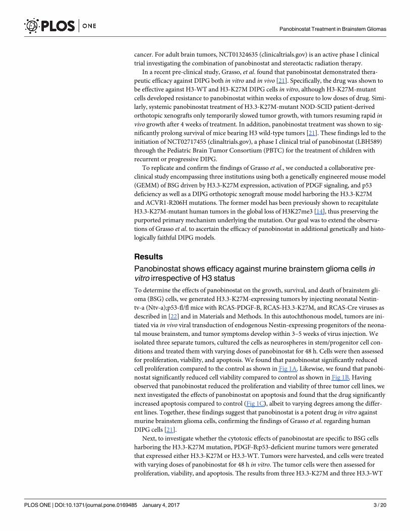
for proliferation, viability, and apoptosis. We found that panobinostat significantly reduced
cell proliferation compared to the control as shown in Fig 1A. Likewise, we found that panobi-
nostat significantly reduced cell viability compared to control as shown in Fig 1B. Having
observed that panobinostat reduced the proliferation and viability of three tumor cell lines, we
next investigated the effects of panobinostat on apoptosis and found that the drug significantly
increased apoptosis compared to control (Fig 1C), albeit to varying degrees among the differ-
ent lines. Together, these findings suggest that panobinostat is a potent drug in vitro against
murine brainstem glioma cells, confirming the findings of Grasso et al. regarding human
DIPG cells [21].
Next, to investigate whether the cytotoxic effects of panobinostat are specific to BSG cells
harboring the H3.3-K27M mutation, PDGF-B;p53-deficient murine tumors were generated
that expressed either H3.3-K27M or H3.3-WT. Tumors were harvested, and cells were treated
with varying doses of panobinostat for 48 h in vitro. The tumor cells were then assessed for
proliferation, viability, and apoptosis. The results from three H3.3-K27M and three H3.3-WT
Panobinostat Treatment in Brainstem Gliomas
PLOS ONE | DOI:10.1371/journal.pone.0169485 January 4, 2017 3 / 20
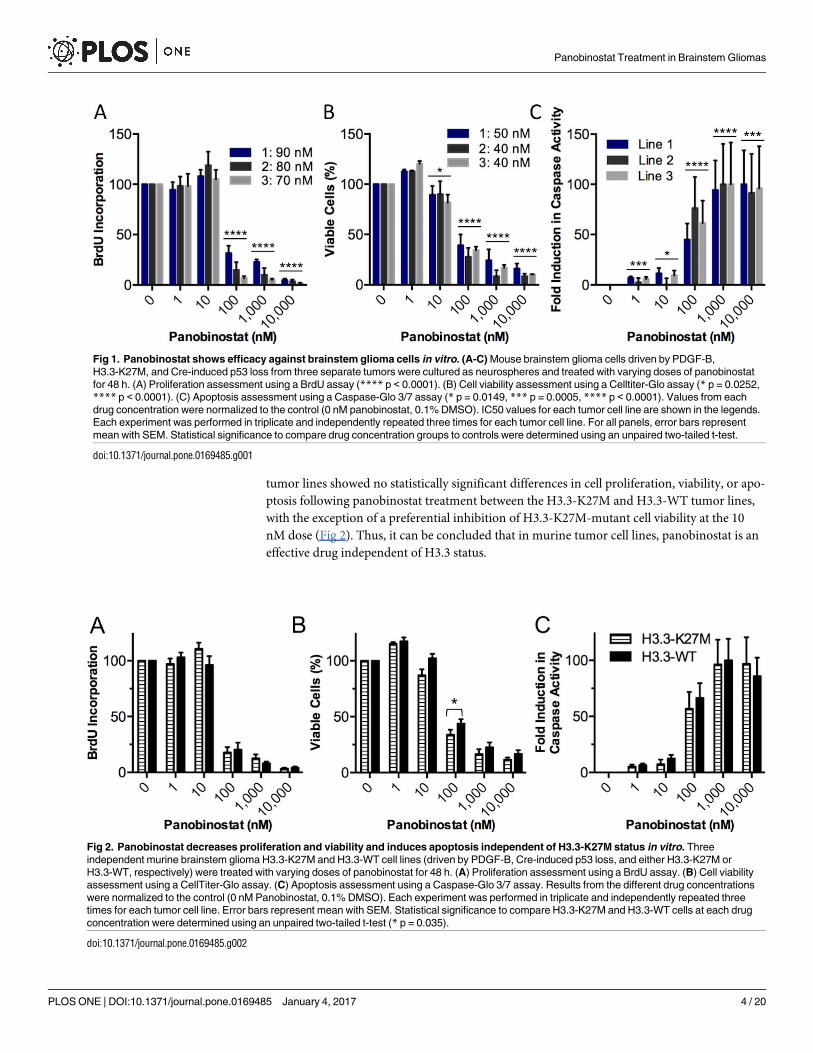
tumor lines showed no statistically significant differences in cell proliferation, viability, or apo-
ptosis following panobinostat treatment between the H3.3-K27M and H3.3-WT tumor lines,
with the exception of a preferential inhibition of H3.3-K27M-mutant cell viability at the 10
nM dose (Fig 2). Thus, it can be concluded that in murine tumor cell lines, panobinostat is an
effective drug independent of H3.3 status.
Fig 1. Panobinostat shows efficacy against brainstem glioma cells in vitro. (A-C) Mouse brainstem glioma cells driven by PDGF-B,
H3.3-K27M, and Cre-induced p53 loss from three separate tumors were cultured as neurospheres and treated with varying doses of panobinostat
for 48 h. (A) Proliferation assessment using a BrdU assay (**** p < 0.0001). (B) Cell viability assessment using a Celltiter-Glo assay (* p = 0.0252,
**** p < 0.0001). (C) Apoptosis assessment using a Caspase-Glo 3/7 assay (* p = 0.0149, *** p = 0.0005, **** p < 0.0001). Values from each
drug concentration were normalized to the control (0 nM panobinostat, 0.1% DMSO). IC50 values for each tumor cell line are shown in the legends.
Each experiment was performed in triplicate and independently repeated three times for each tumor cell line. For all panels, error bars represent
mean with SEM. Statistical significance to compare drug concentration groups to controls were determined using an unpaired two-tailed t-test.
doi:10.1371/journal.pone.0169485.g001
Fig 2. Panobinostat decreases proliferation and viability and induces apoptosis independent of H3.3-K27M status in vitro. Three
independent murine brainstem glioma H3.3-K27M and H3.3-WT cell lines (driven by PDGF-B, Cre-induced p53 loss, and either H3.3-K27M or
H3.3-WT, respectively) were treated with varying doses of panobinostat for 48 h. (A) Proliferation assessment using a BrdU assay. (B) Cell viability
assessment using a CellTiter-Glo assay. (C) Apoptosis assessment using a Caspase-Glo 3/7 assay. Results from the different drug concentrations
were normalized to the control (0 nM Panobinostat, 0.1% DMSO). Each experiment was performed in triplicate and independently repeated three
times for each tumor cell line. Error bars represent mean with SEM. Statistical significance to compare H3.3-K27M and H3.3-WT cells at each drug
concentration were determined using an unpaired two-tailed t-test (* p = 0.035).
doi:10.1371/journal.pone.0169485.g002
Panobinostat Treatment in Brainstem Gliomas
PLOS ONE | DOI:10.1371/journal.pone.0169485 January 4, 2017 4 / 20
Panobinostat potently reduces survival and clonogenicity of human
patient-derived DIPG cells
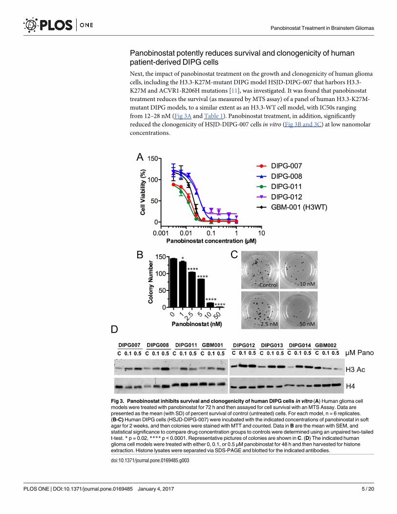
Next, the impact of panobinostat treatment on the growth and clonogenicity of human glioma
cells, including the H3.3-K27M-mutant DIPG model HSJD-DIPG-007 that harbors H3.3-
K27M and ACVR1-R206H mutations [11], was investigated. It was found that panobinostat
treatment reduces the survival (as measured by MTS assay) of a panel of human H3.3-K27M-
mutant DIPG models, to a similar extent as an H3.3-WT cell model, with IC50s ranging
from 12–28 nM (Fig 3A and Table 1). Panobinostat treatment, in addition, significantly
reduced the clonogenicity of HSJD-DIPG-007 cells in vitro (Fig 3B and 3C) at low nanomolar
concentrations.
Fig 3. Panobinostat inhibits survival and clonogenicity of human DIPG cells in vitro (A) Human glioma cell
models were treated with panobinostat for 72 h and then assayed for cell survival with an MTS Assay. Data are
presented as the mean (with SD) of percent survival of control (untreated) cells. For each model, n = 6 replicates.
(B-C) Human DIPG cells (HSJD-DIPG-007) were incubated with the indicated concentrations of panobinostat in soft
agar for 2 weeks, and then colonies were stained with MTT and counted. Data in B are the mean with SEM, and
statistical significance to compare drug concentration groups to controls were determined using an unpaired two-tailed
t-test. * p = 0.02, **** p < 0.0001. Representative pictures of colonies are shown in C. (D) The indicated human
glioma cell models were treated with either 0, 0.1, or 0.5 μM panobinostat for 48 h and then harvested for histone
extraction. Histone lysates were separated via SDS-PAGE and blotted for the indicated antibodies.
doi:10.1371/journal.pone.0169485.g003
Panobinostat Treatment in Brainstem Gliomas
PLOS ONE | DOI:10.1371/journal.pone.0169485 January 4, 2017 5 / 20
Given that panobinostat is an inhibitor of histone deacetylases (HDACs), which remove
acetyl groups from the lysine residues on histone tails, and as Grasso et al. showed increased
H3 acetylation (H3Ac) with panobinostat treatment in vitro [21], we would expect the drug to
increase the levels of histone acetylation within the DIPG cells used here. As shown by western
blot (Fig 3D; for full blots see S1 Fig), panobinostat treatment at 0.1 and 0.5 μM for 48 h dose-
dependently increased the H3 acetylation (H3Ac) levels in 6 different human H3.3-K27M
mutant DIPG lines. Although the same was evident for the H3-WT model HSJD-GBM-001,
this observation did not hold true for the H3.3-G34R mutant model HSJD-GBM-002, in
which H3Ac levels actually decreased with panobinostat treatment.
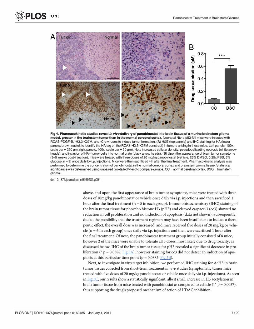
Pharmacokinetic studies reveal delivery of panobinostat into brain
tissue, greater in the brainstem tumor than in the normal cerebral cortex
in vivo.
One of the key obstacles to the treatment of DIPG is the delivery of therapeutic agents across
the blood brain barrier (BBB). To determine if systemically administered panobinostat is
able to cross the BBB and hence potentially target brain tumors, we investigated the pharmaco-
kinetics of panobinostat in brain tissue in vivo. To test this, we used our autochthonous
PDGF-B;H3.3-K27M;p53-deficient BSG GEMM, generated as described above by injecting
RCAS-PDGF-B, -H3.3-K27M, and–Cre into Ntv-a;p53-fl/fl mice. These mice develop symp-
toms of brain tumors within 3 and 5 weeks post-virus injection, and resulting tumors recapitu-
late the high-grade and invasive features of human DIPG (Fig 4A). Upon the first appearance
of brain tumor symptoms, a cohort of these mice were treated with three doses of 20 mg/kg
panobinostat once daily by intraperitoneal (i.p.) injections. Mice were then sacrificed 4 h after
the final treatment. Pharmacokinetic analysis was performed on tissue from the normal cere-
bral cortex and brainstem tumors. Our findings show that panobinostat was successfully
delivered into both normal cerebral cortex and brainstem tumors (Fig 4B). In addition, pano-
binostat levels were significantly higher in brainstem tumor tissue as compared to cerebral cor-
tex tissue of mice treated with drug (Fig 4B, 550±51 nM vs. 70±10 nM, mean±SEM, ��� p =
0.0007). This suggests that in this BSG GEMM, the structural and functional integrity of the
BBB is compromised to a greater extent in the tumors than in the normal cerebral cortex tissue
of tumor-bearing mice. These results provide evidence that systemic delivery of panobinostat
allows for drug concentrations in the brainstem that, according to our in vitro data, are above
potentially active concentrations, precisely targeting the tumor tissue.
Short-term in vivo treatments with panobinostat reduces tumor cell
proliferation and increases H3 acetylation
Given the evidence that panobinostat reaches the brainstem in our BSG GEMM and is cyto-
toxic against tumor cells in vitro, we investigated its short-term in vivo efficacy. To test this,
murine PDGF-B; H3.3-K27M; p53-deficient brainstem tumors were induced as described
Table 1. IC50s of human DIPG cell models with Panobinostat treatment.
Cell model IC50 (nM) 95% CI
HSJD-DIPG-007 15.3 14.6–15.9
HSJD-DIPG-008 28.3 26.0–30.8
HSJD-DIPG-011 12.1 11.1–13.3
HSJD-DIPG-012 26.8 25.3–28.4
HSJD-GBM-001 18.0 16.6–19.6
doi:10.1371/journal.pone.0169485.t001
Panobinostat Treatment in Brainstem Gliomas
PLOS ONE | DOI:10.1371/journal.pone.0169485 January 4, 2017 6 / 20
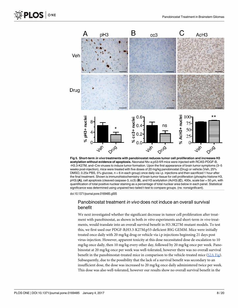
above, and upon the first appearance of brain tumor symptoms, mice were treated with three
doses of 10mg/kg panobinostat or vehicle once daily via i.p. injections and then sacrificed 1
hour after the final treatment (n = 5 in each group). Immunohistochemistry (IHC) staining of
the brain tumor tissue for phospho histone H3 (pH3) and cleaved caspace-3 (cc3) showed no
reduction in cell proliferation and no induction of apoptosis (data not shown). Subsequently,
due to the possibility that the treatment regimen may have been insufficient to induce a thera-
peutic effect, the overall dose was increased, and mice received five doses of 20 mg/kg or vehi-
cle (n = 6 in each group) once daily via i.p. injections and then were sacrificed 1 hour after
the final treatment. Of note, the panobinostat treatment group initially consisted of 8 mice,
however 2 of the mice were unable to tolerate all 5 doses, most likely due to drug toxicity, as
discussed below. IHC of the brain tumor tissue for pH3 revealed a significant decrease in pro-
liferation (� p = 0.0388, Fig 5A), however staining for cc3 did not detect an induction of apo-
ptosis at this particular time point (p = 0.0883, Fig 5B).
Next, to investigate in vivo target inhibition, we performed IHC staining for AcH3 in brain
tumor tissues collected from short-term treatment in vivo studies (symptomatic tumor mice
treated with five doses of 20 mg/kg panobinostat or vehicle once daily via i.p. injections). As seen
in Fig 5C, our results show a statistically significant, albeit small, increase in H3 acetylation in
brain tumor tissue from mice treated with panobinostat as compared to vehicle (�� p = 0.0057),
thus supporting the drug’s proposed mechanism of action of HDAC inhibition.
Fig 4. Pharmacokinetic studies reveal in vivo delivery of panobinostat into brain tissue of a murine brainstem glioma
model, greater in the brainstem tumor than in the normal cerebral cortex. Neonatal Ntv-a;p53-fl/fl mice were injected with
RCAS-PDGF-B, -H3.3-K27M, and -Cre viruses to induce tumor formation. (A) H&E (top panels) and IHC staining for HA (lower
panels, brown nuclei, to identify the HA tag on the RCAS-H3.3-K27M construct) in tumors arising in these mice. Left panels, 100x,
scale bar = 250 μm; right panels, 400x, scale bar = 50 μm). Note increased cellular density, pseudopalisading necrosis (white arrow
heads), and invasion of HA+ tumor cells into normal brain (black arrow heads). (B) Upon the appearance of brain tumor symptoms
(3–5 weeks post-injection), mice were treated with three doses of 20 mg/kg panobinostat (vehicle, 25% DMSO, 0.25x PBS, 5%
glucose, n = 3) once daily by i.p. injections. Mice were then sacrificed 4 h after the final treatment. Pharmacokinetic analysis was
performed to determine the concentration of panobinostat in the normal cerebral cortex and brainstem glioma tissue. Statistical
significance was determined using unpaired two-tailed t-test to compare groups. CC = normal cerebral cortex, BSG = brainstem
glioma.
doi:10.1371/journal.pone.0169485.g004
Panobinostat Treatment in Brainstem Gliomas
PLOS ONE | DOI:10.1371/journal.pone.0169485 January 4, 2017 7 / 20
Panobinostat treatment in vivo does not induce an overall survival
benefit
We next investigated whether the significant decrease in tumor cell proliferation after treat-
ment with panobinostat, as shown in both in vitro experiments and short-term in vivo treat-
ments, would translate into an overall survival benefit in H3.3K27M-mutant models. To test
this, we first used our PDGF-B;H3.3-K27M;p53-deficient BSG GEMM. Mice were initially
treated once daily with 20 mg/kg drug or vehicle via i.p injections beginning 21 days post
virus-injection. However, apparent toxicity at this dose necessitated dose de-escalation to 10
mg/kg once daily, then 10 mg/kg every other day, followed by 20 mg/kg once per week. Pano-
binostat at 20 mg/kg once per week was well-tolerated, however there was no overall survival
benefit in the panobinostat-treated mice in comparison to the vehicle-treated mice (S2A Fig).
Subsequently, due to the possibility that the lack of a survival benefit was secondary to an
insufficient dose, the dose was increased to 20 mg/kg once daily administered twice per week.
This dose was also well-tolerated, however our results show no overall survival benefit in the
Fig 5. Short-term in vivo treatments with panobinostat reduces tumor cell proliferation and increases H3
acetylation without evidence of apoptosis. Neonatal Ntv-a;p53-fl/fl mice were injected with RCAS-PDGF-B,
-H3.3-K27M, and–Cre viruses to induce tumor formation. Upon the first appearance of brain tumor symptoms (3–5
weeks post-injection), mice were treated with five doses of 20 mg/kg panobinostat (Drug) or vehicle (Veh, 25%
DMSO, 0.25x PBS, 5% glucose, n = 6 in each group) once daily via i.p. injections and then sacrificed 1 hour after
the final treatment. Shown is immunohistochemistry of brain tumor tissue for cell proliferation (phospho histone H3,
pH3) (A), cell apoptosis (cleaved caspase-3, cc3) (B), and H3 acetylation (AcH3) (C), 400x, scale bar = 50 μm, with
quantification of total positive nuclear staining as a percentage of total nuclear area below in each panel. Statistical
significance was determined using unpaired two-tailed t-test to compare groups. (ns: nonsignificant).
doi:10.1371/journal.pone.0169485.g005
Panobinostat Treatment in Brainstem Gliomas
PLOS ONE | DOI:10.1371/journal.pone.0169485 January 4, 2017 8 / 20
panobinostat-treated mice in comparison to the vehicle-treated mice (Fig 6A, median survival
35 days versus 34 days, respectively). Tumors in the brainstem were confirmed in all mice by
hematoxylin and eosin (H&E) staining. The treatment dose, frequency, and/or duration was
not further increased due to the high likelihood of panobinostat toxicity. Thus, at the well-tol-
erated dose of 20 mg/kg administered once daily twice per week, panobinostat did not prolong
overall survival of our BSG GEMM.
Next, the efficacy of panobinostat in a human DIPG orthotopic xenograft model was
assessed. HSJD-DIPG-007 cells were orthotopically implanted into the pons of 7-week old
NOD SCID mice using stereotactic coordinates. In this model, mice exhibit symptoms of
brain tumor formation within 60–80 days post tumor cell implantation and harbor tumors
that histologically resemble human DIPG, with tumor cells infiltrating the brainstem as seen
with routine hematoxylin and eosin staining and the cerebellum as shown by vimentin staining
(Fig 6B). Beginning on day 28 post-implantation, mice were treated with 10 mg/kg panobino-
stat or vehicle (5% dextrose) once daily by i.p. injection. All mice exhibited significant toxicity
following the second dose of panobinostat, indicated by decreased motor activity, ruffled fur,
hunched posture, labored breathing, average weight loss of 18%, and death. Autopsy of the
deceased animals revealed a yellow secretion in the stomach cavity. Due to the severe toxicity,
the experiment was continued with the administration of panobinostat on alternating days (3
days per week) for 4 weeks. With this regimen, no severe toxicity was observed. However, mice
suffered from diarrhea and were sustained with mushy food, jelly and 5% i.p. glucose daily for
the duration of the experiment. As shown in Fig 6C, no survival difference was observed
between vehicle-treated control and panobinostat-treated mice (P>0.05, log-rank analysis).
As panobinostat is insoluble in 5% dextrose, it was next solubilized in 2.5% DMSO, 5%
PEG400 and 5% Tween80 in 0.9% saline. Mice harboring intracranial xenografts were treated
with once daily 10 mg/kg panobinostat three times per week (M,W,F) for four weeks starting
at 28 days post-implantation. Using this vehicle and treatment regimen, no signs of toxicity
were observed in panobinostat-treated mice. However, no survival difference was observed
between mice treated with panobinostat and those treated with vehicle (Fig 6D, P>0.05, log-
rank analysis).
Finally, an alternative vehicle and dosing regimen was tested, with mice harboring intracra-
nial HSJD-DIPG-007 xenografts receiving either no treatment or 10 mg/kg panobinostat [in
10% DMSO diluted in PBS-10% hydroxypropyl-beta-cyclodextrin (HPBCD)] once daily via i.
p. injection starting at day 23 post tumor cell implantation for 5 days on followed by 2 days off
and then 5 days on again, for a total of 10 doses (S2B and S2C Fig). This dosing regimen aimed
to reproduce the regimen previously reported by Grasso et al. to temporarily slow H3-K27M
tumor growth and increase survival in an H3-WT DIPG model [21]. However, there was no
survival benefit observed for the H3.3-K27M DIPG model used here with this drug treatment
(median survival of 81.5 and 80 days for treated and control mice, respectively).
Materials and Methods
Duke University Medical Center, USA
Mice. All in vivo experiments associated with Figs 1, 2, 4, 5, 6A and S2A were performed
in accordance with the Duke University Animal Care and Use Committee and Guide for the
Care and Use of Laboratory Animals (protocol # A214-13-08). Nestin-Tv-a (Ntv-a); p53fl/fl
mice have been previously described [22] and were created by breeding Ntv-a mice with
p53-floxed mice (C57BL/6J background) from Jackson Labs.
Autochthonous brainstem glioma GEMM. To generate viruses for the induction of
brainstem gliomas (BSGs) in mice, DF1 virus-producing cells were purchased from ATCC,
Panobinostat Treatment in Brainstem Gliomas
PLOS ONE | DOI:10.1371/journal.pone.0169485 January 4, 2017 9 / 20
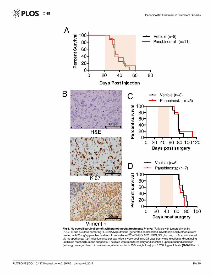
Fig 6. No overall survival benefit with panobinostat treatments in vivo. (A) Mice with tumors driven by
PDGF-B and p53 loss harboring H3.3-K27M mutations (generated as described in Materials and Methods) were
treated with 20 mg/kg panobinostat (n = 11) or vehicle (25% DMSO, 0.25x PBS, 5% glucose, n = 8) administered
via intraperitoneal (i.p.) injection once per day twice a week beginning 21 days post-virus injection and continuing
until mice reached humane endpoints. The mice were monitored daily and sacrificed upon moribund condition
(lethargy, enlarged head circumference, ataxia, and/or > 25% weight loss) (p = 0.756, log rank test). (B-D) Effect of
Panobinostat Treatment in Brainstem Gliomas
PLOS ONE | DOI:10.1371/journal.pone.0169485 January 4, 2017 10 / 20
cultured in DMEM (ATCC) supplemented with 10% FBS, 2mM L-glutamine, 100 units/mL
penicillin and 100 μg/mL streptomycin and incubated at 39˚C and 5% CO2. Cells were trans-
fected with RCAS plasmids (RCAS-PDGF-B, RCAS-Cre, RCAS-H3.3K27M, or RCAS-H3.3WT)
using Fugene 6 or X-TremeGENE 9 (Roche) per the manufacturer’s instructions. Virus-produc-
ing cells were used for injections after being passaged at least 6 times and less than 20 times from
the time of transfection. To generate BSGs, Nestin-tv-a(Ntv-a); p53fl/fl mice (that express tv-a,
the receptor for RCAS viruses, in Nestin-expressing cells) were injected with 1 μL of a 1:1:1 cock-
tail of DF1 cells expressing RCAS-PDGF-B, RCAS-Cre, and RCAS-H3.3-K27M (or RCAS-
H3.3-WT) as previously described [23]. Injections were made 2 mm posterior to the bregma
along the midline using a Hamilton syringe and custom needle. Injections were performed on
postnatal day 2 to postnatal day 5 mice after being anesthetized on ice. Mice were monitored
daily and euthanized with CO2 upon the appearance of signs of brain tumors (lethargy, enlarged
head, ataxia, weight loss up to 25%).
Generation of murine BSG neurospheres. To generate BSG cell lines, tumors were iso-
lated from symptomatic mice (described above) and enzymatically digested in Earl’s Balanced
salt solution containing 4.7mg papain (Worthington) and 60μg /mL DNAse (Sigma Aldrich).
Digestion was inactivated with ovomucoid (0.7 mg/mL) (Worthington) containing 14μg/mL
DNAse. Cells were consecutively washed, triturated, and strained to obtain a single cell sus-
pension. The cells were cultured in neurosphere media [Neurocult media (Stem Cell Technol-
ogies) supplemented with 5 ml cell proliferation supplement (Stem Cell Technologies), 500 μL
penicillin and streptomycin, 10 μL human basic FGF (20 ng/ml), 5 μL human EGF (10 ng/ml),
50 μL heparin] and incubated at 37˚C and 5% CO2.
In vitro assays on murine BSG cells
Cell proliferation. Neurospheres generated from BSGs were plated as single cells in tripli-
cate in a white-walled, clear-bottomed 96-well plate (50,000 cells/well in 100 μL) and allowed
to adhere for 24 h. Cells were then treated with 10 μL working solution [1 μL of panobinostat
(Selleckchem, Catalog #S1030) stock solution (serial dilutions of 10 mM) + 99 μL neurosphere
media (described above)] at increasing panobinostat concentrations (0.001 μM, 0.01 μM,
0.1 μM, 1.0 μM, 10 μM) or 0.1% DMSO (control) for 48 h. A bromodeoxyuridine (BrdU)
based cell proliferation ELISA assay kit (Roche) was used to assay proliferation. Cells were
pulsed with BrdU for 4 h. Absorbance was read using Molecular Devices Versa Max Tunable
Microplate Reader. Normalized proliferation levels were calculated relative to the vehicle (0 μL
panobinostat). The dose response curve was generated with Prism software and analyzed
using nonlinear regression. The values of half-inhibitory concentration (IC50) were calculated
by using log (agonist) versus response including variable slope (four parameters) statistics and
normalized in GraphPad Prism. Each experiment was performed in triplicate and indepen-
dently repeated three times for each tumor cell line. Error bars represent mean with SEM. Sta-
tistical significance to compare drug concentration groups with controls were determined
using an unpaired two-tailed t-test.
panobinostat treatment on the survival of mice-bearing H3.3-K27M HSJD-DIPG-007 orthotopic xenografts.
NOD-SCID mice (7 weeks old) were orthotopically injected with HSJD-DIPG-007 cells (passage 39) into the
brainstem via stereotactic coordinates. (B) Representative H&E (top panel) and IHC staining for Ki67 (middle panel)
and Vimentin (bottom panel) of control mice (treated with vehicle and sacrificed immediately after the last dose, as
described in the Materials and Methods). 400x magnification, scale bar = 50 μm. (C-D) Starting on day 28 post-
implantation, mice were treated with panobinostat prepared in a vehicle containing 5% dextrose (C) or 2.5% DMSO,
5% PEG400 and 5% Tween80 in 0.9% saline (D) via intraperitoneal (i.p.) injection at 10 mg/kg, three times a week
for four weeks (p>0.05, log-rank test). Shaded areas under the curves in A, C-D indicate treatment duration.
doi:10.1371/journal.pone.0169485.g006
Panobinostat Treatment in Brainstem Gliomas
PLOS ONE | DOI:10.1371/journal.pone.0169485 January 4, 2017 11 / 20
Cell viability and apoptosis. BSG neurospheres were generated, cultured, and treated as
above for cell proliferation. For cell viability, the CellTiter-Glo Luminescent Assay (Promega)
was performed, and for apoptosis, the Promega ApoTox-Glo Triplex Assay (# G6321) was
used, both according to the manufacturer’s instructions. Luminescence was read using a
Turner Biosystems Modulus Microplate Reader. Data analysis and statistics were performed as
above for cell proliferation.
Short-term panobinostat treatment of BSG GEMM
Upon the first appearance of brain tumor symptoms, tumor-bearing mice were treated with
either 1) three doses of panobinostat (Selleckchem #S1030) at 10 mg/kg or vehicle (25%
DMSO, 0.25x PBS, 5% glucose) administered once daily by i.p. injections (n = 5 in each
group), or 2) five doses of panobinostat (Selleckchem) at 20 mg/kg or vehicle administered
once daily by i.p. injections (n = 6 in each group). Mice were sacrificed 1 hour after their
final treatment via CO2, and their brains were extracted, fixed in 10% formalin, paraffin
embedded and sectioned on a microtome. Immunohistochemistry for phospho histone H3
(pH3), cleaved caspase-3 (cc3), and H3 acetylation (AcH3) were conducted with subsequent
quantification (described below).
Immunohistochemistry
Formalin fixed brains were paraffin embedded by Duke Pathology Core Services. Sections were
cut 5μm thick using a Leica RM2235 Microtome. Digital images of microscopic fields of brain
tissue sections were acquired with a Leica DMLB microscope, Leica digital camera and Leica
Application Suite Version 3.7 (Leica; Buffalo Grove, IL) at 400x magnification (high powered
field). Hematoxylin and Eosin (H&E) staining was performed using standard protocols. Immu-
nohistochemistry was performed using an automated processor (Discovery XT, Ventana Medi-
cal Systems, Inc.) Antibodies used were as follows: anti-HA (Y-11, 1:250 dilution, Santa Cruz
#sc-805), anti-phospho-Histone H3 (Ser10) (1:1600 dilution, Millipore #04–1093), anti-cleaved
caspase 3 (Asp175) (1:1600 dilution, Cell Signaling #9661), and anti-acetyl-Histone H3 (1:500
dilution, Millipore #06–599). Quantification analysis was performed blindly using MetaMorph
Image Analysis Software. Percentage of positive staining cell nuclei equal to positive nuclear
area (brown staining only with a specific pre-set threshold) / total nuclear area (blue and brown
staining) x 100. Thresholds for positive nuclei and total nuclei were unchanged throughout the
quantification process. Statistical significance was determined using unpaired two-tailed t-test
to compare groups.
Pharmacokinetics
Symptomatic mice (BSG GEMM) were treated with three doses of 20 mg/kg panobinostat
(Selleckchem) or vehicle (25% DMSO, 0.25x PBS, 5% glucose) (n = 3 in each group) adminis-
tered once daily by intraperitoneal injections. Mice were sacrificed 4 h after their final treat-
ment via CO2. Brains from the sacrificed mice were extracted. Half of each brain was fixed in
10% formalin and embedded in paraffin for histological analysis. The other half was SNAP fro-
zen, stored at -80 degrees and sent for pharmacokinetic (PK) studies. Pharmacokinetic analysis
was performed on the cerebral cortex tissue and brainstem tumor tissue of each mouse by the
Pharmacokinetic/Pharmacodynamic (PK/PD) Core Laboratory, Duke Cancer Institute as
described below. Statistical significance was determined using unpaired two-tailed t-test to
compare groups.
Tissue processing. Brain tissue was homogenized with 2 parts water (w/v) by rotary
homogenizer (polyethylene rotor/1.5-mL conical tube). To a 200-μL PP tube, 20 μL of tissue
Panobinostat Treatment in Brainstem Gliomas
PLOS ONE | DOI:10.1371/journal.pone.0169485 January 4, 2017 12 / 20
homogenate and 40 μL of methanol (containing 10 ng/mL PSTAT-d8 internal std.) was added
and vigorously agitated in a FastPrep vortexer (Thermo-Savant) at speed 4 for 20s. After cen-
trifugation at 13,600 g for 5 min at RT, the supernatant was transferred into an injection vial,
and 10 μL was injected into the LC/MS/MS system.
Liquid chromatography tandem-mass spectrometry (LC/MS/MS). The analysis was
performed on a Shimadzu 20A series LC system coupled with an Applied Biosciences/SCIEX
API 4000 QTrap MS/MS spectrometer. Column: Phenomenex, C18 4x3 mm (P/N AJ0-4287)
column at 35˚C. Mobile phase solvents (all MS-grade): A—0.1% formic acid in water, 2% aceto-
nitrile; B–acetonitrile. Elution gradient at 1 mL/min: 0–0.5 min 0–95% B, 0.5–1.0 min 95% B,
1.0–1.2min 95–0% B. Run time: 4min. MRM transitions for PSTAT and PSTAT-d8 (m/z):
350/158 and 358/164, respectively. Positive-ion mode. DP: 76 V, EP: 10 V, ion-spray voltage:
5500 V, curtain gas: 30, ion-source gas1: 30, ion-source gas2: 30. Lower limit of quantification
(LLOQ): 1.2 ng/g wet tissue. Calibration curve samples (n = 6) were prepared by adding increas-
ing amounts of Panobinostat to control brain homogenate obtained from non-treated animals.
Long-Term panobinostat treatment of BSG GEMM
Tumor-bearing mice were randomly assigned to be treated with either 20 mg/kg panobinostat
or vehicle (25% DMSO, 0.25x PBS, 5% glucose) once daily administered once or twice a week
via i.p. injections. This dose was reduced from 20 mg/kg daily to 10 mg/kg daily and 10 mg/kg
every other day due to toxicity concerns. Of note, 25% DMSO was the lowest percentage in
which panobinostat could be successfully and reproducibly solubilized in our hands. Treat-
ments began 21 days post-injection with RCAS-PDGF-B, RCAS-Cre, and RCAS-H3.3-K27M
expressing cells and continued until mice reached humane endpoints. Mice were monitored
daily and sacrificed upon moribund conditions (lethargy, enlarged head circumference, ataxia,
and/or > 25% weight loss) or 12 weeks post-injection in the absence of symptoms. Statistical
significance between the Kaplan-Meier survival curves was determined using a log-rank (Man-
tel-Cox) test.
Children’s Cancer Institute, Australia
Cell lines and reagents. The pediatric autopsy-derived DIPG cell line HSJD-DIPG-007
(DIPG-007) was obtained from Dr. Angel M. Carcaboso (Hospital Sant Joan de Deu Barce-
lona, Barcelona, Spain; for details see below). Cells were cultured as neurospheres in tumor
stem base medium (50:50 DMEM/F12:Neurobasal Life Technologies) supplemented with B-27
(Life technologies), Heparin (Stemcell Technologies) and human growth factors EGF, FGF-
basic, PDGF-AA and PDGF-BB (Jomar Life Research). Cells tested free of mycoplasma con-
tamination. Panobinostat was purchased from Selleckchem.
Clonogenic Assay. Clonogenic assay was performed using a soft agar method. Plates were
coated with 0.5% SeaPlaque agarose (Cambrex) in tumor stem base medium and stored at 4˚C
overnight. The following day, 0.3% agarose containing HSJD-DIPG-007 cells and various con-
centrations of panobinostat were plated over the agar underlay. The plates were incubated at
37˚C for two weeks. Colonies were then stained using MTT (3-[4,5-Dimethylthiazol-2-yl]-
2,5-diphenyltetrazolium bromide; Thiazolyl blue) and counted.
Mice. All in vivo experiments were performed in accordance with the University of New
South Wales Animal Care and Ethics Committee according to the Animal Research Act, 1985
(New South Wales, Australia) and the Australian Code of Practice for Care and Use of Animals
for Scientific Purposes (2013). The animal studies associated with Fig 6B–6D were conducted
according to the protocols approved by the Animal Experimentation Ethics Committee of the
University of New South Wales (ACEC number: 16/7A). NOD/SCID mice (Fig 6B–6D) were
Panobinostat Treatment in Brainstem Gliomas
PLOS ONE | DOI:10.1371/journal.pone.0169485 January 4, 2017 13 / 20
obtained from Animal Resources Centre (ARC, Canning Vale Western Australia) and main-
tained in a temperature-controlled environment with a 12-hour light/dark cycle in Tecniplast
individually vented cages with enviro-dri and igloos for nesting. Mice were given irradiated
feeder pellets and water ad libitum. Mice were euthanized at the completion of the protocol by
carbon dioxide overdose.
Orthotopic injection. To orthotopically implant tumor cells into 7 week old mice, stereo-
tactic surgery was performed using a KOPF small animal stereotactic surgical device. Mice
were sedated using isoflurane gas. The skull of each mouse was exposed by incising the skin,
and a small burr hole was made using a high-speed drill. Exact coordinates that target the pons
region via the IVth ventricle were used (0.5mm lateral to the sagittal suture, 5.4mm posterior
to the bregma and 3.1mm deep). A total of 3x10⁵ HSJD-DIPG-007 cells (passage 39) sus-
pended in 2 μL of matrigel were injected using a point style 2, 25 gauge Hamilton syringe.
Upon sealing the surgery site, mice were given 10μg/kg Buprenorphine intraperitoneally. Mice
were placed on a warming pad and returned to their cages upon full recovery.
Histological analysis of orthotopic xenografts. Mice bearing orthotopic xenografts gen-
erated as described above were treated with vehicle (both Tween/PEG and 5% dextrose via i.p.
injection Monday, Wednesday and Friday for a total of 12 treatments and sacrificed 24 hours
after the last dose. Mouse brains were harvested and following formalin fixation were sec-
tioned in a mouse brain cutter into either coronal or sagittal sections. Processing of tissue was
performed in the Histology Unit at the Garvan Institute of Medical Research. Morphologic
assessment of xenografts is based on routine haematoxylin and eosin (H&E) staining and
immunohistochemical (IHC) staining with Vimentin (1:400; Novocastra NCL-VIM-V9) and
Ki67 (1:400; Thermoscientific; RM-9106S1). Representative photographs of a vehicle control
xenograft (shown in Fig 6B) were taken using an Olympus BX53 light microscope and CD-73
camera with cellSens software.
Panobinostat drug preparation and treatment of orthotopic xenografts. Panobinostat
(Selleckchem) was prepared in two different vehicles. For the first in vivo experiment (Fig 6C)
a stock solution of 1 mg/ml of panobinostat was prepared in 5% dextrose, and for the second
experiment (Fig 6D), panobinostat was dissolved in DMSO at the concentration of 40 mg/ml
and then diluted with 0.9% saline containing 5% PEG400 and 5% Tween80. The final concen-
tration of panobinostat in the solution was 1 mg/ml. The drug was prepared fresh before treat-
ment. In experiment 1, mice were treated daily with 10 mg/kg panobinostat intraperitoneally,
which was decreased to three times a week due to toxicity, for four weeks. For the experiment
2, the animals were treated with 10 mg/kg three times a week (M,W,F) for four weeks intraper-
itoneally. Control groups were administered with the drug-free vehicle. The treatment was
started on day 28 after the tumor implantation. Mice were sacrificed following the develop-
ment of any of the following clinical signs of tumor progression: severe head tilting, severe
ataxia, severe circling, lethargy and/or weight loss�20% of the initial weight.
Statistical analysis. For survival analysis of orthotopic xenografts, log-rank analysis was
used.
Hospital Sant Joan de Deu Barcelona, Spain
Human DIPG cell culture. Patient-derived DIPG cell models were obtained under an
Institutional Review Board-approved protocol and with written informed consent (M-1608-C)
at Hospital Sant Joan de Deu Barcelona, Spain. Models were established either from patient
autopsy (HSJD-DIPG-007; from a patient who survived less than one month after diagnosis
and was treated with one cycle of irinotecan-cisplatin and no radiation therapy; original tumor
sequenced in [11], coded HSJD_DIPG007, with H3.3-K27M and ACVR1-R206H mutations),
Panobinostat Treatment in Brainstem Gliomas
PLOS ONE | DOI:10.1371/journal.pone.0169485 January 4, 2017 14 / 20
or from biopsies at diagnosis (DIPG models HSJD-DIPG-008, HSJD-DIPG-011, HSJD-DIPG-
012, HSJD-DIPG-013, HSJD-DIPG-014, all of which are H3.3-K27M-mutated and were estab-
lished from pons biopsies; and HGG model HSJD-GBM-001, H3-WT, established from tumor
in frontal lobe), or from biopsies at relapse (HGG model HSJD-GBM-002, H3.3-G34R-
mutated, established from a tumor in the left hemisphere). Cells were cultured as tumor-
spheres as previously described in [24].
In vitro experiments with human DIPG cells
MTS Assay. To study the antiproliferative activity of panobinostat on human DIPG and
HGG cells, tumorspheres in culture were disaggregated either mechanically or with TrypLE
Express Stable Trypsin Replacement Enzyme (Life Technologies), and 3,000 cells per well were
plated in 96 well-plates and maintained in culture for 24 h. Cells were then treated with pano-
binostat (Seqchem, Pangbourne, UK; stock 5 mg/mL in DMSO) at concentrations ranging
0.003–1 μM for 72 h, six wells per condition. The final DMSO concentration was� 0.014% for
all conditions; previous data (not shown) indicates that up to 0.1% DMSO does not affect the
growth of these DIPG models. Control cells were untreated. Cell viability was assessed by the
MTS assay (Promega, Fitchburg, WI, USA), and IC50 values were calculated with Graphpad.
Immunoblotting. Tumor cell lines were lysed in EpiSeeker Histone Extraction Kit
(Abcam). Each histone lysate (1 μg) was separated by SDS-PAGE and transferred onto a nitro-
cellulose membrane (Thermo Scientific). Membranes were blocked in Tris-buffered saline
with 5% nonfat dry milk and incubated overnight at 4˚C with rabbit anti-Histone 4 (1:1,000,
Abcam, catalog number ab10158) and rabbit anti-histone H3.3, Acetylated (1:1,000, Millipore,
catalog number: 06–599), followed by horseradish peroxidase-conjugated secondary antibod-
ies (1:10,000, Promega).
Panobinostat treatment of orthotopic xenografts in mice
The xenograft studies of panobinostat activity intended to reproduce the previously reported
preclinical study using an H3-WT DIPG model [21]. NOD.SCID mice were obtained from
Harlan (Barcelona, Spain). Animal studies associated with S2B and S2C Fig were performed
according to the Institutional and European guidelines (EU Directive 2010/63/EU) and were
approved by the local animal care and use committee (Comite Etico de Experimentacion Ani-
mal at Universidad de Barcelona, protocol 135/11). To establish orthotopic tumors, 3-week
old mice (5 per group) were anesthetized with 100 mg/kg ketamine and 10 mg/kg xylazine and
immobilized in a stereotaxic apparatus (Stoelting, Wood Dale, IL). A burr hole was drilled in
the skull at coordinates (from bregma) x+0.5 and y-5.4, and 5 x 10⁵ HSJD-DIPG-007 cells
(p69) suspended in 5 μL matrigel (BD Biosciences) were injected at 3.1 mm depth (targeting
the 4th ventricle) with a dull 22G needle attached to a 50 μL syringe (Hamilton, Bonaduz, Swit-
zerland), using a stereotaxic arm. Animals recovered and treatments were initiated 23 days
after tumor cell implantation. Panobinostat was prepared each day from frozen stocks in
DMSO. Final formulation was 10% DMSO diluted in PBS containing 10% hydroxypropyl-
beta-cyclodextrin (HPBCD) as a solubilizer, for the regimen of 10 mg/kg 5 days on, 2 days off,
5 days on (S2B Fig). Control mice received no treatment. Tumor endpoints used were weight
loss >20% from maximum weight achieved or severe motor symptoms. For survival analysis
of orthotopic xenografts, log-rank analysis was used.
Histological analysis of orthotopic xenografts
Tumors from orthotopic DIPG xenografts, generated as described above, were harvested when
mice reached humane endpoints and formalin-fixed paraffin-embedded. Heat-induced
Panobinostat Treatment in Brainstem Gliomas
PLOS ONE | DOI:10.1371/journal.pone.0169485 January 4, 2017 15 / 20
antigen retrieval with sodium citrate was performed according to standard protocols using an
indirect immunoperoxidase method. The primary antibodies used were mouse anti-human
Nuclei (1:200, MAB4383, Merck Millipore), mouse anti-Ki67 (Bond ready to use reagent,
Clone K2, Leica Biosystems), and mouse anti-Histone H3 trimethyl K27 (1:600, Millipore, cat-
alog number: 07–449). Staining was visualized with Novolink Polymer Detection Systems
from Leica, followed by hematoxylin counterstaining (S2C Fig).
Discussion
Despite numerous efforts to improve the treatment of DIPG the prognosis remains poor. Tar-
geted therapy with panobinostat has recently entered a phase I clinical trial for the treatment
of children with recurrent or progressive DIPG due to promising pre-clinical studies [21]. To
replicate and extend these previously published findings, we conducted a cooperative pre-clin-
ical study encompassing three institutions using both genetically engineered and orthotopic
patient-derived DIPG mouse models to investigate the efficacy of panobinostat in vitro and invivo.
In vitro, we confirmed panobinostat to be highly effective against DIPG tumor cells at low
nanomolar to low micromolar concentrations, depending on the cell and assay type. In addi-
tion, we found the sensitivity of DIPG cells to panobinostat to be independent of the H3.3
mutational status, as both H3.3-K27M and H3.3-WT cells were equally susceptible to treat-
ment. Combined with results reported by Grasso et al. who observed effects of panobinostat
on H3-WT, H3.1-K27M, and H3.3-K27M DIPG cells [21], this confirms that panobinostat
could be an option for the treatment of all DIPG subtypes, even when the H3.3-K27M muta-
tion is not present or if its status is unknown.
Importantly, the study here advances the current literature on the use of panobinostat to
treat DIPG by demonstrating histological evidence of target inhibition in vivo. The short-term
treatment of BSG-bearing mice with panobinostat at 20 mg/kg once daily for 5 days resulted in
a significant decrease in cell proliferation, however, we were unable to detect an induction in
apoptosis via cleaved caspase 3 staining at the time point investigated. It is possible that an
alternative (more sensitive) assay or time point could have revealed an induction of apoptosis;
however the reduced proliferation may have been achieved by an alternate mechanism, such
as epigenetic modifications. For instance, pharmacodynamics analysis showed increased levels
of H3 acetylation in tumor tissue demonstrating target inhibition at this dosing schedule.
Pharmacokinetic studies revealed successful delivery of panobinostat into brain tissue of mice
bearing genetically engineered BSG, providing evidence that it is able to reach potentially
active concentrations in tumor tissue in this model. Furthermore, the distribution of panobi-
nostat was higher in these brainstem tumors as compared to the normal cerebral cortical tis-
sue, likely due to a compromised structural integrity of the BBB, which has been previously
demonstrated in our GEMM [25], thus allowing a greater concentration of panobinostat to
penetrate, and potentially treat, the tumor. Whether panobinostat reached potentially active
levels in the HSJD-DIPG-007 xenograft model was not addressed here.
Grasso et al. reported temporarily reduced H3.3-K27M tumor growth (via biolumines-
cence) with systemic panobinostat administration of 10 mg/kg 3 times per week to a NOD-S-
CID patient-derived orthotopic xenograft DIPG model, with tumor regrowth evident after 4
weeks of therapy; the overall survival of these mice was not reported [21]. In addition, they
observed significantly prolonged survival with systemic panobinostat treatment (10 mg/kg 5
days on 5 days off) as compared to vehicle treated controls using an H3 wild-type DIPG model
[21]. The integrity of the BBB in this xenograft model was not reported, although panobinostat
concentrations were determined to be approximately 200 nM in the pons of nontumor mice
Panobinostat Treatment in Brainstem Gliomas
PLOS ONE | DOI:10.1371/journal.pone.0169485 January 4, 2017 16 / 20
[21]. In our studies, we confirm a short term effect on H3.3-K27M tumor cell growth (via
reduced proliferation after 5 days of treatment) in a BSG GEMM and observe no survival ben-
efit of systemic treatment with panobinostat as compared to vehicle treated controls with vari-
ous dosing regimens in immunocompromised xenograft models and immunocompetent
GEMMs. This lack of efficacy to improve animal survival is inconsistent with our data showing
high drug distribution in the GEMM tumors (above 500 nM) and could be due to the reduced
dosage required to avoid toxicity, the development of drug resistance as was shown previously
in vitro [21], or, for the orthotopic xenograft model, the high passage number of cells trans-
planted, which may have selected for particularly aggressive subclones. Despite the late cell
passage number of this model, however, it represents one of the few molecularly, histologically,
and clinically faithful orthograft DIPG models available. When considered along with the
results of Grasso et al., there may be a discrepancy in drug response in vivo between H3.3-
K27M and H3-WT DIPG models, potentially a result of the mutation, although this will
require further direct investigation to confirm. Although the in vitro studies reported here and
previously showed that panobinostat was potently effective against all tumor cell lines indepen-
dent of H3 status, a multitude of other factors contribute to overall survival in in vivo studies.
It is also important to note that based on the standard deviation of the BSG GEMM used
here, in order for a 5 day-difference in median survival to be statistically significant (as was
shown in a previously published study using an Ink4a-ARF deficient BSG GEMM to test the
CDK4/6 inhibitor PD-033299 [23]), a minimum of 42 (for 90% power) or 32 (for 80% power)
mice would need to be treated with drug. Therefore, our study reported here may have been
insufficiently powered to detect a small survival benefit with systemic panobinostat treatment,
although all data (including GEMM and orthotopic xenograft studies) are sufficiently powered
to detect a clinically-meaningful 10–12 day difference in survival (with 80% or 90% power).
A couple noteworthy potential challenges in using panobinostat for the treatment of DIPG
in children are drug toxicity and inadequate drug delivery. In the in vivo studies shown herein,
the drug concentration and duration of panobinostat treatments required to achieve an overall
survival benefit would have likely resulted in substantial toxicity. As it is not yet known
whether this toxicity translates to children with DIPG, these results should be taken into con-
sideration during the execution of clinical trials with panobinostat. In addition, although data
with our GEMM suggest that the BBB structural integrity is likely compromised in these
tumors, as evidenced by the increased drug concentration in the tumor tissue compared with
normal tissue, this may not hold true for children with DIPGs treated with panobinostat. The
lack of efficacy of systemic chemotherapy or targeted agents in DIPG has been partly attributed
to their poor delivery into the tumor because of a relatively intact BBB, as evidenced by the
minimal contrast enhancement in DIPGs with magnetic resonance imaging [3]. In fact, the
BSG GEMM used here, although compromised relative to normal brain [25], exhibits reduced
permeability as compared with tumors in the cerebral cortex [26]. One possible solution
would be drug delivery via convection enhanced delivery (CED), a neurosurgical technique in
which the drug is delivered through one to several catheters placed stereotactically directly
within the tumor mass or around the tumor or resection cavity [27]. This technique would cir-
cumvent the BBB and deliver panobinostat directly into the brainstem tumor, possibly without
the toxicity associated with systemic administration. Indeed, Grasso et al. reported significant
reduction in H3.3-K27M orthotopic tumor growth after just one administration of panobino-
stat via CED [21].
In summary, our pre-clinical study replicates previously published findings that panobino-
stat is an effective targeted agent against DIPG tumor cells in vitro and in short-term in vivoefficacy studies, however a survival benefit for H3.3-K27M-mutant tumor-bearing mice was
not evident, likely due to toxicity that necessitated dose de-escalation. Combination therapies
Panobinostat Treatment in Brainstem Gliomas
PLOS ONE | DOI:10.1371/journal.pone.0169485 January 4, 2017 17 / 20
that enhance the efficacy of panobinostat at less toxic doses may facilitate further clinical devel-
opment of this therapy for DIPG patients.
Supporting Information
S1 Fig. Panobinostat treatment increases H3 Acetylation in human DIPG cell models. Full
western blots corresponding to Fig 3D. Human glioma cell models were treated with either 0,
0.1, or 0.5 μM panobinostat for 48 h and then harvested for histone extraction. Histone lysates
were separated via SDS-PAGE and blotted for H3 Acetylation (top panels) and total H4 (bot-
tom panels). Arrows indicate the bands representing H3 Acetylation and H4. Western blot 1
and Western blot 2 correspond to the left-hand and right-hand blots, respectively, in Fig 3D.
(TIF)
S2 Fig. No overall survival benefit with panobinostat treatments in vivo. A. Mice with
tumors driven by PDGF-B and p53 loss harboring the H3.3-K27M mutation were treated with
20 mg/kg panobinostat (n = 13) or vehicle (25% DMSO, 0.25x PBS, 5% glucose, n = 10) admin-
istered via intraperitoneal (i.p.) injection once per day once a week beginning 21 days post-
brainstem injection and continuing until mice reached humane endpoints. The mice were
monitored daily and sacrificed upon moribund condition (lethargy, enlarged head circumfer-
ence, ataxia, and/or > 25% weight loss) (p = 0.1176, log rank test). B. NOD-SCID mice (3
weeks old) were orthotopically injected with HSJD-DIPG-007 cells (passage 69) into the brain-
stem via stereotactic coordinates. Starting on day 23 post-implantation, mice were either not
treated or treated with panobinostat prepared in 10% DMSO diluted in PBS-10% hydroxypro-
pyl-beta-cyclodextrin (HPBCD) via i.p. injection at 10 mg/kg 5 days on 2 days off and 5 days
on for a total of 10 doses (p>0.05, log-rank test). Shaded areas under the curves indicate treat-
ment duration. C. Histological analysis of orthotopic xenograft model described in (B),
injected with passage 31 tumor cells, including H&E staining and IHC for Human Nuclei,
H3K27me3, and Ki67 (20x objective, scale bars = 20 μm).
(TIF)
Acknowledgments
We would like to thank Ivan Spasojevic, PhD and Ping Fan (lab research analyst)—PK/PD
Core Laboratory, Duke Cancer and Katherine L. Misuraca, PhD for writing/editing assistance.
Author Contributions
Conceptualization: DSZ MT AE AMC KLB GH TH OJB.
Data curation: DSZ MT AC AE OJB.
Formal analysis: DSZ AC CC MT AE AG AMC NGO KLB GH TH OJB.
Funding acquisition: DSZ AMC OJB TH.
Investigation: DSZ MT AE AMC NGO KLB GH TH OJB.
Methodology: DSZ MT AE AG AMC NGO KLB GH TH OJB.
Project administration: DSZ AMC OJB.
Resources: DSZ AMC OJB.
Software: AC AE.
Panobinostat Treatment in Brainstem Gliomas
PLOS ONE | DOI:10.1371/journal.pone.0169485 January 4, 2017 18 / 20

Supervision: OJB DSZ AMC.
Validation: DSZ AC CC MT AE GH TH.
Visualization: DSZ OJB.
Writing – original draft: DSZ MT AE AMC GH TH OJB.
Writing – review & editing: DSZ AE AMC GH TH OJB.
References1. Hargrave D, Bartels U, Bouffet E. Diffuse brainstem glioma in children: critical review of clinical trials.
The Lancet Oncology. 2006; 7(3):241–8. doi: 10.1016/S1470-2045(06)70615-5 PMID: 16510333
2. Freeman CR, Farmer JP. Pediatric brain stem gliomas: a review. International journal of radiation oncol-
ogy, biology, physics. 1998; 40(2):265–71. PMID: 9457808
3. Schroeder KM, Hoeman CM, Becher OJ. Children are not just little adults: recent advances in under-
standing of diffuse intrinsic pontine glioma biology. Pediatric research. 2014; 75(1–2):205–9. doi: 10.
1038/pr.2013.194 PMID: 24192697
4. Robison NJ, Kieran MW. Diffuse intrinsic pontine glioma: a reassessment. Journal of neuro-oncology.
2014; 119(1):7–15. doi: 10.1007/s11060-014-1448-8 PMID: 24792486
5. Wu G, Broniscer A, McEachron TA, Lu C, Paugh BS, Becksfort J, et al. Somatic histone H3 alterations
in pediatric diffuse intrinsic pontine gliomas and non-brainstem glioblastomas. Nature genetics. 2012;
44(3):251–3. PubMed Central PMCID: PMC3288377. doi: 10.1038/ng.1102 PMID: 22286216
6. Schwartzentruber J, Korshunov A, Liu XY, Jones DT, Pfaff E, Jacob K, et al. Driver mutations in histone
H3.3 and chromatin remodelling genes in paediatric glioblastoma. Nature. 2012; 482(7384):226–31.
doi: 10.1038/nature10833 PMID: 22286061
7. Wu G, Diaz AK, Paugh BS, Rankin SL, Ju B, Li Y, et al. The genomic landscape of diffuse intrinsic pon-
tine glioma and pediatric non-brainstem high-grade glioma. Nature genetics. 2014; 46(5):444–50.
PubMed Central PMCID: PMC4056452. doi: 10.1038/ng.2938 PMID: 24705251
8. Buczkowicz P, Hoeman C, Rakopoulos P, Pajovic S, Letourneau L, Dzamba M, et al. Genomic analysis
of diffuse intrinsic pontine gliomas identifies three molecular subgroups and recurrent activating ACVR1
mutations. Nature genetics. 2014; 46(5):451–6. PubMed Central PMCID: PMC3997489. doi: 10.1038/
ng.2936 PMID: 24705254
9. Fontebasso AM, Papillon-Cavanagh S, Schwartzentruber J, Nikbakht H, Gerges N, Fiset PO, et al.
Recurrent somatic mutations in ACVR1 in pediatric midline high-grade astrocytoma. Nature genetics.
2014; 46(5):462–6. PubMed Central PMCID: PMC4282994. doi: 10.1038/ng.2950 PMID: 24705250
10. Taylor KR, Vinci M, Bullock AN, Jones C. ACVR1 mutations in DIPG: lessons learned from FOP. Can-
cer research. 2014; 74(17):4565–70 PubMed Central PMCID: PMC4154859. doi: 10.1158/0008-5472.
CAN-14-1298 PMID: 25136070
11. Taylor KR, Mackay A, Truffaux N, Butterfield YS, Morozova O, Philippe C, et al. Recurrent activating
ACVR1 mutations in diffuse intrinsic pontine glioma. Nature genetics. 2014; 46(5):457–61. PubMed
Central PMCID: PMCPMC4018681. doi: 10.1038/ng.2925 PMID: 24705252
12. Khuong-Quang DA, Buczkowicz P, Rakopoulos P, Liu XY, Fontebasso AM, Bouffet E, et al. K27M
mutation in histone H3.3 defines clinically and biologically distinct subgroups of pediatric diffuse intrinsic
pontine gliomas. Acta neuropathologica. 2012; 124(3):439–47. PubMed Central PMCID:
PMC3422615. doi: 10.1007/s00401-012-0998-0 PMID: 22661320
13. Sturm D, Witt H, Hovestadt V, Khuong-Quang DA, Jones DT, Konermann C, et al. Hotspot mutations in
H3F3A and IDH1 define distinct epigenetic and biological subgroups of glioblastoma. Cancer cell. 2012;
22(4):425–37. doi: 10.1016/j.ccr.2012.08.024 PMID: 23079654
14. Lewis PW, Muller MM, Koletsky MS, Cordero F, Lin S, Banaszynski LA, et al. Inhibition of PRC2 activity
by a gain-of-function H3 mutation found in pediatric glioblastoma. Science. 2013; 340(6134):857–61
PubMed Central PMCID: PMC3951439. doi: 10.1126/science.1232245 PMID: 23539183
15. Bender S, Tang Y, Lindroth AM, Hovestadt V, Jones DT, Kool M, et al. Reduced H3K27me3 and DNA
hypomethylation are major drivers of gene expression in K27M mutant pediatric high-grade gliomas.
Cancer cell. 2013; 24(5):660–72. doi: 10.1016/j.ccr.2013.10.006 PMID: 24183680
16. Chan KM, Fang D, Gan H, Hashizume R, Yu C, Schroeder M, et al. The histone H3.3K27M mutation in
pediatric glioma reprograms H3K27 methylation and gene expression. Genes & development. 2013; 27
(9):985–90. PubMed Central PMCID: PMC3656328.
Panobinostat Treatment in Brainstem Gliomas
PLOS ONE | DOI:10.1371/journal.pone.0169485 January 4, 2017 19 / 20
17. Castel D, Philippe C, Calmon R, Le Dret L, Truffaux N, Boddaert N, et al. Histone H3F3A and
HIST1H3B K27M mutations define two subgroups of diffuse intrinsic pontine gliomas with different prog-
nosis and phenotypes. Acta neuropathologica. 2015; 130(6):815–27. PubMed Central PMCID:
PMCPMC4654747. doi: 10.1007/s00401-015-1478-0 PMID: 26399631
18. Nikbakht H, Panditharatna E, Mikael LG, Li R, Gayden T, Osmond M, et al. Spatial and temporal homo-
geneity of driver mutations in diffuse intrinsic pontine glioma. Nat Commun. 2016; 7:11185. PubMed
Central PMCID: PMCPMC4823825. doi: 10.1038/ncomms11185 PMID: 27048880
19. New M, Olzscha H, La Thangue NB. HDAC inhibitor-based therapies: can we interpret the code?
Molecular oncology. 2012; 6(6):637–56 doi: 10.1016/j.molonc.2012.09.003 PMID: 23141799
20. Anne M, Sammartino D, Barginear MF, Budman D. Profile of panobinostat and its potential for treatment
in solid tumors: an update. OncoTargets and therapy. 2013; 6:1613–24. PubMed Central PMCID:
PMC3833618. doi: 10.2147/OTT.S30773 PMID: 24265556
21. Grasso CS, Tang Y, Truffaux N, Berlow NE, Liu L, Debily MA, et al. Functionally defined therapeutic tar-
gets in diffuse intrinsic pontine glioma. Nature medicine. 2015; 21(6):555–9. doi: 10.1038/nm.3855
PMID: 25939062
22. Halvorson KG, Barton KL, Schroeder K, Misuraca KL, Hoeman C, Chung A, et al. A high-throughput in
vitro drug screen in a genetically engineered mouse model of diffuse intrinsic pontine glioma identifies
BMS-754807 as a promising therapeutic agent. PloS one. 2015; 10(3):e0118926. PubMed Central
PMCID: PMCPMC4352073. doi: 10.1371/journal.pone.0118926 PMID: 25748921
23. Barton KL, Misuraca K, Cordero F, Dobrikova E, Min HD, Gromeier M, et al. PD-0332991, a CDK4/6
inhibitor, significantly prolongs survival in a genetically engineered mouse model of brainstem glioma.
PloS one. 2013; 8(10):e77639. PubMed Central PMCID: PMC3788718. doi: 10.1371/journal.pone.
0077639 PMID: 24098593
24. Monje M, Mitra SS, Freret ME, Raveh TB, Kim J, Masek M, et al. Hedgehog-responsive candidate cell
of origin for diffuse intrinsic pontine glioma. Proc Natl Acad Sci U S A. 2011; 108(11):4453–8. PubMed
Central PMCID: PMCPMC3060250. doi: 10.1073/pnas.1101657108 PMID: 21368213
25. Mittapalli RK, Chung AH, Parrish KE, Crabtree D, Halvorson KG, Hu G, et al. ABCG2 and ABCB1 limit
the efficacy of dasatinib in a PDGF-B driven brainstem glioma model. Molecular cancer therapeutics.
2016.
26. Subashi E, Cordero FJ, Halvorson KG, Qi Y, Nouls JC, Becher OJ, et al. Tumor location, but not
H3.3K27M, significantly influences the blood-brain-barrier permeability in a genetic mouse model of
pediatric high-grade glioma. Journal of neuro-oncology. 2016; 126(2):243–51. PubMed Central PMCID:
PMC4720569. doi: 10.1007/s11060-015-1969-9 PMID: 26511492
27. Debinski W, Tatter SB. Convection-enhanced delivery for the treatment of brain tumors. Expert review
of neurotherapeutics. 2009; 9(10):1519–27. PubMed Central PMCID: PMC3657605. doi: 10.1586/ern.
09.99 PMID: 19831841
Panobinostat Treatment in Brainstem Gliomas
PLOS ONE | DOI:10.1371/journal.pone.0169485 January 4, 2017 20 / 20