Embed Size (px)
Citation preview
CARDIOVASCULAR PHYSIOLOGY
• Electrical Conduction of the Heart • The Cardiac Cycle • Hemodynamics • Myocardial Performance • Valvular Dysfunction • The Microcirculation • Cardiovascular Control Mechanisms • Shock and Hypertension
ELECTRICAL CONDUCTION OF THE HEART
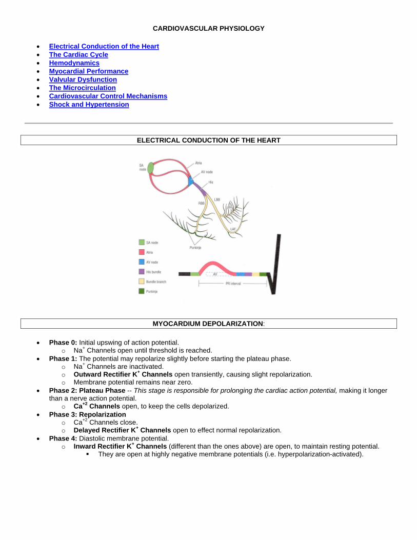
MYOCARDIUM DEPOLARIZATION:
• Phase 0: Initial upswing of action potential. o Na+ Channels open until threshold is reached.
• Phase 1: The potential may repolarize slightly before starting the plateau phase. o Na+ Channels are inactivated. o Outward Rectifier K+ Channels open transiently, causing slight repolarization. o Membrane potential remains near zero.
• Phase 2: Plateau Phase -- This stage is responsible for prolonging the cardiac action potential, making it longer than a nerve action potential.
o Ca+2 Channels open, to keep the cells depolarized. • Phase 3: Repolarization
o Ca+2 Channels close. o Delayed Rectifier K+ Channels open to effect normal repolarization.
• Phase 4: Diastolic membrane potential. o Inward Rectifier K+ Channels (different than the ones above) are open, to maintain resting potential.
They are open at highly negative membrane potentials (i.e. hyperpolarization-activated).
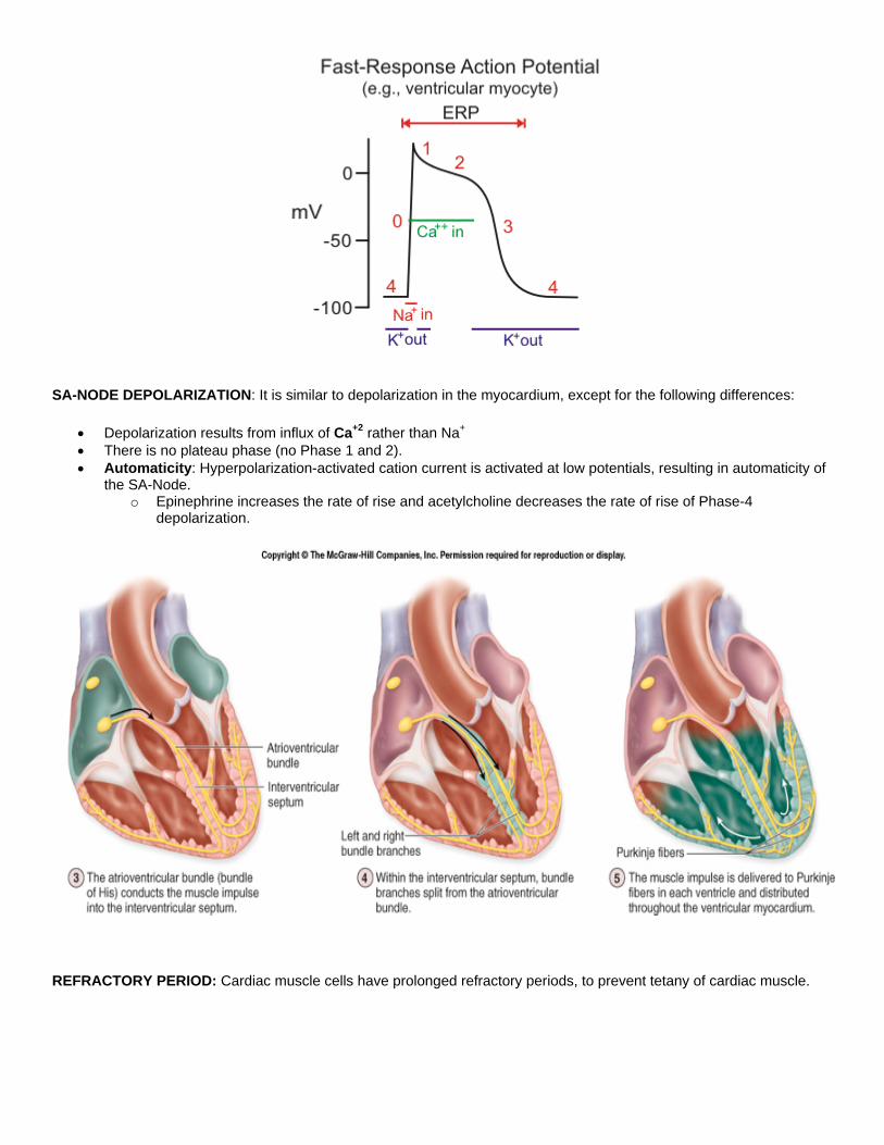
SA-NODE DEPOLARIZATION: It is similar to depolarization in the myocardium, except for the following differences:
• Depolarization results from influx of Ca+2 rather than Na+ • There is no plateau phase (no Phase 1 and 2). • Automaticity: Hyperpolarization-activated cation current is activated at low potentials, resulting in automaticity of
the SA-Node. o Epinephrine increases the rate of rise and acetylcholine decreases the rate of rise of Phase-4
depolarization.
REFRACTORY PERIOD: Cardiac muscle cells have prolonged refractory periods, to prevent tetany of cardiac muscle.
AUTONOMIC REGULATION of HEARTBEAT:
• Acetylcholine slows heart rate by increasing K+ permeability. • Norepinephrine speeds heart rate by increasing the rate of rise of the cardiac action potential during phase 0.
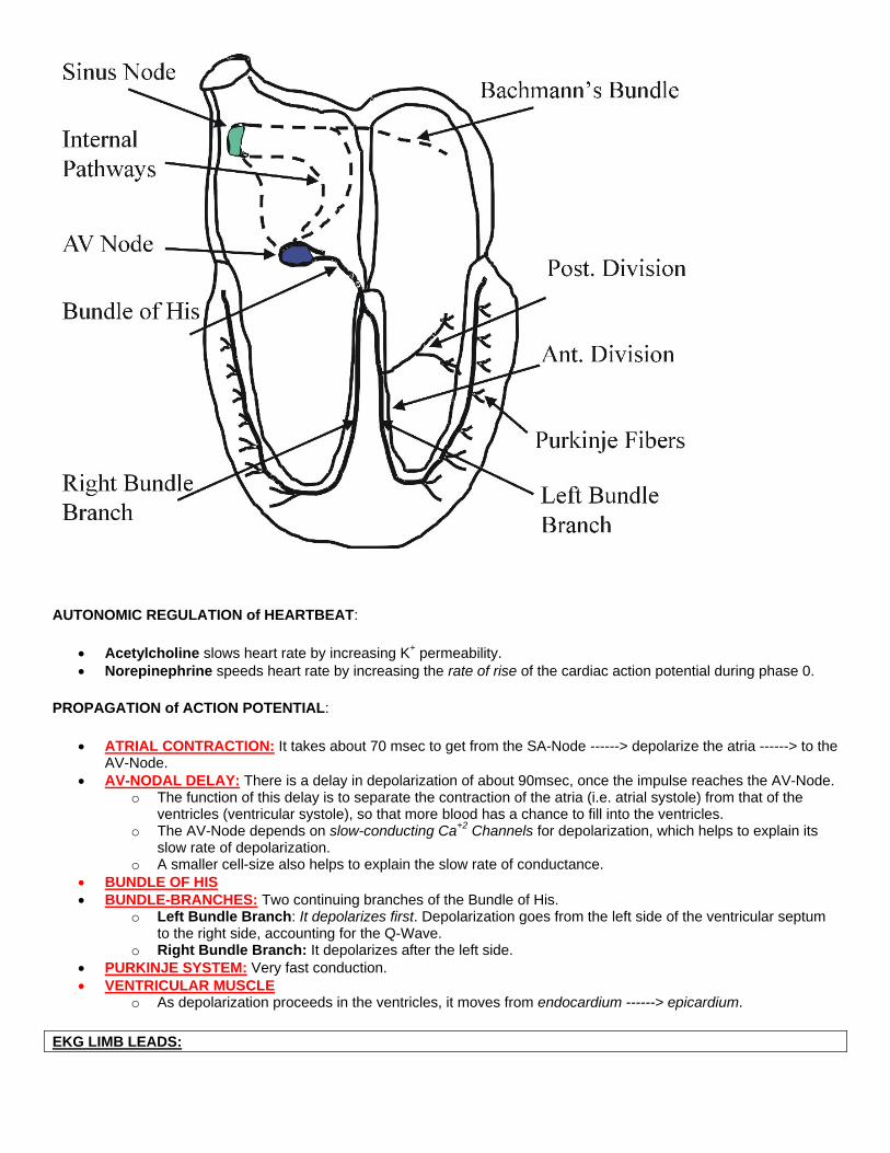
PROPAGATION of ACTION POTENTIAL:
• ATRIAL CONTRACTION: It takes about 70 msec to get from the SA-Node ------> depolarize the atria ------> to the AV-Node.
• AV-NODAL DELAY: There is a delay in depolarization of about 90msec, once the impulse reaches the AV-Node. o The function of this delay is to separate the contraction of the atria (i.e. atrial systole) from that of the
ventricles (ventricular systole), so that more blood has a chance to fill into the ventricles. o The AV-Node depends on slow-conducting Ca+2 Channels for depolarization, which helps to explain its
slow rate of depolarization. o A smaller cell-size also helps to explain the slow rate of conductance.
• BUNDLE OF HIS • BUNDLE-BRANCHES: Two continuing branches of the Bundle of His.
o Left Bundle Branch: It depolarizes first. Depolarization goes from the left side of the ventricular septum to the right side, accounting for the Q-Wave.
o Right Bundle Branch: It depolarizes after the left side. • PURKINJE SYSTEM: Very fast conduction. • VENTRICULAR MUSCLE
o As depolarization proceeds in the ventricles, it moves from endocardium ------> epicardium.
EKG LIMB LEADS:
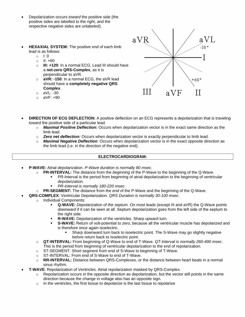
• Depolarization occurs toward the positive side (the positive sides are labelled to the right, and the respective negative sides are unlabeled).
• HEXAXIAL SYSTEM: The positive end of each limb lead is as follows:
o I: 0 o II: +60 o III: +120: In a normal ECG, Lead III should have
a net-zero QRS-Complex, as it is perpendicular to aVR.
o aVR: -150: In a normal ECG, the aVR lead should have a completely negative QRS Complex.
o aVL: -30 o aVF: +90
• DIRECTION OF ECG DEFLECTION: A positive deflection on an ECG represents a depolarization that is traveling toward the positive side of a particular lead.
o Maximal Positive Deflection: Occurs when depolarization vector is in the exact same direction as the limb lead.
o Zero net deflection: Occurs when depolarization vector is exactly perpendicular to limb lead. o Maximal Negative Deflection: Occurs when depolarization vector is in the exact opposite direction as
the limb lead (i.e. in the direction of the negative end).
ELECTROCARDIOGRAM:
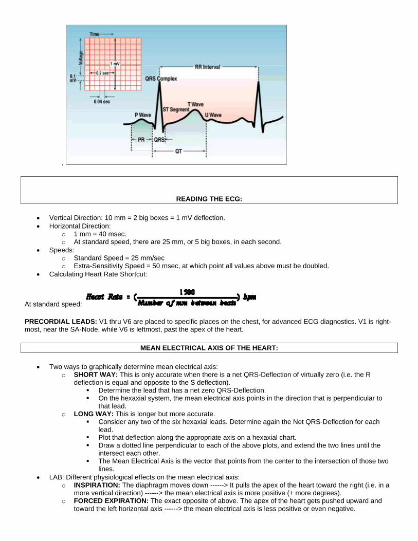
• P-WAVE: Atrial depolarization. P-Wave duration is normally 80 msec. o PR-INTERVAL: The distance from the beginning of the P-Wave to the beginning of the Q-Wave.
PR-Interval is the period from beginning of atrial depolarization to the beginning of ventricular depolarization.
PR-Interval is normally 180-220 msec. o PR-SEGMENT: The distance from the end of the P-Wave and the beginning of the Q-Wave.
• QRS-COMPLEX: Ventricular Depolarization. QRS Duration is normally 30-100 msec. o Individual Components:
Q-WAVE: Depolarization of the septum. On most leads (except III and aVR) the Q-Wave points downward if it can be seen at all. Septum depolarization goes from the left side of the septum to the right side.
R-WAVE: Depolarization of the ventricles. Sharp upward turn. S-WAVE: Return of volt-potential to zero, because all the ventricular muscle has depolarized and
is therefore once again isoelectric. Sharp downward turn back to isoelectric point. The S-Wave may go slightly negative
before return back to isoelectric point. o QT-INTERVAL: From beginning of Q-Wave to end of T-Wave. QT-Interval is normally 260-490 msec.
This is the period from beginning of ventricular depolarization to the end of repolarization. o ST-SEGMENT: Short segment from end of S-Wave to beginning of T-Wave. o ST-INTERVAL: From end of S-Wave to end of T-Wave. o RR-INTERVAL: Distance between QRS-Complexes, or the distance between heart beats in a normal
sinus rhythm. • T-WAVE: Repolarization of Ventricles. Atrial repolarization masked by QRS-Complex.
o Repolarization occurs in the opposite direction as depolarization, but the vector still points in the same direction because the change in voltage also has an opposite sign.
o In the ventricles, the first tissue to depolarize is the last tissue to repolarize
.
READING THE ECG:
• Vertical Direction: 10 mm = 2 big boxes = 1 mV deflection. • Horizontal Direction:
o 1 mm = 40 msec. o At standard speed, there are 25 mm, or 5 big boxes, in each second.
• Speeds: o Standard Speed = 25 mm/sec o Extra-Sensitivity Speed = 50 msec, at which point all values above must be doubled.
• Calculating Heart Rate Shortcut:
At standard speed:
PRECORDIAL LEADS: V1 thru V6 are placed to specific places on the chest, for advanced ECG diagnostics. V1 is right-most, near the SA-Node, while V6 is leftmost, past the apex of the heart.
MEAN ELECTRICAL AXIS OF THE HEART:
• Two ways to graphically determine mean electrical axis: o SHORT WAY: This is only accurate when there is a net QRS-Deflection of virtually zero (i.e. the R
deflection is equal and opposite to the S deflection). Determine the lead that has a net zero QRS-Deflection. On the hexaxial system, the mean electrical axis points in the direction that is perpendicular to
that lead. o LONG WAY: This is longer but more accurate.
Consider any two of the six hexaxial leads. Determine again the Net QRS-Deflection for each lead.
Plot that deflection along the appropriate axis on a hexaxial chart. Draw a dotted line perpendicular to each of the above plots, and extend the two lines until the
intersect each other. The Mean Electrical Axis is the vector that points from the center to the intersection of those two
lines. • LAB: Different physiological effects on the mean electrical axis:
o INSPIRATION: The diaphragm moves down ------> It pulls the apex of the heart toward the right (i.e. in a more vertical direction) ------> the mean electrical axis is more positive (+ more degrees).
o FORCED EXPIRATION: The exact opposite of above. The apex of the heart gets pushed upward and toward the left horizontal axis ------> the mean electrical axis is less positive or even negative.
o PREGNANCY: The mean electrical axis would deviate to the left, within normal limits. The physical presence of the fetus would push up the diaphragm ------> heart leans toward left.
o LEFT VENTRICULAR HYPERTROPHY: Mean axis deviation toward the left. o Pulmonary Valve Stenosis: If we assume that it leads to Right Ventricular Hypertrophy ------> Then we get
(potentially severe) right axis deviation. o INFANCY: Right Axis Deviation, because the infant's right ventricle and left ventricle musculature are
about the same size at birth. Left ventricle becomes larger within a couple months. • NORMAL MEAN AXIS: Anywhere between -30 and +110.
o Anything negative of -30 is left axis deviation, as occurs from left ventricular hypertrophy. o Anything positive of +110 is right axis deviation, as occurs from right ventricular hypertrophy.
ECG ABNORMALITIES:
• SINUS BRADYCARDIA: A heart rate slower than 60 SA-Nodal depolarizations per minute. "Sinus" indicates that the cardiac impulse is originating from the SA-Node as normal.
• SINUS TACHYCARDIA: Heart rate faster than 100 bpm, originating as normal from the SA-Node. o Tachycardia generally means you'll see a shorter RR-Interval (i.e. faster heart rate).
• SINUS ARREST: No SA-Node depolarization. o This can be artificially induced by carotid massage, which results in overstimulation of the Vagus ------>
SA-Node hyperpolarized. • ATRIAL PAROXYSMAL TACHYCARDIA: Faster heart rate resulting from an ectopic pacemaker in the atrial
muscle. o In the example the P-Wave points downward because the atrial depolarization starts in the LA, because
that is where the tissue is leaky. • BUNDLE-BRANCH BLOCKS: There is some conduction block in the Bundle of His (Left or Right Bundle
branches), with results as below: o 1 BLOCK: Partial block. The PR-Interval is longer than normal because it takes longer to conduct the
impulse from SA-Node to AV-Node. o 2 BLOCK: A QRS-Complex occurs only after every other P-Wave. In other words, it takes two P-Waves
to sufficiently excite the AV-Node to conduct the impulse to the ventricles. o 3 BLOCK: There is no temporal relationship between the P-Wave and QRS-Complex. Atrial and
ventricular depolarizations are being controlled by their own independent pacemakers (the SA-Node and AV-Node respectively).
• AV-NODAL TACHYCARDIA: Tachycardia, plus the P-Wave is insignificant or absent. o This is tachycardia, where the impulse originates from the AV-Node. The inherent pacemaker of the AV-
Node is faster than the SA-Node. • PREMATURE VENTRICULAR CONTRACTION (PVC): A premature QRS-Complex, or one that occurs without
being preceded by a P-Wave. o That means that the P-Wave didn't start the impulse, but it started somewhere else. o Ectopic Pacemaker: With PVC, the impulse originates in the ventricular muscle itself, due to leaky
membranes in the muscle. • VENTRICULAR FIBRILLATION: Waves of depolarization traveling in multiple directions all over the ventricular
muscle. The pacemaker activity is lost. • ATRIAL FIBRILLATION: Fibrillation in the atria is not serious in children, but it is serious in old people.
o That's because in old people, atrial systole contributes a greater relative blood volume to cardiac output than in children.
CLINICAL LECTURE: WOLF-PARKINSON-WHITE SYNDROME
• Normally, the AV-Node is the only pathway for conduction of the impulse from the atria to the ventricles. o Bachman's Bundle: Normally conducts the impulse from Right Atrium to Left Atrium during atrial systole. o Moderator Band: Normally conducts the impulse from the right ventricular septal wall to the right free
wall during ventricular systole. • Lupus Erythematosus: Rare condition associated with pediatric bradycardia. Usually pediatric heart problems
result in Tachycardia -- not bradycardia. • PEDIATRIC TACHYCARDIAS: They are divided into two types
o Supraventricular Tachycardia (SVT): One where the problem originates somewhere in the AV-System. o Ventricular Tachycardia (VT): Problem originates in the ventricular system.
• Wolf-Parkinson-White Syndrome: Extra conductive tissue in the myocardium, creating an accessory pathway for conduction from atria to ventricles.
o This accessory pathway ultimately results in a Reentry Tachycardia, or a conduction loop between the normal and accessory pathways.
o The Wolf-Parkinson-White ECG: Shorter PR-Interval due to rapid conduction of signal to ventricles through accessory pathway.
This is the ECG when the patient is healthy and no problems are going on. The P-Wave and the QRS-Complex are scrunched together, creating the appearance of a delta-
wave (hump right before QRS), and a longer overall QRS Complex. • Reentry Tachycardia: You get it from a unidirectional block in one pathway, coupled with slowed conduction of
an alternative pathway. This results in continuous impulse conduction, or circus dysrhythmia. o With WPW, the accessory pathway can get blocked because it hasn't had the time to repolarize, then the
normal pathway provides a mean for retrograde conduction of depolarization. o This results in a conduction loop and severe tachycardia.
• TREATMENT: Slow down the conduction through one pathway or the other. o Use Ca+2-Channel Blockers (such as Verapamil) o Use Digoxin to increase AV-Nodal sensitivity to ACh. o Use beta-Blockers to block the normal NorE sympathetic receptors on the AV-Node and cardiac muscle. o In severe cases, surgically remove the conductive tissue from the myocardium.
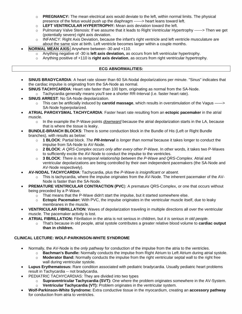
THE CARDIAC CYCLE
VENTRICULAR DIASTOLE:
• ISOVOLUMIC RELAXATION: The very beginning of diastole, right after the aortic valve closes, during which both valves are closed.
o The ventricular muscle is relaxing as ventricular pressure rapidly decreases. o Volume remains constant.
• THREE PHASES OF VENTRICULAR FILLING: o RAPID VENTRICULAR FILLING:
The Mitral Valve opens, when ventricular pressure falls below atrial pressure. Blood rushes into ventricle, very quickly initially. THIRD HEART SOUND (S3): It is turbulent blood flowing past the ventricular wall during early
diastole. It is indicative of pathology. o SLOW VENTRICULAR FILLING: The later period of diastole. The majority of blood has already entered
the ventricles. o TOP-OFF PHASE: The blood contributed to ventricles during atrial systole.
• Diastolic Events Associated with the Atria:
o V-Wave: Small increase in atrial pressure associated with the fact that the mitral valve is closed at the very beginning of diastole.
o Y-Descent: Descent of the V-Wave. Decrease in atrial pressure occurring when the mitral valve opens, right after ventricular isovolumic relaxation.
o A-Wave: Small rise in atrial pressure, occurring right before systole, associated with Atrial Systole and cntrxn of atrial muscle.
o FOURTH HEART SOUND (S4): Vibration of mitral valve leaflets during atrial systole, i.e. during the top-off phase of ventricular filling. This occurs concurrent with the A-Wave and is indicative of pathology.
o Atrial Fibrillation: There is an age difference in the seriousness of this. Again, atrial fibrillation isn't a concern with young people but it is with old people.
YOUNG: Atrial systole contributes about 20mL to stroke volume OLD: Atrial systole contributes about 40mL to stroke volume.
o An Increased heart rate makes the atrial contribution to stroke volume more significant. Shorter time for ventricular filling ------> The top-off phase contributes more relative volume to ventricles.
VENTRICULAR SYSTOLE: QRS-Complex occurs and ventricles start contracting.
• FIRST HEART SOUND (S1): The Mitral Valve Closes, as ventricular pressure exceeds atrial pressure. • ISOVOLUMIC CONTRACTION: Period of contraction during which both valves are closed
o Pressure is increasing. o Volume is constant.
• Systolic Events Associated with the Atria: o C-WAVE: Small increase in atrial pressure. Occurs during isovolumic contraction, as the ventricle pushes
the mitral valve a little upward toward the atrium. o X-DESCENT: The decrease in the C-Wave, due to the change of shape of the ventricle from prolate
spheroid (football-like) to spheroid. This makes the mitral valve move down and the atrial pressure return to normal.
• Aortic Valve Opens, as ventricular pressure exceeds aortic pressure. o Ventricle must achieve systolic arterial pressure in order to open the Aortic valve, so it reaches
pressures around 120 mm Hg. • Ventricular Ejection: 70% of blood is ejected in the first third of systole. • SECOND HEART SOUND (S2): The aortic valve closes, as ventricular pressure falls below aortic pressure.
o DICROTIC (AORTIC) NOTCH: When the Aortic Valve closes, there is a temporary retrograde flow of blood against the Aortic valve cusps. This causes an acute decrease in Aortic pressure at the very beginning of diastole.
o Two Things act in concert to make the Aortic valve close: The left ventricle relaxes so left ventricular pressure decreases. The retrograde blood flow against the leaflets actually aids in the closure of the valve.
HEART SOUNDS: Left Side -vs- Right Side:
• FIRST HEART SOUND (S1): o The mitral valve (left side) closes before the tricuspid valve (right side), because the depolarization begins
on the left side of the septum.
o On the other hand, the Aortic Valve (left side) opens a little after the Pulmonary Valve (right side), because there is so much higher volume in the left side, hence more pressure has to build up before valve will open.
SPLIT SECOND HEART SOUND: During inspiration, You should be able to hear the pulmonic and aortic valves close separately during the second heart sound (i.e. a "split" sound).
• Pulmonic Stenosis: In this case the pulmonic valve is not opening well ------> Wide Splitting during inspiration. • Aortic Stenosis: Causes paradoxical splitting -- i.e. splitting occurs during expiration instead of during
inspiration.
HEMODYNAMICS
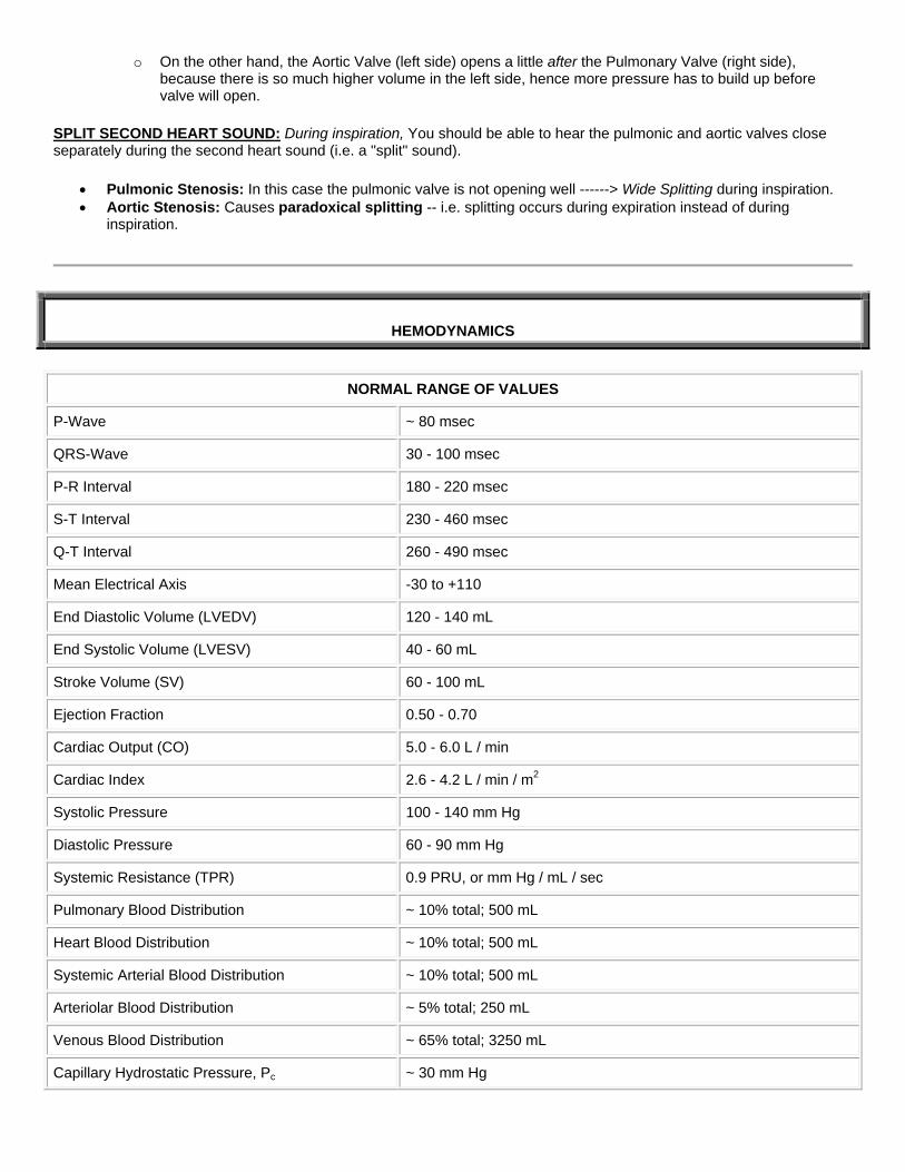
NORMAL RANGE OF VALUES
P-Wave ~ 80 msec
QRS-Wave 30 - 100 msec
P-R Interval 180 - 220 msec
S-T Interval 230 - 460 msec
Q-T Interval 260 - 490 msec
Mean Electrical Axis -30 to +110
End Diastolic Volume (LVEDV) 120 - 140 mL
End Systolic Volume (LVESV) 40 - 60 mL
Stroke Volume (SV) 60 - 100 mL
Ejection Fraction 0.50 - 0.70
Cardiac Output (CO) 5.0 - 6.0 L / min
Cardiac Index 2.6 - 4.2 L / min / m2
Systolic Pressure 100 - 140 mm Hg
Diastolic Pressure 60 - 90 mm Hg
Systemic Resistance (TPR) 0.9 PRU, or mm Hg / mL / sec
Pulmonary Blood Distribution ~ 10% total; 500 mL
Heart Blood Distribution ~ 10% total; 500 mL
Systemic Arterial Blood Distribution ~ 10% total; 500 mL
Arteriolar Blood Distribution ~ 5% total; 250 mL
Venous Blood Distribution ~ 65% total; 3250 mL
Capillary Hydrostatic Pressure, Pc ~ 30 mm Hg
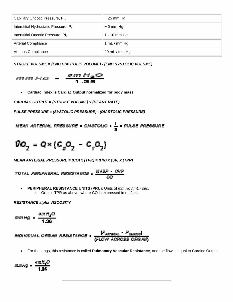
Capillary Oncotic Pressure, PIp ~ 25 mm Hg
Interstitial Hydrostatic Pressure, Pi ~ 0 mm Hg
Interstitial Oncotic Pressure, PIi 1 - 10 mm Hg
Arterial Compliance 1 mL / mm Hg
Venous Compliance 20 mL / mm Hg
STROKE VOLUME = (END DIASTOLIC VOLUME) - (END SYSTOLIC VOLUME)
• Cardiac Index is Cardiac Output normalized for body mass.
CARDIAC OUTPUT = (STROKE VOLUME) x (HEART RATE)
PULSE PRESSURE = (SYSTOLIC PRESSURE) - (DIASTOLIC PRESSURE)
MEAN ARTERIAL PRESSURE = (CO) x (TPR) = (HR) x (SV) x (TPR)
• PERIPHERAL RESISTANCE UNITS (PRU): Units of mm Hg / mL / sec. o Or, it is TPR as above, where CO is expressed in mL/sec.
RESISTANCE alpha VISCOSITY
• For the lungs, this resistance is called Pulmonary Vascular Resistance, and the flow is equal to Cardiac Output.
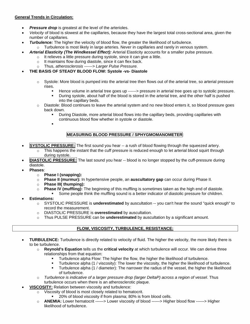
General Trends in Circulation:
• Pressure drop is greatest at the level of the arterioles. • Velocity of blood is slowest at the capillaries, because they have the largest total cross-sectional area, given the
number of capillaries. • Turbulence: The higher the velocity of blood flow, the greater the likelihood of turbulence.
o Turbulence is most likely in large arteries. Never in capillaries and rarely in venous system. • Arterial Elasticity (The Windkessel Effect): Arterial Elasticity accounts for a smaller pulse pressure.
o It relieves a little pressure during systole, since it can give a little. o It maintains flow during diastole, since it can flex back. o Thus, atherosclerosis ------> Larger Pulse Pressure.
• THE BASIS OF STEADY BLOOD FLOW: Systole -vs- Diastole
o Systole: More blood is pumped into the arterial tree then flows out of the arterial tree, so arterial pressure rises.
Hence volume in arterial tree goes up ------> pressure in arterial tree goes up to systolic pressure. During systole, about half of the blood is stored in the arterial tree, and the other half is pushed
into the capillary beds. o Diastole: Blood continues to leave the arterial system and no new blood enters it, so blood pressure goes
back down. During Diastole, more arterial blood flows into the capillary beds, providing capillaries with
continuous blood flow whether in systole or diastole.
MEASURING BLOOD PRESSURE / SPHYGMOMANOMETER:
• SYSTOLIC PRESSURE: The first sound you hear -- a rush of blood flowing through the squeezed artery. o This happens the instant that the cuff pressure is reduced enough to let arterial blood squirt through
during systole. • DIASTOLIC PRESSURE: The last sound you hear -- blood is no longer stopped by the cuff-pressure during
diastole. • Phases:
o Phase I (snapping): o Phase II (murmur): In hypertensive people, an auscultatory gap can occur during Phase II. o Phase III( thumping): o Phase IV (muffling): The beginning of this muffling is sometimes taken as the high end of diastole.
Some people think the muffling sound is a better indicator of diastolic pressure for children. • Estimations:
o SYSTOLIC PRESSURE is underestimated by auscultation -- you can't hear the sound "quick enough" to record the measurement.
o DIASTOLIC PRESSURE is overestimated by auscultation. o Thus PULSE PRESSURE can be underestimated by auscultation by a significant amount.
FLOW, VISCOSITY, TURBULENCE, RESISTANCE:
• TURBULENCE: Turbulence is directly related to velocity of fluid. The higher the velocity, the more likely there is to be turbulence.
o Reynold's Equation tells us the critical velocity at which turbulence will occur. We can derive three relationships from that equation:
Turbulence alpha Flow: The higher the flow, the higher the likelihood of turbulence. Turbulence alpha (1 / viscosity): The lower the viscosity, the higher the likelihood of turbulence. Turbulence alpha (1 / diameter): The narrower the radius of the vessel, the higher the likelihood
of turbulence. o Turbulence is indicative of a larger pressure drop (larger DeltaP) across a region of vessel. Thus
turbulence occurs when there is an atherosclerotic plaque. • VISCOSITY: Relation between viscosity and turbulence:
o Viscosity of blood is most closely related to hematocrit. 20% of blood viscosity if from plasma; 80% is from blood cells.
o ANEMIA: Lower hematocrit ------> Lower viscosity of blood ------> Higher blood flow ------> Higher likelihood of turbulence.
• FLOW: Relation between flow and radius = flow is inversely proportional to r4. • RESISTANCE: The resistance to any organ is greater than the sum of all resistances!
o That's true because the vessels are wired in parallel, and the sum of resistances in parallel is less than its individual parts.
o Systemic Resistance (TPR) is much greater than Pulmonary Resistance. o Pulmonary Resistance = Delta Pulmonary Pressures / CO.
BRUIT: Turbulent flow is detected as a bruit which can be heard by the stethoscope.
• Innocent Ejection Murmur: Children can have high velocity of blood flow without there being any pathology. Bruits are not uncommon.
• Bruits with Anemia: Anemic patients can also have innocent bruits, for two reasons: o Lower hematocrit ------> lower blood viscosity ------> higher likelihood of turbulence. o Anemics tend to compensate their low hematocrit with a higher cardiac output.
• Atherosclerotic Plaque: Turbulence can be heard downstream from the plaque. o Upstream from Plaque: Greater resistance ------> a strong pulse pressure. o Downstream from Plaque: A bruit can be heard.
STANDING BLOOD PRESSURE: Mean Arterial Pressure goes down when standing, because of lower venous return.
• Stand up ------> Venous Pressure in feet goes up ------> capillary hydrostatic pressure goes up ------> fluid flows out of arterial tree and into tissues ------> venous pooling in the feet ------> venous return decreases ------> CO decreases ------> lower MABP.
o Venous pressure goes up in feet because of gravity -- DeltaP = gh • Skeletal Muscle Pump: Tonic contraction of leg muscles while standing aids venous return, because the veins
have valves, so blood is squeezed in only one direction. o Thus prolonged standing can lead to incompetent valves in the veins in the legs.
BLOOD PRESSURE AND THE RESPIRATORY CYCLE:
• INSPIRATION: Systemic blood pressure goes down and pulmonary blood pressure goes up. o The Diaphragm moving down has two effects:
It increases the volume of thoracic airspace and so it decreases intrathoracic pressure. Also the abdominal space becomes smaller, so it increases intra-abdominal pressure.
o The combination of above two effects results in an increased pressure gradient for venous return from the IVC ------> increased venous return ------> More blood to right atrium and more blood to pulmonary circulation ------> less respective blood in left heart and less CO.
o Thus overall result is the following: Lower systemic pressure. Higher pulmonary pressure. Larger Blood Volume in pulmonary circulation.
o The change in MABP from inspiration normally does not exceed 10 mm Hg. • EXPIRATION: Has the exact opposite effect.
o Pulmonary pressure decreases. o Systemic pressure increases.
CENTRAL VENOUS PRESSURE: The pressure going into the right atrium.
• Anything that decreases venous compliance (i.e. sympathetic tone) will increase venous return ------> Higher CVP.
• ESTIMATING CENTRAL VENOUS PRESSURE: You estimate in cm of water. o It is approximately equal to the distance from the end of the distended part (which you can see) to the
sternal angle, plus 5, then convert it into mm Hg.
PRESSURES IN PERIPHERY -vs- AORTA:
• Mean Arterial Pressure is slightly higher in the Aorta than in, for example, the radial artery. • But, Pulse Pressure is greater in the periphery, i.e. the systolic is higher and the diastolic is lower.
o This effect in the periphery is due to constructive interference of reflected waves.
COMPLIANCE: The degree to which a pressure change leads to a corresponding change in volume. Or, Compliance = DeltaV / DeltaP, or the slope of a pressure-volume curve.
• VENOUS COMPLIANCE is about twenty times more than arterial compliance, therefore veins can hold a larger volume of fluid at lower pressure.
o Arterial Compliance is about 1 mL / mm Hg o Venous Compliance is about 20 mL / mm Hg
• EFFECTS OF COMPLIANCE on Blood Pressure: o Higher Venous Compliance ------> higher capacitance in veins ------> less venous return ------> lower
CVP. o Lower Venous Compliance (sympathetic influence) ------> lower capacitance veins ------> more venous
return via the one-way valves ------> higher CVP. o Lower Arterial Compliance results in a higher pulse pressure.
AGE: Arteries in old people have lower compliance. Thus old people have higher pulse pressures.
• Pressure-Volume Curve: The analysis of old -vs- young can be done on the P/V curve. o The slope of the curve is compliance.
o Pressure is on the X-Axis. Volume is on the Y-Axis. o Is you plot systolic and diastolic pressure, and look at the corresponding Y-Values, you can calculate the
following: The difference on the Y-axis (i.e. the volumes corresponding to systolic and diastolic pressures)
is stroke volume. The difference on the X-axis is pulse pressure.
MODULATION OF MEAN ARTERIAL PRESSURE: Under a lot of circumstances, it doesn't change, even when stroke volume and/or pulse pressures do change.
• EFFECT OF STROKE VOLUME: All other factors held constant, a high stroke volume results in a higher pulse pressure, i.e. higher systolic and lower diastolic, but MABP remains constant.
o PULSE PRESSURE IS USUALLY DIRECTLY RELATED TO STROKE VOLUME • EFFECT OF EXERCISE:
o Increased CO and Stroke Volume o Compensatory lower vascular resistance (TPR) o Once again MABP doesn't change (within limits).
• HIGH SYSTOLIC PRESSURE: Tends to occur with higher stroke volume. The more fluid you pump in one beat, the higher the systolic pressure.
• HIGHER DIASTOLIC PRESSURE: CORRELATES WITH HIGH TPR.
MYOCARDIAL PERFORMANCE
General Effects of Autonomic Control on Heart:
• SYMPATHETICS: o Positive chronotropic effect -- faster heart rate. o Positive inotropic effect -- greater contractility for the same fiber length.
• PARASYMPATHETICS: Negative chronotropic effect, but no inotropic effect.
PRELOAD: The diastolic filling pressure, or end-diastolic volume.
AFTERLOAD: Ventricular systolic pressure, which is equal to arterial systolic pressure under normal circumstances.
LAPLACE'S LAW: The stress on the ventricular wall is proportional to the Ventricular Pressure x Ventricular Radius, where the size of the ventricle is determined by stretching, i.e. by ventricular volume.
STARLING'S LAW OF THE HEART: Within limits, increases in end-diastolic volume result in a corresponding increase in stroke volume. Most simplified, within limits, the volume that comes into the heart goes back out.
• MECHANISM: Increased Filling Volume ------> Stretch Ventricular Muscle ------> Augmented ventricular fiber length ------> greater inotropic state ------> faster velocity of ejection ------> Greater Cardiac Output.
• Increased fiber length results in more forceful contraction, within limits. o Optimal muscle fiber length = 2.2 micron. Heart normally works slightly below this level to give room for
optimal filling.
PRESSURE-VOLUME LOOP: P/V graph, with both diastolic and systolic lines plotted on it. You use this graph to plot the pressure and volume at all points in the cardiac cycle.
• END-SYSTOLIC CURVE: The upper limit to the loop. • END-DIASTOLIC CURVE: The lower limit to the loop. • CARDIAC CYCLE in LOOP:
o DIASTOLE: ISOVOLUMIC RELAXATION: Volume is constant while pressure goes straight down. VENTRICULAR FILLING: Pressure remains constant while volume increases.
o SYSTOLE: ISOVOLUMIC CONTRACTION: Volume constant while pressure goes straight up. EJECTION: Pressure continues to increase as blood is ejected from the ventricle. The end-
pressure at this point is systolic arterial pressure. The pressure continues to rise during systole because pressure is rising in the arterial network.
You are putting more blood into the arterial tree then is being put out on the other side. Ventricle must match that rise in pressure to force blood out.
AORTIC VALVE CLOSES: At the end of systole, the ventricular pressure (i.e. fiber length) decreases to the point that the aortic valve can't stay open, so it closes.
• STROKE WORK: The area of the Pressure-Volume Loop. Mathematically, that means: Stroke Work = (Stroke Volume) x (Mean Arterial Pressure)
o STROKE WORK is equivalent to stroke volume, but it is normalized for differences in blood pressure. Thus it is a good indicator of heart performance.
o Because we have normalized for blood pressure, a shift in the curve for stroke work means that there must be an increase in the inotropic state.
VENTRICULAR FUNCTION CURVE: A comparison of End-Diastolic Volume (or Pressure or Fiber Length) and Stroke Volume (or Stroke Work). The curve is essentially a line that levels off at high values. It is a way of expressing Starling's Law.
• If you plot Stroke Work -vs- LVEDV, you will get the same curve for the same inotropic state, regardless of blood pressure. So using Stroke Work normalizes for blood pressure, and it makes the curve represent the inotropic state.
EFFECT OF PRELOAD ON STROKE-WORK:
• Standing at Rest: The least stroke work is performed. • SUPINE ------> Preload (venous return) increases ------> Fiber-length increases ------>------> Higher Stroke Work. • PRONE, with LEGS RISEN: Even more pronounced effect as above ------> higher stroke work.
EFFECT OF AFTERLOAD ON STROKE VOLUME: A higher afterload ------> Higher systolic pressure must be developed ------> Higher end-systolic volume to achieve that pressure, but the end-diastolic volume remains the same ------> lower stroke volume.
AUTOREGULATION OF AFTERLOAD: Due to heterometric autoregulation, within limits, stroke volume will be maintained even in face of a higher blood pressure, but it takes a few beats for the mechanism to kick in.
• High afterload ------> Lower stroke volume ------> Since pulmonary arterial pressure hasn't changed, the right heart continues to pump the same stroke volume as before ------> Pulmonary blood volume increases ------> Higher venous return back to left atrium ------> Higher preload ------> Higher fiber length + velocity of ejection ------> ------> Stroke volume returns to normal
• But a new pressure-volume curve is carved out on the P/V-Loop. Stroke-work overall has increased. • In compensating for the higher blood pressure, we must use some of our Starling Reserve -- the extra capacity
in the heart to do stroke work, strictly because of the Starling mechanism.
TOTAL RESERVE: The total stored capacity the heart has to do extra stroke work. It is equal to Starling Reserve + Inotropic Reserve + Heart-Rate Reserve.
• STARLING RESERVE: The extent to which we can increase Cardiac Output simple by increasing filling, at the same inotropic state.
• INOTROPIC RESERVE • HEART-RATE RESERVE
INOTROPIC STATE: It's the contractile force in the muscle, at any particular fiber-length. That is the same as the Ca+2 concentration in the sarcomeres.
• It increases stroke volume, DUH??
HEART-RATE AND STROKE VOLUME: Heart rate extremes lead to lower stroke volume.
• Bradycardia: Heart-rate slower than 40. Cardiac Output goes way down because the stroke volume can't increase enough to compensate for the lower heart-rate. You've reached the maximum of the heart's inotropic state.
• Tachycardia: Heart-rate faster than 180. Cardiac Output goes way down because there is no longer enough time between beats for sufficient ventricular filling, i.e. the short diastolic time cuts into the "Fast-Filling Phase" of diastole.
VALVULAR DYSFUNCTION
MITRAL INSUFFICIENCY: Insufficiency means the valve can't stay completely closed, so it is leaky. Mitral Insufficiency causes fluid to reflux into the Left Atrium with each systole, leading to a chronically high end-diastolic volume ------> left-ventricular hypertrophy.
• Holosystolic Murmur can be heard throughout systole, as turbulent blood flows through mitral valve. • Third Heart Sound can be heard during diastole, as there is a large excess of atrial blood ------> turbulent flow
during ventricular filling. • Large V-Wave is seen: Higher atrial pressure produced during diastole, because there is higher atrial volume.
MITRAL STENOSIS: Leads to lower filling of the left atrium, as the system backs up. This leads to overload of blood in the pulmonary system.
• High Pulmonary Arterial Pressure from backup of blood. o Pulmonary Edema is a likely complication that can result from the pulmonary hypertension. o Right Ventricular Hypertrophy also commonly comes from high Pulmonary hypertension.
• Heart Sounds: o Pre-Systolic Crescendo Murmur is diagnostic of mitral stenosis. The murmur results from large
increases of pressure during atrial systole, because of the mitral stenosis. o Diastolic (S3) Decrescendo Murmur is also heard, as there is a large pressure difference between
atrium and ventricle during diastole. That pressure difference then becomes smaller (i.e. quieter) as the ventricle fills and the atrium empties.
AORTIC INSUFFICIENCY: Regurgitation back into left-ventricle, on each systole, leads to severe left-ventricular hypertrophy (when the insufficiency is severe).

• Dangerously Large Pulse Pressure results from high systolic pressure (due to compensatory mechanism / inotropic state), and markedly decreased diastolic pressure (due to low stroke volume).
• High LVEDV ------> Left-Ventricular Hypertrophy which can be severe. • Heart Sounds:
o Loud Holo-Diastolic Decrescendo Murmur.
AORTIC STENOSIS: Very common in old people.
• Severe Left Ventricular Hypertrophy. The stenosis results in left ventricular pressure being a lot higher then aortic pressure.
• HEART SOUND: Diamond-Shaped Pansystolic Murmur -- i.e. diamond-shape = crescendo then decrescendo.
THE RIGHT HEART: Tricuspid and Pulmonic Valve problems are similar to those found in the left heart.
MEASUREMENT OF CARDIAC OUTPUT (Last few pages of handout):
• Direct Fick Method: You calculate blood flow through the lungs (rate of O2 uptake) to determine the pulmonary flow. Then you assume that pulmonary blood flow is equal to systemic blood flow (i.e. CO).
o This assumption is true as long as there are no intracardiac shunts. • Indirect Fick (Thermal Dilution) Method: Calculation blood flow essentially by measuring the time that it takes
for the flow of blood to neutralize a temperature difference between injected saline and body temp.
THE MICROCIRCULATION
CAPILLARY FILTRATION AND RESORPTION: STARLING PRINCIPLE FOR CAPILLARY EXCHANGE:
• Filtration: Blood leaving capillary and entering organ. Net flow outward. o Pc, capillary hydrostatic pressure contributes to this outflow. o PIi, interstitial oncotic pressure contributes to this outflow. It is the oncotic (osmotic) pressure created
by insoluble proteins in the interstitial space. • Absorption: Blood leaving organ and entering capillary. Net flow into capillary.
o Pi, interstitial hydrostatic pressure, does not contribute to absorption under normal circumstances. It is ~ 0.
o PIp, capillary oncotic pressure, is the primary contributor to resorption. This is the osmotic pressure created by insoluble proteins in the blood.
• sigma, REFLECTION COEFFICIENT: It is equal to the percentage of proteins that are impermeable to the capillary membrane, i.e. a value between 0 and 1.
o sigma = 1: Proteins are totally impermeable; all of them are "reflected" off the membrane, thus oncotic pressure has the greatest influence possible on net filtration.
o sigma = 0: Proteins are completely permeable, hence no proteins are impermeable and oncotic pressures become zero.
Low reflection coefficient affects resorption but not so much filtration, since the capillary oncotic pressure is the only significant force for resorption.
RESULT: Edema. • Lymphatics: Under normal circumstances, filtration is greater than absorption. Thus more blood is being
deposited in organ systems than is being taken up. The difference is put back into the blood through the lymphatic system.
o The entire blood circulation is turned around through the lymphatic system every 24 hrs.

• Capillary Hydrostatic Pressure: o Ra = arterial resistance is normally much larger than venous resistance. We can usually safely ignore
venous resistance in the calculation. o Pv = venous pressure o Pa = mean arterial pressure
• INFLUENCES ON FILTRATION:
o ARTERIOLAR RESISTANCE: Note that local(arteriolar) changes to vascular tone have an exact opposite effect as systemic (large artery) changes.
All things constant, increased arterial resistance ------> lower capillary hydrostatic pressure + lower filtration
Vasoconstriction or dilation at the level of the arterioles does not affect MABP. Arteriolar Vasoconstriction ------> ------> lower filtration rate. Arteriolar Vasodilation ------>------> higher filtration rate.
o VENOUS PRESSURE: Increased Venous Pressure ------> Higher Capillary Hydrostatic Pressure ------>
Increased Net Filtration, because of the hydrostatic pressure equation: The capillary pressure must increase in order to achieve filtration in face of the increased venous
pressure. o ONCOTIC PRESSURE: Negative nitrogen balance or protein malnutrition (kwashiorkor) will lead to low
plasma albumin ------> low plasma oncotic pressure ------> low or no resorption, which means high net rate of filtration ------> edema, ascites
CAPILLARY PERMEABILITY:
• Three types of capillaries, each having different levels of permeability: o Continuous: Tight Junctions, as in brain, thymus, retina. o Fenestrated: Little diaphragms where diffusion can take place, having somewhat higher permeability. GI-
Tract. o Discontinuous: Liver sinusoids, complete discontinuities in the system.
• Endothelial Cells: When endothelial cells contract, the spaces between them increase ------> higher capillary permeability.
o Mast Cell Degranulation leads to release of Histamine and Platelet-Activating Factor (PAF). o This makes the endothelial cell release Calcium from the SR ------> actin-myosin contraction of
endothelial cell makes the cell change shape ------> more spaces between the cells. • Anaphylactic Shock: High capillary permeability leads to low blood pressure. We can't just give them fluids to
increase blood volume, because the fluids leak right out again.
EDEMA: It can occur from a lot of sources, such as no resorption. Consequences of edema:
• Impair Exchange of Metabolites: It leads to bigger spaces (longer distance) between capillaries and the tissues ------> diffusion becomes impossible.
• THE EDEMA POSITIVE-FEEDBACK CYCLE: Edema can compress venules ------> higher venous return ------> higher CVP ------> higher hydrostatic pressure and filtration rate ------> even more edema.
LYMPHATIC BLOCKAGE: If you block lymphatics, then interstitial fluid along with interstitial proteins will rise ------> increase interstitial oncotic pressure ------> more filtration ------> massive edema.
VASCULAR SMOOTH MUSCLE: Anything that increases intracellular Ca+2 concentration will increase contractility of vascular muscle.
• VASCULAR CONTRACTION: o Mechanism of Contraction, briefly:
Calcium binds to Calmodulin The Ca+2-Calmodulin Complex then binds to Myosin Light-Chain Kinase This results in myosin being free to interact with actin.
o Vascular Tone: The overall rate of cross bridging is much slower than in vascular smooth muscle. There is always a baseline level of activity = vascular tone.
o Norepinephrine will cause vascular contraction by increasing Ca+2 in smooth muscle, via three pathways:
NorE can directly open Ca+2-Channels NorE can bind to alpha1-Receptors to active the alpha-Adrenergic Pathway (DAG/IP3) ------>
higher intracellular Ca+2 Voltage-Gated Ca+2 Channels can further open, in response to the above two.
• VASCULAR RELAXATION: Anything that decreases Ca+2 concentration will cause relaxation. o Epinephrine in the blood causes vascular relaxation.
Epi binds beta2-Receptors to activate beta-Adrenergic Pathway ------> higher levels of cAMP which results in decreased Ca+2 in cytosol.
cAMP will facilitate pumping of Ca+2 back into SR. o ATP: Low levels of ATP will cause vascular relaxation locally, which should allow greater blood flow,
greater perfusion, and hence more ATP to deprived tissue. ATP-Dependent K+-Channels open in response to LOW ATP. This leads to Hyperpolarization ------> Vascular Relaxation ------> greater blood flow to area.
o NO causes relaxation, covered later. • VASOMOTION: Spontaneous action potentials can cause a cyclic change in vascular tone.
o Addition of NorEpi increases the rate of firing of those action potentials ------> more vascular tone. o However, action potentials are not always required to cause sustained contraction.
ENDOTHELIAL-DERIVED FACTORS: Nitric Oxide
• EXPT: Acetylcholine's (i.e. parasympathetic) effect on vessels depends on the presence of the endothelial cells. o Add Ach to vessel with endothelial cells intact ------> relaxation. o Add Ach to vessel with endothelial cells removed ------> actually leads to contraction!
• Process of NO-Mediated Vascular Relaxation: o Ach binds to endothelial cell. o Ca+2 channels open and Ca+2 pours into endothelium. o This makes the endothelial cell produce NO from Arginine, by up-regulating synthesis of the enzyme
Constitutive NO-Synthase. o Endothelium makes NO which diffuses to the underlying vascular smooth muscle. o NO then activates Guanylyl Cyclase, which produces cGMP ------> leads to Ca+2 sequestration and
vascular relaxation. • L-Nitroarginine Methyl Ester (L-NAME): Inhibits NO-Synthase, blocking production of NO ------> arteriolar
constriction ------> lower blood flow to region. • ISCHEMIA-REPERFUSION: The danger in reperfusing ischemic tissue is that massive influx of O2 can lead to
oxidative free radicals which damage endothelial cells. The free radicals have two bad effects: o They react with NO, leading to vasoconstriction and reduced perfusion of the area. o They directly damage the endothelial membrane leading to increased vascular permeability which isn't
good (it can lower blood pressure, etc.) • SEPSIS: Causes vesicle to become less sensitive to vasoconstriction. Phenylephrine has a lesser effect on septic
vessels. o It leads to higher NO via Inducible NO-Synthase. This is not the same enzyme as constitutive NO-
Synthase. o The number of vasoconstrictive alpha-Receptors is decreased. o Basal Ca+2 levels are reduced or Ca+2-channels don't open properly.
• ADHESION MOLECULES: NO protectively prevents expression of adhesion molecules, so that leucocytes don't stick to vessel wall, which can lead to microvascular injury.
o Hence we can't use L-NAME as a treatment for Sepsis -- we need the NO to prevent sticking of blood cells, even if vasodilation is an undesired effect.
o What we need is a drug that blocks only Inducible NO-Synthase (made during sepsis) and not constitutive NO-Synthase. We don't have that (yet).
ENDOTHELIN-1: Vasoconstrictive agent produced by endothelial cells.
• SLOW-RESPONSE: Endothelin is not stored in vesicles. It is synthesized de novo. Thus it is a slow (long-term) response.
o SYNTHESES: Preproendothelin ------> Big Endothelin ------> Endothelin. Multi step synthesis adds to slow response.
• EFFECT: Endothelin causes sustained vasoconstriction. The effect lasts long! It causes increased levels of Ca+2 and thus increased vascular tone.
o It acts via alpha-adrenergic pathway (PIP/DAG ------> Ca+2) o It also acts directly on Ca+2-Channels.
• ISCHEMIA REPERFUSION: Endothelin is bad! It can be released along with inflammatory mediators to cause further vasoconstriction when we want vasodilation.
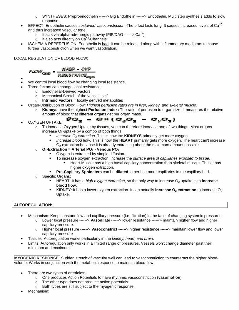
LOCAL REGULATION OF BLOOD FLOW:
• • We control local blood flow by changing local resistance. • Three factors can change local resistance:
o Endothelial-Derived Factors o Mechanical Stretch of the vessel itself o Intrinsic Factors = locally derived metabolites
• Organ-Distribution of Blood Flow: Highest perfusion rates are in liver, kidney, and skeletal muscle. o Kidneys have the highest Perfusion Index: The ratio of perfusion to organ size. It measures the relative
amount of blood that different organs get per organ mass.
• OXYGEN UPTAKE: o To increase Oxygen Uptake by tissues, you can therefore increase one of two things. Most organs
increase O2-uptake by a combo of both things. Increase O2 extraction. This is how the KIDNEYS primarily get more oxygen. Increase blood flow. This is how the HEART primarily gets more oxygen. The heart can't increase
O2-extraction because it is already extracting about the maximum amount possible. o O2-Extraction = Arterial PO2 - Venous PO2
Oxygen is extracted by simple diffusion. To increase oxygen extraction, increase the surface area of capillaries exposed to tissue.
Heart-Muscle has a high basal capillary concentration than skeletal muscle. Thus it has higher oxygen extraction.
Pre-Capillary Sphincters can be dilated to perfuse more capillaries in the capillary bed. o Specific Organs:
HEART: It has a high oxygen extraction, so the only way to increase O2 uptake is to increase blood flow.
KIDNEY: It has a lower oxygen extraction. It can actually increase O2 extraction to increase O2-Uptake.
AUTOREGULATION:
• Mechanism: Keep constant flow and capillary pressure (i.e. filtration) in the face of changing systemic pressures. o Lower local pressure ------> Vasodilate ------> lower resistance ------> maintain higher flow and higher
capillary pressure. o Higher local pressure ------> Vasoconstrict ------> higher resistance ------> maintain lower flow and lower
capillary pressure • Tissues: Autoregulation works particularly in the kidney, heart, and brain. • Limits: Autoregulation only works in a limited range of pressures. Vessels won't change diameter past their
minimum and maximum.
MYOGENIC RESPONSE: Sudden stretch of vascular wall can lead to vasoconstriction to counteract the higher blood-volume. Works in conjunction with the metabolic response to maintain blood flow.
• There are two types of arterioles: o One produces Action Potentials to have rhythmic vasoconstriction (vasomotion) o The other type does not produce action potentials. o Both types are still subject to the myogenic response.
• Mechanism:

o AP-Capable Arterioles: Stretch ------> increased frequency of AP-firing ------> higher vascular tone. o AP-Incapable Arterioles: Stretch ------> depolarization of vascular smooth muscle ------> Ca+2 influx and
higher vascular tone.
METABOLIC RESPONSES: Works in conjunction with the Myogenic Response to maintain blood flow.
• METABOLIC HYPOTHESIS: Vasodilator Metabolites are made locally in response to hypoxia and poor blood flow, in order to increase blood flow. The metabolites are then washed away when blood flow increases again, disposing of their effect.
• HYPOXIA: Hypoxia leads to a decrease in intracellular ATP, which ultimately leads to vasodilation. o K+-ATP CHANNELS: They kick K+ out of the cell in exchange for bringing ATP in. They open in response
to low ATP levels. o Hypoxia ------> low intracellular ATP ------> Open K+-ATP Channels ------> K+ pours out of cell ------>
membrane hyperpolarizes ------> smooth muscle relaxation. o PROSTACYCLIN (PGI2): Prostacyclin may be released by endothelial cells in response to hypoxia ------>
potent vasodilation in a paracrine manner no neighboring smooth muscle. • ACIDOSIS: Acidosis in smooth muscle directly causes hyperpolarization of smooth muscle membrane ------>
vasodilation. o Acidosis means CO2 levels in tissue are high. o CO2 <====> H2CO3 <====> H+ + HCO3
- • ADENOSINE: Adenosine is an indicator that the target tissue is out of ATP (as opposed to the smooth muscle
itself). o Adenosine is membrane-soluble while ATP, ADP, and AMP are not. So when the compounds gets down
to the Adenosine level, it can then leave the cell to affect the neighboring smooth muscle. o Adenosine is a potent vasodilator.
• AUTOCOIDS: Histamine, Bradykinin, Serotonin, Prostaglandins, Leukotrienes. • POTASSIUM: Potassium regulation is especially important in the brain and in skeletal muscle.
o SMALL AMOUNTS OF K+ In both tissues, extracellular K+ concentration goes up because of repeated firing of action
potentials. This results in release of vasodilator-factors (NO, PGI2) and in membrane hyperpolarization of
vascular smooth muscle. o HUGE (PHARMACOLOGICAL) INCREASE IN K+ ------> depolarization of muscle membrane ------>
vasoconstriction. • INTERSTITIAL OSMOLARITY:
ACTIVE HYPEREMIA: Blood flow changes in proportion to changes in metabolic activity of the organ. Occurs in Skeletal Muscle.
• Lactic Acidosis in skeletal muscle ------> Vasodilation of vasculature. • In Active Hyperemia, the metabolic activity of the target tissue (i.e. skeletal muscle) is changing, and that's what
causing the vasodilation.
REACTIVE HYPEREMIA: The short-term increase in flow following temporary ischemia to a region.
• Both myogenic and metabolic effects are playing a role in causing the vasodilation. • In reactive hyperemia, the metabolic activity of the target tissue does not change, whereas in active hyperemia, it
does.
REGIONAL CIRCULATIONS:
• CEREBRAL CIRCULATION: o Cerebrospinal Fluid: Normally has a lower protein content than blood. o Cerebral Vasculatures have very poor sympathetic innervation. Hence in the Cushing Reflex, massive
sympathetics don't cause constriction of vessels in the cerebrum (which they shouldn't!) o Regulation of Flow: It is primarily K+-Mediated. We can get higher extracellular K+ and vascular
hyperpolarization by two sources: Firing of neurons without repolarization. K+-ATPase kicks out K+ in exchange for ATP, at low intracellular ATP levels.
o The brain is very sensitive to changes in PCO2, but not so much to changes in PO2.
• CORONARY CIRCULATION: Regulated almost entirely by local factors. o Increase cardiac work ------> increased coronary blood flow. o SYSTOLE: Coronary blood flow decreases, as the vessels are squeezed as the myocardium contracts.
There may even be some retrograde flow of blood during systole. o DIASTOLE: Coronary blood flow increases.
CARDIOVASCULAR CONTROL MECHANISMS
PARASYMPATHETIC DILATORS: They cause local vascular relaxation. Parasympathetics do not have an important effect on systemic blood pressure.
• Vasoactive Intestinal Peptide (VIP): This neurotransmitter is released directly onto the smooth muscle cells to cause relaxation.
• Nitric Oxide (NO): o The nerve terminals contain Nitric-Oxide Synthase. o NO, when released by nerve terminals, also acts directly on smooth muscle.
NOCICEPTORS: Sensory receptors to noxious chemicals or toxins. They also cause local vasodilation.
• Two peptides are released by Nociceptor Nerves: o Substance P o Calcitonin Gene-Related Peptide (CGRP)
• TRIPLE RESPONSE OF LEWIS: Wheal and flare response to a local irritant. o First, a small red area develops.
This is due to degranulation of mast cells ------> local vasodilation. o Second, a blanched raised area develops around the small red area. o Third, a reddened flare (vasodilation) radiates around the irritated region.
The flare is due to highly branched nociceptor nerves that are distributed through the skin. The Nociceptors release SP and CGRP in the area to cause vasodilation.
• LOCAL NEURAL RESPONSE: The nociceptor reflex does not go through the CNS! o If you cut the Dorsal Root (proximal to the cell body), the neural response still occurs.
This shows that the reflex signal is independent of the CNS. o If you cut the peripheral nerve (distal to the Cell Body), then Wallerian Degeneration occurs and the reflex
no longer happens. • Capsaicin (red-pepper stuff) is an irritant that, if applied to the skin for a period of time, will overuse and numb the
nociceptors. Thus it is a treatment that can prevent the irritant response.
SYMPATHETIC CONTROL OF VASCULAR MUSCLE: This is the primary short-term mediator of TPR and hence arterial blood pressure.
• Sympathetics are of course vasoconstrictive, with two possible exceptions: o beta2-Receptors are vasodilatory. They are most responsive to Epinephrine (which is not a
neurotransmitter), but they are responsive to NorE at huge doses). o Dogs and cats have sympathetic cholinergic nerves (like eccrine sweat glands) that are vasodilatory.
• Norepinephrine: Released from small dense-core vesicles in the sympathetic varicosity. o Norepinephrine is released by any depolarization, of any impulse frequency. o NorE binds to alpha1-Receptors on smooth muscle to increase Ca+2 concentration and effect smooth
muscle contraction. NorE has a very high affinity for alpha1-Receptors. Epinephrine does not.
• ATP: Released from small dense core vesicles in the sympathetic varicosity. o ATP is released by any depolarization. It works at impulse frequencies as low as 2Hz. o ATP binds to Purinoreceptors (P-Receptors) to cause depolarization of the smooth muscle membrane. o Each ATP dense-core vesicle yields +10mV of depolarization. Two simultaneous depolarizations (total of
+20mV) are required to generate smooth muscle action potential. • Neuropeptide-Y (NPY): Released from large dense-core vesicles in the sympathetic varicosity.
o Neuropeptide-Y is only released by repeated depolarizations (i.e. strong sympathetic stimulation). It is only released if the impulse frequency is 8Hz or faster.
o NPY binds to its own Y-Receptor. • COTRANSMISSION: NorE, ATP, and NPY have additive effects.
o At high impulse frequencies, NPY facilitates the release of additional NorE. o The summation of signals will lead to stronger contraction of vascular smooth muscle, up to a point.
MODULATION OF SYMPATHETIC NEUROTRANSMITTERS: Cotransmission principles are based on increased likelihood that a dense-core vesicle will fuse with the pre-synaptic membrane. The higher the impulse frequency, the more likely that is to occur.
• AUTORECEPTORS: Simple negative feedback. There are receptors for NorE, ATP, and NPY. When the respective hormones bind, they inhibit further release of the neurotransmitter (i.e. they decrease the likelihood of vesicle fusion).
• HETERORECEPTORS: These are pre-synaptic receptors that bind to other substances to inhibit or excite release of neurotransmitters.
o Inhibitory Heteroreceptors: Acetylcholine binds to muscarinic receptors on the sympathetic varicosity to inhibit the release
of NorE. Prostaglandins, Serotonin, and Histamine can all bind to inhibitory heteroreceptors as well.
o Excitatory Heteroreceptors: ANGIOTENSIN II will bind to excitatory receptors to promote further release of NorE ------> vasoconstriction.
AUTONOMIC TONE: Heart rate and vascular tone is determined by the relative amounts of sympathetic and parasympathetic continual stimulation.
• PARASYMPATHETIC TONE: The Vagus Nerve (CN X). o Vagal tone for the heart and abdomen originates from:
Nucleus Ambiguus (NA) Dorsal Motor Nerve of CN X (DMV)
o Parasympathetics have the following general effects on CV-System: They increase venous compliance ------> lower venous return. They indirectly decrease systemic resistance by inhibiting sympathetics ------> lower blood
pressure. Vagal Tone on heart slows down the heart-rate at the SA-Node.
• SYMPATHETIC TONE: o In the brain, sympathetics originate from the C1 AREA, which is the Reticular Formation of the closed
medulla. From there, the pathway is Reticular Formation ------> Intermediolateral Column of Thoracic
spinal cord. o Sympathetics have the following general effects on the CV-System:
They decrease venous compliance ------> higher venous return They directly increase systemic resistance ------> higher blood pressure They indirectly speed heart rate by inhibiting Vagal Tone on the SA-Node.
• Miscellaneous Drugs that Affect Heart Rate: o Chlorisondamine: Nicotinic blocker -- it blocks pre-ganglionics of both sympathetics and
parasympathetics. RESULT = a slight increase in HR.
o Atropine: Blocks muscarinic receptors -- i.e. no parasympathetics. RESULT = substantially increase HR.
o Propanolol: It is a beta-Blocker -- it blocks beta1-Sympathetic receptors on the heart. RESULT = Decreased HR.
• VAGAL TONE ON THE HEART: o Parasympathetics (CN X) decrease heart rate by slowing the rate of rise of autodepolarization on the SA-
Node. That is, it directly decreases heart rate. o Sympathetics increase heart rate by inhibiting the release of parasympathetics, i.e. they increase heart
rate indirectly.
VASCULAR BEDS: There are six main vascular beds in the body. Going from supine to upright lowers blood pressure, so blood is conserved for the organs that really need it.

VASCULAR BED SNS DENSITY TONE (SUPINE)
TONE (UPRIGHT) NOTES
Cerebral Moderate Low Low High metabolic requirements; no change
Coronary Low Low Low No Change
Cutaneous High High High Skin doesn't get much blood either way (not much change)
Skeletal Muscle Moderate Low Moderate +
Splanchnic (Mesenteric)
High Low HIGH +++ Blood is pulled away from the GI-System
Renal High Low HIGH +++ Renal blood flow (urine prod.) is cut down.
BARORECEPTOR REFLEX: Short-term modulation of blood-pressure.
• MODE OF ACTION: Baroreceptor firing increases parasympathetic tone and inhibits sympathetic tone. o They decrease heart-rate via increase in vagal tone on the heart. o They decrease blood pressure via inhibition of sympathetic tone on the vessels.
• MODE OF STIMULATION: Baroreceptors are stretch receptors. They are stimulated by high volume and or pressure in the region.
• Three Baroreceptors: o Two Atrial Receptors -- detect "low" (venous) pressures
Locations: At junction of SVC and RA. At junction of pulmonary veins and LA.
It detects high venous return to the RA and goes off as a result ------> increase venous compliance ------> decrease venous return.
o One Aortic Arch Receptor -- modulates "high" (arterial) pressures. o Carotid Sinus: Two high-pressure baroreceptors at the bifurcation of the Common Carotid Artery,
bilaterally. • BARORECEPTOR PATHWAY: The baroreceptor impulse is sent to the Nucleus of the Tractus Solitarius
(NTS). It has two outputs in response to the impulse: o EXCITATORY IMPULSE is sent to the Vagal Nuclei (Dorsal Motor N and the N Ambiguus) ------> higher
parasympathetic tone o INHIBITORY IMPULSE is sent to the C1-Area ------> lower sympathetic tone.
• Short-Term Modulation of Blood-Pressure: o STANDING UP: Blood pools to feet ------> much lower venous return to heart.
The drop in venous return can be as much as 500 mL. That's quite a bit. Baroreceptors stop firing (i.e. are down-regulated) in response to standing up, so that
sympathetics are dis-inhibited (turned on), and b.p. goes back up. o IF PRESSURE FALLS: Baroreceptors are turned off and sympathetics increase ------> faster heart rate
and vasoconstriction. o IF PRESSURE RISES: Baroreceptors are turned on ------> higher activity on NTS ------> slower heart rate
and vasodilation. • Limitations: The Baroreflex is only short-term.
o Autoregulatory Escape: Certain tissues can override the CNS baroreflex if they have been vasoconstricted for too long.
o Baroreceptors do not determine blood pressure. They only modulate it. o They are a buffering system. They operate best between 180 mm Hg and 60 mm Hg.
Bainbridge Reflex: An exception to baroreceptor regulation, where increased stretching actually increases the inotropic state of the heart, i.e. turns on sympathetics.
• It occurs when the Left Atrium is stretched, indicating high preload on the heart.
CHEMORECEPTORS: They have the exact opposite effect as Baroreceptors.
• Two locations: One in the Aortic Region and one at the bifurcation of the Carotid, called the Carotid Body. • Stagnant Hypoxia: Chemoreceptors respond to low O2 levels. The cells have a higher metabolic rate, and when
they run out of O2 they fire. • CHEMORECEPTOR REFLEX: It is the same pathway, but the exact opposite effect as the baroreceptors. They
turn on sympathetics and turn off parasympathetics. o Reflex again goes back to the Nucleus of Tractus Solitarius (NTS) o Afferent signals stimulate the C1 Area (sympathetics) and inhibit the DMV of the Vagus.
• RESULTS: Typical sympathetic CV effects. o Arterial vasoconstriction in the splanchnic beds (alpha1) to divert blood to the brain and heart. o Venous vasoconstriction to increase venous return. o Faster heart-rate from inhibition of Vagus.
• CUSHING REACTION: Happens from high CSF pressure to the point that it occludes cerebral vessels. o This rather quickly causes massive sympathetic outflow and a huge increase in MABP. o Note that this can occur even when systemic b.p. was normal. All that is required is occlusion of cerebral
blood flow due to CSF pressure.
THE SYMPATHO-ADRENAL SYSTEM: Intermediate and long-term modulation of blood pressure.
• Sympathetic Receptors: o alpha1-Receptor: Primary vasoconstrictor found in VASCULAR SMOOTH MUSCLE o alpha2-Receptor: Also found in vascular smooth muscle. o beta1-Receptor: Found in HEART AND KIDNEYS.
Increases heart rate via innervation of SA-Node. Increases inotropic state via innervation of myocardial muscle. Stimulates release of Renin from the kidneys.
o beta2-Receptor: VASODILATOR found in VASCULAR SMOOTH MUSCLE Epinephrine is the primary ligand to bind to these receptors ------> vasodilation ------> lower TPR.
• Sympathetic Neurotransmitters / Neurohormones: o Norepinephrine:
Binds to alpha1 and alpha2 Receptors (vasoconstriction) Binds to beta1-Receptors (Positive inotropy and chronotropy)
o Epinephrine: Binds primarily to beta1 and beta2 receptors: positive inotropic / chronotropic on heart and
VASODILATORY Only at high doses, it also binds to alpha-receptors, which will tend to counteract or even override
the vasodilatory effect of the beta2-Receptors.
ANTI-DIURETIC HORMONE (ADH): It increases Na+-retention in the kidney ------> more water retention ------> high blood volume. It is a "long-term," slow-responding effect.
• CAUSE of Release: ADH is stimulated to be released by lower baroreceptor firing. Not sure of the exact pathway -- but somehow that leads to posterior pituitary being stimulated to release ADH.
• EFFECTS: o Intermediate Effect: There are ADH receptors on arteries and veins. ADH causes vasoconstriction. o Long-Term Effect: ADH increases blood volume via increased Na+-Retention in the kidney.
RENIN-ANGIOTENSIN SYSTEM:
• Renin Release from Kidney: o The Juxtaglomerular Apparatus detects low renal blood flow. It will stimulate release of Renin from the
kidney. o Sympathetic innervation of kidney will also stimulate release of Renin.
• Biosynthetic Pathway of Angiotensin II: o Renin, from the kidney, circulates in the blood stream. o Angiotensinogen ------> Angiotensin I.
This conversion occurs in the bloodstream. This conversion is catalyzed by Renin from the kidney.

o Angiotensin I ------> Angiotensin II (active form) This conversion occurs in the lungs. This conversion is catalyzed by Angiotensin Converting Enzyme (ACE). ACE-INHIBITORS are common drugs to battle hypertension by preventing synthesis of
Angiotensin II. • EFFECTS OF ANGIOTENSIN II:
o It binds heteroreceptors on sympathetic varicosities to cause increased release of NorE onto the vasculature ------> higher arterial resistance.
o It stimulates the release of Aldosterone from adrenal medulla. Aldosterone goes to kidney where it causes Na+-retention and thus increased plasma volume.
o It also directly affects the kidneys to decrease urine production and increase plasma volume.
ATRIAL NATRIURETIC PEPTIDE (ANP): It causes increased Na+-Excretion (opposite effect as ADH) in the kidney.
• It is found in granules in atrial muscle. • RELEASE: Stretch of Atrial Muscle means there is high preload ------> mechanical release of ANP-granules from
Atrium ------> to kidney to increase urine production and decrease plasma volume.
Carotid Sinus Syndrome: Hypersensitivity of the Carotid Sinus, in some old people. Turning their head to the right stimulates parasympathetics and makes them pass out.
HYPOTENSION AND HYPERTENSION
Classifications of Shock: Shock means low blood volume.
• HYPOVOLEMIC SHOCK: Shock from loss of fluid, either blood or vomit, diarrhea, etc. o VITAL SIGNS:
Low CVP: Veins in neck will be flat. High heart rate, breathing rate, and TPR, at least initially. Low urine output and low cardiac index.
• SHOCK FROM TISSUE INJURY • CARDIOGENIC SHOCK: Pump failure, either intrinsic or extrinsic.
o VITAL SIGNS: High CVP: Veins in neck will be distended. High heart rate, breathing rate, and TPR, at least initially. Low urine output and low cardiac index.
o CARDIAC TAMPONADE: Fluid in the pericardial sac. Heart failure can easily result from tamponade, which leads to cardiogenic shock.
• SEPTIC SHOCK: o VITAL SIGNS:
High CVP
Cardiac Index increases initially, then decreases. Peripheral Resistance is initially low and then increases.
o ETIOLOGY: Systemic blood infection can start from compensatory vasoconstriction of the GI-Tract (due to low systemic blood pressure) ------> GI-Ischemia ------> High permeability in intestinal wall ------> bacteria enter blood.
o FOUR STAGES OF SEPTIC SHOCK: STAGE 1: Trauma, inflammation, or infection, leading to hypovolemia and tissue injury. STAGE 2:
CO is markedly increased TPR is reduced.
STAGE 3: CO begins returns to near normal. TPR is markedly reduced. Hypotension
Lactic Acidosis STAGE 4: Irreversible Stage, some say. All of above, except cardiac output is subnormal.
• VASOGENIC / NEUROGENIC SHOCK: Collapse of nervous system and loss of sympathetic tone in blood vessels ------> severe hypotension.
• PROGRESSIVE (IRREVERSIBLE) SHOCK can result from any of the above forms of shock. This is characterized by:
o Ischemia to gut and kidney o High capillary permeability o Marked vasodilation, as mediated by local factors such as bradykinins, serotonin, NO.
• INTERDEPENDENCE: One type of shock begets another. Especially septic shock can result from any of the other types of shock.
HYPERTENSION: Defined as worse than 140 / 90.
• ETIOLOGY: Hypertension is always explained by a combination of either higher preload (blood volume) or Higher TPR.
o STRESS ------> Higher sympathetic tone ------> higher TPR. o GENETIC PREDISPOSITION to high levels of Angiotensin II o EXCESS SODIUM UPTAKE ------> Excess water-retention in kidney ------> higher blood volume
• SYMPTOMS: o Atrial Natriuretic Peptide (ANP) may be released as a compensatory mechanism, from stretch of atrial
muscle ------> Higher Na+ excretion and lower blood volume. o Increased Inotropic State in early hypertension shifts the Systolic Curve up, while maintaining the same
EDV ------> More stroke work and bigger stroke volume. o Continual higher preload will lead to an increased basal level of Ca+2 ------> higher basal TPR.
CONC: Whether the hypertension starts from high blood volume or high TPR, ultimately it will manifest as high TPR.
This further perpetuates vasoconstriction. o Higher basal levels of Endothelin will lead to further vasoconstriction.
• Structural Hypertension: Hypertrophy of vascular muscle, from hypertension. • PROLONGED HYPERTENSION:
o Left Ventricular Hypertrophy ------> Higher End-Diastolic Pressure. o Decrease in the Inotropic State in later hypertension: The Ventricular Function Curve therefore shifts
downward -- A greater end-diastolic pressure is required to achieve the same stroke volume. o Baroreceptors are down-regulated: With chronic hypertension, baroreceptor-firing becomes less than
normal. They are essentially desensitized to the hypertensive condition. • TREATMENT:
o beta-Blockers: Slow heart-rate and decrease contractility ------> Decrease CO o ACE-Inhibitors -- decrease basal levels of Angiotensin II ------> Decrease TPR o Ca+2-Channel Blockers: Decrease contractility of vascular smooth muscle ------> Decrease TPR o Diuretics -- decrease blood volume ------> Decrease CO o alpha-Blockers: Decrease vascular tone (sympathetic influence on alpha1-receptors) ------> Decrease
TPR
CONGESTIVE HEART FAILURE: From chronic hypertension.
• Four Progressive Stages of CHF based on activity-tolerance: Class I has no limitations on activity, and in Class IV, symptoms are present even at rest.
• SYMPTOMS: o LEFT-SIDE CHF SYMPTOMS: Pulmonary Hypertension leading to Pulmonary Edema. o RIGHT-SIDE CHF SYMPTOMS: Central Venous Hypertension leading to Peripheral Edema. o NEGATIVE INOTROPY: NOREPINEPHRINE in blood is HIGH, but NorE in the Heart is low. The Heart is
in a low inotropic state with CHF. There are fewer actin-myosin cross-bridges being made. This leads to a LOWER LEVEL of SYSTOLE on the pressure-volume curve.
o NEGATIVE LUSITROPY: Incomplete relaxation of myocardial muscle. There are high basal levels of Ca+2 in the myocardial cytoplasm. There are low levels of Ca+2 being stored in the myocardial SR, because Ca+2-ATPase Channels
are fewer. This leads to a HIGHER LEVEL of DIASTOLE
o LOWER SYSTOLE + HIGHER DIASTOLE = HEART-FAILURE. Look at the area in the curve now, and it is much lower stroke-work.
o ORTHOSTATIC HYPOTENSION: High levels of Epinephrine make TPR go markedly down, which especially shows up when standing.
Epinephrine can also get stored in sympathetic nerve-terminals, because of perpetually high circulating levels of Epi. This leads to vasodilation when we should have vasoconstriction!
Baroreceptor malfunction also contributes to orthostatic hypotension. • COMPENSATORY MECHANISMS: High Levels of ANP are found in late-stage CHF.
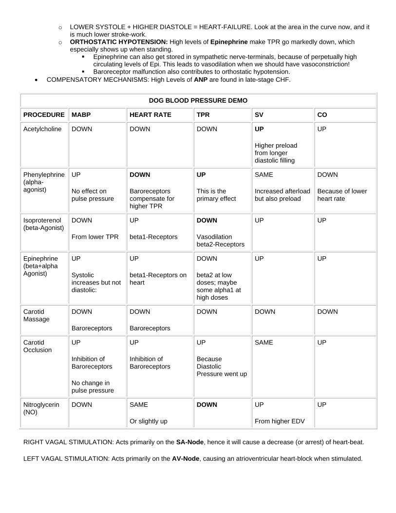
DOG BLOOD PRESSURE DEMO
PROCEDURE MABP HEART RATE TPR SV CO
Acetylcholine DOWN DOWN DOWN UP
Higher preload from longer diastolic filling
UP
Phenylephrine (alpha-agonist)
UP
No effect on pulse pressure
DOWN
Baroreceptors compensate for higher TPR
UP
This is the primary effect
SAME
Increased afterload but also preload
DOWN
Because of lower heart rate
Isoproterenol (beta-Agonist)
DOWN
From lower TPR
UP
beta1-Receptors
DOWN
Vasodilation beta2-Receptors
UP UP
Epinephrine (beta+alpha Agonist)
UP
Systolic increases but not diastolic:
UP
beta1-Receptors on heart
DOWN
beta2 at low doses; maybe some alpha1 at high doses
UP UP
Carotid Massage
DOWN
Baroreceptors
DOWN
Baroreceptors
DOWN DOWN DOWN
Carotid Occlusion
UP
Inhibition of Baroreceptors
No change in pulse pressure
UP
Inhibition of Baroreceptors
UP
Because Diastolic Pressure went up
SAME UP
Nitroglycerin (NO)
DOWN SAME
Or slightly up
DOWN UP
From higher EDV
UP
RIGHT VAGAL STIMULATION: Acts primarily on the SA-Node, hence it will cause a decrease (or arrest) of heart-beat.
LEFT VAGAL STIMULATION: Acts primarily on the AV-Node, causing an atrioventricular heart-block when stimulated.

THE HEART
CORONARY VESSELS:
• LEFT ANTERIOR DESCENDING (LAD) CORONARY ARTERY: Most common artery to occlude. Results in an anterior infarct:
o Anterior wall. o Anterior two thirds of septum. o Entire apex of heart, circumferentially.
• LEFT CIRCUMFLEX CORONARY ARTERY: Occlusion gives you a posterolateral infarct -- posterior, lateral left aspect of heart.
• RIGHT CORONARY ARTERY: Results in a posterior septal infarct -- posterior one third of septum, inferior aspect, and posterior wall of heart.
o Infarction of the Right Ventricle is rare, because the right side has far less demand for oxygen. Right Ventricular infarcts are usually extensions of posterior septal infarcts caused by occlusion of the Right Coronary Artery.
MYOCARDIAL HYPERTROPHY and CONGESTIVE HEART FAILURE:
• CAUSES of HEART FAILURE o Pump Failure: Failure that is intrinsic to the myocardium.
Two types: Systolic Failure: Failure to pump blood out of heart. Low ejection fraction. Diastolic Failure: Failure to distend the heart to fill the ventricles, as in constrictive
pericarditis. Most common reason for pump failure is from myocardial hypertrophy, usually secondary to
hypertension. Myocarditis Cardiomyopathy
o Conduction System Failure: Secondary to MI o Valvular Failure: Inflammatory (endocarditis), autoimmune, or congenital. o Cardiac Malformations: Congenital o Blood Loss / Obstruction of Blood Flow: Extracardiac causes. Pulmonary emboli or bleeding.
• HEART'S RESPONSE TO INJURY o HYPERTROPHY: Normal heart is 250-350g. myocardial cells can hypertrophy to about 3X size, or about
900g. Box Car Nuclei are the characteristic histological appearance of hypertrophied cells.
o DILATATION: Could be caused by pump failure (filled ventricles that can't empty), Aortic Insufficiency, or many other causes.
o NECROSIS: Ischemic Necrosis Contraction Band Necrosis.
o DEGENERATION o INFLAMMATION o RESOLUTION o FIBROSIS o CALCIFICATION: Dystrophic calcification
• PATHOGENESIS o HUMORAL RESPONSES o CELLULAR HYPERTROPHY o ABNORMAL PROTEIN ISOFORMS o ALTERATIONS in CALCIUM HOMEOSTASIS o PROTO-ONCOGENES o EXTRACELLULAR MATRIX o ADRENERGIC DESENSITIZATION
• PATHOLOGY • CLINICAL FEATURES
CONGENITAL HEART DISEASE:

• HEART EMBRYOLOGY: o Openings generally allow blood to pass from right heart to left heart, bypassing the lungs.
FORAMEN OVALE: An opening at the midpoint of the interatrial septum. It is open during fetal life allowing the passage of blood from the right to the left atrium. This natural fetal shunt allows the blood to bypass the fetal lungs.
DUCTUS ARTERIOSUS: Fetal blood vessel that connects the pulmonary artery with the aorta allowing the oxygenated blood from the fetal pulmonary artery to bypass the lungs and enter directly into the aorta.
• INITIAL LEFT-to-RIGHT SHUNT (Late Cyanotic or Non-Cyanotic) o PATHOGENESIS: LATE CYANOSIS is Initial left-to-right shunt will lead to Pulmonary Congestion ------>
Right Ventricular Hypertrophy plus pulmonary hypertension. In the baby, the pulmonary hypertension eventually (over months or years) get so bad as to surpass systemic blood pressure yielding a late Right-to-Left shunt ------> cyanosis.
o VENTRICULAR SEPTAL DEFECT (ROGER DISEASE): Most commonly diagnosed congenital heart disease.
PATHOGENESIS: Maybe membranous or muscular, depending on where it occurs. MEMBRANOUS VSD: Most common, 85% of cases. MUSCULAR VSD: Less common.
MURMUR: Holosystolic Murmur can be heard. CLINICAL: Most commonly diagnosed disorder, and often associated with other defects, like
Tetralogy of Fellot. o ATRIAL SEPTAL DEFECTS:
PATENT FORAMEN OVALE: Most common locale of atrial septal defect. OSTIUM SECONDUM DEFECT: OSTIUM PRIMUM DEFECT: CLINICAL: Small defects are usually asymptomatic, whereas the larger ones may cause shunting
of the blood from left to right. o PATENT DUCTUS ARTERIOSUS: Incomplete involution of the ductus arteriosus.
TREATMENT: Sometimes the ductus will close naturally. Otherwise, two ways to get a patent ductus arteriosus to close:
Indomethacin: Inhibits prostaglandin synthesis in ductus, which forces its closing. The opening is mediated by prostaglandins (PGE2).
Surgically tie it off. o PERSISTENT TRUNCUS ARTERIOSUS: Pulmonary artery and aorta are not separated one from
another but remain a common vessel. • INITIAL RIGHT-to-LEFT SHUNT (Cyanotic)
o TETRALOGY OF FELLOT Classical Diagnostic Symptoms:
Pulmonary Artery Stenosis: The principle etiology responsible for the pulmonary hypertension and right-to-left shunt.
Ventricular Septal Defect Dextroposition of Aorta (overriding): Aorta originates from the septal area, such that it
receives blood originating from both right and left ventricle. Right Ventricular Hypertrophy
Patent Ductus Arteriosus often co-occurs with Tetralogy, although it isn't part of the syndrome. It is helpful in Tetralogy, as it provides a channel for shunted blood to get back into the pulmonary circulation where it belongs.
(Right Atrium ------> Right Ventricle ------> VSD ------> Left Ventricle ------> Aorta ------> through the Ductus Arteriosus ------> Pulmonary Arteries)
• NO SHUNT o TRANSPOSITION of the GREAT ARTERIES (Cyanotic): Aorta and the pulmonary artery are
transposed. The aorta arises from the right ventricle and the pulmonary artery from the left ventricle. It presents with cyanosis of early onset.
o COARCTATION of the AORTA: Congenital constriction of the aorta. PATHOGENESIS: Occurs usually just proximal or distal to the ductus arteriosus (connection of
aorta and pulmonary artery). PRE-DUCTAL COARCTATION: Occurs in infants. Considered to be incompatible with
life. POST-DUCTAL COARCTATION: Occurs in adults. Post-ductal coarctation is associated
with a discrepancy in blood pressure between upper and lower extremities -- arms much higher than legs, as arms get all the blood.
Such patients also develop extensive arterial anastomoses between the subclavian artery and the aorta distal to the constriction.
Internal Mammary Artery Intercostal Arteries -- notching on chest wall
Cerebral hemorrhage is common because of high blood pressure in brain. o PULMONARY STENOSIS o CONGENITAL AORTIC STENOSIS o DEXTROCARDIA: Characterized by inversion of the cardiac chambers. In this condition the left atrium
and ventricle are on the right side and the right atrium and ventricle on the left. It may be associated with situs inversus.
o EBSTEIN MALFORMATION: Congenital Tricuspid Valve Insufficiency. Congenital heart disease characterized by downward displacement of abnormal tricuspid valve
into an underdeveloped right ventricle. o ENDOCARDIAL FIBROELASTOSIS (EFE): Thickening of the endocardium of the heart chambers and
valves most prominent in the left ventricle. PRIMARY EFE: Congenital disease of unknown etiology. SECONDARY EFE: Complication of various other cardiac disease characterized by
interventricular hypertension or turbulent blood flow.
ISCHEMIC HEART DISEASE:
• ANGINA PECTORIS: Pain originating in chest and radiating to left should, typically. But there are variants. o STABLE ANGINA: Exertional angina. o UNSTABLE ANGINA: Late angina carrying poor prognosis. Angina even at rest. o VARIANT (PRINZMETAL) ANGINA: Episodic angina occurring at rest, and due to coronary artery
spasm. • LAB INDICATORS:
o TROPONIN-T: Most recent blood protein has been shown to have very good predictive value for an early MI. Very specific to MI.
Cardiac troponin is its own isotype and is different from troponin of the skeletal muscle. o CAL (Coronary Artery Lesion): Mnemonic indicates the time course of elevated blood enzymes.
CPK (Creatinine Phosphokinase) will go up first (around same time as Troponin). Again, it's the cardiac isoform that does up. Peaks a few hours after the event.
AST (Aspartate aminotransferase) goes up next. Peaks next. LDH (Lactate Dehydrogenase) is the third enzyme to peak, and lasts the longest.
LDH-1 becomes elevated in 6-12 hrs. There is no proportional increase in LDH-2, so the LDH-1:LDH-2 ratio goes up.
• PATHOGENESIS / PATHOLOGY: Coronary atherosclerosis, thrombosis, and coronary artery spasm. o Two types of infarcts
TRANSMURAL INFARCT: Infarct going from epicardium to endocardium, caused by coronary artery occlusion.
Stunned Myocardium is present in the periphery of an infarct zone -- edematous and dysfunctional muscle that is not yet dead and may be recovered.
SUBENDOCARDIAL INFARCT: Diffuse, circumferential infarct, around the subendocardium (just inside the endocardial layer). Caused by shock, CHF, hypotension, or anything that results in inadequate blood supply to the coronary arteries.
Arteries perfuse the myocardium from the outside in. Thus with inadequate blood, the outer layers of muscle will get the blood first, and the inner layers may become ischemic.
The endocardium can gets its blood from the heart chambers itself -- hence the lesion is described as subendocardial.
o GRADES: Coronary atherosclerosis or stenosis is graded as follows. GRADE 1: 0-25% OCCLUSION -- asymptomatic and common GRADE 2: 25-50% OCCLUSION -- possible stable angina GRADE 3: 50-75% OCCLUSION -- stable angina GRADE 4: 75%+ OCCLUSION -- unstable angina, impending MI and thrombosis.
o ENDOTHELIAL INJURY: Early grade atherosclerosis may show paroxysmal coronary artery contractions resulting from endothelial injury.
The substances that were supposed to stimulate release of NO leading to vasodilatation, instead wind up stimulating vasoconstriction, because there are no endothelial cells present to release the NO.
This vasoconstriction can occur with Grades 1 and 2 Can also cause thrombosis in more advanced lesions.
o TIME COURSE of CHANGE: Time after infarct. The myocardium can survive up to one hour of absolute ischemia (center of the infarct) and up to four hours of relative ischemia (periphery of the infarcted area).
1-5 min: Swelling of mitochondria and ER; reversible changes. Glycogen depletion and depletion of ATP are noticeable, but no morphological changes grossly.
20-40 min: Irreversible changes microscopically. Cell necrosis, nuclear changes, plasma membrane rupture.
4-12 hrs: Onset of irreversible coagulative necrosis. Myocardial cells lose striations. Thrombus is still present.
18-24 hrs: Pyknosis, karyolysis, karyohexis. Myocytolysis occurs -- loss of cytoplasm. 1-3 days: Widespread necrosis. Infiltration of PMN's begins. Thrombus has often been lysed
naturally by now. Infarct can be recognized on gross examination -- tissue is mottled, red, and congested.
4-10 days: PMN's move out, and macrophages take over to clean up the debris. Period of greatest weakness, during which rupture and cardiac tamponade may occur. 50% of ruptures occur within first 5 days and 87% within 14 days. Grossly, necrotic tissue is yellow, soft, and pus-like.
1 month: Granulation tissue has developed. The infarct heals from the outside in, with the central area of necrosis being the last to fibrose.
3 months: Scar formation is complete. o CONTRACTION BAND NECROSIS: Hyper contraction of myofilaments. Typically found at the margins of
infarcts or in reperfusion injury. • CLINICAL:
o SYMPTOMS: Either excessive sympathetic or parasympathetic symptoms may be seen. Sympathetic Symptoms: tachycardia, sweating, pallor Parasympathetic Symptoms (Vagal discharge): bradycardia, vomiting. Fever may be seen. Myocardial cells may release endogenous pyrogens such as IL-1 when they
rupture. Cardiogenic Shock: Oliguria, hypotension, pulmonary edema, pale and cold extremities.
o Leukocytosis with left shift will be seen. o Arrhythmias and other EKG changes may be seen.
• COMPLICATIONS o ARRHYTHMIAS: 85% of cases. Most important cause of death from MI, and can occur at any time, either
acute or chronic, after the MI. The arrhythmias are often reversible -- they go away when edema and inflammation disappear.
o CARDIOGENIC SHOCK: 15% of cases, relatively rare. Systemic hypotension due to pump failure. o EXTENSION of the INFARCT o RUPTURE, CARDIAC TAMPONADE: 1-2% of cases. Rare. o ANEURYSM o MURAL THROMBOSIS and EMBOLISM: Arterial embolism may go to distal artery and cause tissue
necrosis (kidney), petechiae, or gangrene. o PERICARDITIS: DRESSLER SYNDROME is a delayed pericarditis that occurs following cardiac surgery
or myocardial infarction.
COR PULMONALE: Right ventricular failure.
• ACUTE COR PULMONALE: May occur from a saddle pulmonary embolus. • CHRONIC COR PULMONALE: From COPD or from left ventricular failure. • CLINICAL: High central venous pressure, jugular venous distension.
ACQUIRED VALVULAR and ENDOCARDIAL DISEASES:
• RHEUMATIC HEART DISEASE: o ACUTE RHEUMATIC FEVER: A hypersensitivity disease typically caused by an abnormal immune
response to Strep-A antigens. PATHOGENESIS: Immune mechanism involves both B and T cells. CLINICAL: Patient must have at least one of the major symptoms, and two of the minor
symptoms of the JONES'S CRITERIA: MAJOR JONES'S SYMPTOMS:
Carditis: Found in 35% of patients. Polyarthritis: found in 75% of patients. Chorea: Rapid jerky, dyskinetic involuntary movements.
Erythema Marginatum Subcutaneous nodules
MINOR JONES'S SYMPTOMS: Fever Arthralgia History of Rheumatic Fever or Rheumatic Heart Disease Acute-Phase Reactants: high sed-rate, or positive C-Reactive Protein, which are
evidence of inflammation. Prolonged PR interval by ECG.
RISK-FACTORS: The more severe the initial Streptococcal infection, the more likely it is for patients to develop Rheumatic Fever.
o CHRONIC RHEUMATIC HEART DISEASE: PANCARDITIS: Rheumatic heart disease is typically a pancarditis, affecting all three layers of the
heart. VALVULITIS: Endocarditis affecting Mitral and Aortic Valves.
MITRAL VALVE is most common: Chordae Tendineae get fused together, resulting in MITRAL STENOSIS. Rheumatic Fever is the most common cause of mitral stenosis.
Stenosis has a fishmouth appearance. You may also at the same time see mitral insufficiency in the same patient.
AORTIC VALVE is second most common, which can lead to AORTIC STENOSIS. PERICARDITIS: Often seen, but described as Bread-and-Butter Pericarditis. This is an
inflammatory process, but it is not Constrictive Pericarditis. HISTOPATHOLOGY: It is aseptic so no bacteria are present.
ASCHOFF BODY is the characteristic finding. A granuloma composed of a central triangular area of fibrinoid necrosis surrounded by histocytes. They heal by scarring.
ANITCHKOFF CELLS: Found around the perimeter of the Aschoff bodies. They are also called Caterpillar cells. Round-to-ovoid nuclei have chromatin disposed in a wavy ribbon resembling a caterpillar.
• BACTERIAL ENDOCARDITIS: o CAUSES:
Congenital Heart Defects lead to stasis of blood in the heart and therefore predispose to them. Bicuspid Aortic Valve predisposes to bacterial endocarditis resulting in Aortic Stenosis.
Sepsis o Types:
ACUTE ENDOCARDITIS SUBACUTE ENDOCARDITIS
o CONSEQUENCES Valvular insufficiency or stenosis Septic Emboli are thrombo-emboli plus bacteria, which may be thrown to a distal artery, causing
gangrene. Heart Failure secondary to valvular insufficiency. Immune complex glomerulonephritis.
• MARANTIC (NON-BACTERIAL THROMBOTIC) ENDOCARDITIS: Occurs in people suffering from malnutrition, marasmus. They cannot repair the little foci of damage that occur with normal wear and tear of the heart (constant contraction and what not).
o Small aggregates of fibrin collect on cardiac valves. It is a form of intravascular thrombosis, and increased coagulability of the blood is seen.
• CALCIFIC AORTIC STENOSIS: o CLINICAL: Multiple causes:
Idiopathic: Can occur without any predisposing conditions in the elderly. Atherosclerotic: As an extension of atherosclerosis of the aorta into the aortic valve Rheumatic Heart Disease: Complication of rheumatic endocarditis. Bicuspid Aortic Valve: Congenital malformation predisposing to infection.
o CLINICAL: Aortic Stenosis can lead to angina as it reduces coronary blood flow. • MITRAL VALVE PROLAPSE: Protrusion of the loosened up mitral valve leaflets into the left atrium during
systole. It may or may not by associated with mitral regurgitation. • CARCINOID HEART DISEASE: Endocardial fibrosis of the right atrium and ventricle, pulmonic and tricuspid
valves. o PATHOGENESIS: Caused by a carcinoid tumor secreting biogenic amines (histamine and serotonin).
Usually the tumor is metastatic to the liver. The left side of the heart is not affected because the biogenic amines are neutralized in the lung.
• COLLAGEN DISEASES
o LUPUS ERYTHEMATOSUS: LIBMAN SACKS ENDOCARDITIS is a complication of SLE. PATHOLOGY: Most often involves the mitral valve.
o RHEUMATOID ARTHRITIS o ANKYLOSING SPONDYLITIS o SCLERODERMA o POLYARTERITIS NODOSA
PRIMARY MYOCARDIAL DISEASE:
• VIRAL MYOCARDITIS: PANCARDITIS, where all three layers of myocardium may be involved --endocarditis, myocarditis, and pericarditis.
• HYPERSENSITIVITY MYOCARDITIS • GIANT CELL MYOCARDITIS
METABOLIC DISEASES:
• HYPERTHYROID HEART DISEASE: o Thyroid hormone does not directly affect the Coronary arteries. o Increased metabolic rate increases the demand for oxygen systemically, which results in tachycardia,
which further increases (exacerbates) the demand for oxygen in the heart. Thus the heart potentially suffers both an increases oxygen demand and an increased work load.
• HYPOTHYROID HEART DISEASE • THIAMIN DEFICIENCY (BERIBERI) FAILURE: High output heart failure.
CARDIOMYOPATHY: Primary idiopathic disease of the myocardium, which is not caused by ischemia, valvular dysfunction, infection, inflammatory disorders, or congenital anomalies.
• IDIOPATHIC DILATED CARDIOMYOPATHY: Also called congestive cardiomyopathy. o ALCOHOLISM is a risk-factor for this form of Cardiomyopathy. You may also Beriberi high-output failure
with Alcoholics. o TOXIC CARDIOMYOPATHY
• HYPERTROPHIC CARDIOMYOPATHY: o PATHOLOGY: Patient will have asymmetrical septal hypertrophy and a disarray of muscle fibers.
This diminishes volume of left ventricle and causes abnormal myocardial contraction. o CLINICAL: The disease is inherited as an autosomal dominant trait.
• RESTRICTIVE CARDIOMYOPATHY o AMYLOIDOSIS: Endomyocardial biopsy is required for a definitive diagnosis. Must demonstrate amyloid
infiltrates in the heart. Amyloidosis can mimic other forms of restrictive cardiomyopathy.
CARDIAC TUMORS:
• CARDIAC MYXOMA: The most common primary benign tumor of the heart. Typically located on the mitral valve. o It is a gelatinous polyp connected to the endocardium.
• RHABDOMYOMA: Primary benign tumor of the myocardium, typically found in children. • PAPILLARY FIBROELASTOMA
PERICARDIAL DISEASES:
• PERICARDIAL EFFUSION: Accumulation of fluid in the pericardial sac in excess of the normal content (50 ml). o PATHOLOGY: Fluid may be of multiple types
Serous effusion Chylous effusion Hemorrhagic effusion
• CONSTRICTIVE PERICARDITIS: Chronic inflammation obliterates the pericardial cavity. Fibrous exudate in pericardium. Results in inability for heart to expand, or diastolic heart failure.
o PATHOLOGY: It leads to Pulmonary Hypertension and elevated CVP. o CLINICAL: It is rare. Rheumatic Heart disease does not lead to constrictive pericarditis.
BLOOD VESSELS
HEMOSTASIS and THROMBOSIS:
• Hemostasis: The arrest of hemorrhage as a response to vascular injury. • BLOOD COAGULATION
o Intrinsic Pathway: Factor XII ------> Factor XI ------> Factor IX o Extrinsic Pathway: Tissue Factor (Thromboplastin) ------> Factor VII + Ca+2 o Common Pathway: Factor X ------> (Prothrombin ------> Thrombin) ------> (Fibrinogen ------> Fibrin)
• PLATELET AGGREGATION: o Von Willebrand Factor from endothelial cells enhances aggregation. o Thromboxane A2 (TXA2) enhances aggregation.
• ENDOTHELIAL FACTORS: Injury to the endothelium is the most important precipitating event of thrombosis. o Function of endothelial cells:
Permeability barrier Vasoactive factors: NO Anti-thrombotic factors: Prostacyclin (PGI2) Clotting Factors:
Factor VIII Von Willebrand Factor
Anti-Coagulant Factors: Thrombomodulin Fibrinolytic Agents: TPA Plasminogen Activator Inhibitor (PAI) inhibits fibrinolysis Inflammatory Mediators: IL-1, Cell adhesion molecules Growth Factors: CSF, FGF, PDGF
• CLOT LYSIS: (Plasminogen ------> Plasmin) ------> (Fibrin ------> Fibrin split products) o Thrombosis itself results in production of TPA, which converts plasminogen to plasmin to effect
fibrinolysis.
ATHEROSCLEROSIS: Progressive accumulation within the intima of smooth muscle cells and lipids.
• PATHOGENESIS and PATHOLOGY: o Progression:
Early on: Proliferation of smooth muscle cells and accumulation of lipid. Smooth muscle cells as well as lipid is required for the atheroma to form.
Later: Infiltration of macrophages, lymphocytes, and connective tissue. Later: Organized thrombus formed, with canals (vaso plaquorum) going through the lesion.
o ELEMENTS of ATHEROSCLEROTIC PLAQUE: Vascular Endothelium Arterial Smooth Muscle Cell Mononuclear Phagocyte Lymphocytes and Neutrophils
• ATHEROGENIC PROCESSES: The overall process of atherogenesis is a combination of the theories below. o Insudation Hypothesis: Lipids in the atheroma are derived from plasma lipoproteins (LDL) in the blood. o Encrustation Hypothesis: Says that small mural thrombi represent the initial event in atherosclerosis.
We now know that this isn't true. Mural thrombi are not the initial event, but they are critical to the later progression of the atheroma, i.e. toward thrombosis.
o Reaction to Injury Hypothesis: Smooth muscle cells accumulate as a response to injury, as a result of release of PDGF and other growth factors.
This theory explains smooth muscle proliferation but not lipid accumulation. o Monoclonal Hypothesis: Points to the fact that many plaques contain cells that are mostly monoclonal.
Perhaps their proliferation was due to a virus or cell-specific mutagen. o Intimal Cell Mass Hypothesis: This is the initial lesion. Accumulation of smooth muscle cells at junctions
and branching points of arteries. o Hemodynamic Hypothesis: Atheromas tend to occur at locations of turbulence, pressure, and shear
forces. Hypertension predisposes to atheromatous formation. o UNIFYING HYPOTHESIS: Likely order of events in Atherogenesis
Intimal cell mass predisposes at branch points. Lipid accumulation occurs.
Lipid Insudation results in cellular injury, leading to accumulation of macrophages and platelets Macrophages and platelets release growth factors Smooth muscle proliferation and endothelial injury may result in loss of anticoagulant properties
of endothelia, and a thrombus results. • MORPHOLOGY:
o INITIAL LESION of ATHEROSCLEROSIS FATTY STREAKS: Can be found in young children as well as adults, and not necessarily at
branch points. INTIMAL CELL MASSES: At branch points, may also be the initial lesion.
o CHARACTERISTIC LESION of ATHEROSCLEROSIS: Fibrous Fatty Plaque FIBROUS CAP: Layer of fibrous connective tissue overlying the atheroma. Contains foam cells
(macrophages) and smooth muscle cells, as well as fibroblasts. ATHEROMA: Necrotic, lipid-laden center of the lesion. Term can also be used to describe the
whole lesion. • COMPLICATIONS
o COMPLICATED LESIONS: Changes in structure of vessel itself. Thrombosis: Damage to endothelial cells leads to loss of anti-coagulant properties (clotting
inhibitors), leading to Thrombosis. Neovascularization: Organization of the thrombus Thinning of the Media: Can lead to aneurysm. Calcification Ulceration
o COMPLICATIONS: Acute occlusion leading to Myocardial Infarct. Usually due to hemorrhage into a plaque, or
thrombosis. Chronic narrowing of vascular lumen, causing chronic ischemia and sometimes atrophy to kidney. Aneurysm formation Cholesterol Embolism: Embolism of atheromatous material
• RISK FACTORS: Any factor with a doubling in the incidence of ischemic heart disease. Atherosclerosis is the most common cause of heart disease in the Western Hemisphere.
o hypertension o blood cholesterol level o cigarette smoking o diabetes o increasing age o male sex o physical inactivity o stressful life patterns.
• MECHANISMS of LESION PROGRESSION: o Cytokines
PDGF and FGF cause proliferation of smooth muscle and endothelial cells. IFN and TGF-beta inhibit cell proliferation and thus could account for endothelial cell
discontinuities. IL-1 and TNF stimulate activation of PAF, Tissue Factor, and PAI (plasminogen activator
inhibitor) in endothelial cells. o T-Lymphocytes o Endothelium: Loss of continuity of endothelial layer
Increase permeability to lipoproteins Permit platelet interaction with vessel wall, and subsequent release of growth factors Blood may enter the wall, allowing interaction, either through an organized thrombus or through a
tear or discontinuity in the endothelial surface. o Thrombosis
• HEREDITARY DISORDERS OF LIPID METABOLISM AND ATHEROSCLEROSIS o FAMILIAL HYPERCHOLESTEROLEMIA: Defect in LDL receptors. o APO-E o HDL: o LIPOPROTEIN(A)
HYPERTENSIVE VASCULAR DISEASE:
• HYPERTENSION: Defined as systolic blood pressure greater than 160mm, or diastolic blood pressure greater than 90mm, or both.
o Subtypes: BENIGN HYPERTENSION: Asymptomatic MALIGNANT HYPERTENSION: Rapidly progressing to end-organ failure.
MORPHOLOGY: Blood vessels show fibrinoid necrosis or concentric hyperplasia of smooth muscle-cells -- onion-skin changes).
o PATHOGENESIS: ESSENTIAL HYPERTENSION: Primary hypertension in which there is no single identifiable
cause. The majority of cases Renin from kidney is a major player.
Renin converts Angiotensinogen ------> Angiotensin I ACE converts Angiotensin I ------> Angiotensin II Angiotensin II effects:
Vasoconstriction. Stimulate secrete of aldosterone from adrenal glands ------> K+
excretion and Na+ retention, fluid retention in kidney ------> higher blood volume.
SECONDARY HYPERTENSION: Hypertension secondary to a disease. Minority of cases. Renal Ischemia: Anything causing ischemia to the kidney (vascular stenosis or
atherosclerosis of Renal Artery or arterioles) will release renin and probably result in hypertension.
Fibromuscular Dysplasia of Renal Artery is a congenital disorder, with progressive concentric thickening of the Renal Artery. Occurs in young females.
Cushing's Syndrome: Primary hypersecretion of cortisol. Conn's Syndrome: Primary hypersecretion of aldosterone. Pheochromocytoma: Adrenal medullary tumor.
Norepinephrine is secreted in spurts, so patient will have wildly fluctuating, paroxysmal elevations in blood pressure.
Thyrotoxicosis RISK FACTORS: Similar as those for atherosclerosis and CAD.
Genetics: often found to be familial Diet Stressful lifestyle Obesity
CLINICAL MANIFESTATIONS: Often asymptomatic Early on: Headache, nosebleeds, tinnitus, dizziness, fainting, visual impairment. Later: End-stage hypertension results in stroke, heart failure (from compensatory
hypertrophy), renal failure • ARTERIOSCLEROSIS: Arterial vascular changes characterized by thickening and loss of elasticity of arterial
walls. It may be seen in patients with chronic hypertension, and, to a lesser degree, as part of the aging process. o BENIGN (HYALINE) ARTERIOSCLEROSIS: Particularly occurs in kidneys. The blood vessel walls take
on a glassy, "hyaline" appearance. This change reflects mild or "benign" hypertension and are particularly seen in kidneys.
o MALIGNANT (HYPERPLASTIC) ARTERIOSCLEROSIS: This change refers to the concentric rings of increased connective tissue and smooth muscle giving the arteries an onion-skin appearance. Such changes signify acceleration of the hypertension.
o ARTERIOLOSCLEROSIS: Smooth muscle proliferation, thickening, and loss of elasticity of walls occurring in the arterioles.
• MONCKEBERG MEDIAL SCLEROSIS: Degenerative calcification of the media of large and medium arteries in old people.
o Usually vessels in the extremities. o Usually is benign and subclinical.
VASCULITIS:
• GENERAL PROPERTIES: Inflammation and necrosis of blood vessels, including arteries, veins and capillaries. The damage may be due to infectious agents, mechanical trauma, radiation or toxins; often no specific etiologic factor is identified. The pathogenesis is thought to involve immune mechanisms such as deposition of immune complexes, direct attack by circulating antibodies etc.
• POLYARTERITIS NODOSA: Systemic vasculitis affecting medium and small muscular arteries.
o PATHOLOGY: Multiple organs involved, but the lungs are characteristically not involved. If lung involvement is present, suspect Wegener's Granulomatosis.
o CLINICAL FEATURES: Condition is associated with Hepatitis-B in about 30% of cases. Treatment: steroids, cyclophosphamide.
• HYPERSENSITIVITY ANGIITIS: o PATHOGENESIS: Maybe hypersensitivity to a drug, a bacterial product, or maybe secondary to an
autoimmune disease like SLE. o PATHOLOGY: Affects mainly the small vessels -- arterioles and capillaries. Will see fibrinoid necrosis.
As opposed to Polyarteritis Nodosa, this disease affects small arteries. Otherwise its similar. As opposed to Malignant Hypertension (which can also show petechiae), this disease is
associated with inflammation whereas malignant hypertension is not. o CLINICAL: Petechiae will be present from capillary hemorrhage. o LEUCO CYTOCLASTIC VASCULITIS: Confined predominantly to the skin and presenting as purpuric
lesions. PATHOLOGY: Fibrinoid necrosis of small vessels, extravasated red cells. CLINICAL: Clinically presents as purpura.
• CHURG-STRAUSS SYNDROME, ALLERGIC GRANULOMATOSIS and ANGIITIS: o PATHOLOGY: Mainly affects the lungs, it is found in young people, and it is associated with asthma.
Will find Eosinophilia in the blood. • GIANT CELL ARTERITIS, TEMPORAL ARTERITIS:
o PATHOLOGY: Giant cells present in the wall of any of the cranial arteries, usually the Temporal Artery. Thrombus found in lumen of the artery. A granulomatous infiltrate will be found in the artery. Giant Cells will be present in the artery, hence the name./=
o CLINICAL FEATURES: Occurs in older population, at least 50 yrs old. Patient presents with headache and temporal pain. Visual symptoms are common, and blindness
is a complication in 50% of cases. Temporal Artery will feel firm and nodular. Treatment: responds well to steroids.
• WEGENER'S GRANULOMATOSIS: o PATHOLOGY:
Two key pathological findings Systemic vasculitis of small arteries and veins. Granulomatous inflammation of nose, sinuses, and lung.
Anti Cytoplasmic Nuclear Antibodies (ANCA) are found in serum. o CLINICAL FEATURES: Patient may have persistent sinusitis, pneumonitis, hematuria, proteinuria.
• TAKAYASU ARTERITIS: An inflammatory disorder of the aortic arch and its major branches, with localized stenosis or occlusion
o PATHOLOGY: Also known as PULSELESS DISEASE, as the obliteration and thickening of the Aortic Arch may result in no pulse in the wrist.
o PATHOGENESIS: An auto-immune basis has been proposed. o CLINICAL FEATURES: It is most common in young women, similar to other auto-immune disorders.
• KAWASAKI DISEASE (MUCOCUTANEOUS LYMPH NODE SYNDROME): o PATHOGENESIS: Thought to be viral etiology. o PATHOLOGY: Acute necrotizing vasculitis.
Coronary Aneurysms are found in 70% of patients -- an unusual finding. o CLINICAL:
Infancy and early childhood Fever, rash, conjunctival and oral lesions Lymphadenitis
• THROMBOANGIITIS OBLITERANS (BUERGER'S DISEASE): An occlusive, inflammatory disease of the medium-sized and small arteries in the distal arms and legs.
o PATHOLOGY: Microscopically, there is acute inflammation of arteries with thrombosis and obliteration of lumen.
o CLINICAL FEATURES: Intermittent Claudication (weakness relieved by rest) is seen in legs. Strongly associated with smoking. Young or middle-aged males.
RAYNAUD'S PHENOMENON: Intermittent attacks of ischemia of fingers or toes due to intense arterial vasospasm, often precipitated by cold or emotional stimuli.
ANEURYSMS:
• SHAPES OF ANEURYSMS: o Fusiform: Most common form. o Saccular: Most common form. o Dissecting: Actually a hematoma, splitting apart of media. o Arteriovenous Fistula: Direct connection of arterioles to venules, bypassing the tissue and causing
necrosis. • ATHEROSCLEROTIC ANEURYSMS: Most common type of aneurysm.
o PATHOLOGY: Usually found in abdominal aorta. Lumen may only be slightly enlarged, but you will see lots of atherosclerosis and thrombosis.
o CLINICAL FEATURES: May be asymptomatic, or may be found coincident to some other X-ray, surgery, or medical procedure.
Ruptured Aneurysm is the most dramatic presentation, surgical emergency. TREATMENT: Put a synthetic graft over the aneurysm.
• CONGENITAL (BERRY) ANEURYSMS: Usually occurs at a main branching point of the basilar artery. o PATHOLOGY: Fairly small aneurysm, saccular in shape.
• DISSECTING ANEURYSMS: Not really an aneurysm, actually it's a hematoma in the muscular media of the Aorta.
o PATHOGENESIS: Results from Marfan's Syndrome or from longstanding hypertension in elderly males. Dissection splits the aortic wall in two. Multiple types have been described.
TYPE-A: Start at Aortic Arch and extend proximally to include the Aortic valves, resulting in hemopericardium. Grave prognosis.
TYPE-B: Start Aortic Arch and extend distally, down through the abdominal Aorta. Double-Barrel Aorta: Dissection penetrates back into the true Aortic lumen, temporarily
relieving the pressure and biding some time. o PATHOLOGY: Cystic Medial Necrosis is the characteristic finding -- cystic changes in media of aorta.
Stains positive for mucopolysaccharide. o CLINICAL FEATURES: Patient describes a severe tearing type of pain.
• SYPHILITIC ANEURYSMS: Aneurysms in Thoracic Aorta resulting from Obliterative Endarteritis of the vaso vasorum supplying the Aorta.
o PATHOLOGY: Characteristic Tree-Bark Appearance of linear and patchy scars is seen on the luminal aspect of the aorta.
o CLINICAL: Aortic calcification, Aortic Insufficiency, and hemopericardium are frequent complications. • MYCOTIC ANEURYSMS: These lesions are caused by significant weakening of the blood vessel wall by
infection.
VEINS:
• VARICOSE VEINS: Enlarged and tortuous blood vessels. Occurs most commonly in the legs. o RISK-FACTORS:
older age female sex heredity posture obesity.
o PATHOLOGY: Dilation and elongation of the veins, incompetence of venous valves. o SITES:
Varicose Veins of Legs is most common. Hemorrhoids Esophageal Varices Varicocele: Varicose veins of pampiniform plexus of scrotum.
• DEEP VENOUS THROMBOSIS: Associated with prolonged bed-rest, blood stasis, and reduced cardiac output. Risk for pulmonary embolism.
o Thrombophlebitis: Inflammation, commonly from a bacterial infection, with secondary thrombosis of deep leg veins.
o Phlebothrombosis: Thrombosis of deep leg veins without initial inflammation.
LYMPHATIC VESSELS:

• LYMPHANGIITIS: • LYMPHATIC OBSTRUCTION
o Lymphedema: o Lymphangiectasia: o Elephantiasis: o Milroy Disease: Inherited form of lymphedema present at birth.
BENIGN BLOOD-VESSEL TUMORS:
• HEMANGIOMAS: Benign tumors of blood vessels. They don't know whether it's a true neoplasia or just a hamartoma.
o Types: CAPILLARY HEMANGIOMA: Containing capillaries. Juvenile Hemangioma: CAVERNOUS HEMANGIOMA: Containing open vascular spaces. Multiple Hemangiomatous Syndromes:
o CLINICAL: Truly benign and not important, unless they are a problem cosmetically. • GRANULOMA PYOGENICUM: Mass of granulation tissue resembling a hemangioma. They are truly benign and
occur after some injury • VASCULAR ECTASIA: Local Dilatation and growth of blood vessels, not a tumor.
o Spider Angiomata is an example. • GLOMUS TUMOR (GLOMANGIOMA): A benign, painful tumor of the glomus body -- neuromyoarterial receptor
which is sensitive to temperature and regulates arterial flow. o PATHOLOGY: Small, reddish blue spots occur most commonly in the distal fingers and toes.
Histologically, there is a mixture of branching vascular channels and nests of glomus cells. • HEMANGIOENDOTHELIOMA: A vascular tumor composed of endothelial cells, considered to be intermediate
between benign hemangiomas and frankly malignant angiosarcomas. Histologically, several variants are described, based on the predominant cell type - spindle cell, epithelioid etc.
o Epithelioid Hemangioendothelioma o Spindle-Cell Hemangioendothelioma o CLINICAL: In general, surgical excision is curative. Rarely do these tumors metastasize.
• MULTIPLE HEMANGIOMATOUS SYNDROMES: Angiomatous lesions present in two or more tissues. o Von Hippel-Lindau Syndrome: Hemangiomas in brain and retina. o Sturge-Weber Syndrome: Vascular lesions in brain and skin.
MALIGNANT BLOOD-VESSEL TUMORS:
• ANGIOSARCOMA: Malignant neoplasm arising from blood vessels. o CARCINOGENS have implicated as causes:
Thorotrast -- radio-opaque dye. PVC Arsenic.
o PATHOLOGY: Most commonly found on scalp, breast, other soft tissues, or liver. Resembles hemangiomas, except the lining endothelial cells are malignant.
o CLINICAL: Variable prognosis, from indolent to high malignant. o LYMPHEDEMA-ASSOCIATED ANGIOSARCOMA: Occurs in 1% of cases, some 20 years after an
axillary-node dissection for breast cancer. Occurs in associated with lymphedema of the upper extremity. Also known Stewart-Treves Syndrome.
• HEMANGIOPERICYTOMA: Rare malignant neoplasm thought to arise from pericytes, smooth muscle cells external to the walls of capillaries and arterioles.
• KAPOSI SARCOMA: o PATHOGENESIS: In AIDS patients, caused by infection of HHV-8 o FOUR TYPES
Classical / European: Found in older men of Poland, Italy. Chronic course and rarely fatal. African: Also chronic, more malignant than above, but still not usually fatal. Transplant-associated: Immunosuppression, particularly with renal transplants. AIDS-ASSOCIATED: Immunosuppression.
Found in highest incidence in homosexual at-risk group, as compared to other groups, perhaps because of sexual transmission of HHV-8 virus.
o PATHOLOGY: Two stages Patch Stage: Early on.
Plaque, Nodular Stage: Later progression. • BACILLARY ANGIOMATOSIS: Vascular infection appearing like Kaposi Sarcoma, and caused by Bartonella
Henselae (same causal agent as Cat Scratch Fever). o AIDS patients may also have this, so this infection should be ruled out before settling on Kaposi Sarcoma.