Embed Size (px)
Citation preview

Copyright © 2015 Wolters Kluwer Health | Lippincott Williams & Wilkins
Chapter 14
The Heart and Heart Disease
Copyright © 2015 Wolters Kluwer • All Rights Reserved
Overview

Copyright © 2015 Wolters Kluwer • All Rights Reserved
Key Terms
angina pectoris echocardiography pacemaker
angiography electrocardiograph pericardium
arrhythmia endocardium plaque
atherosclerosis epicardium septum
atrium fibrillation stenosis
bradycardia infarct systole
cardiac output ischemia tachycardia
coronary mediastinum valve
coronary thrombosis murmur ventricle
diastole myocardium
Copyright © 2015 Wolters Kluwer • All Rights Reserved
Structure of the Heart
Learning Objectives
1. Describe the three tissue layers of the heart wall.
2. Describe the location and structure of the pericardium, and cite its functions.
Copyright © 2015 Wolters Kluwer • All Rights Reserved
Structure of the Heart (cont.)
Learning Objectives
3. Compare the functions of the right and left chambers of the heart.
4. Name the valves at the entrance and exit of each ventricle, and identify the function of each.
5. Briefly describe blood circulation through the myocardium.

Copyright © 2015 Wolters Kluwer • All Rights Reserved
Heart Function
Learning Objectives
6. Briefly describe the cardiac cycle.
7. Name and locate the components of the heart’s conduction system.
8. Explain the effects of the autonomic nervous system (ANS) on the heart rate.
9. List and define several terms that describe variations in heart rates.
10. Explain what produces each of the two normal heart sounds, and identify the usual cause of a murmur.

Copyright © 2015 Wolters Kluwer • All Rights Reserved
Heart Disease
Learning Objective
11. Briefly describe five methods used to study the heart.

Copyright © 2015 Wolters Kluwer • All Rights Reserved
Heart Disease (cont.)
Learning Objectives
12. Describe six types of heart disease.
13. List four risk factors for coronary artery disease that cannot be modified.
14. List seven risk factors for coronary artery disease that can be modified.
15. Describe three approaches to the treatment of heart disease.
Copyright © 2015 Wolters Kluwer • All Rights Reserved
Effects of Aging
Learning Objective
16. List four changes that may occur in the heart with age.

Copyright © 2015 Wolters Kluwer • All Rights Reserved
Case Study
Learning Objective
17. Referring to the case study, list the emergency and surgical procedures commonly performed following a myocardial infarction, and explain why they are done.
Copyright © 2015 Wolters Kluwer • All Rights Reserved
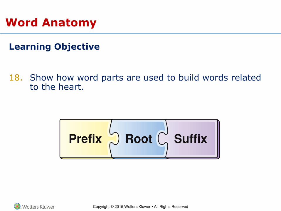
Word Anatomy
Learning Objective
18. Show how word parts are used to build words related to the heart.

Copyright © 2015 Wolters Kluwer • All Rights Reserved
Structure of the Heart
Learning Objectives
1. Describe the three tissue layers of the heart wall.
2. Describe the location and structure of the pericardium, and cite its functions.
Copyright © 2015 Wolters Kluwer • All Rights Reserved
Circulation and the Heart
• The circulatory system is a continuous one-way circuit
of blood vessels, through which blood is pumped by the
heart.
Copyright © 2015 Wolters Kluwer • All Rights Reserved
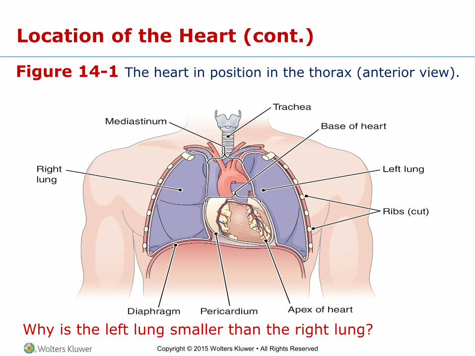
Location of the Heart
• Between the lungs
• Left of the midline of the body
• In mediastinum
• Apex pointed toward left
Copyright © 2015 Wolters Kluwer • All Rights Reserved
Figure 14-1 The heart in position in the thorax (anterior view).
Why is the left lung smaller than the right lung?
Location of the Heart (cont.)
Copyright © 2015 Wolters Kluwer • All Rights Reserved
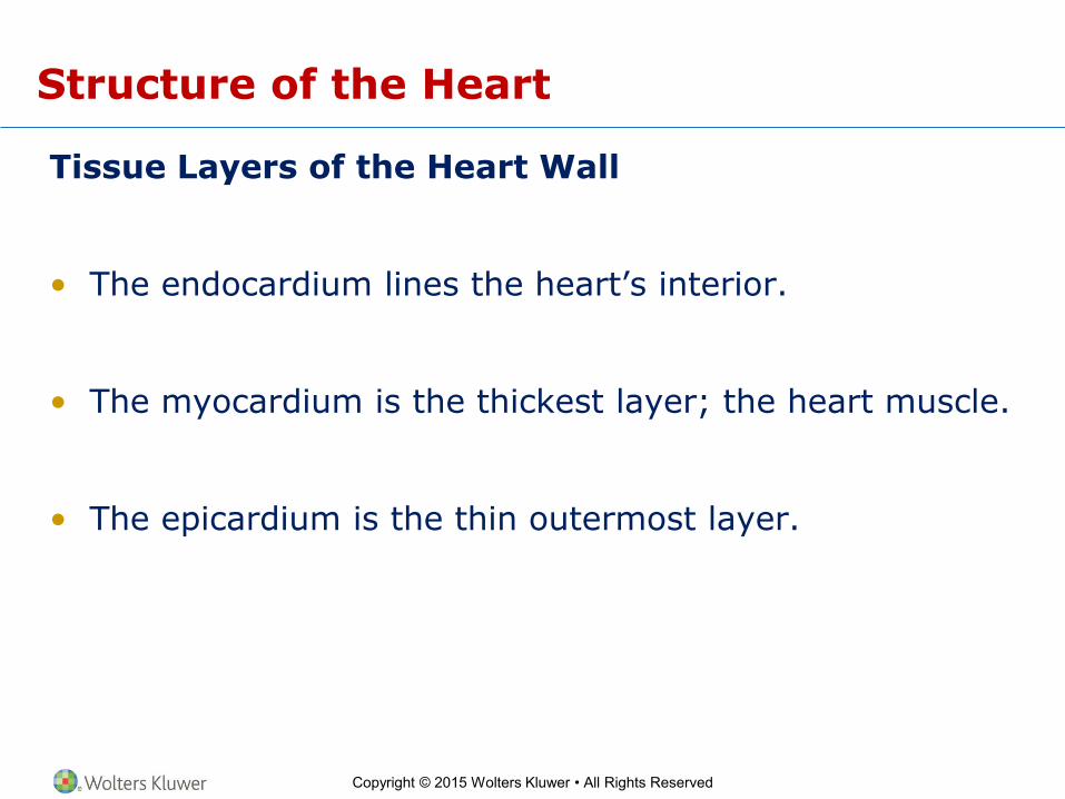
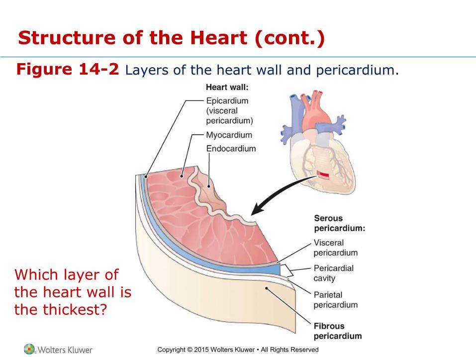
Structure of the Heart
Tissue Layers of the Heart Wall
• The endocardium lines the heart’s interior.
• The myocardium is the thickest layer; the heart muscle.
• The epicardium is the thin outermost layer.
Copyright © 2015 Wolters Kluwer • All Rights Reserved
Figure 14-2 Layers of the heart wall and pericardium.
Which layer of the heart wall is the thickest?
Structure of the Heart (cont.)

Copyright © 2015 Wolters Kluwer • All Rights Reserved
Structure of the Heart (cont.)
The Pericardium
• The sac that encloses the heart
– Outer fibrous pericardium holds the heart in place.
– Serous pericardium:
• Parietal layer fused to fibrous pericardium
• Visceral layer (epicardium) fused to myocardium
– Pericardial cavity is the space between serous layers.

Copyright © 2015 Wolters Kluwer • All Rights Reserved
Structure of the Heart (cont.)
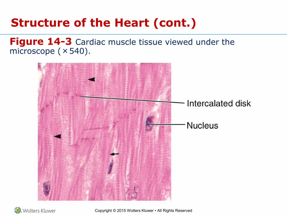
Special Features of the Myocardium
• Composed of cardiac muscle cells
– Are lightly striated (striped)
– Have single nucleus
– Are controlled involuntarily
– Have intercalated disks
– Have branching fibers
Copyright © 2015 Wolters Kluwer • All Rights Reserved
Figure 14-3 Cardiac muscle tissue viewed under the microscope (×540).
Structure of the Heart (cont.)

Copyright © 2015 Wolters Kluwer • All Rights Reserved
Structure of the Heart (cont.)
Learning Objectives
3. Compare the functions of the right and left chambers of the heart.
4. Name the valves at the entrance and exit of each ventricle, and identify the function of each.
5. Briefly describe blood circulation through the myocardium.
Copyright © 2015 Wolters Kluwer • All Rights Reserved
Structure of the Heart (cont.)
Divisions of the Heart
• Double pump
– Right side pumps blood low in oxygen to the lungs via pulmonary circuit.
– Left side pumps oxygenated blood to remainder of body via systemic circuit.
Copyright © 2015 Wolters Kluwer • All Rights Reserved
Structure of the Heart (cont.)
Four Chambers
• Right atrium
– Receives low-oxygen blood returning from body tissue through superior vena cava and inferior vena cava
• Left atrium
– Receives high-oxygen blood from lungs
• Right ventricle
– Pumps blood from right atrium to lungs
• Left ventricle
– Pumps oxygenated blood to body
Copyright © 2015 Wolters Kluwer • All Rights Reserved
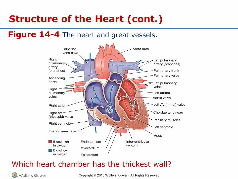
Figure 14-4 The heart and great vessels.
Which heart chamber has the thickest wall?
Structure of the Heart (cont.)
Copyright © 2015 Wolters Kluwer • All Rights Reserved
Structure of the Heart (cont.)
Four Valves
• Atrioventricular valves
– Entrance valves
• Right atrioventricular (AV) valve (tricuspid valve)
• Left atrioventricular (AV) valve (bicuspid valve)
• Semilunar valves
– Exit valves
• Pulmonary valve
• Aortic valve

Copyright © 2015 Wolters Kluwer • All Rights Reserved
Figure 14-5 Heart valves (superior view from posterior, atria removed).
How many cusps does the right AV valve have? The left?
Structure of the Heart (cont.)
Copyright © 2015 Wolters Kluwer • All Rights Reserved
Structure of the Heart (cont.)
Blood Supply to the Myocardium
• Coronary arteries
– Right coronary artery
– Left coronary artery
• Cardiac veins

Copyright © 2015 Wolters Kluwer • All Rights Reserved
Figure 14-6 Blood vessels that supply the myocardium.
What is the largest cardiac vein, and where does it lead?
Structure of the Heart (cont.)

Copyright © 2015 Wolters Kluwer • All Rights Reserved
Figure 14-7 Opening of coronary arteries in the aortic valve (anterior view).
Structure of the Heart (cont.)

Copyright © 2015 Wolters Kluwer • All Rights Reserved
Structure of the Heart (cont.)
Checkpoints
14-1 What are the names of the innermost, middle, and outermost layers of the heart wall?
14-2 What is the name of the sac that encloses the heart?
14-3 What is the heart’s upper receiving chamber on each side called? What is the lower pumping chamber called?
14-4 What is the purpose of the valves in the heart?
14-5 What is the name of the system that supplies blood to the myocardium?
Copyright © 2015 Wolters Kluwer • All Rights Reserved
Heart Function
Learning Objectives
6. Briefly describe the cardiac cycle.
7. Name and locate the components of the heart’s conduction system.
8. Explain the effects of the autonomic nervous system (ANS) on the heart rate.
9. List and define several terms that describe variations in heart rates.
10. Explain what produces each of the two normal heart sounds, and identify the usual cause of a murmur.
Copyright © 2015 Wolters Kluwer • All Rights Reserved
Heart Function (cont.)
Cardiac Cycle
• Series of events occurring in the heart during one heartbeat
– Systole (active phase, contraction)
– Diastole (resting phase)
Copyright © 2015 Wolters Kluwer • All Rights Reserved
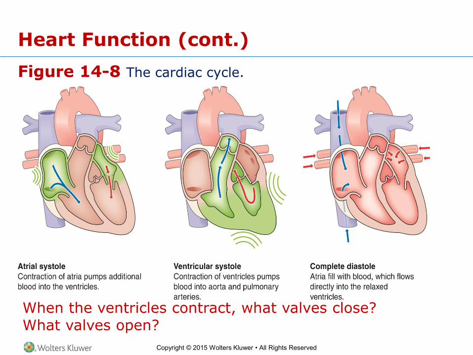
Figure 14-8 The cardiac cycle.
When the ventricles contract, what valves close? What valves open?
Heart Function (cont.)
Copyright © 2015 Wolters Kluwer • All Rights Reserved
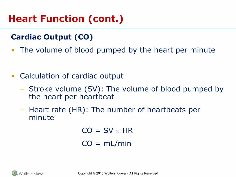
Heart Function (cont.)
Cardiac Output (CO)
• The volume of blood pumped by the heart per minute
• Calculation of cardiac output
– Stroke volume (SV): The volume of blood pumped by the heart per heartbeat
– Heart rate (HR): The number of heartbeats per minute
CO = SV HR
CO = mL/min

Copyright © 2015 Wolters Kluwer • All Rights Reserved
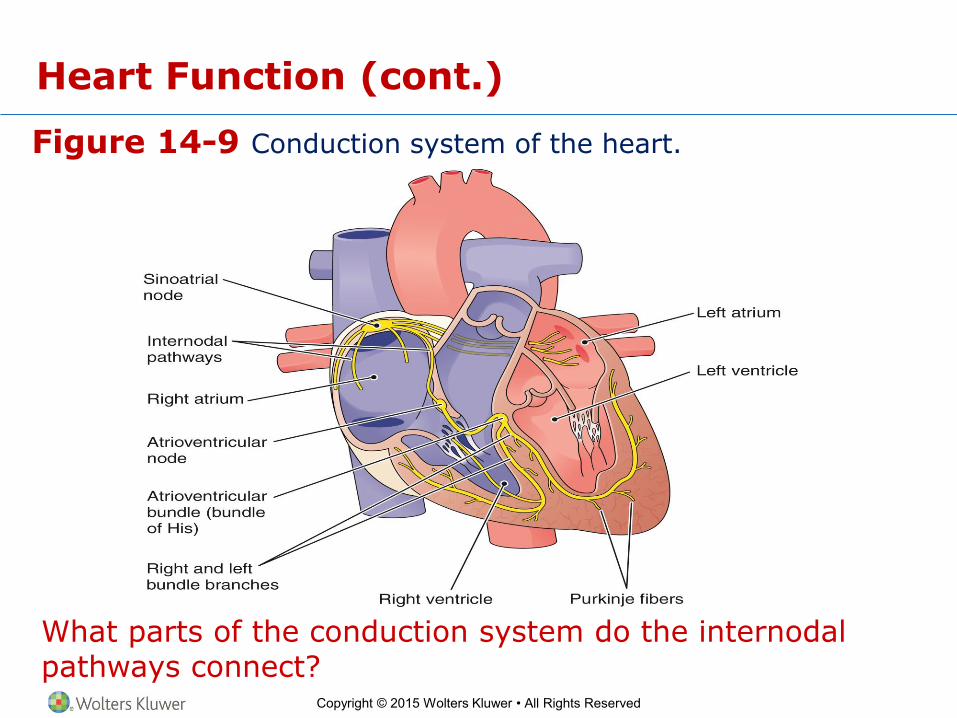
Heart Function (cont.)
The Heart’s Conduction System
• Produces electrical energy, which stimulates cardiac muscle
• Components
– Sinoatrial (SA) node (pacemaker)
– Internodal pathways
– Atrioventricular (AV) node
– Atrioventricular bundle (bundle of His)
– Purkinje fibers (conduction myofibers)
Copyright © 2015 Wolters Kluwer • All Rights Reserved
Figure 14-9 Conduction system of the heart.
What parts of the conduction system do the internodal pathways connect?
Heart Function (cont.)
Copyright © 2015 Wolters Kluwer • All Rights Reserved
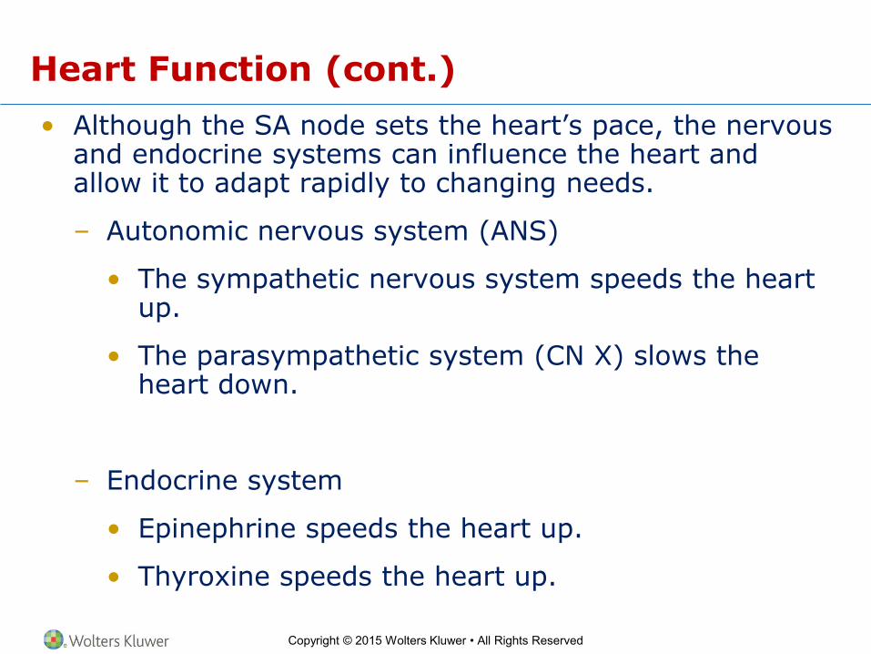
Heart Function (cont.)
• Although the SA node sets the heart’s pace, the nervous and endocrine systems can influence the heart and allow it to adapt rapidly to changing needs.
– Autonomic nervous system (ANS)
• The sympathetic nervous system speeds the heart up.
• The parasympathetic system (CN X) slows the heart down.
– Endocrine system
• Epinephrine speeds the heart up.
• Thyroxine speeds the heart up.

Copyright © 2015 Wolters Kluwer • All Rights Reserved
Figure 14-10 Autonomic nervous system (ANS) regulation of the heart.
Which cranial nerve carries parasympathetic impulses to the heart?
Heart Function (cont.)
Copyright © 2015 Wolters Kluwer • All Rights Reserved
Heart Function (cont.)
Variations in Heart Rates
• Bradycardia
• Tachycardia
• Sinus arrhythmia
• Premature ventricular contraction (PVC)
Copyright © 2015 Wolters Kluwer • All Rights Reserved
Heart Function (cont.)
Normal and Abnormal Heart Sounds
• Normal
– Lub
– Dup
• Abnormal
– Organic murmur
• Functional murmur
– Normal sounds heard as the heart works

Copyright © 2015 Wolters Kluwer • All Rights Reserved
Heart Function (cont.)
Checkpoints
14-6 What name is given to the contraction phase of the cardiac cycle? To the relaxation phase?
14-7 What is cardiac output? What two factors determine cardiac output?
14-8 What is the scientific name of the heart’s pacemaker?
14-9 What system exerts the main influence on the rate and strength of heart contractions?
14-10 What is a heart murmur?
Copyright © 2015 Wolters Kluwer • All Rights Reserved
Heart Studies
Learning Objective
11. Briefly describe five methods used to study the heart.
Copyright © 2015 Wolters Kluwer • All Rights Reserved
Heart Studies (cont.)
Methods of Studying the Heart
• Stethoscope
• Electrocardiograph (ECG or EKG)
– Electrodes
• Catheterization
– Fluoroscope
– Coronary angiography
• Coronary computed tomography angiography (CTA)
• Echocardiography

Copyright © 2015 Wolters Kluwer • All Rights Reserved
Figure 14-11 Normal electrocardiography (ECG) tracing.
What is the length of the cardiac cycle shown in this diagram?
Heart Studies (cont.)
Copyright © 2015 Wolters Kluwer • All Rights Reserved
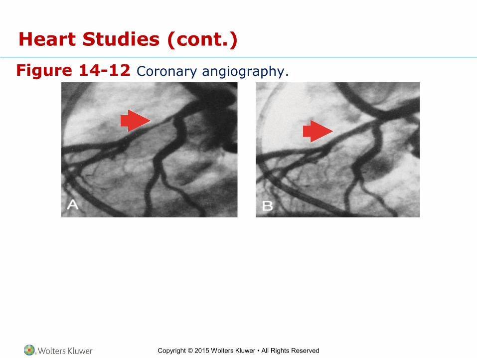
Figure 14-12 Coronary angiography.
Heart Studies (cont.)
Copyright © 2015 Wolters Kluwer • All Rights Reserved
Heart Studies (cont.)
Checkpoints
14-11 What do ECG and EKG stand for?
14-12 What is the general term for using a thin tube threaded through a vessel for diagnosis or repair?
14-13 What techniques use a dye and x-rays to visualize the coronary arteries?

Copyright © 2015 Wolters Kluwer • All Rights Reserved
Heart Disease
Learning Objectives
12. Describe six types of heart disease.
13. List four risk factors for coronary artery disease that cannot be modified.
14. List seven risk factors for coronary artery disease that can be modified.
15. Describe three approaches to the treatment of heart disease.

Copyright © 2015 Wolters Kluwer • All Rights Reserved
Heart Disease (cont.)
• The most common cause of death in industrialized
countries is heart and circulatory system disease.
Copyright © 2015 Wolters Kluwer • All Rights Reserved
Heart Disease (cont.)
Classifications of Heart Disease
• Inflammatory heart disease
– Endocarditis
– Myocarditis
– Pericarditis
• Abnormalities of heart rhythm
– Arrhythmia
– Heart block
Copyright © 2015 Wolters Kluwer • All Rights Reserved
Heart Disease (cont.)
Classifications of Heart Disease (cont.)
• Congenital heart disease
– Atrial septal defect
– Patent ductus arteriosus
– Ventricular septal defect
– Coarctation of the aorta
– Tetralogy of Fallot

Copyright © 2015 Wolters Kluwer • All Rights Reserved
Figure 14-13 Congenital heart defects.
Heart Disease (cont.)

Copyright © 2015 Wolters Kluwer • All Rights Reserved
Heart Disease (cont.)
Classification of Heart Disease (cont.)
• Valve disorders
– Rheumatic heart disease
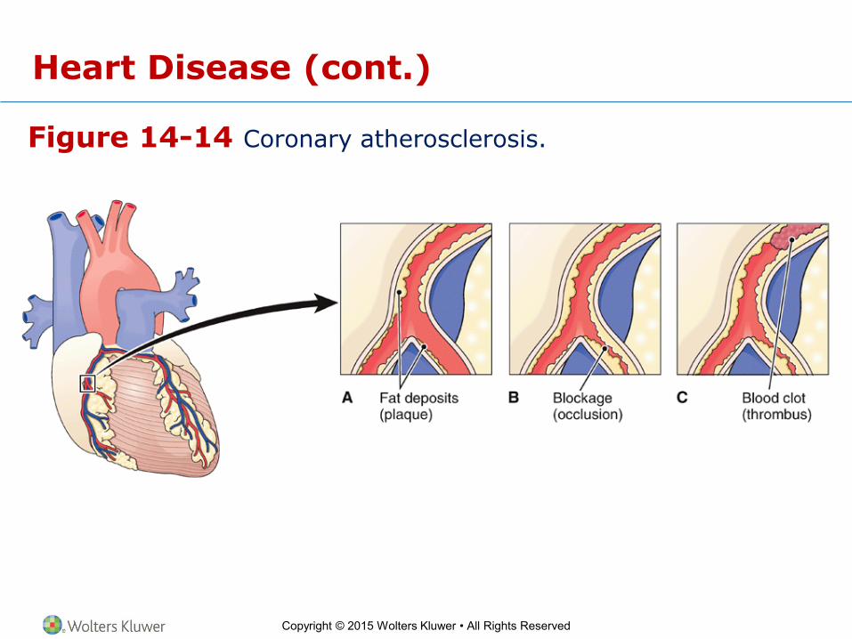
• Coronary artery disease
– Atherosclerosis
• Ischemia
• Thrombosis
– Angina pectoris
– Myocardial infarction
• Coronary thrombosis
• Occlusion
• Congestive heart failure
Copyright © 2015 Wolters Kluwer • All Rights Reserved
Figure 14-14 Coronary atherosclerosis.
Heart Disease (cont.)

Copyright © 2015 Wolters Kluwer • All Rights Reserved
Figure 14-15 Myocardial infarction (MI).
Heart Disease (cont.)
Copyright © 2015 Wolters Kluwer • All Rights Reserved
Treatment of Myocardial Infarction
• Depends on extent and location of the damage
• Cardiopulmonary resuscitation (CPR)
• Defibrillation
– Automated external defibrillator (AED)
• Thrombolytic drugs
• Surgical treatment
– Angioplasty
– Coronary artery bypass surgery
– Pacemakers
• Supportive care
– Morphine
– Oxygen

Copyright © 2015 Wolters Kluwer • All Rights Reserved
Prevention of Coronary Artery Disease
Risk Factors for Coronary Artery Disease
Risk Factors That Cannot Be Modified
Risk Factors That Can Be Modified
Age Smoking and other forms of tobacco use
Gender Physical inactivity
Heredity Overweight
Body type Saturated fat in diet
Hypertension
Type 2 diabetes
Sleep apnea
Copyright © 2015 Wolters Kluwer • All Rights Reserved
Prevention of Coronary Artery Disease (cont.)
• Regular physical examinations
• Control of risk factors
• Monitor “markers”
– C-reactive protein (CRP)
– Homocysteine
– Lipoprotein

Copyright © 2015 Wolters Kluwer • All Rights Reserved
Heart Disease (cont.)
• Congestive heart failure
– The heart is unable to pump sufficient blood.
– Heart chambers enlarge.
– Blood backs up into lungs.
– Ventricular muscles have decreased ability to
contract.
– Fluid accumulates in the lungs, liver, abdomen, legs.

Copyright © 2015 Wolters Kluwer • All Rights Reserved
Heart Disease (cont.)
Checkpoints
14-14 What are the three types of heart inflammation?
14-15 What is an abnormal heart rhythm called?
14-16 What is congenital heart disease?
14-17 What types of organisms cause rheumatic fever?
14-18 What degenerative process commonly causes narrowing of the vessels in coronary artery disease?
14-19 What is the medical term for a “heart attack?”
14-20 What is the general name for a device that restores a normal heart rhythm?
Copyright © 2015 Wolters Kluwer • All Rights Reserved
Treatment of Heart Disease
• Lifestyle changes
• Medications
– Statins
– Anticoagulants
• Aspirin
• Warfarin
– Digitalis
– Beta-adrenergic blockers
– Antiarrhythmic agents
– Slow calcium channel blockers
Copyright © 2015 Wolters Kluwer • All Rights Reserved
Treatment of Heart Disease (cont.)
• Pacemakers
• Heart surgery
– Coronary artery bypass graft (CABG)
– Angioplasty
– Coronary atherectomy
– Cardiac ablation
– Surgical transplantation
Copyright © 2015 Wolters Kluwer • All Rights Reserved
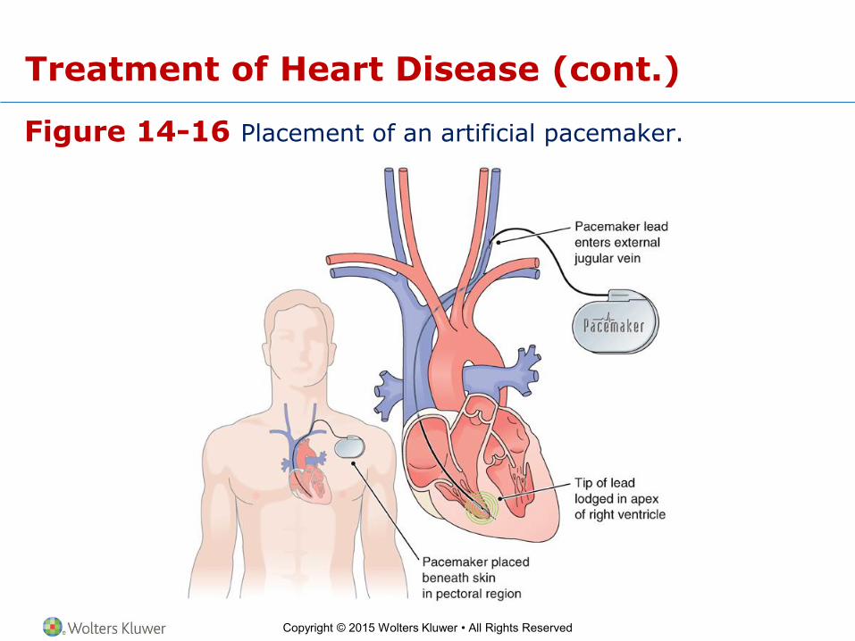
Figure 14-16 Placement of an artificial pacemaker.
Treatment of Heart Disease (cont.)
Copyright © 2015 Wolters Kluwer • All Rights Reserved
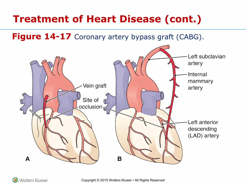
Figure 14-17 Coronary artery bypass graft (CABG).
Treatment of Heart Disease (cont.)
Copyright © 2015 Wolters Kluwer • All Rights Reserved
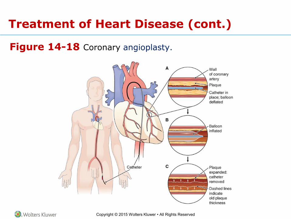
Figure 14-18 Coronary angioplasty.
Treatment of Heart Disease (cont.)
Copyright © 2015 Wolters Kluwer • All Rights Reserved
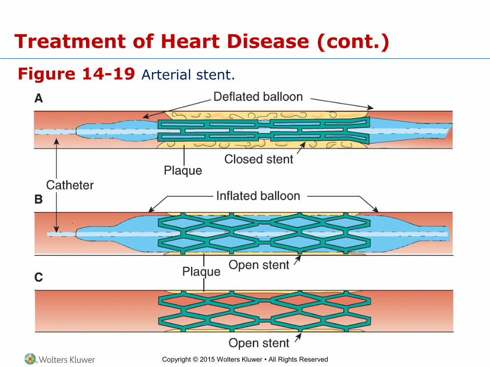
Figure 14-19 Arterial stent.
Treatment of Heart Disease (cont.)

Copyright © 2015 Wolters Kluwer • All Rights Reserved
Treatment of Heart Disease (cont.)
Checkpoints
14-21 How do aspirin and warfarin act to prevent heart attacks?
14-22 What is the name for a device that is wired to the heart to regulate the heartbeat?
14-23 What does CABG stand for?

Copyright © 2015 Wolters Kluwer • All Rights Reserved
Effects of Aging
Learning Objective
16. List four changes that may occur in the heart with age.
Copyright © 2015 Wolters Kluwer • All Rights Reserved
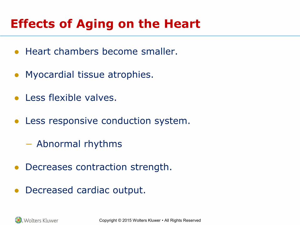
Effects of Aging on the Heart
● Heart chambers become smaller.
● Myocardial tissue atrophies.
● Less flexible valves.
● Less responsive conduction system.
− Abnormal rhythms
● Decreases contraction strength.
● Decreased cardiac output.

Copyright © 2015 Wolters Kluwer • All Rights Reserved
Case Study
Learning Objective
17. Referring to the case study, list the emergency and surgical procedures commonly performed following a myocardial infarction and explain why they are done.

Copyright © 2015 Wolters Kluwer • All Rights Reserved
Case Study (cont.)
Emergency and Surgical Procedures Commonly Performed following a Myocardial Infarction
• Cardiopulmonary resuscitation
• Defibrillation
• Administration of thrombolytic medication and nitroglycerine
• Administration of oxygen
• Administration of morphine
• Monitor assays of specific substances in the blood, that is, cardiac enzymes
• Monitor the cardiac muscle’s electrical activity
Copyright © 2015 Wolters Kluwer • All Rights Reserved
Case Study (cont.)
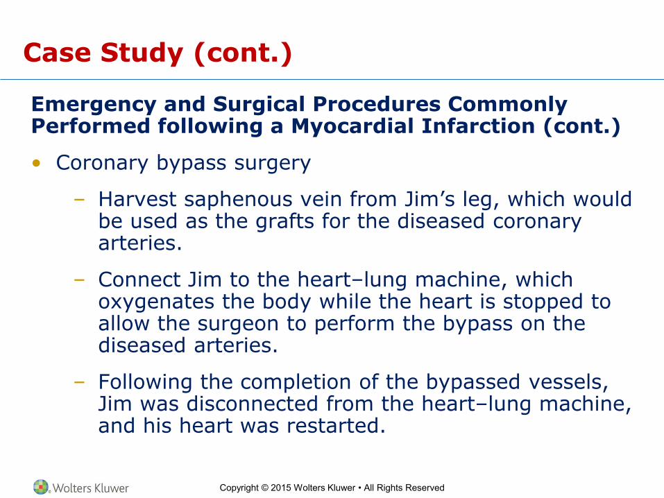
Emergency and Surgical Procedures Commonly Performed following a Myocardial Infarction (cont.)
• Coronary bypass surgery
– Harvest saphenous vein from Jim’s leg, which would be used as the grafts for the diseased coronary arteries.
– Connect Jim to the heart–lung machine, which oxygenates the body while the heart is stopped to allow the surgeon to perform the bypass on the diseased arteries.
– Following the completion of the bypassed vessels, Jim was disconnected from the heart–lung machine, and his heart was restarted.

Copyright © 2015 Wolters Kluwer • All Rights Reserved
Word Anatomy
Learning Objective
18. Show how word parts are used to build words related to the heart.

Copyright © 2015 Wolters Kluwer • All Rights Reserved
Word Anatomy (cont.)
Word Part Meaning Example
Structure of the Heart
cardi/o heart The myocardium is the heart muscle.
pulmon/o lung The pulmonary circuit carries blood to the lungs.
Heart Function
brady/o slow Bradycardia is a slow heart rate.
sin/o sinus The sinoatrial node is in a space (sinus) in the wall of the right atrium.
tachy- rapid Tachycardia is a rapid heart rate.
Copyright © 2015 Wolters Kluwer • All Rights Reserved
Word Anatomy (cont.)
Word Part Meaning Example
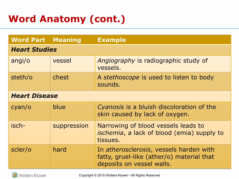
Heart Studies
angi/o vessel Angiography is radiographic study of vessels.
steth/o chest A stethoscope is used to listen to body sounds.
Heart Disease
cyan/o blue Cyanosis is a bluish discoloration of the skin caused by lack of oxygen.
isch- suppression Narrowing of blood vessels leads to ischemia, a lack of blood (emia) supply to tissues.
scler/o hard In atherosclerosis, vessels harden with fatty, gruel-like (ather/o) material that deposits on vessel walls.
Copyright © 2015 Wolters Kluwer • All Rights Reserved
Word Anatomy (cont.)
Word Part Meaning Example
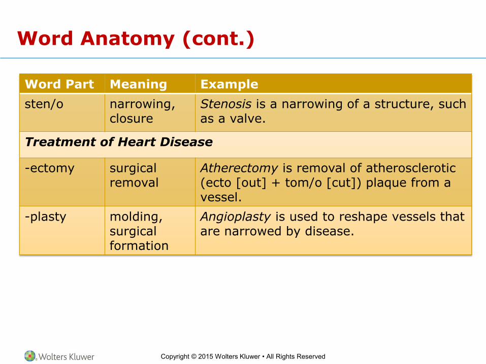
sten/o narrowing, closure
Stenosis is a narrowing of a structure, such as a valve.
Treatment of Heart Disease
-ectomy surgical removal
Atherectomy is removal of atherosclerotic (ecto [out] + tom/o [cut]) plaque from a vessel.
-plasty molding, surgical formation
Angioplasty is used to reshape vessels that are narrowed by disease.