Embed Size (px)
Citation preview

Basic ECGBasic ECG

ObjectivesObjectives
Identify the functions of the electrical Identify the functions of the electrical conduction system of the heart. conduction system of the heart.
Demonstrate accurate ECG rhythm Demonstrate accurate ECG rhythm interpretation.interpretation.
Identify key characteristics of normal Identify key characteristics of normal and abnormal rhythms.and abnormal rhythms.

Sinoatrial NodeSinoatrial Node(SA Node)(SA Node)
Located in RA near SVCLocated in RA near SVC Normal pacemaker of heartNormal pacemaker of heart Initiates an impulseInitiates an impulse Intrinsic rate of 60-100Intrinsic rate of 60-100

Intra-Atrial PathwaysIntra-Atrial Pathways Located in atrial tissue between SA & Located in atrial tissue between SA &
AV nodesAV nodes Conducts impulse from SA Conducts impulse from SA atrial atrial
musculature musculature AV node AV node Anterior, middle, and posterior tractsAnterior, middle, and posterior tracts

Atrioventricular NodeAtrioventricular Node(AV Node)(AV Node)
Located near tricuspid valveLocated near tricuspid valve Delays impulse from atriaDelays impulse from atria Allows for ventricular fillingAllows for ventricular filling Protective mechanism against rapid Protective mechanism against rapid
supraventricular impulsessupraventricular impulses

Junctional TissueJunctional Tissue Tissue in lower AV nodeTissue in lower AV node Back-up pacemakerBack-up pacemaker Intrinsic rate of 40-60Intrinsic rate of 40-60

Bundle of His & R & L Bundle of His & R & L Bundle BranchesBundle Branches
Bundle of His connects AV node to Bundle of His connects AV node to
Right & Left bundle branchesRight & Left bundle branches R bundle carries impulse to RVR bundle carries impulse to RV L bundle carries impulse to LVL bundle carries impulse to LV

Ventricular TissueVentricular Tissue Back-up pacemakerBack-up pacemaker Intrinsic rate 20-40Intrinsic rate 20-40

Purkinje SystemPurkinje System
Distal to the bundle branchesDistal to the bundle branches Rapidly conducts impulses to Rapidly conducts impulses to
ventricular subendocardial ventricular subendocardial layerslayers

ECG PaperECG Paper Horizontal axis represents timeHorizontal axis represents time
small box = 0.04 secondssmall box = 0.04 seconds 5 small boxes = 1 large block = 0.20 5 small boxes = 1 large block = 0.20
secondsseconds 5 large boxes = 1 second5 large boxes = 1 second Normal strip = 30 large boxes = 6 Normal strip = 30 large boxes = 6
secondsseconds

ECG Paper cont...ECG Paper cont... Vertical axis measures:Vertical axis measures:
amplitude in millimeters (mm)amplitude in millimeters (mm) electrical voltage in millivolts (mV)electrical voltage in millivolts (mV) 1 small block = 1 mm or 0.1 mV1 small block = 1 mm or 0.1 mV 1 large block = 5 mm or 0.5 mV1 large block = 5 mm or 0.5 mV

TerminologyTerminology
DepolarizationDepolarization– The electrical activation of a cardiac cellThe electrical activation of a cardiac cell
RepolarizationRepolarizationElectrical recovery of the cardiac cellElectrical recovery of the cardiac cell

The P WaveThe P Wave Atrial depolarization Atrial depolarization Characteristics:Characteristics:
precedes the QRSprecedes the QRS 2-3 mm high2-3 mm high 0.06-0.12 seconds0.06-0.12 seconds round & uprightround & upright

QRS ComplexQRS Complex Ventricular Ventricular
depolarization depolarization Characteristics:Characteristics:
follows PR follows PR intervalinterval
< 0.12 seconds < 0.12 seconds

T WaveT Wave Ventricular Ventricular
repolarization repolarization
Characteristics:Characteristics: follows S wavefollows S wave round & smoothround & smooth
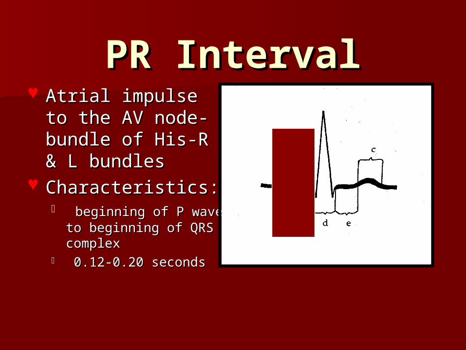
PR IntervalPR Interval Atrial impulse to Atrial impulse to
the AV node-the AV node-bundle of His-R & L bundle of His-R & L bundlesbundles
Characteristics:Characteristics: beginning of P wave to beginning of P wave to
beginning of QRS beginning of QRS complexcomplex
0.12-0.20 seconds0.12-0.20 seconds

ST SegmentST Segment Early Ventricular Early Ventricular
Repolarization Repolarization Characteristics:Characteristics:
from end of QRS from end of QRS complex to complex to beginning of T wavebeginning of T wave

QT IntervalQT Interval Ventricular Ventricular
depolarization & depolarization & repolarizationrepolarization
Characteristics:Characteristics: beginning of QRS beginning of QRS
to end of T waveto end of T wave 0.35 to 0.45 0.35 to 0.45
secondsseconds

MonitoringMonitoringFive leadsFive leads

Electrodes & LeadsElectrodes & Leads Electrodes measure the direction of Electrodes measure the direction of
electrical currentelectrical current The current is transformed into The current is transformed into
waveformswaveforms The ECG records the waveform The ECG records the waveform
information from different views & information from different views & leadsleads

LeadsLeads Provide different views of the Provide different views of the
heart’s electrical activityheart’s electrical activity
LeadLead view between a + pole & a - poleview between a + pole & a - pole the axis refers to the direction of the current the axis refers to the direction of the current
moving through the heartmoving through the heart direction of the waveform on the ECGdirection of the waveform on the ECG

Lead SelectionLead Selection
Based on patient historyBased on patient history

Calculation of Heart Calculation of Heart RatesRates
R-R….. SMALL BOXES….. DIVIDE into 1500R-R….. SMALL BOXES….. DIVIDE into 1500
6 SECOND METHOD6 SECOND METHOD
RATE CHARTRATE CHART
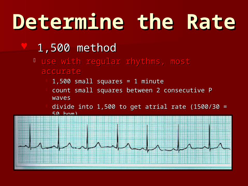
Determine the RateDetermine the Rate 1,500 method1,500 method
use with regular rhythms, most accurateuse with regular rhythms, most accurate 1,500 small squares = 1 minute1,500 small squares = 1 minute count small squares between 2 consecutive P wavescount small squares between 2 consecutive P waves divide into 1,500 to get atrial rate (1500/30 = 50 bpm)divide into 1,500 to get atrial rate (1500/30 = 50 bpm) count small squares between 2 consecutive R wavescount small squares between 2 consecutive R waves divide into 1,500 to get ventricular ratedivide into 1,500 to get ventricular rate

Determine the Rate Determine the Rate cont..cont..
6 Second method6 Second method good method if rhythm is irregulargood method if rhythm is irregular
use a 6 second stripuse a 6 second strip count the number of P waves for the atrial rate and count the number of P waves for the atrial rate and
multiply by 10multiply by 10 count the number of R waves for the ventricular rate count the number of R waves for the ventricular rate
and multiply by 10and multiply by 10

Determine the Rate Determine the Rate cont...cont...
Sequence method-memorization of: Sequence method-memorization of: 300-150-100-75-60-50-43300-150-100-75-60-50-43
for atrial rate, locate a P wave on a heavy black for atrial rate, locate a P wave on a heavy black lineline
assign the next heavy black line 300 & begin assign the next heavy black line 300 & begin counting backwards until you reach the next P counting backwards until you reach the next P wavewave
for ventricular rate, repeat the same sequencefor ventricular rate, repeat the same sequence

ECG - HOW TO READ IT?ECG - HOW TO READ IT?CHECK THE ATRIAL and VENTRICULAR RHYTHMCHECK THE ATRIAL and VENTRICULAR RHYTHM
Is it REGULAR OR IRREGULAR?Is it REGULAR OR IRREGULAR?
CALCULATE THE ATRIAL AND VENTRICULAR RATECALCULATE THE ATRIAL AND VENTRICULAR RATESame or different?Same or different?
LOOK FOR P, Q, R, S, AND TLOOK FOR P, Q, R, S, AND TIs there a P wave for every QRS?Is there a P wave for every QRS?
Is there a QRS for every P?Is there a QRS for every P?
PR Interval? QRS width?PR Interval? QRS width?
FIND THE ORIGINFIND THE ORIGINSinus, Atrial, Junctional, or Ventricular?Sinus, Atrial, Junctional, or Ventricular?
KNOW THE MECHANISMKNOW THE MECHANISMFast, slow, premature, late, fib, flutter or blocked, pacedFast, slow, premature, late, fib, flutter or blocked, paced

Measure the PR Measure the PR IntervalInterval
Is the duration 0.12-0.20 seconds?Is the duration 0.12-0.20 seconds? Is the interval consistent?Is the interval consistent?

Measure the Duration Measure the Duration of the QRS Complexof the QRS Complex Is the duration < 0.12 seconds?Is the duration < 0.12 seconds? Are all of the complexes the same Are all of the complexes the same
size & shape?size & shape? Is there a QRS after each P wave?Is there a QRS after each P wave?

CheckpointCheckpoint1.1. The ECG provides information about:The ECG provides information about:
a.a. the contractility of the heartthe contractility of the heart
b.b. the electrical activity of the heartthe electrical activity of the heart
c. c. cardiac output and resistancecardiac output and resistance
d.d. all of the aboveall of the above
2.2. In the electrocardiogram, the QRS represents:In the electrocardiogram, the QRS represents:
a. ventricular contractiona. ventricular contraction
b. atrial contractionb. atrial contraction
c. ventricular depolarizationc. ventricular depolarization
d. discharge of impulse from the sinus noded. discharge of impulse from the sinus node
3.3. Indicate the intrinsic rate of the following:Indicate the intrinsic rate of the following:
Sinus node Sinus node
Junctional tissue Junctional tissue ______________
Ventricular tissue Ventricular tissue

Sinus RhythmsSinus Rhythms
Normal Sinus Rhythms (NSR)Normal Sinus Rhythms (NSR) Sinus Tachycardia (ST)Sinus Tachycardia (ST) Sinus Bradycardia (SB)Sinus Bradycardia (SB) Sinus Dysrhythmia (SD)Sinus Dysrhythmia (SD) Sinus Pause (Sinus Arrest)Sinus Pause (Sinus Arrest)

Normal Sinus RhythmNormal Sinus Rhythm
The SA node (normal Pacemaker) of the heart is in The SA node (normal Pacemaker) of the heart is in controlcontrol
RhythmRhythm: Regular: Regular Rate:Rate: 60-100/min 60-100/min
P WaveP Wave Precedes each QRS, normal. Precedes each QRS, normal.
PR:PR: 0.12-0.20Sec 0.12-0.20Sec QRS:QRS: < 0.12 sec & Constant < 0.12 sec & Constant

Sinus TachycardiaSinus TachycardiaSA node fires faster than 100/minSA node fires faster than 100/min
Rhythm:Rhythm: Regular Regular Rate:Rate: 100-160/min 100-160/min
P Wave:P Wave: Normal, precedes each QRS Normal, precedes each QRS
PR:PR: 0.12 - 0.20 sec 0.12 - 0.20 sec QRS:QRS: <0.12 sec <0.12 sec

Sinus TachycardiaSinus TachycardiaCauses:Causes:
* Anemia, Hypoxia, Hypovolemia, * Anemia, Hypoxia, Hypovolemia, HypotensionHypotension
* Exercise, Emotion, Anxiety, Pain* Exercise, Emotion, Anxiety, Pain
* Fever* Fever
* Drug related-Caffeine, Epinephrine, Cocaine* Drug related-Caffeine, Epinephrine, Cocaine
* Early sign of CHF* Early sign of CHF
* Theophylline toxicity* Theophylline toxicity
* Hyperthyroidism* Hyperthyroidism
Management:Management:
* Assess for cause & treat it* Assess for cause & treat it
* Beta Blockers for primary tachycardia* Beta Blockers for primary tachycardia

Sinus BradycardiaSinus Bradycardia
SA Node fires slower than 60/minSA Node fires slower than 60/min
Rhythm:Rhythm: Regular Regular Rate:Rate: < 60/min < 60/min
P Wave:P Wave: normal, precedes each QRS normal, precedes each QRS
PR:PR: 0.12 - 0.20 sec 0.12 - 0.20 sec QRS:QRS: < 0.12 sec < 0.12 sec

Sinus Bradycardia
CausesCauses Normal in healthy, Normal in healthy,
young, athletesyoung, athletes Vagal stimulation… MI, Vagal stimulation… MI,
Vomiting, Straining at Vomiting, Straining at stool, Pharyngeal stool, Pharyngeal suctioningsuctioning
Drug effect… Beta Drug effect… Beta blockersblockers
Increased ICPIncreased ICP HypokalemiaHypokalemia Sick Sinus syndromeSick Sinus syndrome
SignificanceSignificance If severe or prolonged, If severe or prolonged,
may cause decrease in may cause decrease in cardiac output and cardiac output and syncopesyncope
At risk for escape rhythm At risk for escape rhythm and or premature beats and or premature beats to gain control due to to gain control due to long pauseslong pauses

Sinus Bradycardia Interventions
Assess patient
Hold digoxin, if digoxin toxicity
Relieve source of vagal stimulation, if possible (treat nausea, shorten periods of suctioning, no valsalvas)May need to adjust drug regimen
If symptomatic, treat with Atropine
May require temporary pacemaker

Sinus Dysrhythmia Sinus Dysrhythmia (Sinus Arrhythmia)(Sinus Arrhythmia)
Irregular heart rate; Sinus node in charge;Irregular heart rate; Sinus node in charge;
usually varies with respiratory cycleusually varies with respiratory cycle
Rhythm :Rhythm : IrregularIrregular Rate :Rate : 60 - 10060 - 100
P wave :P wave : Normal Normal PR : PR : NormalNormal QRS:QRS: Normal Normal
The longest R-R interval - the shortest R-R intervalThe longest R-R interval - the shortest R-R interval = > 0.12 seconds= > 0.12 seconds

Sinus DysrhythmiaSinus DysrhythmiaSignificance: Usually none
Causes:Common in children & outgrown in teensVariation of Sinus rhythmVagal stimulation
Treatment:Usually noneR/O more serious irregular rhythmMinimize vagal stimulation

Sinus Pause (Arrest)Sinus Pause (Arrest)Sinus node fails to generate an impulse for one or Sinus node fails to generate an impulse for one or
more beats; usually reset by sinus node but more beats; usually reset by sinus node but escape beats/rhythms may occurescape beats/rhythms may occur
Rhythm:Rhythm: Regular except for pause Regular except for pause PR:PR: normal normal
Rate:Rate: depends on underlying rhythm depends on underlying rhythm QRS:QRS: normal normal
No PNo P wave preceding pause wave preceding pause

Sinus PauseSinus PauseCAUSES May decrease
CO Duration of
pause determines the seriousness
of dysrhythmia
SIGNIFICANCEVagal stimulationSick Sinus syndromeDig. ToxicityBeta blockersCa channel blockersIschemia of SA
nodePericarditisHyperkalemaAmiodarone
INTERVENTIONSTreat causeAtropine for acute bradycardiaIf asleep, wake
the patientPause over 3 seconds… Evaluation for a
pacemaker (External or permanent)

CheckpointCheckpoint
True or FalseTrue or False1.1. Sinus dysrhythmia is dangerous and requires immediate Sinus dysrhythmia is dangerous and requires immediate
intervention.intervention.
2.2. The treatment of choice for sinus tachycardia is related to The treatment of choice for sinus tachycardia is related to the cause.the cause.
3.3. Sinus dysrhythmia is characterized by a slowing and Sinus dysrhythmia is characterized by a slowing and speeding of the rate.speeding of the rate.
4.4. The first nursing action in relation to a rhythm disturbance The first nursing action in relation to a rhythm disturbance is to check the patient and assess level of consciousness.is to check the patient and assess level of consciousness.

Atrial RhythmsAtrial Rhythms
Premature Atrial Complex (PAC)Premature Atrial Complex (PAC) Supraventricular Tachycardia (SVT)Supraventricular Tachycardia (SVT) Atrial FlutterAtrial Flutter Atrial FibrillationAtrial Fibrillation
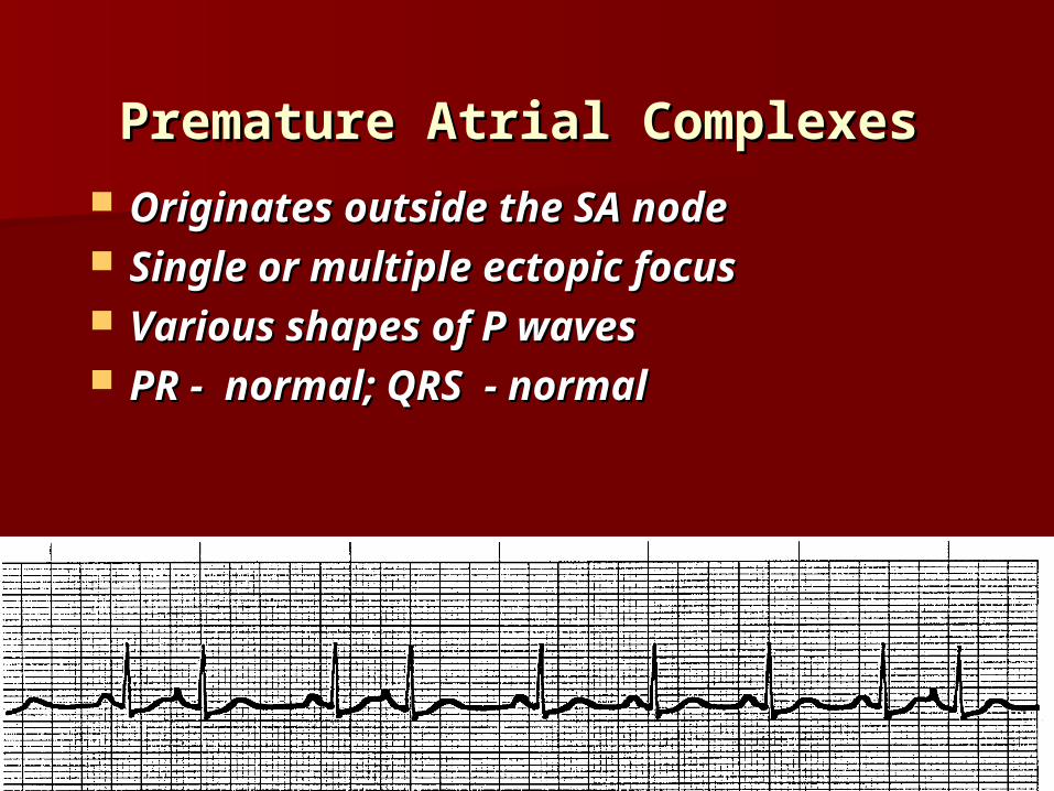
Premature Atrial ComplexesPremature Atrial Complexes Originates outside the SA nodeOriginates outside the SA node Single or multiple ectopic focusSingle or multiple ectopic focus Various shapes of P wavesVarious shapes of P waves PR - normal; QRS - normalPR - normal; QRS - normal

PACsPACsSignificance Cause Interventions
Usually not serious In pts. with heart disease… more serious In MI … early sign of CHF
Common Emotional stress Alcohol, caffeine, tobacco Electrolyte imbalance Hypoxia Digoxin toxicity Hyperthyroidism CV disease
Drugs
Infrequent … No treatment Eliminate cause Drugs

SVTSVT
ABSOLUTELY REGULAR RHYTHM !ABSOLUTELY REGULAR RHYTHM !
USUALLY OCCURS SUDDENLYUSUALLY OCCURS SUDDENLY
RATE: 160-240/minRATE: 160-240/min
P WAVE: MAY NOT BE SEENP WAVE: MAY NOT BE SEEN
PR : NOT MEASURABLEPR : NOT MEASURABLE
QRS: NORMALQRS: NORMAL

Significance - SVTSignificance - SVT •If rapid, can decrease cardiac If rapid, can decrease cardiac outputoutput
•Can causeCan cause•anxietyanxiety•anginaangina•palpitationspalpitations•shortness of breathshortness of breath•decreased level of consciousnessdecreased level of consciousness•decreased BPdecreased BP•shockshock•pulmonary congestionpulmonary congestion•CHFCHF•acute MIacute MI

Causes - SVTCauses - SVT
NORMAL CARDIAC OTHER
Caffeine Recreational drugs Electrolyte imbalance Hypoxia Physical & psychological Stress
MI Cardiomyopathy WPW syndrome Sick-Sinus syndrome
Corpulmonale (COPD) Hyperthyroidism Dig. Toxicity
INTERVENTI ONS Check Digoxin Level Valsalva Maneuver
Carotid sinus massage – physicians only Drugs – Adenosine (drug of choice)
Drugs – Verapamil, Diltiazem, Oxygen; check ABG if needed
Cardioversion Patient education – stress management

Carotid MassageCarotid Massage
By MDs onlyBy MDs only!!
Both diagnose & terminate PSVTBoth diagnose & terminate PSVT
Auscultate first for bruitAuscultate first for bruit
Never compress Never compress
both carotidsboth carotids
simultaneously!simultaneously!

CardioversionCardioversionSynchronized cardioversion delivers Synchronized cardioversion delivers
electrical stimulus during depolarization - electrical stimulus during depolarization - depolarizes all cells simultaneously, depolarizes all cells simultaneously,
allowing SA node to resume the allowing SA node to resume the pacemaker rolepacemaker role
QRS complex must be present QRS complex must be present
Usually electiveUsually elective
Potassium, digoxin level,emergency Potassium, digoxin level,emergency equipment,O2, NPO, IV, TEE, Sedationequipment,O2, NPO, IV, TEE, Sedation
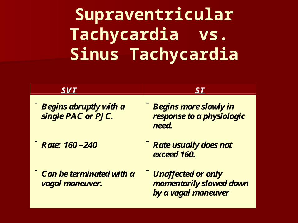
Supraventricular Tachycardia vs.
Sinus Tachycardia
SVT ST
Begins abruptly with asingle PAC or PJC.
Rate: 160 –240
Can be terminated with avagal maneuver.
Begins more slowly inresponse to a physiologicneed.
Rate usually does notexceed 160.
Unaffected or onlymomentarily slowed downby a vagal maneuver

Atrial FlutterAtrial Flutter Atrial rate of 250 - 350 beats / minAtrial rate of 250 - 350 beats / min Originating from a single ectopic Originating from a single ectopic
focusfocus Saw toothed F wavesSaw toothed F waves AV node delays the impulses at AV node delays the impulses at
various ratiosvarious ratios

Atrial FlutterAtrial FlutterCAUSE SIGNIFICANCE INTERVENTION
Conditions that enlarge atrium and elevate atrial pressures
- mitral valve disease - hyperthyroidism - primary myocardial
disease - pericardial disease
Seen in pts with MI, COPD, & hypoxia Occasionally Digoxin toxicity Rare in healthy people
- peripheral & apical pulses are normal
- s/s low cardiac out put if
ventricular rate is high (loss of atrial kick)
- Less stable than A.Fib
- Take a second look at
sinus tachycardia > 150/mt – may be 2:1 conduction!
- More clinical attention
needed in pts with ischemic heart disease
- Assess the pt. - Carotid massage by
MD only (temporary) - Cardioversion
- Drug therapy –
Digoxin, Verapamil, Ibutilide, Diltiazem, beta blockers
- Anticoagulation

Atrial FibrillationAtrial Fibrillation Chaotic, asynchronous electrical activity of Chaotic, asynchronous electrical activity of
atrial tissue atrial tissue
Irregularly irregular rhythmIrregularly irregular rhythm
Rate - 400/min or moreRate - 400/min or more
No distinct P waves - wavy deflection - f No distinct P waves - wavy deflection - f waveswaves
No PR intervalNo PR interval
QRS - normal: ventricular response can be QRS - normal: ventricular response can be rapid, controlled or slowrapid, controlled or slow
Radial & Apical pulse rates may varyRadial & Apical pulse rates may vary

Significance of Atrial Significance of Atrial FibrillationFibrillation
SymptomsSymptomsPalpitation, Pulse deficit, Irregular pulsePalpitation, Pulse deficit, Irregular pulse
Hemodynamic compromiseHemodynamic compromiseLoss of atrial kick ; less ventricular fillingLoss of atrial kick ; less ventricular filling
Low CO - hypotension, syncope, low outputLow CO - hypotension, syncope, low output
S/S heart failureS/S heart failure
Increased risk of Increased risk of thromboembolismthromboembolism

Atrial Fibrillation-CausesAtrial Fibrillation-Causes
HypertensionHypertension
Valvular heart diseaseValvular heart disease
HyperthyroidismHyperthyroidism
CADCAD
Acute MIAcute MI
PericarditisPericarditis
HypoxiaHypoxia
ASDASD

A FIB – InterventionsA FIB – InterventionsControl ventricular responseControl ventricular response
Anticoagulation to prevent embolusAnticoagulation to prevent embolusreturn to sinus rhythmreturn to sinus rhythm
Acute– Medications
Beta Blockers Ca Channel Blockers
– Cardioversion– Anti-coagulation
Subacute– Treat Reversible Causes– Cardioversion– Anticoagulation
ChronicChronic– MedicationsMedications– AnticoagulationAnticoagulation– AblationAblation

CheckpointCheckpoint
1.1. Vagal stimulation which may alter the atrial rhythm Vagal stimulation which may alter the atrial rhythm includes which of the following:includes which of the following:
a.a. Rapid breathingRapid breathing
b.b. Lying downLying down
c.c. Carotid Sinus MassageCarotid Sinus Massage
d.d. Valsalva ManeuverValsalva Maneuver
2.2. Atrial fibrillation decreases cardiac output by which of the Atrial fibrillation decreases cardiac output by which of the following:following:
a.a. Decreasing filling timeDecreasing filling time
b.b. Loss of atrial kickLoss of atrial kick
c.c. Slowing ventricular rateSlowing ventricular rate
d.d. Causing Palpitations. Causing Palpitations.

Atrial Arrhythmias Atrial Arrhythmias Exercise #1Exercise #1

Atrial Arrhythmias
Exercise #2

Atrial Arrhythmias
EXERCISE #3

Atrial Arrhythmias Atrial Arrhythmias EXERCISE #4EXERCISE #4

Atrial Arrhythmia Atrial Arrhythmia Exercise #5Exercise #5

Atrial Arrhythmia Atrial Arrhythmia Exercise #6Exercise #6