Embed Size (px)
Citation preview

RESEARCH Open Access
Bacterial and fungal causes of infectiouskeratitis among patients attendingResearch Institute of OphthalmologyAmal Ibrahim Abouzeid1*, Somaia Abd Ellatif Eissa2, Amal E. Aboelnour3 and Alaa Mohamed Reda Awad2
Abstract
Background: Corneal ulcer is a potentially sight threatening ocular condition and the leading cause of monocularblindness in developing countries. Knowing the predisposing factors and etiologic microorganism can help promptdiagnosis and treatment to prevent devastating outcomesThe aim of this study was to detect the prevalence of bacteria and fungi in infectious keratitis. And to detect theantimicrobial susceptibility pattern against these causative bacterial and fungal pathogens using antibacterial andantifungal disces.
Results: Out of 50 cases (= 50 eyes), fungal growth was predominant 23/50 representing 46% with Aspergillusflavus being the most prevalent 14/23(61%). Bacterial growth was 7/50 (14%), 4/7 was gram-positive cocci(Staphylococcus aureus and Streptococcus pneumonie) and 3/7 was pseudomonas spp. While twenty out of 50 cases(40%) showed no growth.
Conclusion: Ocular trauma was the major cause of infectious keratitis, more in rural population. Fungal growth;mainly Aspergillus spp. was the most prevalent pathogen encountered in all cases. Voriconazole proved to be thefirst choice in the treatment of mould keratitis with 100% susceptibility. While alarmingly, fluconazole should nolonger be used for the empirical therapy as it showed resistance to all the fungal isolates.
Keywords: Keratitis risk factors, Ocular trauma, Antifungal therapy
BackgroundInfectious keratitis is an ocular emergency that requiresprompt and specific management to preserve ocular in-tegrity. It is infection of the cornea by infective organ-isms like bacteria, fungi, viruses, or parasites (Sedhuet al. 2017). It affects both males and females across allage groups worldwide. It presents clinically with pain,photophobia, redness, infiltration, corneal edema, cor-neal ulceration, and anterior chamber reaction. If leftuntreated, it can lead to endophthalmitis and even cor-neal perforation and blindness (Suwal et al. 2016). Kera-titis rarely occurs in the normal eye because of the
cornea’s natural resistance to infection (Suwal et al.2016). However, predisposing factors such as trauma,contact lens wear, dry eyes, ocular surface disorders, andimmune suppression may alter the defense mechanismof the outer eye and permit bacteria to invade the cornea(Lin et al. 2019).Knowing the predisposing factors and etiologic micro-
organism can help control and prevent this problem.Etiologic and epidemiologic pattern of keratitis varieswith the patient population, geographic location and cli-mate. Bacteria and fungi are frequently responsible forsuppurative keratitis especially in the developing coun-tries (Sedhu et al. 2017).Microbial keratitis requires prompt diagnosis and
treatment to prevent devastating outcomes. This isachieved by routine microbiological examination of
© The Author(s). 2020 Open Access This article is licensed under a Creative Commons Attribution 4.0 International License,which permits use, sharing, adaptation, distribution and reproduction in any medium or format, as long as you giveappropriate credit to the original author(s) and the source, provide a link to the Creative Commons licence, and indicate ifchanges were made. The images or other third party material in this article are included in the article's Creative Commonslicence, unless indicated otherwise in a credit line to the material. If material is not included in the article's Creative Commonslicence and your intended use is not permitted by statutory regulation or exceeds the permitted use, you will need to obtainpermission directly from the copyright holder. To view a copy of this licence, visit http://creativecommons.org/licenses/by/4.0/.
* Correspondence: [email protected] Microbiology and Immunology Unit, Department of Microbiologyand Parasitology, Research Institute of Ophthalmology, Giza 12611, EgyptFull list of author information is available at the end of the article
Bulletin of the NationalResearch Centre
Abouzeid et al. Bulletin of the National Research Centre (2020) 44:72 https://doi.org/10.1186/s42269-020-00330-y

patients with keratitis in order to analyze and comparethe changing trends of the etiology and their susceptibil-ity patterns (Ranjini et al. 2016).The aim of this study was to detect the predisposing
risk factors and the causative agents of infectious kera-titis, i.e., bacteria, fungi, and to detect the antimicrobialsusceptibility pattern against these causative bacterialand fungal pathogens using antibacterial and antifungaldisces, since the bacterial sensitivity to various anti-microbial agents varies from place to place and in thesame place from time to time. The changing spectrumof microorganisms involved in ocular infections and theemergence of acquired microbial resistance dictate theneed for continuous surveillance to guide empirical ther-apy (Tesfaye et al. 2013).
Subjects and methodsThis study was carried out on fifty patients (= 50 eyes), pre-sented with symptoms of infectious keratitis to the CorneaOutpatient Clinic of Research Institute of Ophthalmologyin the period from April 2017 to December 2017. Thestudy was approved by the local ethical committee.The demographic characteristics (age, sex, residence,
occupation) and risk factors of the patients were re-corded. After detailed ocular examinations, ophthal-mologist collected a corneal sample after taking thepatient’s consent.
Inclusion and exclusion criteriaInclusion criteriaCases diagnosed clinically as infectious keratitis beforegiving antibiotic therapy or 48 h after discontinuing localor systemic antibiotics and local or systemic antifungal.Including cases with mild or moderate or severe keratitisboth males and females.
Exclusion criteria
Cases with non-infectious keratitis or children. Cor-neal specimens were collected from the edges of theulcer using sterile Kimura spatula under aseptic condi-tions by an ophthalmologist under the magnification ofa slit lamp after instillation of local anesthetic eye drops.The material obtained was directly inoculated ontoblood agar, MacConkey’s agar, chocolate agar, andSabouraud’s dextrose agar medium (SDA) in multiple Cor linear shaped streaks. Sterile cotton swab was used torub gently the ulcer and then directly cut into a tube ofsterile brain heart infusion broth medium (BHI) that wasincubated at 37 °C for 24–48 h and then on the secondday subculture was done on blood agar, chocolate agar,MacConkey agar, and Sabouraud’s dextrose agar. Fordirect smear examination, other corneal scrapings were
taken and carefully spread on a glass slide for gram stainand KOH+Calcofluor white stain (Robinson et al. 2016).Blood agar, MacConkey agar, and broth were incu-
bated aerobically at 37 °C for 24–48 h. Chocolate agarplates were placed into a candle jar for fastidious bacter-ial pathogens, which require CO2 at 37 °C for 24–48 h.The plates were examined after 24 and 48 h. The growthof bacteria or fungus in culture is considered significantif the growth is confluent (more than 10 colonies) onthe site of inoculation on solid media, or the organismwas seen in the smears, or if the same organism wasgrown in more than one medium. SDA plates were incu-bated at room temperature (25 °C) and observed dailyfor the first 7 days and on alternate days for next 14 daysfor observing slow growing fungi.Bacterial growth was identified by their colony morph-
ology, gram staining and conventional biochemical tests.Antimicrobial susceptibility testing was performed byKirby-Bauer disc diffusion method and was interpretedusing the Clinical and Laboratory Standard Institute(CLSI) breakpoints, (CLSI, 2017).The anti-bacterial discs used were erythromycin (10 μg),
aminoglycosides as tobramycin (10 μg), amikacin (30 μg),and gentamicin (10 μg), fluoroquinolones as ciprofloxacin(5 μg), ofloxacin (5 μg), levofloxacin (5 μg), gatifloxacin(5 μg), moxifloxacin (30 μg), polymyxin B 300 unit, chlor-amphenicol (30 μg), teicoplanin (30 μg), trimethoprimsulphamethoxazole (1.25/23.75 μg), and cefoxitin (30 μg).Fungal growth was grossly identified by its colony
morphology, pigment production and microscopically bylacto-phenol cotton blue stain. All filamentous fungalisolates were tested for their antifungal susceptibility bydisc diffusion method against voriconazole (1 μg), flu-conazole (25 μg), itraconazole (50 μg), ketoconazole (10μg), and amphotericin B (20 μg) and interpreted accord-ing to (Sabatelli et al. 2006; Espinel-Ingroff et al. 2007and Johnson, 2008).
Statistical analysisAnalysis of data was done using Statistical Program forSocial Science version 20 (SPSS Inc., Chicago, IL, USA).Quantitative variables were described in the form ofmean and standard deviation. Qualitative variables weredescribed as number and percent. Qualitative variableswere compared using chi-square (χ2) test. P value < 0.05is considered.
ResultsDemographic characteristics of the studied populationare shown in Table 1.Ocular trauma was the most common predisposing
factor observed in 15/50 of the patients. Ocular traumawith organic objects as rice paddy stalks, dust and grasswere reported in 9 patients, while 6 patients received
Abouzeid et al. Bulletin of the National Research Centre (2020) 44:72 Page 2 of 9
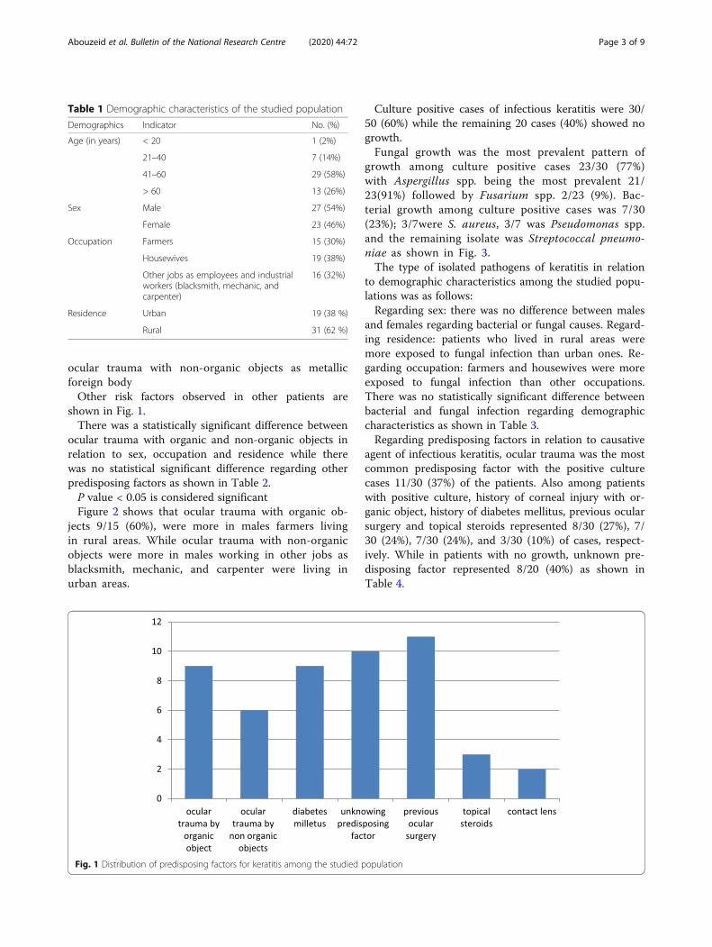
ocular trauma with non-organic objects as metallicforeign bodyOther risk factors observed in other patients are
shown in Fig. 1.There was a statistically significant difference between
ocular trauma with organic and non-organic objects inrelation to sex, occupation and residence while therewas no statistical significant difference regarding otherpredisposing factors as shown in Table 2.P value < 0.05 is considered significantFigure 2 shows that ocular trauma with organic ob-
jects 9/15 (60%), were more in males farmers livingin rural areas. While ocular trauma with non-organicobjects were more in males working in other jobs asblacksmith, mechanic, and carpenter were living inurban areas.
Culture positive cases of infectious keratitis were 30/50 (60%) while the remaining 20 cases (40%) showed nogrowth.Fungal growth was the most prevalent pattern of
growth among culture positive cases 23/30 (77%)with Aspergillus spp. being the most prevalent 21/23(91%) followed by Fusarium spp. 2/23 (9%). Bac-terial growth among culture positive cases was 7/30(23%); 3/7were S. aureus, 3/7 was Pseudomonas spp.and the remaining isolate was Streptococcal pneumo-niae as shown in Fig. 3.The type of isolated pathogens of keratitis in relation
to demographic characteristics among the studied popu-lations was as follows:Regarding sex: there was no difference between males
and females regarding bacterial or fungal causes. Regard-ing residence: patients who lived in rural areas weremore exposed to fungal infection than urban ones. Re-garding occupation: farmers and housewives were moreexposed to fungal infection than other occupations.There was no statistically significant difference betweenbacterial and fungal infection regarding demographiccharacteristics as shown in Table 3.Regarding predisposing factors in relation to causative
agent of infectious keratitis, ocular trauma was the mostcommon predisposing factor with the positive culturecases 11/30 (37%) of the patients. Also among patientswith positive culture, history of corneal injury with or-ganic object, history of diabetes mellitus, previous ocularsurgery and topical steroids represented 8/30 (27%), 7/30 (24%), 7/30 (24%), and 3/30 (10%) of cases, respect-ively. While in patients with no growth, unknown pre-disposing factor represented 8/20 (40%) as shown inTable 4.
Table 1 Demographic characteristics of the studied population
Demographics Indicator No. (%)
Age (in years) < 20 1 (2%)
21–40 7 (14%)
41–60 29 (58%)
> 60 13 (26%)
Sex Male 27 (54%)
Female 23 (46%)
Occupation Farmers 15 (30%)
Housewives 19 (38%)
Other jobs as employees and industrialworkers (blacksmith, mechanic, andcarpenter)
16 (32%)
Residence Urban 19 (38 %)
Rural 31 (62 %)
Fig. 1 Distribution of predisposing factors for keratitis among the studied population
Abouzeid et al. Bulletin of the National Research Centre (2020) 44:72 Page 3 of 9

The antimicrobial susceptibility testing of bacterial iso-lates by disk diffusion method showed that out of the 7cases of bacterial keratitis, the antibiotic susceptibilitypattern of the 3 Pseudomonas isolates revealed that(100%) were susceptible to tobramycin, amikacin and allquinolones (ciprofloxacin, ofloxacin, levofloxacin andgatifloxacin).Whereas two-thirds (66.6%) were suscep-tible to both gentamicin, and polymyxin B. while theremaining isolate showed intermediate susceptibility toboth antibiotics as shown in Fig. 4.The antibiotic susceptibility pattern of the 3 isolates of
S. aureus revealed that all isolates (100%) were suscep-tible to cefoxitin, all quinolones (ciprofloxacin, ofloxacin,levofloxacin, gatifloxacin, and moxifloxacin), chloram-phenicol, tobramycin and gentamicin. Teicoplanin anderythromycin 2/3 (75%) showed intermediate resistance.While one-third (33% and 25%) were resistant to tri-methoprim sulfamethoxazole (SXT) and erythromycin,as respectively shown in Fig. 5.The antibiotic susceptibility pattern of the single iso-
late of S. pneumoniae is shown in Fig. 6.Antifungal susceptibility pattern of the 23 fungal iso-
lates using disk diffusion method revealed that all iso-lates 100% were susceptibile to voriconazole andketoconazole, followed by itraconazole 74% (17/23).While 100% of isolates were resistant to fluconazolefollowed by amphotericin 96% (22/23). Susceptibility to
itraconazole showed different results with different fun-gal species as shown in Fig. 7.
DiscussionIn this study, the majority of patients 29/50 (58%) werebetween 4th and 6th decade. The incidence of infectiouskeratitis is higher in males 27/50 (54%) than in females23/50 (46%). Thirty-one out of 50 (62%) of cases live inrural areas. Housewives represented 38%, farmers 30%,and other jobs represented 32%. In similar findings,(Ravinder et al. 2016) found that infectious keratitis wasmore common in males (62%) than females (38%) morecommonly observed in rural populations (65%), withhigher prevalence in agricultural workers (47.5%)followed by industrial workers (23.75%), housewives(12.5%), and students (8.75%). Sedhu et al. 2017 showedthat 70% of patients with infectious keratitis lived inurban areas mainly housewives (21%) followed byfarmers (16.9%), laborers (13.6%), and carpenters (12.3%)In this study, trauma was the most common predis-
posing factor observed in 15/50 (30%) of infectious kera-titis; ocular trauma with organic material represents 9/15 (60%) and is more in male farmers living in ruralareas. This is in agreement with (Ravinder et al. 2016)and (Choudhury et al. 2017) where ocular trauma wasthe most common predisposing factor with 46% and69%, respectively. Also according to Basak et al. 2005
Table 2 Distribution of predisposing factors according to occupation, sex, and residence
Predisposing factors ♀ ♂ P value Urban Rural P value Farmer Other jobs Housewives P value
Ocular trauma with non organic objects 0 6 < 0.05 2 4 0.367 1 5 0 < 0.05
Ocular trauma with organic objects 1 8 < 0.05 0 9 < 0.05 8 0 1 < 0.05
Previous ocular surgery 7 4 0.365 6 5 0.762 2 2 7 0.103
Idiopathic 4 6 0.527 7 3 0.157 1 4 5 0.272
Diabetes mellitus 6 3 0.317 2 7 0.114 3 1 5 0.263
Contact lens 2 0 0.157 2 0 0.367 0 2 0 0.135
Topical steroids 3 0 0.083 0 3 0.223 0 2 1 0.367
Fig. 2 Ocular trauma with organic and non-organic objects according to sex, occupation, and residence
Abouzeid et al. Bulletin of the National Research Centre (2020) 44:72 Page 4 of 9

and Senthil Vadivu 2013, corneal injury with vegetablematter was the most common factor causing infectiouskeratitis with 59.6% and 47.817%, respectively. Ravinderet al. 2016 reported that farmers were more prone forocular trauma with organic material. El Shabrawy et al.2013 found history of ocular trauma to be the most im-portant predisposing factor for fungal keratitis (63.6%).The above observations clearly show that in developingcountries, agricultural workers are more prone to vege-tative matter-induced ocular trauma as a major cause ofinfectious keratitis in rural populations more than urbanpopulations.While in developed countries wearing contact lense
constitutes a major risk factor for infectious keratitis, ac-cording to Keay L et al. 2006, Jeng B. H. et al. 2010, andMun et al. 2019, in our study, history of wearinf contactlens and using topical corticosteroids represented only2/50 (4%) and 3/50 (6%), respectively.
In the present study, positive culture samples of in-fectious keratitis cases are 60% (30/50). Similar to ourresults, Marasini et al. 2016, found a positive culturerate of 57.8%. However, lower positive culture resultswere reported by Shoja and Manaviat 2004 andAmescua et al. 2012 with 40% and 38%, respectively.While Stefan and Nenciu 2006 and Al-Shakarchiet al. 2015 demonstrated higher results of positivecorneal scraping culture samples with 86.7% and 70%,respectively.Our study showed that fungal growth is the most
prevalent pattern of growth 23/30 (77%), while bacterialgrowth is 7/30 (23%). This is in accordance with a studyby EL Shabrawy et al. (2013) conducted in Egypt. Joshiet al. (2017) and Manikandan et al. (2019) who reportedthat 55%, 65%, and 98%, respectively, of culture positivecases were identified to be due to fungal causes whilethe remaining cases were due to bacterial causes. How-ever, a study by Tewari et al. 2012 and Ghosh et al. 2016reported that out of the positive isolates, 65% and 61%belonged to the bacteria, while 35% and 39% belongedto fungi, respectively.In this study, Aspergillus spp. are being the most
prevalent 21/23(91%), followed by Fusarium spp. 2/23(8%). This finding is in accordance with Al-Shakarchi2007 and Tewari et al. 2012 who demonstrated that As-pergillus spp. was the most common isolate with 57%and 35% followed by Fusarium spp. with 27% and 22%among fungal pathogens, respectively. However, a studyconducted in Egypt by EL-Shabrawy et al. 2013 revealedthat the most frequent fungal pathogens were Penicil-lium spp. (24.2%) followed by Aspergillus fumigatus(21.2%) then Fusarium spp. (9%).The incidence of fungal keratitis is on the rise in the
densely populated continents of Asia and Africa (Ravin-der et al. 2016). This can be explained by the differencein climatic conditions. A study done in the National Re-search Centre in Egypt to detect the association betweenfungal keratitis and the climatic changes concluded thatthe climatic conditions directly affect the frequency offungal keratitis and that the incidence of this disease will
Table 3 Distribution of demographic characteristics according to causative agent of keratitis among the studied populations
Demographic Indicator total Bacterial keratitisN (%)
Fungal keratitisN (%)
No growthN (%)
P value
Sex Male 27 3(11%) 12(44.5%) 12(44.5%) 0.715
Female 23 4(17%) 11(48%) 8(35%)
Residence Urban 19 3(16%) 5(26%) 11(58%) 0.07
Rural 31 4(13%) 18(58%) 9(29%)
Occupation Farmers 15 1(6%) 10(67%) 4(27%) 0.124
Workers 16 2(12%) 4(25%) 10(63%)
Housewives 19 4(21%) 9(47%) 6(32%)
P value < 0.05 is considered significant
Fig. 3 Bacterial and fungal pathogens recovered fromculture-positive cases
Abouzeid et al. Bulletin of the National Research Centre (2020) 44:72 Page 5 of 9

continue to rise as long as the global warming is in-creasing and the greenhouse gases will continue torise (EL Shabrawy et al. 2013). Species of Fusariumand Aspergillus are widespread in nature being im-portant pathogens in fungal keratitis (Manikandanet al. 2019).In the present study, the bacterial growth among posi-
tive culture cases is 7/30 (23%); 3/7 were S. aureus, 3/7was Pseudomonas spp., and the remaining isolate wasS. pneumoniae. A study conducted by AL-Yousuf,2009 in Bahrain reported that Pseudomonas aerugi-nosa, Staphylococcus, and Streptococcus were the mostfrequent pathogens. This is near to a study by Tewariet al. 2012 and Mun et al. 2019 who reported thatthe most frequent bacterial isolates were S. aureusfollowed by Pseudomonas aeruginosa.Proper diagnosis and treatment of bacterial keratitis
are essential to achieve resolution of infection andminimize damage to the cornea. In our study, the anti-biotic susceptibility pattern of the 3 isolates of S. aureusrevealed (100%) susceptibility to cefoxitin (methicillinsensitive) and all quinolones (ciprofloxacin, ofloxacin,levofloxacin, gatifloxacin, and moxifloxacin). This is in
accordance with Senthil Vadivu 2013 who founded 7/9(77%) methicillin-sensitive S. aureus , with 77% sensi-tivity to ciprofloxacin and Mun et al. 2019 who re-ported 11/13(85%) methicillin-sensitive S. aureus withhigher sensitivity to levofloxacin and moxifloxacin;95.8% and 93.8%, respectively. Treatment of P. aerugi-nosa eye infections often becomes a challenge due tothe ability of this bacterium to be resistant to antibi-otics via intrinsic and acquired mechanisms (Subediet al. 2018). In the present study, the 3 Pseudomonasisolates (100%) were susceptible to tobramycin, amika-cin , ciprofloxacin, ofloxacin, levofloxacin, and gatiflox-acin followed by gentamicin and polymyxin B 2/3(66.6%). This is similar to a study conducted by Mara-sini et al. 2016 where all Pseudomonas aeruginosa iso-lates were sensitive to gentamicin and ciprofloxacin.Mun et al. 2019 also found that all Pseudomonas spe-cies isolates were sensitive to amikacin and ciprofloxa-cin. While Cho and Lee, 2018 reported less than 10%resistance in P. aeruginosa to ciprofloxacin and genta-micin. According to SenthilVadivu, 2013, P. aeruginosaisolates showed lower sensitivity to ciprofloxacin (66%)and ofloxacin (83%).
Fig. 4 Antibiotic susceptibility pattern of Pseudomonas by disk diffusion method
Table 4 Distribution of predisposing factors according to causative agents of keratitis
Predisposing factors Total no. Bacterial Keratitis Fungal keratitis No growth(20) P value
Ocular trauma With non organic objects 6 2 1 3 0.606
With organic objects 9 0 8 1 < 0.05
Diabetes mellitus 9 0 7 2 < 0.05
Previous ocular surgery 11 4 3 4 0.913
Idiopathic 10 0 2 8 < 0.05
Contact lens 2 0 0 2 0.135
Topical steroids 3 1 2 0 0.367
P value < 0.05 is considered significant
Abouzeid et al. Bulletin of the National Research Centre (2020) 44:72 Page 6 of 9

Treatment of fungal keratitis is one of the most diffi-cult problems encountered by ophthalmologists due topoor response to the therapy as well as the limited avail-ability of antifungal agents. Although voriconazole andother triazoles have broad-spectrum activity againstcausative fungal isolates, clinically no single drug wasfound to be effective against fungal keratitis (Manikan-dan et al. 2019). In this study, antifungal susceptibilitypattern of the 23 fungal isolates revealed that all isolatesare 100% susceptible to ketoconazole and voriconazole.In accordance with our findings, a study conducted inUpper Egypt by Gharamah et al. 2014 showed that keto-conazole at 0.5% and 1% concentrations was effectiveagainst all fungal isolates, except for three Fusarium spe-cies tested. However, Sirisha et al. 2015 reported lower
percentages of sensitivity to ketoconazole for Aspergillusfumigatus (85%), Fusarium spp. (83%), Aspergillus flavus(73%), and Aspergillus niger (50%).A study conducted by (Saha et al. 2014) showed that
voriconazole had the lowest minimal inhibitory concen-tration (MIC) against Aspergillus spp. and Fusariumspp., followed by amphotericin B, ketoconazole, itraco-nazole, and that it is still the first choice in the treatmentof mould keratitis.In the present study, susceptibility to itraconazole gave
different results. Both A. fumigatus and Fusarium spp.show 100% susceptibility, while A. flavus and A. nigershowed intermediate susceptibility with 21% and 75%,respectively. This finding is in partial agreement withSenthil vadivu 2013 where A. fumigatus and A. niger
Fig. 6 Antibiotic susceptibility pattern of S. pneumoniae using disk diffusion method
Fig. 5 Antibiotic susceptibility pattern of S. aureus using disk diffusion method
Abouzeid et al. Bulletin of the National Research Centre (2020) 44:72 Page 7 of 9

showed 100% susceptibility while A. flavus and Fusar-ium spp. showed 78% and 83%, respectively. Also astudy conducted by Sirisha et al. 2015 showed that all29 fungal isolates (Fusarium spp, A. flavus, andA.niger) were 100% susceptible to itraconazole exceptfor A. fumigatus (85%).In our study, 96% (22/23) are resistant to amphotericin
B. However, Senthilvadivu 2013 reported in his studythat A. fumigatus, Fusarium spp, A. flavus, and A. nigerisolates were sensitive to Amphotericin B (70%), (66%),(64%), and (58%), respectively. Also, a study by Ghara-mah et al. (2014) showed that the (MIC) of amphoteri-cin B was at 0.1% or 0.5% concentrations for mostfungal species tested while there was no effect on the3 Fusarium species. Amphotericin B was quite sensi-tive to genus Aspergillus and Fusarium but due topoor penetration in cornea and the requiring of highdosage, it was not used in such keratitis (Saha et al.2014). In the present study, all isolates were resistantto fluconazole. This is in agreement with Senthilvadivu 2013, Sirisha et al. 2015, and Senthilvadivuand Stalin, 2018.
ConclusionOcular trauma was the major cause of infectiouskeratitis. It was more in the rural population. Fungalgrowth, mainly Aspergillus spp. was the most preva-lent pathogen encountered in all cases. The incidenceof fungal keratitis is on the rise due to increased glo-bal warming. Voriconazole is the first choice in thetreatment of mould keratitis with 100% susceptibility.While alarmingly, fluconazole no longer can be usedfor the empirical therapy as it showed resistance toall fungal isolates
RecommendationFurther studies are recommended on a wider scale ofpopulation to provide more data about epidemiologyand causative agents of infectious keratitis in Egypt. Thepractice of routine microbiological analysis and sensitiv-ity testing for all infectious keratitis is recommended inorder to have enough epidemiological information forempirical therapy.
AcknowledgementsThe authors would like to thank the ophthalmology professors and all theteam of cornea unit in the Research Institute of Ophthalmology (RIO) fortheir great help in sample preparation and sharing needed data of keratitispatients in this study.
Authors’ contributionsAI had carried out the performance of lab work, the collection of data ofcases, and participated in writing the manuscript. SA had made the approvalof work design and research plan, supervising the steps of the work. AMsupervised work steps and participated in writing the manuscript. AEparticipated in writing and approving the article for submission to thejournal as a corresponding author. All authors read and approved the finalmanuscript.
FundingNot applicable
Availability of data and materialsAll data generated or analyzed during this study are included in thispublished article.
Ethics approval and consent to participateThe work is ethically approved by the Scientific Research Committee of theResearch Institute of Ophthalmology RIO, Egypt, prior to the beginning ofthe study. Corneal scrapping samples used in the study were obtained afterpatients’ consent.
Consent for publicationNot applicable.
Competing interestsThe authors declare that they have no competing interests.
Fig. 7 Antifungal susceptibility of fungal isolates to itraconazole
Abouzeid et al. Bulletin of the National Research Centre (2020) 44:72 Page 8 of 9

Author details1Medical Microbiology and Immunology Unit, Department of Microbiologyand Parasitology, Research Institute of Ophthalmology, Giza 12611, Egypt.2Medical Microbiology and Immunology Faculty of Medicine, CairoUniversity, Cairo, Egypt. 3Infection Control Unit, Microbiology andImmunology Unit, Department of Microbiology and Parasitology, (RIO), 2 ElAhram Street, Giza 12611, Egypt.
Received: 6 January 2020 Accepted: 4 May 2020
ReferencesAl-Shakarchi FI, Hussein MA, Al-Shaibani AB (2015) Profile of microbial keratitis at
a referral center in Iraq. Journal of Al-Nahrain University 18(1):141–147Al-Yousuf N (2009) Microbial keratitis in Kingdom of Bahrain: clinical and
microbiology study. Middle East African Journal of Ophthalmology 16(1):3–7.https://doi.org/10.4103/0974-9233.48855
Amescua G, Miller D and Alfonso EC (2012). What is causing the corneal ulcer?Management strategies for unresponsive corneal ulceration. Eye(London,England); 26(2): 228–236. doi: https://doi.org/10.1038/eye.2011.316.
Basak SK, Basak S, Mohanta A, Bhowmick A (2005) Epidemiological andmicrobiological diagnosis of suppurative keratitis in Gangetic West Bengal,eastern India. Indian Journal of Ophthalmology 53(1):17–22
Cho CH and Lee SB. (2018). Comparison of clinical characteristics and antibioticsusceptibility between Pseudomonas aeruginosa and P. putida keratitis at atertiary referral center: a retrospective study. BioMed Central Ophthalmology;18(1):204.
Choudhury RB, Das DK, Das B, Bhuyan J (2017) Comparative study of bacterialprofile of patients with bacterial corneal ulcer with bacterial profile ofconjunctiva of controls-a prospective study. Journal of ContemporaryMedical Research 4(10):2127–2130
Clinical and Laboratory Standard Insitute (2017). Performance standards forantimicrobial susceptibility testing .27th ed. CLSI supplement M100. Wayne,PA,USA,2017.
EL Shabrawy RM, El Badawy NE, Harb AW (2013) The incidence of fungal keratitisin Zagazig University Hospitals, Egypt and the value of direct microscopyand PCR technique in rapid diagnosis. Journal of Microbiology and InfectiousDiseases 3(4):186–191
Espinel-Ingroff A, Arthington-Skaggs B, Iqbal N, Ellis PMA et al (2007) MulticenterEvaluation of a New Disk Agar Diffusion Method for Susceptibility Testing ofFilamentous Fungi with Voriconazole, Posaconazole, Itraconazole,Amphotericin B, and Caspofungin. Journal of Clinical Microbiology 45(6):1811–1820
Gharamah AA, Moharram AM, Ismail MA, Al-Hussaini AK (2014) Bacterial andfungal keratitis in Upper Egypt: in vitro screening of enzymes, toxins andantifungal activity. Indian journal of ophthalmology 62(2):196–203
Ghosh AK, Gupta A, Rudramurthy SM, Paul S, Hallur VK et al (2016) Fungalkeratitis in North India: spectrum of agents, risk factors and treatment.Mycopathologia 181(11-12):843–850
Jeng B. H., Gritz D. C., Kumar A. B. et al.(2010) “Epidemiology of ulcerative keratitisin Northern California,” Archives of Ophthalmology; 128 (8): 1022–1028.Viewat: Publisher Site | Google Scholar
Johnson ME (2008) Issues in antifungal susceptibility testing. Journal ofAntimicrobial Chemotherapy 61(1):i13–i18
Joshi RK, Goyal RK, Kochar A (2017) A prospective study of clinical profile,epidemiology and etiological diagnosis of corneal ulcer in North-WestRajasthan. International Journal of Community Medicine and Public Health4(12):4544–4547
Keay L, Edwards K, Naduvilath T, Taylor HR, Snibson GR et al (2006) Microbialkeratitis: predisposing factors and morbidity. Ophthalmology 113:109–116
Lin A, Rhee MK, Akpek EK, Amescua G, Farid M et al (2019) Bacterial keratitis PPP.The American Academy of Ophthalmology Preferred Practice PatternCornea/External Disease Committee 126(1):1–55
Manikandan P, Abdel-Hadi A, RandhirBabu Singh Y, Revathi R, Anita R et al (2019)Fungal keratitis: epidemiology, rapid detection, and antifungal susceptibilitiesof fusarium and aspergillus isolates from corneal scrapings. BioMed ResearchInternational 2019:6395840. https://doi.org/10.1155/2019/6395840
Marasini S, Swift S, Dean SJ, Ormonde SE, Craig JP (2016) Spectrum andSensitivity of Bacterial Keratitis Isolates in Auckland. Journal ofophthalmology 2016:3769341. https://doi.org/10.1155/2016/3769341
Mun Y, Kim KM, Oh JY (2019) Ten-year analysis of microbiological profile andantibiotic sensitivity for bacterial keratitis in Korea. PLoS ONE 14(3):e0213103
Ranjini CY, Waddepally VV (2016) Microbial Profile of Corneal Ulcers in a TertiaryCare Hospital in South India. Journal of ophthalmic and vision research 11(4):363–367
Ravinder K, Madhav MV, Archana J, Pandurang J (2016) Clinical evaluation ofcorneal ulcer among patients attending teaching hospital. InternationalJournal of Contemporary Medical Research 3(4):949–952
Robinson J, Ellen J, Hadel B and Lighthizer N (2016). Collecting a corneal culture.Review of optometry Acessed online from: https://www.reviewofoptometry.com/article/collecting-a-corneal-culture
Sabatelli F, Patel R, Mann PA, Mendrick CA, Norris CC et al (2006) In Vitroactivities of posaconazole, fluconazole, itraconazole, voriconazole, andamphotericin B against a large collection of clinically important molds andyeasts. Antimicrobial Agents and Chemotherapy 50(6):2009–2015
Saha S, Sengupta J, Banerjee D, Sunayana S, Khetan A et al (2014) Systemicevaluation on antifungal susceptibility of keratitis associated fungalpathogens in Eastern India. Journal of Medical Microbiology and Diagnosis 3:134. https://doi.org/10.4172/2161-0703.1000134
Sedhu PA, Sugathan S, Pushpakaran A, Kurian C (2017). Bacterial and FungalProfile of Infectious Keratitis: A Prospective Study. International Journal ofScientific Study; 5(8): 128-132.DOI: 10.17354/ijss/2017/533.
Senthilvadivu C (2013) Bacterial, fungal and parasitic agents in infectious keratitispatients due to trauma in a tertiary care ophthalmic hospital. Master's thesis,Madras Medical College, Chennai
Senthilvadivu C, Stalin M (2018) Antimicrobial susceptibility pattern of bacterialand fungal ocular isolates from tertiary care ophthalmic hospitals in Chennai.Journal of Evolution of Medical and Dentinsty Sciences 7(15):1868–1871
Shoja MR, Manaviat M (2004) Epidemiology and outcome of corneal ulcer inyazd shahid sadoughi hospital. Acta Medica Iranica 42(2):136–141
Sirisha T, Jayalakshmi L, Ratnakumari G, Viswamitra P (2015) Microbiologicalprofile and their antimicrobial susceptibility in infective keratitis at RegionalEye Hospital, Visakhapatnam. Scholars Journal of Applied Medical Sciences3(3A):1083–1088
Stefan C, Nenciu A (2006) Post traumatic bacterial keratitis – a microbiologicalprospective clinical study. Oftalmologia. 50(3):118–122
Subedi D, Vijay AK, Willcox M (2018) Study of disinfectant resistance genes inocular isolates of Pseudomonas aeruginosa. Antibiotics (Basel) 7(4):88. https://doi.org/10.3390/antibiotics7040088
Suwal S, Bhandari D, Thapa P, Shrestha MK, Amatya J (2016) Microbiologicalprofile of corneal ulcer cases diagnosed in a tertiary care ophthalmologicalinstitute in Nepal. BioMed Central ophthalmology 16(1):209. https://doi.org/10.1186/s12886-016-0388-9
Tesfaye T, Beyene G, Gelaw Y, Bekele S, Saravanan M (2013) Bacterial profile andantimicrobial susceptibility pattern of external ocular infections in JimmaUniversity Specialized Hospital, Southwest Ethiopia. American Journal ofInfectious Diseases and Microbiology 1(1):13–20
Tewari A, Sood N, Vegad MM, Mehta DC (2012) Epidemiological andmicrobiological profile of infective keratitis in Ahmedabad. Indian Journal ofOphthalmology 60(4):267–272
Publisher’s NoteSpringer Nature remains neutral with regard to jurisdictional claims inpublished maps and institutional affiliations.
Abouzeid et al. Bulletin of the National Research Centre (2020) 44:72 Page 9 of 9


![NON INFECTIOUS PERIPHERAL ULCERATIVE KERATITIS [PUK]](https://img.dokumen.tips/doc/110x75/56815e08550346895dcc5f9b/non-infectious-peripheral-ulcerative-keratitis-puk.jpg)