Embed Size (px)
Citation preview
Mitani et al. BMC Research Notes 2014, 7:677http://www.biomedcentral.com/1756-0500/7/677
CASE REPORT Open Access
Fungal keratitis caused by Beauveria bassiana:drug and temperature sensitivity profiles:a case reportArisa Mitani1, Atsushi Shiraishi1,2*, Hitoshi Miyamoto4, Atsuko Sunada5, Akiko Ueda5, Seishi Asari5,Xiaodong Zheng1, Yasuaki Yamamoto1, Yuko Hara1 and Yuichi Ohashi1,3
Abstract
Background: Beauveria bassiana is an entomopathogenic fungus and is a rare cause of keratitis. We present acase of fungal keratitis caused by B. bassiana that was diagnosed by in vivo confocal microscopy and in vitrocorneal cultures. In addition, we determined the temperature- and drug-sensitivities of the isolated strain ofB. bassiana.
Case presentation: A 59-year-old Japanese man with a 2-month history of keratitis was examined by slit-lampbiomicroscopy, in vivo confocal microscopy, and histology and cultures of corneal scrapings. The cornealscrapings were used to determine the minimal inhibitory concentrations of different antifungal drugs and alsoto determine the temperature-sensitivity. In vivo confocal microscopy and histological examinations showedfilamentous fungal keratitis. The characteristics of the fungal growth indicated that the keratitis was caused byB. bassiana. The keratitis responded poorly to systemic and topical voriconazole and to natamycin ointment.However, it was resolved after changing the natamycin to micafungin combined with surgical debridement.The isolated strain was sensitive to itraconazole, miconazole, micafungin, voriconazole, and resistant to flucytosine andfluconazole. It was moderately sensitive to amphotericin B, and natamycin. After 7 days in culture, the isolate grew smallwhite colonies at 25°C, very small colonies at 35°C and 37°C.
Conclusion: The drug-sensitivity and temperature-sensitivity profiles of B. bassiana should be helpful in thetreatment of B. bassiana keratitis. Therapeutic surgery may be helpful for mycotic keratitis poorly responsive tomedical therapy alone.
Keywords: Fungal keratitis, Beauveria bassiana, In vivo confocal microscopy, Drug-sensitivity, Temperaturesensitivity growth
BackgroundBeauveria bassiana is a fungus that is distributed world-wide. It can be isolated from soil, insects, and mites.It is an entomopathogenic fungi and is used as a bio-logical control agent for insect pests as alternativesupplements to chemical insecticides [1-3]. AlthoughB. bassiana is considered to be non-pathogenic to verte-brates, a small number of patients with keratitis caused
* Correspondence: [email protected] of Ophthalmology, Ehime University Graduate School ofMedicine, Shitsukawa, Toon, Ehime 791-0295, Japan2Department of Stem cell Biology, Shitsukawa, Toon, Ehime 791-0295, JapanFull list of author information is available at the end of the article
© 2014 Mitani et al.; licensee BioMed Central LCommons Attribution License (http://creativecreproduction in any medium, provided the orDedication waiver (http://creativecommons.orunless otherwise stated.
by B. bassiana have been reported [4-13]. We presenta case of fungal keratitis caused by B. bassiana thatwas diagnosed by in vivo confocal microscopy andin vitro corneal cultures. In addition, we determinedthe temperature- and drug-sensitivities of the isolatedstrain of B. bassiana.
Case presentationThe patient was a 59-year-old Japanese man who noteda corneal opacity in his right eye in October, 2009.However, he did not seek treatment for the opacity forabout two months. He visited a private ophthalmo-logical clinic on December 11, 2009, because his right
td. This is an Open Access article distributed under the terms of the Creativeommons.org/licenses/by/4.0), which permits unrestricted use, distribution, andiginal work is properly credited. The Creative Commons Public Domaing/publicdomain/zero/1.0/) applies to the data made available in this article,
Mitani et al. BMC Research Notes 2014, 7:677 Page 2 of 5http://www.biomedcentral.com/1756-0500/7/677
eye felt irritated and his vision had decreased. Thepatient was a farm worker, but did not use entomo-pathogenic fungi as a biological control agent. He hadno systemic diseases and no signs or symptoms of im-munosuppression during the clinical course. He was di-agnosed with infectious keratitis and was treated withtopical gatifloxacin and erythromycin eye ointment fortwo weeks. Because the signs and symptoms did notimprove, he was referred to the Ehime University Hospitalon December 28, 2009.On the initial examination, his best-corrected visual
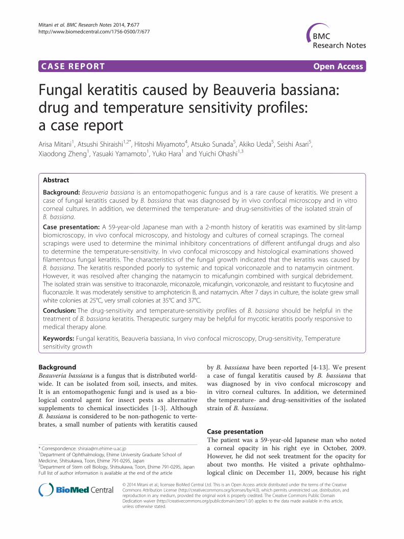
acuity was 20/500 OD and 20/16 OS. Slit-lamp examin-ation of the right eye showed a grayish stromal infiltratewith a dry texture and indistinct margins (Figure 1A).Examination of the cornea with in vivo confocal micros-copy (HRT II-RCM; Heidelberg Engineering, Heidelberg,Germany) showed a mass of branching and interlockingwhite lines in the area of the infiltrate suggesting fila-mentous fungus keratitis (Figure 2A). Microscopicexamination of corneal scrapings showed filamentousfungal hyphal fragments (Figure 2B).Treatment was begun with oral voriconazole (400 mg/day)
p.o., topical 0.1% voriconazole hourly, and natamycineye ointment 5 times/day. Because the lesion did notrespond despite the intensive antifungal therapy, theisolate from the corneal scrapings was sent to the FirstLaboratory in Medical Mycology Research Center, ChibaUniversity for identification and to determine the min-imal inhibitory concentrations (MICs) to different anti-fungal drugs (Table 1).The corneal scrapings grew fungal colonies on Sabouraud
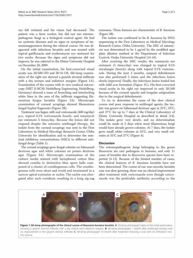
dextrose agar and white colonies on potato dextroseagar (Figure 3A). Microscopic examination of theculture media stained with lactophenol cotton blueshowed conidia in distinctive blue spore balls com-posed of a cluster of conidiogenous cells. The conidio-genous cells were short and ovoid and terminated in anarrow apical extension or rachis. The rachis was elon-gated after each conidium resulting in a long zig-zag
Figure 1 Slit-lamp photographs of the clinical course of Beauveria basshowing a grayish stromal infiltrate with a dry texture and indistinct mano improvement in the grayish stromal infiltrate. C. Slit-lamp photograpthe stroma.
extension. These features are characteristic of B. bassiana(Figure 3B).The isolate was confirmed to be B. bassiana by DNA
sequencing at the First Laboratory in Medical MycologyResearch Center, Chiba University. The MIC of natamy-cin was determined to be 1 μg/ml by the modified agarplate dilution method at the Department of InfectionControl, Osaka University Hospital [14-16].After receiving the MIC results, the natamycin eye
ointment (5 times/day) was changed to topical 0.1%micafungin (hourly) along with topical 0.1% voricona-zole. During the next 3 months, surgical debridementwas also performed 3 times and the infectious lesionslowly improved. Finally, the infectious lesion healed butwith mild scar formation (Figure 1C). His best-correctedvisual acuity in his right eye improved to only 20/100because of the corneal opacity and irregular astigmatismdue to the surgical debridement.To try to determine the cause of the slow clinical
course and poor response to antifungal agents, the iso-late was grown on Sabouraud dextrose agar at 25°C, 35°C,and 37°C for up to 7 days at the Clinical Laboratory ofEhime University Hospital as described in detail [14].The isolate grew very slowly, and no determinationcould be made at 5 days when most filamentous fungiwould have already grown colonies. At 7 days, the isolategrew small white colonies at 25°C, and very small col-onies at 35°C and 37°C (Figure 4).
DiscussionThe entomopathogenic fungi belonging to the genusBeauveria are rare pathogens in humans, and only 11cases of keratitis due to Beauveria species have been re-ported [4-13]. Because of the limited number of cases,the clinical features of B. bassiana keratitis have notbeen determined. The course of our non-necrotic keratitiscase was slow-growing, there was no clinical improvementafter treatment with voriconazole even though vorico-nazole was the preferable antibiotic according to the
siana keratitis. A. Slit-lamp photograph taken at the initial examinationrgins. B. Slit-lamp photograph 1 month after antifungal therapy withh 10 month after treatment showing a scar with no infiltration into
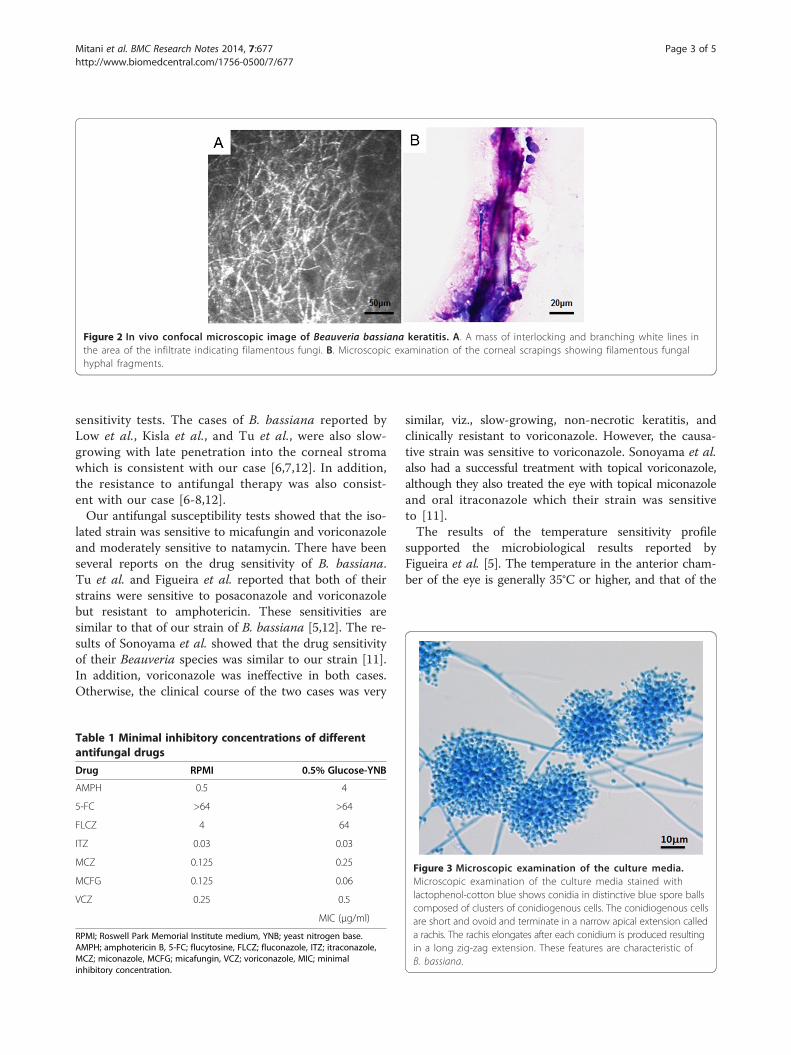
Figure 2 In vivo confocal microscopic image of Beauveria bassiana keratitis. A. A mass of interlocking and branching white lines inthe area of the infiltrate indicating filamentous fungi. B. Microscopic examination of the corneal scrapings showing filamentous fungalhyphal fragments.
Mitani et al. BMC Research Notes 2014, 7:677 Page 3 of 5http://www.biomedcentral.com/1756-0500/7/677
sensitivity tests. The cases of B. bassiana reported byLow et al., Kisla et al., and Tu et al., were also slow-growing with late penetration into the corneal stromawhich is consistent with our case [6,7,12]. In addition,the resistance to antifungal therapy was also consist-ent with our case [6-8,12].Our antifungal susceptibility tests showed that the iso-
lated strain was sensitive to micafungin and voriconazoleand moderately sensitive to natamycin. There have beenseveral reports on the drug sensitivity of B. bassiana.Tu et al. and Figueira et al. reported that both of theirstrains were sensitive to posaconazole and voriconazolebut resistant to amphotericin. These sensitivities aresimilar to that of our strain of B. bassiana [5,12]. The re-sults of Sonoyama et al. showed that the drug sensitivityof their Beauveria species was similar to our strain [11].In addition, voriconazole was ineffective in both cases.Otherwise, the clinical course of the two cases was very
Table 1 Minimal inhibitory concentrations of differentantifungal drugs
Drug RPMI 0.5% Glucose-YNB
AMPH 0.5 4
5-FC >64 >64
FLCZ 4 64
ITZ 0.03 0.03
MCZ 0.125 0.25
MCFG 0.125 0.06
VCZ 0.25 0.5
MIC (μg/ml)
RPMI; Roswell Park Memorial Institute medium, YNB; yeast nitrogen base.AMPH; amphotericin B, 5-FC; flucytosine, FLCZ; fluconazole, ITZ; itraconazole,MCZ; miconazole, MCFG; micafungin, VCZ; voriconazole, MIC; minimalinhibitory concentration.
similar, viz., slow-growing, non-necrotic keratitis, andclinically resistant to voriconazole. However, the causa-tive strain was sensitive to voriconazole. Sonoyama et al.also had a successful treatment with topical voriconazole,although they also treated the eye with topical miconazoleand oral itraconazole which their strain was sensitiveto [11].The results of the temperature sensitivity profile
supported the microbiological results reported byFigueira et al. [5]. The temperature in the anterior cham-ber of the eye is generally 35°C or higher, and that of the
Figure 3 Microscopic examination of the culture media.Microscopic examination of the culture media stained withlactophenol-cotton blue shows conidia in distinctive blue spore ballscomposed of clusters of conidiogenous cells. The conidiogenous cellsare short and ovoid and terminate in a narrow apical extension calleda rachis. The rachis elongates after each conidium is produced resultingin a long zig-zag extension. These features are characteristic ofB. bassiana.
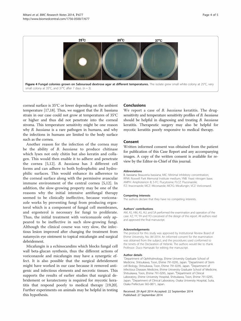
Figure 4 Fungal colonies grown on Sabouraud dextrose agar at different temperatures. The isolate grew small white colony at 25°C, verysmall colony at 35°C, and 37°C after 7 days. (n = 3).
Mitani et al. BMC Research Notes 2014, 7:677 Page 4 of 5http://www.biomedcentral.com/1756-0500/7/677
corneal surface is 35°C or lower depending on the ambienttemperature [17,18]. Thus, we suggest that the B. bassianastrain in our case could not grow at temperatures of 35°Cor higher and thus did not penetrate into the cornealstroma. This temperature sensitivity might be one reasonwhy B. bassiana is a rare pathogen in humans, and whythe infections in humans are limited to the body surfacesuch as the cornea.Another reason for the infection of the cornea may
be the ability of B. bassiana to produce chitinasewhich lyses not only chitin but also keratin and colla-gen. This would then enable it to adhere and penetratethe cornea [5,12]. B. bassiana has 3 different cellforms and can adhere to both hydrophobic and hydro-philic surfaces. This would enhance its adherence tothe corneal surface along with the permissive avascularimmune environment of the central cornea [5,12]. Inaddition, the slow-growing property may be one of thereasons why the initial intensive antifungal therapyseemed to be clinically ineffective, because voricona-zole works by preventing fungi from producing ergos-terol which is a component of fungal cell membranes,and ergosterol is necessary for fungi to proliferate.Thus, the initial treatment with voriconazole only ap-peared to be ineffective in such slow-growing fungi.Although the clinical course was very slow, the infec-tious lesion improved after changing the treatment fromnatamycin eye ointment to topical micafungin and surgicaldebridement.Micafungin is a echinocandins which blocks fungal cell
wall beta-glucan synthesis, thus the different actions ofvoriconazole and micafungin may have a synergetic ef-fect. It is also possible that the surgical debridementmight have worked in our case because it removed anti-genic and infectious elements and necrotic tissues. Thissupports the results of earlier studies that surgical de-bridement or keratectomy is required for mycotic kera-titis that respond poorly to medical therapy [19,20].Further experiments on animals may be helpful in testingthis hypothesis.
ConclusionsWe report a case of B. bassiana keratitis. The drug-sensitivity and temperature sensitivity profiles of B. bassianashould be helpful in diagnosing and treating B. bassianakeratitis. Therapeutic surgery may also be helpful formycotic keratitis poorly responsive to medical therapy.
ConsentWritten informed consent was obtained from the patientfor publication of this Case Report and any accompanyingimages. A copy of the written consent is available for re-view by the Editor-in-Chief of this journal.
AbbreviationsB. bassiana: Beauveria bassiana; MIC: Minimal inhibitory concentration;RPMI: Roswell Park Memorial Institute medium; YNB: Yeast nitrogen base;AMPH: Amphotericin B; 5-FC: Flucytosine; FLCZ: Fluconazole;ITZ: Itraconazole; MCZ: Miconazole; MCFG: Micafungin; VCZ: Voriconazol.
Competing interestsThe authors declare that they have no competing interests.
Authors’ contributionsAM, AS, HM, AS, AU, and SA performed the examination and operation of thecase. XZ, YY, YH and YO conceived of the design of this report. All authors readand approved the final manuscript.
AcknowledgementsThe protocol for this study was approved by Institutional Review Board ofEhime University, No. 0612014. An informed consent for the examinationwas obtained from the subject, and the procedures used conformed tothe tenets of the Declaration of Helsinki. The authors would like to thankProfessor. Duco Hamasaki for editing the manuscript.
Author details1Department of Ophthalmology, Ehime University Graduate School ofMedicine, Shitsukawa, Toon, Ehime 791-0295, Japan. 2Department of Stemcell Biology, Shitsukawa, Toon, Ehime 791-0295, Japan. 3Department ofInfectious Diseases Medicine, Ehime University Graduate School of Medicine,Shitsukawa, Toon, Ehime 791-0295, Japan. 4Department of ClinicalLaboratory, Ehime University Hospital, Shitsukawa, Toon, Ehime 791-0295,Japan. 5Department of Clinical Laboratory, Osaka University Hospital, Suita,Osaka Prefecture 565-0871, Japan.
Received: 29 April 2014 Accepted: 22 September 2014Published: 27 September 2014
Mitani et al. BMC Research Notes 2014, 7:677 Page 5 of 5http://www.biomedcentral.com/1756-0500/7/677
References1. Samish M, Ginsberg H, Glazer I: Biological control of ticks. Parasitology 2004,
129(Suppl):S389–S403.2. St Leger RJ, Wang C: Genetic engineering of fungal biocontrol agents to
achieve greater efficacy against insect pests. Appl Microbiol Biotechnol2010, 85(4):901–907.
3. Wraight SP, Ramos ME, Avery PB, Jaronski ST, Vandenberg JD: Comparativevirulence of Beauveria bassiana isolates against lepidopteran pests ofvegetable crops. J Invertebr Pathol 2010, 103(3):186–199.
4. Ishibashi Y, Matsumoto Y, Takei K: The effects of intravenous miconazoleon fungal keratitis. Am J Ophthalmol 1984, 98(4):433–437.
5. Figueira L, Pinheiro D, Moreira R, Pinto E, Simoes J, Camisa E, Torrao L,Palmares J, Falcao-Reis F: Beauveria bassiana keratitis in bullous keratopathy:antifungal sensitivity testing and management. Eur J Ophthalmol 2012,22(5):814–818.
6. Kisla TA, Cu-Unjieng A, Sigler L, Sugar J: Medical management of Beauveriabassiana keratitis. Cornea 2000, 19(3):405–406.
7. Low CD, Badenoch PR, Coster DJ: Beauveria bassiana keratitis cured bydeep lamellar dissection. Cornea 1997, 16(6):698–699.
8. McDonnell PJ, Werblin TP, Sigler L, Green WR: Mycotic keratitis due toBeauveria alba. Cornea 1984, 3(3):213–216.
9. Oh JY, Lee MJ, Wee WR, Heo JW: A case of necrotizing sclerokeratitis andendophthalmitis caused by Beauveria bassiana. Jpn J Ophthalmol 2009,53(5):551–553.
10. Pariseau B, Nehls S, Ogawa GS, Sutton DA, Wickes BL, Romanelli AM:Beauveria keratitis and biopesticides: case histories and a randomamplification of polymorphic DNA comparison. Cornea 2010,29(2):152–158.
11. Sonoyama H, Araki-Sasaki K, Kazama S, Kawasaki T, Ideta H, Sunada A,Asari S, Inoue Y, Hayashi K: The characteristics of keratomycosis byBeauveria bassiana and its successful treatment with antimycoticagents. Clin Ophthalmol 2008, 2(3):675–678.
12. Tu EY, Park AJ: Recalcitrant Beauveria bassiana keratitis: confocalmicroscopy findings and treatment with posaconazole (Noxafil).Cornea 2007, 26(8):1008–1010.
13. Sachs SW, Baum J, Mies C: Beauvaria bassiana keratitis. Br J Ophthalmol1985, 69(7):548–550.
14. Shiraishi A, Araki-Sasaki K, Mitani A, Miyamoto H, Sunada A, Ueda A, Asari S,Zheng X, Yamamoto Y, Hara Y, Ohashi Y: Clinical Characteristics of KeratitisDue to Colletotrichum gloeosporioides. J Ocul Pharmacol Ther 2011,27(5):487–491.
15. Institute CaLS: Reference Method for Broth Dilution AntifungalSusceptibility Testing of Filamentous Fungi; Approved Standard.CLSI Document 2002, 22(16):M38-A.
16. Institute CaLS: Methods for dilution antimicrobial susceptibility tests forbacteria that grow aerobically. CLSI document 2002, 23(2):M7–A7.
17. Kawasaki S, Mizoue S, Yamaguchi M, Shiraishi A, Zheng X, Hayashi Y,Ohashi Y: Evaluation of filtering bleb function by thermography. Br JOphthalmol 2009, 93(10):1331–1336.
18. Morgan PB, Soh MP, Efron N, Tullo AB: Potential applications of ocularthermography. Optom Vis Sci 1993, 70(7):568–576.
19. Ansari Z, Miller D, Galor A: Current Thoughts in Fungal Keratitis: Diagnosisand Treatment. Curr Fungal Infect Rep 2013, 7(3):209–218.
20. Thomas PA, Kaliamurthy J: Mycotic keratitis: epidemiology, diagnosis andmanagement. Clin Microbiol Infect 2013, 19(3):210–220.
doi:10.1186/1756-0500-7-677Cite this article as: Mitani et al.: Fungal keratitis caused by Beauveriabassiana: drug and temperature sensitivity profiles: a case report. BMCResearch Notes 2014 7:677.
Submit your next manuscript to BioMed Centraland take full advantage of:
• Convenient online submission
• Thorough peer review
• No space constraints or color figure charges
• Immediate publication on acceptance
• Inclusion in PubMed, CAS, Scopus and Google Scholar
• Research which is freely available for redistribution
Submit your manuscript at www.biomedcentral.com/submit