Embed Size (px)
Citation preview
186
Correspondence: Reham Mohamed EL Shabrawy, 11 Attia El Khamry Street, Al Kawmia, Zagazig, Al Sharkia, Egypt Email: [email protected]
Received: 24.04.2013 Accepted: 03.08.2013 Copyright © Journal of Microbiology and Infectious Diseases 2013, All rights reserved
Journal of Microbiology and Infectious Diseases / 2013; 3 (4): 186-191JMID doi: 10.5799/ahinjs.02.2013.04.0106
RESEARCH ARTICLE
The incidence of fungal keratitis in Zagazig University Hospitals, Egypt and the value of direct microscopy and PCR technique in rapid diagnosis
Reham Mohamed EL Shabrawy1, Nissreen El Sayed El Badawy1, Ashraf Wasfy Harb2
1 Department of Medical Microbiology and Immunology, Faculty of Medicine, Zagazig University, Egypt2 Department of Ophthalmology, Faculty of Medicine, Zagazig University, Egypt
ABSTRACT
Objective: To determine the frequency and risk factors of fungal keratitis (FK) and the value of direct microscopy and PCR techniques of corneal smears as appropriate diagnostic methods.Methods: The keratitis cases in Ophthalmology Department of Zagazig University Hospitals, between January and June 2012 were enrolled. Corneal samples were examined by direct microscopic examination of wet mount preparation with KOH (10%), PCR technique and cultured simultaneously. The corneal smear and PCR findings were compared to the culture results to analyze specificity, sensitivity and predictive values of these techniques.Results: A total of 350 patients diagnosed as keratitis and 60 of them were included in the study by a systemic ran-dom sampling method. The FK was proven in 33 (55%) cases with culture results. Ocular trauma (63.6%) was the most prevalent predisposing factor. The cultures revealed that the most frequent fungal pathogen were Penicillium spp. (24.2%) followed by Aspergillus fumigatus (21.2%). Direct microscopic examination had a sensitivity of 100%, specificity of 66.7%, a positive predictive value (PPV) of 78.6% and a negative predictive value (NPV) of 100%. PCR had a sensitivity of 100%, specificity of 88.9%, a PPV of 91.7% and a NPV of 100%.Conclusion: Although, PCR is able to detect fungi in a high proportion of culture negative cases, it is difficult to be used as a routine diagnostic test due to the economic reasons. Therefore, we strongly recommend the use of direct micros-copy of corneal smear as a rapid, economic and sensitive method for screening of FK. J Microbiol Infect Dis 2013;3(4): 186-191
Key words: Fungal keratitis, incidence, predisposing factors, rapid diagnose, direct microscopy, PCR
Mısır, Zagazig Üniversitesi Hastaneleri’nde fungal keratit insidansı ve hızlı tanısında direkt mikroskopi ve PCR tekniğinin yeri
ÖZET
Amaç: Fungal keratit (FK) sıklığının, risk faktörlerinin ve korneal örneklerin direkt mikroskopi ile incelenmesi ve PCR tekniğinin tanı değerinin araştırılması.Yöntemler: Mısır’da, Zagazig Üniversitesi Hastaneleri Göz Hastalıkları Departmanı’nda Ocak-Haziran 2012 arasında keratit tanısı alan hastalar çalışmaya alındı. Korneal örnekler %10 KOH preparasyon yöntemi ile direkt mikroskopi ile incelendi, ayrıca örnekler PCR ile fungal DNA varlığı açısından test edildi vekültür plaklarına ekim yapıldı. Korneal smear direkt mikroskopi ve PCR bulguları kültür sonuçları ile testlerin duyarlılığı, güvenirliği, uygunluğu ve tahmin değerleri açısından karşılaştırıldı.Bulgular: Çalışma süresince toplam 350 hasta keratit olarak değerlendirilerek bunlardan 60 tanesi rastgele örnekleme yöntemiyle çalışmaya dâhil edildi. Toplam 60 hastanın 33’ünde (% 55,0) kültürde fungus üremesi ile FK tanısı doğrulandı. Göz travması en sık predispozan faktör olarak değerlendirildi (% 63,6). En sık fungal ajan Penicillium spp. idi (% 24,2) ve bunu Aspergillus fumigatus (% 21,2) takip etti. Direkt mikroskopinin FK tanısında duyarlılığı % 100, özgüllüğü %66,7, pozitif tahmin değeri (PPV) % 78,6 ve negatif tahmin değeri (NPV) % 100 olarak hesaplandı. PCR tekniğinin ise duyarlılığı % 100, özgüllüğü % 88,9, PPV % 91,7 ve NPV % 100 olarak hesaplandı.Sonuçlar: FK tanısında PCR, kültür negatif olgularda önemli oranda fungal etiyolojiyi gösterebilmesine rağmen, pahalı olması sebebiyle FK tanısında rutin tanısal test yöntemi olarak kullanılamayacağını, bunun yerine daha ekonomik, daha hızlı ve duyarlı bir yöntem olan korneal örneklerin direkt mikroskopik incelemesinin daha uygun olduğunu düşünüyoruz.Anahtar kelimeler: Fungal keratit, insidans, predispozan faktörler, hızlı tanı, direkt mikroskopi, PCR
Shabrawy RME, et al. Fungal keratitis in Egypt 187
J Microbiol Infect Dis www.jmidonline.org Vol 3, No 4, December 2013
INTRODUCTION
Mycotic keratitis is a fungal infection of the cornea. This infection is difficult to treat and it can lead to severe visual impairment and even blindness. It is worldwide in distribution, but is more common in the tropics and subtropical regions. Trauma is the ma-jor predisposing factor, followed by ocular and sys-temic defects, contact lens wear, prior application of corticosteroids, and prolonged use of antibiotic eye-drops.1-3 A recent report showed a steadily increase in the incidence of fungal keratitis in Cairo-Egypt correlating with the climatic changes (rises in mini-mum temperature and the maximum atmospheric humidity) in the region.4
Fungal keratitis can be classified into two forms: Keratitis due to filamentous fungi especially with Fusarium spp and Aspergillus spp, which com-monly occurs in tropical and subtropical zones, and keratitis due to yeast-like and related fungi in areas with lower temperature.5 In fungal keratitis, rapid di-agnosis and early institution of antifungal therapy is necessary to prevent ocular morbidity and blind-ness.6 Although culture helps in definite diagnosis and identification, direct microscopic detection of fungal structures in corneal scrapes or biopsies per-mits a rapid presumptive diagnosis.1 A PCR tech-nique using universal primer for fungi could be an effective rapid method for the diagnosis of fungal keratitis and could be added as the screening di-agnosis test when an early mycotic keratitis is sus-pected.7 In this study, we aimed to determine the frequency and risk factors of fungal keratitis and to compare direct smear with other diagnostic tech-niques including sample cultures and PCR as an appropriate diagnostic method for patients suffering from fungal keratitis admitted to ophthalmology de-partment, Zagazig University Hospitals, Egypt.
METHODS
Study design and patient selectionThis cross sectional study was carried out in oph-thalmology department of Zagazig University Hos-pitals (regional tertiary care hospitals), Zagazig, Egypt, between January 2012 and June 2012. Pa-tient inclusion criteria were the presence of a corne-al ulcer with an epithelial defect of 1 mm at its great-est width, some portion of the infiltrate covering the central third of the cornea, and ability to provide ap-propriate consent. Exclusion criteria were bilateral corneal ulcers, corneal ulcers of viral or parasitic ori-gin (as suggested by history and examination find-ings), presence of endophthalmitis, and inability to give consent. Approving for this study was taken by
the ethics committee and written informed consent was obtained from all participants.
All patients were examined by local experi-enced ophthalmologists to establish the diagnosis of ulcerative keratitis. All patients were then asked to stop antibiotic treatment for at least 72 hours before corneal scrapings were collected. Corneal scrap-ings were obtained by scraping the base and edg-es of the ulcer with a tip of a disposable 23-gauge needle, after instillation of topical anesthetic (0.5% tetracaine). Also a sterile Dacron swab was used to obtain a corneal scrape from the base and leading edge of the corneal ulcer for molecular analysis.7,8
Direct microscopic examination of corneal samplesThe presence of fungi in corneal scrapings was de-termined by direct smearing on glass microscopic slide for potassium hydroxide (KOH) 10% wet mount examination. These smears were examined and the presumptive identification of fungi was done according to the microscopic features of fungi.9 The wet KOH mount was examined immediately under microscope for the presence of any hyphae.
Sample cultures and identification of fungiThree further scrapings were directly inoculated onto Sabouraud’s glucose neopeptone agar (Emmonʼs modification), sheep blood agar and Sabouraud’s dextrose brain heart infusion agar (SABHI) in the form of C-streaks. If the ulcer was very discrete or only a small amount of corneal material was avail-able, a liquid phase medium (Sabouraud’s broth) was also inoculated.5 All media were obtained from market (Oxoid®, UK). Sabouraud’s glucose neopeptone agar and Sabouraud’s dextrose brain heart infusion agar media were supplemented with antibacterial agents (penicillin 20 U/ml and genta-micin 5 µg/ml).9 All fungal media were incubated at 37°C and 25°C for a period of four weeks. Although fungal growth was usually seen within three to four days, a negative culture media required incubation for up to four weeks. Cultures were checked for fun-gal growth daily during the first week and twice a week for the next three weeks. Plates for bacterial culture were kept at 37°C and were observed for seven days before being considered as negative. A significant fungal growth was further re-cultivated on corn meal agar media. Molds growth was iden-tified by the colony characters (growth rate, color, and morphology). Further identification of molds was done by transparency tape preparation from the corn meal agar for observation of their micro-scopic features (Hyphae character, fruiting bodies, and other special structure.9,10 Positive yeast colo-
Shabrawy RME, et al. Fungal keratitis in Egypt188
J Microbiol Infect Dis www.jmidonline.org Vol 3, No 4, December 2013
nies (smooth, and pasty) were further analyzed by standard tests (microscopic features, germ-tube test, sugar fermentation and assimilation tests) until a specific species was identified.11
Molecular analysis of corneal scrapingsA sterile Dacron swab was used to obtain a corneal scrape from the base and leading edge of the cor-neal ulcer. The swab was then placed into a sterile micro centrifuge tube and capped. Each specimen was stirred directly into 200 μl of sterile saline and extracted using a QIAamp DNA mini kit (Qiagen®, Germany) using a protocol adapted for extraction of DNA from fungal cells. In brief, each sample was pre-incubated in cellular lysis buffer at 99°C for 20 min and then processed as suggested by the manu-facturer. An aliquot of 50 μl was taken from each sample and stored at -20°C.
PCR procedure was performed for amplifica-tion of the 28S rDNA using universal primers. The primers were forward: U1 [5′-GTG AAA TTG TTG AAA GGG AA-3′] and reverse: U2 [5′-GAC TCC TTG GTC CGT GTT-3′]). Primers were synthesized by MWG-Biotech (Ebersdorf®, Germany). PCR amplifications were carried out in 20 μl of reaction volumes master-mix beads (Bioron®, USA) with a thermocycler (Bimetra®, Germany). Cycling condi-tions were as follows: initial denaturation at 95°C for 10 min followed by 49 cycles of denaturation at 95°C for 1 min, annealing at 50°C for 1 min, and exten-sion at 72°C for 2 min followed by a final extension phase at 72°C for 10 min. Each PCR run included a positive control with purified DNA of Aspergillus fumigatus, and two negative controls with blank re-agents.12 Amplification products were separated by electrophoresis in a 1% agarose gel, subsequently stained with ethidium bromide, and analyzed with a gel electrophoresis. PCR products of 260 bp in length were interpreted as successful amplification process and indicated presence of fungal infection.
Data collection and statistical methodsPredisposing factors, the results of direct micros-copy, PCR and culture of corneal samples were obtained from chart review and recorded for each keratitis patient. All the data were entered and ana-lyzed by using a Windows® based computer pro-gram, Statistical Package for the Social Sciences (SPSS) version 15.0. The corneal smear and PCR findings were compared to the culture results to analyze specificity, sensitivity, accuracy and predic-tive values of these techniques. An interrater reli-ability analysis using the Kappa statistic was per-formed to determine consistency between the tests. κ value <0.20 was interpreted as poor, 0.20-0.40 as
fair, 0.40-0.60 as moderate, 0.60-0.80 as good and 0.80-1.00 as perfect agreement. Tests were two-tailed and in comparing the sensitivity of the data, P<0.05 was considered significant.
RESULTSDuring the six-month period, 350 patients met the inclusion criteria from which 60 patients were cho-sen by a systemic random sampling method. These 60 patients were included in the study. The fungal etiology was proven in 33 keratitis cases with a rate of 55% based on the culture results. When the pre-disposing factors analyzed, ocular trauma was the first predominant predisposing factor found in 21 (63.6%) out of 33 fungal keratitis cases followed by chronic liver diseases (Table 1).
Table 1. The predisposing factors for fungal keratitis cas-es (n=33)
Predisposing factors Number (%)
History of trauma 21 (63.6)Chronic liver disease 10 (30.3)Diabetes mellitus 6 (18.2)Contact lens use 4 (12.1)Previous steroid use 2 (6.1)
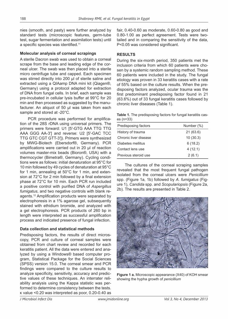
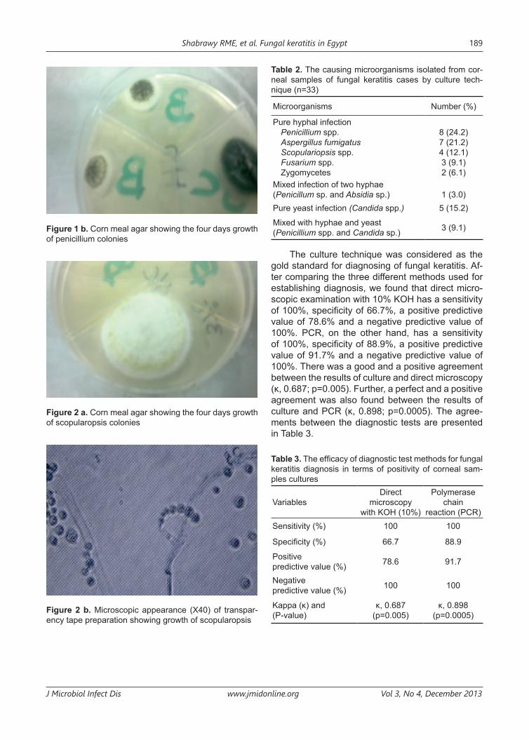
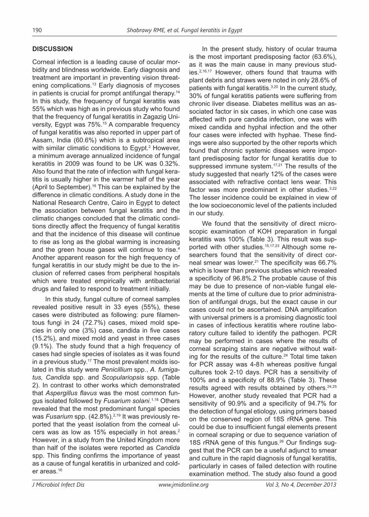
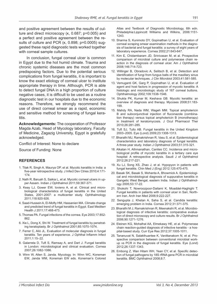
The cultures of the corneal scraping samples revealed that the most frequent fungal pathogen isolated from the corneal ulcers were Penicillium spp. (Figure 1a, 1b) followed by A. fumigatus (Fig-ure 1), Candida spp. and Scopulariopsis (Figure 2a, 2b). The results are presented in Table 2.
Figure 1 a. Microscopic appearance (X40) of KOH smear showing the hypha growth of penicillium
Shabrawy RME, et al. Fungal keratitis in Egypt 189
J Microbiol Infect Dis www.jmidonline.org Vol 3, No 4, December 2013
Figure 1 b. Corn meal agar showing the four days growth of penicillium colonies
Figure 2 a. Corn meal agar showing the four days growth of scopularopsis colonies
Figure 2 b. Microscopic appearance (X40) of transpar-ency tape preparation showing growth of scopularopsis
Table 2. The causing microorganisms isolated from cor-neal samples of fungal keratitis cases by culture tech-nique (n=33)
Microorganisms Number (%)
Pure hyphal infection Penicillium spp.Aspergillus fumigatusScopulariopsis spp.Fusarium spp.Zygomycetes
8 (24.2)7 (21.2)4 (12.1)3 (9.1)2 (6.1)
Mixed infection of two hyphae(Penicillum sp. and Absidia sp.) 1 (3.0)Pure yeast infection (Candida spp.) 5 (15.2)
Mixed with hyphae and yeast (Penicillium spp. and Candida sp.) 3 (9.1)
The culture technique was considered as the gold standard for diagnosing of fungal keratitis. Af-ter comparing the three different methods used for establishing diagnosis, we found that direct micro-scopic examination with 10% KOH has a sensitivity of 100%, specificity of 66.7%, a positive predictive value of 78.6% and a negative predictive value of 100%. PCR, on the other hand, has a sensitivity of 100%, specificity of 88.9%, a positive predictive value of 91.7% and a negative predictive value of 100%. There was a good and a positive agreement between the results of culture and direct microscopy (κ, 0.687; p=0.005). Further, a perfect and a positive agreement was also found between the results of culture and PCR (κ, 0.898; p=0.0005). The agree-ments between the diagnostic tests are presented in Table 3.
Table 3. The efficacy of diagnostic test methods for fungal keratitis diagnosis in terms of positivity of corneal sam-ples cultures
VariablesDirect
microscopywith KOH (10%)
Polymerasechain
reaction (PCR)Sensitivity (%) 100 100
Specificity (%) 66.7 88.9
Positivepredictive value (%) 78.6 91.7
Negativepredictive value (%) 100 100
Kappa (κ) and(P-value)
κ, 0.687(p=0.005)
κ, 0.898(p=0.0005)
Shabrawy RME, et al. Fungal keratitis in Egypt190
J Microbiol Infect Dis www.jmidonline.org Vol 3, No 4, December 2013
DISCUSSION
Corneal infection is a leading cause of ocular mor-bidity and blindness worldwide. Early diagnosis and treatment are important in preventing vision threat-ening complications.13 Early diagnosis of mycoses in patients is crucial for prompt antifungal therapy.14 In this study, the frequency of fungal keratitis was 55% which was high as in previous study who found that the frequency of fungal keratitis in Zagazig Uni-versity, Egypt was 75%.15 A comparable frequency of fungal keratitis was also reported in upper part of Assam, India (60.6%) which is a subtropical area with similar climatic conditions to Egypt.2 However, a minimum average annualized incidence of fungal keratitis in 2009 was found to be UK was 0.32%. Also found that the rate of infection with fungal kera-titis is usually higher in the warmer half of the year (April to September).16 This can be explained by the difference in climatic conditions. A study done in the National Research Centre, Cairo in Egypt to detect the association between fungal keratitis and the climatic changes concluded that the climatic condi-tions directly affect the frequency of fungal keratitis and that the incidence of this disease will continue to rise as long as the global warming is increasing and the green house gases will continue to rise.4 Another apparent reason for the high frequency of fungal keratitis in our study might be due to the in-clusion of referred cases from peripheral hospitals which were treated empirically with antibacterial drugs and failed to respond to treatment initially.
In this study, fungal culture of corneal samples revealed positive result in 33 eyes (55%), these cases were distributed as following: pure filamen-tous fungi in 24 (72.7%) cases, mixed mold spe-cies in only one (3%) case, candida in five cases (15.2%), and mixed mold and yeast in three cases (9.1%). The study found that a high frequency of cases had single species of isolates as it was found in a previous study.17 The most prevalent molds iso-lated in this study were Penicillium spp., A. fumiga-tus, Candida spp. and Scopulariopsis spp. (Table 2). In contrast to other works which demonstrated that Aspergillus flavus was the most common fun-gus isolated followed by Fusarium solani.1,18 Others revealed that the most predominant fungal species was Fusarium spp. (42.8%).2,19 It was previously re-ported that the yeast isolation from the corneal ul-cers was as low as 15% especially in hot areas.2 However, in a study from the United Kingdom more than half of the isolates were reported as Candida spp. This finding confirms the importance of yeast as a cause of fungal keratitis in urbanized and cold-er areas.16
In the present study, history of ocular trauma is the most important predisposing factor (63.6%), as it was the main cause in many previous stud-ies.2,16,17 However, others found that trauma with plant debris and straws were noted in only 28.6% of patients with fungal keratitis.3,20 In the current study, 30% of fungal keratitis patients were suffering from chronic liver disease. Diabetes mellitus was an as-sociated factor in six cases, in which one case was affected with pure candida infection, one was with mixed candida and hyphal infection and the other four cases were infected with hyphae. These find-ings were also supported by the other reports which found that chronic systemic diseases were impor-tant predisposing factor for fungal keratitis due to suppressed immune system.17,21 The results of the study suggested that nearly 12% of the cases were associated with refractive contact lens wear. This factor was more predominant in other studies.3,22 The lesser incidence could be explained in view of the low socioeconomic level of the patients included in our study.
We found that the sensitivity of direct micro-scopic examination of KOH preparation in fungal keratitis was 100% (Table 3). This result was sup-ported with other studies.15,17,23 Although some re-searchers found that the sensitivity of direct cor-neal smear was lower.21 The specificity was 66.7% which is lower than previous studies which revealed a specificity of 96.8%.2 The probable cause of this may be due to presence of non-viable fungal ele-ments at the time of culture due to prior administra-tion of antifungal drugs, but the exact cause in our cases could not be ascertained. DNA amplification with universal primers is a promising diagnostic tool in cases of infectious keratitis where routine labo-ratory culture failed to identify the pathogen. PCR may be performed in cases where the results of corneal scraping stains are negative without wait-ing for the results of the culture.24 Total time taken for PCR assay was 4-8 h whereas positive fungal cultures took 2-10 days. PCR has a sensitivity of 100% and a specificity of 88.9% (Table 3). These results agreed with results obtained by others.24,25 However, another study revealed that PCR had a sensitivity of 90.9% and a specificity of 94.7% for the detection of fungal etiology, using primers based on the conserved region of 18S rRNA gene. This could be due to insufficient fungal elements present in corneal scraping or due to sequence variation of 18S rRNA gene of this fungus.26 Our findings sug-gest that the PCR can be a useful adjunct to smear and culture in the rapid diagnosis of fungal keratitis, particularly in cases of failed detection with routine examination method. The study also found a good
Shabrawy RME, et al. Fungal keratitis in Egypt 191
J Microbiol Infect Dis www.jmidonline.org Vol 3, No 4, December 2013
and positive agreement between the results of cul-ture and direct microscopy (κ, 0.687; p=0.005) and a perfect and positive agreement between the re-sults of culture and PCR (κ, 0.898; p=0.0005) sug-gested these rapid diagnostic tests worked together with corneal sample cultures.
In conclusion, fungal corneal ulcer is common in Egypt due to the hot humid climate. Trauma and chronic systemic diseases are the most common predisposing factors. Due to the potential serious complications from fungal keratitis, it is important to know the exact etiology of corneal ulcer to institute appropriate therapy in time. Although, PCR is able to detect fungal DNA in a high proportion of culture negative cases, it is difficult to be used as a routine diagnostic test in our hospitals due to the economic reasons. Therefore, we strongly recommend the use of direct corneal smear as a rapid, economic and sensitive method for screening of fungal kera-titis.Acknowledgements: The cooperation of Professor Magda Azab, Head of Mycology laboratory, Faculty of Medicine, Zagazig University, Egypt is gratefully acknowledged.Conflict of Interest: None to declare Source of Funding: None
REFERENCES1. Tilak R, Singh A, Maurya OP, et al. Mycotic keratitis in India: a
five-year retrospective study. J Infect Dev Ctries 2010;4:171-174.
2. Nath R, Baruah S, Saikia L, et al. Mycotic corneal ulcers in up-per Assam. Indian J Ophthalmol 2011;59:367-371.
3. Keay LJ, Gower EW, Iovieno A, et al. Clinical and micro-biological characteristics of fungal keratitis in the United States, 2001-2007: a multicenter study. Ophthalmology 2011;118:920-926.
4. Saad-Hussein A, El-Mofty HM, Hassanien MA. Climate change and predicted trend of fungal keratitis in Egypt. East Mediterr Health J 2011;17:468-473.
5. Thomas PA. Fungal infections of the cornea. Eye 2003;17:852-862.
6. Xie L, Dong X, Shi W. Treatment of fungal keratitis by penetrat-ing keratoplasty. Br J Ophthalmol 2001;85:1070-1074.
7. Ferrer C, Alió JL. Evaluation of molecular diagnosis in fungal keratitis. Ten years of experience. J Ophthal Inflamm Infect 2011;1:15–22.
8. Galarreta D, Tuft S, Ramsay A, and Dart J. Fungal keratitis in London: microbiological and clinical evaluation. Cornea 2007;26:1082-1086.
9. Winn W, Allen S, Janda. Mycology. In: Winn WC, Koneman EW, Janda WM, Koneman EW eds. Koneman’s Colored
Atlas and Textbook of Diagnostic Microbiology, 6th edn. Philadelphia:Lippincott Williams and Wilkins, 2006:1151-1243.
10. Sharma S, Kunimoto DY, Gopinathan U, et al. Evaluation of corneal scraping smear examination methods in the diagno-sis of bacterial and fungal keratitis: a survey of eight years of laboratory experience. Cornea 2002;21:643-647.
11. Kim E, Chidambaram JD, Srinivasan M, et al. Prospective comparison of microbial culture and polymerase chain re-action in the diagnosis of corneal ulcer. Am J Ophthalmol 2008;146:714-723.
12. Willinger B, Obradovic A, Selitsch B, et al. Detection and identification of fungi from fungus balls of the maxillary sinus by molecular techniques. J Clin Microbiol 2003;41:581-585.
13. Vemuganti GK, Garg P, Gopinathan U, et al. Evaluation of agent and host factors in progression of mycotic keratitis: A histologic and microbiologic study of 167 corneal buttons. Ophthalmology 2002;109:1538-1546.
14. Shukla PK, Kumar M, Keshava GB. Mycotic keratitis: an overview of diagnosis and therapy. Mycoses 2008;51:183-199.
15. Mahdy RA, Nada WM, Wageh MM. Topical amphotericin B and subconjunctival injection of fluconazole (combina-tion therapy) versus topical amphotericin B (monotherapy) in treatment of keratomycosis. J Ocul Pharmacol Ther 2010;26:281-285.
16. Tuft SJ, Tullo AB. Fungal keratitis in the United Kingdom 2003–2005. Eye (Lond) 2009;23:1308-1313.
17. Bharathi MJ, Ramakrishnan R, Vasu S, et al. Epidemiological characteristics and laboratory diagnosis of fungal keratitis. A three-year study. Indian J Ophthalmol 2003;51:315-321.
18. Alkatan H, Athmanathan, Canites CC. Incidence and micro-biological profile of mycotic keratitis in a tertiary care eye hospital: A retrospective analysis. Saudi J of Ophthalmol 2012;26:217-221.
19. Xu LJ, Song XS, Zhao J, et al. Hypopyon in patients with fungal keratitis. Chin Med J (Eng) 2012;125:470-475.
20. Basak SK, Basak S, Mohanta A, Bhowmick A. Epidemiologi-cal and microbiological diagnosis of suppurative keratitis in Gangetic West Bengal, eastern India. Indian J Ophthalmol-ogy 2005;53:17-22.
21. Shokohi T, Nowroozpoor-Dailami K, Moaddel-Haghighi T. Fungal keratitis in patients with corneal ulcer in Sari, North-ern Iran. Arch Iran Med 2006;9:222-227.
22. Sengupta J, Khetan A, Saha S, et al. Candida keratitis: emerging problem in India. Cornea 2012;31:371-375.
23. Bharathi M J, Ramakrishnan R, Meenakshi R, et al. Microbio-logical diagnosis of infective keratitis: comparative evalua-tion of direct microscopy and culture results. Br J Ophthalmol 2006;90:1271-1276.
24. Eleinen KG, Mohalhal AA, Elmekawy HE, et al. Polymerase chain reaction-guided diagnosis of infective keratitis - a hos-pital-based study. Curr Eye Res 2012;37:1005-1011.
25. Tananuvat N, Salakthuantee K, Vanittanakom N, et al. Pro-spective comparison between conventional microbial work-up vs PCR in the diagnosis of fungal keratitis. Eye (Lond) 2012;26:1337-1343.
26. Embong Z, Wan Hitam WH, Yean CY, et al. Specific detec-tion of fungal pathogens by 18S rRNA gene PCR in microbial keratitis. BMC Ophthalmol 2008;8:7.