Embed Size (px)
Citation preview

AnemiasSupportive module 4 "Essentials of diagnosis, treatment
and prevention of major hematologic diseases"
LECTURE IN INTERNAL MEDICINE FOR IV COURSE STUDENTS
M. Yabluchansky, L. Bogun, L. Martymianova, O. Bychkova, N. Lysenko, N. MakienkoV.N. Karazin National University Medical School’ Internal Medicine Dept.
2016/2017 Spring Semester

Plan of the lecture
• Definition• Epidemiology• Etiology• Mechanisms • Adaptation to anemia• Classification• Clinical investigation• Diagnosis• Treatment • Prognosis• Prophylaxis• Abbreviations • Diagnostic guidelines
http://anemiaofchronicdisease.com/wp-content/uploads/2012/08/anemia-of-chronic-disease1.jpg

Definition Anemia is a disease and/or a clinical syndrome that consist in lowered ability of the blood to carry oxygen (hypoxia) due to decrease quantity and functional capacity and/or structural disturbances of red blood cells (RBCs) or decrease hemoglobin concentration or hematocrit in the blood
A severe form of anemia, in which the hematocrit is below 10%, is called the hyperanemia
WHO criteria is Hb < 13 g/dL in men and Hb < 12 g/dL in women (revised criteria for patient’s with malignancy Hb < 14 g/dL in men and Hb < 12g/dL in women)

Epidemiology 1
https://www.k4health.org/sites/default/files/anemia-map_updated.png

Epidemiology 2
http://img.medscape.com/fullsize/migrated/editorial/conferences/2006/4839/spivak.fig1.jpg

Epidemiology 3
http://www.omicsonline.org/2161-1165/images/2161-1165-2-118-g001.gif

Etiology 1 (basic forms)
Basic forms
• Blood loss
• Deficient erythropoiesis
• Excessive hemolysis (RBC destruction)
• Fluid overload (hypervolemia)
http://www.merckmanuals.com/professional/hematology-and-oncology/approach-to-the-patient-with-anemia/etiology-of-anemia https://en.wikipedia.org/wiki/Anemiahttp://content.onlinejacc.org/data/Journals/JAC/23133/04044_gr2.jpeg
Etiology 2 (blood loss)• Blood loss can be acute or chronic
• Anemia does not develop until several hours after acute blood loss, when interstitial fluid diffuses into the intravascular space and dilutes the remaining RBC mass
• During the first few hours, however, levels of polymorphonuclear granulocytes, platelets, and, in severe hemorrhage, immature WBCs and normoblastsmay rise
• Chronic blood loss results in anemia if loss is more rapid than can be replaced or, more commonly, if accelerated erythropoiesis depletes body iron stores
http://www.merckmanuals.com/professional/hematology-and-oncology/approach-to-the-patient-with-anemia/etiology-of-anemia

Etiology 3a (deficient erythropoiesis )
• Deficient erythropoiesis has myriad causes
• Complete cessation of erythropoiesis results in a decline in RBCs of about 7 to 10%/week (1%/day)
• Impaired erythropoiesis, even if not sufficient to decrease the numbers of RBCs, often causes abnormal RBC size and shape
http://www.merckmanuals.com/professional/hematology-and-oncology/approach-to-the-patient-with-anemia/etiology-of-anemia

Etiology 3b (deficient erythropoiesis )
http://yewbiotech.com/blog/wp-content/uploads/2013/11/F2.large_.jpg
Deficient erythropoiesis has myriad causes

USMLE Step 2 CK` test \1
A 32-year-old woman with type 1 diabetes mellitus has had progressive renal failure over the past 2 years. She has not yet started dialysis. Examination shows no abnormalities. Her hemoglobin concentration is 9 g/dL, hematocrit is 28%, and mean corpuscular volume is 94 μm3. A blood smear shows normochromic, normocytic cells. Which of the following is the most likely cause?
(A) Acute blood loss (B) Chronic lymphocytic leukemia (C) Erythrocyte enzyme deficiency (D) Erythropoietin deficiency (E) Immunohemolysis (F) Microangiopathic hemolysis
usmle-forums.com/usmle-step-2-ck-forum/16583-five-pulmonology-questions-part-2-a.html

Etiology 4a (excessive hemolysis )• Excessive hemolysis can be caused by intrinsic
abnormalities of RBCs or by extrinsic factors, such as the presence of antibodies on their surface, that lead to their early destruction
• An enlarged spleen sequesters and destroys RBCs more rapidly than normal
• Some causes of hemolysis deform as well as destroy RBCs
• Excessive hemolysis does not normally decrease reticulocyte production unless iron or other essential nutrients are depleted
http://www.merckmanuals.com/professional/hematology-and-oncology/approach-to-the-patient-with-anemia/etiology-of-anemia

Etiology 4b (excessive hemolysis )
https://www.healthcare.uiowa.edu/path_handbook/Appendix/Heme/hemolysis.jpg

Etiology 5 (Fluid overload (hypervolemia))
• Fluid overload causes decreased hemoglobin concentration and apparent anemia:
• General causes of hypervolemia include excessive sodium or fluid intake, sodium or water retention and fluid shift into the intravascular space
• Anemia of pregnancy is induced by blood volume expansion
https://en.wikipedia.org/wiki/Anemia

Etiology 6(causes of anemia)
• Blood loss
• Acute
• Chronic
• Deficient erythropoiesis
• Microcytic
• Normochromic-normocytic
• Macrocytic
• Excessive hemolysis due to extrinsic RBC defects
• Reticuloendothelial hyperactivity with splenomegaly
http://www.merckmanuals.com/professional/hematology-and-oncology/approach-to-the-patient-with-anemia/etiology-of-anemia

Etiology 7(causes of anemia)
• Immunologic abnormalities
• Mechanical injury
• Excessive hemolysis due to intrinsic RBC defects
• Membrane alterations, acquired
• Membrane alterations, congenital
• Metabolic disorders (inherited enzyme deficienacies)
• Hemoglobinopathies
http://www.merckmanuals.com/professional/hematology-and-oncology/approach-to-the-patient-with-anemia/etiology-of-anemia

Mechanisms 1a(anemia due to blood loss)
• With anemia due to blood loss, a reduction in oxygen-carrying capacity occurs along with a decrease in intravascular volume, with resultant hypoxia and hypovolemia
• Hypovolemia leads to hypotension, which is detected by stretch receptors (in the carotid bulb, aortic arch, heart, and lungs) and transmitted by their impulses along afferent fibers of the vagal and glossopharyngeal nerves to the medulla oblongata, cerebral cortex, and pituitary gland
http://emedicine.medscape.com/article/198475-overview#a4

Mechanisms 1b(anemia due to blood loss)
A person can have a low hematocrit and not be anemic
http://web2.airmail.net/uthman/anemia/anemia.html
Mechanisms 2a(anemia due to blood loss)
• In the medulla, sympathetic outflow is enhanced, while parasympathetic activity is diminished
• Increased sympathetic outflow leads to norepinephrine release from sympathetic nerve endings and discharge of epinephrine and norepinephrine from the adrenal medulla
• Sympathetic connection to the hypothalamic nuclei increases antidiuretic hormone (ADH) secretion from the pituitary gland
http://emedicine.medscape.com/article/198475-overview#a4

Mechanisms 2b(anemia due to blood loss)
Peptic ulcers extend beyond the lamina propria, whereas erosions are superficial
http://www.zuniv.net/physiology/book/chapter22.html

Mechanisms 3(anemia due to blood loss)
• Antidiuretic hormone (ADH) increases free water reabsorption in the distal collecting tubules
• In response to decreased renal perfusion, juxtaglomerular cells in the afferent arterioles release renin into the renal circulation, leading to increased angiotensin I, which is converted by angiotensin-converting enzyme (ACE) to angiotensin II
• Angiotensin II has a potent pressor effect on arteriolar smooth muscle and stimulates the zona glomerulosa of the adrenal cortex to produce aldosterone
http://emedicine.medscape.com/article/198475-overview#a4

Mechanisms 4(anemia due to blood loss)
• Aldosterone increases sodium reabsorption from the proximal tubules of the kidney, thus increasing intravascular volume
• The primary effect of the sympathetic nervous system is to maintain perfusion to the tissues by increasing systemic vascular resistance (SVR)
• The augmented venous tone increases the preload
http://emedicine.medscape.com/article/198475-overview#a4

Mechanisms 5(anemia due to blood loss)
• In states of hypovolemic hypoxia, the increased venous tone due to sympathetic discharge is thought to dominate the vasodilator effects of hypoxia
• Counterregulatory hormones (e.g., glucagon, epinephrine, cortisol) are thought to shift intracellular water to the intravascular space, perhaps because of the resultant hyperglycemia
http://emedicine.medscape.com/article/198475-overview#a4

Mechanisms 6a(anemia due to blood loss)
• Tissue oxygen delivery is the major controlling factor of erythropoiesis through the synthesis and release of erythropoietin (EPO) by the proximal tubular cells or the peritubular interstitial cells in the kidney
• EPO synthesis is governed by the activation of hypoxia inducible factor-1 (HIF-1), which controls the metabolic responses of multiple gene products to hypoxia
http://www.clevelandclinicmeded.com/medicalpubs/diseasemanagement/hematology-oncology/anemia/

Mechanisms 6b(anemia due to blood loss)
Pathophysiological mechanisms contributing to anemia in patients with Inflammatory Bowel Disease
http://www.haematologica.org/content/95/2/175

Mechanisms 7a(anemia due to blood loss)
• HIF-1 binds and activates the hypoxia-responsive transcriptional enhancer in the EPO gene regulatory region that upregulates EPO expression
• EPO stimulates erythroid precursor cells, leading to increased proliferation and shortening of their maturation time
• The marrow responds to increased EPO maximally in 4 to 7 days if enough iron is available
http://www.clevelandclinicmeded.com/medicalpubs/diseasemanagement/hematology-oncology/anemia/

Mechanisms 7b(anemia due to blood loss)
A. The peripheral blood in severe megaloblastic anemia
B. The bone marrow in severe megaloblastic anemia
http://clinicalgate.com/hemolytic-anemias-and-anemia-due-to-acute-blood-loss/

Mechanisms 8(anemia due to acute blood loss)
• Erythropoiesis can be increased by as much as a factor of 8
• Typical of an endocrine loop feedback mechanism, there is an inverse relation between the hemoglobin and EPO levels measured in the blood
• This relation is somewhat distorted in the anemia associated with inflammation or chronic disease, in which there may be a blunted EPO response
http://www.clevelandclinicmeded.com/medicalpubs/diseasemanagement/hematology-oncology/anemia/

Adaptation to anemia
• Modulation of oxygen affinity—largely mediated by an increase in red blood cell 2,3-biphosphoglycerate
• Increased production of erythropoietin—the main growth factor for red blood cell production
• Redistribution of flow to benefit the myocardium, brain, and muscle
• Increase in cardiac output
• Reduction of mixed venous oxygen tension to increase the arteriovenous oxygen difference
http://www.clevelandclinicmeded.com/medicalpubs/diseasemanagement/hematology-oncology/anemia/

Classification (morphological)
Cell Size Normal RDW High RDW
Microcytosis
MCV<70 µm3
Thalassemia minor, anemia of
chronic disease, some
hemoglobinopathy traits
Iron deficiency, hemoglobin H
disease, some anemia of chronic
disease, some thalassemia minor,
fragmentation hemolysis
Normocytosis:
High reticulocyte
count
Low reticulocyte
count
Anemia of chronic disease,
hereditary spherocytosis, some
hemoglobinopathy traits, acute
bleeding
Early or partially treated iron or
vitamin deficiency, sickle cell disease
Macrocytosis
MCV>100 µm3
Aplastic anemia, some
myelodysplasias
Vitamin B12 or folate deficiency,
autoimmune hemolytic anemia, cold
agglutinin disease, some
myelodysplasias, liver disease,
thyroid disease, alcohol
RDW = red cell distribution width; MCV = mean corpuscular volumehttp://www.ascp.org/PDF/SneekPeekPracDiagofHemDisorders.aspxCacheddefined

Classification (kinetic: red blood cell loss (bleeding) or destruction (hemolysis)
AcquiredMechanical (march hemoglobinuria, artificial heart valves)Microangiopathic (disseminated intravascular hemolysis, thrombotic thrombocytopenic purpura, vasculitis)Parasites and microorganisms (e.g., malaria, bartonellosis, babesiosis, clostridium perfringens )Antibody mediated Warm-type autoimmune hemolytic anemia
Cryopathic syndromes (cold agglutinin disease, paroxysmal cold hemoglobinuria, cryoglobulinemia)
Transfusion reactions
HypersplenismRed cell membrane disorders Spur cell hemolsis
Acquired acanthocytosis and acquired stomatocytosis
Chemical injury and complex chemicals (arsenic, copper, chlorate, spider, scorpion, and snake venoms)Physical injury (heat, oxygen, radiation)
http://www.clevelandclinicmeded.com/medicalpubs/diseasemanagement/hematology-oncology/anemia/#figure1

Classification (kinetic: red blood cell loss (bleeding) or destruction (hemolysis)
http://www.clevelandclinicmeded.com/medicalpubs/diseasemanagement/hematology-oncology/anemia/#figure1
Hereditary
Hemoglobinopathies (sickle cell disease, unstable hemoglobins)
Red cell membrane disorders
Cytoskeletal membrane disorders (hereditary spherocytosis, elliptocytosis, pyropoikilocytosis)
Lipid membrane disorders (hereditary abetalipoproteinemia, hereditary stomatocytosis)
Membrane disorders associated with abnormalities of erythrocyte antigens (McLeod syndrome, Rh deficiency Syndromes)
Membrane disorders associated with abnormal transport (hereditary xerocytosis)
Red cell enzyme defects (pyruvate kinase, 5' nucleotidase, glucose-6-phosphate dehydrogenase deficiencies)Porphyrias (congenital erythropoietic and hepatoerythropoietic porphyrias, rarely congenital erythropoietic protoporphyria)

Classification (kinetic: red blood cell underproduction)
http://www.clevelandclinicmeded.com/medicalpubs/diseasemanagement/hematology-oncology/anemia/#figure1
AcquiredPluripotent stem cell failure Aplastic anemia (radiation induced, drugs and chemicals, viruses, idiopathic)
Anemia of leukemia and of myelodysplastic syndromes
Anemia associated with marrow infiltration (multiple myeloma, myelofibrosis, carcinoma)
Erythroid progenitor cell failure Pure red cell aplasia (parvovirus B19 infection, drugs, associated with thymoma,
autoantibodies)
Endocrine disorders (thyroid, adrenal, pituitary hypofunction)
Acquired sideroblastic anemia (drugs, copper deficiency, etc.)Functional impairment of erythroid and other progenitors due to nutritional and other causes Nutritional deficiencies-iron, vitamin B12, folic acid, pyridoxine
Chronic renal disease
Anemia of chronic disease and inflammation

Classification (kinetic: red blood cell underproduction)
http://www.clevelandclinicmeded.com/medicalpubs/diseasemanagement/hematology-oncology/anemia/#figure1
Hereditary
Pluripotent stem cell failure (Fanconi, Shwachman and dyskeratosis congenital syndromes)Erythroid progenitor cell failure (Diamond-Blackfan, congenital dyserythropoietic syndromes)Functional impairment of erythroid and other progenitors due to nutritional and other causes Megaloblastic anemias (Imerslund-Gräsbeck disease, intrinsic factor deficiency,
transcobalamin II deficiency)
Inborn purine and pyrimidine metabolic defects (Lesch-Nyhan syndrome, hereditary orotic aciduria)
Disorders of iron metabolism (atransferrinemia, divalent metal transporter or DMT-1 mutation)
Hereditary sideroblastic anemia

Classification (clinical)
• Alpha Thalassemia
• Anemia of chronic disease
• Aplastic Anemia
• Beta Thalassemia
• Hemolytic Anemia
• Iron Deficiency Anemia
• Megaloblastic Anemia
• Myelophthisic Anemia
• Pernicious Anemia
• Sickle Cell Anemia
• Spur Cell Anemiahttp://blogs.nejm.org/now/index.php/iron-deficiency-anemia/2015/05/08/
Iron-deficiency anemia is the most common form of anemia in the world

USMLE Step 2 CK` test \2A 43-year-old woman comes to the physician because of fatigue for 6 months. She has had progressively severe dyspnea on exertion for 6 weeks. She had an extensive abdominal operation 5 years ago for Crohn disease. She does not take any medications. Her temperature is 37°C (98.6°F), pulse is 62/min, respirations are 18/min, and blood pressure is 110/65 mm Hg. Examination of the thyroid gland, lungs, heart, abdomen, and extremities shows no abnormalities. Test of the stool for occult blood is negative. Laboratory studies show: Hemoglobin 8 g/dL Mean corpuscular volume 70 μm3 Leukocyte count 9000/mm3 Platelet count 500,000/mm3 . Which of the following is the most likely diagnosis?: (A) Acute leukemia (B) Anemia of chronic disease (C) Folic acid deficiency (D) Iron deficiency (E) Lyme disease (F) Microangiopathic hemolytic anemia (G) Pernicious anemia (H) Sleep apnea
usmle-forums.com/usmle-step-2-ck-forum/16583-five-pulmonology-questions-part-2-a.html

Clinical investigation (symptoms)
• Easy fatigue and loss of energy
• Unusually rapid heart beat, particularly with exercise
• Shortness of breath, particularly with exercise
• Pale skin
• Leg cramps
• Coldness in the hands and feet
• Insomnia
• Light-headedness
• Faintness
• Signs of heart failure
http://emedicine.medscape.com/article/198475-clinical

Clinical investigation (symptoms: anemia caused by iron deficiency)
• A hunger for strange substances such as paper, ice, or dirt (a condition called pica)
• Upward curvature of the nails, referred to as koilonychias
• Soreness of the mouth with cracks at the corners
http://www.webmd.com/a-to-z-guides/understanding-anemia-symptoms http://mystuf123.blogspot.com/p/p-i-c.htmlhttp://www.nhs.uk/conditions/nail-abnormalities/Pages/Introduction.aspx
Pica
Koilonychia

Clinical investigation (symptoms: anemia caused by vitamin B12 deficiency)
• A tingling, "pins and needles" sensation in the hands or feet
• Lost sense of touch
• A wobbly gait and difficulty walking
• Clumsiness and stiffness of the arms and legs
• Dementiahttp://www.webmd.com/a-to-z-guides/understanding-anemia-symptoms http://knitfreedom.com/being-a-knitter/top-5-stretches-for-knitting-pain-relief
Hand stiffness

Clinical investigation (symptoms: anemia caused by chronic lead poisoning)
• A blue-black line on the gums referred to as a lead line
• Abdominal pain
• Constipation
• Vomiting
http://www.webmd.com/a-to-z-guides/understanding-anemia-symptoms http://www.medicaljournals.se/acta/content/?doi=10.2340/00015555-1201&html=1
A blue-black line on the gums referred to as a
lead line

Clinical investigation (symptoms: anemia caused by chronic red blood cell
destruction)
• Jaundice (yellow skin and eyes)
• Brown or red urine
• Leg ulcers
• Failure to thrive in infancy
• Symptoms of gallstones
http://www.webmd.com/a-to-z-guides/understanding-anemia-symptoms http://photos1.blogger.com/x/blogger/5915/3798/1600/202479/500-1a.jpg
Yellow around eyes anemia

Clinical investigation (symptoms: sickle cell anemia)
• Fatigue
• Susceptibility to infection
• Delayed growth and development in children
• Episodes of severe pain, especially in the joints, abdomen, and limbs
http://www.webmd.com/a-to-z-guides/understanding-anemia-symptoms http://www.nhlbi.nih.gov/sites/www.nhlbi.nih.gov/files/images/anemia.jpg
Sickle cell anemia

Clinical investigation (symptoms: anemia caused by sudden red blood cell
destruction)
• Abdominal pain
• Brown or red urine
• Jaundice (yellow skin)
• Small bruises under the skin
• Seizures
• Symptoms of kidney failure
http://www.webmd.com/a-to-z-guides/understanding-anemia-symptoms http://www.wisegeek.org/what-are-the-most-common-causes-of-green-skin.htm
The bruise under the skin

USMLE Step 2 CK` test\3A 15-year-old girl who is a ballet dancer has not had a menstrual period for the past 3 months. Menses were previously regular at 29-day intervals. She has lost weight over the past year; her weight is 70% of that expected for her height. She is afebrile and has purpuric lesions on her extremities and trunk. Platelet, absolute neutrophil, and lymphocyte counts are below the reference range. She has macrocytic anemia. The most likely cause of these symptoms is a deficiency of which of the following nutrients? (A) Folic acid (B) Iron (C) Linoleic acid (D) Magnesium (E) Niacin(Vitamin PP) (F) Protein (G) Vitamin A (H) Vitamin B6 (pyridoxine) (I) Vitamin C (J) Vitamin D (K) Vitamin E (L) Vitamin K (M) Zinc
usmle-forums.com/usmle-step-2-ck-forum/16583-five-pulmonology-questions-part-2-a.html

Clinical investigation (accents on history 1)
• The duration of anemia can be established by obtaining a history of previous blood examinations and, if necessary, by acquiring those records
• Similarly, a history of rejection as a blood donor or prior prescription of hematinic provides clues that anemia was detected previously
http://emedicine.medscape.com/article/198475-clinical
Clinical investigation (accents on history 2)
• Obtain a family history for anemia, jaundice, cholelithiasis, splenectomy, bleeding disorders, and abnormal Hbs
• Document the patient's occupation, hobbies, prior medical treatment, drugs (including over-the-counter medications and vitamins), and household exposures to potentially noxious agents (insecticides, paints, solvents, hair dyes)
http://emedicine.medscape.com/article/198475-clinical

Clinical investigation (accents on history 3)
• In searching for blood loss, carefully document pregnancies, abortions, and menstrual loss
• Patients do not appreciate the significance of tarry stools, but changes in bowel habits can be useful in uncovering neoplasms of the colon
• Hemorrhoidal blood loss is difficult to quantify, and it may be overlooked or overestimated from one patient to another
http://emedicine.medscape.com/article/198475-clinical

Clinical investigation (accents on history 4)
• Seek a history of gastrointestinal (GI) complaints that may suggest gastritis, peptic ulcers, hiatal hernias, or diverticula
• Abnormal urine color can occur in renal and hepatic disease and in hemolytic anemia
• A dietary history must include foods that the patient eats and those that he/she avoids, as well as an estimate of their quantity
http://emedicine.medscape.com/article/198475-clinical

Clinical investigation (accents on history 6)
• Changes in body weight are important with regard to dietary intake and can suggest the presence of malabsorption or an underlying wasting disease of infectious, metabolic, or neoplastic origin
• Obtain a history of fever or identify the presence of fever, because infections, neoplasms, and collagen vascular disease can cause anemia
http://emedicine.medscape.com/article/198475-clinical

Clinical investigation (accents on history 7)
• The occurrence of purpura, ecchymoses, and petechiae suggest the occurrence of either thrombocytopenia or other bleeding disorders; this may be an indication either that more than 1 bone marrow lineage is involved or that coagulopathy is a cause of the anemia because of bleeding
• Cold intolerance can be an important symptom of hypothyroidism or lupus erythematosus, paroxysmal cold hemoglobinuria, and certain macroglobulinemias
http://emedicine.medscape.com/article/198475-clinical

Clinical investigation (accents on history 8)
• The relation of dark urine to either physical activity or time of day can be important in march hemoglobinuria and paroxysmal nocturnal hemoglobinuria
• Explore the presence or the absence of symptoms suggesting an underlying disease, such as cardiac, hepatic, and renal disease; chronic infection; endocrinopathy; or malignancy
• A geographic history can also be important in establishing an etiology
http://emedicine.medscape.com/article/198475-clinical

Clinical investigation (accents on physical examination 1a)
• The skin and mucous membranes are often bypassed, so that pallor, abnormal pigmentation, icterus, spider nevi, petechiae, purpura, angiomas, ulcerations, palmar erythema, coarseness of hair, puffiness of the face, thinning of the lateral aspects of the eyebrows, nail defects, and a usually prominent venous pattern on the abdominal wall are missed in the rush to examine the heart and the lungs
http://emedicine.medscape.com/article/198475-clinical

Clinical investigation (accents on physical examination 1b)
http://emedicine.medscape.com/article/198475-clinical https://en.wikipedia.org/wiki/Purpura
Thinning of the lateral aspects of
the eyebrows
Spider neviPurpura

Clinical investigation (accents on physical examination 2)
• Examine optic fundi carefully but not at the expense of the conjunctivae and the sclerae, which can show pallor, icterus, splinter hemorrhages, petechiae, comma signs in the conjunctival vessels, or telangiectasia that can be helpful in planning additional studies
• Perform systematic examination for palpable enlargement of lymph nodes for evidence of infection or neoplasia
http://emedicine.medscape.com/article/198475-clinical

Clinical investigation (accents on physical examination 3a)
• Bilateral edema is useful in disclosing underlying cardiac, renal, or hepatic disease, whereas unilateral edema may portend lymphatic obstruction due to a malignancy that cannot be observed or palpated
• Carefully search for hepatomegaly and splenomegaly because in patients with chronic disorders, these organs are firm, nontender, and nonnodular, and in patients with carcinoma, they may be hard and nodular
http://emedicine.medscape.com/article/198475-clinical

Clinical investigation (accents on physical examination 3b)
Massive hepatosplenomegaly in a patient with severe malarial anemia due to Plasmodium falciparum
infectionhttp://medicine.academic.ru/pictures/medicine/712.jpg

Clinical investigation (accents on physical examination 4)
• A rectal and pelvic examination cannot be neglected, because tumor or infection of these organs can be the cause of anemia
• The neurologic examination should include tests of position sense and vibratory sense, examination of the cranial nerves, and testing for tendon reflexes
http://emedicine.medscape.com/article/198475-clinical

Clinical investigation (accents on physical examination 5)
• The heart should not be ignored, because enlargement may provide evidence of the duration and the severity of the anemia, and murmurs may be the first evidence of infective endocarditis that could explain the etiology of the anemia
http://emedicine.medscape.com/article/198475-clinical

USMLE Step 2 CK` test \4A 75-year-old woman has increasing shortness of breath on exertion. Findings on physical examination are unremarkable. X-rays of the chest show no abnormalities of the heart or lungs. Pertinent laboratory findings include: Hematocrit 28% Hemoglobin 9 g/dL Mean corpuscular volume 70 μm3
Which of the following is the most likely basis for these findings?
(A) Acquired hemolytic anemia (B) Chronic blood loss (C) Folic acid deficiency (D) β-Thalassemia minor (E) Pernicious anemia usmle-forums.com/usmle-step-2-ck-forum/16583-five-pulmonology-questions-part-2-a.html

Diagnosisinitial assessment
• Patients may present in several ways• The urgency with which anemia is evaluated depends
on the severity at presentation• Patients with an acute severe hemorrhage present with
hypovolaemia and symptoms and signs of the underlying cause
• Many patients with no acute or active bleeding are asymptomatic, and the anaemia is only noted on an full blood count (FBC) taken as part of the assessment of an unrelated condition
• The first step in diagnosis is to identify the type of anemia that is present, using the results of the FBC
http://bestpractice.bmj.com/best-practice/monograph/93/diagnosis.html

Diagnosisalgorithm for the assessment of anemia
http://bestpractice.bmj.com/best-practice/monograph/93/diagnosis.html

Diagnosismicrocytic anemia: abnormal serum iron studies 1/4
• A low serum iron, a high total iron-binding capacity (TIBC), and a low ferritin indicate iron deficiency anaemia
• Iron deficiency produces an associated reactive thrombocytosis that provides an additional clue
• Iron deficiency is not a diagnosis and requires further investigation to elucidate the cause
http://bestpractice.bmj.com/best-practice/monograph/93/diagnosis.html

Diagnosismicrocytic anemia: abnormal serum iron studies 2/4
• Diets low in meat
• Generalized malabsorption and malnutrition (combined vitamin B12 and/or folate deficiency)
• A history of bleeding (excessive menstrual losses in women, upper and lower GI bleeding, peptic ulcer disease, cirrhosis, idiopathic pulmonary hemosiderosis, excessive blood donation, runner's anaemia, etc.)
http://bestpractice.bmj.com/best-practice/monograph/93/diagnosis.html

USMLE Step 2 CK` test \5
usmle-forums.com/usmle-step-2-ck-forum/16583-five-pulmonology-questions-part-2-a.html

Diagnosismicrocytic anemia: abnormal serum iron studies 3/4
• Signs of iron deficiency (koilonychia, angular cheilosis, glossitis, thinning hair)
• Investigations are guided by the history and examination, and include the fecal occult blood testing, upper GI endoscopy, immunoglobulin A-tissue transglutaminase (IgA-tTG) test (positive in coeliac disease), colonoscopy, flow cytometry (if there is a history of passing red urine), transvaginal ultrasound (causes of menorrhagia)
http://bestpractice.bmj.com/best-practice/monograph/93/diagnosis.html

Diagnosismicrocytic anemia: abnormal serum iron studies 4/4
• A low serum iron, a low total iron-binding capacity, and a low ferritin suggest anaemia of chronic disease
• A history of an underlying inflammation (infection, neoplasms, autoimmune reactions, and injury to tissue from trauma and surgery) is usually present
• A serum erythropoietin level is usually normal or mildly elevated
• Hypothyroidism and vitamin C deficiency may produce a falsely low ferritin level
http://bestpractice.bmj.com/best-practice/monograph/93/diagnosis.html

Diagnosismicrocytic anemia: normal serum iron studies 1/5
• The most important cause to exclude is thalassemia
• A family history is usually present
• The disease is more common in individuals of Mediterranean, Middle Eastern, or Southeast Asian descent
• The severity ranges from asymptomatic to severe transfusion-dependent symptoms
http://bestpractice.bmj.com/best-practice/monograph/93/diagnosis.html

Diagnosismicrocytic anemia: normal serum iron studies 2/5
• The examination findings may be normal, or reveal splenomegaly, jaundice, abdominal distension, and icterus
• Morphological changes including skeletal abnormalities, a large head, chipmunk facies, and misaligned teeth are seen in beta-thalassemia intermedia and major
http://bestpractice.bmj.com/best-practice/monograph/93/diagnosis.html

Diagnosismicrocytic anemia: normal serum iron studies 3/5
• Distinct features on the FBC that suggest the diagnosis include a marked decrease in MCV (usually close to 70 femtolitres [fL]) with a low mean corpuscular Hb, target cells on the peripheral smear, and an elevated reticulocyte count (>2%)
• A Mentzer's index (MCV/RBC) <13 is suggestive of thalassemia, and an index >14 suggests iron deficiency
http://bestpractice.bmj.com/best-practice/monograph/93/diagnosis.html

Diagnosismicrocytic anemia: normal serum iron studies 4/5
• Thalassemia is diagnosed using Hb electrophoresis
• The presence of Hb H, Hb Bart, and concomitant hemoglobinopathies (Hb E, Hb S, Hb C, Hb D) is diagnostic of alpha-thalasemia
• A high HbF with minimal or absent HbA and an elevated HbA2 is diagnostic of beta-thalassemia
http://bestpractice.bmj.com/best-practice/monograph/93/diagnosis.html

Diagnosismicrocytic anemia: between IDA and Thalassemia 5/5
http://www.medical-labs.net/wp-content/uploads/2014/03/Microcytic-Anemia-between-IDA-and-Thalassemia.jpg

USMLE Step 2 CK` test \6A 25-year-old woman has a routine, pre-employment physical examination. Laboratory studies include:
Hemoglobin 11.3 g/dL Hematocrit 34% Erythrocyte count 5.2 million/mm3 Mean corpuscular volume 65 μm3
Follow-up laboratory studies show that the serum iron concentration and iron-binding capacity are within the reference ranges. Hemoglobin electrophoresis shows increased hemoglobin A2 (5%). Which of the following is the most likely diagnosis?
(A) Anemia of chronic disease (B) Iron deficiency anemia (C) Sideroblastic anemia (D) α-Thalassemia minor (E) β-Thalassemia minor
usmle-forums.com/usmle-step-2-ck-forum/16583-five-pulmonology-questions-part-2-a.html

Diagnosisnormocytic anemia: hypoproliferative 1/11
• Normocytic anaemia include disorders that decrease RBC production
• Hematological malignancies and aplastic anaemiaare the most important diagnoses to exclude, and are usually associated with multiple cytopenias
• An isolated anemia is usually due to pure red cell aplasia, which may be self-limiting or persistent
http://bestpractice.bmj.com/best-practice/monograph/93/diagnosis.html

Diagnosisnormocytic anemia: hypoproliferative 2/11
• Chronic renal failure or hypothyroidism can cause an isolated anaemia
• Secondary hyperparathyroidism exacerbates the anaemia of chronic renal failure
• Symptoms of bleeding, easy bruising, night sweats, or weight loss suggest hematological malignancy or aplastic anaemia
http://bestpractice.bmj.com/best-practice/monograph/93/diagnosis.html

Diagnosisnormocytic anemia: hypoproliferative 3/11
• Parvovirus infection, infectious mononucleosis, viral hepatitis, malaria, respiratory infections, gastroenteritis, primary atypical pneumonia, and mumps can result in a self-limiting pure red cell aplasia
• Antiepileptic medications (phenytoin, carbamazepine, valproate sodium), azathioprine, sulfonamides, isoniazid, and procainamide cause pure red cell aplasia
http://bestpractice.bmj.com/best-practice/monograph/93/diagnosis.html

Diagnosisnormocytic anemia: hypoproliferative 4/11
• Benzene, penicillamine, and gold can cause aplastic anaemia
• Chloramphenicol can cause either aplastic anaemia or pure red cell aplasia
• Chemotherapy causes pancytopenia.
• Radiotherapy, especially to pelvic or sternal areas, can cause pancytopenia
• A history of immunosuppression or chronic hepatitis suggests persistent pure red cell aplasia
http://bestpractice.bmj.com/best-practice/monograph/93/diagnosis.html

Diagnosisnormocytic anemia: hypoproliferative 5/11
• There may be a history or features of chronic renal failure or hypothyroidism
• Ecchymoses or petechiae due to thrombocytopenia suggest hematological malignancy, myelodysplastic syndrome, or aplastic anaemia
• Lymphadenopathy or fever suggest malignancy or infections (e.g., infectious mononucleosis)
http://bestpractice.bmj.com/best-practice/monograph/93/diagnosis.html

Diagnosisnormocytic anemia: hypoproliferative 6/11
• Splenomegaly may be seen in hematological malignancies
• Clinical features of systemic lupus erythematosus (SLE), rheumatoid arthritis, dermatomyositis, polyarteritis nodosa, or scleroderma resulting in persistent pure red cell aplasia may be present
• Abnormal lung examination (if lung cancer is the primary cancer) or a breast mass (if breast cancer is the primary) may be present
http://bestpractice.bmj.com/best-practice/monograph/93/diagnosis.html

Diagnosisnormocytic anemia: hypoproliferative 7/11
• A positive Trousseau's sign or Chvostek's sign in patients with chronic renal failure indicates hypocalcemia, probably due to associated secondary hyperparathyroidism
• FBC may show an associated cytopenia and characteristic changes specific to a hematological malignancy
• A pancytopenia suggests aplastic anaemia, or may be due to chemotherapy or radiotherapy
http://bestpractice.bmj.com/best-practice/monograph/93/diagnosis.html

Diagnosisnormocytic anemia: hypoproliferative 8/11
• An isolated anemia suggests pure red cell aplasia or anemia due to chronic renal failure
• Bone marrow aspiration provides a definitive diagnosis of aplastic anemia, acute leukemia, chronic myelogenous leukemia (CML), or bone marrow metastases
• Antiparvovirus antibodies are positive in parvovirus infection, the most common infectious cause of pure red cell aplasia
http://bestpractice.bmj.com/best-practice/monograph/93/diagnosis.html

Diagnosisnormocytic anemia: hypoproliferative 9/11
Other tests to consider:
• Hepatitis serology, to exclude an active hepatitis
• Monospot test or Epstein-Barr virus (EBV) IgM, to exclude infectious mononucleosis
• Thick and thin peripheral smear, to exclude malaria if history and findings suggest it
• Thyroid function tests; TSH is elevated and free T4 reduced in hypothyroidism
• Antinuclear antibodies, which are positive in SLE or scleroderma
http://bestpractice.bmj.com/best-practice/monograph/93/diagnosis.html

Diagnosisnormocytic anemia: hypoproliferative 10/11
Other tests to consider:
• Rheumatoid factor, which is positive in rheumatoid arthritis
• Serum CK, which is elevated in dermatomyositis
• Chest x-ray, which may show infiltrates in atypical pneumonia or a smooth mass in thymoma
• Erythropoietin levels, which may be decreased in patients with chronic renal failure
• Serum calcium and parathyroid hormone levels should be considered if associated secondary hyperparathyroidism is suspected
http://bestpractice.bmj.com/best-practice/monograph/93/diagnosis.html

Diagnosisnormocytic anemia: hypoproliferative 11/11
http://accessmedicine.mhmedical.com/data/Books/tint/tint_c626f003.gif

Diagnosisnormocytic anemia: hyperproliferative 1 /15
• Potential diagnoses include hemolytic anemias (microangiopathic hemolytic anemias, autoimmune hemolytic anemia, drugs, infections, inherited conditions, transfusion reactions, or burns)
• Drugs that can cause hemolysis include penicillin, methyldopa, levodopa, quinidines, cephalosporins, and some NSAIDs
http://bestpractice.bmj.com/best-practice/monograph/93/diagnosis.html

Diagnosisnormocytic anemia: hyperproliferative 2/15
• Cyclosporine, tacrolimus, clopidogrel, oral contraceptive pills, and some chemotherapy drugs may cause hemolytic uremic syndrome
• The triggers of disseminated intravascular coagulation (DIC), that include ongoing severe infection, sepsis, malignancy, obstetric emergency, trauma, burns, envenomation, drug overdose, or any cause of endothelial damage
http://bestpractice.bmj.com/best-practice/monograph/93/diagnosis.html

Diagnosisnormocytic anemia: hyperproliferative 3/15
• The presence of acute-onset neurological symptoms, including headache, confusion, focal weakness, seizures, or coma, should prompt suspicion of thrombotic thrombocytopenic purpura (TTP)
• Female patients may have associated menorrhagia
http://bestpractice.bmj.com/best-practice/monograph/93/diagnosis.html

Diagnosisnormocytic anemia: hyperproliferative 4/15
• Sudden-onset dizziness, headache, mental status changes, loss of sensation or motor strength, chest pain or pressure, dyspnoea, or oedema in a patient with known hypertension should prompt suspicion of malignant hypertension; a history of renal failure or eclampsia may also be present
• An expanding vascular skin lesion in a young infant or child should prompt suspicion of a hemangioma
http://bestpractice.bmj.com/best-practice/monograph/93/diagnosis.html

Diagnosisnormocytic anemia: hyperproliferative 5/15
• A history of prosthetic valve replacement may indicate hemolysis induced by the prosthesis
• Cutaneous burns affecting more than 10% of the body surface area can cause a hemolytic anaemia, or trigger DIC
• Infective causes include cytomegalovirus (CMV), infectious mononucleosis, toxoplasmosis, and leishmaniosis
http://bestpractice.bmj.com/best-practice/monograph/93/diagnosis.html

Diagnosisnormocytic anemia: hyperproliferative 6/15
• Bloody diarrhoea should prompt suspicion of Escherichia coli infection and hemolytic uraemic syndrome
• Patients with inherited hemolytic anemias such as sickle cell anaemia, hereditary spherocytosis, or glucose-6-phosphate dehydrogenase (G6PD) deficiency may have a positive family history
http://bestpractice.bmj.com/best-practice/monograph/93/diagnosis.html

Diagnosisnormocytic anemia: hyperproliferative 7/15
• Persistent pain in the skeleton, chest, or abdomen; priapism; lower-extremity skin ulcers; or an acute pneumonia-like syndrome suggest sickle cell anemia
• There may be a previous history of autoimmune disease (e.g., SLE, rheumatoid arthritis, scleroderma) or lymphoproliferative disorders (non-Hodgkin's lymphoma, chronic lymphocytic leukemia), which can lead to autoimmune hemolytic anemia
http://bestpractice.bmj.com/best-practice/monograph/93/diagnosis.html

Diagnosisnormocytic anemia: hyperproliferative 8/15
• The autoimmune diseases may also cause pure red cell aplasia, in which case the reticulocyte count would be low, with normal lactate dehydrogenase, haptoglobin, and bilirubin levels
• Recent blood transfusion may indicate hemolysis due to a transfusion reaction
• Occupational or home exposure to lead should prompt suspicion of lead toxicity
http://bestpractice.bmj.com/best-practice/monograph/93/diagnosis.html

Diagnosisnormocytic anemia: hyperproliferative 9/15
• Features of microangiopathic disease: purpura or ecchymoses, malignant hypertension, edema, oliguria or polyuria, focal neurological signs, and hypertensive retinopathy
• Splenomegaly is seen in hereditary spherocytosis
• Lymphadenopathy may indicate infectious mononucleosis, leukemia, lymphoma, or autoimmune disease
http://bestpractice.bmj.com/best-practice/monograph/93/diagnosis.html

Diagnosisnormocytic anemia: hyperproliferative 10/15
• A thrombocytopenia with schistocytes strongly suggests a microangiopathic hemolytic anemia
• Spherocytes suggest autoimmune hemolytic anaemia or hereditary spherocytosis
• Hereditary spherocytosis is associated with increased mean corpuscular Hb
• Sickling of RBCs is diagnostic of sickle cell anemia
http://bestpractice.bmj.com/best-practice/monograph/93/diagnosis.html

Diagnosisnormocytic anemia: hyperproliferative 11/15
• Heinz bodies, eccentrocytes, or bite cells are seen in G6PD deficiency
• Elevated lactate dehydrogenase and bilirubin levels with a decreased haptoglobin are strongly suggestive of a hemolytic anemia
• Serum creatinine may be elevated in patients with hemolytic uraemic syndrome or malignant hypertension
http://bestpractice.bmj.com/best-practice/monograph/93/diagnosis.html

Diagnosisnormocytic anemia: hyperproliferative 12/15
• Prothrombin time and activated partial prothrombin time, which are prolonged in DIC but normal in other microangiopathic hemolytic anemias
• DIC panel shows elevated D-dimers and fibrin degradation products with low fibrinogen in patients with DIC
• X-rays and MRI scanning of suspected regions reveal internal hemangiomas
http://bestpractice.bmj.com/best-practice/monograph/93/diagnosis.html

Diagnosisnormocytic anemia: hyperproliferative 13/15
• Direct antiglobulin (Coombs') test is positive in autoimmune hemolytic anemia
• Sickle cell anaemia is diagnosed on FBC
• Osmotic fragility test is positive in hereditary spherocytosis
• G6PD assays identify deficiencies of the enzyme
http://bestpractice.bmj.com/best-practice/monograph/93/diagnosis.html

Diagnosisnormocytic anemia: hyperproliferative 14/15
• Monospot test or EBV IgM is positive in infectious mononucleosis
• CMV IgM is positive in CMV infection
• Double-sandwich IgM ELISA or IgG avidity test is positive for IgM in acute toxoplasmosis
• Splenic or bone marrow aspirate shows amastigotes of the parasite in leishmaniosis
• Blood lead levels, which are elevated in lead toxicity
http://bestpractice.bmj.com/best-practice/monograph/93/diagnosis.html

Diagnosisnormocytic anemia: hyperproliferative 15/15
http://bestpractice.bmj.com/best-practice/images/bp/en-gb/93-5-iline_default.gif

Diagnosismacrocytic anemia: megaloblastic 1/5
• The main causes to consider are vitamin B12 or folate deficiency, or drugs that interfere with DNA synthesis
• Discontinuation of causative medications leads to resolution of the anaemia
• Poor intake due to malnutrition, alcohol abuse, or strict vegan or low-protein diets can produce deficiency of vitamin B12 and/or folate
http://bestpractice.bmj.com/best-practice/monograph/93/diagnosis.html

Diagnosismacrocytic anemia: megaloblastic 2/5
• A history of coeliac disease, tropical sprue, Crohn's disease, previous gastric or intestinal surgery, or bacterial overgrowth may indicate malabsorption
• A swollen, red, painful tongue; angular stomatitis; patchy hyperpigmentation of the skin and mucous membranes; and a persistent mild pyrexia are symptoms of folate deficiency
http://bestpractice.bmj.com/best-practice/monograph/93/diagnosis.html

Diagnosismacrocytic anemia: megaloblastic 3/5
• Known causative medications include purine analogues, pyrimidine analogues, reductase inhibitors, methotrexate, trimethoprim, anticonvulsants, oral contraceptives, cycloserine, p-aminosalicylic acid, metformin, colchicine, neomycin, and biguanides
• Hydroxyurea, in particular, is known to cause oval macrocytosis with MCV >110 femtolitres (fL)
http://bestpractice.bmj.com/best-practice/monograph/93/diagnosis.html

Diagnosismacrocytic anemia: megaloblastic 4/5
• Serum vitamin B12 levels are decreased and serum methylmalonic acid levels are elevated in vitamin B12 deficiency
• Normal serum homocysteine levels make folate deficiency unlikely
• Anti-intrinsic factor and parietal cell antibodies are positive in pernicious anemia
http://bestpractice.bmj.com/best-practice/monograph/93/diagnosis.html

Diagnosismacrocytic anemia: megaloblastic 5/5
http://www.medical-labs.net/wp-content/uploads/2014/03/Macrocytic-Anemia-Interpretation-Diagram.jpg

USMLE Step 2 CK` test \7
usmle-forums.com/usmle-step-2-ck-forum/16583-five-pulmonology-questions-part-2-a.html

Diagnosismacrocytic anemia: non-megaloblastic 1/6
• Causes to consider non-megaloblastic macrocytic anemia include alcohol abuse, myelodysplastic syndrome, chronic liver disease, and congenital bone marrow failure
• High alcohol intake indicates alcohol-induced anemia, which usually persists for months after total abstinence
• A history of chronic liver disease indicates liver disease-induced anaemia
http://bestpractice.bmj.com/best-practice/monograph/93/diagnosis.html

Diagnosismacrocytic anemia: non-megaloblastic 2/6
• History of prior exposure to petroleum distillates (especially benzene), chemotherapy, or radiotherapy should prompt suspicion of myelodysplastic syndrome
• A history of fever, chills, fatigue, weakness, recurrent infection, anorexia, night sweats, shortness of breath, and easy bruising should prompt suspicion of myelodysplastic syndrome
http://bestpractice.bmj.com/best-practice/monograph/93/diagnosis.html

Diagnosismacrocytic anemia: non-megaloblastic 3/6
• Recurrent infections in an infant should prompt suspicion of congenital bone marrow failure syndromes
• Examination may reveal stigmata of chronic alcoholism or chronic liver disease
• Dyskeratosis congenital is characterised by the triad of abnormal nails, reticulated skin rash, and leukoplakia
http://bestpractice.bmj.com/best-practice/monograph/93/diagnosis.html

Diagnosismacrocytic anemia: non-megaloblastic 4/6
• Skeletal abnormalities and growth retardation are seen in Shwachman-Diamond syndrome
• FBC shows associated neutropenia and thrombocytopenia with macro-ovalocytes in myelodysplastic syndrome
• Bone marrow aspiration and biopsy shows myeloblasts with immature precursors in myelodysplastic syndrome
http://bestpractice.bmj.com/best-practice/monograph/93/diagnosis.html

Diagnosismacrocytic anemia: non-megaloblastic 5/6
• Diagnostic features of congenital bone marrow failure syndromes are also identified
• Cytogenetics reveal chromosomal translocations in myelodysplastic syndrome
• Additional tests for congenital bone marrow syndromes: diepoxybutane or mitomycin-c fragility test is positive in Fanconi anaemia
• Genetic testing reveals underlying mutations
http://bestpractice.bmj.com/best-practice/monograph/93/diagnosis.html

Diagnosismacrocytic anemia: megaloblastic 6/6
http://www.medical-labs.net/wp-content/uploads/2014/03/Macrocytic-Anemia-Interpretation-Diagram.jpg

USMLE Step 2 CK` test\8
usmle-forums.com/usmle-step-2-ck-forum/16583-five-pulmonology-questions-part-2-a.html

Treatmentpatient education
• Inform patients of the etiology of their anemia, the significance of their medical condition, and the therapeutic options available for treatment
• If no effective specific treatment of the underlying disease exists, educate patients requiring periodic transfusions about the symptoms that herald the need for transfusion
• Likewise, they should be aware of the potential complications of transfusion
http://emedicine.medscape.com/article/198475-overview#a3

Treatmentiron deficiency anemia
• Iron deficiency anemia is treated with changes in diet and iron supplements
• If the underlying cause of iron deficiency is loss of blood — other than from menstruation — the source of the bleeding must be located and stopped
http://www.mayoclinic.org/diseases-conditions/anemia/basics/treatment/con-20026209

Treatmentvitamin deficiency anemias
• Folic acid and vitamin C deficiency anemias are treated with dietary supplements and increasing these nutrients in diet
• If patient’s digestive system has trouble absorbing vitamin B-12 from the food, he may receive vitamin B-12 injections
http://www.mayoclinic.org/diseases-conditions/anemia/basics/treatment/con-20026209

Treatmentanemia of chronic disease
• There's no specific treatment for anemia of chronic disease
• If symptoms become severe, a blood transfusion or injections of synthetic erythropoietin, a hormone normally produced by your kidneys, may help stimulate red blood cell production and ease fatigue
http://www.mayoclinic.org/diseases-conditions/anemia/basics/treatment/con-20026209

Treatmentaplastic anemia
• Treatment for aplastic anemia may include blood transfusions to boost levels of red blood cells
• Patient may need a bone marrow transplantation if bone marrow can't make healthy blood cells
http://www.mayoclinic.org/diseases-conditions/anemia/basics/treatment/con-20026209
Treatmentanemias associated with bone marrow disease
Treatment of anemias associated with bone marrow disease can include simple medication, chemotherapy or bone marrow transplantation
http://www.mayoclinic.org/diseases-conditions/anemia/basics/treatment/con-20026209

Treatmenthemolytic anemias
Managing hemolytic anemias includes avoiding suspect medications, treating related infections and taking drugs that suppress immune system, which may be attacking your red blood cells
Depending on the severity of anemia, a blood transfusion or plasmapheresis may be necessary
Plasmapheresis is a type of blood-filtering procedure
In certain cases, removal of the spleen can be helpful
http://www.mayoclinic.org/diseases-conditions/anemia/basics/treatment/con-20026209

Treatmentsickle cell anemia
• Treatment for sickle cell anemia may include the administration of oxygen, pain-relieving drugs, oral and intravenous fluids, blood transfusions, folic acid supplements and antibiotics
• A bone marrow transplant may be an effective treatment in some circumstances
• A cancer drug called hydroxyurea (Droxia, Hydrea) also is used to treat sickle cell anemia
http://www.mayoclinic.org/diseases-conditions/anemia/basics/treatment/con-20026209

Treatmentthalassemia
• Thalassemia may be treated with blood transfusions, folic acid supplements, removal of the spleen (splenectomy), a bone marrow transplant or a another drug
http://www.mayoclinic.org/diseases-conditions/anemia/basics/treatment/con-20026209

Treatmenterythropoiesis-stimulating agent
• The motive for the administration of an erythropoiesis-stimulating agent (ESA) is to maintain hemoglobin at the lowest level that both minimizes transfusions and meets the individual persons needs
• They should not be used for mild or moderate anemia and are not recommended in people with chronic kidney disease unless hemoglobin levels are less than 10 g/dL or they have symptoms of anemia
• Their use should be along with parenteral iron
https://en.wikipedia.org/wiki/Anemia

Treatmentoral iron
• Mild to moderate iron-deficiency anemia is treated by oral iron supplementation with ferrous sulfate, ferrous fumarate, or ferrous gluconate
• When taking iron supplements, stomach upset and/or darkening of the feces are commonly experienced
• The stomach upset can be alleviated by taking the iron with food; however, this decreases the amount of iron absorbed.
• Vitamin C aids in the body's ability to absorb iron, so taking oral iron supplements with orange juice is of benefit
https://en.wikipedia.org/wiki/Anemia

Treatmentoral iron
http://image.slidesharecdn.com/irondeficiencyanemiafinal-111212143737-phpapp01/95/iron-deficiency-anemia-21-728.jpg?cb=1323706014
Treatmentinjectable iron
• In cases where oral iron has either proven ineffective, would be too slow (for example, pre-operatively) or where absorption is impeded (for example in cases of inflammation), parenteral iron can be used
• The body can absorb up to 6 mg of iron daily from the gastrointestinal tract
• In many cases the patient has a deficit of over 1,000 mg of iron which would require several months to replace
• This can be given concurrently with erythropoietin to ensure sufficient iron for increased rate of erythropoiesis
https://en.wikipedia.org/wiki/Anemia
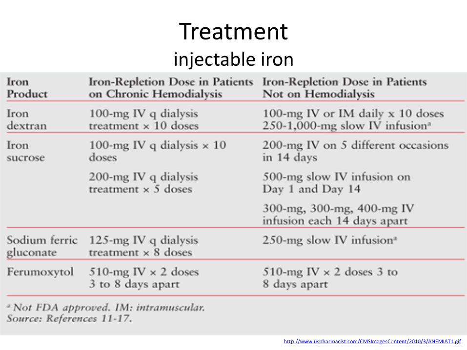
Treatmentinjectable iron
http://www.uspharmacist.com/CMSImagesContent/2010/3/ANEMIAT1.gif

Treatmentblood transfusions
• Blood transfusions in those without symptoms is not recommended until the hemoglobin is below 60 to 80 g/L (6 to 8 g/dL)
• In those with coronary artery disease who are not actively bleeding transfusions are only recommended when the hemoglobin is below 70 to 80g/L (7 to 8 g/dL)
• Transfusing earlier does not improve survival
• Transfusions otherwise should only be undertaken in cases of cardiovascular instability
https://en.wikipedia.org/wiki/Anemia

Treatmentblood transfusions
http://www.henrysurteesfoundation.com/wp-content/uploads/2014/02/BloodTransfusionsMoulage.jpg

Treatmenterythropoiesis-stimulating agent
http://www.searchhomeremedy.com/drugs-and-medications-to-treat-anemia/
Recombinant human erythropoietin injections

Treatmenthyperbaric oxygen
• Treatment of exceptional blood loss (anemia) is recognized as an indication for hyperbaric oxygen (HBO) by the Undersea and Hyperbaric Medical Society
• The use of HBO is indicated when oxygen delivery to tissue is not sufficient in patients who cannot be given blood transfusions for medical or religious reasons
• HBO may be used for medical reasons when threat of blood product incompatibility or concern for transmissible disease are factors
https://en.wikipedia.org/wiki/Anemia

Treatmenthyperbaric oxygen
http://www.aboutcancer.com/hyperbaric.jpg

Prognosis
• The prognosis depends on the underlying cause of the anemia
• The severity of the anemia, its etiology, and the rapidity with which it develops can each play a significant role in the prognosis
• Similarly, the age of the patient and the existence of other comorbid conditions influence outcome
http://emedicine.medscape.com/article/198475-overview#a4

Prophylaxis
• Many types of anemia can't be prevented
• Iron deficiency anemia and vitamin deficiency anemias can be prevented by a diet that includes:
• Iron (meats, beans, lentils, iron-fortified cereals, dark green leafy vegetables, and dried fruit)
• Folate (citrus fruits and juices, bananas, dark green leafy vegetables, legumes, and fortified breads, cereals and pasta)
• Vitamin B-12 (meat and dairy products)
• Vitamin C (citrus fruits, melons and berries)
http://emedicine.medscape.com/article/198475-overview#a4

Abbreviations
• ADH - antidiuretic hormone• EPO - erythropoietin • FBC - full blood count• GI - gastrointestinal • Hct - hematocrit• HIF-1 - hypoxia inducible factor-1 • IVF - intravenous fluid• MCV - mean corpuscular volume• NSAID - non-steroidal anti-inflammatory drug• TBV - total blood volume

Diagnostic guidelines
• Anaemia management in people with chronic kidneydisease(external link)
• Clinical practice guidelines for evaluation of anemia(external link)
• Guidelines for the diagnosis and management of adultaplastic anaemia(external link)
• Guideline for the laboratory diagnosis of functional irondeficiency(external link)
• British consensus guidelines on intravenous fluid therapy foradult surgical patients (GIFTASUP)(external link)
• Guidelines for the management of iron deficiencyanaemia(external link)
• Significant haemoglobinopathies: guidelines for screening and diagnosis(external link)