Embed Size (px)
Citation preview

Dowlati, E., Adams, S. E., Stiles, A. B., & Moran, R. J. (2016). Aginginto Perceptual Control: A Dynamic Causal Modeling for fMRI Study ofBistable Perception. Frontiers in Human Neuroscience, 10, [141].https://doi.org/10.3389/fnhum.2016.00141
Publisher's PDF, also known as Version of recordLicense (if available):CC BYLink to published version (if available):10.3389/fnhum.2016.00141
Link to publication record in Explore Bristol ResearchPDF-document
This is the final published version of the article (version of record). It first appeared online via Frontiers Media athttp://journal.frontiersin.org/article/10.3389/fnhum.2016.00141/full. Please refer to any applicable terms of use ofthe publisher.
University of Bristol - Explore Bristol ResearchGeneral rights
This document is made available in accordance with publisher policies. Please cite only thepublished version using the reference above. Full terms of use are available:http://www.bristol.ac.uk/red/research-policy/pure/user-guides/ebr-terms/

ORIGINAL RESEARCHpublished: 31 March 2016
doi: 10.3389/fnhum.2016.00141
Aging into Perceptual Control: ADynamic Causal Modeling for fMRIStudy of Bistable PerceptionEhsan Dowlati 1†, Sarah E. Adams 2†, Alexandra B. Stiles 3 and Rosalyn J. Moran 1,2,4*
1 Virginia Tech Carilion School of Medicine, Roanoke, VA, USA, 2 Virginia Tech Carilion Research Institute, Roanoke, VA, USA,3 Virginia Commonwealth University School of Medicine, Richmond, VA, USA, 4 Bradley Department of Electrical andComputer Engineering, Virginia Tech, Blacksburg, VA, USA
Edited by:Adeel Razi,
University College London, UK
Reviewed by:Kamen Atanasov Tsvetanov,University of Camrbidge, UK
Gerald Cooray,Karolinska Institute, Sweden
*Correspondence:Rosalyn J. Moran
†These authors have contributedequally to this work.
Received: 18 January 2016Accepted: 15 March 2016Published: 31 March 2016
Citation:Dowlati E, Adams SE, Stiles AB and
Moran RJ (2016) Aging intoPerceptual Control: A Dynamic
Causal Modeling for fMRI Studyof Bistable Perception.
Front. Hum. Neurosci. 10:141.doi: 10.3389/fnhum.2016.00141
Aging is accompanied by stereotyped changes in functional brain activations, forexample a cortical shift in activity patterns from posterior to anterior regions is onehallmark revealed by functional magnetic resonance imaging (fMRI) of aging cognition.Whether these neuronal effects of aging could potentially contribute to an ameliorationof or resistance to the cognitive symptoms associated with psychopathology remains tobe explored. We used a visual illusion paradigm to address whether aging affects thecortical control of perceptual beliefs and biases. Our aim was to understand the effectiveconnectivity associated with volitional control of ambiguous visual stimuli and to testwhether greater top-down control of early visual networks emerged with advancing age.Using a bias training paradigm for ambiguous images we found that older participants(n = 16) resisted experimenter-induced visual bias compared to a younger cohort(n = 14) and that this resistance was associated with greater activity in prefrontaland temporal cortices. By applying Dynamic Causal Models for fMRI we uncovereda selective recruitment of top-down connections from the middle temporal to Lingualgyrus (LIN) by the older cohort during the perceptual switch decision following biastraining. In contrast, our younger cohort did not exhibit any consistent connectivity effectsbut instead showed a loss of driving inputs to orbitofrontal sources following training.These findings suggest that perceptual beliefs are more readily controlled by top-downstrategies in older adults and introduce age-dependent neural mechanisms that may beimportant for understanding aberrant belief states associated with psychopathology.
Keywords: visual illusion, visual processing, aging, dynamic causal modeling, fMRI
INTRODUCTION
Several studies have demonstrated that later patient age-at-onset is a predictor of greaterremission rates and better outcome prognosis in psychopathologies including schizophrenia(Häfner et al., 1998; Ho et al., 2000; Jeste et al., 2003), first-episode psychosis (Malla et al.,2006) and bipolar disorder (Carlson et al., 2002; Carter et al., 2003), independent of othercontributing factors such as illness duration. Age also influences the relative symptomspectrum in these psychopathologies (Gur et al., 1996; Topor et al., 2013). In schizophrenia,for example the trajectories of positive, negative and thought-disorder symptom dimensionshave been shown to display differential age effects, with advancing age associated withdecreases in positive symptoms including hallucinations, delusions and bizarre behavior
Frontiers in Human Neuroscience | www.frontiersin.org 1 March 2016 | Volume 10 | Article 141

Dowlati et al. Age and Control of Visual Beliefs
(Schultz et al., 1997). However, the putative neural mechanismsunderlying adaptive effects of aging have been relativelyunexplored in the neuroimaging and neuropsychiatricliterature.
For this special issue on psychopathology, we aimed toaddress the basic mechanisms of brain networks that underlieage-dependent changes in constructive perception. A methodof examining conscious perception is to take advantage of thevisual system by instigating bistable perception. This allows usto study the underlying neural networks related to perceptionformation rather than stimulus-driven visual processing. Illusoryvisual paradigms have proved useful in probing the neuralmechanisms associated with impaired perceptual inference andaberrant beliefs in psychosis and schizophrenia (Foxe et al., 2005;Dima et al., 2009, 2010; Notredame et al., 2014). Ambiguousvisual stimuli such as the Necker’s cube, Rubin’s face-vase,or Boring’s Old-Young lady, where images have two distinctinterpretations (Leopold and Logothetis, 1999), in particular lendthemselves to the study of volitional inference and subjectiveperception (Sundareswara and Schrater, 2008; Wang et al.,2013). Moreover, these paradigms are often designed to illicitactivations across distributed cortical networks or hierarchies.Earlier theories of switching perceptions focused on neuronaladaptation as a key mediator (Blake, 1989) however these havebeen superseded by connectivity analyses which demonstratethat bottom-up and top-down connections to early visual cortices(Cardin et al., 2011; Wang et al., 2013) and endogenousneuronal oscillations (Kloosterman et al., 2015) also contributeto the bistability of a percept. Bayesian decision theory, usedto construct models of perception (Kersten and Schrater, 2002)support the role of networked cortical communication. In theseaccounts, reverses in perception between competing alternativesare posed as an active process that involves multiple regionsof the brain seeking to understand the stimulus, where oneparticular perception emerges as the result of bottom-up andtop-down interplay that suppresses one interpretation in favorof the other (Dayan, 1998). Modeling accounts have alsodemonstrated a potential impact from noisy neuronal firing asa possible bottom-up influence in perceptual switches (Shapiroet al., 2009). These computational accounts appeal to priors onwhat might be perceived—on our visual beliefs (Cardin et al.,2011).
In terms of the prior beliefs that encourage perceptualswitching and image stability, opposing behaviors have beenobserved which support both bottom-up and top-down neuronalmediators. Some studies reveal that the most prevalent percept inthe recent past is the one that is most likely favored when theambiguous image is shown (Leopold et al., 2002), suggesting thatimplicit perceptual memory may affect perception of ambiguousfigures (de Jong et al., 2014). Other studies have shown thatprolonged viewing of an ambiguous stimuli leads to preferenceof the novel perception vs. past perceptual experience (de Jonget al., 2012). Importantly, these images can also be manipulatedto induce stability of a particular percept, for example movingbistable stimuli can be stabilized by motion of backgroundelements (Kramer and Yantis, 1997) and the Necker cube,which elicits viewpoint ambiguity, can be manipulated with
color enhancement of particular sides so that one viewpointis predominantly perceived (Wang et al., 2013). This enablesthe investigation of perceptual priors and their volitionalcontrol.
We have previously shown that alterations in perceptualpriors by short-term changes in environmental statistics arelinked to adjusted ratios of bottom-up to top-down signalpropagation in neural hierarchies that exhibit a pronounced ageeffect, with older adults less likely to adjust their beliefs (Moranet al., 2014). In the current study, we build upon these findingsto test whether advanced age is associated with greater control ofwhat is perceived.
The aim of the study was to establish a perceptualpreference based on external stimulus manipulations and to usedynamic causal modeling (DCM) to assess changes in effectiveconnectivity that arise from bias training. Our training consistedof a modified Rubin vase as a non-ambiguous image used toinduce bias within subjects. We intended to elicit this effect toobserve a change in percept duration in the younger individualsbehaviorally. For older adults, we hypothesized that they wouldresist biasing by the training stimulus (Moran et al., 2014) andmore actively control perceptual states when viewing bistableimages. We were interested specifically in whether there was anage-dependence in post-training constructive perception.
MATERIALS AND METHODS
ParticipantsA total of 30 participants (16 females) partook in our fMRIexperiment. The average age of the participants was 44.9, rangingfrom 18–76. Participants were divided into two groups: a youngcohort with an average age of 23.9 (n = 14, 18–29 years,7 females) and an older cohort with an average age of 63.7(n = 16, 54–76 years, 9 females). All were screened for MRIcontraindication and psychiatric or neurological disorders, hadnormal or corrected-to-normal visual acuity, and were fluent inEnglish. Study protocols were approved by the Virginia TechInstitutional Review Board and written informed consent wasobtained from each participant. Participants were compensatedfor their time.
Experimental ProtocolEach participant received task instructions and completed aninstruction quiz prior to the scanning session. The fMRI taskconsisted of three blocks: ambiguous Block 1, ‘‘Biasing’’ Non-ambiguous Block 2, and ambiguous Block 3 (Figure 1A). Inambiguous Block 1, the Rubin vase was presented for 60 s,followed by a fixation cross displayed for 6 s (Rubin, 1921).Participants were instructed to indicate via button press whetherthey perceived two faces or a vase initially as well as every timetheir perception switched over the 60-s trial. This experimentaldesign was similar to that employed in Sterzer et al. (2009) inthat participants were not given instructions to focus on oneperception over the other. All button presses were recorded andthis was repeated for a total of six trials. Participants were thenshown amodified, non-ambiguous stimulus during the ‘‘Biasing’’
Frontiers in Human Neuroscience | www.frontiersin.org 2 March 2016 | Volume 10 | Article 141

Dowlati et al. Age and Control of Visual Beliefs
FIGURE 1 | Experimental design and age effects on trained stimulus. (A) Block 1: the ambiguous Rubin vase was shown for 60 s, where participantsindicated their perception, faces or vase, with a button press. This was repeated 6 times and each trial was separated by a 6 s fixation cross. Block 2: anon-ambiguous, modified Rubin vase was shown for 16 s, where participants indicated when the fixation-cross appeared on either the left or right of the image. Thiswas repeated 16 times and each trial was separated by a 4 s fixation cross. Block 3 was identical in design to Block 1. (B) Left: the average duration in viewing faces(the biased percept) in Block 3 compared to Block 1 for the young cohort (light red) and older cohort (dark red). Right: the ratio of these durations—i.e., theperceptual biasing effect, was significantly different between the younger and older groups ∗p < 0.05.
Non-ambiguous Block 2. This non-ambiguous stimulus wasintended to explicitly portray two faces by modifying it ina way that the two faces was the most likely perceptiongained from looking at the stimulus. By presenting such animage, we intended to ‘‘train’’ or ‘‘bias’’ participants towardthe perception of the faces vs. a vase when they viewed theambiguous figure. The non-ambiguous stimulus was presentedfor a total of 16 s, followed by a fixation cross displayedfor 4 s. This was repeated for a total of 16 trials. When thenon-ambiguous stimuli were presented, a fixation cross wouldappear at random to either the left or right of the screen andparticipants were instructed to indicate via button press whenthe fixation cross appeared. In Ambiguous Block 3, participantswere again presented the ambiguous Rubin vase image for 60 s,followed by a fixation cross displayed for 6 s and instructedto indicate via button press their initial perception and theirsubsequent perceptual switches. This repeated for a total of
six trials. To summarize: in two blocks (Blocks 1 and 3), weshowed participants a non-modified ambiguous Rubin vasefigure. The non-ambiguous block (block 2) was the ‘‘training’’block in which the participant was shown a modified versionof the Rubin vase diagram eliciting a stable perception showingtwo faces, where the top and bottom borders were removed.This was a similar modification to the image as presentedin Wang et al. (2013). The non-ambiguous image was alsochosen as a result of pilot data (not reported) which suggestedthe Rubin image modified to elicit a face-bias was a strongernon-ambiguous image than the Rubin image modified to elicita vase-bias.
Button presses indicating percept switches, their times, andperceptual durations were recorded for behavioral data analysis.Total percept duration throughout the trials and average perceptduration for each perception, i.e., face or vase, was analyzedacross age and block (pre- vs. post-training).
Frontiers in Human Neuroscience | www.frontiersin.org 3 March 2016 | Volume 10 | Article 141
Dowlati et al. Age and Control of Visual Beliefs
fMRI Data AcquisitionAnatomical and functional images were acquired using a3-T Siemens MAGNETOM Trio scanner. High-resolutionT1-weighted structural images were collected using MPRAGEsequence with a repetition time (TR) = 1200 ms, echo time(TE) = 2.66 ms, field of view (FOV) = 245 mm, 1.0 mm slicethickness. Echo planar image data were acquired with a TRof 2000 ms, TE = 25 ms, field of view (FOV) = 220 mm,with 37 slices acquired at a slice thickness of 4.0 mm. Sliceswere oriented 30◦ superior-caudal to the plane through theanterior and posterior commissures to reduce signal drop-out.Headphones were used to reduce scanner noise. Participantsused a mirror to view the stimuli projected behind themin the scanner. Participants were provided with additionalitems such as blankets and noise-cancelling ear plugs uponrequest.
fMRI Data AnalysisPreprocessing and data analysis were performed using statisticalparametric mapping software implemented in Matlab (SPM12bbeta; Wellcome Trust Centre for Neuroimaging, London, UK).The first five functional images of the acquisition were discardedto allow for equilibrium magnetization. The mean scan wasused as the reference for EPI blood-oxygen-level dependent(BOLD) images which were realigned with a six parameter spatialtransformation. The structural image was co-registered to themean resliced image. The unified segmentation routine wasthen used to perform segmentation bias correction and spatialnormalization. Images were normalized to MNI space using theICBM template. Then, the data was smoothed using a kernel with8 mm full-width at half maximum (FWHM).
Individual participant BOLD responses were analyzed usinga General Linear Model (GLM). There were nine totalregressors: (1) ambiguous stimuli presentation AmbiguousBlock 1; (2) ambiguous stimuli presentation Ambiguous Block 3;(3) non-ambiguous image presentation Non-ambiguous Block 2;(4) button press responses for Block 1; (5) pre-switch event, a2000 ms time period immediately prior to button press, duringBlock 1; (6) button press responses for Block 3; (7) pre-switchevent, a 2000 ms time period immediately prior to button press,during Block 3; (8) button press responses in Block 2; and(9) pre-press, 2000 ms prior to button press, during the Block 2.All regressors were convolved with a canonical hemodynamicresponse function. In the first level GLM, estimated motionparameters were used as nuisance regressors. Once all regressorsfor all individual GLMs had been created, contrasts werecreated at the first-level to identify activation differences betweenambiguous and non-ambiguous stimuli and between the pre- andpost-training ambiguous stimuli. We assigned 2000 ms prior tothe button press as the ‘‘pre-switch’’ event. This was motivatedby previous research suggesting that subjective decisions can beobserved in fMRI activity up to 10 s prior to a motor report (Soonet al., 2008).
We then used a summary statistic approach to assess group-level whole-brain peak activations to identify regions of interest.An F-contrast was used to identify positive or negative responsesto the ambiguous stimuli compared to non-ambiguous stimuli
(Table 1). An F-contrast was also applied to identify trainingeffects—examining positive or negative response differences toambiguous stimuli before (Block 1) and after biasing (Block 3;Table 1). A 2 × 2 analysis of variance (ANOVA) was preformedto test interactions between age and training effects as well.
Dynamic Causal ModelingDCM for fMRI provides a model-based investigation ofeffective connectivity (Friston et al., 2003), where effectiveconnectivity represents directional and modulatory interactionsbetween multiple brain regions using separate neuronal andhemodynamic parameterizations. At the neuronal level theDCMs comprise a set of differential equations with parametersthat control the drive of external inputs and of inter-regionalneuronal influences. Given our interests in endogenous driversof perceptual switches, we applied stochastic DCM for fMRIswhich explicitly parameterizes non-stimulus linked fluctuationsin neuronal activity (Li et al., 2011). We chose this over thealternative counterpart, deterministic DCM, due to its abilityto parameterize and formally incorporate random neuronalfluctuations (Friston et al., 2014). Bayesian Model Selection wasapplied to find the best—most probable—model to explain theobserved hemodynamics (Stephan et al., 2007). Our aim wasto identify the neuronal connections associated with perceptualchanges—i.e., pre-switch events.
We used our second-level summary statistics to identifyregions of interest which responded differentially to ambiguousand non-ambiguous stimuli. We further used two age covariatesto identify within these regions, specific nodes that exhibitedpositive and negative correlations with age. The regionsof interest (ROIs) were identified around the group peakcoordinates of the Lingual gyrus (LIN) [−6 −68 −2] andthe Precuneus (PRE) [−12 −70 36]-these regions exhibited anegative correlation with age. ROIs were identified around thegroup peak coordinates of the Middle Temporal gyrus (MTG)[50 30−6] and Inferior Orbitofrontal Cortex (IOF) [62−22−6]-with a positive correlation with age (group peaks are summarizedin Table 1).
Given these coordinates, we extracted BOLD time seriesfrom each participant’s fMRI data individually. Time series wereextracted using an F-contrast mask that tested for differencesbetween ambiguous and non-ambiguous stimuli with a p-valuethreshold of p < 0.05, uncorrected with a sphere radius of8 mm (note: p-values here are used to define the voxel clusterfrom which the principal eigenvariate will be extracted, theyare not involved in the final DCM statistics). The principaleigenvariate within a sphere of 8 mm was extracted forthe model-based analysis. To correct for confounding motionand button-press contributions to our ROI time series, theseextractions were corrected for ‘‘effects of interest’’ using anF contrast to partition data variance in order to incorporateeffects from just four regressors including: (1) ‘‘AmbiguousBlock 1’’, (2) ‘‘Ambiguous Block 3’’, (5) ‘‘Pre-Switch Block 1’’and (7) ‘‘Pre-Switch Block 3’’. By using this F contrast, weare partitioning out any effects that could be due to allthe other regressors, which include head motion and buttonpresses.
Frontiers in Human Neuroscience | www.frontiersin.org 4 March 2016 | Volume 10 | Article 141

Dowlati et al. Age and Control of Visual Beliefs
TABLE 1 | fMRI second level group statistics: effects of ambiguity and age correlations.
Peak activation region (MNI) X Y Z F statistic Puncorrected PFWE−corrected
(A) Significant voxels with positive or negative response to onset of ambiguousvs. non-ambiguous images, unmasked, extended threshold 10 voxelsR Lingual 6 −68 −4 6.26 0 0R Sup occipital 14 −96 18 5.99 0 0L Cerebelum −25 −58 −25 5.92 0 0L Mid temporal −52 −48 14 5.81 0 0L Angular −42 −64 36 5.47 0 0.001
(B) Significant voxels with positive or negative response to onset of ambiguousimages Block 1 vs. Block 3, unmasked, extended threshold 10 voxelsL Lingual −6 −68 −2 5.88 0 0R Lingual 6 −64 −2 5.85 0 0R Cuneus 14 −96 16 5.83 0 0L Precentral −60 2 28 5.83 0 0L Mid temporal −50 −60 −2 5.78 0 0
(C) Significant voxels with positive or negative response to onset of ambiguousvs. non-ambiguous images, masked inclusively with a negative correlationcontrast of age, extended threshold 10 voxels: Lingual gyrus and PrecuneusLingual −6 −68 −2 5.90 0 0L Precuneus −12 −70 36 5.17 0 0.004
Significant voxels with positive or negative response to onset of ambiguousvs. non-ambiguous images, masked inclusively with a positive correlationcontrast of age, extended threshold 10 voxels: Mid Temporal gyrus and InferiorOrbitofrontal cortexR Mid temp 50 30 −6 4.94 0 0.011R Inf orb 62 −22 −6 4.85 0 0.016
(A) The peak activations were identified as positive or negative response to the onset of ambiguous compared to non-ambiguous images and image onsets of ambiguous
images before and after biasing. Both comparisons were unmasked and extended thresholds were at 20 voxels. (B) The effects of perceptual biases—comparing post
and pre-training responses to ambiguous stimuli. (C) The peak activations with positive or negative response to onset of ambiguous compared to non-ambiguous images
were used to mask age covariation. Lingual and precuneus activations were found when testing for decreasing with age. Right middle temporal gyrus and right inferior
orbitofrontal cortex activations were found with a covariate of increasing age.
To test the effective connections across the network weconstructed fourmodels of potential interactions among our fourregions of interest. There were intrinsic connections within allregions and between all regions (DCM’s A matrix), except forthe IOF and MTG. Inputs from ambiguous stimuli onsets droveall regions (DCM’s C matrix). Modulatory connections (DCM’sB matrix) were used to test network connections associatedwith pre-switch events. In model 1 we placed these modulationsonly on bottom-up connections for block 1 (LIN to MTG, LINto IOF, PRE to MTG, and PRE to IOF) and only on top-down connections for post-training block 3 (MTG to LIN, MTGto PRE, IOF to LIN, IOF to PRE). For model 2 we allowedmodulation of pre-switch events for pre and post trainingblocks on both sets of bottom-up and top-down connections.For model 3 we allowed modulation of pre-switch events onlyon bottom-up connections. Finally, for model 4 we allowedmodulation of pre-switch events only on top-down connections.
RESULTS
Behavioral Effects of BiasingBehavioral data were analyzed to test for perceptual biasingeffects. For this we compared the average duration of perceptionof trained stimulus (i.e., two faces, see Figure 1A) between
pre- and post-training blocks. All percepts throughout the six60-s trials were examined, regardless of number of switchesmade within the trial and of the initial percept. Furthermore,we ensured that all participants had at least three or moreswitches within a single trial of 60 s. From these data weestablished a simple bias ratio—the ratio of average timedurations for perceptions where the stimulus was viewedas two faces for post-training (block 3) relative to pre-training (block 1; Figure 1). Although we were not interestedin the initial percept at each trial during the ambiguousblocks, it is important to note that there was no significantdifference on the effect of training or age in the initialpercept.
Overall, our hypothesized effects of age on biasing wereevidenced. Average percept duration of the trained stimuliwas similar between the young and older group (Figure 1B),however, during the post-training block, the average durationof the ‘‘faces’’ percept significantly differed between the youngand older cohort (young (n = 14): 7064 ms; older (n = 16):4209 ms) showing a positive bias for the trained stimulus forthe young relative to older cohort (p < 0.05, Figure 1B).There was a medium effect size in this comparison (Cohen’sd = 0.6). In addition, analysis of total percept duration forvases compared to faces post-training showed preference in olderindividuals towards the novel, non-trained percept significantly
Frontiers in Human Neuroscience | www.frontiersin.org 5 March 2016 | Volume 10 | Article 141
Dowlati et al. Age and Control of Visual Beliefs
(p < 0.001). These data demonstrate a preference toward thenovel or untrained percept in block 3 for the older cohort relativeto a trained or biased prior in the younger cohort.
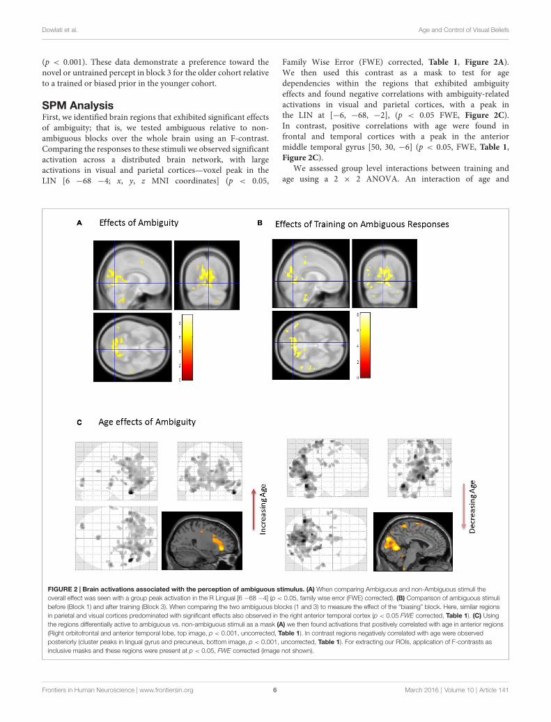
SPM AnalysisFirst, we identified brain regions that exhibited significant effectsof ambiguity; that is, we tested ambiguous relative to non-ambiguous blocks over the whole brain using an F-contrast.Comparing the responses to these stimuli we observed significantactivation across a distributed brain network, with largeactivations in visual and parietal cortices—voxel peak in theLIN [6 −68 −4; x, y, z MNI coordinates] (p < 0.05,
Family Wise Error (FWE) corrected, Table 1, Figure 2A).We then used this contrast as a mask to test for agedependencies within the regions that exhibited ambiguityeffects and found negative correlations with ambiguity-relatedactivations in visual and parietal cortices, with a peak inthe LIN at [−6, −68, −2], (p < 0.05 FWE, Figure 2C).In contrast, positive correlations with age were found infrontal and temporal cortices with a peak in the anteriormiddle temporal gyrus [50, 30, −6] (p < 0.05, FWE, Table 1,Figure 2C).
We assessed group level interactions between training andage using a 2 × 2 ANOVA. An interaction of age and
FIGURE 2 | Brain activations associated with the perception of ambiguous stimulus. (A) When comparing Ambiguous and non-Ambiguous stimuli theoverall effect was seen with a group peak activation in the R Lingual [6 −68 −4] (p < 0.05, family wise error (FWE) corrected). (B) Comparison of ambiguous stimulibefore (Block 1) and after training (Block 3). When comparing the two ambiguous blocks (1 and 3) to measure the effect of the “biasing” block. Here, similar regionsin parietal and visual cortices predominated with significant effects also observed in the right anterior temporal cortex (p < 0.05 FWE corrected, Table 1). (C) Usingthe regions differentially active to ambiguous vs. non-ambiguous stimuli as a mask (A) we then found activations that positively correlated with age in anterior regions(Right orbitofrontal and anterior temporal lobe, top image, p < 0.001, uncorrected, Table 1). In contrast regions negatively correlated with age were observedposteriorly (cluster peaks in lingual gyrus and precuneus, bottom image, p < 0.001, uncorrected, Table 1). For extracting our ROIs, application of F-contrasts asinclusive masks and these regions were present at p < 0.05, FWE corrected (image not shown).
Frontiers in Human Neuroscience | www.frontiersin.org 6 March 2016 | Volume 10 | Article 141

Dowlati et al. Age and Control of Visual Beliefs
training was seen in posterior regions with peak activationin the right LIN [4 −64 6] (p < 0.05, FWE). To unpackthis result we performed a ‘‘simple main effects’’ analysisspecifically for training in Figure 2B. Here, we tested only forthe effects of the non-ambiguous training block we comparedambiguous responses pre and post training. This contrastshowed that similar regions exhibited the biasing effects asprevious seen for ambiguous processing generally (ambiguouspre-trained vs. ambiguous post-training), including LIN, PREand middle temporal cortices (p < 0.05, FWE, Table 1).Furthermore, we tested for other covariates that may beimplicated in the context of psychopathology, including genderand education. These covariates did not show any significanteffects in activation between male or female participants orin terms of education level categorized by some high school,high school graduate, some college, college graduate (data notshown).
DCM of Ambiguous Visual Processing andAge-Related Connectivity EffectsWe used those activations associated with ambiguous comparedto non-ambiguous stimuli to study perceptual belief networksusing DCM (see ‘‘Materials and Methods’’ Section, Figure 3A).We were particularly interested in the mechanisms subtendingswitches in subjective perceptual beliefs and the effects biastraining had on the network. To analyze switch responses wedefined ‘‘pre-switch events’’, a 2000 ms period immediatelyprior to a button press indicating the percept had switched.We chose this timing due to the possibility of activenetworks present before the action of a button press, withoutoverlapping button press responses. This was motivated byprevious research suggesting that subjective decisions can beobserved in fMRI activity up to 10 s prior to a motor report(Soon et al., 2008). Our network comprised four regionsincluding LIN, PRE, mid-temporal gyrus (MTG) and IOF,with intrinsic connections arranged reciprocally among theseregions (with the exception of IOF to MTG). The percentvariances explained by our fMRI data in the four extractedprincipal eigenvariates over an 8 mm-radius sphere weredetermined and averages across all subjects were calculated(Table 2).
TABLE 2 | Average percent variation explained in regions of interest(ROIs).
Average percent variance ± SEM
LIN 79.00% ± 1.91PRE 79.18% ± 1.89MTG 74.75% ± 2.06IOF 77.58% ± 2.32
For our DCMs, we extracted the principal eigenvariate for an 8 mm-radius
sphere around a group peak at the four regions of interest (LIN: [−6, −68, −2];
PRE: [−12, −70, 36]; MTG: [50, 30, −6]; IOF: [62, −22, −6]) at p < 0.05,
uncorrected. This principal eigenvariates explain the above percent variances on
average over each region for all subjects. The table shows average ± standard
error mean of these regions across all 30 subjects.
We constructed four models to test for training-relateddifferences in top-down vs. bottom-up perceptual control.In model 1, pre-switch modulations during the pre-trainingblock were confined to bottom-up connections and pre-switchmodulations during the post-training block were confinedto top-down connections. In model 2, we allowed for bothbottom-up and top-down pre-switch modulations in bothpre- and post-training blocks (Figure 3B). In model 3, onlybottom-up pre-switch modulations were present in both pre-and post-training blocks. In contrast, in model 4, only top-down pre-switch modulations were present in both pre- andpost-training blocks. We show the equations representingthe models (Figure 3C). Using a random-effects Bayesianmodel comparison across participants and within each cohortseparately we found that both the young and older cohortspreferred model 1 (with a model exceedance probability (MEP)of 0.9993 for all subjects, Figure 3D, MEP = 0.9882 in theyoung cohort, and MEP = 0.9494 in the older cohort). Theeffect across individuals was consistent with 11 participants inthe younger cohort preferring model 1, and the other threepreferring model 3. Nine participants in the older cohortpreferred model 1, five preferred model 4, and two preferredmodel 3.
Equipped with this winning model we tested for trainingor biasing effects within each cohort. To test for effect size,the average coefficients of determination for the model fitwere determined in each cohort. There is a medium effectsize for the lingual region in the young cohort, a mediumeffect size for the middle temporal regions in both cohorts,and a small effect size for PRE and inferior orbitofrontalregions (Table 3). Interestingly, we observed that the youngcohort exhibited no significant modulations in connectionsrelated to training. Rather, we found that the arrangementof driving inputs differed between pre- and post-training.Specifically, the initial pre-training block was associatedwith a negative driving input to both lingual (student’st test; p = 0.0016) and PRE sources (p = 0.014), whilein the post-training block these negative driving inputswere confined to the lingual source only (p = 0.0017).These negative driving inputs will suppress endogenousnoise in each region under the stochastic DCM. In theolder cohort however significant effects of training wereobserved—with the emergence of a significant top-downconnection from the middle temporal gyrus to LIN onthe post-training block (p < 0.05; Figure 4A). This cohortalso exhibited negative input drive into lingual and frontalsources post training (p < 0.05). Within both cohorts, theDCMs adequately recapitulated the measured data features(Figure 4B).
Hemodynamic changes with age may alter BOLD activityand contribute to second-level group statistics (Tsvetanov et al.,2015). With DCM we were able to separate the hemodynamicparameters and test whether they exhibited age-dependenteffects. However no effects of age on hemodynamic parameterswere observed where we tested decay and transit time differencesbetween the two age groups for all four regions (p > 0.1uncorrected for eight tests).
Frontiers in Human Neuroscience | www.frontiersin.org 7 March 2016 | Volume 10 | Article 141

Dowlati et al. Age and Control of Visual Beliefs
FIGURE 3 | Dynamic causal model and Bayesian model selection. (A) Sources for the Dynamic Causal Modelings (DCMs) were obtained from the second levelanalysis, displayed here. Regions of interest were identified around the group peak coordinates for Lingual [−6 −68 −2], Precuneus [−12 −70 36], Mid Temporal [5030 −6], and Inf Orbitofrontal [62 −22 −6]. (B) The four regions of interest (ROIs) were used to create a stochastic DCM. There were intrinsic connections within allregions and between all regions, except for the inferior orbitofrontal cortex (IOF) and middle temporal gyrus (MTG). (B) Inputs from ambiguous image onsets enteredall regions. The modulations in connections associated with pre-switch events were tested using Bayesian model comparison. In model 1 pre-switch connections inthe pre-training block were confined to bottom-up connections (light gray), i.e., from Lingual and PRE to Inferior Orbitofrontal and Mid Temporal Lobe. While inmodel 1 post-training switches were modeled via top-down connection modulations only (dark gray), i.e., from Inferior Orbitofrontal and Mid Temporal Lobe toLingual and PRE. Model 2 comprised pre-switch, pre- and post-training modulations in both directions. Model 3 consisted of bottom-up pre-switch modulations forboth pre- and post-training modulations. Finally, model 4 involved of top-down pre-switch modulations for both pre- and post-training modulations. (C) We alsodisplay the equations used to define each of these models. A is the intrinsic connection parameters matrix. B is the input-dependent or modulatory connectionparameter matrix. z denotes the regions. C is the extrinsic influences or input connection parameter matrix. u Represents the inputs. (D) Bayesian model comparisonrevealed that both younger and older cohorts preferred model 1 (see “Results” Section) and these fixed effects were consistent across most subjects. Here, weillustrate the exceedance probabilities for a comparison including all models from both age groups.
TABLE 3 | Average coefficient of determination for DCM fits in ROIs.
Average coefficient of determination (R2) ± SEM
LIN PRE MTG IOF
Young (n = 14) 0.30 ± 0.03 0.12 ± 0.02 0.29 ± 0.04 0.076 ± 0.02Older (n = 16) 0.20 ± 0.02 0.11 ± 0.03 0.28 ± 0.03 0.073 ± 0.02Overall 0.24 ± 0.02 0.12 ± 0.02 0.29 ± 0.02 0.074 ± 0.01
Coefficients of determination for DCM fits were calculated for all 30 subjects for the
winning model. We present these as effect size for our DCM data extraction and
model fit. We compared predicted response with observed response. Coefficients
of determination show a medium effect size for the LIN region in the young cohort,
a medium effect size for the MTG region in both cohorts, and a small effect size for
PRE and IOF regions.
DISCUSSION
Despite theoretical and imaging-driven advances inunderstanding bistable perception, its interaction with anaging neurobiology has received little attention. Motivatedby the ubiquitous role age plays in psychopathological status(Häfner et al., 1998; Ho et al., 2000; Jeste et al., 2003; Toporet al., 2013; Lin et al., 2006), the present study addressesthe age-dependency of neuronal connectivity underlyingvolitional control of perceptual beliefs. In our study, weinvestigated the brain regions associated with fluctuatingperceptual content, whether these brain regions interactduring perceptual rivalry, and how stimulus-driven biasing canaffect subsequent subjective perceptual beliefs and neuronal
Frontiers in Human Neuroscience | www.frontiersin.org 8 March 2016 | Volume 10 | Article 141

Dowlati et al. Age and Control of Visual Beliefs
FIGURE 4 | Age-effects on connectivity mediating volitional perception. (A) Only in the older cohort did we see pre-switch modulations of effectiveconnectivity. Specifically we observed an emergence of a top-down control connection (MTG→ LIN) following the bias or training block. (B) DCM fits here from onesubject and displayed for two regions accurately recapitulate the extracted time-series.
connectivity. In our study, we used the Rubin vase diagramand manipulated the image in order to bias perception andtested the underlying processing networks using fMRI andDCM. In summary our findings reveal that consistent withour hypothesized training effect, older cohorts exhibited aresistance to perceptual biasing compared to the younger cohortand these effects were found to be mediated by an increase intop-down connections from temporal to visual cortical sourcespost training.
Our study was motivated by predictive coding theories ofcortico-cortical interactions which has been explored recentlyin the context of visual illusory processing (Brown and Friston,2012; Chopin and Mamassian, 2012). Our aim was to determinewhether prior beliefs could resist external manipulation inan age-dependent manner. Our paradigm was suited to thisconnectivity hypothesis given recent work by Kok et al. (2016)who show that top-down connections selectively activate earlyvisual regions during the perception of illusory figures such as
the Kanizsa stimulus. In our study, we used the non-ambiguousblock for training to test whether inference networks withinthe brain became more robust to environmental perturbationsas we age. This fits within larger theoretical frameworkssuch as the Free-energy principle (Brown and Friston, 2012),which appeals to the Bayesian brain hypothesis and laminarspecific connectivity which optimizes to better predict futuresensory inputs (Moran et al., 2014). With this in mind, wesuggest that perceptual switches in the aging population can bedescribed as changes in connectivity between regions, generatedby an internal predictive model. In the context of visualprocessing and perceptual competition, binocular rivalry isanother phenomenon explained in the framework of a brainthat is engaged in Bayesian inference (Hohwy et al., 2008).Furthermore our motivation for this framework relates topsychopathology where studies such as Shergill et al. (2005) haveimplicated predictive coding abnormalities in diseases such asschizophrenia.
Frontiers in Human Neuroscience | www.frontiersin.org 9 March 2016 | Volume 10 | Article 141

Dowlati et al. Age and Control of Visual Beliefs
Whole-brain analysis from the fMRI study identified anetwork of cortical regions involved in viewing the ambiguousfigures that included the LIN and precuneus, regions typicallyassociated with perceptual changes in ambiguous figures (Sterzerand Kleinschmidt, 2007; Wang et al., 2013). Within theseactivated regions we found a striking correlation with aging, asage increases the ambiguity-associated activations predominatedin anterior regions, while younger age was associated withgreater posterior activity. This is consistent with general agingeffects observed in fMRI-neurocognitive experiments whichdemonstrate a posterior to anterior shift in activation (PASA)patterns (Cabeza, 2001; Davis et al., 2008). With regards toPASA, there is reduced neural specialization in the visual cortexwith age as well as an increase in distributed processing infrontal areas (Cabeza, 2001), with these anterior shifts notedin visual processing tasks (Ansado et al., 2012). However, sucha paradigm has not been considered in bistable perceptionvisual processing, making our study unique in that matter.In our study, we show that this shift to anterior regionsof the brain can be associated with visual processing andperceptual control and not attributed to any specific defaultnetwork, which has been shown to undergo reallocationwith aging as a compensatory mechanism (Davis et al.,2008). We are unable to provide evidence for or against acompensatory mechanism in our study since we do not havea metric of ‘‘good’’ or ‘‘poor’’ performance. Instead, we areinterested in Bayesian predictive coding leading to differencesin connectivity. Exploring our activations using DCM wefound that younger participants did recruit frontal regionsduring ambiguous stimulus processing but that this droppedoffline following a biasing session. In contrast, our oldercohorts resisted biasing and furthermore recruited top-downconnections to control their perceptual beliefs following training.In the context of psychopathology it may be useful to controlperceptual beliefs internally and to resist model updating basedon spurious environmental stimuli. An inaccurate assignmentof one’s environmental experiences may contribute to theunderlying pathology in diseases such as schizophrenia (Kapur,2003).
Previous behavioral studies using binocular rivalry haveshown that perceptual stability increases with increasing age(Ukai et al., 2003; Beers et al., 2013). However, binocularrivalry, compared to bistable perception with ambiguousfigures, involves a more automatic and stimulus driven formof visual competition occurring at the lower levels of thevisual pathway (Tong et al., 2006). We do not assume thatour findings extend to studies of binocular rivalry. Bistableperception with ambiguous figures occurs at a higher levelin the visual pathway (Tong and Engel, 2001). This providesa method of intentional control, making it more suitable forthe larger goal of our analysis, which is the study of theactive process of perception. Using multisensory sound flash-illusions, studies have also demonstrated that aging presentswith stronger illusory percepts compared to younger adults(DeLoss et al., 2013), but that training to avoid the temporaloverlap illusion can be accomplished by older cohorts (Settiet al., 2014). Few studies however have sought to establish
the neural correlates of these effects. In our study we usedstochastic DCM for fMRI (Daunizeau et al., 2011; Li et al.,2011) in order to account for the internally-generated dynamicsthat cause endogenous percept fluctuations as well as task-dependent changes (deterministic effects; Friston et al., 2014).This is in contradistinction to other spectral DCMs whichmay present a more accurate and parsimonious account ofconnectivity in studies examining complete resting or stationarystates (Razi et al., 2015). The optimized parameter sets ofour stochastic models revealed interesting dynamics particularlyin the driving inputs (Friston et al., 2003). We found thatnegative driving inputs were observed in posterior and frontalsources for the older subjects post-training whereas for theyounger subjects these patterns were seen pre-training with adropout of frontal inhibitory drive post-training. The polarityof these driving inputs are reasonable in the setting ofstochastic DCMs since they would dampen endogenous noisyfluctuations in their respective regions and in the case ofthe older cohort enable top-down control via long-rangeconnections.
Our results complement previous studies involving bistableperception, which have shown a decline in attentional selection oflow-salient stimuli (Tsvetanov et al., 2013). Additionally, Aydinet al. (2013) examined perceptual switching of the Rubin vaseshowing that older individuals are less likely to attend to visualstimuli after holding a specific percept. In fact, the older groupprefers the novel percept. However, we do not use distractors orperceptual holding in our experiment but rather assess control inthe context of biasing effects.
Overall, our analysis provides a holistic account of bistableperceptual processing in aging given the combination of fMRIand stochastic DCMs. Our observed network involving thefrontal and temporal regions was derived from our wholebrain analysis. Our regions in these models are supportedby previous research suggesting significant modulation ofinferior frontal cortex to medial temporal regions during theperceptual transitions of the ambiguous rotating Lissajous figure(Weilnhammer et al., 2013). All of our four ROIs have beenshown to respond to bistable percepts in previous studies(Wang et al., 2013). An electroencephalogram (EEG) study onbistable perception using ambiguous images showed activityin the posterior visual regions in addition to higher-orderfronto-parietal and temporal regions of the brain (Britz et al.,2009). In terms of psychopathology, frontal and temporalcortices, specifically the inferior frontal gyrus and superiortemporal gyrus, is implicated in schizophrenia showing alteredconnectivity in resting state fMRI study (Zaytseva et al.,2015). Orbitofrontal cortices and middle temporal gyrus arefurthermore areas of disruption in perspective-taking tasks inschizophrenia (Eack et al., 2013). Brain networks such as thedefault network or salience network may play a role in bistableperception and show differences in age. Future work can use task-based independent component analysis (ICA; Hyett et al., 2015;Tsvetanov et al., 2015) to characterize the network topology incontrol to better understand perceptual changes.
Limitations of the study include other covariates thatare affected with normal aging. For example, potential time
Frontiers in Human Neuroscience | www.frontiersin.org 10 March 2016 | Volume 10 | Article 141

Dowlati et al. Age and Control of Visual Beliefs
differences may exist in the pre-switch event with age. Inour study, we allotted the same pre-switch period durationfor both the younger and older group. Given that ouranalysis relies on subjective recording of perceptual switches,this is an inherent limitation in the study of bistableperception since the only objective marker to assume achange in perception is the button press. Other limitationsof the study include putative effects on bistability notaccounted for in our design including eye position (Einhäuseret al., 2004) or attention (van Ee et al., 2005). Futurestudies could address these and more fine-grained featuresof aging control dynamics, using electrophysiological DCMs(Legon et al., 2015). For example, GABA levels in thevisual cortex have been linked to bistable perception, withhigher concentrations resulting in slower perceptual dynamics(van Loon et al., 2013)-an effect used to simulate aging
differences in computational modeling studies of multistableperception (Hoshino, 2013). These simple visual paradigmsmay uncover further neurobiological correlates of perceptualcontrol, and provide important clues for developmental andaging dependencies in psychopathology.
AUTHOR CONTRIBUTIONS
ED and RJM designed the experiment, performed analysis. ED,SEA andABS collected the data. ED, SEA, ABS and RJMpreparedthe manuscript.
FUNDING
This work was supported by a start-up grant from VTCRI toRJM.
REFERENCES
Ansado, J., Monchi, O., Ennabil, N., Faure, S., and Joanette, Y. (2012). Load-dependent posterior-anterior shift in aging in complex visual selective attentionsituations. Brain Res. 1454, 14–22. doi: 10.1016/j.brainres.2012.02.061
Aydin, S., Strang, N. C., and Manahilov, V. (2013). Age-related deficits inattentional control of perceptual rivalry. Vision Res. 77, 32–40. doi: 10.1016/j.visres.2012.11.010
Beers, A. M., Bennett, P. J., and Sekuler, A. B. (2013). Age-related effects of sizeand contrast on binocular rivalry. J. Vis. 13, 546–546. doi: 10.1167/13.9.546
Blake, R. (1989). A neural theory of binocular rivalry. Psychol. Rev. 96, 145–167.doi: 10.1037/0033-295x.96.1.145
Britz, J., Landis, T., andMichel, C. M. (2009). Right parietal brain activity precedesperceptual alternation of bistable stimuli. Cereb. Cortex 19, 55–65. doi: 10.1093/cercor/bhn056
Brown, H., and Friston, K. J. (2012). Free-energy and illusions: the cornsweeteffect. Front. Psychol. 3:43. doi: 10.3389/fpsyg.2012.00043
Cabeza, R. (2001). Cognitive neuroscience of aging: contributions of functionalneuroimaging. Scand. J. Psychol. 42, 277–286. doi: 10.1111/1467-9450.00237
Cardin, V., Friston, K. J., and Zeki, S. (2011). Top-down modulations in thevisual form pathway revealed with dynamic causal modeling. Cereb. Cortex 21,550–562. doi: 10.1093/cercor/bhq122
Carlson, G. A., Bromet, E. J., Driessens, C., Mojtabai, R., and Schwartz, J. E. (2002).Age at onset, childhood psychopathology and 2-year outcome in psychoticbipolar disorder. Am. J. Psychiatry 159, 307–309. doi: 10.1176/appi.ajp.159.2.307
Carter, T. D. C., Mundo, E., Parikh, S. V., and Kennedy, J. L. (2003). Early age atonset as a risk factor for poor outcome of bipolar disorder. J. Psychiatr. Res. 37,297–303. doi: 10.1016/s0022-3956(03)00052-9
Chopin, A., and Mamassian, P. (2012). Predictive properties of visual adaptation.Curr. Biol. 22, 622–626. doi: 10.1016/j.cub.2012.02.021
Daunizeau, J., David, O., and Stephan, K. E. (2011). Dynamic causal modelling:a critical review of the biophysical and statistical foundations. Neuroimage 58,312–322. doi: 10.1016/j.neuroimage.2009.11.062
Davis, S. W., Dennis, N. A., Daselaar, S. M., Fleck, M. S., and Cabeza, R. (2008).Qué PASA? the posterior-anterior shift in aging. Cereb. Cortex 18, 1201–1209.doi: 10.1093/cercor/bhm155
Dayan, P. (1998). A hierarchical model of binocular rivalry. Neural Comput. 10,1119–1135. doi: 10.1162/089976698300017377
de Jong, M. C., Brascamp, J. W., Kemner, C., van Ee, R., and Verstraten,F. A. (2014). Implicit perceptual memory modulates early visual processing ofambiguous images. J. Neurosci. 34, 9970–9981. doi: 10.1523/JNEUROSCI.2413-13.2014
de Jong, M. C., Knapen, T., and van Ee, R. (2012). Opposite influence of perceptualmemory on initial and prolonged perception of sensory ambiguity. PLoS One7:e30595. doi: 10.1371/journal.pone.0030595
DeLoss, D. J., Pierce, R. S., and Andersen, G. J. (2013). Multisensory integration,aging and the sound-induced flash illusion. Psychol. Aging 28, 802–812. doi: 10.1037/a0033289
Dima, D., Roiser, J. P., Dietrich, D. E., Bonnemann, C., Lanfermann, H.,Emrich, H. M., et al. (2009). Understanding why patients with schizophreniado not perceive the hollow-mask illusion using dynamic causalmodelling. Neuroimage 46, 1180–1186. doi: 10.1016/j.neuroimage.2009.03.033
Dima, D., Dietrich, D. E., Dillo,W., and Emrich, H.M. (2010). Impaired top-downprocesses in schizophrenia: a DCM study of ERPs. Neuroimage 52, 824–832.doi: 10.1016/j.neuroimage.2009.12.086
Eack, S. M., Wojtalik, J. A., Newhill, C. E., Keshavan, M. S., and Phillips,M. L. (2013). Prefrontal cortical dysfunction during visual perspective-takingin schizophrenia. Schizophr. Res. 150, 491–497. doi: 10.1016/j.schres.2013.08.022
Einhäuser, W., Martin, K. A., and König, P. (2004). Are switches in perception ofthe Necker cube related to eye position? Eur. J. Neurosci. 20, 2811–2818. doi: 10.1111/j.1460-9568.2004.03722.x
Foxe, J. J., Murray, M. M., and Javitt, D. C. (2005). Filling-in in schizophrenia:a high-density electrical mapping and source-analysis investigation of illusorycontour processing. Cereb. Cortex 15, 1914–1927. doi: 10.1093/cercor/bhi069
Friston, K. J., Harrison, L., and Penny, W. (2003). Dynamic causal modelling.Neuroimage 19, 1273–1302. doi: 10.1016/s1053-8119(03)00202-7
Friston, K. J., Kahan, J., Biswal, B., and Razi, A. (2014). A DCM for resting statefMRI. Neuroimage 94, 396–407. doi: 10.1016/j.neuroimage.2013.12.009
Gur, R. E., Petty, R. G., Turetsky, B. I., and Gur, R. C. (1996). Schizophreniathroughout life: sex differences in severity and profile of symptoms. Schizophr.Res. 21, 1–12. doi: 10.1016/0920-9964(96)00023-0
Häfner, H., Hambrecht, M., Löffler, W., Munk-Jørgensen, P., and Riecher-Rössler, A. (1998). Is schizophrenia a disorder of all ages? A comparison offirst episodes and early course across the life-cycle. Psychol. Med. 28, 351–365.doi: 10.1017/s0033291797006399
Ho, B. C., Andreasen, N. C., Flaum, M., Nopoulos, P., and Miller, D. (2000).Untreated initial psychosis: its relation to quality of life and symptom remissionin first-episode schizophrenia. Am. J. Psychiatry 157, 808–815. doi: 10.1176/appi.ajp.157.5.808
Hohwy, J., Roepstorff, A., and Friston, K. (2008). Predictive coding explainsbinocular rivalry: an epistemological review. Cognition 108, 687–701. doi: 10.1016/j.cognition.2008.05.010
Hoshino, O. (2013). Ambient GABA responsible for age-related changesin multistable perception. Neural Comput. 25, 1164–1190. doi: 10.1162/NECO_a_00431
Hyett, M. P., Breakspear, M. J., Friston, K. J., Guo, C. C., and Parker, G. B. (2015).Disrupted effective connectivity of cortical systems supporting attentionand interoception in melancholia. JAMA Psychiatry 72, 350–358. doi: 10.1001/jamapsychiatry.2014.2490
Frontiers in Human Neuroscience | www.frontiersin.org 11 March 2016 | Volume 10 | Article 141

Dowlati et al. Age and Control of Visual Beliefs
Jeste, D. V., Twamley, E. W., Eyler Zorrilla, L. T., Golshan, S., Patterson, T. L.,and Palmer, B. W. (2003). Aging and outcome in schizophrenia.Acta Psychiatr. Scand. 107, 336–343. doi: 10.1034/j.1600-0447.2003.01434.x
Kapur, S. (2003). Psychosis as a state of aberrant salience: a framework linkingbiology, phenomenology and pharmacology in schizophrenia.Am. J. Psychiatry160, 13–23. doi: 10.1176/appi.ajp.160.1.13
Kersten, D., and Schrater, P. R. (2002). ‘‘Pattern inference theory: a probabilisticapproach to vision,’’ in Perception and the Physical World, eds R. Mausfeld andD. Heyer (Chichester: John Wiley & Sons), 191–228.
Kloosterman, N. A., Meindertsma, T., Hillebrand, A., van Dijk, B. W.,Lamme, V. A., and Donner, T. H. (2015). Top-down modulation in humanvisual cortex predicts the stability of a perceptual illusion. J. Neurophysiol. 113,1063–1076. doi: 10.1152/jn.00338.2014
Kok, P., Bains, L. J., van Mourik, T., Norris, D. G., and de Lange, F. P.(2016). Selective activation of the deep layers of the human primary visualcortex by top-down feedback. Curr. Biol. 26, 371–376. doi: 10.1016/j.cub.2015.12.038
Kramer, P., and Yantis, S. (1997). Perceptual grouping in space and time:evidence from the ternus display. Percept. Psychophys. 59, 87–99. doi: 10.3758/bf03206851
Legon, W., Punzell, S., Dowlati, E., Adams, S. E., Stiles, A. B., and Moran,R. J. (2015). Altered prefrontal excitation/inhibition balance and prefrontaloutput: markers of aging in human memory networks. Cereb. Cortex. doi: 10.1093/cercor/bhv200 [Epub ahead of print].
Leopold, D. A., and Logothetis, N. K. (1999). Multistable phenomena: changingviews in perception. Trends Cogn. Sci. 3, 254–264. doi: 10.1016/s1364-6613(99)01332-7
Leopold, D. A., Wilke, M., Maier, A., and Logothetis, N. K. (2002). Stableperception of visually ambiguous patterns. Nat. Neurosci. 5, 605–609. doi: 10.1038/nn851
Li, B., Daunizeau, J., Stephan, K. E., Penny, W., Hu, D., and Friston, K. (2011).Generalised filtering and stochastic DCM for fMRI. Neuroimage 58, 442–457.doi: 10.1016/j.neuroimage.2011.01.085
Lin, P. I., McInnis, M. G., Potash, J. B., Willour, V., MacKinnon, D. F.,DePaulo, J. R., et al. (2006). Clinical correlates and familial aggregation of ageat onset in bipolar disorder. Am. J. Psychiatry 163, 240–246. doi: 10.1176/appi.ajp.163.2.240
Malla, A., Norman, R., Schmitz, N., Manchanda, R., BÉChard-Evans, L., Takhar, J.,et al. (2006). Predictors of rate and time to remission in first-episodepsychosis: a two-year outcome study. Psychol. Med. 36, 649–658. doi: 10.1017/s0033291706007379
Moran, R. J., Symmonds, M., Dolan, R. J., and Friston, K. J. (2014). The brainages optimally to model its environment: evidence from sensory learning overthe adult lifespan. PLoS Comput. Biol. 10:e1003422. doi: 10.1371/journal.pcbi.1003422
Notredame, C. E., Pins, D., Deneve, S., and Jardri, R. (2014). What visual illusionsteach us about schizophrenia. Front. Integr. Neurosci. 8:63. doi: 10.3389/fnint.2014.00063
Razi, A., Kahan, J., Rees, G., and Friston, K. J. (2015). Construct validation of aDCM for resting state fMRI.Neuroimage 106, 1–14. doi: 10.1016/j.neuroimage.2014.11.027
Rubin, E. (1921). Visuell Wahrgenommene Figuren: Studien in PsychologischerAnalyse. Copenhagen: Gyldendalske boghandel.
Schultz, S. K., Miller, D. D., Oliver, S. E., Arndt, S., Flaum, M., andAndreasen, N. C. (1997). The life course of schizophrenia: age andsymptom dimensions. Schizophr. Res. 23, 15–23. doi: 10.1016/s0920-9964(96)00087-4
Setti, A., Stapleton, J., Leahy, D., Walsh, C., Kenny, R. A., and Newell, F. N.(2014). Improving the efficiency of multisensory integration in older adults:audio-visual temporal discrimination training reduces susceptibility to thesound-induced flash illusion. Neuropsychologia 61, 259–268. doi: 10.1016/j.neuropsychologia.2014.06.027
Shapiro, A., Moreno-Bote, R., Rubin, N., and Rinzel, J. (2009). Balancebetween noise and adaptation in competition models of perceptual bistability.J. Comput. Neurosci. 27, 37–54. doi: 10.1007/s10827-008-0125-3
Shergill, S. S., Samson, G., Bays, P. M., Frith, C. D., and Wolpert, D. M. (2005).Evidence for sensory prediction deficits in schizophrenia.Am. J. Psychiatry 162,2384–2386. doi: 10.1176/appi.ajp.162.12.2384
Soon, C. S., Brass, M., Heinze, H. J., and Haynes, J. D. (2008). Unconsciousdeterminants of free decisions in the human brain. Nat. Neurosci. 11, 543–545.doi: 10.1038/nn.2112
Stephan, K. E., Weiskopf, N., Drysdale, P. M., Robinson, P. A., and Friston, K. J.(2007). Comparing hemodynamic models with DCM. Neuroimage 38,387–401. doi: 10.1016/j.neuroimage.2007.07.040
Sterzer, P., and Kleinschmidt, A. (2007). A neural basis for inference in perceptualambiguity. Proc. Natl. Acad. Sci. U S A 104, 323–328. doi: 10.1073/pnas.0609006104
Sterzer, P., Kleinschmidt, A., and Rees, G. (2009). The neural bases of multistableperception. Trends Cogn. Sci. 13, 310–318. doi: 10.1016/j.tics.2009.04.006
Sundareswara, R., and Schrater, P. R. (2008). Perceptual multistability predictedby search model for Bayesian decisions. J. Vis. 8, 12.1–12.19. doi: 10.1167/8.5.12
Tong, F., and Engel, S. A. (2001). Interocular rivalry revealed in the human corticalblind-spot representation. Nature 411, 195–199. doi: 10.1038/35075583
Tong, F., Meng, M., and Blake, R. (2006). Neural bases of binocular rivalry. TrendsCogn. Sci. 10, 502–511. doi: 10.1016/j.tics.2006.09.003
Topor, D. R., Swenson, L., Hunt, J. I., Birmaher, B., Strober, M., Yen, S., et al.(2013). Manic symptoms in youth with bipolar disorder: factor analysis by ageof symptom onset and current age. J. Affect. Disord. 145, 409–412. doi: 10.1016/j.jad.2012.06.024
Tsvetanov, K. A., Henson, R. N. A., Tyler, L. K., Davis, S. W., Shafto, M. A.,Taylor, J. R., et al. (2015). The effect of ageing on fMRI: correction for theconfounding effects of vascular reactivity evaluated by joint fMRI and MEGin 335 adults. Hum. Brain Mapp. 36, 2248–2269. doi: 10.1002/hbm.22768
Tsvetanov, K. A., Mevorach, C., Allen, H., and Humphreys, G. W. (2013). Age-related differences in selection by visual saliency. Atten. Percept. Psychophys 75,1382–1394. doi: 10.3758/s13414-013-0499-9
Ukai, K., Ando, H., and Kuze, J. (2003). Binocular rivalry alternation rate declineswith age. Percept. Mot. Skills 97, 393–397. doi: 10.2466/pms.97.5.393-397
van Ee, R., van Dam, L. C. J., and Brouwer, G. J. (2005). Voluntary control and thedynamics of perceptual bi-stability. Vision Res. 45, 41–55. doi: 10.1016/j.visres.2004.07.030
van Loon, A. M., Knapen, T., Scholte, H. S., St. John-Saaltink, E. , Donner, T. H.,and Lamme, V. A. (2013). GABA shapes the dynamics of bistable perception.Curr. Biol. 23, 823–827. doi: 10.1016/j.cub.2013.03.067
Wang,M., Arteaga, D., andHe, B. (2013). Brainmechanisms for simple perceptionand bistable perception. Proc. Natl. Acad. Sci. U S A 10, E3350–E3359. doi: 10.1073/pnas.1221945110
Weilnhammer, V. A., Ludwig, K., Hesselmann, G., and Sterzer, P. (2013).Frontoparietal cortex mediates perceptual transitions in bistable perception. J.Neurosci. 33, 16009–16015. doi: 10.1523/JNEUROSCI.1418-13.2013
Zaytseva, Y., Chan, R. C., Pöppel, E., and Heinz, A. (2015). Luria revisited:cognitive research in schizophrenia, past implications and future challenges.Philos. Ethics Humanit. Med. 10:4. doi: 10.1186/s13010-015-0026-9
Conflict of Interest Statement: The authors declare that the research wasconducted in the absence of any commercial or financial relationships that couldbe construed as a potential conflict of interest.
Copyright © 2016 Dowlati, Adams, Stiles and Moran. This is an open-access articledistributed under the terms of the Creative Commons Attribution License (CC BY).The use, distribution and reproduction in other forums is permitted, provided theoriginal author(s) or licensor are credited and that the original publication in thisjournal is cited, in accordance with accepted academic practice. No use, distributionor reproduction is permitted which does not comply with these terms.
Frontiers in Human Neuroscience | www.frontiersin.org 12 March 2016 | Volume 10 | Article 141


![The causal role of α-oscillations in feature binding · 1.711, P > 0.208] and the interactions between perceptual state and electrode [all F(60, 1,020) < 0.965, P > 0.363]](https://img.dokumen.tips/doc/110x75/5fd699928bb6a060a327b33b/the-causal-role-of-oscillations-in-feature-binding-1711-p-0208-and-the.jpg)