Embed Size (px)
Citation preview
BIOSYNTHESIS AND REGULATION OF CYCLIC LIPOPEPTIDES IN PSEUDOMONAS FLUORESCENS
IRENE DE BRUIJN

Promotor: Prof. dr. ir. P.J.G.M. de Wit Hoogleraar in de Fytopathologie Wageningen Universiteit
Co-Promoter: Dr. J,M. Raaijmakers Universitair docent bij het Laboratorium voor Fytopathologie
Promotiecommissie: Prof. dr. J. van der Oost, Wageningen Universiteit Prof. dr. O. Nybroe, Copenhagen University, Denmark Dr. ir. E.W. van der Heijden, Bejo Zaden BV, Warmenhuizen Dr. W. Bitter, VUMC Amsterdam
Dit onderzoek is uitgevoerd binnen de onderzoekschool ‘Experimental Plant Sciences’.
IRENE DE BRUIJN
BIOSYNTHESIS AND REGULATION OF CYCLIC LIPOPEPTIDES IN PSEUDOMONAS FLUORESCENS
Proefschriftter verkrijging van de graad van doctor
op gezag van de rector magnificus van Wageningen Universiteit,
Prof. Dr. M.J. Kropff,in het openbaar te verdedigen
op dinsdag 31 maart 2009des namiddags te half twee in de Aula

Biosynthesis and regulation of cyclic lipopeptides in Pseudomonas fluorescens. I. de Bruijn. 2009
Thesis Wageningen University, The NetherlandsWith references - with summaries in English and Dutch
ISBN 978-90-8585-358-9
CONTENTS
Chapter 1. General introduction and thesis outline. 7 Cyclic lipopeptide production by plant-associated Pseudomonas spp.: diversity, activity, biosynthesis, and regulation.
Chapter 2. Genome-based discovery, structure prediction and 41 functional analysis of cyclic lipopeptide antibiotics in Pseudomonas species.
Chapter 3. Massetolide A biosynthesis in Pseudomonas fluorescens. 65
Chapter 4. Heterologous expression of regulatory and biosynthesis 91 genes directing cyclic lipopeptide antibiotic production in Pseudomonas fluorescens.
Chapter 5. Regulation of cyclic lipopeptide biosynthesis 113 in Pseudomonas fluorescens by the ClpP protease.
Chapter 6. Cyclic lipopeptides protect bacteria from protozoan 143 predators.
Chapter 7. Natural roles of cyclic lipopeptides and other bacterial 153 biosurfactants.
Chapter 8. Summarizing discussion 181
Summary 195Samenvatting 199Acknowledgements 203About the Author 207Graduate School certificate 209

7
CHAPTER 1
Molecular Plant-Microbe Interactions Vol. 19, No. 7, 2006, pp. 699–710. DOI: 10.1094/MPMI -19-0699.
REVIEW
Cyclic lipopeptide productionby plant-associated Pseudomonas spp.:diversity, activity, biosynthesis, and regulation
Jos M. Raaijmakers, Irene de Bruijn, and Maarten J. D. de KockLaboratory of Phytopathology, Wageningen University, Binnenhaven 5, 6709 PD Wageningen, The Netherlands
Accepted 21 February 2006.
8
ABSTRACTCyclic lipopeptides (CLPs) are versatile molecules produced by a variety of bacterial genera, including plant-associated Pseudomonas spp. CLPs are composed of a fatty acid tail linked to a short oligopeptide, which is cyclized to form a lactone ring between two amino acids in the peptide chain. CLPs are very diverse both structurally and in terms of their biological activity. The structural diversity is due to differences in the length and composition of the fatty acid tail and to variations in the number, type, and configuration of the amino acids in the peptide moiety. CLPs have received considerable attention for their antimicrobial, cytotoxic, and surfactant properties. For plant-pathogenic Pseudomonas spp., CLPs constitute important virulence factors, and pore formation, followed by cell lysis, is their main mode of action. For the antagonistic Pseudomonas sp., CLPs play a key role in antimicrobial activity, motility, and biofilm formation. CLPs are produced via nonribosomal synthesis on large, multifunctional peptide synthetases. Both the structural organization of the CLP synthetic templates and the presence of specific domains and signature sequences within peptide synthetase genes will be described for both pathogenic and antagonistic Pseudomonas spp. Finally, the role of various genes and regulatory mechanisms in CLP production by Pseudomonas spp., including two-component regulation and quorum sensing, will be discussed in detail.

9
The genus Pseudomonas not only harbors plant- and human pathogenic species, but also accommodates species that promote plant growth, degrade xenobiotic compounds, antagonize plant pathogenic fungi, or induce resistance in plants (Compant et al. 2005; Haas and Défago 2005; Pieterse et al. 2002; Ryu et al. 2003). The diverse life styles of Pseudomonas spp. and the complexity of their interactions with multiple hosts have been the subject of numerous molecular, biochemical, and ecological studies (Ramos 2004). Recently, whole genome sequences of multiple Pseudomonas strains representing various species have become available and allow a more in-depth study of metabolic pathways and regulatory networks as well as the exploration and exploitation of yet-unknown genes and gene products. The extensive interest in Pseudomonas spp. is due, in part, to the production of a wide variety of metabolites, including enzymes, volatiles, bacteriocins, toxins, antibiotics, and cyclic lipopeptides (Haas and Défago 2005; Raaijmakers et al. 2002). Cyclic lipopeptides (CLPs) are versatile molecules with antimicrobial, cytotoxic, and surfactant properties. CLPs are produced by several plant-associated Pseudomonas spp., including pathogenic Pseudomonas syringae, P. tolaasii, P. fuscovaginae, P. corrugata, and P. fluorescens (Bender et al. 1999), and by multiple strains classified as antagonistic P. fluorescens and P. putida (Nielsen et al. 2002; Nybroe and Sørensen 2004). In a recent review by Nybroe and Sørensen (2004), several chemical and biological aspects of CLP production in fluorescent pseudomonads were described. Also, CLP production by plant-pathogenic P. syringae has been the subject of several reviews (Bender et al. 1999; Bender and Scholz-Schroeder 2004; Willis and Kinscherf 2004). The purpose of this review is to provide an up-to-date overview of the current knowledge of the structural diversity and activity of CLPs produced by plant-associated Pseudomonas spp., in particular the antagonistic Pseudomonas spp. A detailed description of genes involved in the regulation and biosynthesis of CLPs will be presented, with emphasis on specific domains and signature sequences within CLP biosynthetic templates. These analyses provide insights into the specificity-conferring codes in peptide synthetase genes of Pseudomonas spp. and allow a prediction of the final structure of the CLP molecule based on sequence data. Whenever necessary, comparisons will be made with CLPs of other bacteria and, in particular, with surfactin produced by Bacillus spp.

10
STRUCTURAL DIVERSITYA variety of methods have been used to elucidate the primary and secondary structures of CLPs produced by bacteria (Ballio et al. 1996; Bare et al. 1999; De Souza et al. 2003; Henriksen et al. 2000; Koumoutsi et al. 2004; Monti et al. 2001; Nielsen et al. 1999, 2002; Sørensen et al. 2001). CLPs produced by Pseudomonas spp. are composed of a fatty acid tail linked to a short oligopeptide, which is cyclized to form a lactone ring between two amino acids in the peptide chain. Based on the length and composition of the fatty acid tail as well as the number, type, and configuration of the amino acids in the peptide moiety, CLPs of Pseudomonas spp. were classified into four major groups (i.e., the viscosin, amphisin, tolaasin, and syringomycin groups) (Table 1). In brief, the viscosin group harbors CLPs with 9 amino acids linked at the N-terminus to, in most
Table 1. Simplified primary structures of cyclic lipopeptides (CLPs) produced by plant-associated Pseudomonas spp.a
Adapted from Ballio et al. 1991; Ballio et al. 1995; Coiro et al. 1998; Gerard et al. 1997; Kuiper et al. 2004; Morikawa et al. 2000; Nybroe and Sørensen, 2004; Paulsen et al. 2006; Quail et al. 2002; and Scaloni et al. 2004. The lines represent the lactone ring of the cyclic lipopeptide. ⊗: isomer not identified; Asp(3-OH): 3-hydroxyaspartic acid; Dab: 2,4-diaminobutyric acid; Dhb, 2,3-dihydro-2-aminobutyric acid; Glx: glutamic acid or glutamine; Hse: homoserine; aIle: allo-isoleucine; Orn: ornithine; Thr(4-Cl): 4-chlorothreonine; aThr: allo-threonine; Xle: isoleucine or leucine; PFL2145/46/47 refers to the predicted lipodecapeptide of P. fluorescens strain Pf-5.
11
cases, 3-hydroxy decanoic acid (3-HDA). Pseudomonas spp. producing CLPs in this group are from a variety of origins, including soil, rhizosphere, phyllosphere, and marine environments. For example, viscosin production has been described for pectolytic strains of P. fluorescens causing head rot of broccoli (Hildebrand et al. 1998) and for a Pseudomonas isolate obtained from a tube worm collected from a marine environment (Gerard et al. 1997). Similarly, massetolide A production originally was described for a yet-unidentified Pseudomonas sp. isolated from the surface of leafy red algae collected at –15 m in the sea near the coast of British Columbia (Gerard et al. 1997), and later was described for a P. fluorescens strain isolated from the rhizosphere of wheat grown in a soil suppressive to take-all disease of wheat (De Souza et al. 2003). In a comprehensive survey, Nielsen and associates (2002) showed that, among the Pseudomonas spp. isolated from the rhizosphere of sugar beet, viscosin-like CLPs were produced primarily by strains belonging to P. fluorescens biovar I. Whether members of specific CLP groups are produced predominantly by specific biovars of fluorescent Pseudomonas spp. remains to be further investigated using large collections of isolates from different environmental origins. CLPs in the amphisin group consist of 11 amino acids in the peptide part coupled to 3-HDA (Table 1). For several members of this group, including amphisin and tensin, the crystal structure has been resolved (Henriksen et al. 2000; Sørensen et al. 2001). For both tensin and amphisin, the structures are mainly helical, with the cyclic peptide wrapping around a water molecule. Based on the distance between two negatively charged residues of the molecule, Henriksen and associates (2000) suggested that tensin most likely cannot form complexes with Ca2+, as was shown for surfactin produced by Bacillus spp. (Peypoux et al. 1999). Crystallography further showed that, in amphisin, the 3-HDA fatty acid tail protrudes from the hydrophilic side of the molecule (Nybroe and Sørensen 2004). For the P. fluorescens strains producing amphisin-like CLPs, Nielsen and associates (2002) identified several other interesting characteristics, including the production of chitinases and the volatile hydrogen cyanide (HCN), two traits that have been associated with biological control of plant-pathogenic fungi (Haas and Défago 2005; Whipps 2001). These traits and, in particular, HCN production were not common among the P. fluorescens strains producing viscosin-like CLPs (Nielsen et al. 2002). Compared with the viscosin and amphisin groups, CLPs in the tolaasin group are much more diverse due to multiple variations in both the composition and
12
length of the peptide chain (19 to 25 amino acids) and the lipid tail [3-HDA or 3-hydroxyoctanoic acid (3-HOA)]. The peptide part of the CLPs in this group contains several unusual amino acids, including 2,3-dihydro-2-aminobutyric acid (Dhb) and homoserine (Hse), the first always being in front of the allo-Thr residue (Table 1). The cyclic part of the peptide moiety contains five to eight amino acids and the lactone ring is formed between the C-terminal amino acid and the allo-Thr residue. Several tolaasin-like CLPs are produced by plant-pathogenic strains of Pseudomonas and constitute important virulence factors. In terms of shear numbers of amino acids in the peptide moiety, CLPs in the syringomycin group are structurally similar to the CLPs in the viscosin group (Table 1). However, syringomycin-like CLPs harbor unusual amino acids, including Dhb, 2,4-diamino butyric acid (Dab), and the C-terminal 4-chlorothreonine [Thr(4-Cl)], the latter being important for the antifungal activity of syringomycin (Grgurina et al. 1994). Furthermore, the lactone ring is formed between the N-terminal Ser and the C-terminal Thr(4-Cl); whereas, for members of the viscosin group, the ring usually is formed between the C-terminal amino acid and the D-allo-Thr at the third amino acid position in the peptide chain (Table 1). The fatty acid tail of CLPs in the syringomycin group may consist of a 3-hydroxy or 3,4-dihydroxy fatty acid composed of 10 to 14 carbon atoms. For example, three forms of syringomycin have been described which differ only in the length of the fatty acid moiety, being either decanoic, dodecanoic, or tetradecanoic acid (Bender et al. 1999; Bender and Scholz-Schroeder 2004). The structures of a number of other CLPs from Pseudomonas spp. have been elucidated in the past years. These include arthrofactin produced by Pseudomonas sp. strain MIS38 (Morikawa et al. 1993; Roongsawang et al. 2003) and putisolvin I and II produced by P. putida PCL1445 (Kuiper et al. 2004). Arthrofactin contains an 11–amino acid peptide moiety linked to a β-hydroxydecanoyl tail. Based on its chemical structure, arthrofactin falls within the amphisin group and is most similar to lokisin (Table 1). Putisolvin I and II harbor some unique features, including a peptide moiety of 12 amino acids and a hexanoic lipid tail. The cyclization in the two putisolvins also is different from other CLPs because the linkage occurs between the C-terminal carboxyl group and the ninth amino acid residue (serine) instead of the first or third amino acid as described previously for many of the other CLPs
13
(Table 1). Recently, in silico analysis of the whole genome sequence of the biocontrol strain P. fluorescens Pf-5 has resulted in the identification of a gene cluster that most likely encodes a cyclic lipodecapeptide (Paulsen et al. 2005). Although the amino acid composition of the peptide moiety of the predicted CLP of strain Pf-5 (Table 1) as well as the identity of the fatty acid tail needs to be confirmed by chemical analyses, this approach elegantly illustrates that knowledge of whole genome sequences allows the discovery of yet-unknown genes and traits which may be biologically relevant for the activity of antagonistic Pseudomonas spp.
ACTIVITYGiven the structural diversity of CLPs produced by Pseudomonas spp. and other bacterial genera, Ron and Rosenberg (2001) postulated that CLPs and, more generally, microbial surface-active compounds (biosurfactants) also have different natural roles, some of which may be unique to the physiology and ecology of the producing microorganism. Several natural roles for CLPs and other biosurfactants were proposed (Ron and Rosenberg 2001), including their function in i) pathogenicity, ii) antimicrobial activity, iii) regulation of attachment and detachment to and from surfaces, and iv) motility. Another natural role, possibly limited to spore-forming bacteria, is that CLPs may function as signal molecules for coordinated growth and differentiation (Marahiel et al. 1997). To date, most of the hypotheses for the natural roles of CLPs and other biosurfactants have been derived mainly from their physical and chemical properties. In recent years, mutants deficient in CLP production have been obtained for several Pseudomonas strains and currently are being used to further investigate the natural roles of CLPs. In this section, several of the potential functions of CLPs produced by plant-associated Pseudomonas spp. will be discussed. Whenever possible, details on structure-function relationships will be included.
Role in plant pathogenesis. CLPs produced by plant-pathogenic P. syringae and P. fluorescens function as virulence factors and, as such, are not solely required for pathogenicity (Bender et al. 1999). For example, the production of syringomycin and syringopeptin by P. syringae pv. syringae substantially increases disease severity, although disease may still occur in their absence (Scholz-Schroeder et al. 2001a). Similarly, a viscosin-deficient mutant of a pectolytic strain of P. fluorescens still caused decay of
14
wounded broccoli florets, but the decay failed to spread to adjacent nonwounded florets (Hildebrand et al. 1998). One of the main modes of action of CLPs produced by plant-pathogenic Pseudomonas spp., including syringomycin, syringopeptin, and tolaasin, involves the formation of ion channels in the host plasma membrane leading to cytolysis (Dalla Serra et al. 1999; Hutchison and Gross 1997; Hutchison et al. 1995; Mott and Takemoto 1989; Rainey et al. 1991). Pore formation results in the alkalization of the intercellular fluid and in the release of multiple cellular compounds, conditions that are favorable for further multiplication and increased CLP production by the invading bacteria. At high concentrations [well above the critical micelle concentration (CMC value)], CLPs can directly solubilize plasma membranes. In several studies (Bender et al. 1999), solubilization of membranes also was demonstrated for erythrocytes. Interestingly, erythrocyte hemolysis by tolaasin can be inhibited by addition of divalent metal ions such as Zn2+, Ca2+, and Mg2+ (Rainey et al. 1991). Given that pre-incubation of tolaasin with Zn2+ did not affect its ability to lyse erythrocytes and that cells pretreated with Zn2+ were no less susceptible to tolaasin-induced lysis than untreated cells, Rainey and associates (1991) proposed that the inhibitory effect of divalent metal ions operates by binding to negatively charged groups on the extracellular side of the plasma membrane close to the site of pore formation. Recent studies with surfactin (Dufour et al. 2005) further indicated that the cyclic character of the peptide moiety is important for erythrocyte hemolysis because linear products of surfactin failed to cause lysis. In addition to the direct toxic effects of CLPs from pathogenic Pseudomonas spp. on plant cells, their surfactant properties play an important indirect role in virulence by facilitating colonization of plant tissue and enhancing physical access of cell-wall-degrading enzymes (CWDE) to the plant surface (Hildebrand et al. 1998; Hutchison and Johnstone 1993; Lindow and Brandl 2003). Although the order in which CLPs and CWDE operate is subject to discussion (Fogliano et al. 2002), studies on the indirect and direct effects of CLPs on plant cells, including their impact on physiological processes and signaling pathways (Bender et al. 1999), clearly exemplify the versatile functions of CLPs in pathogenicity. From a more applied perspective, knowledge of the detrimental effects of CLPs on plant cells has led to an increased interest in the potential use of these CLPs or the producing Pseudomonas spp. as microbial herbicides (Li et al. 2003; Pedras et al. 2003).
15
Antimicrobial activity. CLPs produced by Pseudomonas and Bacillus spp. also have received considerable attention for their activity against a range of other microorganisms, including viruses, mycoplasmas, trypanosomes, bacteria, fungi, and oomycetes. Studies on the antiviral activity of CLPs produced by Pseudomonas spp. date back to work by Groupé and colleagues in 1951 (Nybroe and Sørensen 2004), in which viscosin was shown to have activity against enveloped human-pathogenic viruses, including the infectious bronchitis virus. Similarly, surfactin was shown to inactivate multiple enveloped viruses, including herpes viruses and retroviruses (Vollenbroich et al. 1997a). Electron microscopy indicated that surfactin acts directly on the lipid envelope, leading to disintegration of the virus particles (Vollenbroich et al. 1997a). Additional studies revealed that surfactins with fatty acid chains of 13 carbon atoms showed low antiviral activity compared with C14 and C15 isoforms, indicating that the hydrophobicity is an important determinant of the antiviral activity of surfactin (Kracht et al. 1999). Disintegration of membranes also was shown to be the primary activity of surfactin against several mycoplasmas (Vollenbroich et al. 1997b). Surfactin was active only at concentrations above the CMC value, suggesting that disruption of mycoplasma membranes is due to nonspecific detergent activity. In studies on the human pathogen Trypanosoma cruzi, the causal agent of Chagas’s disease, Mercado and Colon-Whitt (1982) observed lysis of trypomastigotes isolated from the blood of infected mice. Further analysis of the parasite samples led to the isolation of a P. fluorescens isolate that produces viscosin, which itself also causes trypanosomal lysis (Burke et al. 1999). An interesting microscopic observation was that, in direct interactions, the P. fluorescens isolate approached a single flagellate of T. cruzi at the posterior end, the site of the flagellar origin, resulting in immobilization (Mercado and Colonwhitt 1982). Observations further revealed rounding up of the parasite with the flagellum coiled around the cell body, followed by disintegration. Similar stages were observed in the disintegration process of zoospores of oomycete pathogens exposed to massetolide A and viscosin produced by antagonistic strains of P. fluorescens (De Souza et al. 2003; J. M. Raaijmakers, personal communication). Studies on the antibacterial activity of CLPs have focused primarily on human-pathogenic bacteria and, in particular, the gram positives. In summary, activity was shown for corpeptins, syringopeptins, and tolaasin against Bacillus megaterium (Emanuele et al. 1998; Lavermicocca et al. 1997; Soler-Rivas et al. 1999), and for massetolides, viscosin,
16
syringopeptin, and syringomycins against Mycobacterium tuberculosis, M. avium-intercellulare, and M. smegmalis (Buber et al. 2002; El Sayed et al. 2000; Gerard et al. 1997). A two-fold difference between the activity of massetolide A and viscosin against M. tuberculosis and M. avium-intercellulare (Gerard et al. 1997) illustrated that a difference of only one amino acid residue in the peptide moiety (Table 1) may have a significant impact on the antibacterial activity. For massetolide A and viscosin, no activity was observed against a panel of other human-pathogenic bacteria, including Escherichia coli and Staphylococcus aureus (Gerard et al. 1997). Also, for putisolvin produced by P. putida PCL1445, no antibacterial effects against P. fluorescens and P. aeruginosa were observed when these strains were grown together on agar plates (Kuiper et al. 2004). The apparent lack of activity of several CLPs against gram-negative bacteria has been ascribed to protective effects of the outer membrane (Nybroe and Sørensen 2004). However, recent studies by Bais and associates (2004) indicate that this general observation does not apply to all CLPs; in their study, surfactin was shown to have bactericidal activity against P. syringae pv. tomato. The antifungal activity has been studied for many different CLPs and for a wide variety of plant- and human-pathogenic fungi and yeasts, including Rhizoctonia solani, Phoma lingam, Alternaria brassicae, Sclerotinia sclerotiorum, Geotrichum candidum, Botrytis cinerea, Ophiostoma ulmi, Aspergillus and Fusarium spp., Penicillium digitatum, Cryptococcus neoformans, Candida albicans, and C. glabrans (Nybroe and Sørensen 2004). In particular, the studies with viscosinamide produced by antagonistic Pseudomonas sp. strain DR54 provide several lines of evidence that CLPs are important constituents in the biological control of plant-pathogenic fungi. In vitro studies showed that viscosinamide adversely affected mycelium of R. solani and the oomycete Pythium ultimum, causing reduced growth and intracellular activity, hyphal swellings, increased branching, and rosette formation (Hansen et al. 2000; Thrane et al. 1999, 2000a). Viscosinamide is produced by strain DR54 in situ (Nielsen and Sorensen 2003), and several of the adverse effects on R. solani and P. ultimum recorded in vitro, including reduced mycelium density and intracellular activity of P. ultimum and reduced sclerotia formation by R. solani, also were observed in situ (Thrane et al. 1999, 2000b, 2001). However, in these and several other studies with antagonistic CLP-producing Pseudomonas strains, a comparison between the biocontrol activity of a wild type strain and CLP-deficient mutants was not included. Advances in the understanding of CLP biosynthesis have resulted in the construction of CLP-
17
deficient mutants and also mutants overproducing CLPs. For example, a mutant of Bacillus subtilis 6051 defective in surfactin production was substantially less effective than the wild type strain in controlling root infection of Arabidopsis by Pseudomonas syringae (Bais et al. 2004). Furthermore, a derivative of B. subtilis BBG100 that overproduces mycosubtilin showed an increased activity against Pythium spp. on tomato seedlings (Leclère et al. 2005). Studies with Pseudomonas fluorescens SS101 also showed reduced activity of massetolide A-deficient mutants against several plant-pathogenic fungi and oomycetes (A. Ficke and J. M. Raaijmakers, personal communications). The activity of massetolide A produced by strain SS101 against Pythium spp. has, so far, been attributed mainly to their zoosporicidal activity (De Souza et al. 2003). The zoosporicidal activity of massetolide A also may explain, at least in part, the biocontrol activity of strain SS101 against a range of other oomycete pathogens, including Phytophthora spp. However, the observation that strain SS101 and purified massetolide A also exert significant biocontrol activity against Pythium spp. that do not produce zoospores point to other modes of action (J. M. Raaijmakers, personal communications). A complicating factor in studying the role of CLPs in interactions between antagonistic Pseudomonas strains and plant pathogens is that CLPs have a dual function, as described previously for their role in pathogenesis. In addition to their direct effects on pathogen membranes, their surface activity also may enhance or even be essential for the delivery of and exposure of target pathogens to other antagonistic traits. In this context, Fogliano and associates (2002) demonstrated that, in combination, with CWDE of Trichoderma atroviride, CLPs produced by Pseudomonas syringae acted synergistically in antagonism toward several plant-pathogenic fungi.
Attachment and detachment. Attachment and detachment of bacteria to synthetic and natural surfaces has been the subject of numerous studies in the past and present, especially because of the importance of these processes in biofilm formation (Neu 1996; O’Toole et al. 2000). The role of CLPs in attachment or biofilm formation has been studied for several bacterial genera, including Pseudomonas and Bacillus spp. Arthrofactin produced by Pseudomonas MIS38 was shown to play an important role in developing mature biofilms (Roongsawang et al. 2003). An arthrofactin-deficient mutant formed unstable, but more biofilms than the wild type strain; furthermore, the structure of the biofilm was different, being more flat and fused for the mutant in

18
contrast to the well-developed biofilms separated by channels for the wild type strain. For P. putida PCL1445, Kuiper and associates (2004) showed that putisolvins I and II also influenced biofilm formation. The biofilm formed by a putisolvin-deficient mutant was strongly increased, containing more cells which formed aggregates that were not distributed as regularly as those of the parental strain. Addition of purified putisolvin I to the growth medium prior to incubation reduced biofilm formation by the wild type strain in a concentration-dependent manner. Furthermore, putisolvins I and II also adversely affected biofilm formation of at least two other Pseudomonas strains and were shown to break down existing biofilms (Kuiper et al. 2004). In addition, surfactin inhibits biofilm formation of several human-pathogenic bacteria, including Salmonella spp. (Mireles et al. 2001). This capacity to destroy existing biofilms emphasizes the potential use of these CLPs in removal of biofilms formed by hazardous bacteria. Furthermore, surfactin was shown to play an essential role in adherence of B. subtilis 6051 to a synthetic surface and in biofilm formation on Arabidopsis roots (Bais et al. 2004). They postulated that biofilm formation and CLP production may enable bacteria to efficiently colonize plant roots, thereby providing protection to their host. Recent studies by Nielsen and associates (2005) showed that the production of amphisin is an important trait for Pseudomonas sp. strain DSS73 to colonize sugar beet seed and barley straw residues. However, given that CLP production impairs biofilm formation for several Pseudomonas strains (discussed above), Nielsen and associates (2005) questioned the role of amphisin in biofilm formation during colonization of seed and straw by strain DSS73. Instead, they postulated that amphisin contributes to cell proliferation of strain DSS73 on the germinating seed and supports motility toward the plant material. Other putative roles of CLPs in colonization and proliferation suggested by Nielsen and associates (2005) included relieve of a nutrient limitation or a toxic effect of unknown seed components.
Motility. As described previously, CLPs facilitate the movement of plant-pathogenic Pseudomonas spp. on the phylloplane (Hildebrand et al. 1998; Lindow and Brandl 2003). Also, for antagonistic Pseudomonas strains, efficient colonization of the phytosphere (spermosphere, rhizosphere, and phyllosphere) as well as in situ production and delivery of the antagonistic traits at the right time and place are key aspects of their success to control plant pathogens (Lugtenberg et al. 2001; Whipps 2001). Several studies have
19
shown that CLPs produced by antagonistic Pseudomonas strains and other bacterial genera are essential for surface motility (Andersen et al. 2003; Kuiper et al. 2004; Lindum et al. 1998; Nielsen et al. 2002; Roongsawang et al. 2003). For Pseudomonas sp. strain DSS73, amphisin production plays an important role in surface motility and in efficient containment of mycelial growth of root-infecting fungi, which most likely results from a combination of spatial limitation and delivery of antifungal compounds (Andersen et al. 2003). The reduced surface motility of amphisin-deficient mutants could be restored by addition of amphisin to the medium and also by addition of other CLPs, including tensin, viscosinamide, and serrawettin. Interestingly, several synthetic surfactants failed to complement the deficiency in surface motility of the mutants, suggesting that not only the reduction of surface tension is essential in motility but also the physical-chemical nature of the surfactant (Andersen et al. 2003).
BIOSYNTHESISCLPs are produced nonribosomally on large, multifunctional peptide synthetases via a thiotemplate process. Nonribosomal peptide synthesis has been the subject of numerous biochemical and molecular studies. For a more detailed understanding of the structural and functional characteristics of the enzymes involved, we refer to several reviews (Challis and Naismith 2004; Challis et al. 2000; Finking and Marahiel 2004; Marahiel et al. 1997; Stachelhaus et al. 1999; von Dohren et al. 1999). As elegantly described by Gewolb (2002), nonribosomal peptide synthetases (NRPSs) are not bound by the restrictions of the “ribosome’s rulebooks” and are involved not only in CLP production but also in the synthesis of an array of other unusual and potent peptides, including several clinically important drugs such as penicillin, vancomycin, and cyclosporin. NRPSs possess a modular structure and each module is a building block resulting in the stepwise incorporation and modification of one amino acid unit. Their substrates are not restricted to the usual proteinogenic amino acids but also can incorporate multiple nonproteinogenic D-amino acids, carboxy acids, or fatty acids (Gewolb 2002; Sieber and Marahiel 2005). The order and number of the modules of an NRPS protein are, in many cases, colinear to the amino acid sequence of the corresponding peptide moiety of the final CLP molecule (“colinearity rule”). A typical NRPS module consists of an adenylation (A) domain responsible for amino acid activation, a thiolation (T or, alternatively, PCP) domain for thioesterification of the activated amino acid, and a condensation (C) domain for peptide bond formation between
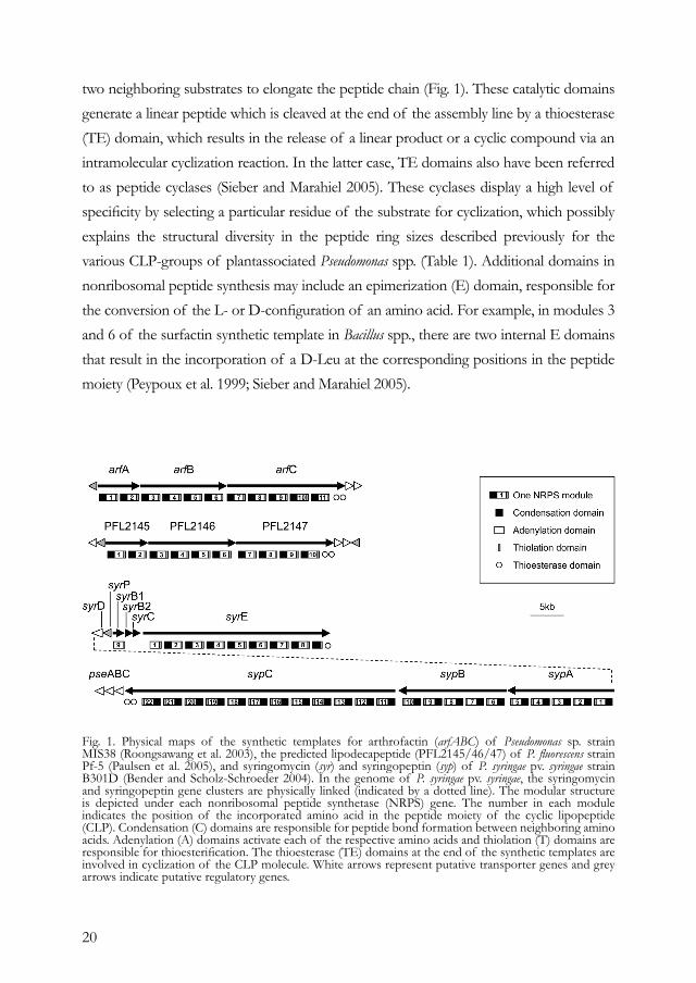
20
two neighboring substrates to elongate the peptide chain (Fig. 1). These catalytic domains generate a linear peptide which is cleaved at the end of the assembly line by a thioesterase (TE) domain, which results in the release of a linear product or a cyclic compound via an intramolecular cyclization reaction. In the latter case, TE domains also have been referred to as peptide cyclases (Sieber and Marahiel 2005). These cyclases display a high level of specificity by selecting a particular residue of the substrate for cyclization, which possibly explains the structural diversity in the peptide ring sizes described previously for the various CLP-groups of plantassociated Pseudomonas spp. (Table 1). Additional domains in nonribosomal peptide synthesis may include an epimerization (E) domain, responsible for the conversion of the L- or D-configuration of an amino acid. For example, in modules 3 and 6 of the surfactin synthetic template in Bacillus spp., there are two internal E domains that result in the incorporation of a D-Leu at the corresponding positions in the peptide moiety (Peypoux et al. 1999; Sieber and Marahiel 2005).
Fig. 1. Physical maps of the synthetic templates for arthrofactin (arfABC) of Pseudomonas sp. strain MIS38 (Roongsawang et al. 2003), the predicted lipodecapeptide (PFL2145/46/47) of P. fluorescens strain Pf-5 (Paulsen et al. 2005), and syringomycin (syr) and syringopeptin (syp) of P. syringae pv. syringae strain B301D (Bender and Scholz-Schroeder 2004). In the genome of P. syringae pv. syringae, the syringomycin and syringopeptin gene clusters are physically linked (indicated by a dotted line). The modular structure is depicted under each nonribosomal peptide synthetase (NRPS) gene. The number in each module indicates the position of the incorporated amino acid in the peptide moiety of the cyclic lipopeptide (CLP). Condensation (C) domains are responsible for peptide bond formation between neighboring amino acids. Adenylation (A) domains activate each of the respective amino acids and thiolation (T) domains are responsible for thioesterification. The thioesterase (TE) domains at the end of the synthetic templates are involved in cyclization of the CLP molecule. White arrows represent putative transporter genes and grey arrows indicate putative regulatory genes.
21
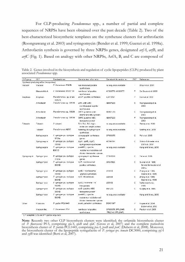
For CLP-producing Pseudomonas spp., a number of partial and complete sequences of NRPSs have been obtained over the past decade (Table 2). Two of the best-characterized biosynthetic templates are the synthetase clusters for arthrofactin (Roongsawang et al. 2003) and syringomycin (Bender et al. 1999; Guenzi et al. 1998a). Arthrofactin synthesis is governed by three NRPSs genes, designated arfA, arfB, and arfC (Fig. 1). Based on analogy with other NRPSs, ArfA, B, and C are composed of
Note: Recently two other CLP biosynthesis clusters were identified; the orfamide biosynthesis cluster of P. fluorescens Pf-5, comprising ofaA, ofaB and ofaC (Gross et al, 2007) and the complete putisolvin biosynthesis cluster of P. putida PCL1445, comprising psoA, psoB and psoC (Dubern et al, 2008). Moreover, the biosynthesis cluster of the lipopeptide syringafactin of P. syringae pv. tomato DC3000, comprising syfA and syfB was identified (Berti et al, 2007).
Table 2. Genes involved in the biosynthesis and regulation of cyclic lipopeptides (CLPs) produced by plant associated Pseudomonas spp.
22
a total of 11 modules, which is consistent with the number of amino acids in the peptide moiety (Fig. 1; Table 1). Each module contains the typical features of NRPSs, including the A, C, and T domains. In this respect, the arthrofactin synthetase cluster is a classic example of the colinearity rule. Although seven of the 11 amino acids in the peptide moiety of arthrofactin have the D-configuration (Table 1), none of the 11 modules in the arf synthetic cluster harbor an internal E domain for the conversion from L to D form. Also, in the synthetic templates of syringopeptin (Scholz-Schroeder et al. 2003), the predicted lipodecapeptide of strain Pf-5 (Paulsen et al. 2005), and in the gene clusters for massetolide A and viscosin production by antagonistic P. fluorescens strains (M. J. D. de Kock and J. M. Raaijmakers, personal communications), internal E domains were not found. Based on additional ATP-PPi exchange assays with a total of 20 typical L amino acids, Roongsawang and associates (2003) showed that the D-Leu1 A domain of the arthrofactin synthetic template adopted only L-Leu as a substrate and not D-Leu, suggesting that an external racemase is responsible for the L to D conversion. This suggestion for an external racemase was further supported by specific sequence motifs in the T domains of the arf synthetic template. T domains that are associated with an internal E domain, as in the surfactin synthetic template in Bacillus spp., have the highly conserved motif (F[F/Y]XXGGDSIKA[I/ L]Q), in which the aspartate residue in front of the serine residue is important in the interaction between the T and internal E domains (Linne et al. 2001). In the arthrofactin synthetic template, the T domains contain a different conserved sequence motif (FFELGGHSLLA[V/M]) and can be further classified into the T(L) and T(D) domains, responsible for transferring L or D amino acids, respectively, to the intermediate peptide moiety (Roongsawang et al. 2003). Roongsawang and associates (2003) suggested that these differences may reflect the different topologies between A and T domains or, alternatively, be important for the recognition by an external racemase that is necessary for D-amino acid incorporation. The C-terminal regions of CLP synthetic templates in Pseudomonas spp., including SypC in P. syringae and PFL2147 of P. fluorescens Pf-5, usually harbor one or two TE domains (Fig. 1). Also, in the arthrofactin synthetic template, the C-terminal region of ArfC contains two putative TE domains designated ArfCTe1 and ArfCTe2 (Fig. 1). Based on phylogenetic analyses and alignments of the amino acid sequences, it was postulated that ArfCTe1 and ArfCTe2 have a separate or, possibly, a coordinated
23
function in hydrolysis and cyclization of the final product (Roongsawang et al. 2003). Further analysis of the regions downstream of arfC resulted in the identification of two other genes, designated ORF5 and ORF6 (Roongsawang et al. 2003) (Fig. 1). Based on the direction of transcription of these two open reading frames (ORFs) and their homology to ATP-dependent transport proteins, these two ORFs were postulated to play a role in the secretion of arthrofactin (Roongsawang et al. 2003). The physical link between CLP synthetic templates and putative efflux genes also is the case for the predicted lipodecapeptide of P. fluorescens Pf-5 (Paulsen et al. 2005) (Fig. 1) and for syringopeptin and syringomycin of P. syringae pv. syringae (Feil et al. 2005; Scholz-Schroeder et al. 2001b) (Fig. 1). In the plant pathogen P. syringae pv. syringae, the gene cluster for syringomycin synthesis makes up a substantial portion of the total genome. Together with the syringopeptin cluster, it composes a region of 132 kb which accounts for approximately 2% of the genome (Feil et al. 2005; Scholz-Schroeder et al. 2001b, 2003; Wang et al. in press; D. C. Gross, personal communication). This cluster, referred to as the syr-syp genomic island (Scholz-Schroeder et al. 2001b), was not found in the genome of P. syringae pv. tomato DC3000 (Feil et al. 2005). In contrast to the arthrofactin gene cluster discussed above, the syringomycin gene cluster has several different and unique features. First of all, the organization of the structural synthetase genes does not respect the colinearity rule because the syrB1 gene, responsible for the incorporation of the ninth amino acid in the peptide moiety, is located upstream of syrE, which encodes the first eight amino acids (Fig. 1). Another interesting structural feature is the positioning of the single TE domain, which is not fused to the C-terminus of the last amino acid binding domain SyrB1 but, instead, to the C-terminus of SyrE-M8, the module required for the incorporation of the eighth amino acid (Bender and Scholz-Schroeder 2004; Guenzi et al. 1998a). The C-terminus of SyrE does contain elements of a ninth module, including condensation and thiolation domains, but lacks an adenylation domain which is proposed to be provided by SyrB1 (Bender et al. 1999; Bender and Scholz-Schroeder 2004). Recent studies have further indicated that SyrB1 activates the amino acid L-Thr, after which SyrB2-mediated chlorination takes place to yield the biologically active ninth amino acid residue 4-chlorothreonine (Vaillancourt et al. 2005). The fourth protein essential in syringomycin synthesis is SyrC, which is proposed to hydrolyze 3-hydroxydodecanoyl-CoA, leading to subsequent transfer of the 3-hydroxy fatty acid to the amino group of serine bound to SyrE-M1.
24
For the secretion of syringomycin, studies initially focused on SyrD, a protein with homology to membrane proteins of the ABC transporter family (Quigley et al. 1993; Scholz-Schroeder et al. 2001b). A mutation in syrD resulted in a significant reduction in secretion of syringomycin and also syringopeptin and, concurrently, in a 70% reduction in virulence on cherry fruit. However, the syrD mutation did not lead to a complete loss of CLP secretion. Kang and Gross (2005) recently discovered an additional efflux system, designated PseABC, located at the left border of the syr-syp genomic island of P. syringae pv. syringae B301D (Fig. 1). This tripartite transporter system, with homology to the resistance-nodulation-cell division (RND) efflux system of Ralstonia solanacearum, was shown to be involved in syringomycin and syringopeptin secretion. Also, in B. subtilis, an “RND-like” gene, designated yerP, was shown to be at least partially involved in efflux of surfactin (Tsuge et al. 2001). Interestingly, expression of yerP was not induced by surfactin, but associated with the growth phase (late logarithmic) of the producing strain. Furthermore, YerP also appeared to play a role in resistance of B. subtilis to other antibiotics, including acriflavine (Tsuge et al. 2001). Although a role of PseABC of P. syringae pv. syringae in resistance to acriflavine could not be demonstrated, heterologous expression of the pseABC genes in a drug-supersensitive mutant of E. coli did result in a significant increase in resistance to acriflavine and other antibiotics, supporting their functional relationship with RND-type transporter genes (Kang and Gross 2005). Based on the unusual and nonlinear genetic organization of the synthetic template of syringomycin and the templates of several other nonribosomal peptide antibiotics (Finking and Marahiel 2004), Guenzi and associates (1998a) postulated that, for the coordinated transcription of the enzyme subunits, individual modules can be dissociated without affecting CLP synthesis at both the qualitative and quantitative level. To further test this hypothesis, Guenzi and associates (1998b) designed two mutants of surfactin-producing B. subtilis. In one mutant, the operon structure of the surfactin synthetic template was altered by integration of a constitutive promoter; whereas, in the other mutant, the thioesterase domain was physically dissociated from the third amino acid binding domain and expressed independently. The results showed that both mutants produced surfactin at levels comparable with the wild type strain (Guenzi et al. 1998b). These studies indicated that domain–domain interactions rather than coordinated transcription and translation are essential for the correct assembly and activity of the NRPSs.
25
Substrate selection and selectivity-conferring elements of NRPSs. Comparative analysis of multiple NRPS sequences has shown that several of the modules harbor conserved motifs that allow the prediction of the sequence and configuration of the amino acids in the peptide moiety of the CLP molecule. A number of studies and reviews have addressed this issue in detail from a fundamental scientific perspective and also because it provides a means for targeted alteration of the structure and activity spectrum and, ultimately, enables the design of artificial nonribosomal peptide products (Challis et al. 2000; Gruenewald et al. 2004; Lautru and Challis 2004; Stachelhaus et al. 1999). One of the key questions in this context is how NRPSs select from the wide variety of precursors to be incorporated into the final CLP molecule (Lautru and Challis 2004). Most of the studies have focused on the selectivity-conferring codes within the adenylation (A) domains, because these domains select the amino acids and are regarded as the “gatekeeper” units of the repeated modules (Stachelhaus et al. 1999). Following the elucidation of the crystal structure of the phenylalanine-activating A domain (PheA) of gramicidin-S-synthetase from B. brevis (Conti et al. 1997), Stachelhaus and associates (1999) compared the amino acid residues of a large number of A domains from prokaryotic and eukaryotic origins with those lining the binding pocket of PheA. The in silico analyses provided a large number of signature sequences within A domains that allowed a prediction of the amino acid composition of the peptide moiety. This information, supplemented with new sequence data, has resulted in the design of web-based interfaces for analysis of NRPS sequences and polyketide (PKS) synthetase genes (Ansari et al. 2004; Challis et al. 2000). In addition to in silico analyses, Stachelhaus and associates (1999) provided experimental evidence for the specificity-conferring codes of A domains within NRPSs by introducing mutations in PheA, which led to the activation of L-Leu instead of phenylalanine. This and other studies also revealed that the same amino acid can be activated by A domains with different predicted selectivity pocket residues, which suggests some level of degeneracy in substrate recognition (Challis et al. 2000; Lautru and Challis 2004; Stachelhaus et al. 1999). Furthermore, certain signature sequences in the A domains contain so-called “wobble-like” positions which can lead to flexibility in amino acid usage (Stachelhaus et al. 1999). In other words, the signature sequences of A domains permit, to some degree, the incorporation of an “incorrect” amino acid, which may explain the presence of several less abundant “isoforms” of massetolide- or amphisin-like CLPs in extracts from cell cultures of antagonistic P.

26
fluorescens strains (De Souza et al. 2003; Nielsen et al. 2002). Also C, E, and TE domains play an important role in substrate recognition (Finking and Marahiel 2004; Lautru and Challis 2004; Linne et al. 2004; Roongsawang et al. 2005; Stein et al. 2005), although specificity studies have not been conducted as extensively as for A domains. Based on the increased availability of NRPS gene sequences of Pseudomonas and other species, the list of signature sequences specific for the various A domains now can be further expanded and will provide a valuable tool in annotation of NRPS sequences and in the prediction of the CLP structures based on genome data. This information can be exploited further for targeted alteration of CLP structures and activities via modification or exchange of NRPS modules, and for combinatorial synthesis of artificial nonribosomal peptide products (Gruenewald et al. 2004; Lautru and Challis 2004).
REGULATIONCompared with our understanding of the biosynthesis of CLPs in Pseudomonas and other bacterial genera, relatively little is known about the genetic network involved in the perception of external factors (abiotic and biotic) and the signal transduction pathways that drive transcription of the CLP biosynthetic genes. A number of studies have focused on the effects of abiotic conditions (e.g., temperature, pH, and oxygen), nutritional factors (e.g., carbon and nitrogen sources and trace elements), and plant signal molecules (glycosides and phenolics) on CLP production in Pseudomonas spp. (Bender et al. 1999; Bender and Scholz-Schroeder 2004; Nybroe and Sørensen 2004). This section focuses primarily on CLP regulatory genes and mechanisms identified so far for plant-associated Pseudomonas spp. Similar to regulatory mechanisms identified for CLP production in Bacillus spp. (Duitman 2003; Sullivan 1998), two-component systems and cell density also play a key role in the regulation of CLP production in Pseudomonas spp. (Table 2). For several CLPs, including syringomycin (Bender et al. 1999; Willis and Kinscherf 2004), amphisin (Koch et al. 2002), putisolvin I and II (Dubern et al. 2005), massetolide A, and viscosin (I. de Bruijn and J. M. Raaijmakers, personal communication), the GacS/GacA two-component regulatory system appears to serve as a master switch; mutants disrupted in either one of the two genes are deficient in CLP production. Although the GacA/GacS system has been the subject of numerous studies (Heeb and Haas 2001), very little still is known about signals that serve as a trigger. Studies by Koch and associates (2002) with antagonistic Pseudomonas DSS73 elegantly demonstrated that exudates of sugar beet seed contain triggers for amphisin production and
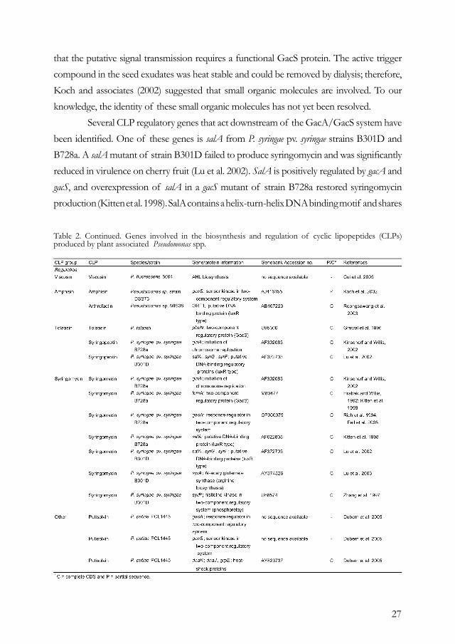
27
that the putative signal transmission requires a functional GacS protein. The active trigger compound in the seed exudates was heat stable and could be removed by dialysis; therefore, Koch and associates (2002) suggested that small organic molecules are involved. To our knowledge, the identity of these small organic molecules has not yet been resolved. Several CLP regulatory genes that act downstream of the GacA/GacS system have been identified. One of these genes is salA from P. syringae pv. syringae strains B301D and B728a. A salA mutant of strain B301D failed to produce syringomycin and was significantly reduced in virulence on cherry fruit (Lu et al. 2002). SalA is positively regulated by gacA and gacS, and overexpression of salA in a gacS mutant of strain B728a restored syringomycin production (Kitten et al. 1998). SalA contains a helix-turn-helix DNA binding motif and shares
Table 2. Continued. Genes involved in the biosynthesis and regulation of cyclic lipopeptides (CLPs) produced by plant associated Pseudomonas spp.
28
homology with members of the LuxR family of regulatory proteins (Lu et al. 2002). SalA activates the expression of syrB1 (Kitten et al. 1998) and syrF, another LuxR-type regulatory gene involved in syringomycin synthesis (Lu et al. 2002). To further identify the members of the SalA regulon, Lu and associates (2005) designed an oligonucleotide microarray representing genes in the syr-syp genomic island as well as genes involved in the Hrp type III secretion system, phytohormone synthesis, siderophore and syringolin production, quorum sensing, and global regulation, including gacS, gacA, rpoN, and rpoS. Comparative analysis of the transcription profiles of a salA mutant and the wild type strain B301D showed that genes involved in the synthesis, secretion, and regulation of syringomycin and syringopeptin were upregulated by the salA gene. No significant effects were observed for the genes located outside the syr-syp genomic island, except for the sylD gene, which is involved in syringolin synthesis (Lu et al. 2005). This “genome-wide” study serves as an excellent model to further elucidate regulatory elements and networks in CLP synthesis in other Pseudomonas spp. DnaK is another gene that is positively regulated by the GacA/GacS system and involved in regulation of CLP synthesis in P. putida (Dubern et al. 2005). In a dnaK mutant of strain PCL1445, production of putisolvin I and II was strongly reduced. Given that DnaK is a member of the Hsp70 heat shock protein family, Dubern and associates (2005) subsequently investigated the role of temperature on putisolvin production. Their results showed that putisolvin production is upregulated at low temperatures and that dnaK is required for putisolvin production at low temperatures. Sequencing both up- and downstream of the dnaK gene further led to the identification of two other genes, dnaJ and grpE, that both adversely affected the synthesis of putisolvin I and II. Although the exact role of these three genes in regulation of putisolvin synthesis is not yet known, the authors speculated that they may be required for the proper folding or activity of other positive regulators of the putisolvin synthetic gene psoA. Two other possible mechanisms proposed were interaction of the DnaK complex with the rpoS encoded sigma factor σs, or the requirement of the DnaK complex for proper assembly of the peptide synthetase complex. In addition to two-component regulation, cell density plays an important role in global regulation of CLP production in Pseudomonas spp. For many antagonistic P. fluorescens strains, CLP production occurs in the late exponential or stationary growth phase, but the underlying molecular mechanisms are far from being understood. In this context, Nybroe and Sørensen (2004) emphasized that, although CLP production is

29
affected by the growth phase and nutritional conditions that lead to increased growth, the specific impact of these factors may differ considerably among strains. Quorum sensing via N-acyl homoserine lactones (N-AHL) does not appear to play a role in amphisin and syringomycin production (Andersen et al. 2003; Kinscherf and Willis 1999; Quinones et al. 2005). Studies with antagonistic P. fluorescens SS101 indicated that N-AHL-mediated quorum sensing does not seem to be involved in massetolide A production (I. de Bruijn and J. M. Raaijmakers, personal communication). For the plant-pathogenic P. fluorescens strain 5064, recent studies by Cui and associates (2005) provided, for the first time, evidence for N-AHL-mediated quorum sensing in viscosin synthesis. Screening of a mutant collection of strain 5064 resulted in the identification of six mutants affected in quorum sensing signal (QSS) production. The QSS was identified as N-3-acyl-hydroxyoctanoyl-HSL, and addition of QSS extracts or the synthetic signal molecule restored viscosin synthesis in the mutant. Cui and associates (2005) also observed that the mutation did not lead to a complete blockage in N-AHL expression and they speculated that there may be more than one quorum sensing system in strain 5064. Although the sequences of the genes involved in quorum sensing-mediated viscosin synthesis in strain 5064 were not described in this study (Cui et al. 2005), they may provide a useful tool to assess the presence (or absence) of homologues in other CLP-producing Pseudomonas strains. Next to two-component regulatory systems and cell density, Kinscherf and Willis (2002) showed that the gidA gene is involved in global regulation of syringomycin and syringopeptin synthesis in P. syringae. Next to the inability to produce CLPs, mutations in gidA (glucose-inhibited division) affected a variety of other phenotypes, including pyoverdine synthesis and surface motility on low-agar media. As was shown for gacS mutants of P. syringae, overexpression of salA restored syringomycin synthesis in gidA mutants, suggesting that gidA plays a role in salA regulation (Kinscherf and Willis 2002). Finally, analyses of regions physically linked to the CLP synthetic templates revealed a number of other interesting regulatory genes (Table 2). For syringomycin, these include the phosphate-regulated gene syrP (Fig. 1) and yet another LuxR-type gene syrG, although their exact roles remain to be determined (Bender and Scholz-Schroeder 2004).
30
CONCLUDING REMARKSBased on this survey, we conclude that CLPs produced by plant-associated Pseudomonas spp. are enormously diverse, both structurally and in terms of their biological activity. Although a number of regulatory genes have been identified, the understanding of the regulatory networks involved in CLP synthesis by plant-associated Pseudomonas spp. is still in its infancy. Therefore, identification of genes and mechanisms involved in perception and signal transduction is a major challenge for the near future. Also, the synthesis of the fatty acid moiety of the CLP molecule has received little attention. At present, many other specific questions remain to be addressed. For example, which external racemases in Pseudomonas spp. play a role in epimerization of the amino acids in the CLP peptide moiety? Which other natural roles and modes of action can be assigned to CLPs? How can we modulate, both quantitatively and qualitatively, in situ production of CLPs by antagonistic and pathogenic Pseudomonas spp.? Is regulation of CLP synthesis fundamentally different between pathogenic and antagonistic Pseudomonas spp.? Significant advances in the analyses and mining of whole genome sequences, the design of microarrays, and developments in metabolomics and proteomics will enable a better understanding of the synthesis, regulation, and activity of CLPs produced by Pseudomonas and other bacterial genera.
ACKNOWLEDGMENTSThis work is supported financially by the Dutch Technology Foundation (STW), the applied science division of NWO, and by the Royal Netherlands Academy of Arts and Science. We are grateful to D. Gross for critically reviewing this manuscript and for his valuable suggestions.
REFERENCESAndersen, J. B., Koch, B., Nielsen, T. H., Sørensen, D., Hansen, M., Nybroe, O., Christophersen, C., Sørensen, J., Molin, S., and Givskov, M. 2003. Surface motility in Pseudomonas sp. DSS73 is required for efficient biological containment of the root-pathogenic microfungi Rhizoctonia solani and Pythium ultimum. Microbiology 149:37-46.Ansari, M. Z., Yadav, G., Gokhale, R. S., and Mohanty, D. 2004. NRPSPKS: A knowledge-based resource for analysis of NRPS/PKS megasynthases. Nucleic Acids Res. 32:W405-413. Bais, H. P., Fall, R., and Vivanco, J. M. 2004.
Biocontrol of Bacillus subtilis against infection of Arabidopsis roots by Pseudomonas syringae is facilitated by biofilm formation and surfactin production. Plant Physiol. 134:307-319.Ballio, A., Barra, D., Bossa, F., Collina, A., Grgurina, I., Marino, G., Moneti, G., Paci, M., Pucci, P., Segre, A., and Simmaco, M. 1991. Syringopeptins, new phytotoxic lipodepsipeptides of Pseudomonas syringae pv. syringae. FEBS (Fed. Eur. Biol. Soc.) Lett. 291:109-112. Ballio, A., Bossa, F., Camoni, L., Di Giorgio, D., Flamand, M. C., Maraite, H., Nitti, G., Pucci, P., and Scaloni, A. 1996. Structure

31
of fuscopeptins, phytotoxic metabolites of Pseudomonas fuscovaginae. FEBS Lett. 381:213-216.Bare, S., Coiro, V. M., Scaloni, A., Di Nola, A., Paci, M., Segre, A. L., and Ballio, A. 1999. Conformations in solution of the fuscopeptins—phytotoxic metabolites of Pseudomonas fuscovaginae. Eur. J. Biochem. 266:484-492.Bender, C. L., Alarcon-Chaidez, F., and Gross, D. C. 1999. Pseudomonas syringae phytotoxins: Mode of action, regulation, and biosynthesis by peptide and polyketide synthetases. Microbiol. Mol. Biol. Rev. 63:266-292. Bender, C. L., and Scholz-Schroeder, B. K. 2004. New insights into the biosynthesis, mode of action, and regulation of syringomycin, syringopeptin, and coronatine. Pages 125-158 in: Pseudomonas Vol. 2, Virulence and Gene Regulation. J.-L. Ramos, ed. Kluwer Academic/Plenum Publishers, New York, U.S.A.Berti, A.D., Greve, N. D., Christensen Q.H., Thomas M.G. 2007. Identification of a biosynthetic gene cluster and the six associated lipopeptides involved in swarming motility of Pseudomonas syringae pv. tomato DC3000. J. Bacteriol. 189:6312-23Braun, P. G., Hildebrand, P. D., Ells, T. C., and Kobayashi, D. Y. 2001. Evidence and characterization of a gene cluster required for the production of viscosin, a lipopeptide biosurfactant, by a strain of Pseudomonas fluorescens. Can. J. Microbiol. 47:294-301.Buber, E., Stindl, A., Acan, N. L., Kocagoz, T., and Zocher, R. 2002. Antimycobacterial activity of lipodepsipeptides produced by Pseudomonas syringae pv. syringae B359. Nat. Prod. Lett. 16:419-423.Burke, T. Jr., Chandrasekhar, B., and Knight, M. 1999. Analogs of viscosin and uses thereof. U.S. Patent Number 5,965,524. Challis, G. L., and Naismith, J. H. 2004. Structural aspects of nonribosomal peptide biosynthesis. Curr. Opin. Struct. Biol. 14:748-756.Challis, G. L., Ravel, J., and Townsend, C. A. 2000. Predictive, structurebase model of amino acid recognition by nonribosomal peptide synthetase adenylation domains. Chem. Biol. 7:211-224.Coiro, V. M., Segre, A. L., Di Nola, A., Paci, M., Grottesi, A., Veglia, G., and Ballio, A. 1998. Solution conformation of the Pseudomonas syringae MSU 16H phytotoxic lipodepsipeptide Pseudomycin A determined by computer simulations using distance geometry and molecular dynamics from NMR data. Eur. J. Biochem. 257:449-456.Compant, S., Duffy, B., Nowak, J., Clement, C., and Barka, E. A. 2005. Use of plant growth-promoting bacteria for biocontrol of plant diseases: Principles, mechanisms of action, and future prospects. Appl. Environ. Microbiol. 71:4951-4959.Conti, E., Stachelhaus, T., Marahiel, M. A., and Brick, P. 1997. Structural basis for the activation of phenylalanine in the nonribosomal biosynthesis of gramicidin S. EMBO J. 16:4174-4183.
Cui, X., Harling, R., Mutch, P., and Darling, D. 2005. Identification of N-3-hydroxyoctanoyl-homoserine lactone production in Pseudomonas fluorescens 5064, pathogenic to broccoli, and controlling biosurfactant production by quorum sensing. Eur. J. Plant Pathol. 111:297-308.Dalla Serra, M., Fagiuoli, G., Nordera, P., Bernhart, I., Della Volpe, C., Di Giorgio, D., Ballio, A., and Menestrina, G. 1999. The interaction of lipodepsipeptide toxins from Pseudomonas syringae pv. syringae with biological and model membranes: A comparison of syringotoxin, syringomycin, and two syringopeptins. Mol. Plant-Microbe Interact. 12:391-400.De Souza, J. T., De Boer, M., De Waard, P., Van Beek, T. A., and Raaijmakers, J. M. 2003. Biochemical, genetic, and zoosporicidal properties of cyclic lipopeptide surfactants produced by Pseudomonas fluorescens. Appl. Environ. Microbiol. 69:7161-7172.Dubern J.F., Coppoolse E.R., Stiekema W.J., Bloemberg G.V. 2008. Genetic and functional characterization of the gene cluster directing the biosynthesis of putisolvin I and II in Pseudomonas putida strain PCL1445. Microbiology 154:2070-83.Dubern, J.-F., Lagendijk, E. L., Lugtenberg, B. J. J., and Bloemberg, G. V. 2005. The heat shock genes dnaK, dnaJ, and grpE are involved in regulation of putisolvin biosynthesis in Pseudomonas putida PCL1445. J. Bacteriol. 187:5967-5976.Dufour, S., Deleu, M., Nott, K., Wathelet, B., Thonart, P., and Paquot, M. 2005. Hemolytic activity of new linear surfactin analogs in relation to their physico-chemical properties. Biochim. Biophys. Acta. 1726:87-95Duitman, E. H. 2003. Nonribosomal peptide synthesis in Bacillus subtilis. Ph.D. thesis, University of Groningen, Groningen, The Netherlands. El Sayed, K. A., Bartyzel, P., Shen, X. Y., Perry, T. L., Kjawiony, J. K., and Hamann, M. T. 2000. Marine natural products as anti-tuberculosis agents. Tetrahedron 56:949-953.Emanuele, M. C., Scaloni, A., Lavermicocca, P., Jacobellis, N. S., Camoni, L., Di Giorgio, D., Pucci, P., Paci, M., Segre, A., and Ballio, A. 1998. Corpeptins, new bioactive lipodepsipeptides from cultures of Pseudomonas corrugata. FEBS Lett. 433:317.Feil, H., Feil, W. S., Chain, P., Larimer, F., Dibartolo, G., Copeland, A., Lykidis, A., Trong, S., Nolan, M., Goltsman, E., Thiel, J., Malfatti, S., Loper, J. E., Lapidus, A., Detter, J. C., Land, M., Richardson, P. M., Kyrpides, N. C., Ivanova, N., and Lindow, S. E. 2005. Comparison of the complete genome sequences of Pseudomonas syringae pv. syringae B728a and pv. tomato DC3000. Proc. Natl. Acad. Sci. USA 102:11064-11069. Finking, R., and Marahiel, M. A. 2004. Biosynthesis of nonribosomal peptides. Annu. Rev. Microbiol. 58:453-488.Fogliano, V., Ballio, A., Gallo, M., Woo, S., Scala, F., and Lorito, M. 2002. Pseudomonas

32
lipodepsipeptides and fungal cell wall-degrading enzymes act synergistically in biological control. Mol. Plant-Microbe Interact. 15:323-333.Gerard, J., Lloyd, R., Barsby, T., Haden, P., Kelly, M. T., and Andersen, R. J. 1997. Massetolides A-H, antimycobacterial cyclic depsipeptides produced by two pseudomonads isolated from marine habitats. J. Nat. Prod. 60:223-229.Gewolb, J. 2002. Bioengineering. Working outside the protein-synthesis rules. Science 295:2205-2207.Godfrey, S. A. C., Marshall, J. W., and Klena, J. D. 2001. Genetic characterization of Pseudomonas ‘NZ17—a novel pathogen that results in a brown blotch disease of Agaricus bisporus. J. Appl. Microbiol. 91:412-420.Grewal, S. I. S., Han, B., and Johnstone, K. 1995. Identification and characterization of a locus which regulates multiple functions in Pseudomonas tolaasii, the cause of brown blotch disease of Agaricus bisporus. J. Bacteriol. 177:4658-4668.Grgurina, I., Barca, A., Cervigni, S., Gallo, M., Scaloni, A., and Pucci, P. 1994. Relevance of chlorine-substituent for the antifungal activity of syringomycin and syringotoxin, metabolites of the phytopathogenic bacterium Pseudomonas syringae pv. syringae. Experientia 50:130-133. Gross H., Stockwell V.O., Henkels M.D., Nowak-Thompson B., Loper J.E., Gerwick W.H. 2007. The genomisotopic approach: a systematic method to isolate products of orphan biosynthetic gene clusters. Chem. Biol. 14:53-63.Gruenewald, S., Mootz, H. D., Stehmeier, P., and Stachelhaus, T. 2004. In vivo production of artificial nonribosomal peptide products in the heterologous host Escherichia coli. Appl. Environ. Microbiol. 70:3282-3291.Guenzi, E., Galli, G., Grgurina, I., Gross, D. C., and Grandi, G. 1998a. Characterization of the syringomycin synthetase gene cluster. A link between prokaryotic and eukaryotic peptide synthetases. J. Biol. Chem. 273:32857-32863.Guenzi, E., Galli, G., Grgurina, I., Pace, E., Ferranti, P., and Grandi, G. 1998b. Coordinate transcription and physical linkage of domains in surfactin synthetase are not essential for proper assembly and activity of the multienzyme complex. J. Biol. Chem. 273:14403-14410. Haas, D., and Défago, G. 2005. Biological control of soil-borne pathogens by fluorescent pseudomonads. Nat. Rev. Microbiol. 3:307-319. Hansen, M., Thrane, C., Olsson, S., and Sorensen, J. 2000. Confocal imaging of living fungal hyphae challenged with the fungal antagonist viscosinamide. Mycologia 92:216-221.Heeb, S., and Haas, D. 2001. Regulatory roles of the GacS/GacA two-component system in plant-associated and other gram-negative bacteria. Mol. Plant-Microbe Interact. 14:1351-1363.Henriksen, A., Anthoni, U., Nielsen, T. H., Sorensen, J., Christophersen, C., and Gajhede, M. 2000. Cyclic lipoundecapeptide
tensin from Pseudomonas fluorescens strain 96.578. Acta Crystal. 56:113-115.Hildebrand, P. D., Braun, P. G., McRae, K. B., and Lu, X. 1998. Role of the biosurfactant viscosin in broccoli head rot caused by a pectolytic strain of Pseudomonas fluorescens. Can. J. Plant Pathol. 20:296-303.Hrabak, E. M., and Willis, D. K. 1992. The lemA gene required for pathogenicity of Pseudomonas syringae pv. syringae on bean is a member of a family of 2-component regulators. J. Bacteriol. 174:3011-3020.Hutchison, M. L., and Gross, D. C. 1997. Lipopeptide phytotoxins produced by Pseudomonas syringae pv. syringae: Comparison of the biosurfactant and ion channel-forming activities of syringopeptin and syringomycin. Mol. Plant-Microbe Interact. 10:347-354.Hutchison, M. L., and Johnstone, K. 1993. Evidence for the involvement of the surface-active properties of the extracellular toxin tolaasin in the manifestation of brown blotch disease symptoms by Pseudomonas tolaasii on Agaricus bisporus. Physiol. Mol. Plant Pathol. 42:373-384.Hutchison, M. L., Tester, M. A., and Gross, D. C. 1995. Role of biosurfactant and ion channel-forming activities of syringomycin in transmembrane ion flux: A model for the mechanism of action in the plant-pathogen interaction. Mol. Plant-Microbe Interact. 8:610-620.Kang, H., and Gross, D. C. 2005. Characterization of a resistance-nodulation-cell division transporter system associated with the syr-syp genomic island of Pseudomonas syringae pv. syringae. Appl. Environ. Microbiol. 71:5056-5065.Kinscherf, T. G., and Willis, D. K. 1999. Swarming by Pseudomonas syringae B728a requires gacS (lemA) and gacA but not the acyl-homoserine lactone biosynthetic gene ahlI. J. Bacteriol. 181:4133-4136.Kinscherf, T. G., and Willis, D. K. 2002. Global regulation by gidA in Pseudomonas syringae. J. Bacteriol. 184:2281-2286.Kitten, T., Kinscherf, T. G., McEvoy, J. L., and Willis, D. K. 1998. A newly identified regulator is required for virulence and toxin production in Pseudomonas syringae. Mol. Microbiol. 28:917-929. Koch, B., Nielsen, T. H., Sorensen, D., Andersen, J. B., Christophersen, C., Molin, S., Givskov, M., Sorensen, J., and Nybroe, O. 2002. Lipopeptide production in Pseudomonas sp. strain DSS73 is regulated by components of sugar beet seed exudate via the Gac two-component regulatory system. Appl. Environ. Microbiol. 68:4509-4516.Koumoutsi, A., Chen, X. H., Henne, A., Liesegang, H., Hitzeroth, G., Franke, P., Vater, J., and Borriss, R. 2004. Structural and functional characterization of gene clusters directing nonribosomal synthesis of bioactive cyclic lipopeptides in Bacillus amyloliquefaciens strain FZB42. J. Bacteriol. 186:1084-1096.Kracht, M., Rokos, H., Ozel, M., Kowall,
33
M., Pauli, G., and Vater, J. 1999. Antiviral and hemolytic activities of surfactin isoforms and their methyl ester derivatives. J. Antibiot. 52:613-619.Kuiper, I., Lagendijk, E. L., Pickford, R., Derrick, J. P., Lamers, G. E., Thomas-Oates, J. E., Lugtenberg, B. J., and Bloemberg, G. V. 2004. Characterization of two Pseudomonas putida lipopeptide biosurfactants, putisolvin I and II, which inhibit biofilm formation and break down existing biofilms. Mol. Microbiol. 51:97-113.Lautru, S., and Challis, G. L. 2004. Substrate recognition by nonribosomal peptide synthetase multi-enzymes. Microbiology 150:1629-1636.Lavermicocca, P., Iacobellis, N. S., Simmaco, M., and Graniti, A. 1997. Biological properties and spectrum of activity of Pseudomonas syringae pv. syringae toxins. Physiol. Mol. Plant Pathol. 50:129-140.Leclère, V., Bechet, M., Adam, A., Guez, J.-S., Wathelet, B., Ongena, M., Thonart, P., Gancel, F., Chollet-Imbert, M., and Jacques, P. 2005. Mycosubtilin overproduction by Bacillus subtilis BBG100 enhances the organism’s antagonistic and biocontrol activities. Appl. Environ. Microbiol. 71:4577-4584.Li, Y. Q., Sun, Z. L., Zhuang, X. F., Xu, L., Chen, S. F., and Li, M. Z. 2003. Research progress on microbial herbicides. Crop Prot. 22:247-252. Lindow, S. E., and Brandl, M. T. 2003. Microbiology of the phyllosphere. Appl. Environ. Microbiol. 69:1875-1883.Lindum, P. W., Anthoni, U., Christophersen, C., Eberl, L., Molin, S., and Givskov, M. 1998. N-Acyl-L-homoserine lactone autoinducers control production of an extracellular lipopeptide biosurfactant required for swarming motility of Serratia liquefaciens MG1. J. Bacteriol. 180:6384-6388.Linne, U., Doekel, S., and Marahiel, M. A. 2001. Portability of epimerization domain and role of peptidyl carrier protein on epimerization activity in nonribosomal peptide synthetases. Biochemistry 40:15824-15834.Linne, U., Schwarzer, D., Schroeder, G. N., and Marahiel, M. A. 2004. Mutational analysis of a type II thioesterase associated with nonribosomal peptide synthesis. Eur. J. Biochem. 271:1536-1545.Lu, S. E., Scholz-Schroeder, B. K., and Gross, D. C. 2002. Characterization of the salA, syrF, and syrG regulatory genes located at the right border of the syringomycin gene cluster of Pseudomonas syringae pv. syringae. Mol. Plant-Microbe Interact. 15:43-53.Lu, S. E., Soule, J. D., and Gross, D. C. 2003. Characterization of the argA gene required for arginine biosynthesis and syringomycin production by Pseudomonas syringae pv. syringae. Appl. Environ. Microbiol. 69:7273-7280.Lu, S. E., Wang, N., Wang, J., Chen, Z. J., and Gross, D. C. 2005. Oligonucleotide microarray analysis of the salA regulon controlling phytotoxin production by Pseudomonas syringae pv.
syringae. Mol. Plant-Microbe Interact. 18:324-333.Lugtenberg, B. J. J., Dekkers, L., and Bloemberg, G. V. 2001. Molecular determinants of rhizosphere colonization by Pseudomonas. Annu. Rev. Phytopathol. 39:461-490.Marahiel, M. A., Stachelhaus, T., and Mootz, H. D. 1997. Modular peptide synthetases involved in nonribosomal peptide synthesis. Chem. Rev. 97:2651-2673.Mercado, T. I., and Colon-Whitt, A. 1982. Lysis of Trypanosoma cruzi by Pseudomonas fluorescens. Antimicrob. Agents Chemother. 22:1051-1057.Mireles, J. R., Toguchi, A., and Harshey, R. M. 2001. Salmonella enterica serovar Typhimurium swarming mutants with altered biofilm-forming abilities: Surfactin inhibits biofilm formation. J. Bacteriol. 183:5848-5854.Monti, S. M., Gallo, M., Ferracane, R., Borrelli, R. C., Ritieni, A., Greco, M. L., Graniti, A., and Fogliano, V. 2001. Analysis of bacterial lipodepsipeptides by matrix-assisted laser desorption/ionisation time-of-flight and high-performance liquid chromatography with electrospray mass spectrometry. Rapid Commun. Mass Spectrom. 15:623-628.Morikawa, M., Daido, H., Takao, T., Murata, S., Shimonishi, Y., and Imanaka, T. 1993. A new lipopeptide biosurfactant produced by Arthrobacter sp. strain MIS38. J. Bacteriol. 175:6459-6466.Morikawa, M., Hirata, Y., and Imanaka, T. 2000. A study on the structure function relationship of lipopeptide biosurfactants. Biochim. Biophys. Acta 1488:211-218.Mott, K. A., and Takemoto, J. Y. 1989. Syringomycin, a bacterial phytotoxin, closes stomata. Plant Physiol. 90:1435-1439.Neu, T. R. 1996. Significance of bacterial surface-active compounds in interaction of bacteria with interfaces. Microbiol. Rev. 60:151-166.Nielsen, T. H., Christophersen, C., Anthoni, U., and Sorensen, J. 1999. Viscosinamide, a new cyclic depsipeptide with surfactant and antifungal properties produced by Pseudomonas fluorescens DR54. J. Appl. Microbiol. 87:80-90.Nielsen, T. H., Sorensen, D., Tobiasen, C., Andersen, J. B., Christophersen, C., Givskov, M., and Sorensen, J. 2002. Antibiotic and biosurfactant properties of cyclic lipopeptides produced by fluorescent Pseudomonas spp. from the sugar beet rhizosphere. Appl. Environ. Microbiol. 68:3416-3423.Nielsen, T. H., and Sorensen, J. 2003. Production of cyclic lipopeptides by Pseudomonas fluorescens strains in bulk soil and in the sugar beet rhizosphere. Appl. Environ. Microbiol. 69:861-868.Nielsen, T. H., Nybroe, O., Koch, B., Hansen, M., and Sorensen, J. 2005. Genes involved in cyclic lipopeptide production are important for seed and straw colonization by Pseudomonas sp strain DSS73. Appl. Environ. Microbiol. 71:4112-4116.Nybroe, O., and Sorensen, J. 2004. Production of cyclic lipopeptides by fluorescent
34
pseudomonads. Pages 147-172 in: Pseudomonas, Biosynthesis of Macromolecules and Molecular Metabolism. J.-L. Ramos, ed., Kluwer Academic/Plenum Publishers, New York, U.S.A.O’Toole, G., Kaplan, H. B., and Kolter, R. 2000. Biofilm formation as microbial development. Annu. Rev. Microbiol. 54:49-79.Paulsen, I. T., Press, C. M., Ravel, J., Kobayashi, D. Y., Myers, G. S. A., Mavrodi, D. V., DeBoy, R. T., Seshadri, R., Ren, Q. H., Madupu, R., Dodson, R. J., Durkin, A. S., Brinkac, L. M., Daugherty, S. C., Sullivan, S. A., Rosovitz, M. J., Gwinn, M. L., Zhou, L. W., Schneider, D. J., Cartinhour, S. W., Nelson, W. C., Weidman, J., Watkins, K., Tran, K., Khouri, H., Pierson, E. A., Pierson, L. S., Thomashow, L. S., and Loper, J. E. 2005. Complete genome sequence of the plant commensal Pseudomonas fluorescens Pf-5. Nat. Biotechnol. 23:873-878.Paulsen, I. T., Press, C. M., Ravel, J., Kobayashi, D. Y., Myers, G. S. A.,Mavrodi, D. V., DeBoy, R. T., Seshadri, R., Ren, Q. H., Madupu, R., Dodson, R. J., Durkin, A. S., Brinkac, L. M., Daugherty, S. C., Sullivan, S. A., Rosovitz, M. J., Gwinn, M. L., Zhou, L. W., Schneider, D. J., Cartinhour, S. W., Nelson, W. C., Weidman, J., Watkins, K., Tran, K., Khouri, H., Pierson, E. A., Pierson, L. S., Thomashow, L. S., and Loper, J. E. 2006. Corrigendum: Biocontrol genome deciphered. Nat. Bioltechnol. 27 March 2006. Published online. Pedras, M. S. C., Ismail, N., Quail, J. W., and Boyetchko, S. M. 2003. Structure, chemistry, and biological activity of pseudophomins A and B, new cyclic lipodepsipeptides isolated from the biocontrol bacterium Pseudomonas fluorescens. Phytochemistry 62:1105-1114.Peypoux, F., Bonmatin, J. M., and Wallach, J. 1999. Recent trends in the biochemistry of surfactin. Appl. Microbiol. Biotechnol. 51:553-563.Pieterse, C. M. J., Van Wees, S. C. M., Ton, J., Van Pelt, J. A., and Van Loon, L. C. 2002. Signaling in rhizobacteria-induced systemic resistance in Arabidopsis thaliana. Plant Biol. 4:535-544.Quail, J. W., Ismail, N., Pedras, M. S. C., and Boyetchko, S. M. 2002. Pseudophomins A and B, a class of cyclic lipodepsipeptides isolated from a Pseudomonas species. Acta Crystallogr. Sect. C 58:268-271.Quigley, N. B., Mo, Y. Y., and Gross, D. C. 1993. SyrD is required for syringomycin production by Pseudomonas syringae pathovar syringae and is related to a family of ATP-binding secretion proteins. Mol. Microbiol. 9:787-801.Quinones, B., Dulla, G., and Lindow, S. E. 2005. Quorum sensing regulates exopolysaccharide production, motility, and virulence in Pseudomonas syringae. Mol. Plant-Microbe Interact. 18:682-693.Raaijmakers, J. M., Vlami, M., and de Souza, J. T. 2002. Antibiotic production by bacterial biocontrol agents. Antonie Leeuwenhoek Int. J. Gen. Mol. Microbiol. 81:537-547.
Rainey, P. B., Brodey, C. L., and Johnstone, K. 1991. Biological properties and spectrum of activity of tolaasin, a lipodepsipeptide toxin produced by the mushroom pathogen Pseudomonas tolaasii. Physiol. Mol. Plant Pathol. 39:57-70.Rainey, P. B., Brodey, C. L., and Johnstone, K. 1993. Identification of a gene-cluster encoding 3 high-molecular-weight proteins, which is required for synthesis of tolaasin by the mushroom pathogen Pseudomonas tolaasii. Mol. Microbiol. 8:643-652.Ramos, J.-L. 2004. Pseudomonas, Vol. 1–3. Kluwer Academic/Plenum Publishers, New York, U.S.A.Rich, J. J., Kinscherf, T. G., Kitten, T., and Willis, D. K. 1994. Genetic evidence that the gacA gene encodes the cognate response regulator for the lemA sensor in Pseudomonas syringae. J. Bacteriol. 176:7468-7475.Ron, E. Z., and Rosenberg, E. 2001. Natural roles of biosurfactants. Environ. Microbiol. 3:229-236.Roongsawang, N., Hase, K., Haruki, M., Imanaka, T., Morikawa, M., and Kanaya, S. 2003. Cloning and characterization of the gene cluster encoding arthrofactin synthetase from Pseudomonas sp. MIS38. Chem. Biol. 10:869-880.Roongsawang, N., Lim, S. P., Washio, K., Takano, K., Kanaya, S., and Morikawa, M. 2005. Phylogenetic analysis of condensation domains in the nonribosomal peptide synthetases. FEMS Microbiol. Lett. 252:143-151.Ryu, C.-M., Hu, C.-H., Reddy, M. S., and Kloepper, J. W. 2003. Different signaling pathways of induced resistance by rhizobacteria in Arabidopsis thaliana against two pathovars of Pseudomonas syringae. New Phytol. 160:413-420.Scaloni, A., Dalla Serra, M., Amodeo, P., Mannina, L., Vitale, R. M., Segre, A. L., Cruciani, O., Lodovichetti, F., Greco, M. L., Fiore, A., Gallo, M., D’Ambrosio, C., Coraiola, M., Menestrina, G., Granit, A., and Fogliano, V. 2004. Structure, conformation and biological activity of a novel lipodepsipeptide from Pseudomonas corrugata: Cormycin A. Biochem. J. 384:25-36.Scholz-Schroeder, B. K., Hutchison, M. L., Grgurina, I., and Gross, D. C. 2001a. The contribution of syringopeptin and syringomycin to virulence of Pseudomonas syringae pv. syringae strain B301D on the basis of sypA and syrB1 biosynthesis mutant analysis. Mol. Plant-Microbe Interact. 14:336-348. Scholz-Schroeder, B. K., Soule, J. D., and Gross, D. C. 2003. The sypA, sypB and sypC synthetase genes encode twenty-two modules involved in the nonribosomal peptide synthesis of syringopeptin by Pseudomonas syringae pv. syringae B301D. Mol. Plant-Microbe Interact. 16:271-280. Scholz-Schroeder, B. K., Soule, J. D., Lu, S. E., Grgurina, I., and Gross, D. C. 2001b. A physical map of the syringomycin and syringopeptin gene clusters localized to an approximately 145-kb DNA region of Pseudomonas syringae pv. syringae strain B301D. Mol. Plant-Microbe Interact. 14:1426-1435.Sieber, S. A., and Marahiel, M. A. 2005. Molecular

35
mechanisms underlying nonribosomal peptide synthesis: Approaches to new antibiotics. Chem. Rev. 105:715-738.Soler-Rivas, C., Arpin, N., Olivier, J. M., and Wichers, H. J. 1999. WLIP, a lipodepsipeptide of Pseudomonas ‘reactans’, as inhibitor of the symptoms of the brown blotch disease of Agaricus bisporus. J. Appl. Microbiol. 86:635-641.Sorensen, D., Nielsen, T. H., Christophersen, C., Sorensen, J., and Gajhede, M. 2001. Cyclic lipoundecapeptide amphisin from Pseudomonas sp. strain DSS73. Acta Crystallogr. 57:1123-1124.Stachelhaus, T., Mootz , H. D., and Marahiel, M. A. 1999. The specificity-conferring code of adenylation domains in nonribosomal peptide synthetases. Chem. Biol. 6:493-505.Stein, D. B., Linne, U., and Marahiel, M. A. 2005. Utility of epimerization domains for the redesign of nonribosomal peptide synthetases. FEBS (Fed. Eur. Biol. Soc.) J. 272:4506-4520.Sullivan, E. R. 1998. Molecular genetics of biosurfactant production. Curr. Opin. Biotechnol. 9:263-269.Thrane, C., Harder Nielsen, T., Neiendam Nielsen, M., Sorensen, J., and Olsson, S. 2000a. Viscosinamide-producing Pseudomonas fluorescens DR54 exerts a biocontrol effect on Pythium ultimum in sugar beet rhizosphere. FEMS Microbiol. Ecol. 33:139-146.Thrane, C., Nielsen, M. N., Sorensen, J., and Olsson, S. 2001. Pseudomonas fluorescens DR54 reduces sclerotia formation, biomass development, and disease incidence of Rhizoctonia solani causing damping-off in sugar beet. Microb. Ecol. 42:438-445.Thrane, C., Nielsen, T. H., Nielsen, M. N., Sorensen, J., and Olsson, S. 2000b. Viscosinamide-producing Pseudomonas fluorescens DR54 exerts a biocontrol effect on Pythium ultimum in sugar beet rhizosphere. FEMS Microbiol. Ecol. 33:139-146.Thrane, C., Olsson, S., Nielsen, T. H., and Sorensen, J. 1999. Vital fluorescent stains for detection of stress in Pythium ultimum and Rhizoctonia solani challenged with viscosinamide from Pseudomonas fluorescens DR54. FEMS Microbiol. Ecol. 30:11-23.Tsuge, K., Ohata, Y., and Shoda, M. 2001. Gene yerP, involved in surfactin self-resistance in Bacillus subtilis. Antimicrob. Agents Chemother. 45:3566-3573.Vaillancourt, F. H., Yin, J., and Walsh, C. T.
2005. SyrB2 in syringomycin E biosynthesis is a nonheme FeII {alpha}-ketoglutarate-and O2-dependent halogenase. Proc. Natl. Acad. Sci. USA 102:10111-10116.Vollenbroich, D., Ozel, M., Vater, J., Kamp, R. M., and Pauli, G. 1997a. Mechanism of inactivation of enveloped viruses by the biosurfactant surfactin from Bacillus subtilis. Biologicals 25:289-297. Vollenbroich, D., Pauli, G., Ozel, M., and Vater, J. 1997b. Antimycoplasma properties and application in cell culture of surfactin, a lipopeptide antibiotic from Bacillus subtilis. Appl. Environ. Microbiol. 63:44-49.von Dohren, H., Dieckmann, R., and Pavela-Vrancic, M. 1999. The nonribosomal code. Chem. Biol. 6:R273-R279.Wang, N., Lu, S.-E., Yang, Q., Sze, S.-H., and Gross D.C. 2006. Identification of the syr-syp box in the promoter regions of genes dedicated to syringomycin and syringopeptin production by Pseudomonas syringae pv. syringae B301D. J. Bacteriol. 188: 160-168.Whipps, J. M. 2001. Microbial interactions and biocontrol in the rhizosphere. J. Exp. Bot. 52:487-511. Willis, D. K., and Kinscherf, T. G. 2004. Global regulation in Pseudomonas syringae. Pages 223-238 in: Pseudomonas Vol. 2, Virulence and Gene Regulation. J.-L. Ramos, ed. Kluwer Academic/Plenum Publishers, New York, U.S.A.Zhang, J. H., Quigley, N. B., and Gross, D. C. 1995. Analysis of the syrB and syrC genes of Pseudomonas syringae pv. syringae indicates that syringomycin is synthesized by a thiotemplate mechanism. J. Bacteriol. 177:4009-4020.Zhang, J. H., Quigley, N. B., and Gross, D. C. 1997. Analysis of the syrP gene, which regulates syringomycin synthesis by Pseudomonas syringae pv. syringae. Appl. Environ. Microbiol. 63:2771-2778.
AU T H O R - R E C O M M E N D E D INTERNET RESOURCES
Pseudomonas Genome Database V2: v2.Pseudomonas.com; PseudoDB database: pseudo.bham.ac.uk/

36
THESIS OUTLINEBiosurfactants are surface-active compounds produced by a variety of bacterial genera. Among the biosurfactants produced by Pseudomonas species, the cyclic lipopeptides (CLPs) have received considerable attention for their antimicrobial activity and roles in virulence and motility. CLPs consist of a hydrophilic cyclic peptide linked to a hydrophobic fatty acid tail. Depending on the type and number of amino acids in the peptide ring as well as the length and side-chains of the fatty acid, the physical and biological properties of CLPs may change. The biosynthesis of CLPs is relatively well studied, but the regulation and biological functions of CLPs have received considerably less attention. This Ph.D. thesis focuses on CLP biosynthesis and regulation in two Pseudomonas fluorescens strains. P. fluorescens strain SBW25 was isolated from the phylloplane of sugar beet and has activity against damping-off diseases caused by Pythium species. The genome of strain SBW25 is fully sequenced, providing a valuable tool in bioinformatic and molecular analyses. P. fluorescens strain SS101 was isolated from the rhizosphere of wheat and is effective in controlling plant diseases caused by oomycete pathogens, including Pythium species and the late blight pathogen Phytophthora infestans. In chapter 1, an overview is given on the current knowledge of the structural diversity of CLPs produced by pathogenic and beneficial Pseudomonas species, their activity spectrum and biological functions. This chapter also describes in detail which genes are known to be involved in CLP biosynthesis and regulation. Special emphasis is given to the modular structure of the nonribosomal peptide synthetases (NRPSs) that govern CLP biosynthesis and the specificity of the adenylation domains involved in selection and activation of the amino acids in the CLP peptidic ring. In chapter 2, the genomes of fully sequenced Pseudomonas species and strains were analyzed for the presence of NRPS genes involved in CLP biosynthesis. Bioinformatic analysis led to the discovery of several novel CLP biosynthesis genes and structure prediction of the CLP peptide moieties. To provide proof-of-principle, molecular and biochemical analyses revealed that P. fluorescens strain SBW25 harbors three NRPS genes, designated viscA, viscB and viscC, which govern the production of the CLP viscosin. This study provides one of the first examples of genome mining in Pseudomonas to identify novel biosynthesis pathways of unknown antibiotic compounds.
37
In chapter 3, the genes involved in the biosynthesis of the CLP massetolide A of P. fluorescens SS101 were identified. For that purpose, random mutagenesis was performed followed by identification and genetic characterization of CLP-deficient mutants. Three NRPS genes, designated massA, massB and massC, were identified, which govern massetolide biosynthesis. Transcriptional analysis with real time quantitative PCR (Q-PCR) revealed that expression of massA is independent of massBC, and that massB and massC function in an operon. The mass genes showed high resemblance with the viscosin biosynthesis genes in P. fluorescens strain SBW25, which are also located in two separate clusters with viscA being disconnected from viscBC. In chapter 4, a comparative analysis on the biosynthesis of the two closely related CLPs viscosin and massetolide A was performed. Both massetolide A and viscosin consist of a 9-amino acid peptide moiety linked to 3-hydroxydecanoic acid. Viscosin differs from massetolide A in only one amino acid: the fourth amino acid residue in the peptide moiety of viscosin is valine instead of isoleucine in massetolide A. Various genetic constructs were made and exchanged between the two strains. The results showed that NRPS genes can be exchanged between different Pseudomonas strains, which can be exploited for the design and biosynthesis of structurally new CLPs. The results further showed that massetolide A and viscosin have several biological functions: both are potent biosurfactants and are essential for surface motility and biofilm formation in P. fluorescens strains SS101 and SBW25. This thesis also focused on identifying genes involved in regulation of CLP biosynthesis in P. fluorescens. In both strains SS101 and SBW25, the two-component regulatory system GacA/GacS is involved in CLP biosynthesis. Concerning cell density-dependent regulation of CLP biosynthesis in strains SS101 and SBW25, no indications were found for quorum sensing via N-acyl homoserine lactones (N-AHLs). To identify other regulatory genes, two approaches were adopted: (i) functional analysis of putative regulatory genes flanking the biosynthesis genes (chapter 4), and (ii) random mutagenesis followed by genetic and biochemical analyses of CLP-deficient mutants (chapter 5). Following the first approach, LuxR-type transcriptional regulators were identified upstream of the massA and viscA genes. Overexpression of the luxR-mA gene in strain SS101 resulted in an increased massetolide A production. Moreover, site-directed mutagenesis of luxR-vA in strain SBW25 resulted in reduced viscA transcript levels and a lack of viscosin production. Transfer of luxR-mA from strain SS101 to

38
the luxR-vA mutant of strain SBW25 restored viscosin production, indicating that this LuxR-type regulator has a similar function in both P. fluorescens strains and plays an important role in regulation of CLP biosynthesis (Chapter 4). Following the second approach to identify regulatory genes, CLP-deficient mutants of strain SS101 were analyzed (chapter 5). Specific emphasis was given to the role of the clpP protease (caseinolytic protease) as a potential regulator of massetolide A biosynthesis. The results showed that mass transcript levels are strongly reduced in a clpP mutant and that massetolide production, swarming motility and biofilm formation are adversely affected. The ClpP peptidase subunit is known to form a proteolytic complex with the ATPase ClpX. However, site-directed mutagenesis showed that clpX is not involved in the regulation of massetolide A production and Q-PCR analyses indicated that clpP and clpX are transcribed independently. Further analysis revealed that expression of the luxR-mA regulatory gene upstream of the massA biosynthesis gene is down-regulated in the clpP mutant and that massetolide A production could be restored by introduction of extra copies of luxR-mA. These results show, for the first time, that the clpP protease affects (directly or indirectly) the expression of a LuxR-type transcriptional regulator and thereby regulates surfactant biosynthesis in P. fluorescens. The results described in chapter 5 further showed that specific amino acids increase transcript levels of luxR-mA and thereby partly restore massetolide A biosynthesis in the clpP mutant. Based on these results, a hypothetical model is presented on the role of the clpP protease in regulation of surfactant biosynthesis in P. fluorescens. In chapter 6, the activity of viscosin and massetolide A against protozoan predators was studied, both in vitro as well as in soil environments. Depending on the concentrations, both CLPs induced a range of reponses in trophozoites of the protozoan predator Naegleria americana, including encystment and lysis. In vitro, protozoan feeding on wild type strains SS101 and SBW25 was severely restricted whereas enhanced feeding was observed on the CLP-deficient mutants. Genetic complementation of the mutants restored CLP biosynthesis and resistance against protozoan feeding. Also in soil environment, P. fluorescens SS101 and SBW25 exhibited superior persistence relative to their CLP-deficient mutants when confronted with N. americana. The results further showed that in response to protozoa, expression of the massetolide and viscosin biosynthesis genes is significantly up-regulated in the feeding margin, indicating that grazing activates signal transduction cascade(s) in strain SS101 and SBW25 that trigger

39
CLP biosynthesis. The signals and regulatory cascade involved in this protozoan-induced regulation of CLP biosynthesis remain to be investigated. These results indicate that next to their role in swarming motility and biofilm formation, CLPs play an important role in bacterial defense against protozoan grazing and survival of P. fluorescens in soil environments. Chapter 7 further expands on the biological functions of CLPs in Pseudomonas and other bacterial genera. Special emphasis is given to their role in virulence, increasing nutrient availability and antimicrobial activity. In chapter 8, a general and summarizing discussion is given on the results obtained in this thesis. Additional information is provided on the role of CLP production in the biocontrol activity of P. fluorescens SS101.

40
41
CHAPTER 2
Molecular Microbiology (2007) 63(2), pp. 417-428 DOI. 10.11/j.1365-2958.2006.05525.x
Genome-based discovery, structure prediction and functional analysis of cyclic lipopeptide antibiotics in Pseudomonas species.
Irene de Bruijn 1, Maarten J. D. de Kock 1, Meng Yang 1, Pieter de Waard 2, Teris A. van Beek3, and Jos M. Raaijmakers1
1Laboratory of Phytopathology, Wageningen University, 6709 PD Wageningen, the Netherlands.2Wageningen NMR Centre, Wageningen University, 6700 ET Wageningen, the Netherlands.3Natural Products Chemistry Group, Laboratory of Organic Chemistry, Wageningen University, the Netherlands.
Accepted 9 November 2006.
42
ABSTRACTAnalysis of microbial genome sequences has revealed numerous genes involved in antibiotic biosynthesis. In pseudomonads, several gene clusters encoding nonribosomal peptide synthetases (NRPSs) were predicted to be involved in the synthesis of cyclic lipopeptide (CLP) antibiotics. Most of these predictions, however, are untested and the association between genome sequence and biological function of the predicted metabolite is lacking. Here we report the genome-based identification of previously unknown CLP gene clusters in plant pathogenic Pseudomonas syringae strains B728a and DC3000 and in plant beneficial Pseudomonas fluorescens Pf0-1 and SBW25. For P. fluorescens SBW25, a model strain in studying bacterial evolution and adaptation, the structure of the CLP with a predicted 9-amino acid peptide moiety was confirmed by chemical analyses. Mutagenesis confirmed that the three identified NRPS genes are essential for CLP synthesis in strain SBW25. CLP production was shown to play a key role in motility, biofilm formation and in activity of SBW25 against zoospores of Phytophthora infestans. This is the first time that an antimicrobial metabolite is identified from strain SBW25. The results indicate that genome mining may enable the discovery of unknown gene clusters and traits that are highly relevant in the lifestyle of plant beneficial and plant pathogenic bacteria.
43
INTRODUCTIONThe tremendous increase in the availability of whole genome sequences of a wide variety of microorganisms has provided a significant advance in the understanding of microbial evolution, ecology, pathogenicity and physiology. Currently, more than 300 bacterial genome sequences are available, covering hundreds of species and multiple strains of the same species (Raskin et al., 2006). For the genus Pseudomonas, 10 complete genome sequences are currently available in public databases and sequencing of at least 12 genomes of other species and strains is in progress or nearly completed. Interest in Pseudomonas species is due to their diverse lifestyles and complex interactions with multiple hosts, and to their ability to produce an array of metabolites, including enzymes, siderophores, antibiotics, toxins and cyclic lipopeptides (CLPs) (Haas and Defago, 2005; Jorgensen et al., 2005; Paulsen et al., 2005; Raaijmakers et al., 2006). Cyclic lipopeptides are composed of a short oligopeptide with a linked fatty acid tail. They are produced by several Pseudomonas species, including plant pathogenic and saprophytic species, and play an important role in virulence and motility (Nybroe and Sorensen, 2004; Raaijmakers et al., 2006). CLPs produced by Pseudomonas species have inhibitory activity against a wide range of human pathogenic microorganisms, including enveloped viruses, mycoplasmas and Gram-positive bacteria (Raaijmakers et al., 2006). This broad-spectrum antimicrobial activity also has been exploited for the biological control of plant pathogenic fungi and oomycetes by application of CLP-producing Pseudomonas strains to seeds, plant tissue and fruits (Nybroe and Sorensen, 2004). Important determinants of the activity spectrum of CLPs include the number, nature, position and configuration (L or D form) of the amino acids in the macrocyclic peptide ring as well as the type and length of the fatty acid tail (Raaijmakers et al., 2006). CLPs are produced nonribosomally on large peptide synthetases via a thiotemplate process. Nonribosomal peptide synthetases (NRPS) possess a modular structure and each module is a building block resulting in the stepwise incorporation of one amino acid in the peptide chain (Gewolb, 2002; Sieber and Marahiel, 2005). The order and number of the modules of a NRPS protein are, in most cases, colinear to the amino acid sequence of the corresponding CLP peptide moiety (‘colinearity rule’). Modules can be further subdivided into initiation and elongation modules (Finking and Marahiel, 2004). A typical NRPS elongation module consists of an adenylation (A)
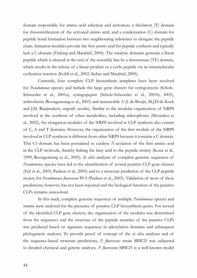
44
domain responsible for amino acid selection and activation, a thiolation (T) domain for thioesterification of the activated amino acid, and a condensation (C) domain for peptide bond formation between two neighbouring substrates to elongate the peptide chain. Initiation modules provide the first amino acid for peptide synthesis and typically lack a C-domain (Finking and Marahiel, 2004). The catalytic domains generate a linear peptide which is cleaved at the end of the assembly line by a thioesterase (TE) domain, which results in the release of a linear product or a cyclic peptide via an intramolecular cyclization reaction (Kohli et al., 2002; Sieber and Marahiel, 2005). Currently, four complete CLP biosynthetic templates have been resolved for Pseudomonas species and include the large gene clusters for syringomycin (Scholz-Schroeder et al., 2001a), syringopeptin (Scholz-Schroeder et al., 2001b; 2003), arthrofactin (Roongsawang et al., 2003) and massetolide A (I. de Bruijn, M.J.D de Kock and J.M. Raaijmakers, unpubl. results). Similar to the modular organization of NRPS involved in the synthesis of other metabolites, including siderophores (Mossialos et al., 2002), the elongation modules of the NRPS involved in CLP synthesis also consist of C, A and T domains. However, the organization of the first module of the NRPS involved in CLP synthesis is different from other NRPS because it contains a C domain. This C1-domain has been postulated to catalyse N-acylation of the first amino acid in the CLP molecule, thereby linking the fatty acid to the peptide moiety (Konz et al., 1999; Roongsawang et al., 2005). In silico analyses of complete genome sequences of Pseudomonas species have led to the identification of several putative CLP gene clusters (Feil et al., 2005; Paulsen et al., 2005) and to a structure prediction of the CLP peptide moiety for Pseudomonas fluorescens Pf-5 (Paulsen et al., 2005). Validation of most of these predictions, however, has not been reported and the biological function of the putative CLPs remains unresolved. In this study, complete genome sequences of multiple Pseudomonas species and strains were analysed for the presence of putative CLP biosynthesis genes. For several of the identified CLP gene clusters, the organization of the modules was determined from the sequences and the structure of the peptide moieties of the putative CLPs was predicted based on signature sequences in adenylation domains and subsequent phylogenetic analyses. To provide proof of concept of the in silico analyses and of the sequence-based structure predictions, P. fluorescens strain SBW25 was subjected to detailed chemical and genetic analyses. P. fluorescens SBW25 is a well-known model
45
strain often used in studies on bacterial evolution, adaptation and plant colonization (Kassen et al., 2004; Rainey and Rainey, 2003). It also received considerable attention as a biocontrol agent of plant diseases (Timms-Wilson et al., 2000), but as yet no metabolites with activity against fungi or oomycetes have been chemically identified for SBW25 (M.J. Bailey, pers. comm.). In this study, site-directed mutagenesis of the in silico identified genes and chemical analyses provided, for the first time, evidence that P. fluorescens SBW25 produces a viscosin-like CLP. The identified CLP was shown to play a role in motility and biofilm formation by SBW25, and to have lytic activity against zoospores of the economically important plant pathogen Phytopthora infestans.
RESULTS AND DISCUSSIONIdentification of CLP gene clusters in Pseudomonas. Complete genome sequences of multiple Pseudomonas species and strains were analysed for the presence of NRPS genes that have two features specific for NRPS genes involved in CLP synthesis. The first feature was the C1-domain, the condensation (C) domain of the first NRPS module that catalyses N-acylation of the first amino acid in the CLP molecule. Phylogenetic analyses revealed that C1-domains of CLP genes in Pseudomonas form a distinct group, distant from C-domains present in other NRPS modules (Fig. S1). The second feature included in the analyses was the TE domain involved in cyclization of the CLP molecule. In Pseudomonas, the CLP gene clusters for syringopeptin, arthrofactin and massetolide A synthesis contain two TE domains in the last NRPS module, whereas the syringomycin template contains one TE domain (Raaijmakers et al., 2006). The BLASTP searches with the C1 and TE domains of the massetolide A gene cluster resulted in the identification of multiple putative CLP gene clusters in several fully sequenced Pseudomonas strains, including Pseudomonas syringae pv. syringae B728a, P. syringae pv. tomato DC3000 and P. fluorescens strains Pf-5, Pf0-1 and SBW25 (Fig. 1; Table S1). For Pseudomonas aeruginosa PAO1, Pseudomonas putida KT2440 and P. syringae pv. phaseolicola 1448A, the identified NRPS genes did not fit all of the criteria used in our analyses, in spite of significant E-values (< e-5) and identity scores (ranging from 27 to 87%). For example, in the BLASTP search with the TE domain, the peptide synthetase gene ppsD of strain KT2440 was identified (4e-14, identity 41%), but in phylogenetic analysis this gene clustered in the branch with NRPS genes involved in siderophore biosynthesis rather than with NRPS genes involved in CLP synthesis (Fig. S2). Also
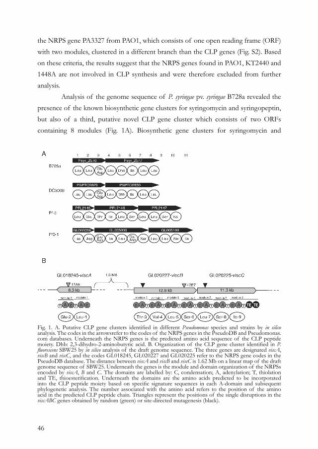
46
the NRPS gene PA3327 from PAO1, which consists of one open reading frame (ORF) with two modules, clustered in a different branch than the CLP genes (Fig. S2). Based on these criteria, the results suggest that the NRPS genes found in PAO1, KT2440 and 1448A are not involved in CLP synthesis and were therefore excluded from further analysis. Analysis of the genome sequence of P. syringae pv. syringae B728a revealed the presence of the known biosynthetic gene clusters for syringomycin and syringopeptin, but also of a third, putative novel CLP gene cluster which consists of two ORFs containing 8 modules (Fig. 1A). Biosynthetic gene clusters for syringomycin and
Fig. 1. A. Putative CLP gene clusters identified in different Pseudomonas species and strains by in silico analysis. The codes in the arrowsrefer to the codes of the NRPS genes in the PseudoDB and Pseudomonas.com databases. Underneath the NRPS genes is the predicted amino acid sequence of the CLP peptide moiety. Dhb: 2,3-dihydro-2-aminobutyric acid. B. Organization of the CLP gene cluster identified in P. fluorescens SBW25 by in silico analysis of the draft genome sequence. The three genes are designated viscA, viscB and viscC, and the codes GL018245, GL020227 and GL020225 refer to the NRPS gene codes in the PseudoDB database. The distance between viscA and viscB and viscC is 1.62 Mb on a linear map of the draft genome sequence of SBW25. Underneath the genes is the module and domain organization of the NRPSs encoded by viscA, B and C. The domains are labelled by: C, condensation; A, adenylation; T, thiolation and TE, thioesterification. Underneath the domains are the amino acids predicted to be incorporated into the CLP peptide moiety based on specific signature sequences in each A-domain and subsequent phylogenetic analysis. The number associated with the amino acid refers to the position of the amino acid in the predicted CLP peptide chain. Triangles represent the positions of the single disruptions in the viscABC genes obtained by random (green) or site-directed mutagenesis (black).
47
syringopeptin synthesis, referred to as the syr–syp genomic island (Scholz-Schroeder et al., 2001b), make up a substantial portion of the total genome (Feil et al., 2005; Scholz-Schroeder et al., 2001b; 2003). Both syringomycin and syringopeptin constitute important virulence factors and pore formation leading to cell lysis is their main mode of action (Bender et al., 1999). Virulence assays with immature cherry fruits indicated that a double mutant deficient in syringomycin and syringopeptin synthesis was reduced in virulence by 76% (Scholz-Schroeder et al., 2001a). Whether the third putative CLP gene cluster identified in this study plays a role in virulence remains to be tested. In P. syringae pv. tomato DC3000, the syr–syp gene cluster was not found, which is consistent with the analyses of Feil et al. (2005). However, also in DC3000 a novel putative CLP gene cluster was identified which consists of two ORFs with 8 modules (Fig. 1A). In B728a, another putative CLP synthetic template, designated GL029167, was found with a truncated orthologue in DC3000, designated PS015499 (Fig. S2). The CLP gene cluster identified for P. fluorescens Pf-5 was identical to that described by Paulsen et al. (2005). For the plant beneficial P. fluorescens strain Pf0-1, a previously unknown CLP gene cluster was found containing three ORFs with 11 modules (Fig. 1A). Analysis of the draft genome sequence of P. fluorescens SBW25 resulted in the identification of a putative CLP gene cluster containing three genes with a total of nine modules (Fig. 1B). Other NRPS genes identified in SBW25 clustered with siderophore synthetase genes but not with CLP genes, despite significant E-values (<e-5) and identity scores (>24%). Similar results were obtained when the C1 and TE domains of the arthrofactin gene cluster (Roongsawang et al., 2003) were used in the in silico analyses. In the putative CLP synthetic template in SBW25, each module consists of C, A and T domains and the ninth module also harbours two TE domains (Fig. 1B). In contrast to CLP gene clusters in Bacillus (Konz et al., 1999; Peypoux et al., 1999; Sieber and Marahiel, 2005), no internal epimerization (E) domains for conversion of amino acids from L- to D-form were found in the newly identified CLP clusters in Pseudomonas (Fig. 1). Also in the known biosynthetic templates for syringomycin (Scholz-Schroeder et al., 2001b), syringopeptin (Scholz-Schroeder et al., 2003) and arthrofactin (Roongsawang et al., 2003), internal E domains were not found in spite of the presence of several D-amino acids in the peptide moieties of these CLPs. Based on additional ATP-PPi exchange assays with L-amino acids, Roongsawang et al. (2003) showed that the D-Leu1 A domain of the arthrofactin synthetic template only adopted L-Leu as a
48
substrate and not D-Leu, suggesting that an external racemase is responsible for the L to D conversion. Although several amino acid racemases have been characterized from various microorganisms, including Pseudomonas (Yoshimura and Esak, 2003; Ju et al., 2005), their role in CLP synthesis awaits further experimental confirmation. For arthrofactin, epimerization was attributed to C domains with dual catalytic activity (Balibar et al., 2005).
Sequence-based structure prediction. For subsequent prediction of the identity of the amino acids in the peptide moiety of the putative CLPs, signature sequences were identified in the adenylation (A) domains using the phenylalanine-activating A domain (PheA) of gramicidin-S-synthetase (GrsA) from Bacillus brevis as the anchor sequence. The results of our analyses confirmed the predictive models of Stachelhaus et al. (1999) and Challis et al. (2000) and revealed that also in Pseudomonas specific signature sequences in A-domains are linked to the incorporation of a particular amino acid in the CLP molecule (Table S2). Combined with phylogenetic analyses of A domains (Fig. 2), the signature sequences enabled prediction of the amino acid composition of the peptide moiety of the putative CLPs in Pseudomonas species (Fig. 1). For one specific signature sequence (DAQVGVVD) present in the third module of the CLP gene cluster of strain B728a and in the third and sixth modules of the CLP gene cluster of strain Pf0-1, no amino acid could be assigned by our analysis. Based on phylogenetic analysis of the A domains, the amino acid that is activated by this particular A domain is most likely Glu or Asp (Fig. 2). Selectivity-conferring codes for the unusual amino acid Dhb were only found in the two pathogenic P. syringae strains, but not in the beneficial P. fluorescens strains (Fig. 1 and Table S2). The predicted amino acid composition of the CLP of P. fluorescens Pf-5 (Fig. 1A) was identical to that predicted by Paulsen et al. (2005). For plant beneficial P. fluorescens strain SBW25, the putative CLP was predicted to consist of a peptide moiety of nine amino acids (Fig. 1B).
Chemical confirmation of genome-based prediction. To provide experimental proof of the genome-based identification and structure prediction, P. fluorescens strain SBW25 was chosen for detailed chemical and genetic analyses. Cell suspensions and cell-free culture supernatants of SBW25 showed a strongly reduced surface tension, indicating the production of extracellular surface active compounds (Fig. 3A). Subsequent extraction
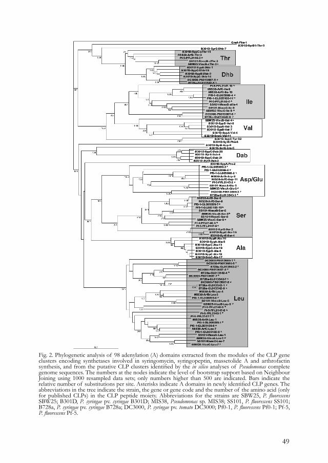
49
Fig. 2. Phylogenetic analysis of 98 adenylation (A) domains extracted from the modules of the CLP gene clusters encoding synthetases involved in syringomycin, syringopeptin, massetolide A and arthrofactin synthesis, and from the putative CLP clusters identified by the in silico analyses of Pseudomonas complete genome sequences. The numbers at the nodes indicate the level of bootstrap support based on Neighbour Joining using 1000 resampled data sets; only numbers higher than 500 are indicated. Bars indicate the relative number of substitutions per site. Asterisks indicate A domains in newly identified CLP genes. The abbreviations in the tree indicate the strain, the gene or gene code and the number of the amino acid (only for published CLPs) in the CLP peptide moiety. Abbreviations for the strains are SBW25, P. fluorescens SBW25; B301D, P. syringae pv. syringae B301D; MIS38, Pseudomonas sp. MIS38; SS101, P. fluorescens SS101; B728a, P. syringae pv. syringae B728a; DC3000, P. syringae pv. tomato DC3000; Pf0-1, P. fluorescens Pf0-1; Pf-5, P. fluorescens Pf-5.
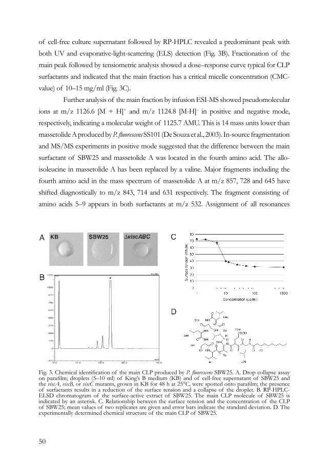
50
of cell-free culture supernatant followed by RP-HPLC revealed a predominant peak with both UV and evaporative-light-scattering (ELS) detection (Fig. 3B). Fractionation of the main peak followed by tensiometric analysis showed a dose–response curve typical for CLP surfactants and indicated that the main fraction has a critical micelle concentration (CMC-value) of 10–15 mg/ml (Fig. 3C). Further analysis of the main fraction by infusion ESI-MS showed pseudomolecular ions at m/z 1126.6 [M + H]+ and m/z 1124.8 [M-H]– in positive and negative mode, respectively, indicating a molecular weight of 1125.7 AMU. This is 14 mass units lower than massetolide A produced by P. fluorescens SS101 (De Souza et al., 2003). In-source fragmentation and MS/MS experiments in positive mode suggested that the difference between the main surfactant of SBW25 and massetolide A was located in the fourth amino acid. The allo-isoleucine in massetolide A has been replaced by a valine. Major fragments including the fourth amino acid in the mass spectrum of massetolide A at m/z 857, 728 and 645 have shifted diagnostically to m/z 843, 714 and 631 respectively. The fragment consisting of amino acids 5–9 appears in both surfactants at m/z 532. Assignment of all resonances
Fig. 3. Chemical identification of the main CLP produced by P. fluorescens SBW25. A. Drop collapse assay on parafilm; droplets (5–10 ml) of King’s B medium (KB) and of cell-free supernatant of SBW25 and the viscA, viscB, or viscC mutants, grown in KB for 48 h at 25°C, were spotted onto parafilm; the presence of surfactants results in a reduction of the surface tension and a collapse of the droplet. B. RP-HPLC-ELSD chromatogram of the surface-active extract of SBW25. The main CLP molecule of SBW25 is indicated by an asterisk. C. Relationship between the surface tension and the concentration of the CLP of SBW25; mean values of two replicates are given and error bars indicate the standard deviation. D. The experimentally determined chemical structure of the main CLP of SBW25.
51
and crosspeaks in the 1D and 2D NMR spectra resulted in a full assignment of the amino acids and the fatty acid tail (Table S3). In the NOESY and HMBC spectra a complete sequential assignment of the amino acids and the fatty acid was possible. A crosspeak in the HMBC spectrum between H3β of threonine and the C = O of isoleucine-9 confirmed the formation of the macrocyclic lactone ring between residues 3 and 9. Collectively, these analyses indicated that the main surface-active compound produced by SBW25 is a CLP with a 10-carbon hydroxy fatty acid tail linked to a 9-amino acid peptide moiety (Fig. 3D). The results show that the experimentally determined structure of the CLP peptide moiety is identical to the genome-based predicted structure (Figs 1B and 3D) and demonstrate that, similar to Streptomyces coelicolor (Challis and Ravel, 2000; Lautru et al., 2005), also for Pseudomonas substrate selectivity of adenylation domains can be predicted accurately. Based on the structural characteristics, the main CLP produced by SBW25 falls within the viscosin group, which comprises CLPs with 9 amino acids linked at the N-terminus to, in most cases, 3-hydroxydecanoic acid (Nybroe and Sorensen, 2004). The fatty acid, amino acid sequence and composition of the CLP of SBW25 are identical to those in viscosin and White Line Inducing Principle (WLIP), two CLPs that differ only in the L-D configuration of Leu-5, the fifth amino acid in the peptide chain (Raaijmakers et al., 2006). Viscosin was first isolated as an antibiotic compound from Pseudomonas viscosa (Kochi et al., 1951). WLIP was described for Pseudomonas ‘reactans’ (Mortishire-Smith et al., 1991) and was shown to inhibit brown blotch disease of mushrooms caused by Pseudomonas tolaasii (Soler-Rivas et al., 1999). MS experiments of the two minor peaks in the SBW25 extract with a retention time of 18.4 min and 20.6 min (Fig. 3B) suggested that they may be very closely related to viscosin and massetolide A (Gerard et al., 1997). The minor representation of these two isoforms in the surface-active extract of SBW25 is most likely the result of the relaxed substrate specificity of certain A domains in amino acid activation (further discussed below).
Genetic confirmation of genome-based prediction. To provide evidence that the three putative CLP genes in SBW25 (Fig. 1B), designated viscA (GL018245), viscB (GL020227) and viscC (GL020225), are indeed required for CLP production, mutants were generated by either random transposon mutagenesis or site-directed mutagenesis. From a random TnMod plasposon mutant library of SBW25, containing 4384 plasposon mutants, three single insertion mutants deficient in CLP synthesis were
52
identified. Sequencing of the disrupted genes showed that in mutants 17A8 and 42E7 the plasposon had integrated in viscA and viscB respectively (Fig. 1B). For the third single insertion mutant, the plasposon was located in gacS (GL018559), the sensor kinase of the Gac two-component regulatory system, a well-known global regulator for the production of antibiotics and extracellular enzymes in pseudomonads (Haas and Defago, 2005). Site-directed mutagenesis was successful for viscB and viscC. All viscA, viscB, viscC mutants and the gacS mutant showed a surfactant-deficient phenotype based on the drop-collapse assay and the lack of CLP production was confirmed by RP-HPLC (data not shown). In these mutants, also the two minor fractions (retention times 18.4 and 20.6 min) were absent, further supporting the hypothesis that these two viscosin isoforms result from the relaxed substrate specificity of A domains rather than being products from a different gene cluster in SBW25. Collectively, these results confirm that each of the three identified visc genes is essential for CLP production and indicate that GacS is a key element in the regulation of CLP synthesis in SBW25, confirming and extending the results obtained for the CLPs amphisin (Koch et al., 2002) and putisolvin (Dubern et al., 2005). The genomic organization of the viscA, viscB and viscC genes is different from other CLP gene clusters described to date. In most CLP gene clusters, including the newly identified templates in other Pseudomonas strains (Fig. 1A), the NRPS genes are physically linked (Raaijmakers et al., 2006). In contrast, viscA is located approximately 1.62 Mb from viscB and viscC (Fig. 1B). Also in the massetolide A gene cluster, massA is disconnected from massB and massC (I. de Bruijn, M.J.D. de Kock and J.M. Raaijmakers, unpubl. results). In spite of the physical disconnection, however, CLP synthesis by the viscA, viscB and viscC genes does obey the colinearity rule. Preliminary analysis indicated that each of the three visc genes contains a putative promoter sequence, suggesting that domain–domain interactions are more important for CLP synthesis than physical linkage and co-ordinated transcription of the genes, as was shown for surfactin synthesis in Bacillus subtilis (Guenzi et al., 1998).
Biological role of CLP synthesis in P. fluorescens SBW25. Several biological roles for CLPs have been proposed, including functions in pathogenicity, antimicrobial activity, regulation of attachment-detachment to and from surfaces, and swarming (Nybroe and Sorensen, 2004; Raaijmakers et al., 2006). For plant pathogenic Pseudomonas
53
species, including P. syringae pv. syringae and pectolytic P. fluorescens, CLPs are important virulence factors (Bender et al., 1999). Their surfactant properties also may play an indirect role in virulence by facilitating colonization of plant tissue and enhancing physical access of cell wall degrading enzymes to the plant surface (Hildebrand et al., 1998; Lindow and Brandl, 2003). For the beneficial Pseudomonas species, a range of functions and activities have been reported (Raaijmakers et al., 2006). For example, viscosin was shown to have antiviral activity (Groupe et al., 1951) and to cause lysis of trypomastigotes of the human pathogen Trypanosoma cruzi, the causal agent of Chagas’ disease (Mercado and Colon-Whitt, 1982). For strain P. fluorescens SBW25, we focused on the role of CLP synthesis in surface motility, biofilm formation and activity against zoospores of P. infestans, the causal agent of late blight of tomato and potato. The results showed that the viscA, viscB and viscC mutants were impaired in surface motility on soft agar medium (Fig. 4A) and in biofilm formation on an artificial surface (Fig. 4B and C).When the CLP of SBW25 was added to the growth medium, biofilm formation by the visc mutants was restored, but more localized at the liquid–solid interface than at the liquid–air interface (Fig. 4B and C). Biofilm formation at the liquid–solid interface was also observed when cultures of wild type SBW25 were amended with the pure CLP (Fig. 4B and C), which may be explained by a more homogeneous distribution of the CLP surfactant when added to the medium as compared with a more localized CLP production by the wild type at the liquid–air interface in non-amended cultures. Attachment and detachment of bacteria to artificial and natural surfaces has been the subject of numerous studies because of the importance of these processes in biofilm formation (Neu, 1996; O’Toole et al., 2000). The results with SBW25 indicate that CLP synthesis is an important determinant of biofilm formation. This is in contrast to results obtained previously for arthrofactin (Roongsawang et al., 2003) and for putisolvins I and II (Kuiper et al., 2004). Mutants deficient in the synthesis of these CLPs produced more, but differently structured biofilms than their wild type strains. The results obtained for SBW25 are more in line with the study of Bais et al. (2004) who showed that the CLP surfactin plays an essential role in adherence of B. subtilis strain 6051 to an artificial surface. Given that surfactin was also involved in biofilm formation by B. subtilis on Arabidopsis roots, Bais et al. (2004) postulated that CLP production may enable bacteria to efficiently colonize plant roots thereby providing protection to their host.
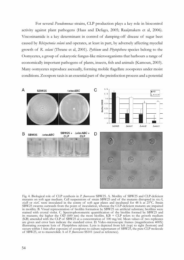
54
For several Pseudomonas strains, CLP production plays a key role in biocontrol activity against plant pathogens (Haas and Defago, 2005; Raaijmakers et al, 2006). Viscosinamide is a key determinant in control of damping-off disease of sugar beet caused by Rhizoctonia solani and operates, at least in part, by adversely affecting mycelial growth of R. solani (Thrane et al, 2001). Pythium and Phytopthora species belong to the Oomycetes, a group of eukaryotic fungus-like microorganisms that harbours a range of economically important pathogens of plants, insects, fish and animals (Kamoun, 2003). Many oomycetes reproduce asexually, forming mobile flagellate zoospores under moist conditions. Zoospore taxis is an essential part of the preinfection process and a potential
Fig. 4. Biological role of CLP synthesis in P. fluorescens SBW25. A. Motility of SBW25 and CLP-deficient mutants on soft agar medium. Cell suspensions of strain SBW25 and of the mutants disrupted in viscA, viscB or viscC were inoculated in the centre of soft agar plates and incubated for 48 h at 25°C. Strain SBW25 swarms outwards from the point of inoculation, whereas the CLP-deficient mutants are impaired in motility. B. Visual representation of biofilm formation by SBW25 on artificial substrate; biofilms were stained with crystal violet. C. Spectrophotometric quantification of the biofilm formed by SBW25 and its mutants; the higher the OD (600 nm) the more biofilm. KB + CLP refers to the growth medium (KB) amended with the CLP of SBW25 at a concentration of 100 mg/ml. Mean values of two replicates are given and error bars indicate the standard error. D. Video-microscopic frames (magnification 400X) illustrating zoospore lysis of Phytophthora infestans. Lysis is depicted from left (top) to right (bottom) and occurs within 1 min after exposure of zoospores to culture supernatant of SBW25, the pure CLP molecule of SBW25, or to massetolide A of P. fluorescens SS101 (used as reference).
C
55
target for controlling diseases caused by this group of pathogens. For P. fluorescens SBW25, the assays with P. infestans showed that cell suspensions and cell-free culture supernatant of SBW25, and the purified CLP cause immobilization and subsequent lysis of entire zoospore populations within 1 min (Fig. 4D; Movie S1), whereas cell-free culture supernatant of the visc mutants did not cause these responses. Immobilization and rounding-off of the zoospores (Fig. 4D) occurred at a CLP concentration of 25 mg/ml, whereas lysis occurred at CLP concentrations of 50 mg/ml and higher. Because these concentrations are above the CMC-value, the zoosporicidal activity is most likely due to solubilization of the zoospore membranes. Identical responses of zoospores have been described for massetolide A of P. fluorescens SS101 (De Souza et al., 2003) and for rhamnolipids produced by strains of P. aeruginosa (Stanghellini and Miller, 1997; Kim et al. 2000). Also for the human pathogen T. cruzi, similar microscopic observations have been made (Mercado and Colon-Whitt, 1982). In their study, the viscosin-producing P. fluorescens isolate approached a single flagellate of T. cruzi at the posterior end, the site of the flagellar origin, resulting in immobilization. Observations further revealed rounding-off of the parasite with the flagellum coiled around the cell body followed by disintegration. The fact that the closely related CLP viscosinamide has no zoosporicidal activity but instead induces encystment of zoospores of Pythium (Thrane et al., 2000), suggests that not only the reduction of surface tension is essential but also the chemical nature of the CLP peptide moiety.
CONCLUSIONThe results of this study have demonstrated that microbial genomes provide a valuable source for the discovery of previously unknown biosynthetic gene clusters encoding traits that are highly relevant in the biology of the organism. Discovery of unknown antibiotic compounds by genome analyses has also been demonstrated for Streptomyces species (Challis and Ravel, 2000; Lautru et al., 2005; McAlpine et al., 2005) and shows that genomics is a valuable complementary tool in resolving complex structures of novel compounds. The integration of different disciplines and approaches used in this study, ranging from in silico and phylogenetic analyses, to chemistry, genetics and microbial ecology, established a link between genome sequence, chemical structure and biological function of the first identified antimicrobial metabolite and biosurfactant in P. fluorescens SBW25. How the identified viscosin-like CLP interacts with other traits involved in

56
biofilm formation by SBW25, including lipopolysaccharides and acetylated cellulose (Spiers and Rainey, 2005), is yet unknown. This study also enables the identification of regulatory networks involved in CLP biosynthesis and offers new opportunities to assess the role of CLP synthesis in adaptation, plant colonization and biocontrol activity of beneficial P. fluorescens strain SBW25.
MATERIALS AND METHODSIdentification and phylogeny of NRPS genes and domains. For identification of putative NRPS genes involved in CLP synthesis, the C1-domain of massA (GenBank DQ835588) as well as the first TE domain of the ninth module of massC (GenBank DQ835590) were used in BLASTP comparisons with whole genome sequences of Pseudomonas spp. available in the databases ‘Pseudomonas.com’ (http://v2.Pseudomonas.com) and ‘PseudoDB’ (http://pseudo.bham.ac.uk). Adenylation (A), thiolation (T), condensation (C) and thioesterase (TE) domains of the NRPS genes were identified by PFAM (http://www.sanger.ac.uk/Software/Pfam/). For phylogenetic analyses, alignments were made with CLUSTALX (version 1.81) and software available at http://www.ebi.ac.uk/clustalw/. Trees were inferred by Neighbour Joining in CLUSTALX using 1000 bootstrap replicates.
Extraction and chemical identification of the CLP produced by P. fluorescens SBW25. Strain SBW25 was grown on King’s medium B (KB) for 48 h at 25°C and the CLP was extracted according to the method described by De Souza et al. (2003) and Gerard et al. (1997). Chemical identification was performed according to methods described by De Souza et al. (2003) and the CLP massetolide A was used as a reference. For details on reagents, equipment and procedures used, we refer to the Supporting Information. Surface tension measurements were carried out with a K6 tensiometer (Krüss GmbH, Hamburg, Germany) and were performed at 25°C.
Mutagenesis of P. fluorescens SBW25. A spontaneous rifampicin-resistant derivative of strain SBW25 was subjected to random plasposon mutagenesis with plasmid pTnModOkm (Dennis and Zylstra, 1998). Transformants were screened for single insertions by Southern blot analysis. Single insertion mutants were screened for surfactant production by resuspending cells from agar plate cultures in sterile demineralized water to a density of approximately 109 cells/ml followed by a drop collapse assay on parafilm (Fig. 3A). Plasmid rescue and sequence analysis of the regions flanking the plasposon was performed according to the methods described by Dennis and Zylstra (1998). Site-directed mutagenesis of the visc genes was performed with the pKnockout-G suicide vector (Windgassen et al., 2000). Fragments of the genes of interest were amplified by polymerase chain reaction (PCR) with the primers 5’CCACCTTGCTCAACCACGAA3’(viscB forward primer), 5’ATCACCTGGCGCAGGAATG3’ (viscB reverse primer), 5’CAGGCCGTTCCGTTTCGCAA3‘ (viscC forward primer), 5’TGATCACCGGCACATCCAGG3’ (viscC reverse primer) and cloned into pGEM-T Easy (Promega). Inserts were subcloned by ApaI/SacI digestions into pKnockout-G and transferred into SBW25 by triparental mating. Integrations in the target gene were verified by PCR.
Motility, biofilm and zoospore assays. Motility of wild type strain SBW25 and the CLP-deficient mutants was assessed on soft (0.6% agar, w/v) standard succinate medium (SSM). Overnight cultures of SBW25 and the CLP-deficient mutants were washed once and 5 ml of a cell suspension (1*1010 cells/ml) were spotted in the centre of the soft SSM agar plates and incubated for 48 h at 25°C. Biofilm formation was assessed according to the method described by O’Toole et al. (1999) using flat-bottom 96-wells plates (Greiner) with 200 ml of King’s medium B broth per well. For obtaining zoospores, P. infestans strain PIC97757 was grown on Rye Sucrose Agar for 10 days at 18°C. Plates were flooded with sterile distilled water and incubated at 4°C; after 4 h, zoospores were collected and exposed to cell-free supernatant of SBW25, viscA, viscB and visC mutants or to a concentration series of the purified CLP of SBW25. Zoospore behaviour was observed microscopically. Massetolide A producing strain P. fluorescens SS101 (De Souza et al., 2003) was used as a reference.
ACKNOWLEDGEMENTSWe are grateful to Marc Lemmers and Elbert van der Klift for their assistance in RP-HPLC and MS, to Andrea Ficke for video-microscopy of zoospores, to Martijn Staats en Peter van Baarlen for their advice
57
on the phylogenetic analyses, and to Mark Bailey, Tracey Timms-Wilson and Andrew Spiers for fruitful discussions and strain SBW25. We thank Wilbert Flier (PRI, Wageningen) for P. infestans strain PIC97757 and are grateful to Francine Govers for critically reviewing the manuscript and her valuable suggestions. This work was funded in part by the Royal Dutch Academy of Arts and Sciences, and by the Dutch Technology Foundation (STW), the applied science division of NWO.
REFERENCESBais, H.P., Fall, R., and Vivanco, J.M. 2004.Biocontrol of Bacillus subtilis against infection of Arabidopsis roots by Pseudomonas syringae is facilitated by biofilm formation and surfactin production. Plant Physiol. 134: 307–319.Balibar, C.J., Vaillancourt, F.H., and Walsh, C.T. 2005. Generation of D amino acid residues in assembly of arthrofactin by dual condensation/epimerization domains. Chem. Biol. 12: 1189–1200.Bender, C.L., Alarcon-Chaidez, F., and Gross, D.C. 1999. Pseudomonas syringae phytotoxins: mode of action, regulation, and biosynthesis by peptide and polyketide synthetases. Microbiol. Mol. Biol. Rev. 63: 266–292.Challis, G.L., and Ravel, J. 2000. Coelichelin, a new peptide siderophore encoded by the Streptomyces coelicolor genome: structure prediction from the sequence of its nonribosomal peptide synthetase. FEMS Microbiol. Lett. 187: 111–114.Challis, G.L., Ravel, J., and Townsend, C.A. 2000. Predictive, structure-based model of amino acid recognition by nonribosomal peptide synthetase adenylation domains. Chem. Biol. 7: 211–224.De Souza, J.T., De Boer, M., De Waard, P., Van Beek, T.A., and Raaijmakers, J.M. 2003. Biochemical, genetic, and zoosporicidal properties of cyclic lipopeptide surfactants produced by Pseudomonas fluorescens. Appl. Environ. Microbiol. 69: 7161–7172.Dennis, J.J., and Zylstra, G.J. 1998. Plasposons: modular self-cloning minitransposon derivatives for rapid genetic analysis of Gram-negative bacterial genomes. Appl. Environ. Microbiol. 64: 2710–2715.Dubern, J.F., Lagendijk, E.L., Lugtenberg, B.J., and Bloemberg, G.V. 2005. The heat shock genes dnaK, dnaJ, and grpE are involved in regulation of putisolvin biosynthesis in Pseudomonas putida PCL1445. J. Bacteriol. 187: 5967–5976.Feil, H., Feil, W. S., Chain, P., Larimer, F., Dibartolo, G., Copeland, A., Lykidis, A., Trong, S., Nolan, M., Goltsman, E., Thiel, J., Malfatti, S., Loper, J. E., Lapidus, A., Detter, J. C., Land, M., Richardson, P. M., Kyrpides, N. C., Ivanova, N., and Lindow, S. E. 2005. Comparison of the complete genome sequences of Pseudomonas syringae pv. syringae B728a and pv. tomato DC3000. Proc. Natl. Acad. Sci. USA 102:11064-11069. Finking, R., and Marahiel, M.A. 2004. Biosynthesis of nonribosomal peptides. Annu. Rev. Microbiol 58: 453–488. Gerard, J., Lloyd, R., Barsby, T., Haden,
P., Kelly, M.T., and Andersen, R.J. 1997. Massetolides A-H, antimycobacterial cyclic depsipeptides produced by two pseudomonads isolated from marine habitats. J. Nat. Prod. 60: 223–229.Gewolb, J. 2002. Bioengineering. Working outside the protein-synthesis rules. Science 295: 2205–2207.Groupe, V., Pugh, L.H., Weiss, D., and Kochi, M. 1951. Observations on antiviral activity of viscosin. Proc. Soc. Exp. Biol. Med. 78: 354–358.Guenzi, E., Galli, G., Grgurina, I., Pace, E., Ferranti, P., and Grandi, G. 1998. Coordinate transcription and physical linkage of domains in surfactin synthetase are not essential for proper assembly and activity of the multienzyme complex. J. Biol. Chem. 273: 14403–14410.Haas, D., and Defago, G. 2005. Biological control of soilborne pathogens by fluorescent pseudomonads. Nat. Rev. Microbiol. 3: 307–319.Hildebrand, P.D., Braun, P.G., McRae, K.B., and Lu, X. 1998. Role of biosurfactant viscosin in broccoli head rot caused by a pectolytic strain of Pseudomonas fluorescens. Can. J. Plant Pathol. 20: 296–303.Jorgensen, R., Merrill, A.R., Yates, S.P., Marquez, V.E., Schwan, A.L., Boesen, T., and Andersen, G.R. 2005. Exotoxin A-eEF2 complex structure indicates ADP ribosylation by ribosome mimicry. Nature 436: 979–984. Ju, J., Yokoigawa, K., Misono, H., and Ohnishi, K. 2005. Cloning of alanine racemase genes from Pseudomonas fluorescens strains and oligomerization states of gene products expressed in Escherichia coli. J. Biosci. Bioeng. 100: 409–417.Kamoun, S. 2003. Molecular genetics of pathogenic oomycetes. Eukaryot. Cell 2: 191–199.Kassen, R., Llewellyn, M., and Rainey, P.B. 2004. Ecological constraints on diversification in a model adaptive radiation. Nature 431: 984–988.Kim, B.S., Lee, J.Y., and Hwang, B.K. 2000. In vivo control and in vitro antifungal activity of rhamnolipid B, a glycilipid antibiotic, against Phytopthora capsici and Colletotrichum orbiculare. Pest Manag. Sci. 56: 1029–1035.Koch, B., Nielsen, T. H., Sorensen, D., Andersen, J. B., Christophersen, C., Molin, S., Givskov, M., Sorensen, J., and Nybroe, O. 2002. Lipopeptide production in Pseudomonas sp. strain DSS73 is regulated by components of sugar beet seed exudate via the Gac two-component regulatory system. Appl. Environ. Microbiol. 68:4509-4516.Kochi, M., Weiss, D.W., Pugh, L.H., and Groupé, V. 1951. Viscosin, a new antibiotic.
58
Bacteriol. Proc. 1: 29–30. Kohli, R.M., Walsh, C.T., and Burkart, M.D. 2002. Biomimetic synthesis and optimization of cyclic peptide antibiotics. Nature 418: 658–661.Konz, D., Doekel, S., and Marahiel, M.A. 1999. Molecular and biochemical characterization of the protein template controlling biosynthesis of the lipopeptide lichenysin. J. Bacteriol. 181: 133–140.Kuiper, I., Lagendijk, E. L., Pickford, R., Derrick, J. P., Lamers, G. E., Thomas-Oates, J. E., Lugtenberg, B. J., and Bloemberg, G. V. 2004. Characterization of two Pseudomonas putida lipopeptide biosurfactants, putisolvin I and II, which inhibit biofilm formation and break down existing biofilms. Mol. Microbiol. 51:97-113.Lautru, S., Deeth, R.J., Bailey, L.M., and Challis, G.L. 2005. Discovery of a new peptide natural product by Streptomyces coelicolor genome mining. Nat. Chem. Biol. 1: 265–269.Lindow, S.E., and Brandl, M.T. 2003. Microbiology of the phyllosphere. Appl. Environ. Microbiol. 69: 1875–1883. McAlpine, J.B., Bachmann, B.O., Piraee, M., Tremblay, S., Alarco, A.M., Zazopoulos, E., and Farnet, C.M. 2005. Microbial genomics as a guide to drug discovery and structural elucidation: ECO-02301, a novel antifungal agent, as an example. J. Nat. Prod. 68: 493–496.Mercado, T.I., and Colon-Whitt, A. 1982. Lysis of Trypanosoma cruzi by Pseudomonas fluorescens. Antimicrob. Agents Chemother. 22: 1051–1057.Mortishire-Smith, R.J., Drake, A.F., Nutkins, J.C., and Williams, D.H. 1991. Left handed alpha-helix formation by a bacterial peptide. FEBS Lett. 278: 244–246.Mossialos, D., Ochsner, U., Baysse, C., Chablain, P., Pirnay, J.P., Koedam, N., Budzikiewicz H, Fernández DU, Schäfer M, Ravel J, and Cornelis P. 2002. Identification of new, conserved, nonribosomal peptide synthetases from fluorescent pseudomonads involved in the biosynthesis of the siderophore pyoverdine. Mol. Microbiol. 45: 1673–1685.Neu, T.R. 1996. Significance of bacterial surface-active compounds in interaction of bacteria with interfaces. Microbiol. Rev. 60: 151–166.Nybroe, O., and Sorensen, J. 2004. Production of cyclic lipopeptides by fluorescent pseudomonads. In Pseudomonas, Biosynthesis of Macromolecules and Molecular Metabolism. Ramos, J.-L. (ed.). New York: Kluwer Academic/Plenum Publishers, pp. 147–172.O’Toole, G.A., Pratt, L.A., Watnick, P.I., Newman, D.K., Weaver, V.B., and Kolter, R. 1999. Genetic approaches to study of biofilms. Methods Enzymol. 310: 91–109. O’Toole, G., Kaplan, H.B., and Kolter, R. 2000. Biofilm formation as microbial development. Annu. Rev. Microbiol. 54: 49–79.Paulsen, I. T., Press, C. M., Ravel, J., Kobayashi, D. Y., Myers, G. S. A., Mavrodi, D. V., DeBoy, R. T., Seshadri, R., Ren, Q. H., Madupu, R., Dodson, R. J., Durkin, A. S., Brinkac, L. M., Daugherty, S. C., Sullivan,
S. A., Rosovitz, M. J., Gwinn, M. L., Zhou, L. W., Schneider, D. J., Cartinhour, S. W., Nelson, W. C., Weidman, J., Watkins, K., Tran, K., Khouri, H., Pierson, E. A., Pierson, L. S., Thomashow, L. S., and Loper, J. E. 2005. Complete genome sequence of the plant commensal Pseudomonas fluorescens Pf-5. Nat. Biotechnol. 23:873-878.Peypoux, F., Bonmatin, J.M., and Wallach, J. 1999. Recent trends in the biochemistry of surfactin. Appl. Microbiol. Biotechnol. 51: 553–563.Raaijmakers, J.M., De Bruijn, I., and De Kock, M.J.D. 2006. Cyclic lipopeptide production by plant-associated Pseudomonas spp.: diversity, activity, biosynthesis, and regulation. Mol. Plant-Microbe Interact. 19: 699–710.Rainey, P.B., and Rainey, K. 2003. Evolution of cooperation and conflict in experimental bacterial populations. Nature 425: 72–74.Raskin, D.M., Seshadri, R., Pukatzki, S.U., and Mekalanos, J.J. 2006. Bacterial genomics and pathogen evolution. Cell 124: 703–714.Roongsawang, N., Hase, K., Haruki, M., Imanaka, T., Morikawa, M., and Kanaya, S. 2003. Cloning and characterization of the gene cluster encoding arthrofactin synthetase from Pseudomonas sp. MIS38. Chem. Biol. 10: 869–880.Roongsawang, N., Ping Lim, S., Washio, K., Takano, K., Kanaya, S., and Morikwa, M. 2005. Phylogenetic analysis of condensation domains in the nonribosomal peptide synthetases. FEMS Microbiol. Lett. 252: 143–151.Scholz-Schroeder, B.K., Hutchison, M.L., Grgurina, I., and Gross, D.C. 2001a. The contribution of syringopeptin and syringomycin to virulence of Pseudomonas syringae pv. syringae strain B301D on the basis of sypA and syrB1 biosynthesis mutant analysis. Mol. Plant-Microbe Interact. 14: 336–348.Scholz-Schroeder, B.K., Soule, J.D., Lu, S.E., Grgurina, I., and Gross, D.C. 2001b. A physical map of the syringomycin and syringopeptin gene clusters localized to an approximately 145-kb DNA region of Pseudomonas syringae pv. syringae strain B301D. Mol. Plant-Microbe Interact. 14: 1426–1435.Scholz-Schroeder, B.K., Soule, J.D., and Gross, D.C. 2003. The sypA, sypB, and sypC synthetase genes encode twenty-two modules involved in the nonribosomal peptide synthesis of syringopeptin by Pseudomonas syringae pv. syringae B301D. Mol. Plant-Microbe Interact. 16: 271–280.Sieber, S.A., and Marahiel, M.A. 2005. Molecular mechanisms underlying nonribosomal peptide synthesis: approaches to new antibiotics. Chem. Rev. 105: 715–738. Soler-Rivas, C., Jolivet, S., Arpin, N., Olivier, J.M., and Wichers, H.J. 1999. Biochemical and physiological aspects of brown blotch disease of Agaricus bisporus. FEMS Microbiol. Rev. 23: 591–614.Spiers, A.J., and Rainey, P.B. 2005. The Pseudomonas fluorescens SBW25 wrinkly spreader biofilm requires attachment factor, cellulose fibre
59
and LPS interactions to maintain strength and integrity. Microbiology 151: 2829–2839.Stachelhaus, T., Mootz, H.D., and Marahiel, M.A. 1999. The specificity-conferring code of adenylation domains in nonribosomal peptide synthetases. Chem. Biol. 6: 493–505. Stanghellini, M.E., and Miller, R.M. 1997. Biosurfactants. Their identity and potential efficacy in the biological control of zoosporic plant pathogens. Plant Dis. 81: 4–12.Thrane, C., Harder Nielsen, T., Neiendam Nielsen, M., Sorensen, J., and Olsson, S. 2000. Viscosinamide-producing Pseudomonas fluorescens DR54 exerts a biocontrol effect on Pythium ultimum in sugar beet rhizosphere. FEMS Microbiol. Ecol. 33: 139–146.Thrane, C., Nielsen, M.N., Sorensen, J., and Olsson, S. 2001. Pseudomonas fluorescens
DR54 reduces sclerotia formation, biomass development, and disease incidence of Rhizoctonia solani causing damping-off in sugar beet. Microb. Ecol. 42: 438–445.Timms-Wilson, T.M., Ellis, R.J., Renwick, A., Rhodes, D.J., Mavrodi, D.V., Weller, D.M., Thomashow LS, and Bailey MJ 2000. Chromosomal insertion of phenazine-1-carboxylic acid biosynthetic pathway enhances efficacy of damping-off disease control by Pseudomonas fluorescens. Mol. Plant-Microbe Interact.13: 1293–1300.Windgassen, M., Urban, A., and Jaeger, K.E. 2000. Rapid gene inactivation in Pseudomonas aeruginosa. FEMS Microbiol. Lett. 193: 201–205.Yoshimura, T., and Esak, N. 2003. Amino acid racemases: functions and mechanisms. J. Biosci. Bioeng. 96: 103–109.

60
Fig. S1. Phylogenetic analysis of 97 condensation (C) domains identified in the CLPclusters encoding for synthetases involved in syringomycin, syringopeptin, massetolide A, arthrofactinproduction and in the putative CLP clusters identified in this study by in silico analyses ofPseudomonas genome sequences. The numbers at the nodes indicate the level of bootstrap supportbased on Neighbour Joining analysis of 1000 resampled data sets; only numbers higher than 500 areindicated. Bars indicate the relative number of substitutions per site. The abbreviations in the treeindicate the strain, the gene, and the C domain number (i.e. the number refers to the module in theCLP cluster). Asterisks indicate C domains in newly identified CLP genes. C-1 domains are boxed.The gene codes correspond to the codes given in the databases PseudoDB or Pseudomonas.com.
SUPPLEMENTAL INFORMATION
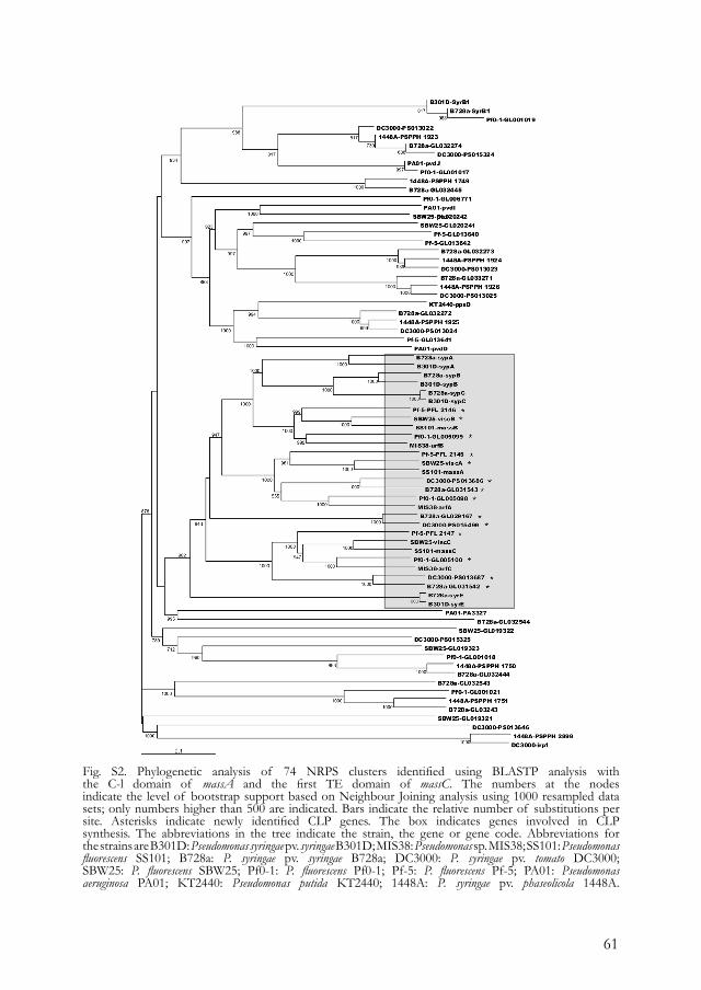
61
Fig. S2. Phylogenetic analysis of 74 NRPS clusters identified using BLASTP analysis with the C-l domain of massA and the first TE domain of massC. The numbers at the nodesindicate the level of bootstrap support based on Neighbour Joining analysis using 1000 resampled datasets; only numbers higher than 500 are indicated. Bars indicate the relative number of substitutions persite. Asterisks indicate newly identified CLP genes. The box indicates genes involved in CLPsynthesis. The abbreviations in the tree indicate the strain, the gene or gene code. Abbreviations forthe strains are B301D: Pseudomonas syringae pv. syringae B301D; MIS38: Pseudomonas sp. MIS38; SS101: Pseudomonasfluorescens SS101; B728a: P. syringae pv. syringae B728a; DC3000: P. syringae pv. tomato DC3000;SBW25: P. fluorescens SBW25; Pf0-1: P. fluorescens Pf0-1; Pf-5: P. fluorescens Pf-5; PA01: Pseudomonasaeruginosa PA01; KT2440: Pseudomonas putida KT2440; 1448A: P. syringae pv. phaseolicola 1448A.
62
Table S1. Codes of the putative CLP genes identified by in silico analysis of the complete genomes of Pseudomonas species. The gene codes refer to the codes given in the databases Pseudomonas.com and PseudoDB. Abbreviation for the strains are SBW25: Pseudomonas fluorescens SBW25; Pf0-1: P. fluorescens Pf0-1; Pf-5: P. fluorescens Pf-5; DC3000: Pseudomonas syringae pv. tomato DC3000; B728a: P. syringae pv. syringae B728a.
Table S2. The selectivity-conferring codes of adenylation domains in known CLP biosynthetic templates of Pseudomonas spp. Signature sequences were identified in the adenylation domains for syringomycin (SyrE, SyrB1), syringopeptin (SypA, SypB , SypC), arthrofactin (Arfa, ArfB, ArfC) and massetolide A (MassA, MassB , MassC). The phenylalanine-activating adenylation domain (PheA) of the gramicidine S synthetase (GrsA) was used as the anchor sequince in the alignment.
Movie S1. Real time microscopic movie (magnification 100¥) that illustrates the typical behaviour of zoospores when exposed to a cell suspension of P. fluorescens SBW25, cell-free culture supernatant of SBW25, the CLP compound of SBW25, or massetolide A of P. fluorescens SS101 (shown here as a representative example). Within 1 min zoospores become immobilized, round-off and fully disintegrate.
This material is available as part of the online article from http://www.blackwell-synergy.com
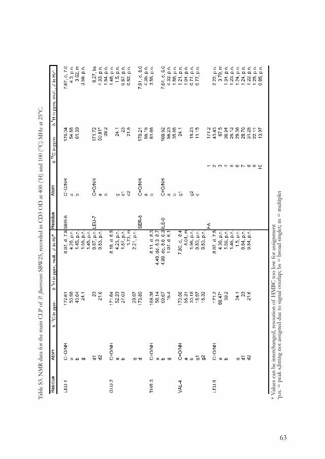
63
Tabl
e S3
. NM
R da
ta fo
r the
mai
n C
LP o
f P.
fluo
rescen
s SBW
25, r
ecor
ded
in C
D3
OD
at 4
00 (1 H
) and
100
(13C
) MH
z at
25°
C.
* Va
lues
can
be
inte
rcha
nged
, res
outio
n of
HM
BC to
o lo
w fo
r ass
ignm
ent
a p.n
. = p
eak
s;litt
ing
not a
ssig
ned
due
to si
gnal
ove
rlap;
bs =
bro
ad si
ngle
t; m
= m
ultip
let

64
65
CHAPTER 3
Journal of Bacteriology (2008) 190(8), 2777-2789 DOI. 10.1128/JB.01563-07
Massetolide A biosynthesis in Pseudomonas fl uorescens.
Irene de Bruijn,1 Maarten J. D. de Kock,1 Pieter de Waard,2 Teris A. van Beek3, and Jos M. Raaijmakers1
1Laboratory of Phytopathology, Wageningen University, 6709 PD Wageningen, the Netherlands.2Wageningen NMR Centre, Wageningen University, 6700 ET Wageningen, the Netherlands.3Natural Products Chemistry Group, Laboratory of Organic Chemistry, Wageningen University, the Netherlands.
Accepted 29 October 2007.

66
ABSTRACTMassetolide A is a cyclic lipopeptide (CLP) antibiotic produced by various Pseudomonas strains from diverse environments. Cloning, sequencing, site-directed mutagenesis and complementation showed that massetolide A biosynthesis in P. fluorescens SS101 is governed by three nonribosomal peptide synthetase (NRPS) genes, designated massA, massB and massC, spanning approximately 30 kb. Prediction of the nature and configuration of the amino acids by in silico analysis of adenylation and condensation domains of the NRPSs was consistent with the chemically determined structure of the peptide moiety of massetolide A. Structural analysis of massetolide A derivatives produced by SS101 indicated that most of the variations in the peptide moiety occur at amino acid positions 4 and 9. Regions flanking the mass genes contained several genes found in other Pseudomonas CLP biosynthesis clusters, which encode LuxR-type transcriptional regulators, ABC transporters and an RND-like outer membrane protein. In contrast to most Pseudomonas CLP gene clusters known to date, the mass genes are not physically linked but are organized in two separate clusters with massA disconnected from massB and massC. Quantitative real time PCR analysis indicated that transcription of massC is strongly reduced when massB is mutated, suggesting that these two genes function in an operon, whereas transcription of massA is independent of massBC and vice versa. Massetolide A is produced in the early exponential growth phase and biosynthesis appears not to be regulated by N-acylhomoserine lactone-based quorum sensing. Massetolide A production is essential in swarming motility of P. fluorescens SS101 and plays an important role in biofilm formation.
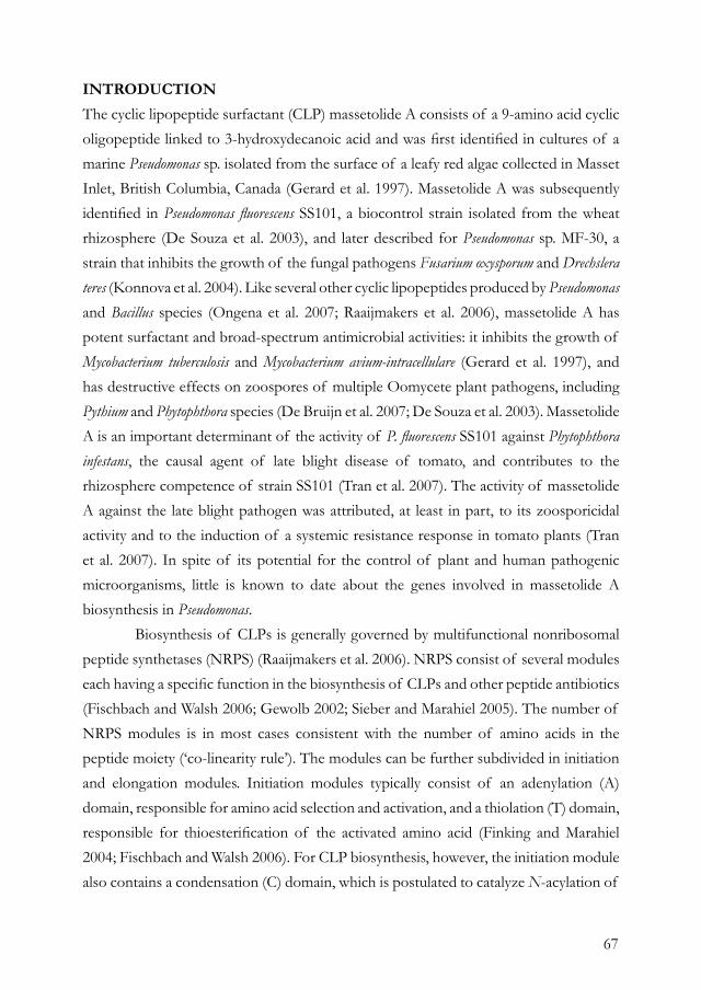
67
INTRODUCTIONThe cyclic lipopeptide surfactant (CLP) massetolide A consists of a 9-amino acid cyclic oligopeptide linked to 3-hydroxydecanoic acid and was first identified in cultures of a marine Pseudomonas sp. isolated from the surface of a leafy red algae collected in Masset Inlet, British Columbia, Canada (Gerard et al. 1997). Massetolide A was subsequently identified in Pseudomonas fluorescens SS101, a biocontrol strain isolated from the wheat rhizosphere (De Souza et al. 2003), and later described for Pseudomonas sp. MF-30, a strain that inhibits the growth of the fungal pathogens Fusarium oxysporum and Drechslera teres (Konnova et al. 2004). Like several other cyclic lipopeptides produced by Pseudomonas and Bacillus species (Ongena et al. 2007; Raaijmakers et al. 2006), massetolide A has potent surfactant and broad-spectrum antimicrobial activities: it inhibits the growth of Mycobacterium tuberculosis and Mycobacterium avium-intracellulare (Gerard et al. 1997), and has destructive effects on zoospores of multiple Oomycete plant pathogens, including Pythium and Phytophthora species (De Bruijn et al. 2007; De Souza et al. 2003). Massetolide A is an important determinant of the activity of P. fluorescens SS101 against Phytophthora infestans, the causal agent of late blight disease of tomato, and contributes to the rhizosphere competence of strain SS101 (Tran et al. 2007). The activity of massetolide A against the late blight pathogen was attributed, at least in part, to its zoosporicidal activity and to the induction of a systemic resistance response in tomato plants (Tran et al. 2007). In spite of its potential for the control of plant and human pathogenic microorganisms, little is known to date about the genes involved in massetolide A biosynthesis in Pseudomonas. Biosynthesis of CLPs is generally governed by multifunctional nonribosomal peptide synthetases (NRPS) (Raaijmakers et al. 2006). NRPS consist of several modules each having a specific function in the biosynthesis of CLPs and other peptide antibiotics (Fischbach and Walsh 2006; Gewolb 2002; Sieber and Marahiel 2005). The number of NRPS modules is in most cases consistent with the number of amino acids in the peptide moiety (‘co-linearity rule’). The modules can be further subdivided in initiation and elongation modules. Initiation modules typically consist of an adenylation (A) domain, responsible for amino acid selection and activation, and a thiolation (T) domain, responsible for thioesterification of the activated amino acid (Finking and Marahiel 2004; Fischbach and Walsh 2006). For CLP biosynthesis, however, the initiation module also contains a condensation (C) domain, which is postulated to catalyze N-acylation of
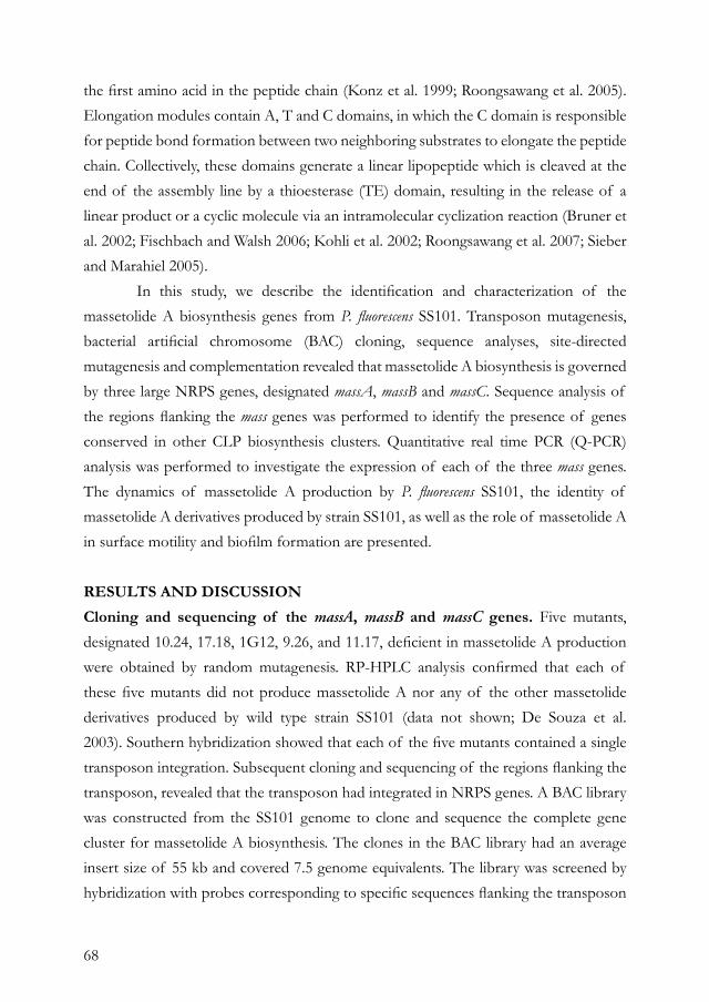
68
the first amino acid in the peptide chain (Konz et al. 1999; Roongsawang et al. 2005). Elongation modules contain A, T and C domains, in which the C domain is responsible for peptide bond formation between two neighboring substrates to elongate the peptide chain. Collectively, these domains generate a linear lipopeptide which is cleaved at the end of the assembly line by a thioesterase (TE) domain, resulting in the release of a linear product or a cyclic molecule via an intramolecular cyclization reaction (Bruner et al. 2002; Fischbach and Walsh 2006; Kohli et al. 2002; Roongsawang et al. 2007; Sieber and Marahiel 2005). In this study, we describe the identification and characterization of the massetolide A biosynthesis genes from P. fluorescens SS101. Transposon mutagenesis, bacterial artificial chromosome (BAC) cloning, sequence analyses, site-directed mutagenesis and complementation revealed that massetolide A biosynthesis is governed by three large NRPS genes, designated massA, massB and massC. Sequence analysis of the regions flanking the mass genes was performed to identify the presence of genes conserved in other CLP biosynthesis clusters. Quantitative real time PCR (Q-PCR) analysis was performed to investigate the expression of each of the three mass genes. The dynamics of massetolide A production by P. fluorescens SS101, the identity of massetolide A derivatives produced by strain SS101, as well as the role of massetolide A in surface motility and biofilm formation are presented.
RESULTS AND DISCUSSIONCloning and sequencing of the massA, massB and massC genes. Five mutants, designated 10.24, 17.18, 1G12, 9.26, and 11.17, deficient in massetolide A production were obtained by random mutagenesis. RP-HPLC analysis confirmed that each of these five mutants did not produce massetolide A nor any of the other massetolide derivatives produced by wild type strain SS101 (data not shown; De Souza et al. 2003). Southern hybridization showed that each of the five mutants contained a single transposon integration. Subsequent cloning and sequencing of the regions flanking the transposon, revealed that the transposon had integrated in NRPS genes. A BAC library was constructed from the SS101 genome to clone and sequence the complete gene cluster for massetolide A biosynthesis. The clones in the BAC library had an average insert size of 55 kb and covered 7.5 genome equivalents. The library was screened by hybridization with probes corresponding to specific sequences flanking the transposon
69
insertions, resulting in seven positive BAC clones (Fig. 1A). Based on restriction analysis, hybridization patterns and BAC-end sequencing, the seven clones aligned in two contigs, of which clones 2H12 and 7B4 were completely sequenced (Fig. 1A). On the first contig (64 kb), one large open reading frame (ORF) of 6270 bp, designated massA, was identified and predicted to encode an NRPS of 2089 amino acids (aa) with a mass of 230 kDa. On the second contig (80 kb), two large ORFs were identified: one ORF of 12924 bp, designated massB, was predicted to encode an NRPS protein of 4307 aa with a mass of 469 kDa; the second ORF of 11328 bp, designated massC, was predicted to encode an NRPS protein of 3775 aa with a mass of 410 kDa. No overlap was found between the two contigs, indicating that massA is disconnected from massB and massC (Fig. 1A).
Fig. 1. (A) Representation of the contig assembly of BAC clones 2F11, 3G1, 2H12, 7F11, 6F12, 8G2, and 7B4. The first contig (64 kb) harbors the massA gene, and the second contig (80 kb) contains the massB and massC genes. (B) Organization of the CLP gene cluster identified in P. fluorescens SS101 by sequence analysis of BAC clones 2H12 and 7B4. The three genes designated massA, massB, and massC are responsible for massetolide A biosynthesis. In the genome of strain SS101, massA is disconnected from massB and massC. Underneath the genes are the module and domain organization of MassA, MassB, and MassC. The domains are as follows: C, condensation; A, adenylation; T, thiolation; and TE, thioesterification. Underneath the domains are the amino acids that are incorporated into the CLP peptide moiety. The number associated with the amino acid refers to the position of the amino acid in the CLP peptide chain. Triangles represent the positions of the single transposon disruptions in the massABC genes obtained by random mutagenesis. The arrows indicate putative promoter sequences, and the closed circles represent putative terminator sequences. For each of the three mass genes, double arrows indicate the locations of the primers used for Q-PCR.
70
Collectively, massA, massB and massC span a region of 30.5 kb (Fig. 1). Site-directed mutagenesis of each of these three NRPS genes with pKnockout-G (Windgassen et al. 2000) generated mutants that were deficient in massetolide A biosynthesis. In addition, complementation of the transposon mutants 10.24 (ΔmassA), 17.18 (ΔmassB) and 9.26 (ΔmassC) with pME6031-massA and pME6031-massBC, respectively, restored massetolide A biosynthesis, which was confirmed by a drop collapse assay and RP-HPLC analysis. These results confirm the role of massA, massB and massC in massetolide A biosynthesis. Putative promoter and terminator sequences were identified for each of the three mass genes (Fig. 1B). For massA, the -35 (TTGATG) and -10 (TTATAAAAT) putative promoter regions were identified at 221 and 213 bp upstream of massA, respectively. A putative terminator sequence was identified by RNA secondary structure analysis; a sense (TGTAGGAGCGAGCTTGCTCGCGAAAA) and an antisense stem loop were identified with a dG of 51.6 kcal/mol at 500 and 574 bp downstream of massA, respectively. For massB, the -35 (TTACCA) and -10 (CGGCAGACT) putative promoter regions were identified at 412 and 394 bp upstream of massB, respectively. Sense (GCCTGGCGC) and antisense stem loops were identified with a dG of 23 kcal/mol at 520 and 548 bp downstream of massB, respectively. For massC, the -35 (CTCACT) and -10 (CTATGTGAT) putative promoter regions were identified at 1290 and 1310 bp bp upstream of massC, respectively, and are located in the 3’ region of massB. Sense (GCCCCACCACTCGGCACCTCGCCTAGGCTCGGTGTGCCCG) and antisense stem loops with a dG of 94.9 kcal/mol were identified at 144 and 567 bp downstream of massC.
Characteristics of massetolide A synthetases. Analysis of the deduced NRPS amino acid sequences revealed two modules in MassA, four in MassB and three in MassC. Each module consists of C, A and T domains, and in MassC also two TE domains were identified (Fig. 1B). The N-terminal C domain in MassA clusters closely with C1 domains of other NRPS involved in CLP biosynthesis (Fig. 2) and is presumably involved in N-acylation of the first amino acid of the CLP molecule (De Bruijn et al. 2007; Roongsawang et al. 2005). In silico analysis of the substrate specificity of the nine A domains and subsequent prediction of the amino acids in the CLP peptide moiety based on signature sequences (Challis et al. 2000; De Bruijn et al. 2007; Stachelhaus et al. 1999), were consistent with the
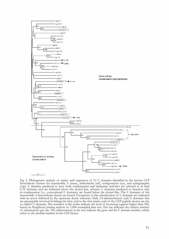
71
Fig. 2. Phylogenetic analysis of amino acid sequences of 51 C domains identified in the known CLP biosynthesis clusters for massetolide A (mass), arthrofactin (arf), syringomycin (syr), and syringopeptin (syp). C domains predicted to have both condensation and epimerase activities are referred to as dual C/E domains and are indicated above the dotted line, whereas C domains predicted to function only in condensation (i.e., conventional C domains) are found below the dotted line. The C domains of the massetolide A biosynthesis cluster are boxed. Exceptions in this classification of C domains are indicated with an arrow followed by the upstream donor substrate. Dab, 2.4-diaminobutyric acid. C domains that are presumably involved in linking the fatty acid to the first amino acid of the CLP peptide moiety are the so-called C1 domains. The numbers at the nodes indicate the level of bootstrap support higher than 500, based on Neighbour Joining analysis of 1,000 resampled data sets. The bar indicates the relative number of substitutions per site. The abbreviations in the tree indicate the gene and the C domain number, which refers to the module number in the CLP cluster.
72
chemically determined structure of the peptide moiety of massetolide A (Fig. 1B). These results indicate that massA is the first and massC the last gene involved in massetolide A biosynthesis and that the nine modules in the massetolide biosynthetic template are colinear with the number of amino acids in massetolide A. Chiral GC-analysis performed in this study confirmed that the peptide moiety of massetolide A produced by P. fluorescens SS101 contains 4 aa in the L-configuration (aa 1, 5, 7 and 9) and 5 amino acids in the D-configuration (aa 2, 3, 4, 6 and 8) (Fig. 1B). In contrast to CLP biosynthetic templates in Bacillus species (Peypoux et al. 1999; Sieber and Marahiel 2005), no epimerization (E) domains were found in the massetolide A synthetases (Fig. 1B), nor in any of the other six CLP biosynthetic templates described to date for Pseudomonas (Berti et al. 2007; De Bruijn et al. 2007; Gross et al. 2007; Guenzi et al. 1998; Paulsen et al. 2005; Roongsawang et al. 2003; Scholz-Schroeder et al. 2003; Zhang et al. 1995). Roongsawang et al. (2003) initially postulated that external racemases may be responsible for the D-configuration of the amino acids in arthrofactin and that sequence differences downstream of a conserved core motif (FFELGGHSLLA(V/M) in the T domains might reflect the recognition by these external racemases. However, when sequences of the T domains of massetolide A were aligned, alone or together with T domains of other CLP biosynthesis clusters including arthrofactin, no relationship could be established between this sequence motif and the amino acid configuration (data not shown). Subsequent studies on arthrofactin biosynthesis by Balibar et al. (2005) had indicated that the D-configuration of the amino acids is generated by specific C domains that have dual catalytic activities, i.e. condensation and epimerization. In their study, they showed that this subclass of C domains, referred to as C/E domains, is involved in epimerization of the amino acid that is loaded onto the T domain of the preceding module. Given that aa 2, 3, 4, 6 and 8 have the D-configuration in massetolide A (Fig. 1B), this would suggest that the C-domains of the third, fourth, fifth, seventh, and ninth module should fall within this subclass of C/E-domains. Subsequent alignment of the primary sequence of each of the individual C domains from NRPS genes involved in massetolide A, arthrofactin, syringomycin, and syringopeptin biosynthesis showed that C-domains 3, 4, 5, 7 and 9 from massetolide A indeed cluster with the C/E domains of arthrofactin, syringomycin and syringopeptin (Fig. 2). There were, however, two exceptions: based on the alignment, the second and sixth C-domain from massetolide A were also predicted to have dual catalytic activity, but follow an L-Leu residue (aa
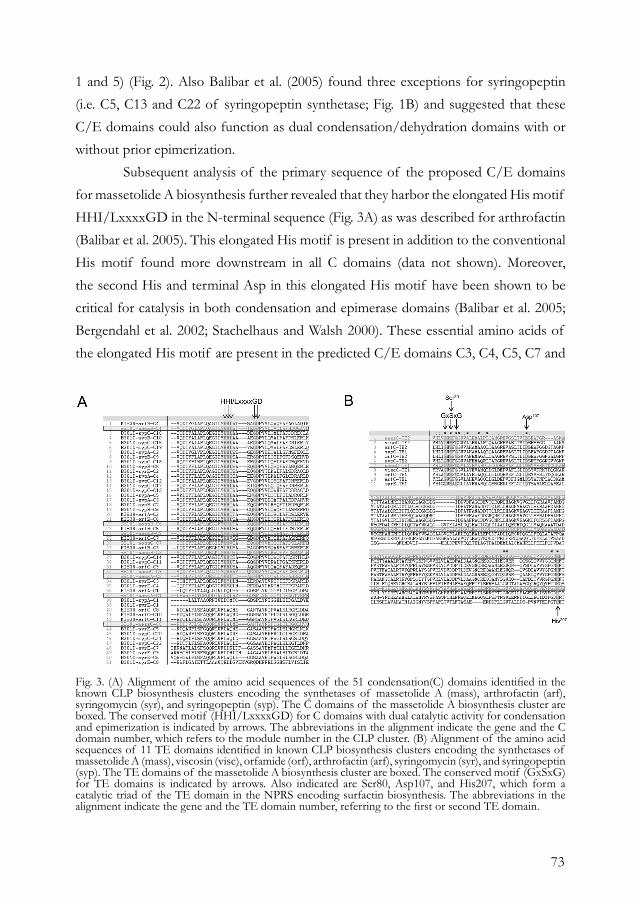
73
1 and 5) (Fig. 2). Also Balibar et al. (2005) found three exceptions for syringopeptin (i.e. C5, C13 and C22 of syringopeptin synthetase; Fig. 1B) and suggested that these C/E domains could also function as dual condensation/dehydration domains with or without prior epimerization. Subsequent analysis of the primary sequence of the proposed C/E domains for massetolide A biosynthesis further revealed that they harbor the elongated His motif HHI/LxxxxGD in the N-terminal sequence (Fig. 3A) as was described for arthrofactin (Balibar et al. 2005). This elongated His motif is present in addition to the conventional His motif found more downstream in all C domains (data not shown). Moreover, the second His and terminal Asp in this elongated His motif have been shown to be critical for catalysis in both condensation and epimerase domains (Balibar et al. 2005; Bergendahl et al. 2002; Stachelhaus and Walsh 2000). These essential amino acids of the elongated His motif are present in the predicted C/E domains C3, C4, C5, C7 and
Fig. 3. (A) Alignment of the amino acid sequences of the 51 condensation(C) domains identified in the known CLP biosynthesis clusters encoding the synthetases of massetolide A (mass), arthrofactin (arf), syringomycin (syr), and syringopeptin (syp). The C domains of the massetolide A biosynthesis cluster are boxed. The conserved motif (HHI/LxxxxGD) for C domains with dual catalytic activity for condensation and epimerization is indicated by arrows. The abbreviations in the alignment indicate the gene and the C domain number, which refers to the module number in the CLP cluster. (B) Alignment of the amino acid sequences of 11 TE domains identified in known CLP biosynthesis clusters encoding the synthetases of massetolide A (mass), viscosin (visc), orfamide (orf), arthrofactin (arf), syringomycin (syr), and syringopeptin (syp). The TE domains of the massetolide A biosynthesis cluster are boxed. The conserved motif (GxSxG) for TE domains is indicated by arrows. Also indicated are Ser80, Asp107, and His207, which form a catalytic triad of the TE domain in the NPRS encoding surfactin biosynthesis. The abbreviations in the alignment indicate the gene and the TE domain number, referring to the first or second TE domain.
74
C9 of the massetolide A synthetases (Fig. 3A). The second and sixth domain in the massetolide A synthetases also have an elongated His motif, but in the C2 domain the Gly in front of the Asp is missing and in the C6 domain the second His is replaced by a Tyr (Fig. 3A). Whether these deficiencies affect or eliminate epimerase activity is not known. MassC terminates with two thioesterase (TE) domains of approximately 250 aa, each containing the conserved GxSxG sequence motif (Fig. 3B; (Roongsawang et al. 2003; Roongsawang et al. 2007)). Both TE domains of MassC contain the residues Ser80, Asp107 and His207 (Fig. 3B), which form a catalytic triad in the TE domain of SrfA-C, the synthetases involved in surfactin biosynthesis in Bacillus (Bruner et al. 2002). These results suggest that both TE domains in MassC are likely to be active in massetolide A biosynthesis and may function in tandem to enhance the rate of product release from the NRPS assembly line, as was shown for the two TE domains in arthrofactin biosynthesis (Roongsawang et al. 2003; Roongsawang et al. 2007).
Organization of the massetolide biosynthesis genes and identification of flanking regions. Compared with other Pseudomonas CLP biosynthesis genes described to date (Berti et al. 2007; De Bruijn et al. 2007; Gross et al. 2007; Guenzi et al. 1998; Paulsen et al. 2005; Roongsawang et al. 2003; Scholz-Schroeder et al. 2003; Zhang et al. 1995), the massA, massB and massC genes showed highest similarity (81-84 % identity) to viscA, viscB and viscC of P. fluorescens SBW25, respectively (Fig. 4). Upstream of massA, two additional ORFs were found (Fig. 4): the first ORF of 1424 bp showed 75% identity with the outer membrane protein NodT (Rivilla et al. 1995) of P. fluorescens Pf0-1, belonging to the family of resistance nodulation and cell division (RND) efflux systems. The tripartite RND-efflux system PseABC, identified at the left border of the syr-syp genomic island in Pseudomonas syringae pv. syringae, encodes an outer membrane protein (PseA), a periplasmic membrane fusion protein (PseB) and a cytoplasmic membrane protein (PseC) (Kang and Gross 2005). Mutations in each of the pseABC genes resulted in a significant decrease (40-60%) in syringomycin and syringopeptin production (Kang and Gross 2005). Interestingly, the predicted RND-like outer membrane protein flanking massA showed only 30% identity to PseA (Psyr_2620), but 69% identity to another outer membrane protein found in P. syringae pv. syringae. This other membrane protein, designated Psyr_2606, is also located close to the syr-syp genomic island (Scholz-Schroeder et al. 2001),
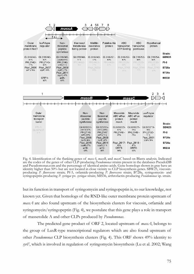
75
but its function in transport of syringomycin and syringopeptin is, to our knowledge, not known yet. Given that homologs of the RND-like outer membrane protein upstream of massA are also found upstream of the biosynthesis clusters for viscosin, orfamide and syringomycin/syringopeptin (Fig. 4), we postulate that this gene plays a role in transport of massetolide A and other CLPs produced by Pseudomonas. The predicted gene product of ORF 2, located upstream of massA, belongs to the group of LuxR-type transcriptional regulators which are also found upstream of other Pseudomonas CLP biosynthesis clusters (Fig. 4). This ORF shows 49% identity to syrF, which is involved in regulation of syringomycin biosynthesis (Lu et al. 2002; Wang
Fig. 4. Identification of the flanking genes of massA, massB, and massC based on Blastx analysis. Indicated are the codes of the genes of other CLP-producing Pseudomonas strains present in the databases PseudoDB and Pseudomonas.com and the percentage of identical amino acids. Gene homologs shown in gray have an identity higher than 50% but are not located in close vicinity to CLP biosynthesis genes. SBW25, viscosin-producing P. fluorescens strain; Pf-5, orfamide-producing P. fluorescens strain; B728a, syringomycin- and syringopeptin-producing P. syringae pv. syringae strain; MIS36, arthrofactin-producing Pseudomonas sp. strain.
76
et al. 2006). The role of this LuxR-type regulator in massetolide A biosynthesis is yet unknown, but preliminary results showed that overexpression of this gene resulted in an increased production of massetolide A in strain SS101 (data not shown). Downstream of massA, ORFs 3, 4 and 5 were identified with 76% identity to mannosyltransferase, 69 % identity to the melittin resistance protein PqaB, and 50% identity to the Ais protein (aluminum induced protein), respectively (Fig. 4). These three ORFs are also found downstream of viscA in P. fluorescens SBW25, but their role in massetolide A or viscosin biosynthesis, if any, has not been resolved. Located even more downstream of massA are two ABC transporters (ORFs 6 and 7) and a hypothetical protein (ORF 8). Although close homologs of ORFs 6 through 8 were identified downstream of viscA in P. fluorescens SBW25 and in the genomes of other CLP producing Pseudomonas strains, their role in CLP biosynthesis remains elusive. The ABC transporters flanking massA have relatively low identity (<25%) to SyrD, an ABC transporter involved in virulence and lipopeptide transport in Pseudomonas syringae pv. syringae (Quigley et al. 1993). Upstream of massB and massC, an 11.3-kb ORF showing 61% identity to an outer membrane transport barrel of P. fluorescens Pf0-1 was identified. The presence of this gene close to the CLP biosynthesis cluster seems to be unique for strain SS101, since it is not found near any of the other CLP biosynthesis clusters described to date. Downstream of massB and massC, two ORFs with 84-85% identity to the ABC-type macrolide efflux proteins MacA and MacB of P. fluorescens Pf-5 were identified. In Escherichia coli, this transport system confers resistance against the macrolides erythromycin and azithromycin (Kobayashi et al. 2001). The presence of these genes downstream of the CLP biosynthesis cluster is conserved among CLP-producing pseudomonads (Fig. 4), suggesting that the MacA and MacB homologs may play a role in CLP transport. ORF 4, located downstream of the macA and macB genes, has 63% identity with a LuxR-type transcriptional regulator of P. fluorescens Pf0-1. Homologs of this gene are also found downstream of the viscosin and orfamide biosynthesis clusters in P. fluorescens strains SBW25 and Pf-5, respectively, but shows relatively low identity (39%) to salA, an important LuxR-type transcriptional regulator of syringomycin and syringopeptin biosynthesis (Kinscherf and Willis 2002; Kitten et al. 1998; Lu et al. 2002; Lu et al. 2005). Site-directed mutagenesis of the LuxR-type regulators of the massetolide A biosynthesis cluster, as performed for strain DC3000 (Berti et al. 2007), as well as expression analyses should be conducted to more conclusively assess their functions
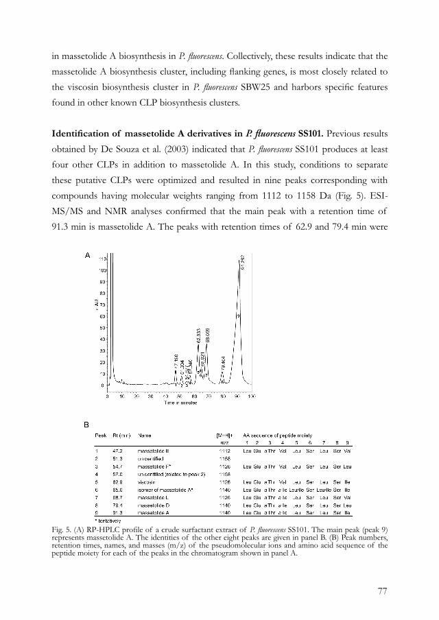
77
in massetolide A biosynthesis in P. fluorescens. Collectively, these results indicate that the massetolide A biosynthesis cluster, including flanking genes, is most closely related to the viscosin biosynthesis cluster in P. fluorescens SBW25 and harbors specific features found in other known CLP biosynthesis clusters.
Identification of massetolide A derivatives in P. fluorescens SS101. Previous results obtained by De Souza et al. (2003) indicated that P. fluorescens SS101 produces at least four other CLPs in addition to massetolide A. In this study, conditions to separate these putative CLPs were optimized and resulted in nine peaks corresponding with compounds having molecular weights ranging from 1112 to 1158 Da (Fig. 5). ESI-MS/MS and NMR analyses confirmed that the main peak with a retention time of 91.3 min is massetolide A. The peaks with retention times of 62.9 and 79.4 min were
Fig. 5. (A) RP-HPLC profile of a crude surfactant extract of P. fluorescens SS101. The main peak (peak 9) represents massetolide A. The identities of the other eight peaks are given in panel B. (B) Peak numbers, retention times, names, and masses (m/z) of the pseudomolecular ions and amino acid sequence of the peptide moiety for each of the peaks in the chromatogram shown in panel A.
78
identified by ESI-MS/MS and NMR as viscosin and massetolide D, respectively (Fig. 5). Viscosin differs from massetolide A by the replacement of the allo-isoleucine at position number four (aa 4) with a valine, and massetolide D differs from massetolide A by the replacement of the isoleucine at aa 9 with a leucine (Fig. 5). Based on ESI-MS/MS studies, the small peaks at 47.2 and 54.7 min were tentatively identified as massetolide E and F, respectively. Massetolide E and F resemble viscosin, the only difference being that at aa 9 a valine replaces the isoleucine in massetolide E, and a leucine replaces the isoleucine in massetolide F. The molecular weight and MS/MS of massetolide F are identical to that of viscosin. The small amounts available precluded the recording of HMBC NMR spectra for further confirmation. Also for the peaks at 51.3 and 57.0 min, the amounts available were too small for NMR, and assignment of a putative structure was not possible on basis of MS data alone. The peak at 65.0 min showed exactly the same MS/MS fragmentation as massetolide A, which suggests that the leucine at either aa 5 or 7 is replaced by an isoleucine. The peak at 68.7 min has a molecular mass of 1126. MS/MS and NMR data indicated that it has a valine as aa 9 instead of an isoleucine in massetolide A. This compound has, to our knowledge, not been described before and was given the name massetolide L (Fig. 5). For all eight fractions, the lipid tail was identified as 3-hydroxydecanoic acid. None of the five massetolide A biosynthesis mutants (Fig. 1B) produces any of the derivatives of massetolide A, indicating that these derivatives are the result of flexibility in amino acid selection and activation by the A domains of the massetolide A synthetases, and in particular A domains 4 and 9. Substrate flexibility of A domains is a common phenomenon in nonribosomal peptide synthesis, resulting in the production of a range of structural analogs that may have different biological functions or activities. For example, Gerard et al. (1997) showed that for the massetolides produced by the marine Pseudomonas strain, variations at the fourth and ninth amino acid position resulted in a significant change in antibacterial activity. Although massetolide A is the most predominant CLP produced by P. fluorescens SS101, culture conditions may have a significant effect on the production of these structural analogs. For example, carbon sources and the nature of amino acids supplemented to the culture medium may greatly affect the production levels and structural diversity of CLPs (Dubern and Bloemberg 2006) and may also be exploited in precursor-directed biosynthesis to generate structurally novel derivatives with different activities (Gerard et al. 1997).
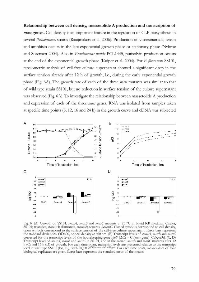
79
Relationship between cell density, massetolide A production and transcription of mass genes. Cell density is an important feature in the regulation of CLP biosynthesis in several Pseudomonas strains (Raaijmakers et al. 2006). Production of viscosinamide, tensin and amphisin occurs in the late exponential growth phase or stationary phase (Nybroe and Sorensen 2004). Also in Pseudomonas putida PCL1445, putisolvin production occurs at the end of the exponential growth phase (Kuiper et al. 2004). For P. fluorescens SS101, tensiometric analysis of cell-free culture supernatant showed a significant drop in the surface tension already after 12 h of growth, i.e., during the early exponential growth phase (Fig. 6A). The growth rate of each of the three mass mutants was similar to that of wild type strain SS101, but no reduction in surface tension of the culture supernatant was observed (Fig. 6A). To investigate the relationship between massetolide A production and expression of each of the three mass genes, RNA was isolated from samples taken at specific time points (8, 12, 16 and 24 h) in the growth curve and cDNA was subjected
Fig. 6. (A) Growth of SS101, massA, massB and massC mutants at 25 °C in liquid KB medium. Circles, SS101; triangles, ΔmassA; diamonds, ΔmassB; squares, ΔmassC. Closed symbols correspond to cell density, open symbols correspond to the surface tension of the cell-free culture supernatant. Error bars represent the standard deviations. OD600, optical density at 600 nm. (B) Transcript levels of massA, massB and massC corrected for the transcript levels of the housekeeping gene rpoD [∆Ct = Ct(mass gene)–Ct(rpoD)]. (C, D) Transcript level of massA, massB and massC in SS101, and in the massA, massB and massC mutants after 12 h (C) and 16 h (D) of growth. For each time point, transcript levels are presented relative to the transcript level in wild type SS101 (log RQ) with RQ = 2-[∆Ct(mutant) - ∆Ct(wildtype)]. For each time point, mean values of four biological replicates are given. Error bars represent the standard error of the means.
C
80
to Q-PCR with different primers specific for each of the three mass genes (positions of the primers are indicated with arrows in Fig. 1B). Transcript levels were determined in four independent RNA isolations and related to transcript levels of the housekeeping gene rpoD to correct for small differences in template concentration; correction with 16S-rDNA transcript levels gave similar results (data not shown). During growth of wild type strain SS101, transcript levels of massA, massB and massC increased in time, reaching a maximum after 16 h of growth (Fig. 6B). Analysis of transcript levels of the mass genes in each of the three mutants was performed after 12 and 16 h of growth. The results show that at both time points, massC transcript levels were significantly and consistently decreased in the massB mutant, but were not affected in the massA mutant (Fig. 6C and D). Transcript levels of massA and massB were variable between both time points, but were not substantially and consistently changed in all three mutants, indicating that initiation of transcription was not affected (Fig. 6C, D). These results show that transcript levels of massA, massB and massC follow the same dynamics as the growth and biosurfactant production by strain SS101. A mutation in massB strongly reduces massC expression, suggesting that these two genes function in an operon. Collectively, these results indicate that under the culture conditions used in this study, massetolide A biosynthesis is initiated in the early exponential growth phase. Although the results obtained with strain SS101 do not point to cell density-dependent regulation of massetolide A production, various methods were adopted to investigate whether N-acylhomoserine lactone (N-AHL)-mediated quorum sensing plays a role. When strain SS101 was coinoculated with the N-AHL-reporter strains Chromobacterium violaceum O26 (McClean et al. 1997), Pseudomonas aureofaciens 30-84I (Pierson et al. 1994), Agrobacterium tumefaciens NTLR4 (McClean et al. 1997; Smadja et al. 2004), E. coli pSCR1 (Aguilar et al. 2003) or E. coli pSB401 (Winson et al. 1998), no response of these N-AHL reporter strains was observed (data not shown). Furthermore, to separate putative metabolites produced by SS101 that may inhibit the induction of the N-AHL reporters, cell-free supernatant from stationary phase-cultures of SS101 were extracted with ethyl acetate and separated by RP-HPLC, followed by overlay thin-layer chromatography analysis with C. violaceum CV026 as the reporter strain (Laue et al. 2000; Shaw et al. 1997). This approach also did not provide any indications of N-AHL production by strain SS101 (data not shown). Taken together, these results strongly suggest that, under the conditions tested, N-AHL-dependent quorum sensing does not appear to play a role in massetolide A biosynthesis in P. fluorescens SS101. Similar results were found for
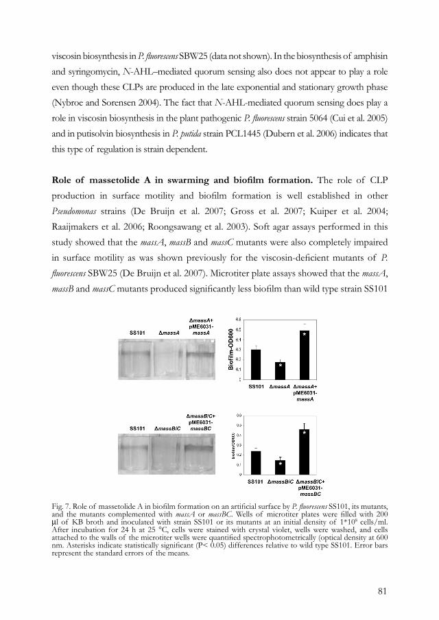
81
viscosin biosynthesis in P. fluorescens SBW25 (data not shown). In the biosynthesis of amphisin and syringomycin, N-AHL–mediated quorum sensing also does not appear to play a role even though these CLPs are produced in the late exponential and stationary growth phase (Nybroe and Sorensen 2004). The fact that N-AHL-mediated quorum sensing does play a role in viscosin biosynthesis in the plant pathogenic P. fluorescens strain 5064 (Cui et al. 2005) and in putisolvin biosynthesis in P. putida strain PCL1445 (Dubern et al. 2006) indicates that this type of regulation is strain dependent.
Role of massetolide A in swarming and biofilm formation. The role of CLP production in surface motility and biofilm formation is well established in other Pseudomonas strains (De Bruijn et al. 2007; Gross et al. 2007; Kuiper et al. 2004; Raaijmakers et al. 2006; Roongsawang et al. 2003). Soft agar assays performed in this study showed that the massA, massB and massC mutants were also completely impaired in surface motility as was shown previously for the viscosin-deficient mutants of P. fluorescens SBW25 (De Bruijn et al. 2007). Microtiter plate assays showed that the massA, massB and massC mutants produced significantly less biofilm than wild type strain SS101
Fig. 7. Role of massetolide A in biofilm formation on an artificial surface by P. fluorescens SS101, its mutants, and the mutants complemented with massA or massBC. Wells of microtiter plates were filled with 200 µl of KB broth and inoculated with strain SS101 or its mutants at an initial density of 1*108 cells/ml. After incubation for 24 h at 25 °C, cells were stained with crystal violet, wells were washed, and cells attached to the walls of the microtiter wells were quantified spectrophotometrically (optical density at 600 nm. Asterisks indicate statistically significant (P< 0.05) differences relative to wild type SS101. Error bars represent the standard errors of the means.
82
(Fig. 7). The biofilm formed by SS101 was located mostly at the air-liquid interface. The deficiency in biofilm formation of the mutants was restored by complementation (Fig. 7). The complemented mutants produced even more biofilm than wild type strain SS101, which is most likely due to the copy number [n=5 to 7; (Heeb et al. 2000)] of the vector used to reintroduce massA and massBC in the mutants. The role of CLPs in biofilm formation can differ considerably between different strains. For example, arthrofactin and putisolvin were shown to adversely affect biofilm formation since mutants deficient in the biosynthesis of these CLPs produced more and differently structured biofilms than their respective parental strains (Kuiper et al. 2004; Roongsawang et al. 2003). How CLPs influence biofilm formation is still unclear, but their effect on cell surface hydrophobicity may play an important role in this process. Hydrophobic interactions and surface-active compounds, including CLPs, have been widely suggested to play a role in the adherence of cells to surfaces (Neu 1996; O’Toole et al. 2000; Palmer et al. 2007). More specifically, biosurfactants may be oriented with the hydrophilic part to the cell surface, thereby exposing the hydrophobic part to the outside and facilitating attachment to hydrophobic surfaces; the other way around, i.e. when the hydrophobic part of the biosurfactant is anchored in the outer layers of the cell surface, the cell can interact with a hydrophilic surface but not with a hydrophobic interface (Neu 1996). Given the diversity in structures and hydrophobicity of various CLPs produced by Pseudomonas strains, we postulate that depending on the cell surface of the producing strain as well as the structure and hydrophobicity of the CLP produced, the role in biofilm formation may be entirely different.
CONCLUSION Cyclic lipopeptides are produced by a variety of microorganisms and have activity against a wide range of plant and human pathogenic microorganisms, including fungi, oomycetes, enveloped viruses, mycoplasmas, trypanosomes and gram-positive bacteria (Raaijmakers et al. 2006). Insight into the biosynthesis, regulation and transport of these versatile compounds can ultimately be exploited in combinatorial biosynthesis to generate new derivatives with more specific or different activities (Fischbach and Walsh 2006; Raaijmakers et al. 2006). The mutagenesis, BAC cloning, sequencing, complementation, and in silico analysis performed in this study revealed that massetolide A biosynthesis is governed by three large NRPS genes. Although other CLP biosynthesis genes have been
83
identified previously for Pseudomonas species, the complete massetolide A biosynthesis cluster was unknown. Furthermore, in contrast to other Pseudomonas CLP biosynthesis clusters, the mass genes are not physically linked in the SS101 genome but organized in two separate clusters that are transcribed independently. The only Pseudomonas CLP biosynthesis cluster known to date, for which the genes are also physically disconnected, is the viscosin biosynthesis cluster in P. fluorescens SBW25, where viscA is separated from viscBC by 1.6 Mb on the physical map of the draft genome sequence (De Bruijn et al. 2007). Several flanking genes of the massetolide A biosynthesis cluster are conserved among other Pseudomonas CLP biosynthesis clusters and include LuxR-type transcriptional regulators, ABC transport carriers and an RND-like outer membrane protein. Although their function in massetolide A biosynthesis needs to be assessed, the conserved positioning of these genes suggests an important role in CLP biosynthesis. Interestingly, no genes involved in the biosynthesis of the lipid side chain nor genes involved in acyl transfer were found up- or downstream of the massetolide biosynthesis genes. Similar observations were made for the orfamide biosynthesis cluster (Gross et al. 2007) and other CLP gene clusters (Raaijmakers et al. 2006). Gross et al. (2007) postulated that the hydroxy fatty acids composing the lipid side chains of these CLPs may be produced by the primary metabolism, i.e. the type II fatty acid synthase systems.
MATERIAL AND METHODS Bacterial strains and culture conditions. P. fluorescens SS101 was grown on Pseudomonas agar F (Difco) (PSA) plates or in liquid King’s medium B (KB) at 25 °C. The transposon mutants were obtained as described by De Souza et al. (2003) and plasposon mutants were obtained with plasmid pTnModOKm (Dennis and Zylstra, 1998). Escherichia coli strain DH5α and EPI3000 were used as hosts for the plasmids for site-directed mutagenesis, complementation and construction of the BAC library. E. coli strains were grown on Luria-Bertani (LB) plates or in LB broth amended with the appropriate antibiotics.
Site-directed mutagenesis. Site-directed mutagenesis of the mass genes was performed with the pKnockout-G suicide vector (Windgassen et al. 2000). Fragments of the genes of interest were amplified by PCR with the primers 5’-CATTCCTGGCGTTGGCTGG-3’ (massA forward primer), 5’-TGCAGCATTCCTCCAGCCTG-3’ (massA reverse primer), 5’-AAATTCACGGGCGCTGGCAT-3’ (massB forward primer), 5’-ACATGCCTCGTTGTCCCTGG-3’ (massB reverse primer), 5’-TCCTGGCGTTGATGGAAGG-3’ (massC forward primer), 5’-AACGACAGGTCGAACTTGGC-3’ (massC reverse primer) and first cloned into pGEM-T Easy Vector Systems I (Promega) according to the manufacturer’s instructions. Inserts were subcloned by ApaI/SacI digestions into pKnockout-G and transferred into SS101 by triparental mating with helper strain E. coli HB101 carrying plasmid pRK2013. Transformants were selected on KB agar plates supplemented with rifampicin (100 μg/ml) and gentamicin (75 μg/ml). Integrations in the target gene were verified by PCR using one primer specific for the insert and one primer specific for the gene fragment flanking the pKnockout insertion site.
Construction of BAC library. P. fluorescens SS101 cells from an 25 ml overnight culture grown at 25 °C were washed twice with sterile demineralized water and embedded in 1% low-melting point agarose (Invitrogen) dissolved in TAE buffer (40 mM Tris, 20 mM acetic acid, 1 mM EDTA, pH 8.0). The cell-
84
agarose mixture was taken up into a 1-ml syringe and cooled down to 4 °C to solidify. The cell-agarose worm was extruded from the syringe and incubated with 10 ml of lysis buffer (10 mM Tris, 50 mM NaCl, 0.2 M EDTA, 1% sarkosyl, 0.2 % sodium deoxycholate, 1 mg/ml lysozyme, pH 8.0) with gentle agitation for 3 hrs at 37 °C. The agarose worms were subsequently transferred to 40 ml of 1% Sarkosyl and proteinase K (1 mg/ml) and incubated with gentle agitation for 16 hrs at 55 °C. After refreshing the buffer and incubation for 1 hour, agarose worms were washed three times with 50 ml T10E1 buffer (10 mM Tris, 1 mM EDTA, pH 8.0) and subsequently incubated for 1 hour at room temperature in T10E1 buffer supplemented with 1 mM phenylmethylsulfonyl fluoride. Agarose worms were washed three times with T10E1 buffer and incubated overnight in storage buffer (10 mM Tris, 50 mM EDTA, pH 8.0). Pulse-field gel electrophoresis (PFGE) was performed at 6 V/cm for 16 hours with a 5- to 15-s switch time at a 120° angle to analyse DNA yield and quality. Plugs were preincubated with Sau3AI digestion buffer (New England Biolabs) and subsequently treated with Sau3AI. Partially digested DNA was fractionated by pulse-field gel electrophoresis, and fragments of 50-250 kb were isolated from agarose by Gelase (Epicentre) treatment according to manufacturer’s instructions; 225 ng of DNA fragments were ligated to 25 ng of BamHI-digested and dephosphorylated pCC1BAC vector DNA (Epicentre) according to supplier’s protocol and electroporated into E. coli EPI3000 cells (Epicentre). Cells were plated on LB with chloramphenicol (12.5 μg/ml).
Identification and sequencing of the mass genes. Library clones were blotted onto Hybond-N+ membranes (Amersham) and hybridized with 32P-labelled probes amplified by PCR with the same primers as described for the site-directed mutagenesis (see above). Hybridization was performed overnight, and membranes were washed at 65 °C with 0.5x SSC (75 mM NaCl, 7.5 mM sodium citrate)-0.1 % SDS). Hybridization-positive clones were subjected to detailed restriction digestion and hybridization analysis using PstI, EcoRV, and previously described probes. Contigs were constructed by cluster analysis of these experimental data by the unweighted-pair group method using average linkages. Clones 2H12, containing massA, and 7B4, containing massB and massC, were sent for shotgun sequencing (Macrogen, Seoul, Korea). Sequence gaps were closed by primer walking and by sequencing the PCR products overlapping the gaps. Bacterial operons and genes were subsequently predicted by the Softberry FGENESB program (Softberry, Inc., Mount Kiscco, NY), and the identified open reading frames (ORFs) were analyzed using Blastx in the NCBI database and PseudoDB (http://xbase.bham.ac.uk/pseudodb/). Putative promoter sequences were identified by the Softberry BPROM program, and putative terminator sequences were identified by the RNA secondary structure prediction program of Genebee (http://www.genebee.msu.su/). Specific domains in the deduced protein sequences of the mass genes were analyzed with PFAM (http://pfam.sanger.ac.uk/search?tab=searchSequenceBlock). Protein sequences of specific domains were aligned in ClustalX (version 1.81). Trees were inferred by Neighbour Joining using 1000 bootstrap replicates. Identification of the flanking genes of massA, massB and massC was performed by Blastx analysis in NCBI, Pseudomonas.com (http://v2.Pseudomonas.com/) or PseudoDB and by comparison with genes flanking the known CLP biosynthesis clusters for syringomycin, syringopeptin, viscosin, orfamide and arthrofactin.
Construction of pME6031-based vectors for complementation. A 7.7 kb fragment containing the massA gene, including promoter and terminator, was obtained by PCR (forward primer: CAGACAAATCCTTCTTCACC; reverse primer: GCGAGCTGCTGGATAACCCA) with Phusion DNA polymerase (Finnzymes). This PCR fragment was subcloned in pGEM-T Easy Vector Systems I (Promega) according to manufacturer’s instructions and the obtained plasmid was digested with EcoRI. Restriction analysis of the BAC clone containing massB and massC revealed unique restriction sites for BamHI. Digestion with this enzyme resulted in a fragment of approximately 30 kb containing massB, massC, and homologs of macA and macB. The fragments containing the mass genes were obtained by excising the fragments from gel and isolating the DNA with the NucleoTrap kit (Macherey-Nagel). These fragments were cloned into the shuttle vector pME6031 (Heeb et al. 2000) which was digested, dephosphorylated (shrimp alkaline phosphatase; Promega) and purified with the NucleoTrap kit according to manufacturer’s instructions. E. coli DH5α was transformed with the obtained plasmids pME6031-massA and pME6031-massBC by heat shock transformation (Inoue et al. 1990) and transformed colonies were selected on LB agar plates supplemented with tetracycline (25 μg/ml). Integration of the inserts was verified by PCR analysis and restriction analysis of isolated plasmids. The correct pME6031-massA and pME6031-massBC constructs were subsequently electroporated into the massetolide-deficient massA, massB and massC transposon mutants. Electrocompetent cells were obtained by washing the cells three times with 1 mM MOPS (morpholinepropanesulfonic acid) and 15% glycerol from a 5-ml overnight culture and finally dissolving the cells in 100 μl of the washing buffer. Cells were stored at -80 °C for at least 1 hour prior to electroporation. Electroporation occurred at 2.4 kV, 200 μF, and after incubation in SOC medium (2% bacto tryptone [Difco], 0.5% bacto yeast extract [Difco], 10 mM NaCl, 2.5 mM KCl, 10 mM MgCl2, 10 mM MgSO4, 20 mM glucose [pH 7]) for 2 hrs at 25 °C, cells were plated on KB supplemented with tetracycline (25 μg/ml). Verification of transformation was performed by PCR analysis using one primer specific for the insert and one primer specific for the pME6031 vector. Massetolide A production in the complemented mutants was tested with a drop collapse assay followed by high-pressure liquid chromatography (RP-HPLC) analysis and liquid chromatograph-tandem mass spectrometry (LC-MS-MS).
85
Chemical identification of massetolide A derivatives produced by P. fluorescens SS101. Analytical RP-HPLC separations were carried out on Alltech end-capped 5- μm C18 columns 250 mm length and of 4.6 mm (UV [210 nm] detection and evaporative light-scattering detection) or 2.1 mm (LC-MS) in diameter at a flow rate of 1.2 or 0.20 ml/min, respectively. For the separations on the 4.6- mm column, a Midas autosampler (Spark), two Spectroflow 400 pumps (Kratos), a Spectroflow UV detector 785 (Applied Biosystems), a Sedex 55 evaporative light-scattering detector (Sedere) and Dionex Chromeleon software were used. Preparative separations were performed with an Alltech end-capped 5- μm C18 column (250 by 22 mm) at 23 ml/min with methyl cyanide-methanol-H2O (3800:1925:4275) containing 0.1% trifluoracetic acid as solvent on a Shimadzu autopreparative system. Two hundred milligrams of crude surfactant extract of strain SS101, obtained as described by De Souza et al. (2003), were dissolved in 6 ml dimethyl sulfoxide. After membrane filtration, five injections of 1150 μl each were carried out. The fractions eluting at 47.2, 51.3, 57.0, 62.9, 65.0, 68.7, 79.4 and 91.3 min were collected on the basis of the UV signal in round-bottom flasks, and the eluent was removed with a rotary evaporator (Büchi) in vacuo. All fractions were investigated by means of analytical RP-HPLC and infusion (+)- electrospray ionization (ESI)-MS/MS. The fractions at 57.0 and 65.0 min were shown to contain a second minor compound. Sufficient amounts of the fractions at 62.9, 68.7, 79.4 and 91.3 min were collected to perform two-dimensional nuclear magnetic resonance (NMR) experiments. The MS system consisted of a Finnigan LCQ ion trap mass spectrometer equipped with a Finnigan electrospray ionisation (ESI) interface. Data were processed by Finnigan Xcalibur software system (ThermoQuest, Breda, The Netherlands). For off-line MS studies, all peaks of the preparative separation were introduced by continuous infusion using a syringe pump (Hamilton, Nevada) at a flow rate of 5 μl/min. Spectra were recorded in positive mode over a period of 2 min and averaged. The scan range was m/z 310-1200 at a scan rate of 0.20 sec. For the MS/MS experiments, helium was used as collision gas. Only a single parent ion was kept in resonance (isolation width m/z 3); all other ions were ejected from the trap without mass analysis. The ion was then agitated and allowed to fragment by collision-induced dissociation. A collision energy of 22% was used to give > 90% yield of fragmentation. For NMR, fractions were dissolved in CD3OD (99.9 atom% D, Acros) and transferred to a standard 5-mm NMR tube. NMR spectra were recorded at a probe temperature of 25 °C on a Bruker DPX-400 (1H, correlation [COSY], total correlation [TOCSY], nuclear Overhauser effect [NOESY], 13C, distortionless enhancement by polarization transfer [DEPT], and heteronuclear multiple bond correlation [HMBC] spectroscopy) or a Bruker AMX 500 (1H, heteronuclear multiple quantum coherence [HMQC]) spectrometer, both from Wageningen NMR Centre. Chemical shifts are expressed in ppm relative to dimethyl sulfoxide (δ 1H 2.50, δ 13C 39.52). One- and two-dimensional double quantum filtered COSY, -TOCSY, -NOESY, -HMBC, and -HMQC spectra were acquired using standard pulse sequences delivered by Bruker. The mixing time for the TOCSY was 80 ms and for the NOESY 200 ms. The analysis of the D/L-configuration of the constituting amino acids was carried out as described by Gerard et al. (1997) with some modifications. In short, 2 mg of purified lipopeptide was dissolved in 4 ml of 6 M HCl and heated at 110 °C during 24 hr in a closed glass vial. The HCl solution was removed in a fume hood by blowing a stream of N2 over the solution at 45 °C. The residue was dissolved in 250 µl of isopropanol saturated with HCl gas, and heated in a closed vial at 110 °C during 45 min. After cooling the solvent was removed with N2 as before. The residue was dissolved in 250 µl of CH2Cl2, and 100 µl of pentafluoropropionyl anhydride was added. After closing the vial, it was heated at 110 °C during 15 min. The reagent solution was removed with N2 as before, re-dissolved in 200 µl of CH2Cl2, and 1 µl was injected (split 1:100) onto a 25-m Chiralsil-Val Heliflex gas chromatography (GC) column installed in an Agilent 6890 GC equipped with both a mass selective detector and an flame ionization detector. Other parameters were as followed: injector, cooled injection system, starting at 100 °C and going to 240 °C at 10 °C/sec; temperature program, 60 °C (hold 2 min) to 170 °C at 4 °C/min. Helium was used a carrier gas. Amino acids were identified by comparing the retention time and mass spectrum with authentic reference D and L-amino acids.
Massetolide A production and transcription of massA, massB and massC. Cells were grown in a 24-well plate with 1.25 ml KB broth per well and shaking at 220 rpm at 25 °C. At specific time points, 1 ml of cell culture was collected and spun down. The cells were frozen in liquid N2 and stored at -80 °C. For the RNA isolations and cDNA synthsis, four biological replicates were used for each time point. Massetolide A production was measured qualitatively by the drop collapse assay and quantitatively by tensiometric analysis of the cell-free supernatant (K6 tensiometer, Krüss GmbH, Hamburg, Germany) at room temperature. To get sufficient volume for the tensiometric analysis, the supernatant of four biological replicates was collected and pooled for each time point. Surface tension of each sample was measured in triplicate. RNA was isolated from the frozen bacterial cells with Trizol Reagent (Invitrogen), followed by DNase I (GE Healthcare) treatment. One microgram RNA was used for cDNA synthesis with Superscript III (Invitrogen) according to the manufacturer’s protocol. For the Q-PCR, conducted with the 7300SDS system of Applied Biosystems, the SYBR Green Core kit (Eurogentec) with a final concentration of 3.5 mM MgCl2 was used according to the manufacturer’s protocol. The concentration of the primers was optimized (400 nM final concentration for all) and a dissociation curve was performed to check the specificity of the primers. The primers used for the Q-PCR for massA are: forward 5’- GCTGTACAACATTGGCGGCT-3’, reverse 5’- GGTATGCAGTTGAGTGCGTAGC-3’, for massB: forward 5’-AACAACGACCGGAGATGCC-3’, reverse 5’-AAGGTGTGCAGCAAGTGATGG-3’, for massC: forward 5’- GTCGACCCTCAACGCGTCT-3’, reverse
86
5’-CCACCGACAGTTGGTCAAGC-3’, for 16S-rRNA: forward 5’-GCGCAACCCTTGTCCTTAGTT-3’, reverse 5’-TGTGTAGCCCAGGCCGTAA-3’, for rpoD: forward 5’-GCAGCTCTGTGTCCGTGATG-3’, reverse 5’-TCTACTTCGTTGCCAGGGAATT-3’. To correct for small differences in template concentration, 16S-rRNA and rpoD were used as housekeeping genes. The cycle where the SYBR Green fluorescence crosses a manually set threshold cycle (Ct) was used to determine transcript levels. For each gene the threshold was fixed based on the exponential segment of the PCR curve. The Ct value of massA was corrected for the housekeeping gene as follows: ∆Ct = Ct(massA)–Ct(rpoD); the same formula was used for massB and massC. The relative quantification (RQ) values, were calculated by the following formula: RQ = 2-[∆Ct(mutant) - ∆Ct(wildtype)]. If there is no difference in transcript level between mutant and wild type, than the RQ=1 (20) and the logRQ=0. Q-PCR analysis was performed in duplicate (technical replicates) on four independent RNA isolations (biological replicates). Statistically significant differences were determined for log-transformed RQ values by analysis of variance (p<0.05) followed by the Bonferroni and Dunnet post hoc multiple comparisons.
Motility and biofilm. Motility of wild type strain SS101 and the massetolide A-deficient mutants was assessed on soft (0.6% agar, w/v) standard succinate medium (SSM) (32.8 mM K2HPO4, 22 mM KH2PO4, 7.6mM (NH4)2SO4, 0.8 mM MgSO4, 34 mM succinic acid, adjusted pH to 7 with NaOH). Overnight cultures of SS101 and the mutants were washed three times and 5 μl of a cell suspension (1*1010 cells/ml) were spotted in the centre of the soft SSM agar plates and incubated for 48 to 72 h at 25 °C. Biofilm formation was assessed according to the method described by O’Toole et al. ( 1999) using flat-bottom 96-well plates made of transparent polystyrene (Greiner) with 200 μl KB broth per well. Statistically significant differences were determined with the Student’s t-test (p<0.05).
Nucleotide sequence accession numbers. The sequences of the contig containing massA and the contig containing massB and massC have been deposited in GenBank under accession numbers EU199080 and EU199081, respectively
ACKNOWLEDGEMENTSWe are grateful to Marc Lemmers, Elbert van der Klift and Frank Claasen for their assistance in reverse-phase HPLC, LC-MS/MS and chiral GC analysis, to Andreas Untergasser for his help with the pME6031-massBC construct. The N-AHL reporter strain P. aureofaciens 30-84I was kindly provided by Leland S. Pierson III, A. tumefaciens NTLR4 was kindly provided by Yves Dessaux, E. coli pSCR1 and E. coli pSB401 and C. violaceum CV026 were kindly provided by Paul Williams and Vittorio Venturi. This work was funded by the Dutch Technology Foundation (STW), the applied science division of NWO.
REFERENCESAguilar, C., Bertani, I., and Venturi, V. 2003. Quorum-sensing system and stationary-phase sigma factor (rpoS) of the onion pathogen Burkholderia cepacia genomovar I type strain, ATCC 25416. Appl. Environ. Microbiol. 69:1739-1747.Balibar, C. J., Vaillancourt, F. H., and Walsh, C. T. 2005. Generation of D amino acid residues in assembly of arthrofactin by dual condensation/epimerization domains. Chem. Biol. 12:1189-1200.Bergendahl, V., Linne, U., and Marahiel, M. A. 2002. Mutational analysis of the C-domain in nonribosomal peptide synthesis. Eur. J. Biochem. 269:620-629.Berti, A. D., Greve, N. J., Christensen, Q. H., and Thomas, M. G. 2007. Identification of a biosynthetic gene cluster and the six associated lipopeptides involved in swarming motility of Pseudomonas syringae pv. tomato DC3000. J. Bacteriol. 189:6312-6323.Bruner, S. D., Weber, T., Kohli, R. M., Schwarzer, D., Marahiel, M. A., Walsh, C. T., and Stubbs, M. T. 2002. Structural basis for the cyclization of the lipopeptide antibiotic surfactin by the thioesterase domain SrfTE.
Structure 10:301-310.Challis, G. L., Ravel, J., and Townsend, C. A. 2000. Predictive, structure-based model of amino acid recognition by nonribosomal peptide synthetase adenylation domains. Chem. Biol. 7:211-24.Cui, X., Harling, R., Mutch, P., and Darling, D. 2005. Identification of N-3-hydroxyoctanoyl-homoserine lactone production in Pseudomonas fluorescens 5064, pathogenic to broccoli, and controlling biosurfactant production by quorum sensing. Eur. J. Plant Pathol. 111:297-308.De Bruijn, I., de Kock, M. J. D., Yang, M., de Waard, P., van Beek, T. A., and Raaijmakers, J. M. 2007. Genome-based discovery, structure prediction and functional analysis of cyclic lipopeptide antibiotics in Pseudomonas species. Mol. Microbiol. 63:417-428.De Souza, J. T., De Boer, M., De Waard, P., Van Beek, T. A., and Raaijmakers, J. M. 2003. Biochemical, genetic, and zoosporicidal properties of cyclic lipopeptide surfactants produced by Pseudomonas fluorescens. Appl. Environ. Microbiol. 69:7161-7172.Dennis, J. J., and Zylstra, G. J. 1998. Plasposons: modular self-cloning minitransposon derivatives
87
for rapid genetic analysis of gram-negative bacterial genomes. Appl. Environ. Microbiol. 64:2710-2715.Dubern, J. F., and Bloemberg, G. V. 2006. Influence of environmental conditions on putisolvins I and II production in Pseudomonas putida strain PCL1445. FEMS Microbiol. Lett. 263:169-175.Dubern, J. F., Lugtenberg, B. J. J., and Bloemberg, G. V. 2006. The ppuI-rsaL-ppuR quorum-sensing system regulates biofilm formation of Pseudomonas putida PCL1445 by controlling biosynthesis of the cyclic lipopeptides putisolvins I and II. J Bacteriol 188:2898-2906.Finking, R., and Marahiel, M. A. 2004. Biosynthesis of nonribosomal peptides. Annu. Rev. Microbiol. 58:453-488.Fischbach, M. A., and Walsh, C. T. 2006. Assembly-line enzymology for polyketide and nonribosomal peptide antibiotics: logic, machinery, and mechanisms. Chem. Rev. 106:3468-3496.Gerard, J., Lloyd, R., Barsby, T., Haden, P., Kelly, M. T., and Andersen, R. J. 1997. Massetolides A-H, antimycobacterial cyclic depsipeptides produced by two pseudomonads isolated from marine habitats. J. Nat. Prod. 60:223-229.Gewolb, J. 2002. Bioengineering. Working outside the protein-synthesis rules. Science 295:2205-2207.Gross, H., Stockwell, V. O., Henkels, M. D., Nowak-Thompson, B., Loper, J. E., and Gerwick, W. H. 2007. The genomisotopic approach: a systematic method to isolate products of orphan biosynthetic gene clusters. Chem. Biol. 14:53-63.Guenzi, E., Galli, G., Grgurina, I., Gross, D. C., and Grandi, G. 1998. Characterization of the syringomycin synthetase gene cluster. A link between prokaryotic and eukaryotic peptide synthetases. J. Biol. Chem. 273:32857-32863.Heeb, S., Itoh, Y., Nishijyo, T., Schnider, U., Keel, C., Wade, J., Walsh, U., O’Gara, F., and Haas, D. 2000. Small, stable shuttle vectors based on the minimal pVS1 replicon for use in gram-negative, plant-associated bacteria. Mol. Plant-Microbe Interact. 13:232-237.Inoue, H., Nojima, H., and Okayama, H. 1990. High efficiency transformation of Escherichia coli with plasmids. Gene 96:23-28.Kang, H., and Gross, D. C. 2005. Characterization of a resistance-nodulation-cell division transporter system associated with the syr-syp genomic island of Pseudomonas syringae pv. syringae. Appl. Environ. Microbiol. 71:5056-5065.Kinscherf, T. G., and Willis, D. K. 2002. Global regulation by gidA in Pseudomonas syringae. J. Bacteriol. 184:2281-6.Kitten, T., Kinscherf, T. G., McEvoy, J. L., and Willis, D. K. 1998. A newly identified regulator is required for virulence and toxin production in Pseudomonas syringae. Mol. Microbiol. 28:917-929.Kobayashi, N., Nishino, K., and Yamaguchi,
A. 2001. Novel macrolide-specific ABC-type efflux transporter in Escherichia coli. J. Bacteriol. 183:5639-5644.Kohli, R. M., Walsh, C. T., and Burkart, M. D. 2002. Biomimetic synthesis and optimization of cyclic peptide antibiotics. Nature 418:658-661.Konnova, E. V., Hedman, R., Welch, C. J., and Gerhardson, B. 2004. Character of massetolide A production by biocontrol strain Pseudomonas sp. MF-30. Intl Society for Plant-Microbe, pages 565-568 in Biology of Plant-Microbe Interactions volume 4.Konz, D., Doekel, S., and Marahiel, M. A. 1999. Molecular and biochemical characterization of the protein template controlling biosynthesis of the lipopeptide lichenysin. J. Bacteriol. 181:133-140.Kuiper, I., Lagendijk, E. L., Pickford, R., Derrick, J. P., Lamers, G. E. M., Thomas-Oates, J. E., Lugtenberg, B. J. J., and Bloemberg, G. V. 2004. Characterization of two Pseudomonas putida lipopeptide biosurfactants, putisolvin I and II, which inhibit biofilm formation and break down existing biofilms. Mol. Microbiol. 51:97-113.Laue, R. E., Jiang, Y., Chhabra, S. R., Jacob, S., Stewart, G. S. A. B., Hardman, A., Downie, J. A., O’Gara, F., and Williams, P. 2000. The biocontrol strain Pseudomonas fluorescens F113 produces the Rhizobium small bacteriocin, N-(3-hydroxy-7-cis-tetradecenoyl)homoserine lactone, via HdtS, a putative novel N-acylhomoserine lactone synthase. Microbiology 146 2469-2480.Lu, S. E., Scholz-Schroeder, B. K., and Gross, D. C. 2002. Characterization of the salA, syrF, and syrG regulatory genes located at the right border of the syringomycin gene cluster of Pseudomonas syringae pv. syringae. Mol. Plant-Microbe Interact. 15:43-53.Lu, S. E., Wang, N., Wang, J., Chen, Z. J., and Gross, D. C. 2005. Oligonucleotide microarray analysis of the salA regulon controlling phytotoxin production by Pseudomonas syringae pv. syringae. Mol. Plant-Microbe Interact. 18:324-333.McClean, K. H., Winson, M. K., Fish, L., Taylor, A., Chhabra, S. R., Camara, M., Daykin, M., Lamb, J. H., Swift, S., Bycroft, B. W., Stewart, G. S. A. B., and Williams, P. 1997. Quorum sensing and Chromobacterium violaceum: exploitation of violacein production and inhibition for the detection of N-acylhomoserine lactones. Microbiology 143: 3703-3711.Neu, T. R. 1996. Significance of bacterial surface-active compounds in interaction of bacteria with interfaces. Microbiol. Rev. 60:151-166.Nybroe, O., and Sorensen, J. 2004. Production of cyclic lipopeptides by fluorescent pseudomonads. Pages 147-172 in: Pseudomonas volume 3, Biosynthesis of Macromolecules and Molecular Metabolism, Ramos, J-L., ed., Kluwer Academic/Plenum Publishers, New York.O’Toole, G., Kaplan, H. B., and Kolter, R. 2000. Biofilm formation as microbial development. Annu. Rev. Microbiol. 54:49-79.
88
O’Toole, G. A., Pratt, L. A., Watnick, P. I., Newman, D. K., Weaver, V. B., and Kolter, R. 1999. Genetic approaches to study of biofilms. Methods Enzymol. 310:91-109.Ongena, M., Jourdan, E., Adam, A., Paquot, M., Brans, A., Joris, B., Arpigny, J. L., and Thonart, P. 2007. Surfactin and fengycin lipopeptides of Bacillus subtilis as elicitors of induced systemic resistance in plants. Environ. Microbiol. 9:1084-1090.Palmer, J., Flint, S., and Brooks, J. 2007. Bacterial cell attachment, the beginning of a biofilm. J. Ind. Microbiol. Biotechnol. 34:577-588.Paulsen, I. T., Press, C. M., Ravel, J., Kobayashi, D. Y., Myers, G. S. A., Mavrodi, D. V., DeBoy, R. T., Seshadri, R., Ren, Q., Madupu, R., Dodson, R. J., Durkin, A. S., Brinkac, L. M., Daugherty, S. C., Sullivan, S. A., Rosovitz, M. J., Gwinn, M. L., Zhou, L., Schneider, D. J., Cartinhour, S. W., Nelson, W. C., Weidman, J., Watkins, K., Tran, K., Khouri, H., Pierson, E. A., Pierson, L. S., 3rd, Thomashow, L. S., and Loper, J. E. 2005. Complete genome sequence of the plant commensal Pseudomonas fluorescens Pf-5. Nat. Biotechnol. 23:873-878.Peypoux, F., Bonmatin, J. M., and Wallach, J. 1999. Recent trends in the biochemistry of surfactin. Appl. Microbiol. Biotechnol. 51:553-563.Pierson, L. S., 3rd, Keppenne, V. D., and Wood, D. W. 1994. Phenazine antibiotic biosynthesis in Pseudomonas aureofaciens 30-84 is regulated by PhzR in response to cell density. J. Bacteriol. 176:3966-3974.Quigley, N. B., Mo, Y. Y., and Gross, D. C. 1993. SyrD is required for syringomycin production by Pseudomonas syringae pathovar syringae and is related to a family of ATP-binding secretion proteins. Mol. Microbiol. 9:787-801.Raaijmakers, J. M., De Bruijn, I., and De Kock, M. J. D. 2006. Cyclic lipopeptide production by plant-associated Pseudomonas spp.: diversity, activity, biosynthesis, and regulation. Mol. Plant-Microbe Interact. 19:699-710.Rivilla, R., Sutton, J. M., and Downie, J. A. 1995. Rhizobium leguminosarum NodT is related to a family of outer-membrane transport proteins that includes TolC, PrtF, CyaE and AprF. Gene 161:27-31.Roongsawang, N., Hase, K., Haruki, M., Imanaka, T., Morikawa, M., and Kanaya, S. 2003. Cloning and characterization of the gene cluster encoding arthrofactin synthetase from Pseudomonas sp. MIS38. Chem. Biol. 10:869-880.Roongsawang, N., Lim, S. P., Washio, K., Takano, K., Kanaya, S., and Morikawa, M. 2005. Phylogenetic analysis of condensation domains in the nonribosomal peptide synthetases. FEMS Microbiol. Lett. 252:143-151.Roongsawang, N., Washio, K., and Morikawa, M. 2007. In vivo characterization of tandem C-terminal thioesterase domains in arthrofactin synthetase. Chem. Bio. Chem. 8:501-512.Scholz-Schroeder, B. K., Soule, J. D., and
Gross, D. C. 2003. The sypA, sypB, and sypC synthetase genes encode twenty-two modules involved in the nonribosomal peptide synthesis of syringopeptin by Pseudomonas syringae pv. syringae B301D. Mol. Plant-Microbe Interact. 16:271-280.Scholz-Schroeder, B. K., Soule, J. D., Lu, S. E., Grgurina, I., and Gross, D. C. 2001. A physical map of the syringomycin and syringopeptin gene clusters localized to an approximately 145-kb DNA region of Pseudomonas syringae pv. syringae strain B301D. Mol. Plant-Microbe Interact. 14:1426-1435.Shaw, P. D., Ping, G., Daly, S. L., Cha, C., Cronan, J. E., Jr., Rinehart, K. L., and Farrand, S. K. 1997. Detecting and characterizing N-acyl-homoserine lactone signal molecules by thin-layer chromatography. Proc. Natl. Acad. Sci. USA 94:6036-6041.Sieber, S. A., and Marahiel, M. A. 2005. Molecular mechanisms underlying nonribosomal peptide synthesis: approaches to new antibiotics. Chem. Rev. 105:715-738.Smadja, B., Latour, X., Faure, D., Chevalier, S., Dessaux, Y., and Orange, N. 2004. Involvement of N-acylhomoserine lactones throughout plant infection by Erwinia carotovora subsp. atroseptica (Pectobacterium atrosepticum). Mol. Plant-Microbe Interact. 17:1269-1278.Stachelhaus, T., Mootz, H. D., and Marahiel, M. A. 1999. The specificity-conferring code of adenylation domains in nonribosomal peptide synthetases. Chem. Biol. 6:493-505.Stachelhaus, T., and Walsh, C. T. 2000. Mutational analysis of the epimerization domain in the initiation module PheATE of gramicidin S synthetase. Biochemistry 39:5775-5787.Tran, H., Ficke, A., Asiimwe, T., Hofte, M., and Raaijmakers, J. M. 2007. Role of the cyclic lipopeptide massetolide A in biological control of Phytophthora infestans and in colonization of tomato plants by Pseudomonas fluorescens. New Phytol. 175:731-742.Wang, N., Lu, S. E., Records, A. R., and Gross, D. C. 2006. Characterization of the transcriptional activators SalA and SyrF, which are required for syringomycin and syringopeptin production by Pseudomonas syringae pv. syringae. J. Bacteriol. 188:3290-3298.Windgassen, M., Urban, A., and Jaeger, K. E. 2000. Rapid gene inactivation in Pseudomonas aeruginosa. FEMS Microbiol. Lett. 193:201-205.Winson, M. K., Swift, S., Fish, L., Throup, J. P., Jorgensen, F., Chhabra, S. R., Bycroft, B. W., Williams, P., and Stewart, G. S. A. B. 1998. Construction and analysis of luxCDABE-based plasmid sensors for investigating N-acyl homoserine lactone-mediated quorum sensing. FEMS Microbiol. Lett. 163:185-192.Zhang, J. H., Quigley, N. B., and Gross, D. C. 1995. Analysis of the syrB and syrC genes of Pseudomonas syringae pv. syringae indicates that syringomycin is synthesized by a thiotemplate mechanism. J. Bacteriol. 177:4009-4020.

89

90
91
CHAPTER 4
Heterologous expression of regulatory and biosynthesis genes directing cyclic lipopeptide antibiotic production in Pseudomonas fl uorescens.
Irene de Bruijn1 and Jos M. Raaijmakers1
1Laboratory of Phytopathology, Wageningen University, 6709 PD Wageningen, the Netherlands.
Submitted.
92
ABSTRACTCyclic lipopeptides (CLPs) are produced by many Pseudomonas species and have several biological functions, including facilitating surface motility, biofilm formation, virulence, and antibiotic activity. To improve or alter the antimicrobial properties of CLPs via combinatorial biosynthesis, fundamental knowledge of genetic regulation and heterologous expression of CLP biosynthesis genes is essential. This study focused on the role of LuxR-type transcriptional regulators in the biosynthesis of the CLP viscosin in Pseudomonas fluorescens strain SBW25. Genetic analyses showed that LuxR-type regulators flanking the CLP biosynthesis clusters in SBW25 and various other Pseudomonas strains contain a DNA-binding helix-turn-helix domain, but lack autoinducer binding or response regulator domains, suggesting that they belong to a separate subfamily of LuxR-type regulators. For strain SBW25, the results showed that each of the two LuxR-type regulators, designated LuxR-vA and LuxR-vBC, flanking the viscABC biosynthesis genes is essential for viscosin production. Site-directed mutagenesis of the luxR-vA and luxR-vBC genes strongly reduced transcript levels of the viscABC genes and resulted in loss of viscosin production. Transformation of the luxR-vA mutant of SBW25 with luxR-mA from massetolide-producing P. fluorescens strain SS101 restored transcription of the viscABC genes and viscosin production to wild type levels. Also expression of the massetolide biosynthesis genes of strain SS101 in strain SBW25 resulted in the production of both massetolide and viscosin. Collectively, these results show that LuxR-type transcriptional regulators and CLP biosynthesis genes can be exchanged and expressed in different Pseudomonas strains, thereby regulating the production of native and non-native CLPs.
93
INTRODUCTIONCyclic lipopeptides (CLPs) have diverse functions for soil and plant-associated Pseudomonas species, including a role in motility, biofilm formation, virulence, and defense against competing (micro)organisms (Raaijmakers et al. 2006). CLPs are synthesized by large nonribosomal peptide synthetases (NRPS), which form a microbial assembly line of various enzymatic steps resulting in the modification and stepwise incorporation of amino acids in the peptide moiety (Sieber and Marahiel 2005). For Pseudomonas species, the biosynthesis genes for syringomycin, syringopeptin, syringafactin, amphisin, arthrofactin, viscosin, massetolide A and putisolvin are now sequenced (Berti et al. 2007; De Bruijn et al. 2008; De Bruijn et al. 2007; Dubern et al. 2008; Gross et al. 2007; Raaijmakers et al. 2006). Compared to the understanding of CLP biosynthesis, however, knowledge about the genetic regulation is limited and fragmentary. For various Pseudomonas strains studied to date, the GacA/GacS two-component system is a key regulator of CLP biosynthesis as a mutation in either one of the two encoding genes results in loss of CLP production (De Bruijn et al. 2008; De Bruijn et al. 2007; Dubern et al. 2005; Kitten et al. 1998; Koch et al. 2002). For plant pathogenic P. fluorescens strain 5064 and saprophytic P. putida strain PCL1445, also N-acylhomoserine lactone (N-AHL)-mediated quorum sensing was shown to be required for viscosin and putisolvin biosynthesis, respectively (Cui et al. 2005; Dubern et al. 2006). In many other pathogenic and saprophytic Pseudomonas strains, however, CLP production is not regulated via N-AHL-mediated quorum sensing (Andersen et al. 2003; De Bruijn et al. 2008; Dumenyo et al. 1998; Kinscherf and Willis 1999). Downstream of the Gac-system, the heat shock proteins DnaK and DnaJ were shown to regulate putisolvin biosynthesis in P. putida strain PCL1445 (Dubern et al. 2005). Although their exact role is not yet resolved, it was suggested that DnaK and DnaJ may be required for proper folding or activity of other regulators or involved in assembly of the peptide synthetase complex. Downstream of the Gac-system in plant pathogenic P. syringae pv. syringae, two LuxR-type genes, designated salA and syrF, were identified as transcriptional regulators of syringomycin and syringopeptin biosynthesis (Kitten et al. 1998; Lu et al. 2002; Lu et al. 2005; Wang et al. 2006a). In the salA mutant, syringomycin/syringopeptin production was completely abolished, whereas production of these
94
CLPs was reduced by 88% in the syrF mutant based on results of activity assays with Geotrichum candidum (Lu et al. 2002). LuxR-type transcriptional regulators were also shown to be involved in the biosynthesis of syringafactin and putisolvin: mutations in syrF or psoR resulted in loss of syringafactin and putisolvin production in P. syringae pv. tomato and P. putida, respectively (Berti et al. 2007; Dubern et al. 2008). The LuxR superfamily consists of transcriptional regulators that contain a DNA-binding helix-turn-helix motif in the C-terminal domain. Based on their activation mechanism, LuxR-type regulators can be divided into two major groups: i) regulators that belong to a two-component sensory transduction system and are activated upon phosphorylation, i.e. FixJ of Sinorhizobium meliloti (Birck et al. 2002), and ii) regulators that are activated via binding to an autoinducer, i.e. LuxR of Vibrio fischeri (Fuqua et al. 1996). In addition, there are various LuxR-type regulators that contain the C-terminal helix-turn-helix motif, but do not belong to these two subfamilies. Examples are regulators that are activated upon ligand binding, i.e. MalT of Escherichia coli induced by maltotriose (Schlegel et al. 2002), and so-called autonomous effector domain LuxR-type regulators, i.e. GerE which is involved in regulating spore formation in Bacillus subtilis (Ducros et al. 2001). With respect to CLP biosynthesis, two types of LuxR-type regulators have been identified to date. These include PpuR, a LuxR-type regulator involved in putisolvin production by P. putida PCL1445 and which harbors a putative autoinducer binding domain (Dubern et al. 2006), and the LuxR-type regulators SalA, SyrF and SyrG in P. syringae pv. syringae; these latter LuxR-type regulators do not contain N-AHL-binding nor response regulator domains and therefore do not belong to one of the two major LuxR-type subfamilies (Kitten et al. 1998; Lu et al. 2002; Lu et al. 2005; Wang et al. 2006a). This study focuses on the function of specific LuxR-type regulators in P. fluorescens SBW25, a strain isolated from the phyllosphere of sugar beet (Timms-Wilson et al. 2000) and extensively used as a model for bacterial evolution and adaptation (Rainey and Rainey 2003). Strain SBW25 produces the CLP viscosin, which plays an important role in swarming motility, biofilm formation and activity against oomycete plant pathogens (De Bruijn et al. 2007). Viscosin biosynthesis is governed by the NRPS genes viscA, viscB and viscC, which are very similar to the mass genes that direct the biosynthesis of massetolide A, a viscosin-related

95
CLP produced by P. fluorescens strain SS101 (Fig. 1). The biosynthesis of viscosin and massetolide A is regulated by the Gac-system, but not by N-AHL-mediated quorum sensing (De Bruijn et al. 2007; 2008). Analysis of the genes flanking the viscosin biosynthesis cluster in strain SBW25 revealed the presence of two LuxR-type transcriptional regulators. In this study, the role of both LuxR-type regulators in viscosin production by strain SBW25 was investigated. Given the similarity of the viscosin and massetolide biosynthesis clusters, we also determined if the LuxR-type regulator genes from strains SBW25 and SS101 can direct either viscosin or massetolide biosynthesis and if the massetolide biosynthesis genes can be expressed in strain SBW25, leading to the production of non-native CLPs.
Fig. 1. Comparison between viscosin biosynthesis in P. fluorescens SBW25 and massetolide A biosynthesis in P. fluorescens SS101. (A) structures of viscosin and massetolide A. (B) Comparison of the viscosin and massetolide A biosynthesis clusters. Indicated are the gene names and the module and domain organization. The domains are as follows: C, condensation; A, adenylation; T, thiolation; TE, thioesterification. Underneath the domains are the amino acids that are incorporated into the CLP peptide moiety. The number associated with the amino acid refers to the position in the CLP peptide chain. In between the viscosin and massetolide biosynthesis clusters, the percentage amino acid identity is shown based on BlastP analyses.
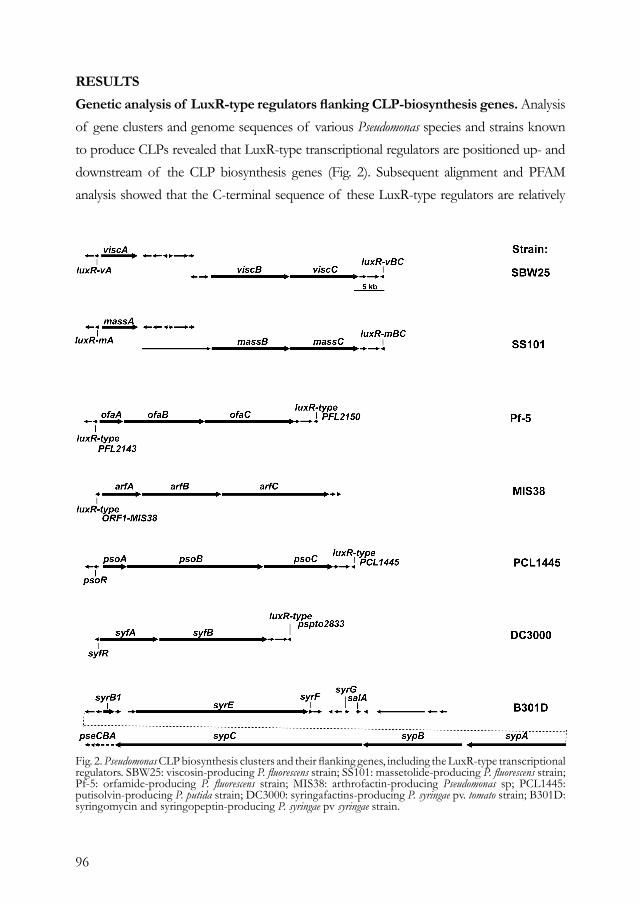
96
RESULTS Genetic analysis of LuxR-type regulators flanking CLP-biosynthesis genes. Analysis of gene clusters and genome sequences of various Pseudomonas species and strains known to produce CLPs revealed that LuxR-type transcriptional regulators are positioned up- and downstream of the CLP biosynthesis genes (Fig. 2). Subsequent alignment and PFAM analysis showed that the C-terminal sequence of these LuxR-type regulators are relatively
Fig. 2. Pseudomonas CLP biosynthesis clusters and their flanking genes, including the LuxR-type transcriptional regulators. SBW25: viscosin-producing P. fluorescens strain; SS101: massetolide-producing P. fluorescens strain; Pf-5: orfamide-producing P. fluorescens strain; MIS38: arthrofactin-producing Pseudomonas sp; PCL1445: putisolvin-producing P. putida strain; DC3000: syringafactins-producing P. syringae pv. tomato strain; B301D: syringomycin and syringopeptin-producing P. syringae pv syringae strain.
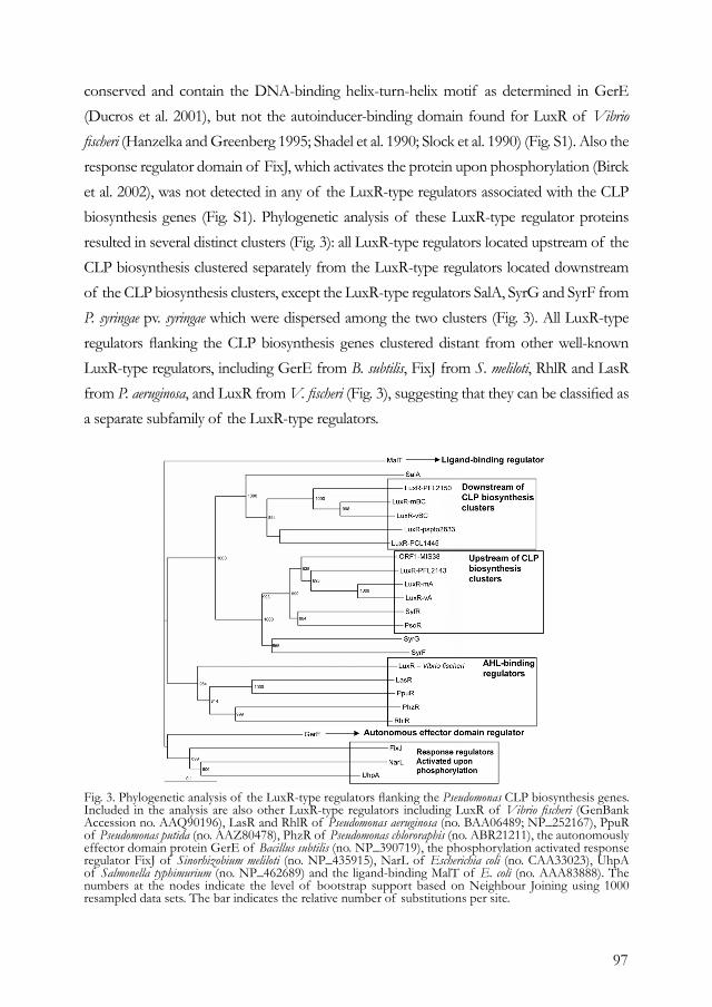
97
conserved and contain the DNA-binding helix-turn-helix motif as determined in GerE (Ducros et al. 2001), but not the autoinducer-binding domain found for LuxR of Vibrio fischeri (Hanzelka and Greenberg 1995; Shadel et al. 1990; Slock et al. 1990) (Fig. S1). Also the response regulator domain of FixJ, which activates the protein upon phosphorylation (Birck et al. 2002), was not detected in any of the LuxR-type regulators associated with the CLP biosynthesis genes (Fig. S1). Phylogenetic analysis of these LuxR-type regulator proteins resulted in several distinct clusters (Fig. 3): all LuxR-type regulators located upstream of the CLP biosynthesis clustered separately from the LuxR-type regulators located downstream of the CLP biosynthesis clusters, except the LuxR-type regulators SalA, SyrG and SyrF from P. syringae pv. syringae which were dispersed among the two clusters (Fig. 3). All LuxR-type regulators flanking the CLP biosynthesis genes clustered distant from other well-known LuxR-type regulators, including GerE from B. subtilis, FixJ from S. meliloti, RhlR and LasR from P. aeruginosa, and LuxR from V. fischeri (Fig. 3), suggesting that they can be classified as a separate subfamily of the LuxR-type regulators.
Fig. 3. Phylogenetic analysis of the LuxR-type regulators flanking the Pseudomonas CLP biosynthesis genes. Included in the analysis are also other LuxR-type regulators including LuxR of Vibrio fischeri (GenBank Accession no. AAQ90196), LasR and RhlR of Pseudomonas aeruginosa (no. BAA06489; NP_252167), PpuR of Pseudomonas putida (no. AAZ80478), PhzR of Pseudomonas chlororaphis (no. ABR21211), the autonomously effector domain protein GerE of Bacillus subtilis (no. NP_390719), the phosphorylation activated response regulator FixJ of Sinorhizobium meliloti (no. NP_435915), NarL of Escherichia coli (no. CAA33023), UhpA of Salmonella typhimurium (no. NP_462689) and the ligand-binding MalT of E. coli (no. AAA83888). The numbers at the nodes indicate the level of bootstrap support based on Neighbour Joining using 1000 resampled data sets. The bar indicates the relative number of substitutions per site.
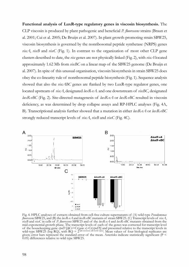
98
Functional analysis of LuxR-type regulatory genes in viscosin biosynthesis. The CLP viscosin is produced by plant pathogenic and beneficial P. fluorescens strains (Braun et al. 2001; Cui et al. 2005; De Bruijn et al. 2007). In plant growth-promoting strain SBW25, viscosin biosynthesis is governed by the nonribosomal peptide synthetase (NRPS) genes viscA, viscB and viscC (Fig. 1). In contrast to the organization of most other CLP gene clusters described to date, the visc genes are not physically linked (Fig. 2), with viscA located approximately 1.62 Mb from viscBC on a linear map of the SBW25 genome (De Bruijn et al. 2007). In spite of this unusual organisation, viscosin biosynthesis in strain SBW25 does obey the co-linearity rule of nonribosomal peptide biosynthesis (Fig. 1). Sequence analysis showed that also the viscABC genes are flanked by two LuxR-type regulator genes, one located upstream of viscA, designated luxR-vA. and one downstream of viscBC, designated luxR-vBC (Fig. 2). Site-directed mutagenesis of luxR-vA or luxR-vBC resulted in viscosin deficiency, as was determined by drop collapse assays and RP-HPLC analyses (Fig. 4A, B). Transcriptional analysis further showed that a mutation in either luxR-vA or luxR-vBC strongly reduced transcript levels of viscA, viscB and viscC (Fig. 4C).
Fig. 4. HPLC analyses of extracts obtained from cell-free culture supernatants of (A) wild type Pseudomonas fluorescens SBW25, and (B) the luxR-vA and luxR-vBC mutants of strain SBW25. (C) Transcript levels of viscA, viscB and viscC in cells of P. fluorescens SBW25 and of the luxR-vA and luxR-vBC mutants obtained from the mid-exponential growth phase. The transcript levels of each of the genes was corrected for transcript level of the housekeeping gene rpoD [ΔCt=Ct(gene x)-Ct(rpoD)] and presented relative to the transcript levels in wild-type SBW25 (log RQ), with RQ = 2-[ΔCt(mutant)-ΔCt(wild type)]. Mean values of four biological replicates are given; error bars represent the standard error of the mean. Asterisks indicate statistically significant (P < 0.05) differences relative to wild type SBW25.
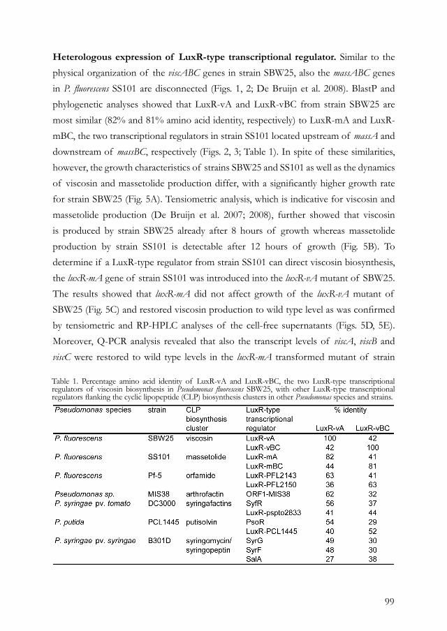
99
Heterologous expression of LuxR-type transcriptional regulator. Similar to the physical organization of the viscABC genes in strain SBW25, also the massABC genes in P. fluorescens SS101 are disconnected (Figs. 1, 2; De Bruijn et al. 2008). BlastP and phylogenetic analyses showed that LuxR-vA and LuxR-vBC from strain SBW25 are most similar (82% and 81% amino acid identity, respectively) to LuxR-mA and LuxR-mBC, the two transcriptional regulators in strain SS101 located upstream of massA and downstream of massBC, respectively (Figs. 2, 3; Table 1). In spite of these similarities, however, the growth characteristics of strains SBW25 and SS101 as well as the dynamics of viscosin and massetolide production differ, with a significantly higher growth rate for strain SBW25 (Fig. 5A). Tensiometric analysis, which is indicative for viscosin and massetolide production (De Bruijn et al. 2007; 2008), further showed that viscosin is produced by strain SBW25 already after 8 hours of growth whereas massetolide production by strain SS101 is detectable after 12 hours of growth (Fig. 5B). To determine if a LuxR-type regulator from strain SS101 can direct viscosin biosynthesis, the luxR-mA gene of strain SS101 was introduced into the luxR-vA mutant of SBW25. The results showed that luxR-mA did not affect growth of the luxR-vA mutant of SBW25 (Fig. 5C) and restored viscosin production to wild type level as was confirmed by tensiometric and RP-HPLC analyses of the cell-free supernatants (Figs. 5D, 5E). Moreover, Q-PCR analysis revealed that also the transcript levels of viscA, viscB and viscC were restored to wild type levels in the luxR-mA transformed mutant of strain
Table 1. Percentage amino acid identity of LuxR-vA and LuxR-vBC, the two LuxR-type transcriptional regulators of viscosin biosynthesis in Pseudomonas fluorescens SBW25, with other LuxR-type transcriptional regulators flanking the cyclic lipopeptide (CLP) biosynthesis clusters in other Pseudomonas species and strains.
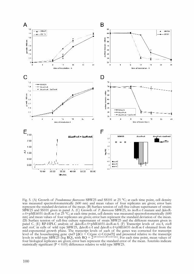
100
Fig. 5. (A) Growth of Pseudomonas fluorescens SBW25 and SS101 at 25 °C; at each time point, cell density was measured spectrofotometrically (600 nm) and mean values of four replicates are given; error bars represent the standard deviation of the mean. (B) Surface tension of cell-free culture supernatant of strains SBW25 and SS101 given in panel A. (C) Growth of P. fluorescens SBW25, its luxR-vA mutant and ΔluxR-vA+pME6031-luxR-mA at 25 °C; at each time point, cell density was measured spectrofotometrically (600 nm) and mean values of four replicates are given; error bars represent the standard deviation of the mean. (D) Surface tension of cell-free culture supernatant of strain SBW25 and the different mutants given in panel C. (E) RP-HPLC analysis of ΔluxR-vA+pME6031-luxR-mA. (F) Transcript levels of viscA, viscB and viscC in cells of wild type SBW25, ΔluxR-vA and ΔluxR-vA+pME6031-luxR-mA obtained from the mid-exponential growth phase. The transcript levels of each of the genes was corrected for transcript level of the housekeeping gene rpoD [ΔCt = Ct(gene x)-Ct(rpoD)] and presented relative to the transcript levels in wild-type SBW25 (log RQ), with RQ = 2-[ΔCt(mutant)-ΔCt(wild type)]. For each time point, mean values of four biological replicates are given; error bars represent the standard error of the mean. Asterisks indicate statistically significant (P < 0.05) differences relative to wild type SBW25.
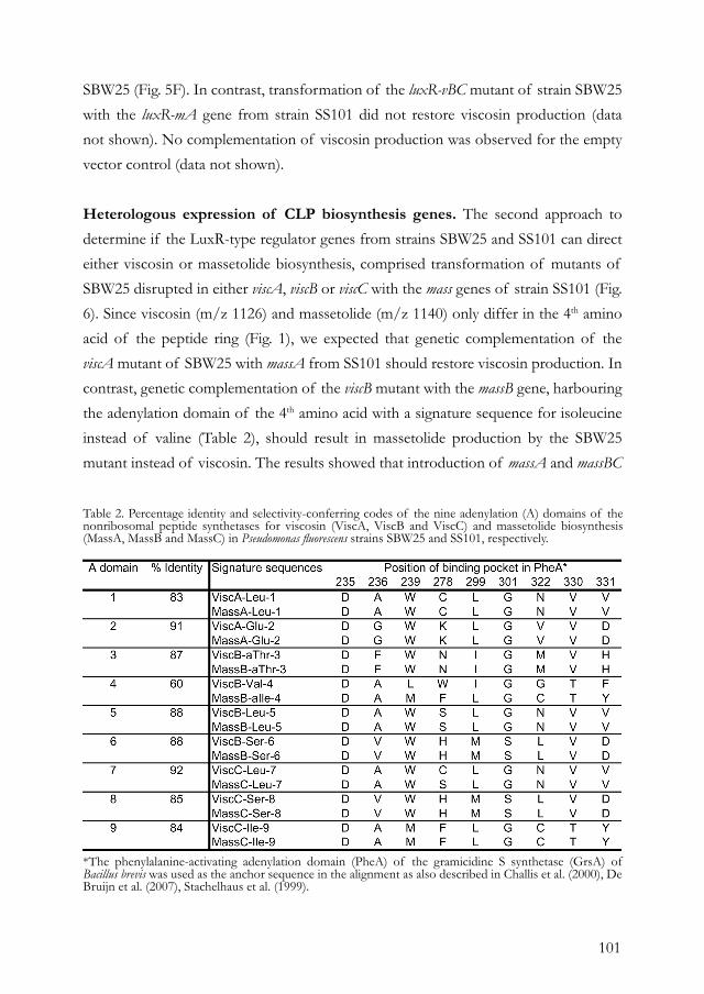
101
SBW25 (Fig. 5F). In contrast, transformation of the luxR-vBC mutant of strain SBW25 with the luxR-mA gene from strain SS101 did not restore viscosin production (data not shown). No complementation of viscosin production was observed for the empty vector control (data not shown).
Heterologous expression of CLP biosynthesis genes. The second approach to determine if the LuxR-type regulator genes from strains SBW25 and SS101 can direct either viscosin or massetolide biosynthesis, comprised transformation of mutants of SBW25 disrupted in either viscA, viscB or viscC with the mass genes of strain SS101 (Fig. 6). Since viscosin (m/z 1126) and massetolide (m/z 1140) only differ in the 4th amino acid of the peptide ring (Fig. 1), we expected that genetic complementation of the viscA mutant of SBW25 with massA from SS101 should restore viscosin production. In contrast, genetic complementation of the viscB mutant with the massB gene, harbouring the adenylation domain of the 4th amino acid with a signature sequence for isoleucine instead of valine (Table 2), should result in massetolide production by the SBW25 mutant instead of viscosin. The results showed that introduction of massA and massBC
Table 2. Percentage identity and selectivity-conferring codes of the nine adenylation (A) domains of the nonribosomal peptide synthetases for viscosin (ViscA, ViscB and ViscC) and massetolide biosynthesis (MassA, MassB and MassC) in Pseudomonas fluorescens strains SBW25 and SS101, respectively.
*The phenylalanine-activating adenylation domain (PheA) of the gramicidine S synthetase (GrsA) of Bacillus brevis was used as the anchor sequence in the alignment as also described in Challis et al. (2000), De Bruijn et al. (2007), Stachelhaus et al. (1999).
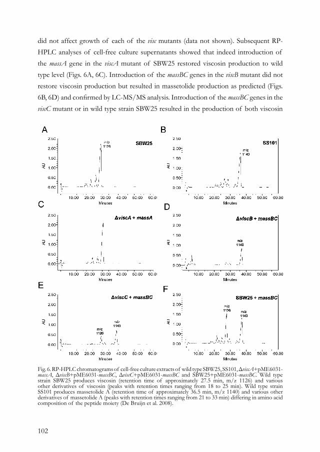
102
did not affect growth of each of the visc mutants (data not shown). Subsequent RP-HPLC analyses of cell-free culture supernatants showed that indeed introduction of the massA gene in the viscA mutant of SBW25 restored viscosin production to wild type level (Figs. 6A, 6C). Introduction of the massBC genes in the viscB mutant did not restore viscosin production but resulted in massetolide production as predicted (Figs. 6B, 6D) and confirmed by LC-MS/MS analysis. Introduction of the massBC genes in the viscC mutant or in wild type strain SBW25 resulted in the production of both viscosin
Fig. 6. RP-HPLC chromatograms of cell-free culture extracts of wild type SBW25, SS101, ΔviscA+pME6031-massA, ΔviscB+pME6031-massBC, ΔviscC+pME6031-massBC and SBW25+pME6031-massBC. Wild type strain SBW25 produces viscosin (retention time of approximately 27.5 min, m/z 1126) and various other derivatives of viscosin (peaks with retention times ranging from 18 to 25 min). Wild type strain SS101 produces massetolide A (retention time of approximately 36.5 min, m/z 1140) and various other derivatives of massetolide A (peaks with retention times ranging from 21 to 33 min) differing in amino acid composition of the peptide moiety (De Bruijn et al. 2008).
103
and massetolide (Figs. 6E, 6F). Transformation of the luxR-vA mutant with massA or massBC did not restore viscosin production (data not shown), indicating that a functional luxR-vA gene is required for heterologous expression of the mass genes. These results also show that large NRPS genes encoding CLPs can be exchanged between strains and lead to the production of non-native compounds.
DISCUSSIONThe superfamily of LuxR-type transcriptional regulators consists of proteins with a DNA-binding helix-turn-helix domain in the C-terminus. Based on specific domains in the N-terminal part, these proteins are grouped into two major subfamilies: those containing autoinducer-binding domains and those harbouring response regulator domains. This study focused specifically on LuxR-type transcriptional regulators flanking CLP biosynthesis genes in various Pseudomonas species and strains. The results showed that all these LuxR-type regulators contain a DNA-binding helix-turn-helix domain, but do not contain an autoinducer-binding domain nor a response regulator domain, suggesting that they belong to another subfamily of LuxR-type regulators. For P. fluorescens strain SBW25, we subsequently showed that each of the two LuxR-type regulator genes, designated luxR-vA and luxR-vBC, flanking the viscosin biosynthesis genes are essential for viscosin production. Site-directed mutagenesis of luxR-vA or luxR-vBC strongly reduced transcript levels of each of the three visc biosynthesis genes and resulted in loss of viscosin production. These results confirm in part previous studies with P. syringae (Berti et al. 2007; Kitten et al. 1998; Lu et al. 2002) and P. putida (Dubern et al. 2008) where the importance of LuxR-type regulatory genes for CLP biosynthesis was addressed. In P. syringae pv. tomato DC3000, however, only the LuxR-type regulator SyfR upstream of the syringafactin genes appeared to be essential, whereas a mutation of the LuxR-type regulator located downstream did not affect syringafactin biosynthesis (Berti et al. 2007). In P. putida PCL1445, only the LuxR-type regulator psoR, located upstream of the putisolvin genes, was investigated for its role in putisolvin production (Dubern et al. 2008), but the function of the other LuxR-type regulator downstream of the putisolvin genes has, to our knowledge, not been resolved yet. It is intriguing that the viscosin biosynthesis genes in strain SBW25 are regulated by two LuxR-type regulators. Whether this is due to the unusual physical organization of the visc genes is not known. In P. syringae pv. syringae, three LuxR-type

104
regulators, designated SalA, SyrF and SyrG, were identified flanking the syringomycin/syringopeptin biosynthesis clusters (Lu et al. 2002) of which only SalA and SyrF were shown to be essential for syringomycin/syringopeptin biosynthesis (Kitten et al. 1998; Lu et al. 2002; Lu et al. 2005). Like most LuxR-type regulators, both SalA and SyrF act as transcriptional activators regulating all of the syr-syp genes, including genes involved in biosynthesis, regulation and efflux (Lu et al. 2005; Wang et al. 2006a). Moreover, it was shown that SyrF binds to the region containing the syr-syp box, which was identified around the -35 region of the syr-syp genes and is required for syrB1 expression (Wang et al. 2006b). However, SalA does not bind to the syr-syp box, but only to the promoter region of SyrF, showing that the control of expression of the syr-syp genes by SalA is mediated through SyrF (Wang et al. 2006a). Upon activation, multimerization of the LuxR-type transcriptional regulators is critical for DNA-binding (Ducros et al. 2001; Pristovsek et al. 2003; Zhang et al. 2002). It was shown that SalA and SyrF form dimers in vitro, in absence of an autoinducer, which again shows that these LuxR-type regulators do not belong to the autoinducer subfamily. SalA and SyrF also do not contain the phosphorylation domains of response regulators, although their phosphorylation status is not known. Whether LuxR-vA in P. fluorescens strain SBW25 binds to the promoter region of LuxR-vBC and regulates its expression or vice versa, was not addressed in this study and will require promoter binding studies as performed by Wang et al (2006). Comparative analyses of the different CLP biosynthesis clusters in different Pseudomonas strains further showed a high level of similarity, both in physical organization and in amino acid identities, between the viscABC genes of P. fluorescens SBW25 and the massABC genes of P. fluorescens SS101. Also the LuxR-type transcriptional regulators flanking these two CLP biosynthesis clusters showed high amino acid identities. Results further showed that transformation of the luxR-vA mutant of SBW25 with the luxR-mA gene of strain SS101 restored transcription of the viscABC genes and viscosin production to wild type levels. These results indicate that these LuxR-type transcriptional regulators can be exchanged between different strains, thereby regulating the biosynthesis of structurally different CLPs. However, what the prerequisites (i.e. amino acid identity, specific residues) are for successful heterologous expression of LuxR-type regulators is yet unclear. The assembly line by which the NRPSs build the CLPs consists of several enzymatic steps, including amino acid selection, activation and incorporation followed

105
by cyclization of the peptide moiety (Finking and Marahiel 2004). Because of the modular structure of the NRPSs and the co-linearity of the assembly process, the difference between viscosin and massetolide A is conferred by the fourth adenylation (A) domain located in ViscB and MassB, respectively. This was confirmed in this study where transfer of the massB gene of strain SS101 into a viscB mutant of strain SBW25 resulted in the production of massetolide A instead of viscosin. This result also demonstrated that the assembly line for CLP biosynthesis in Pseudomonas can be altered leading to the production of non-native products. Another example of this type of combinatorial biosynthesis was performed by Eppelman et al. (2001) who showed that expression of the complete bacitracin biosynthesis cluster of the slow growing Bacillus licheniformis in a surfactin-deficient (srfA) mutant of B. subtilis resulted in elevated levels of bacitracin as compared to the natural producer. Combinatorial biosynthesis is not only used to improve production in a suitable host or enable expression of biosynthesis clusters from unculturable organisms, but it also allows the generation of structurally novel compounds (Bode and Muller 2005). For example, genetic engineering of the NRPS biosynthetic gene cluster of daptomycin and carotenoids resulted in a library of novel compounds, which have substantial potential for the medical, pharmaceutical and food industry (Nguyen et al. 2006; Schmidt-Dannert et al. 2000). Other examples of exchanging parts or complete biosynthesis clusters have been described previously (Cha et al. 2008; Lee et al. 2007; Stachelhaus et al. 1995) and showed that product yields of the non-native or structurally novel NRPS/PKS products are in many cases relatively low. Whether this is related to differences in genetic regulation of the genes, poor communication between the different NRPSs, or due to limitations in the efflux of the final products are intriguing questions to address. With respect to communication between NRPSs, specific domains were identified in Bacillus brevis and Bacillus subtilis for intermolecular communication between NRPS proteins. These so-called COM domains are located at the C-terminus of the aminoacyl-donating NRPS (donor COM) and at the N-terminus of the accepting partner NRPS (acceptor COM) (Chiocchini et al. 2006; Hahn and Stachelhaus 2004; 2006; Weissman and Muller 2008a). However, alignment of the N- and C-termini of the massetolide and viscosin NRPSs with the tyrocidine and surfactin NRPSs showed that the COM domains found in Bacillus were absent in Pseudomonas (data not shown). Also the higher proportion of acidic amino acids in the donor COM domains and the polar amino acids in the acceptor
106
COM domains described for Bacillus (Weissman and Muller 2008b) were absent in the Visc and Mass NRPSs, suggesting that the mechanisms underlying intermolecular communication between NRPSs involved in CLP biosynthesis in Pseudomonas are different from Bacillus.
MATERIALS AND METHODSBacterial strains and culture conditions. P. fluorescens strains SBW25 and SS101 were grown on Pseudomonas agar F (Difco) plates or in liquid King’s medium B (KB) at 25°C. The biosynthesis mutants ΔviscA, ΔviscB and ΔviscC were obtained as described previously (De Bruijn et al. 2007). Escherichia coli strain DH5α was used as a host for the plasmids used in site-directed mutagenesis and genetic complementation. E. coli strains were grown on Luria-Bertani (LB) plates or in LB broth amended with the appropriate antibiotics.
In silico analyses. Identification of the genes flanking the CLP biosynthesis genes was performed by either BlastX or BlastP analysis. For phylogenetic analyses, alignments were made with CLUSTALX (version 1.81) and trees were inferred by Neighbour Joining using 1000 bootstrap replicates. Autoinducer and response regulator domains were identified by PFAM analysis (http://pfam.sanger.ac.uk/search?tab=searchSequenceBlock).
Site-directed mutagenesis. Site-directed mutagenesis of the luxR-vA and luxR-vBC genes was performed based on the method described by Choi and Schweizer (2005). For each construct used in site-directed mutagenesis, three fragments were amplified: a 5’ (Up) fragment, the FRT-Gm-FRT cassette and a 3’ (Dn) fragment. The primers used for amplification are described in Table 3. In the 1st round PCR, the FRT-Gm-FRT cassette, the 5’ and 3’ fragments were amplified. In the 2nd round PCR, these three fragments were coupled by overlap extension PCR. The 5’ and 3’ fragments were chosen in such a way that when homologous recombination in Pseudomonas takes place, the FRT-Gm-FRT cassette is inserted approximately 383 bp after the start of the luxR-vA and 192 bp after the start of luxR-vBC open reading frames (ORF). The FRT-Gm-FRT cassette was amplified with a derivative of pPS854 (Hoang et al. 1998), pPS854-GM was used as a template and FRT-F and FRT-R were used as primers (Table 3). The 1st round PCR was performed with KOD polymerase (Novagen) according to the manufacturer’s protocol. The program used for the PCR reaction consists of 2 min of denaturation at 95 °C, followed by 5 cycli of subsequently 95 °C, 55 °C and 68 °C, each for 20 s. The PCR amplification was proceeded with 25 cycli of subsequently 95 °C, 60 °C and 68 °C, each for 20 s. The last step of the PCR was 68 °C for 7 min. All fragments were run on a 1% agarose gel and purified with NucleoSpin kit (Macherey-Nagel). The 2nd round PCR was performed by adding equimolar amounts of the 5’ fragment, FRT-Gm-FRT and 3’ fragments to milliQ, dNTPS, KOD buffer and KOD polymerase to a total of 47 μl. The PCR was started by 2 min of denaturation at 95 °C, followed by 3 cycli of subsequently 95 °C, 55 °C and 68 °C, for 20, 30 and 60 sec, respectively. In the 3rd extension cycle, 1.5 μl of the Up forward and Dn reverse primer (10 μM) was added. The PCR amplification was proceeded with 25 cycli of subsequently 95 °C, 58 °C and 68 °C, for 20, 20 and 120 s, respectively. The last step of the PCR was 68 °C for 7 min. All fragments were run on a 1% agarose gel and bands of the predicted size were purified with NucleoSpin kit. The fragments were digested with BamHI or KpnI and cloned into BamHI- or KpnI-digested NucleoSpin purified plasmid pEX18Tc. E. coli DH5α was transformed with the obtained pEX18Tc-luxR-vA and pEX18Tc-luxR-vBC plasmids by heat-shock transformation according to Innoue et al (1990) and transformed colonies were selected on LB supplemented with 25 μg/ml gentamicin (Sigma). Integration of the inserts was verified by PCR analysis with pEX18Tc primers (Table 1) and by restriction analysis of isolated plasmids. The plasmids were verified by sequencing by BaseClear (Leiden, the Netherlands). The correct pEX18Tc-luxR-vA and pEX18Tc-luxR-vBC constructs were subsequently electroporated into strain SBW25. Electrocompetent cells were obtained according to Choi et al (2006) by washing the cells three times with 300 mM sucrose from a 6 ml overnight culture and finally dissolving the cells in 100 μl of 300 mM sucrose. Electroporation occurred at 2.4 kV, 200 μF, and after incubation in SOC medium (2% Bacto tryptone [Difco], 0.5% Bacto yeast extract [Difco], 10 mM NaCl, 2.5 mM KCl, 10 mM MgCl2, 10 mM MgSO4, 20 mM glucose [pH 7]) for 2 hrs at 25 °C, cells were plated on KB supplemented with gentamicin (25 μg/ml). Obtained colonies were grown in LB for 1 hr at 25 °C and plated on LB supplemented with gentamicin (25 μg/ml) and 5% sucrose to accomplish double crossing over. Plates were incubated at 25 °C for at least 48 hrs and colonies were re-streaked on LB supplemented with gentamicin and 5% sucrose and were streaked on LB supplemented with tetracycline (25 μg/ml). Colonies that did grow on LB with gentamicin, but not on LB with tetracycline were selected and subjected to colony PCR to confirm the presence of the gentamicin resistance cassette and the absence of the tetracycline resistance cassette. Positive colonies were confirmed by sequencing the PCR fragments obtained with the Up forward and Dn reverse primers. The obtained luxR-vA and luxR-vBC mutants were tested for viscosin production by a drop collapse assay and by RP-HPLC as described in De Bruijn et al (2008).
107
Construction of pME6031-based vectors for heterologous expression. Generation of the pME6031-massA and pME6031-massBC constructs are described in De Bruijn et al (2008). The pME6031-luxR-mA construct was generated as follows: a 1817 bp fragment was obtained by PCR (Table 3) with Phusion DNA polymerase (Finnzymes). This PCR fragment was subcloned in pGEM-T Easy vector systems I (Promega) according to the manufacturer’s instructions, and the obtained plasmid was digested with EcoRI. The fragment was excised from gel and isolated with the NucleoTrap kit (Macherey-Nagel). Fragments were cloned into the shuttle vector pME6031 (Heeb et al. 2000), which was digested, dephosphorylated (by shrimp alkaline phosphatase; Promega), and purified with the NucleoTrap kit according to the manufacturer’s instructions. E. coli DH5α was transformed with the obtained plasmid pME6031-luxR-mA by heat shock transformation (Inoue et al. 1990), and transformed colonies were selected on LB agar plates supplemented with tetracycline (25 μg/ml). Integration of the inserts was verified by PCR analysis and restriction analysis of isolated plasmids. The correct pME6031-massA, pME6031-massBC and pME6031-luxR-mA were subsequently electroporated into strain SBW25, and the viscA, viscB, viscC and luxR-vA mutants. Electrocompetent cells were obtained by washing the cells three times with 1 mM MOPS (morpholinepropanesulfonic acid) and 15% glycerol from a 5-ml overnight culture and finally dissolving the cells in 100 μl of the washing buffer. Cells were stored at -80°C for at least 1 hour prior to electroporation. Electroporation occurred at 2.4 kV and 200 μF, and after incubation in SOC medium for 2 h at 25°C, cells were plated on KB supplemented with tetracycline (25 μg/ml). Verification of transformation was performed by PCR analysis using one primer specific for the insert and one primer specific for the pME6031 vector. Viscosin and massetolide A production in the transformed strain SBW25 and mutants was tested with a drop collapse assay followed by RP-HPLC and LC-MS/MS analyses as described previously (De Bruijn et al. 2008).
Table 3. Primers used in this study. The 5’ end of the Up reverse and Dn forward primers for site-directed mutagenesis contain a 25-bp sequence (small letters) complementary to the FRT-F and FRT-R primers for overlap extension in the 2nd round PCR. The 5’ end of the Up forward and Dn reverse primers contain a restriction site (underlined) for BamHI, which is required for cloning into pEX18Tc.
108
Surface tension measurements and transcriptional analysis. Cells were grown in a 24-well plate with 1.25 ml KB broth per well and shaken at 220 rpm at 25°C. At specific time points, growth was determined by measuring 100 μl in a 96-wells plate in a Biorad-680 Microplate Reader at 600 nm. From each culture, 1 ml was collected and spun down. The cells were frozen in liquid N2 and stored at -80°C. For the RNA isolations and cDNA synthesis, four biological replicates were used for each time point. Biosurfactant production was measured qualitatively by the drop collapse assay and quantitatively by tensiometric analysis of the cell-free supernatant (K6 tensiometer; Kruss GmbH, Hamburg, Germany) at room temperature. To get sufficient volume for the tensiometric analysis, the supernatants of four biological replicates were collected and pooled for each time point. The surface tension of each sample was measured in triplicate. RNA isolation and Q-PCR analysis were performed as described previously (De Bruijn et al. 2008). The primers used for the Q-PCR are described in Table 3. To correct for small differences in template concentration, rpoD was used as a housekeeping gene. The cycle where the SYBR green fluorescence crosses a manually set threshold cycle (CT) was used to determine transcript levels. For each gene the threshold was fixed based on the exponential segment of the PCR curve. The CT value for viscA was corrected for the housekeeping gene as follows: ΔCt= Ct(viscA)-Ct(rpoD). The same formula was used for the other genes. The relative quantification (RQ) values were calculated by the formula RQ = 2-[ΔCt(mutant)-
ΔCt(wild type)]. If there is no difference in transcript level between mutant and wild type, than RQ = 1 (20) and log RQ = 0. Q-PCR analysis was performed in duplicate (technical replicates) on four independent RNA isolations (biological replicates). Statistically significant differences were determined for log-transformed RQ values by analysis of variance (P < 0.05) followed by the Bonferroni post hoc multiple comparisons.
ACKNOWLEDGEMENTSWe are grateful to Teris van Beek and Frank Claasen for their assistance in reverse-phase HPLC and LC-MS/MS. We are also grateful to Teresa Sweat (USDA, Corvallis, OR) and Dimitri Mavrodi (USDA, Pullman, WA) for providing the plasmids (pEX18Tc and pPS854-Gm), protocols and advice for the site-directed mutagenesis. This work was funded by the Dutch Technology Foundation (STW) and the applied science division of NWO and Productschap Tuinbouw.
REFERENCESAndersen, J. B., Koch, B., Nielsen, T. H., Sorensen, D., Hansen, M., Nybroe, O., Christophersen, C., Sorensen, J., Molin, S., and Givskov, M. 2003. Surface motility in Pseudomonas sp. DSS73 is required for efficient biological containment of the root-pathogenic microfungi Rhizoctonia solani and Pythium ultimum. Microbiology 149:37-46.Berti, A. D., Greve, N. J., Christensen, Q. H., and Thomas, M. G. 2007. Identification of a biosynthetic gene cluster and the six associated lipopeptides involved in swarming motility of Pseudomonas syringae pv. tomato DC3000. J. Bacteriol. 189:6312-6323.Birck, C., Malfois, M., Svergun, D., and Samama, J. 2002. Insights into signal transduction revealed by the low resolution structure of the FixJ response regulator. J. Mol. Biol. 321:447-57.Bode, H. B., and Muller, R. 2005. The impact of bacterial genomics on natural product research. Angew. Chem. Int. Ed. Engl. 44:6828-46.Braun, P. G., Hildebrand, P. D., Ells, T. C., and Kobayashi, D. Y. 2001. Evidence and characterization of a gene cluster required for the production of viscosin, a lipopeptide biosurfactant, by a strain of Pseudomonas fluorescens. Can. J. Microbiol. 47:294-301.Cha, M., Lee, N., Kim, M., Kim, M., and Lee, S. 2008. Heterologous production of Pseudomonas aeruginosa EMS1 biosurfactant in Pseudomonas putida. Bioresour. Technol. 99:2192-9.
Challis, G.L., and Ravel, J. 2000. Predictive, structure-based model of amino acid recognition by nonribosomal peptide synthetase adenylation domains. Chem. Biol. 7: 211-224.Chiocchini, C., Linne, U., and Stachelhaus, T. 2006. In vivo biocombinatorial synthesis of lipopeptides by COM domain-mediated reprogramming of the surfactin biosynthetic complex. Chem. Biol. 13:899-908.Choi, K. H., Kumar, A., and Schweizer, H. P. 2006. A 10-min method for preparation of highly electrocompetent Pseudomonas aeruginosa cells: application for DNA fragment transfer between chromosomes and plasmid transformation. J. Microbiol. Methods. 64:391-7.Choi, K. H., and Schweizer, H. P. 2005. An improved method for rapid generation of unmarked Pseudomonas aeruginosa deletion mutants. BMC Microbiol. 5:30.Cui, X., Harling, R., Mutch, P., and Darling, D. 2005. Identification of N-3-hydroxyoctanoyl-homoserine lactone production in Pseudomonas fluorescens 5064, pathogenic to broccoli, and controlling biosurfactant production by quorum sensing. Eur. J. Plant Pathol. 111:297-308.De Bruijn, I., de Kock, M. J., de Waard, P., van Beek, T. A., and Raaijmakers, J. M. 2008. Massetolide A biosynthesis in Pseudomonas fluorescens. J. Bacteriol. 190:2777-2789.De Bruijn, I., de Kock, M. J. D., Yang, M., de Waard, P., van Beek, T. A., and Raaijmakers,
109
J. M. 2007. Genome-based discovery, structure prediction and functional analysis of cyclic lipopeptide antibiotics in Pseudomonas species. Mol. Microbiol. 63:417-428.Dubern, J.-F., Lagendijk, E. L., Lugtenberg, B. J. J., and Bloemberg, G. V. 2005. The heat shock genes dnaK, dnaJ, and grpE are involved in regulation of putisolvin biosynthesis in Pseudomonas putida PCL1445. J. Bacteriol. 187:5967-5976.Dubern, J. F., Coppoolse, E. R., Stiekema, W. J., and Bloemberg, G. V. 2008. Genetic and functional characterization of the gene cluster directing the biosynthesis of putisolvin I and II in Pseudomonas putida strain PCL1445. Microbiology 154:2070-83.Dubern, J. F., Lugtenberg, B. J. J., and Bloemberg, G. V. 2006. The ppuI-rsaL-ppuR quorum-sensing system regulates biofilm formation of Pseudomonas putida PCL1445 by controlling biosynthesis of the cyclic lipopeptides putisolvins I and II. J. Bacteriol. 188:2898-2906.Ducros, V. M., Lewis, R. J., Verma, C. S., Dodson, E. J., Leonard, G., Turkenburg, J. P., Murshudov, G. N., Wilkinson, A. J., and Brannigan, J. A. 2001. Crystal structure of GerE, the ultimate transcriptional regulator of spore formation in Bacillus subtilis. J. Mol. Biol. 306:759-71.Dumenyo, C. K., Mukherjee, A., Chun, W., and Chatterjee, A. K. 1998. Genetic and physiological evidence for the production of N-acyl homoserine lactones by Pseudomonas syringae pv. syringae and other fluorescent plant pathogenic Pseudomonas species. Eur. J. Plant Pathol. 104:569-582.Eppelmann, K., Doekel, S., and Marahiel, M. A. 2001. Engineered biosynthesis of the peptide antibiotic bacitracin in the surrogate host Bacillus subtilis. J. Biol. Chem. 276:34824-31.Finking, R., and Marahiel, M. A. 2004. Biosynthesis of nonribosomal peptides. Annu. Rev. Microbiol. 58:453-488.Fuqua, C., Winans, S. C., and Greenberg, E. P. 1996. Census and consensus in bacterial ecosystems: the LuxR-LuxI family of quorum-sensing transcriptional regulators. Annu. Rev. Microbiol. 50:727-51.Gross, H., Stockwell, V. O., Henkels, M. D., Nowak-Thompson, B., Loper, J. E., and Gerwick, W. H. 2007. The genomisotopic approach: a systematic method to isolate products of orphan biosynthetic gene clusters. Chem. Biol. 14:53-63.Hahn, M., and Stachelhaus, T. 2004. Selective interaction between nonribosomal peptide synthetases is facilitated by short communication-mediating domains. Proc. Natl. Acad. Sci. USA 101:15585-90.Hahn, M., and Stachelhaus, T. 2006. Harnessing the potential of communication-mediating domains for the biocombinatorial synthesis of nonribosomal peptides. Proc. Natl. Acad. Sci. USA 103:275-80.Hanzelka, B. L., and Greenberg, E. P. 1995. Evidence that the N-terminal region of the Vibrio
fischeri LuxR protein constitutes an autoinducer-binding domain. J. Bacteriol. 177:815-7.Heeb, S., Itoh, Y., Nishijyo, T., Schnider, U., Keel, C., Wade, J., Walsh, U., O‘Gara, F., and Haas, D. 2000. Small, stable shuttle vectors based on the minimal pVS1 replicon for use in gram-negative, plant-associated bacteria. Mol. Plant-Microbe Interact. 13:232-237.Hoang, T. T., Karkhoff-Schweizer, R. R., Kutchma, A. J., and Schweizer, H. P. 1998. A broad-host-range Flp-FRT recombination system for site-specific excision of chromosomally-located DNA sequences: application for isolation of unmarked Pseudomonas aeruginosa mutants. Gene 212:77-86.Inoue, H., Nojima, H., and Okayama, H. 1990. High efficiency transformation of Escherichia coli with plasmids. Gene 96:23-28.Kinscherf, T. G., and Willis, D. K. 1999. Swarming by Pseudomonas syringae B728a requires gacS (lemA) and gacA but not the acyl-homoserine lactone biosynthetic gene ahlI. J. Bacteriol. 181:4133-6.Kitten, T., Kinscherf, T. G., McEvoy, J. L., and Willis, D. K. 1998. A newly identified regulator is required for virulence and toxin production in Pseudomonas syringae. Mol. Microbiol. 28:917-929.Koch, B., Nielsen, T. H., Sorensen, D., Andersen, J. B., Christophersen, C., Molin, S., Givskov, M., Sorensen, J., and Nybroe, O. 2002. Lipopeptide production in Pseudomonas sp. strain DSS73 is regulated by components of sugar beet seed exudate via the Gac two-component regulatory system. Appl. Environ. Microbiol. 68:4509-16.Lee, Y. K., Yoon, B. D., Yoon, J. H., Lee, S. G., Song, J. J., Kim, J. G., Oh, H. M., and Kim, H. S. 2007. Cloning of srfA operon from Bacillus subtilis C9 and its expression in E. coli. Appl. Microbiol. Biotechnol. 75:567-72.Lu, S. E., Scholz-Schroeder, B. K., and Gross, D. C. 2002. Characterization of the salA, syrF, and syrG regulatory genes located at the right border of the syringomycin gene cluster of Pseudomonas syringae pv. syringae. Mol. Plant-Microbe Interact.15:43-53.Lu, S. E., Wang, N., Wang, J., Chen, Z. J., and Gross, D. C. 2005. Oligonucleotide microarray analysis of the salA regulon controlling phytotoxin production by Pseudomonas syringae pv. syringae. Mol. Plant-Microbe Interact. 18:324-333.Nguyen, K. T., Ritz, D., Gu, J. Q., Alexander, D., Chu, M., Miao, V., Brian, P., and Baltz, R. H. 2006. Combinatorial biosynthesis of novel antibiotics related to daptomycin. Proc. Natl. Acad. Sci. USA 103:17462-7.Pristovsek, P., Sengupta, K., Lohr, F., Schafer, B., von Trebra, M. W., Ruterjans, H., and Bernhard, F. 2003. Structural analysis of the DNA-binding domain of the Erwinia amylovora RcsB protein and its interaction with the RcsAB box. J. Biol. Chem. 278:17752-9.Raaijmakers, J. M., De Bruijn, I., and De Kock, M. J. D. 2006. Cyclic lipopeptide production by plant-associated Pseudomonas spp.:
110
diversity, activity, biosynthesis, and regulation. Mol. Plant-Microbe Interact. 19:699-710.Rainey, P. B., and Rainey, K. 2003. Evolution of cooperation and conflict in experimental bacterial populations. Nature 425:72-4.Schlegel, A., Bohm, A., Lee, S. J., Peist, R., Decker, K., and Boos, W. 2002. Network regulation of the Escherichia coli maltose system. J. Mol. Microbiol. Biotechnol. 4:301-7.Schmidt-Dannert, C., Umeno, D., and Arnold, F. H. 2000. Molecular breeding of carotenoid biosynthetic pathways. Nat. Biotechnol. 18:750-3.Shadel, G. S., Young, R., and Baldwin, T. O. 1990. Use of regulated cell lysis in a lethal genetic selection in Escherichia coli: identification of the autoinducer-binding region of the LuxR protein from Vibrio fischeri ATCC 7744. J. Bacteriol. 172:3980-7.Sieber, S. A., and Marahiel, M. A. 2005. Molecular mechanisms underlying nonribosomal peptide synthesis: approaches to new antibiotics. Chem. Rev. 105:715-738.Slock, J., VanRiet, D., Kolibachuk, D., and Greenberg, E. P. 1990. Critical regions of the Vibrio fischeri LuxR protein defined by mutational analysis. J. Bacteriol. 172:3974-9.Stachelhaus, T., Mootz, H.D., and Marahiel, M.A. 1999. The specificity-conferring code of adenylation domains in nonribosomal peptide synthetases. Chem. Biol. 6: 493-505.Stachelhaus, T., Schneider, A., and Marahiel, M. A. 1995. Rational design of peptide antibiotics by targeted replacement of bacterial
and fungal domains. Science 269:69-72.Timms-Wilson, T. M., Ellis, R. J., Renwick, A., Rhodes, D. J., Mavrodi, D. V., Weller, D. M., Thomashow, L. S., and Bailey, M. J. 2000. Chromosomal insertion of phenazine-1-carboxylic acid biosynthetic pathway enhances efficacy of damping-off disease control by Pseudomonas fluorescens. Mol. Plant-Microbe Interact. 13:1293-300.Wang, N., Lu, S. E., Records, A. R., and Gross, D. C. 2006a. Characterization of the transcriptional activators SalA and SyrF, which are required for syringomycin and syringopeptin production by Pseudomonas syringae pv. syringae. J. Bacteriol. 188:3290-3298.Wang, N., Lu, S. E., Yang, Q., Sze, S. H., and Gross, D. C. 2006b. Identification of the syr-syp box in the promoter regions of genes dedicated to syringomycin and syringopeptin production by Pseudomonas syringae pv. syringae B301D. J. Bacteriol. 188:160-8.Weissman, K. J., and Muller, R. 2008a. Crystal structure of a molecular assembly line. Angew. Chem. Int. Ed. Engl. 47:8344-8346.Weissman, K. J., and Muller, R. 2008b. Protein-protein interactions in multienzyme megasynthetases. Chembiochem 9:826-48.Zhang, R. G., Pappas, T., Brace, J. L., Miller, P. C., Oulmassov, T., Molyneaux, J. M., Anderson, J. C., Bashkin, J. K., Winans, S. C., and Joachimiak, A. 2002. Structure of a bacterial quorum-sensing transcription factor complexed with pheromone and DNA. Nature 417:971-4.
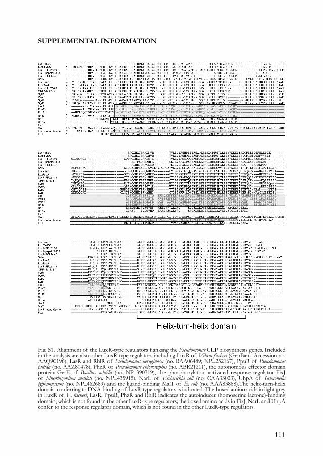
111
SUPPLEMENTAL INFORMATION
Fig. S1. Alignment of the LuxR-type regulators flanking the Pseudomonas CLP biosynthesis genes. Included in the analysis are also other LuxR-type regulators including LuxR of Vibrio fischeri (GenBank Accession no. AAQ90196), LasR and RhlR of Pseudomonas aeruginosa (no. BAA06489; NP_252167), PpuR of Pseudomonas putida (no. AAZ80478), PhzR of Pseudomonas chlororaphis (no. ABR21211), the autonomous effector domain protein GerE of Bacillus subtilis (no. NP_390719), the phosphorylation activated response regulator FixJ of Sinorhizobium meliloti (no. NP_435915), NarL of Escherichia coli (no. CAA33023), UhpA of Salmonella typhimurium (no. NP_462689) and the ligand-binding MalT of E. coli (no. AAA83888).The helix-turn-helix domain conferring to DNA-binding of LuxR-type regulators is indicated. The boxed amino acids in light grey in LuxR of V. fischeri, LasR, PpuR, PhzR and RhlR indicates the autoinducer (homoserine lactone)-binding domain, which is not found in the other LuxR-type regulators; the boxed amino acids in FixJ, NarL and UhpA confer to the response regulator domain, which is not found in the other LuxR-type regulators.

112
113
CHAPTER 5
Journal of Bacteriology (2008)
Regulation of cyclic lipopeptide biosynthesis in Pseudomonas fl uorescens by the ClpP protease.
Irene de Bruijn1 and Jos M. Raaijmakers1
1Laboratory of Phytopathology, Wageningen University, 6709 PD Wageningen, the Netherlands.
Accepted 23 December 2008.
114
ABSTRACT Cyclic lipopeptides produced by Pseudomonas species exhibit potent surfactant and broad-spectrum antibiotic properties. Their biosynthesis is governed by large multimodular nonribosomal peptide synthetases, but little is known about the genetic regulatory network. This study provides, for the first time, evidence that the serine protease ClpP regulates the biosynthesis of massetolides, cyclic lipopeptides involved in swarming motility, biofilm formation and antimicrobial activity of Pseudomonas fluorescens SS101. The results show that ClpP affects expression of luxR-mA, the transcriptional regulator of the massetolide biosynthesis genes massABC, thereby regulating biofilm formation and swarming motility of P. fluorescens SS101. Transcription of luxR-mA was significantly repressed in the clpP mutant and introduction of luxR-mA restored, in part, massetolide biosynthesis and swarming motility of the clpP mutant. Site-directed mutagenesis and expression analyses indicated that the chaperone subunit ClpX and the Lon protease are not involved in regulation of massetolide biosynthesis and are transcribed independently from clpP. Addition of casamino acids enhanced transcription of luxR-mA and massABC in the clpP mutant, leading to a partial rescue of massetolide production and swarming motility. Results further suggested that, at the transcriptional level, ClpP-mediated regulation of massetolide biosynthesis operates independently from regulation by the GacA/GacS two-component system. The role of amino acid metabolism and putative mechanisms underlying ClpP-mediated regulation of cyclic lipopeptide biosynthesis, swarming motility and growth in P. fluorescens are discussed.
115
INTRODUCTIONCyclic lipopeptides are versatile metabolites produced by a variety of bacterial genera, including Pseudomonas and Bacillus (Nybroe and Sorensen 2004; Ongena and Jacques 2008; Raaijmakers et al. 2006). They are composed of a short cyclic oligopeptide linked to a fatty acid tail and exhibit potent surfactant properties (Raaijmakers et al. 2006). Cyclic lipopeptides have received considerable attention for their antibiotic activity against a range of human and plant pathogenic organisms, including enveloped viruses, mycoplasmas, trypanosomes, bacteria, fungi, and oomycetes (Raaijmakers et al. 2006). For plant-associated Pseudomonas species, cyclic lipopeptides play an important role in swarming motility, biofilm formation and virulence (Andersen et al. 2003; Berti et al. 2007; De Bruijn et al. 2008; De Bruijn et al. 2007; Deziel et al. 2003; Gross et al. 2007; Kuiper et al. 2004; Raaijmakers et al. 2006; Roongsawang et al. 2003). Cyclic lipopeptide biosynthesis is governed by large, multimodular nonribosomal peptide synthetases (NRPS) via a thiotemplate process (Finking and Marahiel 2004; Raaijmakers et al. 2006). Compared to the understanding of cyclic lipopeptide biosynthesis in Pseudomonas and other bacterial genera, however, relatively little is known about the genetic network involved in the perception of external factors and the signal transduction pathways that drive transcription of the cyclic lipopeptide biosynthesis genes. For pathogenic and saprophytic Pseudomonas species, only a few regulatory genes and mechanisms have been identified to date. The GacA/GacS two-component system functions as a master switch as a mutation in either one of the two genes results in loss of cyclic lipopeptide production (De Bruijn et al. 2008; De Bruijn et al. 2007; Dubern et al. 2005; Kitten et al. 1998; Koch et al. 2002). For pathogenic Pseudomonas syringae pv. syringae, regulatory genes identified downstream of the Gac-system include salA and syrF, two LuxR-type transcriptional regulators involved in syringomycin and syringopeptin biosynthesis (Kitten et al. 1998; Lu et al. 2002; Lu et al. 2005; Wang et al. 2006). For saprophytic Pseudomonas putida strain PCL1445, also DnaK and DnaJ were shown to regulate putisolvin biosynthesis (Dubern et al. 2005). Although the exact role of these heat shock proteins is not yet resolved, the authors speculated that they may be required for proper folding or activity of other regulators of the putisolvin biosynthesis gene psoA or that DnaK is required for proper assembly of the peptide synthetase complex (Dubern et al. 2005). In addition, cell density plays a role in cyclic lipopeptide biosynthesis in some Pseudomonas strains. For plant pathogenic Pseudomonas fluorescens
116
strain 5064, Cui et al. (2005) provided evidence that N-acyl homoserine lactone (N-AHL)-mediated quorum sensing is required for viscosin biosynthesis. Also for P. putida strain PCL1445, it was shown that putisolvin production was regulated by the quorum sensing system composed of ppuI, rsaL and ppuR (Dubern et al. 2006). In many other pathogenic and saprophytic Pseudomonas species and strains, however, cyclic lipopeptide production is not regulated via N-AHL-mediated quorum sensing (Andersen et al. 2003; De Bruijn et al. 2008; De Bruijn et al. 2007; Kinscherf and Willis 2002; Quinones et al. 2005). In this context, Nybroe and Sørensen (2004) emphasized that although cyclic lipopeptide production is affected by the growth phase and nutritional conditions, the specific impact of these factors and the underlying molecular mechanisms in relation to cyclic lipopeptide biosynthesis are yet unknown and may differ considerably among species and strains. This study focuses on regulation of cyclic lipopeptide biosynthesis in plant growth-promoting strain P. fluorescens SS101. Strain SS101 produces massetolide A which consists of a 9-amino acid cyclic peptide moiety linked to 3-hydroxydecanoic acid (De Bruijn et al. 2008). Massetolide A was first identified in a marine Pseudomonas sp. isolated from Masset Inlet, BC, Canada (Gerard et al. 1997) and showed surfactant and broad-spectrum antimicrobial activities. Massetolide A inhibits the growth of Mycobacterium tuberculosis and Mycobacterium avium-intracellulare (Gerard et al. 1997), and has destructive effects on zoospores of multiple Oomycete plant pathogens (De Bruijn et al. 2007; De Souza et al. 2003). Furthermore, massetolide A induces a systemic resistance response in tomato plants and contributes to root colonization of strain SS101 (Tran et al. 2007). Massetolide A is produced in the early exponential growth phase and is essential for swarming motility and biofilm formation of strain SS101 (De Bruijn et al. 2008). Its biosynthesis is governed by three nonribosomal peptide synthetases, designated MassA, MassB and MassC, and not regulated via N-AHL-based quorum sensing (De Bruijn et al. 2008). Due to flexibility in amino acid selection by the nonribosomal peptide synthetases (NRPSs), strain SS101 produces several massetolide A derivatives which differ in amino acid composition of the peptide moiety (De Bruijn et al. 2008). To begin to identify the genetic networks and mechanisms underlying regulation of cyclic lipopeptide biosynthesis, P. fluorescens strain SS101 was subjected to random mutagenesis. Among the massetolide-deficient mutants obtained, one mutant harbored a Tn5 insertion in the caseinolytic protease gene clpP. The clpP gene of strain
117
SS101 was cloned and sequenced, and its genomic context assessed by primer walking. Site-directed mutagenesis, genetic complementation, phenotypic and transcriptional analyses were performed to assess the functions of the ClpP protease in regulation of massetolide biosynthesis and other bacterial traits, including swarming motility, growth and biofilm formation. The effect of the clpP mutation on expression of two LuxR-type transcriptional regulators, as well as the role of amino acids in ClpP-mediated regulation of massetolide biosynthesis were investigated in detail.
RESULTSRole of clpP in regulation of massetolide biosynthesis. Screening of an initial number of 520 random transposon mutants of P. fluorescens SS101 for loss of massetolide production by a drop collapse assay (Fig. 1A) resulted in the selection of six putative mutants. All six mutants contained a single Tn5 transposon insertion as was determined by Southern blot analysis of their genomic DNA with the Km gene as a probe (data not shown). The regions flanking the Tn5 transposon insertion were cloned and sequenced for all six massetolide-deficient mutants. In five mutants, the Tn5 insertion was located in the massA, massB or massC genes (De Bruijn et al. 2008). In the sixth mutant, designated mutant 13.3, the transposon was inserted in the caseinolytic protease gene clpP. The complete clpP gene comprised 636 bp and blastX analysis showed 80-98% identity to clpP in other Pseudomonas genomes and 72% identity to clpP in Escherichia coli. To confirm the role of clpP in regulation of massetolide biosynthesis, site-directed mutagenesis of clpP was performed. Consistent with the phenotype of transposon mutant 13.3, also the site-directed clpP mutant lacked the ability to collapse a droplet of water (Fig. 1A). RP-HPLC analysis confirmed that the clpP mutants obtained by random or site-directed mutagenesis did not produce detectable levels of massetolide A nor its derivatives (Fig. 1B). Complementation of the clpP transposon mutant with the stable vector pME6031-clpP restored massetolide production to wild type level, whereas the empty vector control had no effect (Fig. 1B). Taken together, these results indicate that clpP is required for massetolide biosynthesis in P. fluorescens SS101.
Genomic context of clpP in P. fluorescens SS101. By primer walking up- and downstream of the transposon insertion, a total of 8670-bp sequence was obtained from the regions flanking the clpP gene in strain SS101. Several ORFs were identified (Fig. 2) including
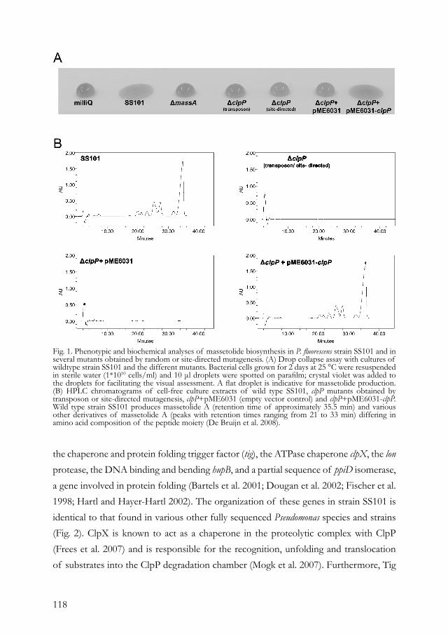
118
Fig. 1. Phenotypic and biochemical analyses of massetolide biosynthesis in P. fluorescens strain SS101 and in several mutants obtained by random or site-directed mutagenesis. (A) Drop collapse assay with cultures of wildtype strain SS101 and the different mutants. Bacterial cells grown for 2 days at 25 °C were resuspended in sterile water (1*1010 cells/ml) and 10 μl droplets were spotted on parafilm; crystal violet was added to the droplets for facilitating the visual assessment. A flat droplet is indicative for massetolide production. (B) HPLC chromatograms of cell-free culture extracts of wild type SS101, clpP mutants obtained by transposon or site-directed mutagenesis, clpP+pME6031 (empty vector control) and clpP+pME6031-clpP. Wild type strain SS101 produces massetolide A (retention time of approximately 35.5 min) and various other derivatives of massetolide A (peaks with retention times ranging from 21 to 33 min) differing in amino acid composition of the peptide moiety (De Bruijn et al. 2008).
the chaperone and protein folding trigger factor (tig), the ATPase chaperone clpX, the lon protease, the DNA binding and bending hupB, and a partial sequence of ppiD isomerase, a gene involved in protein folding (Bartels et al. 2001; Dougan et al. 2002; Fischer et al. 1998; Hartl and Hayer-Hartl 2002). The organization of these genes in strain SS101 is identical to that found in various other fully sequenced Pseudomonas species and strains (Fig. 2). ClpX is known to act as a chaperone in the proteolytic complex with ClpP (Frees et al. 2007) and is responsible for the recognition, unfolding and translocation of substrates into the ClpP degradation chamber (Mogk et al. 2007). Furthermore, Tig
119
Fig. 2. Genomic organisation of clpP and flanking genes in P. fluorescens SS101 (top) and other fully sequenced Pseudomonas strains. For each of the genes of strain SS101, the percentages of amino acid identity with their corresponding genes in the other Pseudomonas strains are given. The reference strains used are P. fluorescens strains SBW25 (De Bruijn et al. 2007), Pf0-1 (De Bruijn et al. 2007), Pf-5 (Gross et al. 2007), P. syringae pv. tomato strain DC3000 (Berti et al. 2007), P. syringae pv. syringae strain B728a (Scholz-Schroeder et al. 2003; Zhang et al. 1995), P. putida strain KT2440, and P. aeruginosa strain PAO1. The codes of the genes of these reference strains correspond to those in the PseudoDB and NCBI databases.
and Lon were shown to be substrates for Clp-dependent proteolysis (Flynn et al. 2003; Gerth et al. 2008). To determine wheteher the genes flanking clpP also play a role in regulation of massetolide biosynthesis, site-directed mutagenesis was performed for tig, clpX and lon. Drop collapse assays and RP-HPLC analyses showed that disruptions of these three genes did not affect massetolide production (data not shown), suggesting that ClpP acts independently from ClpX in regulating massetolide biosynthesis. Phenotypic characterization of the clpP mutant of P. fluorescens. Consistent with observations made previously for E. coli, Staphylococcus aureus and Pseudomonas aeruginosa (Damerau and St John 1993; Shanks et al. 2006; Wang et al. 2007), a mutation in clpP adversely affected growth of P. fluorescens SS101 (Fig. 3A). This reduced growth of the clpP mutant was not due to a lack of massetolide production, because the massA biosynthesis mutant showed comparable growth to wild type strain SS101 (Fig. 3A). Complementation of the clpP mutant with pME6031-clpP restored growth to wild type level whereas the empty vector control had no effect (Fig. 3A). Tensiometric analysis of cell-free culture supernatants of strain
120
Fig. 3. Phenotypic characteristics of P. fluorescens strain SS101, the massA mutant, clpP mutant, clpP+pME6031 (empty vector control) and clpP+pME6031-clpP. (A) Growth at 25 °C; at each time point, cell density was measured spectrofotometrically (600 nm) and mean values of four replicates are given; error bars represent the standard error of the mean. (B) Surface tension of cell-free culture supernatant of strain SS101 and the different mutants given in panel A. (C) Biofilm formation by strain SS101 and the different mutants in a polystyrene 96-wells plate containing 200 μl growth medium; cells firmly attached to the wall of the wells were stained with crystal violet and their density quantified spectrofotometrically at 600 nm. Mean values of 4 replictes are given. For each of the mutants, asterisks indicate statistically significant (P < 0.05) differences relative to wild type SS101. (D) Swarming motility of wild type strain SS101 and mutants on soft (0.6% w/v) agar plates. Five μl of washed overnight cultures of wild type SS101 and mutants was spot-inoculated in the center of the soft agar plate and incubated for 48 to 72 h at 25°C.
121
SS101 and mutants indicated that wild type strain SS101 starts producing the massetolide surfactants between 12 h and 16 h of incubation (Fig. 3B). A reduction in surface tension of the growth medium was not observed for the massA mutant nor for the clpP mutant, but was restored by complementation with pME6031-clpP (Fig. 3B). Massetolide biosynthesis is essential for biofilm formation and swarming motility of strain SS101 (De Bruijn et al. 2008). The capacity to form a biofilm was strongly reduced in the clpP mutant to a level similar as that observed for the massA biosynthesis mutant (Fig. 3C). Biofilm formation was fully restored to wild type level by complementation of the clpP mutant with pME6031-clpP (Fig. 3C). The clpP mutant also lost its ability to swarm on a soft agar surface (Fig. 3D). Swarming motility was restored in the clpP mutant by introduction of pME6031-clpP, although the extent of complementation as well as the swarming pattern was not identical to that of the wild type strain (Fig 3D). Introduction of pME6031-clpP into wild type SS101 also resulted in reduced swarming (data not shown), suggesting that the altered swarming pattern of the complemented clpP mutant may have resulted from multiple copies of the clpP gene. In contrast to a complete loss of swarming motility, the clpP mutant was still able to swim on soft (0.25 %) agar plates (Fig. S1). The observation that swimming motility was similar to that of the massA biosynthesis mutant, but reduced compared to wild type strain SS101 and the complemented clpP mutant (Fig. S1), indicates that massetolide production also plays a (partial) role in swimming motility.
Transcriptional analysis of the clpP mutant of P. fluorescens. Q-PCR analyses were performed to study the effect of the clpP mutation on expression of a range of genes, including the biosynthesis genes massA, massB and massC. To prevent that differences in growth rates between wild type strain SS101 and the clpP mutant would interfere with gene expression measurements, cells used for the RNA isolation were collected when wild type and mutant reached a specific density, i.e. early exponential (OD600~0.2) and mid-exponential (OD600~0.6). Consistent with previous results (De Bruijn et al. 2008), massA, massB and massC are expressed in wild type strain SS101 during the early exponential and mid-exponential growth phases (data not shown). The transcript levels of all three mass genes were significantly decreased in the clpP mutant especially in the mid-exponential growth phase (Fig. 4A). Mutations in massA, massB or massC did not affect transcription of clpP (data not shown). Collectively, these results indicate that clpP regulates transcription of the mass biosynthesis genes.
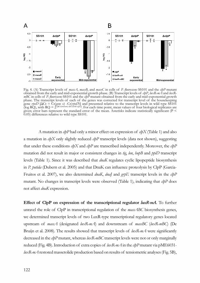
122
A mutation in clpP had only a minor effect on expression of clpX (Table 1) and also a mutation in clpX only slightly reduced clpP transcript levels (data not shown), suggesting that under these conditions clpX and clpP are transcribed independently. Moreover, the clpP mutation did not result in major or consistent changes in tig, lon, hupB and ppiD transcript levels (Table 1). Since it was described that dnaK regulates cyclic lipopeptide biosynthesis in P. putida (Dubern et al. 2005) and that DnaK can influence proteolysis by ClpP (Garcia-Fruitos et al. 2007), we also determined dnaK, dnaJ and grpE transcript levels in the clpP mutant. No changes in transcript levels were observed (Table 1), indicating that clpP does not affect dnaK expression.
Effect of ClpP on expression of the transcriptional regulator luxR-mA. To further unravel the role of ClpP in transcriptional regulation of the massABC biosynthesis genes, we determined transcript levels of two LuxR-type transcriptional regulatory genes located upstream of massA (designated luxR-mA) and downstream of massBC (luxR-mBC) (De Bruijn et al. 2008). The results showed that transcript levels of luxR-mA were significantly decreased in the clpP mutant, whereas luxR-mBC transcript levels were not or only marginally reduced (Fig. 4B). Introduction of extra copies of luxR-mA in the clpP mutant via pME6031-luxR-mA restored massetolide production based on results of tensiometric analyses (Fig. 5B),
Fig. 4. (A) Transcript levels of massA, massB, and massC in cells of P. fluorescens SS101 and the clpP mutant obtained from the early and mid-exponential growth phase. (B) Transcript levels of clpP, luxR-mA and luxR-mBC in cells of P. fluorescens SS101 and the clpP mutant obtained from the early and mid-exponential growth phase. The transcript levels of each of the genes was corrected for transcript level of the housekeeping gene rpoD [ΔCt = Ct(gene x) -Ct(rpoD)] and presented relative to the transcript levels in wild type SS101 (log RQ), with RQ = 2-[ΔCt(mutant)-ΔCt(wild type)]. For each time point, mean values of four biological replicates are given; error bars represent the standard error of the mean. Asterisks indicate statistically significant (P < 0.05) differences relative to wild type SS101.
A
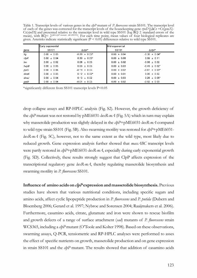
123
drop collapse assays and RP-HPLC analysis (Fig. S2). However, the growth deficiency of the clpP mutant was not restored by pME6031-luxR-mA (Fig. 5A) which in turn may explain why massetolide production was slightly delayed in the clpP+pME6031-luxR-mA compared to wild type strain SS101 (Fig. 5B). Also swarming motility was restored for clpP+pME6031-luxR-mA (Fig. 5C), however, not to the same extent as the wild type, most likely due to reduced growth. Gene expression analysis further showed that massABC transcript levels were partly restored in clpP+pME6031-luxR-mA, especially during early exponential growth (Fig. 5D). Collectively, these results strongly suggest that ClpP affects expression of the transcriptional regulatory gene luxR-mA, thereby regulating massetolide biosynthesis and swarming motility in P. fluorescens SS101.
Influence of amino acids on clpP expression and massetolide biosynthesis. Previous studies have shown that various nutritional conditions, including specific sugars and amino acids, affect cyclic lipopeptide production in P. fluorescens and P. putida (Dubern and Bloemberg 2006; Gerard et al. 1997; Nybroe and Sorensen 2004; Raaijmakers et al. 2006). Furthermore, casamino acids, citrate, glutamate and iron were shown to rescue biofilm and growth defects of a range of surface attachment (sad) mutants of P. fluorescens strain WCS365, including a clpP mutant (O’Toole and Kolter 1998). Based on these observations, swarming assays, Q-PCR, tensiometric and RP-HPLC analyses were performed to asses the effect of specific nutrients on growth, massetolide production and on gene expression in strain SS101 and the clpP mutant. The results showed that addition of casamino acids
Table 1. Transcript levels of various genes in the clpP mutant of P. fluorescens strain SS101. The transcript level of each of the genes was corrected for the transcript levels of the housekeeping gene rpoD [(ΔCt =Ct(geneX)-Ct(rpoD)] and presented relative to the transcript level in wild type SS101 (log RQ ± standard errors of the mean), with RQ= 2-[ΔCt(clpP mutant)- ΔCt(SS101)]. For each time point, mean values of four biological replicates are given. Asterisks indicate statistically significant (P < 0.05) differences relative to wild type SS101.
*significantly different from SS101 transcript levels P<0.05

124
Fig. 5. (A) Growth of P. fluorescens strain SS101, the clpP mutant and clpP+ pME6031-luxR-mA at 25 °C. At each time point, cell density was measured spectrofotometrically (600 nm) and mean values of four replicates are given; error bars represent the standard error of the mean. (B) Surface tension of cell-free culture supernatant of strain SS101 and the different mutants given in panel A. (C) Swarming motility of wild type strain SS101 and mutants on soft (0.6% w/v) agar plates. Five μl of washed overnight cultures of wild type SS101 and mutants was spot-inoculated in the center of the soft agar plate and incubated for 48 to 72 h at 25°C. (D) Transcript levels of the massA, massB and massC genes in wild type strain SS101, the clpP mutant and clpP+ pME6031-luxR-mA in early and mid-exponential growth phase. The transcript levels of each of the genes was corrected for transcript levels of rpoD [ΔCt = Ct(gene x) -Ct(rpoD)] and presented relative to the transcript level in wild-type SS101 (log RQ), with RQ = 2-[ΔCt(mutant)-ΔCt(wild type)]. For each time point, mean values of four biological replicates are given; error bars represent standard errors of the means. Asterisks indicate statistically significant (P < 0.05) differences relative to wild type SS101.
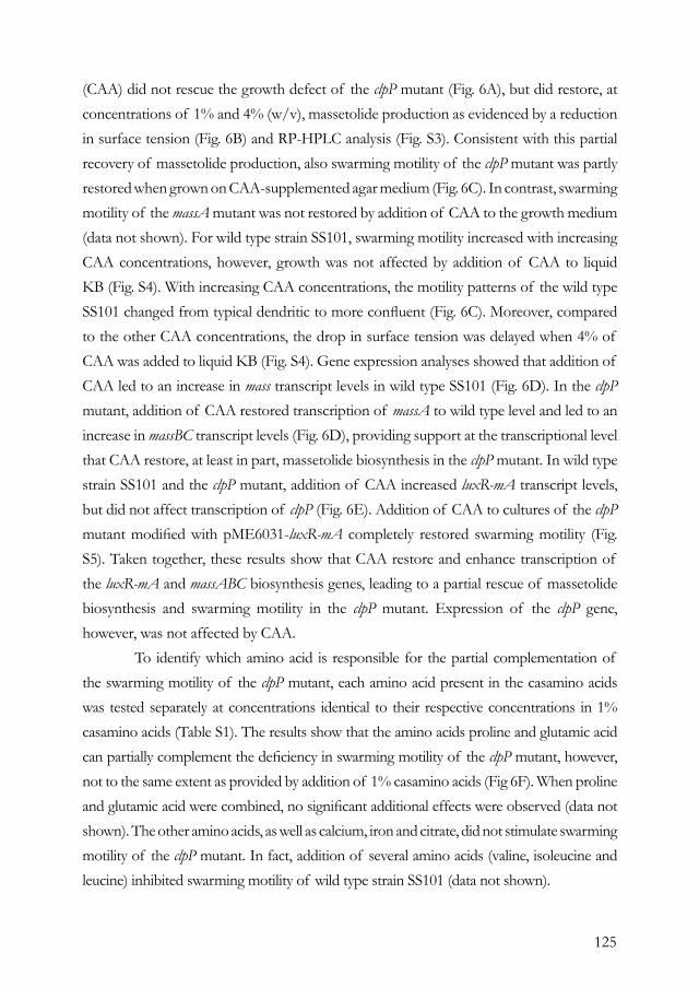
125
(CAA) did not rescue the growth defect of the clpP mutant (Fig. 6A), but did restore, at concentrations of 1% and 4% (w/v), massetolide production as evidenced by a reduction in surface tension (Fig. 6B) and RP-HPLC analysis (Fig. S3). Consistent with this partial recovery of massetolide production, also swarming motility of the clpP mutant was partly restored when grown on CAA-supplemented agar medium (Fig. 6C). In contrast, swarming motility of the massA mutant was not restored by addition of CAA to the growth medium (data not shown). For wild type strain SS101, swarming motility increased with increasing CAA concentrations, however, growth was not affected by addition of CAA to liquid KB (Fig. S4). With increasing CAA concentrations, the motility patterns of the wild type SS101 changed from typical dendritic to more confluent (Fig. 6C). Moreover, compared to the other CAA concentrations, the drop in surface tension was delayed when 4% of CAA was added to liquid KB (Fig. S4). Gene expression analyses showed that addition of CAA led to an increase in mass transcript levels in wild type SS101 (Fig. 6D). In the clpP mutant, addition of CAA restored transcription of massA to wild type level and led to an increase in massBC transcript levels (Fig. 6D), providing support at the transcriptional level that CAA restore, at least in part, massetolide biosynthesis in the clpP mutant. In wild type strain SS101 and the clpP mutant, addition of CAA increased luxR-mA transcript levels, but did not affect transcription of clpP (Fig. 6E). Addition of CAA to cultures of the clpP mutant modified with pME6031-luxR-mA completely restored swarming motility (Fig. S5). Taken together, these results show that CAA restore and enhance transcription of the luxR-mA and massABC biosynthesis genes, leading to a partial rescue of massetolide biosynthesis and swarming motility in the clpP mutant. Expression of the clpP gene, however, was not affected by CAA. To identify which amino acid is responsible for the partial complementation of the swarming motility of the clpP mutant, each amino acid present in the casamino acids was tested separately at concentrations identical to their respective concentrations in 1% casamino acids (Table S1). The results show that the amino acids proline and glutamic acid can partially complement the deficiency in swarming motility of the clpP mutant, however, not to the same extent as provided by addition of 1% casamino acids (Fig 6F). When proline and glutamic acid were combined, no significant additional effects were observed (data not shown). The other amino acids, as well as calcium, iron and citrate, did not stimulate swarming motility of the clpP mutant. In fact, addition of several amino acids (valine, isoleucine and leucine) inhibited swarming motility of wild type strain SS101 (data not shown).
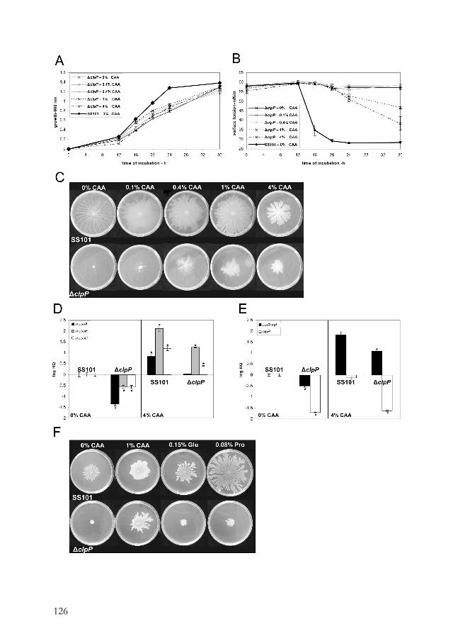
126
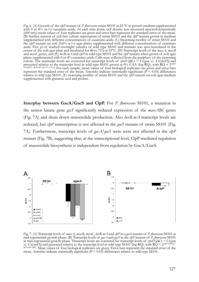
127
Interplay between GacA/GacS and ClpP. For P. fluorescens SS101, a mutation in the sensor kinase gene gacS significantly reduced expression of the massABC genes (Fig. 7A) and shuts down massetolide production. Also luxR-mA transcript levels are reduced, but clpP transcription is not affected in the gacS mutant of strain SS101 (Fig. 7A). Furthermore, transcript levels of gacA/gacS were were not affected in the clpP mutant (Fig. 7B), suggesting that, at the transcriptional level, ClpP-mediated regulation of massetolide biosynthesis is independent from regulation by GacA/GacS.
Fig. 6. (A) Growth of the clpP mutant of P. fluorescens strain SS101 at 25 °C in growth medium supplemented with 0 to 4% (w/v) casamino acids. At each time point, cell density was measured spectrofotometrically (600 nm); mean values of four replicates are given and error bars represent the standard error of the mean. (B) Surface tension of cell-free culture supernatant of strain SS101 and the clpP mutant grown in medium supplemented with different concentrations of casamino acids. (C) Swarming motility of strain SS101 and the clpP mutant on soft (0.6% w/v) agar plates supplemented with different concentrations of casamino acids. Five μl of washed overnight cultures of wild type SS101 and mutants was spot-inoculated in the center of the soft agar plate and incubated for 48 to 72 h at 25°C. (D) Transcript levels of the massA, massB and massC genes, and (E) luxR-mA and clpP in wild type SS101 and the clpP mutant when grown of soft agar plates supplemented with 0 or 4% casamino acids. Cells were collected from the periphery of the swarming colony. The transcript levels are corrected for transcript levels of rpoD [ΔCt = Ct(gene x) -Ct(rpoD)] and presented relative to the transcript level in wild-type SS101 grown at 0% CAA (log RQ), with RQ = 2-[ΔCt
(sampleX)- ΔCt(wild type 0% CAA)]. For each sample, mean values of four biological replicates are given and error bars represent the standard error of the mean. Asterisks indicate statistically significant (P < 0.05) differences relative to wild type SS101. (F) swarming motility of strain SS101 and the clpP mutant on soft agar medium supplemented with glutamic acid and proline.
Fig. 7. (A) Transcript levels of massA, massB, massC, luxR-mA and clpP in a gacS mutant of P. fluorescens SS101 at mid-exponential growth phase. (B) Transcript levels of gacA and gacS in the clpP mutant of P. fluorescens SS101 at mid-exponential growth phase. Transcript levels are corrected for transcript levels of rpoD [ΔCt = Ct(gene x) -Ct(rpoD)] and presented relative to the transcript level in wild type SS101 (log RQ), with RQ = 2-[ΔCt(mutant)-
ΔCt(wild type)]. Mean values of four biological replicates are given. Error bars represent the standard error of the mean. Asterisks indicate statistically significant (P < 0.05) differences relative to wild type SS101.

128
DISCUSSIONClpP is a serine protease that is highly conserved in bacteria and eukaryotes (Wong and Houry 2004; Yu and Houry 2007). Together with other proteases, ClpP plays a crucial role in intracellular refolding and degradation of proteins, which is an essential process for the viability and growth of cells. In this study, we cloned and sequenced clpP from plant growth-promoting P. fluorescens strain SS101 and show that clpP plays an important role in regulation of cyclic lipopeptide biosynthesis, swarming motility, biofilm formation and growth. These results confirm and extend observations made for other Pseudomonas species and bacterial genera. For example, biofilm formation was reduced in clpP mutants of P. fluorescens WCS365 and S. aureus, but enhanced in a clpP mutant of P. aeruginosa (Frees et al. 2004; O’Toole and Kolter 1998; Shanks et al. 2006; Wang et al. 2007). ClpP is also important for virulence of several bacterial pathogens, like Streptococcus pneumoniae, S. aureus, Salmonella typhimurium, Yersinia entrocolitica, Listeria monocytogenes and Porphyromonas gingivalis (Capestany et al. 2008; Gaillot et al. 2000; Ibrahim et al. 2005; Porankiewicz et al. 1999). In Listeria, the haemolytic activity, but not the production, of the virulence factor listeriolysin O was strongly reduced in a clpP mutant (Gaillot et al. 2000). In Bacillus subtilis, ClpP plays a role in competence development, motility and sporulation (Msadek et al. 1998). Although specific extracelullar metabolites of Pseudomonas strains are known to play a role in swarming motility and biofilm formation, the involvement of ClpP in regulation of the biosynthesis genes encoding these metabolites has, to our knowledge, not been demonstrated conclusively. This study provides, for the first time, evidence that the ClpP protease regulates the biosynthesis of cyclic lipopeptide surfactants that play an important role in swarming motility, biofilm formation and antimicrobial activity of P. fluorescens. More specifically, ClpP was shown to affect expression of the transcriptional regulatory gene luxR-mA, thereby regulating massetolide biosynthesis and concomitantly biofilm formation and swarming motility in P. fluorescens SS101. Whether this is typical for the Pseudomonas strain under study remains to be addressed, but the observation by Nakano et al. (2000) that expression of the surfactin gene srfA in B. subtilis is affected in a clpP mutant suggests that a similar role of ClpP may apply to other bacterial genera and species producing lipopeptide antibiotics. Based on the results of this and previous studies, several hypotheses can be proposed for the mechanisms underlying ClpP-mediated regulation of luxR-mA
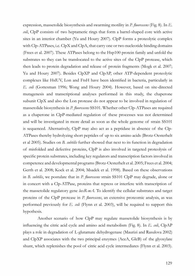
129
expression, massetolide biosynthesis and swarming motility in P. fluorescens (Fig. 8). In E. coli, ClpP consists of two heptameric rings that form a barrel-shaped core with active sites in an interior chamber (Yu and Houry 2007). ClpP forms a proteolytic complex with Clp-ATPases, i.e. ClpX and ClpA, that carry one or two nucleotide binding domains (Frees et al. 2007). These ATPases belong to the Hsp100 protein family and unfold the substrates so they can be translocated to the active sites of the ClpP protease, which then leads to protein degradation and release of protein fragments (Mogk et al. 2007; Yu and Houry 2007). Besides ClpXP and ClpAP, other ATP-dependent proteolytic complexes like HslUV, Lon and FtsH have been identified in bacteria, particularly in E. coli (Gottesman 1996; Wong and Houry 2004). However, based on site-directed mutagenesis and transcriptional analyses performed in this study, the chaperone subunit ClpX and also the Lon protease do not appear to be involved in regulation of massetolide biosynthesis in P. fluorescens SS101. Whether other Clp-ATPases are required as a chaperone in ClpP-mediated regulation of these processes was not determined and will be investigated in more detail as soon as the whole genome of strain SS101 is sequenced. Alternatively, ClpP may also act as a peptidase in absence of the Clp-ATPases thereby hydrolyzing short peptides of up to six amino acids (Brotz-Oesterhelt et al 2005). Studies on B. subtilis further showed that next to its function in degradation of misfolded and defective proteins, ClpP is also involved in targeted proteolysis of specific protein substrates, including key regulators and transcription factors involved in competence and developmental programs (Brotz-Oesterhelt et al. 2005; Frees et al. 2004; Gerth et al. 2008; Kock et al. 2004; Msadek et al. 1998). Based on these observations in B. subtilis, we postulate that in P. fluorescens strain SS101 ClpP may degrade, alone or in concert with a Clp-ATPase, proteins that repress or interfere with transcription of the massetolide regulatory gene luxR-mA. To identify the cellular substrates and target proteins of the ClpP protease in P. fluorescens, an extensive proteomic analysis, as was performed previously for E. coli (Flynn et al. 2003), will be required to support this hypothesis. Another scenario of how ClpP may regulate massetolide biosynthesis is by influencing the citric acid cycle and amino acid metabolism (Fig. 8). In E. coli, ClpAP plays a role in degradation of L-glutamate dehydrogenase (Maurizi and Rasulova 2002) and ClpXP associates with the two principal enzymes (AceA, GlcB) of the glyoxylate shunt, which replenishes the pool of citric acid cycle intermediates (Flynn et al. 2003).
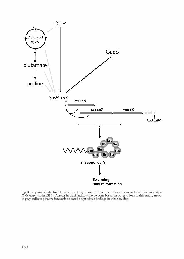
130
Fig. 8. Proposed model for ClpP-mediated regulation of massetolide biosynthesis and swarming motility in P. fluorescens strain SS101. Arrows in black indicate interactions based on observations in this study; arrows in grey indicate putative interactions based on previous findings in other studies.
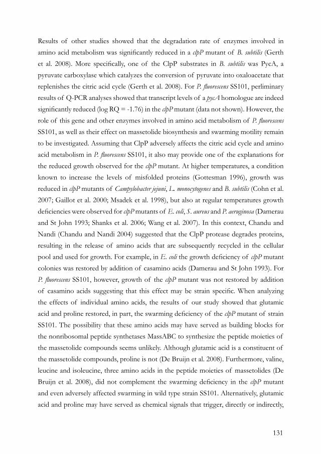
131
Results of other studies showed that the degradation rate of enzymes involved in amino acid metabolism was significantly reduced in a clpP mutant of B. subtilis (Gerth et al. 2008). More specifically, one of the ClpP substrates in B. subtilis was PycA, a pyruvate carboxylase which catalyzes the conversion of pyruvate into oxaloacetate that replenishes the citric acid cycle (Gerth et al. 2008). For P. fluorescens SS101, perliminary results of Q-PCR analyses showed that transcript levels of a pycA homologue are indeed significantly reduced (log RQ = -1.76) in the clpP mutant (data not shown). However, the role of this gene and other enzymes involved in amino acid metabolism of P. fluorescens SS101, as well as their effect on massetolide biosynthesis and swarming motility remain to be investigated. Assuming that ClpP adversely affects the citric acid cycle and amino acid metabolism in P. fluorescens SS101, it also may provide one of the explanations for the reduced growth observed for the clpP mutant. At higher temperatures, a condition known to increase the levels of misfolded proteins (Gottesman 1996), growth was reduced in clpP mutants of Campylobacter jejuni, L. monocytogenes and B. subtilis (Cohn et al. 2007; Gaillot et al. 2000; Msadek et al. 1998), but also at regular temperatures growth deficiencies were observed for clpP mutants of E. coli, S. aureus and P. aeruginosa (Damerau and St John 1993; Shanks et al. 2006; Wang et al. 2007). In this context, Chandu and Nandi (Chandu and Nandi 2004) suggested that the ClpP protease degrades proteins, resulting in the release of amino acids that are subsequently recycled in the cellular pool and used for growth. For example, in E. coli the growth deficiency of clpP mutant colonies was restored by addition of casamino acids (Damerau and St John 1993). For P. fluorescens SS101, however, growth of the clpP mutant was not restored by addition of casamino acids suggesting that this effect may be strain specific. When analyzing the effects of individual amino acids, the results of our study showed that glutamic acid and proline restored, in part, the swarming deficiency of the clpP mutant of strain SS101. The possibility that these amino acids may have served as building blocks for the nonribosomal peptide synthetases MassABC to synthesize the peptide moieties of the massetolide compounds seems unlikely. Although glutamic acid is a constituent of the massetolide compounds, proline is not (De Bruijn et al. 2008). Furthermore, valine, leucine and isoleucine, three amino acids in the peptide moieties of massetolides (De Bruijn et al. 2008), did not complement the swarming deficiency in the clpP mutant and even adversely affected swarming in wild type strain SS101. Alternatively, glutamic acid and proline may have served as chemical signals that trigger, directly or indirectly,
132
the expression of luxR-mA and the mass biosynthesis genes, leading to a partial rescue of massetolide biosynthesis and swarming motility of the clpP mutant (Fig. 8). It is well known that specific amino acids, including glutamate and proline, can promote swarming in P. aeruginosa (Kohler et al. 2000), Proteus mirabilis (Allison et al. 1993), and act as a chemoattractant (Allison et al. 1993). Moreover, glutamine can serve as a signal for the cellular nitrogen state; glutamine is sensed by enzymes which trigger a signal transduction cascade that activate the glutamine synthase gene glnA in E. coli (Leigh and Dodsworth, 2007). Also, exogenously provided proline can release the transcriptional repressor PutA from the proline utilization genes (Brown and Wood, 1993; Zhou et al. 2008). These studies exemplifie that these amino acids can induce gene transcription. Finally, we looked into the possible interplay between ClpP and the two-component regulatory system GacA/GacS (Fig. 8). In other systems, ClpP affects global regulation. For example, in S. aureus, the global regulator agr was repressed in the clpP mutant, which resulted in a reduced α-toxin and extracellular protease activity (Frees et al. 2003; Mogk et al. 2007). Also in Bacillus, ClpP-dependent proteolysis is regulated in response to environmental signals (nutrients) and transmitted via the two-component signal transduction system ComK/ComS (Frees et al. 2007). For P. fluorescens SS101, gacS regulates transcription of the massABC and luxR-mA genes and thereby massetolide production, but clpP transcription is not affected. Furthermore, transcript levels of gacA/gacS were not affected in the clpP mutant, suggesting that, at the transcriptional level, ClpP-mediated regulation of massetolide biosynthesis is independent from regulation by GacA/GacS.
MATERIALS AND METHODSBacterial strains and culture conditions. P. fluorescens SS101 was grown on Pseudomonas agar F (Difco) (PSA) plates or in liquid King’s medium B (KB) at 25 °C. The transposon mutants were obtained as described by De Souza et al. (2003) and plasposon mutants were obtained with plasmid pTnModOKm (Dennis and Zylstra 1998). Escherichia coli strain DH5α was used as a host for the plasmids for site-directed mutagenesis and complementation. E. coli strains were grown on Luria-Bertani (LB) plates or in LB broth amended with the appropriate antibiotics. Identification clpP cluster. ClpP was identified by sequencing the regions flanking the transposon insertions as described by De Sousa et al (2003). The flanking regions of clpP were sequenced by primer walking and open reading frames (ORFs) were identified with the Softberry FGENESB program (http://www.softberry.com/berry.phtml). The ORFs werer analyzed using Blastx in the NCBI database, PseudoDB (http://xbase.bham.ac.uk/pseudodb/) and Pseudomonas.com (http://Pseudomonas.com).
Site-directed mutagenesis. Site-directed mutagenesis of the lon and tig genes was performed with the pKnockout-G suicide vector (67) and performed as described in De Bruijn et al. (14). Primers used for site-directed mutagenesis are listed in Table 2. Site-directed mutagenesis of the clpP and clpX genes was performed based on the method described by Choi and Schweizer (10). For each mutant construct, three

133
fragments were amplified: a 5’ fragment, FRT-Gm-FRT cassette and a 3’ fragment. In the 1st round PCR, the FRT-Gm-FRT cassette and the 5’ and 3’ fragments were amplified. In the 2nd round PCR, these three fragments were coupled by overlap extension PCR. The 5’ and 3’ fragments were chosen in such a way that after homologous recombination in Pseudomonas, the FRT-Gm-FRT cassette is inserted around position 170 bp of the clpP or clpX genes. For amplification of the FRT-Gm-FRT cassette, pPS854-GM, a derivative of pPS854 (37) was used as a template in the PCR with primers FRT-F and FRT-R. The 1st round PCR was performed with KOD polymerase (Novagen) according to the manufacturer’s protocol, but with the addition of 1-10% DMSO for the clpP and clpX fragments. The program used for the PCR reaction contains 2 min of denaturation at 95 °C, followed by 5 cycles of subsequently 95 °C, 55 °C and 68 °C, each for 20 s. The PCR amplification was proceeded with 25 cycles of subsequently 95 °C, 60 °C and 68 °C, each for 20 s. The last step of the PCR was 68 °C for 7 min. All fragments were separated on a 1% (w/v) agarose gel and purified with NucleoSpin kit (Macherey-Nagel). The 2nd round PCR was performed by mixing equimolar amounts of the 5’ fragment, FRT-Gm-FRT and 3’ fragments with milliQ, dNTPs, KOD buffer and KOD polymerase to a total of 47 μl. The PCR was started by 2 min of denaturation at 95 °C, followed by 3 cycles of subsequently 95 °C, 55 °C and 68 °C, for 20, 30 and 60 sec, respectively. In the 3rd extension cycle, 1.5 μl of the Up forward and Dn reverse primer (10 μM stock) was added. The PCR amplification was proceeded with 25 cycles of subsequently 95 °C, 58 °C and 68 °C, for 20, 20 and 120 s, respectively. The last step of the PCR was 68 °C for 7 min. All fragments were separated on a 1% agarose gel and bands of the right size were purified with NucleoSpin kit. The fragments were digested with BamHI and cloned into pEX18Tc. E. coli DH5α was transformed with pEX18Tc-clpP or pEX18Tc-clpX plasmids by heat-shock transformation according to Innoue et al (39) and transformed colonies were selected on LB supplemented with 25 μg/ml gentamicin (Sigma). Integration of the inserts was verified by PCR analysis with pEX18Tc primers and by restriction analysis of the isolated plasmids. The plasmid inserts were verified by sequencing (BaseClear, Leiden, the Netherlands). The correct pEX18Tc-clpP and pEX18Tc-clpX constructs were subsequently electroporated into P. fluorescens strain SS101. Electrocompetent cells were obtained according to Choi et al. (9) and electroporation occurred at 2.4 kV, 200 μF. After incubation in SOC medium for 2 hrs at 25 °C, cells were plated on KB supplemented with gentamicin (25 μg/ml) and rifampicin (50 μg/ml). Obtained colonies were grown in LB for 1 hr at 25 °C and plated on LB supplemented with gentamicin (25 μg/ml) and 5% sucrose to accomplish the double cross-over. Plates were incubated at 25 °C for at least 48 hrs and colonies were re-streaked on LB supplemented with gentamicin plus 5% sucrose and on LB supplemented with tetracycline (25 μg/ml). Colonies that did grow on LB with gentamicin plus sucrose, but not on LB with tetracycline were selected and subjected to colony PCR to confirm the presence of the gentamicin resistance cassette and the absence of the tetracycline resistance cassette. Positive colonies were confirmed by sequencing the PCR fragments obtained with the Up forward and Dn reverse primers. The obtained clpP and clpX mutants were tested for massetolide production in a drop collapse assay and by RP-HPLC analysis. RP-HPLC analyses were performed as described previously (14) with the exception that in this study samples of the crude surfactant extract (1 mg/ml) were analysed isocratically (flow rate 0.5 ml/min) using a solution of 45% acetonitrile and 15% milliQ, both containing 0.1% trifluoracetic acid, and 40% methanol as eluens.
Construction of pME6031-based vectors for genetic complementation. A fragment of approximately 2-kb containing the clpP gene, including promoter and terminator, was obtained by PCR (Table 1) with the KOD polymerase. The pME6031-luxR-mA construct was generated as follows: a 1817 bp fragment was obtained by PCR with specific primers (Table 2) with Phusion DNA polymerase (Finnzymes). The PCR fragments were subcloned in pGEM-T Easy (Promega) and the obtained plasmids were digested with EcoRI. The clpP and luxR-mA fragments were obtained from gel with the NucleoSpin kit and cloned into the shuttle vector pME6031 (36), which was digested, dephosphorylated (Shrimp alkaline phosphatase; Promega) and purified with the NucleoSpin kit according to manufacturer’s instructions. E. coli DH5α was transformed with the obtained plasmid pME6031-clpP or pME6031-luxR-mA by heat shock transformation (39) and transformed colonies were selected on LB agar plates supplemented with tetracycline (25 μg/ml). Correct integration of the fragments was verified by PCR analysis and restriction analysis of isolated plasmids. The pME6031-clpP and pME6031-luxR-mA constructs were subsequently electroporated into the clpP mutant and wild type strain SS101. Transformed cells were plated on KB supplemented with tetracycline (25 μg/ml) and the presence of pME6031-clpP or pME6031-luxR-mA was verified by PCR analysis with primers specific for pME6031.
Surface tension measurements and transcriptional analysis. Cells were grown at 25 °C (220 rpm) in a 24-wells plate with 1.25 ml KB broth per well. At specific time points during growth, 100 μl culture was transferred to a 96-wells plate and cell density was measured at 600 nm with a microplate reader (BioRad). Subsequently, 1 ml of cell culture was collected and spun down. The cells were frozen in liquid N2 and stored at -80 °C. For the RNA isolations and cDNA synthesis, four biological replicates were used for each time point. Massetolide production was measured qualitatively by the drop collapse assay and quantitatively by tensiometric analysis of the cell-free supernatant (K6 tensiometer, Krüss GmbH, Hamburg, Germany) at room temperature. To get sufficient volume for the tensiometric analysis, the supernatant of four biological replicates was collected and pooled for each time point. Surface tension of each sample was measured in
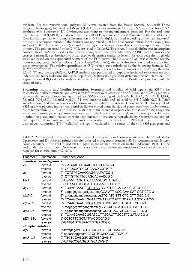
134
triplicate. For the transcriptional analyses, RNA was isolated from the frozen bacterial cells with Trizol Reagent (Invitrogen), followed by DNase I (GE Healthcare) treatment. One μg RNA was used for cDNA synthesis with Superscript III (Invitrogen) according to the manufacturer’s protocol. For the real time quantitative PCR (Q-PCR), conducted with the 7300SDS system of Applied Biosystems, the SYBR Green Core kit (Eurogentec) with a final concentration of 3.5 mM MgCl2 was used according to the manufacturer’s protocol. The concentration of the primers was optimized (400 nM final concentration for the mass genes and rpoD, 500 nM for clpP and clpX) and a melting curve was performed to check the specificity of the primers. The primers used for the Q-PCR are listed in Table S2. To correct for small differences in template concentration, rpoD was used as the housekeeping gene. The cycle where the SYBR Green fluorescence crosses a manually set threshold (Ct) was used to determine transcript levels. For each gene the threshold was fixed based on the exponential segment of the PCR curve. The Ct value of clpP was corrected for the housekeeping gene rpoD as follows: ∆Ct = Ct(clpP)–Ct(rpoD); the same formula was used for the other genes investigated. The relative quantification (RQ) values, were calculated by the following formula: RQ = 2-[∆Ct(mutant)-∆Ct(wildtype)]. If there is no difference in transcript level between mutant and wild type, than the RQ=1 (20) and the log (RQ)=0. Q-PCR analysis was performed in duplicate (technical replicates) on four independent RNA isolations (biological replicates). Statistically significant differences were determined for log-transformed RQ values by analysis of variance (p<0.05) followed by the Bonferroni post hoc multiple comparisons.
Swarming motility and biofilm formation. Swarming and motility of wild type strain SS101, the massetolide-deficient mutants and several transformants were assessed on soft (0.6% and 0.25% agar, w/v, respectively) standard succinate agar medium (SSM) consisting of 32.8 mM K2HPO4, 22 mM KH2PO4, 7.6 mM (NH4)2SO4, 0.8 mM MgSO4, 34 mM succinic acid, and adjusted to pH 7 with NaOH. After autoclavation, SSM medium was cooled down in a waterbath for at least 1 hour to 55 °C. Twenty ml of SSM agar was pipetted into a 9-cm-petridish, lid was closed immediately and plates were kept for 24 hours at room temperature (~20 °C) prior to inoculation with the bacterial suspensions. For all swarming assays, the same conditions (agar temperature, temperature at which plates were stored, and the time period between pouring the plates and inoculation) were kept constant to maximize reproducibility. Overnight cultures of wild type SS101, mutants and transformants were washed three times with 0.9% NaCl, and 5 μl of the washed cell suspension (1*1010 cells/ml) was spot-inoculated in the center of the soft SSM agar plate and
Table 2. Primers used in this study for site-directed mutagenesis and complementation. The 5’ end of the Up reverse and Dn forward primers for site-directed mutagenesis contain a 25-bp sequence (small letters) complementary to the FRT-F and FRT-R primers for overlap extension in the 2nd round PCR. The 5’ end of the Up forward and Dn reverse primers contain a restriction site (underlined) for BamHI, which is required for cloning into pEX18Tc.
135
incubated for 48 to 72 h at 25°C. For the assays with the casamino acids, a filter-sterile stock solution of 20% casamino acids (Difco; BD: Becton, Dickinson and Co.) was prepared and diluted in SSM to obtain final concentrations of 0.1, 0.4, 1 and 4%. To test each amino acid present in the casamino acids separately, the amounts used were equivalent to those present in 1% CAA (Table S1). Also the effects of citric acid (citrate; 0.4%), CaCl2 (14.7 μM) and FeCl3 (0.24 μM) on swarming motility of strain SS101 were tested. Biofilm formation was assessed according to the method described by De Bruijn et al. (14) and O’Toole et al. (56) using flat-bottom 96-well plates made of transparent polystyrene (Greiner) with 200 μl KB broth per well. Statistically significant differences were determined with the Student’s t test (P <0.05).
Nucleotide sequence accession number. The sequence of clpP and its flanking genes have been deposited in GenBank under accession number FJ403110.
ACKNOWLEDGEMENTSThis work was funded by the Dutch Technology Foundation (STW), the applied science division of NWO and Productschap Tuinbouw. We also would like to thank Dr. Jorge de Souza, who initially isolated the clpP mutant of P. fluorescens. We are very grateful to Dr. Teresa Sweat (USDA, Corvallis, OR) and Dr. Dimitri Mavrodi (USDA, Pullman, WA) for providing the plasmids (pEX18Tc and pPS854-Gm), protocols and advice for the site-directed mutagenesis.
REFERENCESAllison, C., Lai, H. C., Gygi, D., and Hughes, C. 1993. Cell differentiation of Proteus mirabilis is initiated by glutamine, a specific chemoattractant for swarming cells. Mol. Microbiol. 8:53-60.Andersen, J. B., Koch, B., Nielsen, T. H., Sorensen, D., Hansen, M., Nybroe, O., Christophersen, C., Sorensen, J., Molin, S., and Givskov, M. 2003. Surface motility in Pseudomonas sp. DSS73 is required for efficient biological containment of the root-pathogenic microfungi Rhizoctonia solani and Pythium ultimum. Microbiology 149:37-46.Bartels, F., Fernandez, S., Holtel, A., Timmis, K. N., and de Lorenzo, V. 2001. The essential HupB and HupN proteins of Pseudomonas putida provide redundant and nonspecific DNA-bending functions. J. Biol. Chem. 276:16641-8.Berti, A. D., Greve, N. J., Christensen, Q. H., and Thomas, M. G. 2007. Identification of a biosynthetic gene cluster and the six associated lipopeptides involved in swarming motility of Pseudomonas syringae pv. tomato DC3000. J. Bacteriol. 189:6312-6323.Brotz-Oesterhelt, H., Beyer, D., Kroll, H. P., Endermann, R., Ladel, C., Schroeder, W., Hinzen, B., Raddatz, S., Paulsen, H., Henninger, K., Bandow, J. E., Sahl, H. G., and Labischinski, H. 2005. Dysregulation of bacterial proteolytic machinery by a new class of antibiotics. Nat. Med. 11:1082-7.Brown, E. D., and J. M. Wood. 1993. Conformational change and membrane association of the PutA protein are coincident with reduction of its FAD cofactor by proline. J. Biol. Chem. 268:8972-9. Capestany, C. A., Tribble, G. D., Maeda, K., Demuth, D. R., and Lamont, R. J. 2008. Role of the Clp system in stress tolerance, biofilm formation, and intracellular invasion in Porphyromonas gingivalis. J. Bacteriol. 190:1436-46.
Chandu, D., and Nandi, D. 2004. Comparative genomics and functional roles of the ATP-dependent proteases Lon and Clp during cytosolic protein degradation. Res. Microbiol. 155:710-9.Choi, K. H., Kumar, A., and Schweizer, H. P. 2006. A 10-min method for preparation of highly electrocompetent Pseudomonas aeruginosa cells: application for DNA fragment transfer between chromosomes and plasmid transformation. J. Microbiol. Methods. 64:391-7.Choi, K. H., and Schweizer, H. P. 2005. An improved method for rapid generation of unmarked Pseudomonas aeruginosa deletion mutants. BMC Microbiol. 5:30.Cohn, M. T., Ingmer, H., Mulholland, F., Jorgensen, K., Wells, J. M., and Brondsted, L. 2007. Contribution of conserved ATP-dependent proteases of Campylobacter jejuni to stress tolerance and virulence. Appl. Environ. Microbiol. 73:7803-13.Cui, X., Harling, R., Mutch, P., and Darling, D. 2005. Identification of N-3-hydroxyoctanoyl-homoserine lactone production in Pseudomonas fluorescens 5064, pathogenic to broccoli, and controlling biosurfactant production by quorum sensing. Eur. J. Plant Pathol. 111:297-308.Damerau, K., and St John, A. C. 1993. Role of Clp protease subunits in degradation of carbon starvation proteins in Escherichia coli. J. Bacteriol. 175:53-63.De Bruijn, I., de Kock, M. J., de Waard, P., van Beek, T. A., and Raaijmakers, J. M. 2008. Massetolide A biosynthesis in Pseudomonas fluorescens. J. Bacteriol. 190:2777-2789.De Bruijn, I., de Kock, M. J. D., Yang, M., de Waard, P., van Beek, T. A., and Raaijmakers, J. M. 2007. Genome-based discovery, structure prediction and functional analysis of cyclic lipopeptide antibiotics in Pseudomonas species. Mol. Microbiol. 63:417-428.
136
De Souza, J. T., De Boer, M., De Waard, P., Van Beek, T. A., and Raaijmakers, J. M. 2003. Biochemical, genetic, and zoosporicidal properties of cyclic lipopeptide surfactants produced by Pseudomonas fluorescens. Appl. Environ. Microbiol. 69:7161-7172.Dennis, J. J., and Zylstra, G. J. 1998. Plasposons: modular self-cloning minitransposon derivatives for rapid genetic analysis of gram-negative bacterial genomes. Appl. Environ. Microbiol. 64:2710-2715.Deziel, E., Lepine, F., Milot, S., and Villemur, R. 2003. RhlA is required for the production of a novel biosurfactant promoting swarming motility in Pseudomonas aeruginosa: 3-(3-hydroxyalkanoyloxy)-alkanoic acids (HAAs), the precursors of rhamnolipids. Microbiology 149:2005-13.Dougan, D. A., Mogk, A., and Bukau, B. 2002. Protein folding and degradation in bacteria: to degrade or not to degrade? That is the question. Cell. Mol. Life Sci. 59:1607-16.Dubern, J.-F., Lagendijk, E. L., Lugtenberg, B. J. J., and Bloemberg, G. V. 2005. The heat shock genes dnaK, dnaJ, and grpE are involved in regulation of putisolvin biosynthesis in Pseudomonas putida PCL1445. J. Bacteriol. 187:5967-5976.Dubern, J. F., and Bloemberg, G. V. 2006. Influence of environmental conditions on putisolvins I and II production in Pseudomonas putida strain PCL1445. FEMS Microbiol. Lett. 263:169-75.Dubern, J. F., Lugtenberg, B. J. J., and Bloemberg, G. V. 2006. The ppuI-rsaL-ppuR quorum-sensing system regulates biofilm formation of Pseudomonas putida PCL1445 by controlling biosynthesis of the cyclic lipopeptides putisolvins I and II. J. Bacteriol. 188:2898-2906.Finking, R., and Marahiel, M. A. 2004. Biosynthesis of nonribosomal peptides. Annu. Rev. Microbiol. 58:453-88.Fischer, G., Tradler, T., and Zarnt, T. 1998. The mode of action of peptidyl prolyl cis/trans isomerases in vivo: binding vs. catalysis. FEBS Lett. 426:17-20.Flynn, J. M., Neher, S. B., Kim, Y. I., Sauer, R. T., and Baker, T. A. 2003. Proteomic discovery of cellular substrates of the ClpXP protease reveals five classes of ClpX-recognition signals. Mol. Cell. 11:671-83.Frees, D., Chastanet, A., Qazi, S., Sorensen, K., Hill, P., Msadek, T., and Ingmer, H. 2004. Clp ATPases are required for stress tolerance, intracellular replication and biofilm formation in Staphylococcus aureus. Mol. Microbiol. 54:1445-62.Frees, D., Qazi, S. N., Hill, P. J., and Ingmer, H. 2003. Alternative roles of ClpX and ClpP in Staphylococcus aureus stress tolerance and virulence. Mol. Microbiol. 48:1565-78.Frees, D., Savijoki, K., Varmanen, P., and Ingmer, H. 2007. Clp ATPases and ClpP proteolytic complexes regulate vital biological processes in low GC, Gram-positive bacteria. Mol. Microbiol. 63:1285-95.Gaillot, O., Pellegrini, E., Bregenholt, S., Nair, S., and Berche, P. 2000. The ClpP serine protease is essential for the intracellular parasitism and virulence of Listeria monocytogenes. Mol. Microbiol. 35:1286-94.
Garcia-Fruitos, E., Martinez-Alonso, M., Gonzalez-Montalban, N., Valli, M., Mattanovich, D., and Villaverde, A. 2007. Divergent genetic control of protein solubility and conformational quality in Escherichia coli. J. Mol. Biol. 374:195-205.Gerard, J., Lloyd, R., Barsby, T., Haden, P., Kelly, M. T., and Andersen, R. J. 1997. Massetolides A-H, antimycobacterial cyclic depsipeptides produced by two pseudomonads isolated from marine habitats. J. Nat. Prod. 60:223-229.Gerth, U., Kock, H., Kusters, I., Michalik, S., Switzer, R. L., and Hecker, M. 2008. Clp-dependent proteolysis down-regulates central metabolic pathways in glucose-starved Bacillus subtilis. J. Bacteriol. 190:321-31.Gottesman, S. 1996. Proteases and their targets in Escherichia coli. Annu. Rev. Genet. 30:465-506.Gross, H., Stockwell, V. O., Henkels, M. D., Nowak-Thompson, B., Loper, J. E., and Gerwick, W. H. 2007. The genomisotopic approach: a systematic method to isolate products of orphan biosynthetic gene clusters. Chem. Biol. 14:53-63.Hartl, F. U., and Hayer-Hartl, M. 2002. Molecular chaperones in the cytosol: from nascent chain to folded protein. Science 295:1852-8.Heeb, S., Itoh, Y., Nishijyo, T., Schnider, U., Keel, C., Wade, J., Walsh, U., O’Gara, F., and Haas, D. 2000. Small, stable shuttle vectors based on the minimal pVS1 replicon for use in gram-negative, plant-associated bacteria. Mol. Plant-Microbe Interact. 13:232-237.Hoang, T. T., Karkhoff-Schweizer, R. R., Kutchma, A. J., and Schweizer, H. P. 1998. A broad-host-range Flp-FRT recombination system for site-specific excision of chromosomally-located DNA sequences: application for isolation of unmarked Pseudomonas aeruginosa mutants. Gene 212:77-86.Ibrahim, Y. M., Kerr, A. R., Silva, N. A., and Mitchell, T. J. 2005. Contribution of the ATP-dependent protease ClpCP to the autolysis and virulence of Streptococcus pneumoniae. Infect. Immun. 73:730-40.Inoue, H., Nojima, H., and Okayama, H. 1990. High efficiency transformation of Escherichia coli with plasmids. Gene 96:23-28.Kinscherf, T. G., and Willis, D. K. 2002. Global regulation by gidA in Pseudomonas syringae. J. Bacteriol. 184:2281-6.Kitten, T., Kinscherf, T. G., McEvoy, J. L., and Willis, D. K. 1998. A newly identified regulator is required for virulence and toxin production in Pseudomonas syringae. Mol. Microbiol. 28:917-929.Koch, B., Nielsen, T. H., Sorensen, D., Andersen, J. B., Christophersen, C., Molin, S., Givskov, M., Sorensen, J., and Nybroe, O. 2002. Lipopeptide production in Pseudomonas sp. strain DSS73 is regulated by components of sugar beet seed exudate via the Gac two-component regulatory system. Appl. Environ. Microbiol. 68:4509-16.Kock, H., Gerth, U., and Hecker, M. 2004. MurAA, catalysing the first committed step in peptidoglycan biosynthesis, is a target of Clp-dependent proteolysis in Bacillus subtilis. Mol. Microbiol. 51:1087-102.

137
Kohler, T., Curty, L. K., Barja, F., van Delden, C., and Pechere, J. C. 2000. Swarming of Pseudomonas aeruginosa is dependent on cell-to-cell signaling and requires flagella and pili. J. Bacteriol. 182:5990-5996.Kuiper, I., Lagendijk, E. L., Pickford, R., Derrick, J. P., Lamers, G. E. M., Thomas-Oates, J. E., Lugtenberg, B. J. J., and Bloemberg, G. V. 2004. Characterization of two Pseudomonas putida lipopeptide biosurfactants, putisolvin I and II, which inhibit biofilm formation and break down existing biofilms. Mol. Microbiol. 51:97-113.Leigh, J. A., and J. A. Dodsworth. 2007. Nitrogen regulation in bacteria and archaea. Annu Rev. Microbiol. 61:349-77.Lu, S. E., Scholz-Schroeder, B. K., and Gross, D. C. 2002. Characterization of the salA, syrF, and syrG regulatory genes located at the right border of the syringomycin gene cluster of Pseudomonas syringae pv. syringae. Mol. Plant-Microbe Interact. 15:43-53.Lu, S. E., Wang, N., Wang, J., Chen, Z. J., and Gross, D. C. 2005. Oligonucleotide microarray analysis of the salA regulon controlling phytotoxin production by Pseudomonas syringae pv. syringae. Mol. Plant-Microbe Interact. 18:324-333.Maurizi, M. R., and Rasulova, F. 2002. Degradation of L-glutamate dehydrogenase from Escherichia coli: allosteric regulation of enzyme stability. Arch. Biochem. Biophys. 397:206-16.Michel, A., Agerer, F., Hauck, C. R., Herrmann, M., Ullrich, J., Hacker, J., and Ohlsen, K. 2006. Global regulatory impact of ClpP protease of Staphylococcus aureus on regulons involved in virulence, oxidative stress response, autolysis, and DNA repair. J. Bacteriol. 188:5783-96.Mogk, A., Schmidt, R., and Bukau, B. 2007. The N-end rule pathway for regulated proteolysis: prokaryotic and eukaryotic strategies. Trends Cell Biol. 17:165-72.Msadek, T., Dartois, V., Kunst, F., Herbaud, M. L., Denizot, F., and Rapoport, G. 1998. ClpP of Bacillus subtilis is required for competence development, motility, degradative enzyme synthesis, growth at high temperature and sporulation. Mol. Microbiol. 27:899-914.Nakano, M. M., Zhu, Y., Liu, J., Reyes, D. Y., Yoshikawa, H., and Zuber, P. 2000. Mutations conferring amino acid residue substitutions in the carboxy-terminal domain of RNA polymerase alpha can suppress clpX and clpP with respect to developmentally regulated transcription in Bacillus subtilis. Mol. Microbiol. 37:869-84.Nybroe, O., and Sorensen, J. 2004. Production of cyclic lipopeptides by fluorescent pseudomonads. Pages 147-172 in: Pseudomonas volume 3, Biosynthesis of Macromolecules and Molecular Metabolism, Ramos, J-L., ed., Kluwer Academic/Plenum Publishers, New York.O’Toole, G. A., and Kolter, R. 1998. Initiation of biofilm formation in Pseudomonas fluorescens WCS365 proceeds via multiple, convergent signalling pathways: a genetic analysis. Mol. Microbiol. 28:449-61.O’Toole, G. A., Pratt, L. A., Watnick, P. I., Newman, D. K., Weaver, V. B., and Kolter, R. 1999. Genetic approaches to study of biofilms. Methods Enzymol. 310:91-109.
Ongena, M., and Jacques, P. 2008. Bacillus lipopeptides: versatile weapons for plant disease biocontrol. Trends Microbiol. 16:115-125.Porankiewicz, J., Wang, J., and Clarke, A. K. 1999. New insights into the ATP-dependent Clp protease: Escherichia coli and beyond. Mol. Microbiol. 32:449-58.Quinones, B., Dulla, G., and Lindow, S. E. 2005. Quorum sensing regulates exopolysaccharide production, motility, and virulence in Pseudomonas syringae. Mol. Plant-Microbe Interact. 18:682-693.Raaijmakers, J. M., De Bruijn, I., and De Kock, M. J. D. 2006. Cyclic lipopeptide production by plant-associated Pseudomonas spp.: diversity, activity, biosynthesis, and regulation. Mol. Plant-Microbe Interact. 19:699-710.Roongsawang, N., Hase, K., Haruki, M., Imanaka, T., Morikawa, M., and Kanaya, S. 2003. Cloning and characterization of the gene cluster encoding arthrofactin synthetase from Pseudomonas sp. MIS38. Chem. Biol. 10:869-80.Scholz-Schroeder, B. K., Soule, J. D., and Gross, D. C. 2003. The sypA, sypB, and sypC synthetase genes encode twenty-two modules involved in the nonribosomal peptide synthesis of syringopeptin by Pseudomonas syringae pv. syringae B301D. Mol. Plant-Microbe Interact. 16:271-280.Shanks, R. M., Caiazza, N. C., Hinsa, S. M., Toutain, C. M., and O’Toole, G. A. 2006. Saccharomyces cerevisiae-based molecular tool kit for manipulation of genes from gram-negative bacteria. Appl. Environ. Microbiol. 72:5027-36.Tran, H., Ficke, A., Asiimwe, T., Hofte, M., and Raaijmakers, J. M. 2007. Role of the cyclic lipopeptide massetolide A in biological control of Phytophthora infestans and in colonization of tomato plants by Pseudomonas fluorescens. New Phytol. 175:731-742.Wang, C., Li, M., Dong, D., Wang, J., Ren, J., Otto, M., and Gao, Q. 2007. Role of ClpP in biofilm formation and virulence of Staphylococcus epidermidis. Microbes Infect. 9:1376-83.Wang, N., Lu, S. E., Records, A. R., and Gross, D. C. 2006. Characterization of the transcriptional activators SalA and SyrF, which are required for syringomycin and syringopeptin production by Pseudomonas syringae pv. syringae. J. Bacteriol. 188:3290-3298.Windgassen, M., Urban, A., and Jaeger, K. E. 2000. Rapid gene inactivation in Pseudomonas aeruginosa. FEMS Microbiol. Lett. 193:201-205.Wong, P., and Houry, W. A. 2004. Chaperone networks in bacteria: analysis of protein homeostasis in minimal cells. J. Struct. Biol. 146:79-89.Yu, A. Y., and Houry, W. A. 2007. ClpP: a distinctive family of cylindrical energy-dependent serine proteases. FEBS Lett. 581:3749-57.Zhang, J. H., Quigley, N. B., and Gross, D. C. 1995. Analysis of the syrB and syrC genes of Pseudomonas syringae pv. syringae indicates that syringomycin is synthesized by a thiotemplate mechanism. J. Bacteriol. 177:4009-4020. Zhou, Y., W. Zhu, P. S. Bellur, D. Rewinkel, and D. F. Becker. 2008. Direct linking of metabolism and gene expression in the proline utilization A protein from Escherichia coli. Amino Acids 35:711-8.
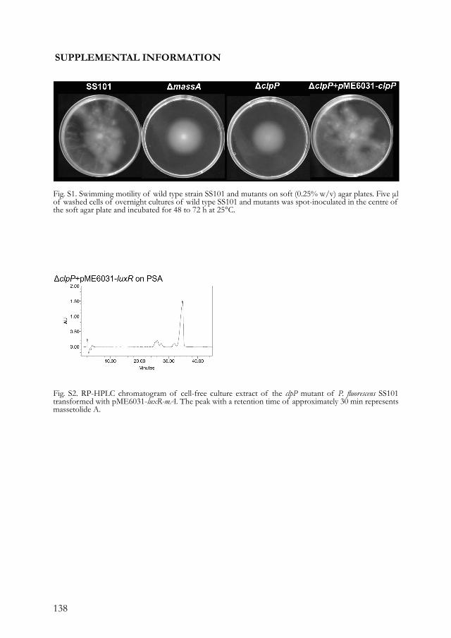
138
SUPPLEMENTAL INFORMATION
Fig. S2. RP-HPLC chromatogram of cell-free culture extract of the clpP mutant of P. fluorescens SS101 transformed with pME6031-luxR-mA. The peak with a retention time of approximately 30 min represents massetolide A.
Fig. S1. Swimming motility of wild type strain SS101 and mutants on soft (0.25% w/v) agar plates. Five μl of washed cells of overnight cultures of wild type SS101 and mutants was spot-inoculated in the centre of the soft agar plate and incubated for 48 to 72 h at 25°C.
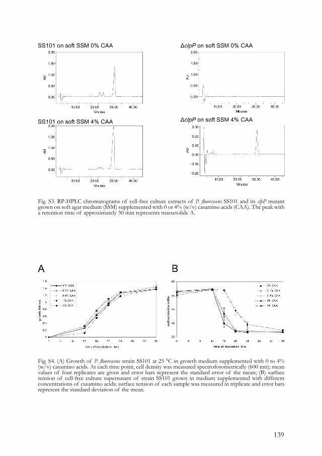
139
Fig. S4. (A) Growth of P. fluorescens strain SS101 at 25 °C in growth medium supplemented with 0 to 4% (w/v) casamino acids. At each time point, cell density was measured spectrofotometrically (600 nm); mean values of four replicates are given and error bars represent the standard error of the mean; (B) surface tension of cell-free culture supernatant of strain SS101 grown in medium supplemented with different concentrations of casamino acids; surface tension of each sample was measured in triplicate and error bars represent the standard deviation of the mean.
Fig. S3. RP-HPLC chromatograms of cell-free culture extracts of P. fluorescens SS101 and its clpP mutant grown on soft agar medium (SSM) supplemented with 0 or 4% (w/v) casamino acids (CAA). The peak with a retention time of approximately 30 min represents massetolide A.
A

140
Fig. S5. Swarming motility of the clpP mutant transformed with pME6031-luxR-mA on soft agar medium supplemented with 0 to 4% (w/v) casamino acids. Five μl of a washed overnight culture was spot-inoculated in the center of the soft agar plate and incubated for 48 to 72 h at 25°C.
Table S1. Percentages used of the amino acids present in casamino acids (CAA)
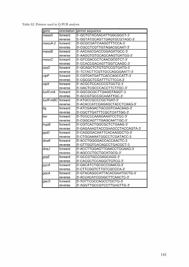
141
Table S2. Primers used in Q-PCR analysis

142
143
CHAPTER 6
Cyclic lipopeptides protect bacteria from protozoan predators.
Mark Mazzola1, Irene de Bruijn2, Michael F. Cohen3, and Jos M. Raaijmakers2
1USDA-ARS Tree Fruit Research Lab, Wenatchee, WA, USA2Laboratory of Phytopathology, Wageningen University, 6709 PD Wageningen, the Netherlands.3Department of Biology, Sonoma State University, Rohnert Park, CA USACorresponding author: Mark Mazzola; [email protected]
Submitted.

144
ABSTRACTEnvironmental bacteria are exposed to a myriad of biotic interactions. The grazing activity of protozoan predators significantly impacts the dynamics, diversification and evolution of bacterial communities in soil ecosystems. To evade protozoan predation, bacteria employ various defense strategies. Here, we show that soil bacteria are triggered at the molecular level by protozoan predators to produce specific detergent-like compounds, cyclic lipopeptide surfactants that protect them from being attacked and consumed. These findings highlight the functional versatility of these natural detergents and document the existence of a previously unknown sensing-signaling mechanism in the molecular dialogue between bacteria and protozoan predators.
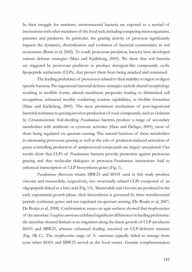
145
In their struggle for nutrients, environmental bacteria are exposed to a myriad of interactions with other members of the food web, including competing microorganisms, parasites and predators. In particular, the grazing activity of protozoa significantly impacts the dynamics, diversification and evolution of bacterial communities in soil ecosystems (Rønn et al, 2002). To evade protozoan predation, bacteria have developed various defense strategies (Matz and Kjelleberg, 2005). We show that soil bacteria are triggered by protozoan predators to produce detergent-like compounds, cyclic lipopeptide surfactants (CLPs), that protect them from being attacked and consumed. The feeding preference of protozoa is related to their inability to ingest or digest specific bacteria. Pre-ingestional bacterial defense strategies include altered morphology resulting in inedible forms, altered membrane properties leading to diminished cell recognition, enhanced motility conferring evasion capabilities, or biofilm formation (Matz and Kjelleberg, 2005). The most prominent mechanism of post-ingestional bacterial resistance to grazing involves production of toxic compounds, such as violacein by Chromobacterium. Soil-dwelling Pseudomonas bacteria produce a range of secondary metabolites with antibiotic or cytotoxic activities (Haas and Defago, 2005), most of them being regulated via quorum sensing. The natural function of these metabolites in attenuating protozoan grazing as well as the role of predation-induced activation of genes controlling production of antiprotozoal compounds are largely unexplored. Our results show that CLPs of Pseudomonas bacteria provide protection against protozoan grazing and that molecular dialogues in protozoa-Pseudomonas interactions lead to enhanced transcription of CLP biosynthesis genes (Fig. 1). Pseudomonas fluorescens strains SBW25 and SS101 used in this study produce viscosin and massetolide, respectively, two structurally related CLPs composed of an oligopeptide linked to a fatty acid (Fig. 1A). Massetolide and viscosin are produced in the early exponential growth phase; their biosynthesis is governed by three nonribosomal peptide synthetase genes and not regulated via quorum sensing (De Bruijn et al, 2007; De Bruijn et al, 2008). Confrontation assays on agar surfaces showed that trophozoites of the amoebae Naegleria americana exhibited significant differences in feeding preference: the amoebae showed limited or no migration along the linear growth of CLP-producers SS101 and SBW25, whereas enhanced feeding occurred on CLP-deficient mutants (Fig. 1B, C). The trophozoite stage of N. americana typically failed to emerge from cysts when SS101 and SBW25 served as the food source. Genetic complementation
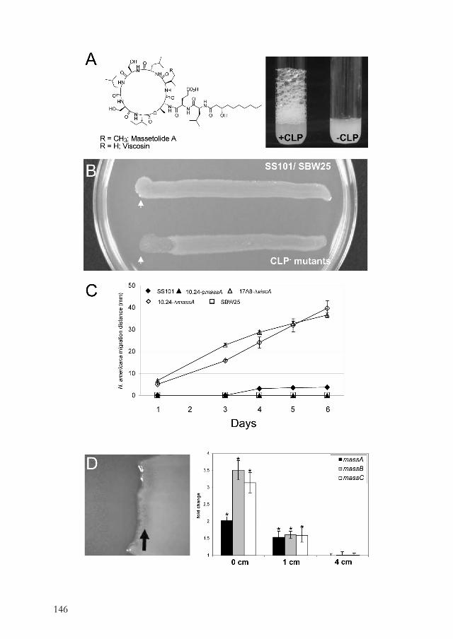
146
147
of the massetolide biosynthesis mutant of strain SS101 restored CLP production and resistance to feeding by N. americana (Fig. 1C). Also in soil environments, P. fluorescens SS101 exhibited superior persistence relative to its CLP-deficient mutant when confronted with N. americana (Fig. S1A). When exposed to different concentrations of purified viscosin, trophozoite lysis occurred rapidly. Purified massetolide and producing strain SS101 caused death of N. americana (Fig. S1B), but cell lysis was delayed relative to that observed for viscosin (Fig. S1C). These results exemplify that small structural differences between CLPs affect antiprotozoal activity. In vitro assays with a CLP-mutant of strain SS101 harbouring a biosensor that visualizes expression of the biosynthesis gene massA, showed that interaction with actively feeding N. americana triggered gene expression (Fig. 1D). Real-time quantitative PCR analyses confirmed that N. americana modulated expression of CLP-biosynthesis genes in P. fluorescens: bacterial cells collected at the feeding margin exhibited mass and visc expression levels that were significantly higher (up to 3.5 fold) than in cells collected at 4-cm distant (Fig. 1D). Parallel to recent work on amoebae-induced transcriptional changes in the human pathogen P. aeruginosa (Matz et al, 2008), our study documents the existence of a previously unknown sensing-signaling mechanism in bacteria-protozoa interactions that results in activation of an antipredator response in prey populations. Potential signals resulting in up-regulation of CLP-genes in P. fluorescens
Fig. 1. Structures of the cyclic lipopeptide surfactants (CLPs) viscosin and massetolide produced by Pseudomonas fluorescens strains SBW25 and SS101, respectively (left panel). The detergent-like properties of these CLPs are visualized (right panel) by shaking bacterial suspensions of strains SS101 and SBW25 (+CLP) and CLP-deficient mutants (-CLP); (B) In vitro assessment of the effect of CLP production on bacterial resistance to protozoan grazing. After 3 days of incubation, migration of Naegleria americana from the inoculation point (indicated by an arrow) can be discerned by the clearing of confluent growth of CLP-deficient mutants of P. fluorescens resulting in an obvious feeding margin, which is absent in CLP-producing wild type strains; (C) Migration distance for N. americana along confluent growth of CLP-producing strains SS101 and SBW25 and the corresponding CLP-deficient mutants 10.24-ΔmassA and 17A8-ΔviscA; genetic complementation of mutant 10.24 with pmassA (10.24-pmassA) restored massetolide production and resistance to feeding by N. americana; (D) In vitro assay conducted with N. americana and P. fluorescens 10.24-ΔmassA containing a promoterless lacZ fusion to massA. The feeding assay was conducted on agar plates supplemented with Xgal to visualise β-galactosidase activity in the bacterial cells. The protozoan predator was spotted on confluent growth of the bacterium and is grazing from left (cleared) to right in this diagram. The enhanced blue coloration just to the rear of the protozoan feeding margin (arrow) is indicative of enhanced transcriptional activity of the lacZ gene (left panel). Transcript levels of massA, massB, and massC in SS101 cells collected at distances of 0, 1 and 4 cm from the feeding front of N. americana trophozoites when incubated for 72 h (right panel). Similar results were obtained for transcript levels of viscA, viscB and viscC in strain SBW25. Transcript levels of the CLP biosynthesis genes were corrected for the transcript levels of the housekeeping gene rpoD [ΔCt=Ct(CLP gene) - Ct(rpoD)] and presented relative to the transcript level of cells taken at 4 cm distance from the feeding front (fold change), with fold change=2-[ΔCt(0 or 1 cm)-ΔCt(4 cm)]. For each distance, mean values of two technical replicates from two independent biological samples are given. Error bars represent the standard error of the mean. Asterisks indicate a statistically significant (P<0.05) difference from samples taken at 4 cm.

148
were not determined, however, it may not require physical contact as bacterial cells 1-cm distant from the amoebae also often exhibited enhanced gene expression (Fig. 1D). Thus, Pseudomonas-Naegleria signalling does not inevitably require intimate contact as was reported for the killing effect of biofilm-associated P. aeruginosa toward Acanthamoeba castellanii (Matz et al, 2008). Considering the importance of CLPs in bacterial motility and biofilm formation (De Bruijn et al, 2007; De Bruijn et al, 2008), this study highlights the functional versatility of these natural detergent-like compounds and provides novel insights into the regulation and evolution of antiprotozoal genes and compounds in environmental bacteria. Understanding the mechanisms underlying soil bacteria-protozoa interaction may also be instrumental for developing strategies to control human and animal pathogenic protozoa.
MATERIALS AND METHODS Organisms and cultivation. Strains SS101 and SBW25 produce the cyclic lipopeptide surfactants (CLPs) massetolide and viscosin, respectively (De Bruijn et al, 2007, 2008). The massetolide-deficient mutant 10.24-ΔmassA of strain SS101 has a Tn5 transposon insertion in massA (De Souza et al, 2003; De Bruijn et al, 2008). This mutation was complemented through introduction of plasmid pmassA (pME6031-massA; De Bruijn et al, 2008) to yield derivative strain 10.24-pmassA. The viscosin-deficient mutant 17A8-ΔviscA of strain SBW25 contains a TnMod plasposon insertion in viscA (De Bruijn et al, 2008). All bacterial strains were routinely cultured on King’s medium B (King et al, 1954) agar at 28°C. The bacteriovorous amoeba-flagellate Naegleria americana C1 (Cohen and Mazzola, 2006) was utilized as the protozoan predator in this study. Amoebae were amplified by cultivation on heat-killed Escherichia coli. Bacterial cells (≈1010) were spread evenly over a water agar surface in a 9-cm diameter Petri plate and inoculated with 200 μl of a 2 x 10² μl-1 N. americana cyst suspension, overlayed with 3 ml Page’s modified Neff ’s amoeba saline (PAS). Plates were sealed with parafilm, incubated at 20°C and 2 ml PAS was added to plates at 7-d intervals.
Feeding assay. Inocula of individual CLP-producing strains and corresponding mutants prepared on KB agar were lined across the surface of 1/5th-strength nutrient broth-yeast extract (NBY; Vidaver, 1967) agar plates at a width of 4 mm. After overnight incubation at 24°C, 3.5 μl of a N. americana suspension (300 cyst/μl) was spotted at one end of the linear bacterial growth and plates were incubated at 24°C, with 15 replicates for each strain. Linear migration of N. americana trophozoites along the confluent bacterial growth was monitored over a period of six days.
CLP antiprotozoal activity. The CLPs massetolide and viscosin were purified from cultures of strains SS101 and SBW25, respectively, according to the methods described by De Souza et al. (2003). The individual surfactants were resuspended in PAS to achieve concentrations of 1, 10, 25, 100 and 500 μg/ml. A 5-μl aliquot of a N. americana trophozoite suspension was added to 50 μl of the CLP suspension and the phenotypic responses of the amoebae were monitored microscopically (100X) for up to 1 hour.
Effect of N. americana feeding on CLP gene transcription. SS101 and SBW25 cells were cultured in a linear band on 1/5th-strength NBY agar, with four replicate bands per agar plate, each 4 mm in width. Cultures were incubated at 28°C for 3 h and then a 5-μl spot of a N. americana suspension (100 cysts/μl) was placed at one end of each bacterial band. After 48 h co-incubation at 28°C, bacterial cells were collected from the entire 4 mm width of the bacterial culture using an RNase-free pipet tip at distances of 0, 1 and 4 cm from the N. americana trophozoite feeding margin. RNA was extracted from cells using a Qiagen RNAeasy kit using the manufacturer’s protocol or as described by De Bruijn et al. (2008). cDNA synthesis was conducted as previously described (De Bruijn et al, 2008). CLP gene expression was monitored by real-time quantitative PCR (Q-PCR) as described in detail by De Bruijn et al. (2008). The primers used in Q-PCR for the massetolide biosynthesis genes were, massA: forward 5’- GCTGTACAACATTGGCGGCT-3’, reverse 5’- GGTATGCAGTTGAGTGCGTAGC-3’, massB: forward 5’-AACAACGACCGGAGATGCC-3’, reverse 5’-AAGGTGTGCAGCAAGTGATGG-3’, massC: forward 5’-AAGGTGTGCAGCAAGTGATGG-3’, reverse 5’- CCACCGACAGTTGGTCAAGC-3’. Primers used
149
in Q-PCR for viscosin biosynthetic genes were, viscA: forward 5’ CCGGATGGCAATCTTGAGTTT-3’, reverse 5’-GTGACTCGATTTCCCCCAGTT-3’, viscB: forward 5′- ATCCGTGGCCTGCGTATC-3′, reverse 5′- CCTTGACCGATGCGTGTTC-3′ , viscC: forward 5′- CGGACCTCTTGAGCTTTATCGA-3′, reverse 5′- AGAATCACTGCGTCGTGACAAC-3′. For the housekeeping gene rpoD, the primers used were forward 5’-GCAGCTCTGTGTCCGTGATG-3’, reverse 5’-TCTACTTCGTTGCCAGGGAATT-3’. The concentration of the primers was optimized (400 nM final concentration for all) and a melting curve was performed to check the specificity of the primers. Q-PCR analysis was performed in duplicate (technical replicates) on two independent RNA isolations (biological replicates) and repeated for two sets of feeding assays. Transcript levels of the CLP biosynthesis genes were corrected for the transcript levels of the housekeeping gene rpoD [ΔCt=Ct(CLP gene)-Ct(rpoD)] and presented relative to the transcript level of cells taken at 4 cm distant from the feeding front (fold change), with fold change=2-[ΔCt(0 or 1 cm)-ΔCt(4 cm)]. For each distance, mean values for three biological replicates are given. Error bars represent the standard errors of the means. Statistically significant differences were determined for log-transformed fold change values by analysis of variance (p<0.05) followed by Bonferroni post hoc multiple comparisons.
Effect of CLP production on bacterial-protozoan interaction in soil. Bacterial cultures were scraped from the surface of a KB agar plate, resuspended in sterile distilled water and adjusted to a concentration of approximately 5 x 10⁹ cfu/ml. Cells were dispensed into the chamber of a 50-ml chromatography sprayer and applied as an atomized mist to individual 1.4 kg samples of soil (Adkins very fine sandy loam; pH 7.6) that had been pasteurized at 102°C for 3 h prior to inoculation. Cysts from two-week-old cultures of N. americana were collected from water agar plates and adjusted to a concentration of 200 μl-1 in PAS. The cyst suspension was applied to soil using a 50-ml spray bottle. The volume of treated soil was adjusted to attain an initial protozoa population of approximately 104 cysts/g soil. Treated soils were decanted into conical tubes (20.5-by-4 cm top diameter) with 12 replicates per soil treatment arranged in a complete randomized design. Assays were conducted in environmental growth chambers with a 16-h photoperiod and a 24/18°C day/night temperature regime. At weekly intervals, a 1-g soil sample was collected from six tubes for each soil treatment. For determination of populations of SS101 and 10.24-ΔmassA, the 1-g sample was resuspended in 10 ml sterile distilled water, vortexed for 60 s, and serial dilutions were plated onto KB agar amended with rifampicin (100 μg/ml) and cycloheximide (75 μg/ml).
REFERENCES Cohen, M. F., and Mazzola, M. 2006. Effects of Brassica napus seed meal amendment on soil populations of resident bacteria and Naegleria americana, and the unsuitability of arachidonic acid as a protozoan-specific marker. J. Protozool. Res. 16:16-25.De Bruijn, I., de Kock, M. J., de Waard, P., van Beek, T. A., and Raaijmakers, J. M. 2008. Massetolide A biosynthesis in Pseudomonas fluorescens. J. Bacteriol. 190:2777-2789.De Bruijn, I., de Kock, M. J. D., Yang, M., de Waard, P., van Beek, T. A., and Raaijmakers, J. M. 2007. Genome-based discovery, structure prediction and functional analysis of cyclic lipopeptide antibiotics in Pseudomonas species. Mol. Microbiol. 63:417-428.De Souza, J. T., De Boer, M., De Waard, P., Van Beek, T. A., and Raaijmakers, J. M. 2003. Biochemical, genetic, and zoosporicidal properties of cyclic lipopeptide surfactants produced by Pseudomonas fluorescens. Appl. Environ. Microbiol. 69:7161-7172.Haas, D., and Defago, G. 2005. Biological
control of soil-borne pathogens by fluorescent pseudomonads. Nat. Rev. Microbiol. 3:307-19.King, E. O., Ward, M. K., and Raney, D. E. 1954. Two simple media for the demonstration of pyocyanin and fluorescin. J. Lab. Clin. Med. 44:301-307.Matz, C., and Kjelleberg, S. 2005. Off the hook - how bacteria survive protozoan grazing. Trends Microbiol. 13:302-307.Matz, C., Moreno, A. M., Alhede, M., Manefield, M., Hauser, A. R., Givskov, M., and Kjelleberg, S. 2008. Pseudomonas aeruginosa uses type III secretion system to kill biofilm-associated amoebae. ISME J. 2:843-52.Ronn, R., McCaig, A. E., Griffiths, B. S., and Prosser, J. I. 2002. Impact of protozoan grazing on bacterial community structure in soil microcosms. Appl. Environ. Microbiol. 68:6094-6105.Vidaver, A. K. 1967. Synthetic and complex media for the rapid detection of fluorescence of phytopathogenic pseudomonads: effect of the carbon source. Appl. Microbiol. 15:1523-1524.
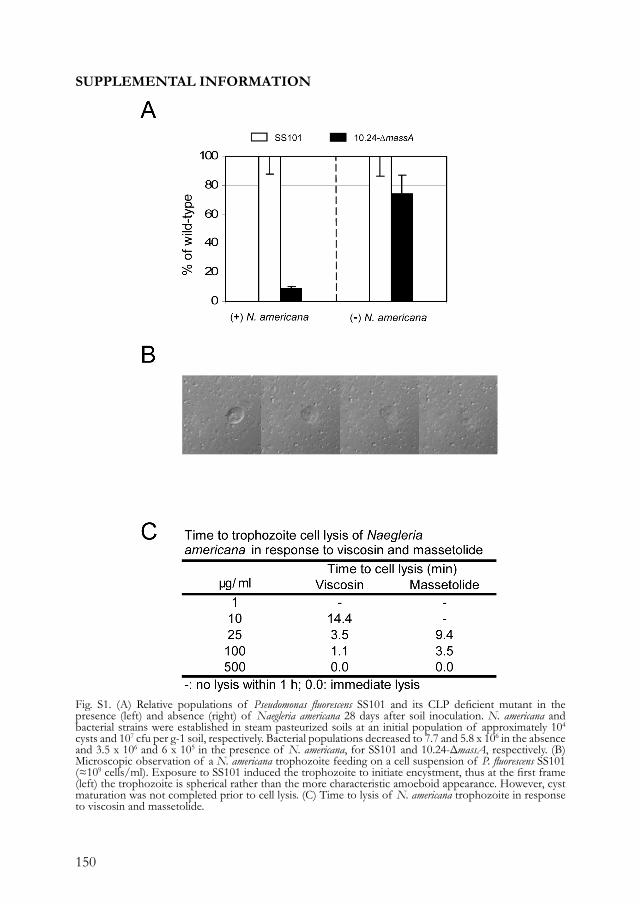
150
SUPPLEMENTAL INFORMATION
Fig. S1. (A) Relative populations of Pseudomonas fluorescens SS101 and its CLP deficient mutant in the presence (left) and absence (right) of Naegleria americana 28 days after soil inoculation. N. americana and bacterial strains were established in steam pasteurized soils at an initial population of approximately 104 cysts and 107 cfu per g-1 soil, respectively. Bacterial populations decreased to 7.7 and 5.8 x 10⁶ in the absence and 3.5 x 10⁶ and 6 x 10⁵ in the presence of N. americana, for SS101 and 10.24-ΔmassA, respectively. (B) Microscopic observation of a N. americana trophozoite feeding on a cell suspension of P. fluorescens SS101 (≈109 cells/ml). Exposure to SS101 induced the trophozoite to initiate encystment, thus at the first frame (left) the trophozoite is spherical rather than the more characteristic amoeboid appearance. However, cyst maturation was not completed prior to cell lysis. (C) Time to lysis of N. americana trophozoite in response to viscosin and massetolide.

151

152
153
CHAPTER 7
Natural roles of cyclic lipopeptides and other bacterial biosurfacants.
Irene de Bruijn1 and Jos M. Raaijmakers1
1Laboratory of Phytopathology, Wageningen University, 6709 PD Wageningen, the Netherlands.

154
155
INTRODUCTIONBiosurfactants are soap-like compounds produced by a variety of microorganisms, including yeasts, fungi and bacteria (Table 1; Banat et al. 2000; Maier 2003; Mulligan 2005; Raaijmakers et al. 2006). A range of structurally different biosurfactants have been identified, including the low-molecular weight biosurfactants such as glycolipids and cyclic lipopeptides, and the high-molecular weight biosurfactants composed of polysaccharides, proteins, lipopolysaccharides, lipoproteins or mixtures thereof (Banat et al. 2000; Desai and Banat 1997; Maier 2003; Mulligan 2005; Muthusamy et al. 2008; Neu 1996; Ron and Rosenberg 2001). The amphipathic properties as well as the antimicrobial activities of biosurfactants have received considerable interest from chemical and biomedical industries (Banat et al. 2000; Desai and Banat 1997; Mulligan 2005; Muthusamy et al. 2008; Ongena and Jacques 2008; Ron and Rosenberg 2002). In contrast, the natural roles of these metabolites in the ecology of the producing microorganisms have received less attention and the available information is fragmentary and limited to relatively few microbial groups. The purpose of this mini-review is to give a detailed overview of the biological functions of biosurfactants produced by pathogenic and beneficial bacteria. Specific attention will be given to the cyclic lipopeptides (CLPs) produced by Pseudomonas and Bacillus species, but whenever possible also the functions of other biosurfactants will be presented and discussed. Prior to addressing the different natural roles of bacterial biosurfactants, we also describe briefly the different methods to detect biosurfactants as well as their diversity, stability and regulation.
DETECTION AND CHARACTERIZATION OF BIOSURFACTANTSPhenotypic analyses. One of the first tests to determine if a specific microorganism produces a biosurfactant is the drop collapse assay: in this qualitative assay, a droplet of a cell suspension or culture supernatant is spotted onto a hydrophobic surface (e.g. parafilm); when a biosurfactant is present, the droplet collapses (Fig. 1). Subsequently, the reduction in surface tension of cell suspensions can be measured quantitatively by tensiometric analysis or by measuring the contact angle and shape of the droplet. Other relatively simple phenotypic assays include testing the emulsifying or haemolytic properties of cell suspensions (Johnson and Boese-Marrazzo, 1980), or measuring the transmission and reflection of a light beam through a droplet, which are distorted in presence of a biosurfactant (Chen et al. 2007). Although these methods are very useful
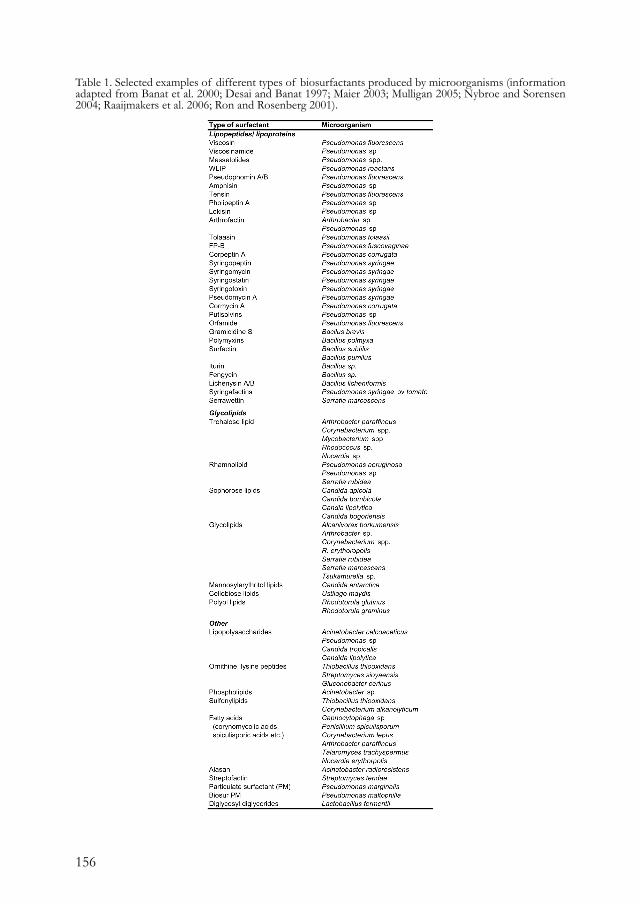
156
Table 1. Selected examples of different types of biosurfactants produced by microorganisms (information adapted from Banat et al. 2000; Desai and Banat 1997; Maier 2003; Mulligan 2005; Nybroe and Sorensen 2004; Raaijmakers et al. 2006; Ron and Rosenberg 2001).
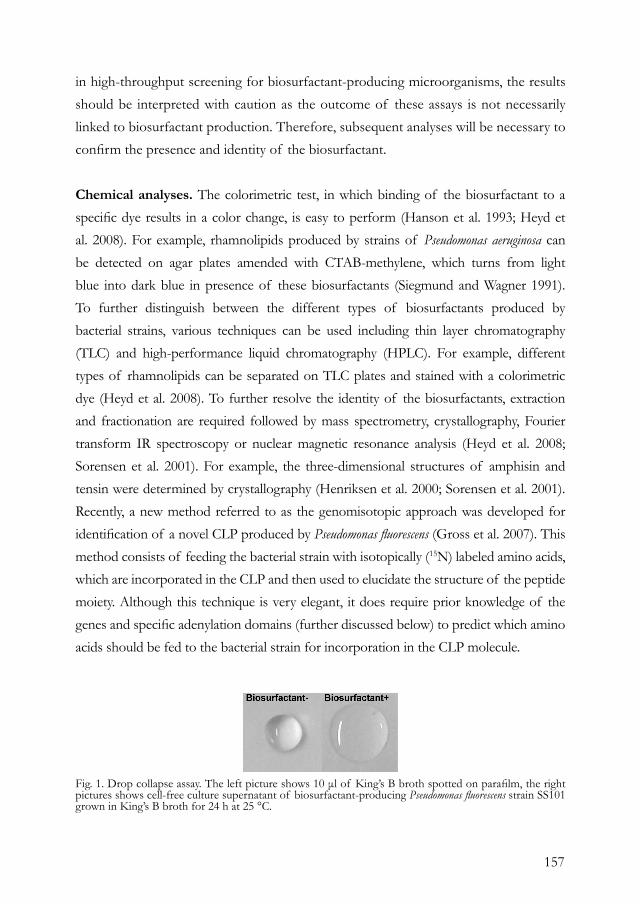
157
in high-throughput screening for biosurfactant-producing microorganisms, the results should be interpreted with caution as the outcome of these assays is not necessarily linked to biosurfactant production. Therefore, subsequent analyses will be necessary to confirm the presence and identity of the biosurfactant.
Chemical analyses. The colorimetric test, in which binding of the biosurfactant to a specific dye results in a color change, is easy to perform (Hanson et al. 1993; Heyd et al. 2008). For example, rhamnolipids produced by strains of Pseudomonas aeruginosa can be detected on agar plates amended with CTAB-methylene, which turns from light blue into dark blue in presence of these biosurfactants (Siegmund and Wagner 1991). To further distinguish between the different types of biosurfactants produced by bacterial strains, various techniques can be used including thin layer chromatography (TLC) and high-performance liquid chromatography (HPLC). For example, different types of rhamnolipids can be separated on TLC plates and stained with a colorimetric dye (Heyd et al. 2008). To further resolve the identity of the biosurfactants, extraction and fractionation are required followed by mass spectrometry, crystallography, Fourier transform IR spectroscopy or nuclear magnetic resonance analysis (Heyd et al. 2008; Sorensen et al. 2001). For example, the three-dimensional structures of amphisin and tensin were determined by crystallography (Henriksen et al. 2000; Sorensen et al. 2001). Recently, a new method referred to as the genomisotopic approach was developed for identification of a novel CLP produced by Pseudomonas fluorescens (Gross et al. 2007). This method consists of feeding the bacterial strain with isotopically (15N) labeled amino acids, which are incorporated in the CLP and then used to elucidate the structure of the peptide moiety. Although this technique is very elegant, it does require prior knowledge of the genes and specific adenylation domains (further discussed below) to predict which amino acids should be fed to the bacterial strain for incorporation in the CLP molecule.
Fig. 1. Drop collapse assay. The left picture shows 10 μl of King’s B broth spotted on parafilm, the right pictures shows cell-free culture supernatant of biosurfactant-producing Pseudomonas fluorescens strain SS101 grown in King’s B broth for 24 h at 25 °C.
158
Immunological detection. Compared to most of the phenotypic and chemical techniques described above, immunological assays are more suited to detect and quantify biosurfactants, especially in plant-associated environments where relatively low amounts of the biosurfactants are produced and where plant-derived compounds may interfere with chemical detection. Immunological detection has been successfully adopted for syringopeptins produced in situ by Pseudomonas syringae pv. lachrymans, the causal agent of angular leaf spot: the competitive ELISA assay appeared to be approximately 100 times more sensitive than HPLC analysis and did not require extraction of plant material with organic solvents (Fogliano et al. 1999). Specific antibodies will also be highly instrumental to study the localization and stability of biosurfactants produced in situ, or to monitor their fate after their application to soil or plant tissues.
PCR-based detection. The tremendous increase in the availability of whole genome sequences of a wide variety of microorganisms has provided a significant advance in the identification of genes involved in biosurfactant biosynthesis. The biosynthesis genes for rhamnolipids (Ochsner et al. 1994a) and for many CLPs (De Bruijn et al. 2008; De Bruijn et al. 2007; Dubern et al. 2008; Gross et al. 2007; Raaijmakers et al. 2006) are now available and can be used to develop specific primers and probes for detection. For example, Lee et al (2002) developed PCR primers directed against specific sequences in the tolaasin biosynthesis gene for detection and identification of the brown blotch pathogen Pseudomonas tolaasii in various mushroom samples. Also for identification of P. syringae strains producing syringomycin, a PCR-based method was developed using primers for the syrD gene (Bultreys and Gheysen 1999; Quigley and Gross 1994; Sorensen et al. 1998). However, the syrD primers were not always specific for syringomycin-producing strains (Sorensen et al. 1998), possibly due to the fact that syrD encodes for an ATP-binding transporter (Quigley et al. 1993) with close homologues also in non-syringomycin producers. The use of primers specific for syrB, one of the syringomycin biosynthesis genes, resulted in more specific detection of syringomycin producers with a positive result for 27 out of 39 strains tested. Among these 27 isolates, however, some did not produce syringomycin but the related syringostatin or syringotoxin (Sorensen et al. 1998), showing that the close genetic relatedness between the CLP biosynthesis genes makes it difficult to design primers for detection of a specific CLP.
159
Whole genome sequences not only provide a tool to develop primers and probes for detection, but also a means to predict the structure of biosurfactants, in particular CLPs. Structure prediction of the CLP peptide moiety is based on specific signature sequences in the adenylation domains of the NRPS proteins (Challis et al. 2000; De Bruijn et al. 2007; Stachelhaus et al. 1999). Moreover, also the L-or D- configuration of the amino acid in the peptide chain of CLPs can be predicted (Balibar et al. 2005). Structure prediction of metabolites produced by NRPS or polyketide synthetases (PKS) can be performed manually or with the use of online databases, i.e. ANSARI (http://www.nii.res.in/nrps-pks.html) and NORINE (http://bioinfo.lifl.fr/norine/). Using this genome-based approach, several CLPs were recently identified in Pseudomonas species and confirmed by chemical analyses, including viscosin in P. fluorescens SBW25 (De Bruijn et al. 2007), orfamide in P. fluorescens Pf-5 (Gross et al. 2007) and syringafactins in P. syringae pv. tomato DC3000 (Berti et al. 2007).
DIVERSITY AND STABILITY OF BIOSURFACTANTSOver the past decades, many biosurfactants and biosurfactant-producing microorganisms have been identified (Banat et al. 2000; Desai and Banat 1997; Maier 2003; Mulligan 2005; Muthusamy et al. 2008; Raaijmakers et al. 2006). Among CLPs produced by Bacillus and Pseudomonas species, there is considerable structural diversity due to differences in the length and composition of the fatty acid tails, as well as in the type, number and configuration of the amino acids in the peptide moiety (Nybroe and Sorensen 2004; Raaijmakers et al. 2006). However, little is known about the frequency of biosurfactant-producing microorganisms in diverse environments. One of the few studies on the frequency of CLP-producing bacteria in natural environments is the work by Nielsen et al. (2002), who showed that in a Danish sandy loam soil 6% of the fluorescent pseudomonads produced biosurfactants, whereas in a loamy sand soil approximately 60% of the Pseudomonas population were positive in the drop collapse assay. Although the pH and soil texture was significantly different between the two Danish soils, the actual factors contributing to this difference in frequency of CLP producers were not identified (Nielsen et al. 2002). In Dutch agricultural soils, De Souza et al. (2003) showed that on average 1-5% of the fluorescent pseudomonads isolated from the wheat rhizosphere produced a biosurfactant based on the drop collapse assay. A similar
160
percentage of biosurfactant producers was found among the Pseudomonas population obtained from the rhizosphere of black pepper plants grown in Vietnam (Tran et al. 2008) and in the rhizosphere of sugar beets grown in the Danisco experimental field (Nielsen et al. 1998). Not only in soil, but also in other environments biosurfactant producers were detected and quantified. From a drinking water reservoir in Russia, one out of 46 Pseudomonas isolates produced a CLP (Ivanova et al. 2002). Taken together, these results indicate that CLP production is not uncommon among indigenous Pseudomonas isolates although the frequency is relatively low compared to the frequency found in the genome-based analyses, which indicated that 5 out of 7 Pseudomonas strains harbor biosynthesis genes for CLP production (De Bruijn et al. 2007). This discrepancy may be explained, in part, by the fact that the conditions used to grow the indigenous bacteria prior to the drop collapse assay may not be favorable for biosurfactant production in all isolates tested. The study by Nielsen et al. (2002) further showed that the CLPs produced by approximately 170 isolates obtained from the Danish soils clustered in eight distinct groups based on the LC-MS analyses. In contrast, genotypically diverse populations of biosurfactant-producing Pseudomonas isolates from the rhizosphere of black pepper were shown to all produce the same biosurfactants (Tran et al. 2008). More experiments will be required to determine the specific effects of host plant and soil type on the frequency of biosurfactant producers and the structural diversity of the biosurfactants.
In situ production and stability. Several biosurfactants are produced in situ, including viscosinamide by P. fluorescens strain DR54 (Nielsen and Sorensen 2003), iturin A and surfactin by Bacillus subtilis RB14-C (Asaka and Shoda 1996), and xanthobaccin A by Stenotrophomonas strain SB-K88 (Nakayama et al. 1999). In bulk soil, amphisin and tensin were not produced by P. fluorescens sp. DSS73 and strain 96.578 at detectable levels. However, when the bacterial strains were introduced onto sugar beet seeds both CLPs were detected in soil (Nielsen and Sorensen 2003). This study and the work by Koch et al. (2002) suggest that specific signals and/or exudates from the sugar beet seeds induce biosurfactant production. The stability of biosurfactants in complex environments has not been studied extensively. For the CLPs produced by Bacillus, Asaka and Shoda (1996) showed that iturin levels declined within 25 days in sterile soil, while surfactin levels

161
remained stable. The authors suggested that iturin may have leached from soil by watering, was degraded by microorganisms or was not detectable due to irreversible binding to soil particles (Asaka and Shoda 1996). When the CLPs viscosinamide, tensin and amphisin were introduced in sterile soil, their concentrations remained stable during the two-week incubation period, but declined after one to two days in non-sterile bulk soil and only minimal amounts could be recovered after two weeks. Nielsen and Sorensen (2003) suggested that in natural soil the biosurfactants are degraded by indigenous microorganisms. When the Pseudomonas strains producing viscosinamide, tensin or amphisin were introduced into bulk soil, viscosinamide was detected at an initial concentration of 0.3 μg/g of dry soil, but declined to 17% over the next 15 days although the population density of the producing strain remained stable. Unlike viscosinamide, tensin and amphisin have a limited binding to the producing cells and washing of the bacterial inocula prior to addition to soil resulted in undetectable levels of tensin and amphisin during the course of the experiment (Nielsen and Sorensen 2003). Whether tensin and amphisin are more sensitive to microbial degradation than viscosinamide was not determined. For Staphylococcus aureus, it was shown that an endoprotease hydrolyzed surfactin, thereby generating a linear peptide (Grangemard et al. 1999). Also Nitschke and Pastore (2004) suggested that protease activity contributed to a decrease of the surfactin levels in culture medium of B. subtilis strain 21332. Enzymatic degradation was studied in more detail for syringomycin and syringopeptin to determine potential harmful effects of these CLPs after human ingestion; both CLPs were degraded and showed a reduced haemolytic activity during upper-gut and gut digestion, but not during stomach digestion (Fiore et al. 2008). The authors suggested that these biosurfactants are not substrates for pepsin, but that pancreatic enzymes strongly reduced their toxic activity. Additional tests revealed that syringomycin did not pass the intestinal cell layer, indicating that when ingested, syringomycin will not reach the blood stream and cause harmful effects (Fiore et al. 2008).
Cell-density dependent regulation of biosurfactant production. Nybroe and Sørensen (2004) emphasized that although biosurfactant production is affected by the growth phase and nutritional conditions, the specific impact of these factors and the underlying molecular mechanisms in relation to CLP biosynthesis are yet
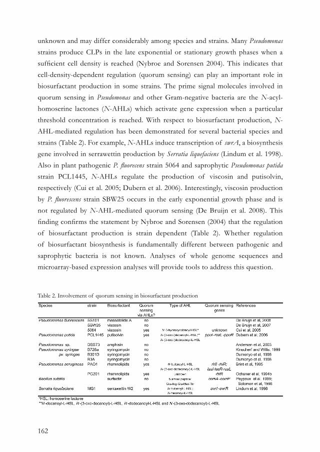
162
unknown and may differ considerably among species and strains. Many Pseudomonas strains produce CLPs in the late exponential or stationary growth phases when a sufficient cell density is reached (Nybroe and Sorensen 2004). This indicates that cell-density-dependent regulation (quorum sensing) can play an important role in biosurfactant production in some strains. The prime signal molecules involved in quorum sensing in Pseudomonas and other Gram-negative bacteria are the N-acyl-homoserine lactones (N-AHLs) which activate gene expression when a particular threshold concentration is reached. With respect to biosurfactant production, N-AHL-mediated regulation has been demonstrated for several bacterial species and strains (Table 2). For example, N-AHLs induce transcription of swrA, a biosynthesis gene involved in serrawettin production by Serratia liquefaciens (Lindum et al. 1998). Also in plant pathogenic P. fluorecens strain 5064 and saprophytic Pseudomonas putida strain PCL1445, N-AHLs regulate the production of viscosin and putisolvin, respectively (Cui et al. 2005; Dubern et al. 2006). Interestingly, viscosin production by P. fluorescens strain SBW25 occurs in the early exponential growth phase and is not regulated by N-AHL-mediated quorum sensing (De Bruijn et al. 2008). This finding confirms the statement by Nybroe and Sorensen (2004) that the regulation of biosurfactant production is strain dependent (Table 2). Whether regulation of biosurfactant biosynthesis is fundamentally different between pathogenic and saprophytic bacteria is not known. Analyses of whole genome sequences and microarray-based expression analyses will provide tools to address this question.
Table 2. Involvement of quorum sensing in biosurfactant production

163
NATURAL FUNCTIONS OF BIOSURFACTANTSBiosurfactants exhibit enormous structural diversity, suggesting that they also have different ecological roles (Ron and Rosenberg, 2001). However, biosurfactants have many functions in common, including a role in motility, attachment and detachment to and from surfaces, and antimicrobial activity. To date, most of the hypotheses for the natural roles of biosurfactants have been derived from their physical and chemical properties. In recent years, mutants deficient in biosurfactant production were obtained for several bacterial species and strains, and are now being used to further investigate the biological functions of biosurfactants. In this section, several of the potential functions of biosurfactants, in particular CLPs, will be discussed. Motility. Movement of bacteria on a surface has been extensively studied in vitro and several distinct forms were recognized, including swimming, swarming and twitching (Henrichsen 1972). For swimming and swarming, the flagella coalesce into a bundle and by the rotating flagellar motor push the bacteria forward (Harshey 2003). With swimming, the bacterial cells move individually, while with swarming the cells move in groups forming very distinct phenotypes on soft agar plates (Fig. 2). During swarming, the vegetative cells differentiate into specialized swarmer cells, which are hyperflagellated and generally longer (Harshey 2003). The viscosity of the surface can regulate this cell differentiation: for example, when branched polymers such as Ficoll 400 or polyvinylpyrrolidone 360 are added to liquid medium, a viscous matrix was created and resulted in cell differentiation in Proteus mirabilis and Vibrio parahaemolyticus (Allison et al. 1993; McCarter and Silverman 1990). Moreover, the polar flagella seem to be able to sense the viscosity and trigger a response of the lateral flagella (McCarter et al. 1988). Biosurfactants can act as wetting agents and change the viscosity of surfaces,
Fig. 2. Different phenotypes of swarming motility of Pseudomonas fluorescens strain SS101 and a massetolide-deficient mutant (complete right). Washed cells of strain SS101 are spot-inoculated in the centre of soft agar (0.6% w/v) plates and incubated for 3 days at 25 °C.
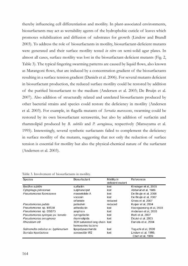
164
thereby influencing cell differentiation and motility. In plant-associated environments, biosurfactants may act as wettability agents of the hydrophobic cuticle of leaves which promotes solubilization and diffusion of substrates for growth (Lindow and Brandl 2003). To address the role of biosurfactants in motility, biosurfactant-deficient mutants were generated and their surface motility tested in vitro on semi-solid agar plates. In almost all cases, surface motility was lost in the biosurfactant-deficient mutants (Fig. 2, Table 3). The typical fingering swarming patterns are caused by liquid flows, also known as Marangoni flows, that are induced by a concentration gradient of the biosurfactants resulting in a surface tension gradient (Daniels et al. 2006). For several mutants deficient in biosurfactant production, the reduced surface motility could be restored by addition of the purified biosurfactant to the medium (Andersen et al. 2003; De Bruijn et al. 2007). Also addition of structurally related and unrelated biosurfactants produced by other bacterial strains and species could restore the deficiency in motility (Andersen et al. 2003). For example, in flagella mutants of Serratia marcescens, swarming could be restored by its own biosurfactant serrawettin, but also by addition of surfactin and rhamnolipid produced by B. subtilis and P. aeruginosa, respectively (Matsuyama et al. 1995). Interestingly, several synthetic surfactants failed to complement the deficiency in surface motility of the mutants, suggesting that not only the reduction of surface tension is essential for motility but also the physical-chemical nature of the surfactant (Andersen et al. 2003).
Table 3. Involvement of biosurfactants in motility.
165
Surface attachment and colonization. Single bacterial cells can attach to surfaces and, after cell division and proliferation, form dense, highly hydrated aggregates called biofilms. The cells secrete polymers like polysaccharides and proteins that form a hydrated gel-like slime which holds the biofilm together (Stewart and Franklin 2008). The extracellular matrix of the biofilm protects bacteria against adverse environmental conditions: for example, biofilms are less sensitive to antibiotic treatment or shearing (Drenkard and Ausubel 2002; Hall-Stoodley et al. 2004). Biofilms also provide protection against protozoan predation and are a niche for horizontal gene transfer (Danhorn and Fuqua 2007). Within mature biofilms, there is substantial structural, chemical and biological heterogeneity with concentration gradients of metabolic substrates or products (reviewed in Stewart and Franklin (2008). For example, the oxygen concentration varies within and between biofilms; some are completely anaerobic, while others show a high oxygen concentration at the biofilm-liquid interface that decreases with increasing depth (Stewart and Franklin 2008). Within a biofilm, also genetic variation can occur and a small percentage of the colonies can have a morphology different from that of the parental strain. For example, in static liquid microcosms of P. fluorescens SBW25, wrinkly variants arose which were able to form a cellulose-based biofilm at the air-liquid interface and had distinct colony morphology compared to the wild type (Spiers and Rainey 2005). Biosurfactants play an important role in the attachment and detachment to and from surfaces (Fig. 3, Table 4). Rhamnolipids produced by P. aeruginosa were shown to be essential for the initiation of biofilm formation, but also for migration of a subpopulation within the biofilm (Pamp and Tolker-Nielsen 2007). In addition, Davey et al. (2003) and Klausen et al. (2003) proposed that biosurfactants can maintain liquid-channels in biofilms to facilitate the distribution of nutrients and oxygen. For various Pseudomonas strains, CLPs play an important role in biofilm formation although the outcome may differ depending on the type of CLP: some CLPs are essential for biofilm formation, whereas others adversely affect biofilm formation (Fig. 3, Table 4). Pseudomonas sp. MIS38 produces the biosurfactant arthrofactin and forms a biofilm on polypropylene microcentrifuge tubes. However, arthrofactin-deficient mutants formed more, but unstable biofilms (Roongsawang et al. 2003). Similar results were found for putisolvin produced by P. putida strains (Kuiper et al. 2004; Kruijt et al. 2008). For P. fluorescens strains SBW25 and SS101, however, viscosin and massetolide are essential
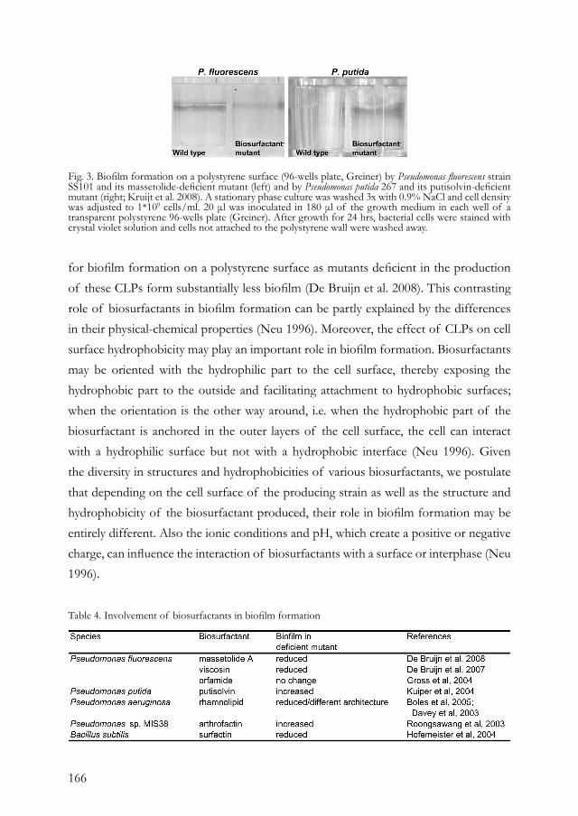
166
for biofilm formation on a polystyrene surface as mutants deficient in the production of these CLPs form substantially less biofilm (De Bruijn et al. 2008). This contrasting role of biosurfactants in biofilm formation can be partly explained by the differences in their physical-chemical properties (Neu 1996). Moreover, the effect of CLPs on cell surface hydrophobicity may play an important role in biofilm formation. Biosurfactants may be oriented with the hydrophilic part to the cell surface, thereby exposing the hydrophobic part to the outside and facilitating attachment to hydrophobic surfaces; when the orientation is the other way around, i.e. when the hydrophobic part of the biosurfactant is anchored in the outer layers of the cell surface, the cell can interact with a hydrophilic surface but not with a hydrophobic interface (Neu 1996). Given the diversity in structures and hydrophobicities of various biosurfactants, we postulate that depending on the cell surface of the producing strain as well as the structure and hydrophobicity of the biosurfactant produced, their role in biofilm formation may be entirely different. Also the ionic conditions and pH, which create a positive or negative charge, can influence the interaction of biosurfactants with a surface or interphase (Neu 1996).
Fig. 3. Biofilm formation on a polystyrene surface (96-wells plate, Greiner) by Pseudomonas fluorescens strain SS101 and its massetolide-deficient mutant (left) and by Pseudomonas putida 267 and its putisolvin-deficient mutant (right; Kruijt et al. 2008). A stationary phase culture was washed 3x with 0.9% NaCl and cell density was adjusted to 1*109 cells/ml. 20 μl was inoculated in 180 μl of the growth medium in each well of a transparent polystyrene 96-wells plate (Greiner). After growth for 24 hrs, bacterial cells were stained with crystal violet solution and cells not attached to the polystyrene wall were washed away.
Table 4. Involvement of biosurfactants in biofilm formation
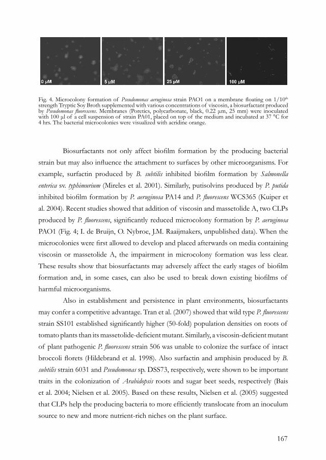
167
Biosurfactants not only affect biofilm formation by the producing bacterial strain but may also influence the attachment to surfaces by other microorganisms. For example, surfactin produced by B. subtilis inhibited biofilm formation by Salmonella enterica sv. typhimurium (Mireles et al. 2001). Similarly, putisolvins produced by P. putida inhibited biofilm formation by P. aeruginosa PA14 and P. fluorescens WCS365 (Kuiper et al. 2004). Recent studies showed that addition of viscosin and massetolide A, two CLPs produced by P. fluorescens, significantly reduced microcolony formation by P. aeruginosa PAO1 (Fig. 4; I. de Bruijn, O. Nybroe, J.M. Raaijmakers, unpublished data). When the microcolonies were first allowed to develop and placed afterwards on media containing viscosin or massetolide A, the impairment in microcolony formation was less clear. These results show that biosurfactants may adversely affect the early stages of biofilm formation and, in some cases, can also be used to break down existing biofilms of harmful microorganisms. Also in establishment and persistence in plant environments, biosurfactants may confer a competitive advantage. Tran et al. (2007) showed that wild type P. fluorescens strain SS101 established significantly higher (50-fold) population densities on roots of tomato plants than its massetolide-deficient mutant. Similarly, a viscosin-deficient mutant of plant pathogenic P. fluorescens strain 506 was unable to colonize the surface of intact broccoli florets (Hildebrand et al. 1998). Also surfactin and amphisin produced by B. subtilis strain 6031 and Pseudomonas sp. DSS73, respectively, were shown to be important traits in the colonization of Arabidopsis roots and sugar beet seeds, respectively (Bais et al. 2004; Nielsen et al. 2005). Based on these results, Nielsen et al. (2005) suggested that CLPs help the producing bacteria to more efficiently translocate from an inoculum source to new and more nutrient-rich niches on the plant surface.
Fig. 4. Microcolony formation of Pseudomonas aeruginosa strain PAO1 on a membrane floating on 1/10th
strength Tryptic Soy Broth supplemented with various concentrations of viscosin, a biosurfactant produced by Pseudomonas fluorescens. Membranes (Poretics, polycarbonate, black, 0.22 μm, 25 mm) were inoculated with 100 μl of a cell suspension of strain PA01, placed on top of the medium and incubated at 37 °C for 4 hrs. The bacterial microcolonies were visualized with acridine orange.
168
Virulence. Since biosurfactants play a role in colonization, they can also indirectly influence virulence of a pathogenic bacterium. For example, the plant-pathogenic P. fluorescens 5064, producing the biosurfactant viscosin, causes crown rot in broccoli. Although a viscosin-deficient mutant was still able to cause decay of the broccoli tissue, it remained confined to the wounded florets only and did not cause disease on other parts (Hildebrand et al. 1998). Also for the opportunistic human pathogenic P. aeruginosa, that causes chronic respiratory infections in cystic fibrosis and immunocompromised individuals, rhamnolipids are important for virulence as they alter the epithelial barrier by reducing the transepithelial resistance and permeability of reconstituted epithelium, thereby helping the bacterial cells to infiltrate the epithelium (Zulianello et al. 2006). For plant pathogenic P. syringae pv. syringae, syringomycin and syringopeptin are important virulence traits, although not essential. On immature sweet cherry fruits, the diameter of the lesions caused by syringomycin- and syringopeptin-deficient mutants were reduced by 26 and 59%, respectively, and for a syringomycin-syringopeptin double mutant even 76% (Scholz-Schroeder et al. 2001). Subsequent studies revealed that the activity of syringomycin and syringopeptin differs: syringopeptin causes electrolyte leakage at much lower concentrations and is therefore considered more phytotoxic than syringomycin (Iacobellis et al. 1992). This can be due to the fact that syringopeptin is much larger than syringomycin and is more hydrophobic causing a lower accessibility to host plasma membranes (Fogliano et al. 2002). Also for P. tolaasii, the causal agent of brown blotch in mushroom, the CLP tolaasin is a virulence factor as it disrupts the plasma and vacuole membranes of its host (Rainey et al. 1991). The primary mode of action of these and other CLPs is their ability to form pores in membranes, thereby causing an inbalance in transmembrane ion fluxes that lead to cell death (Bender et al. 1999). In more detail, syringomycin was shown to form ion channels in an artificial lipid bilayer and caused an influx of Ca2+ across the plasma membrane of tobacco protoplast (Hutchison and Gross 1997). Moreover, studies with red blood cells and Saccharomyces cerevisiae showed that the chloresterol and ergosterol levels in membranes are important for the syringomycin binding and membrane-disrupting activity (Blasko et al. 1998; Cliften et al. 1996; Taguchi et al. 1994; Takemoto et al. 1993). Disruption of cell membranes was also observed for many biosurfactants produced by beneficial bacteria. These include zoospore lysis by massetolide A and viscosin (Fig. 5, De Bruijn et al. 2007; De Souza et al. 2003), and erythrocyte lysis by syringomycin, syringopeptin,
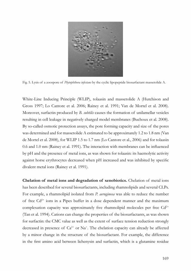
169
White-Line Inducing Principle (WLIP), tolaasin and massetolide A (Hutchison and Gross 1997; Lo Cantore et al. 2006; Rainey et al. 1991; Van de Mortel et al. 2008). Moreover, surfactin produced by B. subtilis causes the formation of unilamellar vesicles resulting in cell leakage in negatively charged model membranes (Buchoux et al. 2008). By so-called osmotic protection assays, the pore forming capacity and size of the pores was determined and for massetolide A estimated to be approximately 1.2 to 1.8 nm (Van de Mortel et al. 2008), for WLIP 1.5 to 1.7 nm (Lo Cantore et al., 2006) and for tolaasin 0.6 and 1.0 nm (Rainey et al. 1991). The interaction with membranes can be influenced by pH and the presence of metal ions, as was shown for tolaasin: its haemolytic activity against horse erythrocytes decreased when pH increased and was inhibited by specific divalent metal ions (Rainey et al. 1991).
Chelation of metal ions and degradation of xenobiotics. Chelation of metal ions has been described for several biosurfactants, including rhamnolipids and several CLPs. For example, a rhamnolipid isolated from P. aeruginosa was able to reduce the number of free Cd2+ ions in a Pipes buffer in a dose dependent manner and the maximum complexation capacity was approximately five rhamnolipid molecules per free Cd2+ (Tan et al. 1994). Cations can change the properties of the biosurfactants, as was shown for surfactin: the CMC value as well as the extent of surface tension reduction strongly decreased in presence of Ca2+ or Na+. The chelation capacity can already be affected by a minor change in the structure of the biosurfactant. For example, the difference in the first amino acid between lichenysin and surfactin, which is a glutamine residue
Fig. 5. Lysis of a zoospore of Phytophthora infestans by the cyclic lipopeptide biosurfactant massetolide A.
170
in lichenysin instead of a glutamic acid in surfactin, resulted in a better chelation of Ca2+ by lichenysin. To form a biosurfactant-cation complex, two molecules of lichenysin are required to form the biosurfactant-Ca2+ complex, suggesting the formation of lichenysin dimers and intermolecular salt bridges. In contrast, one molecule of surfactin is required to form the biosurfactant-Ca2+ complex, suggesting the formation of an intramolecular complex (Grangemard et al. 2001) by which the Ca2+ molecule is stabilized in the two polar side chains of the ‘claw’ conformation of surfactin (Bonmatin et al. 1994). For surfactin, it was also shown that when the leucine at position number two is substituted by an isoleucine, a three-fold increase in affinity for Ca2+ occurred, possibly by an increase in accessibility of the acidic side chains and carboxylate groups which constitute the calcium binding site (Grangemard et al. 1997). Moreover, binding of surfactin with Ca2+ results in a conformational change of the peptide moiety by which it can incorporate deeper into a phospholipid bilayer (Maget-Dana and Ptak 1995). Both iturin and gramicidin S have an affinity for metal cations like Na+, Rb+ and K+. For iturin, the order of cation selectivity is Na+>K+>Rb+, indicating a size limitation in the interaction cavity or cavities (Rautenbach et al. 2000). Due to these metal chelation properties, biosurfactants can be used for bioremediation of soils contaminated with heavy metals. After application of the biosurfactants to the heavy metal contaminated soil, the biosurfactant detaches the metal ion from the soil particle and the metal ion is incorporated in the micelles (Mulligan et al. 2001). The metal-biosurfactant complex can then be washed from the contaminated soil, but also air can be applied and, due to the foaming properties of the biosurfactant, metal and soil can be separated (reviewed in Banat et al. 2000; Mulligan 2005). The preference and efficiency of chelating a specific metal ion depend on the properties of the biosurfactant. For example, washing sediment, obtained from a canal contaminated with industrial waste, with rhamnolipid resulted in a decrease in the Cu2+ and Zn2+ content of 65% and 18%, respectively; washing with sophorolipid reduced the Cu2+ and Zn2+ content with 25% and 60%, respectively, whereas surfactin was less effective (Mulligan et al. 2001). Other metals that can be chelated by rhamnolipids are Cd2+ and Pb2+ (Herman et al. 1997). What the actual function of metal chelation by biosurfactants is for the bacterium itself, is to our knowledge not known. One could postulate that the bacteria use the biosurfactants for sequestering metal ions for activation of several enzymes as was described for the Fe3+ chelating siderophores.
171
In addition to their use for bioremediation of heavy metal contaminated soils, biosurfactants also have potential for degradation of aromatic compounds (Harvey et al. 1990; Mulligan 2005). Various microbes are able to degrade persistent aromatic compounds with low solubility in water and utilize these compounds as a carbon or nitrogen source (Ron and Rosenberg 2002). Aromatic compounds are aerobically degraded by oxidizing the aromatic ring, making them more susceptible to cleavage by dioxygenases (Phale et al. 2007). The limiting step in the degradation of partially insoluble aromatic compounds is bioavailability which can be substantially improved by adding surface active compounds or biosurfactants (Phale et al. 2007). Some P. aeruginosa strains that did not produce a biosurfactant, showed slow growth on insoluble hydrocarbons, but when rhamnolipid was added, the hydrocarbons could be utilized and growth was restored (Desai and Banat 1997). Moreover, some strains need this type of hydrocarbons to produce biosurfactants. For example, Rhodococcus erythropolis isolated from seaside soil required hexadecane as a source for its biosurfactant production, while glucose, sucrose or glycerol did not result in biosurfactant production (Peng et al. 2007).
Antimicrobial activity. Biosurfactants generally have antibiotic activity against a wide range of other microorganisms, including plant, human and animal pathogens. CLPs have activity against plant pathogenic oomycetes, including Pythium and Phytophthora species, but also against human pathogenic Gram-posititve bacteria like Mycobacterium and Staphylococcus spp (Raaijmakers et al. 2006). The main mode of action is most likely interference with cell membrane integrity, which causes leakage and in several cases cytolysis. The best known example of a biosurfactant developed for medical applications is daptomycin, which is a CLP produced by Streptomyces roseosporus (McHenney et al. 1998). Daptomycin has recently acquired marketing approval for the treatment of complicated skin and skin-structure infections caused by Gram-positive bacteria like S. aureus. Daptomycin binds to and inserts in the bacterial cell membrane which leads to pore or ion channel formation, causing disruption of the ion concentration gradient (Silverman et al. 2003). Intravenous application of daptomycin showed that approximately 44% of the patients were cured from infections caused by methicillin-resistant S. aureus (MRSA). These results were comparable to treatment with vancomycin, an antibiotic generally used to treat MRSA but less effective due to the occurrence of vancomycin-resistant strains
172
(Eisenstein, 2008). Also several other biosurfactants, including iturin and viscosin, were proposed as effective agents for treatment of mycosis and Chagas’ disease, respectively (Singh and Cameotra 2004; Burke et al. 1999). Surfactin produced by B. subtilis has activity against a variety of enveloped viruses, including herpes and retroviruses by interfering with the viral lipid envelope, causing leakage and disintegration (Vollenbroich et al. 1997). Most biosurfactants have activity against Gram-positive bacteria (Raaijmakers et al. 2006), but also some reports described activity against Gram-negative bacteria. An uncharacterised biosurfactant isolated from a marine Bacillus circulans was tested against a variety of bacteria and shown to inhibit the growth of Echerichi coli, S. marcescens, and other Gram-negative bacteria (Das et al. 2008). The activity of biosurfactants against human and plant pathogens can be further enhanced by the presence of specific extracellular enzymes. For example, addition of an endochitinase or glucanase purified from Trichoderma atroviride significantly enhanced the activity of syringomycin and syringopeptin against a variety of fungal plant pathogens (Fogliano et al. 2002). It was shown that cell wall degrading enzymes interfere with binding of syringomycin to the fungal cell wall constituents chitin, β-1.3-glucan and mannan (3-10%) (De Lucca et al. 1999), thereby enhancing the effect of syringomycin on the plasma membrane (Fogliano et al. 2002). Moreover, the effectiveness of various combinations of biosurfactants and cell wall degrading enzymes was correlated with the cell wall composition of each target fungus (Fogliano et al. 2002). Also addition of sub-MIC concentrations of the peptide antibiotic polymyxin B (Landman et al. 2008) to the CLP tolaasin resulted in sensitivity of Gram-negative bacteria to tolaasin (Rainey et al. 1991). Collectively, these studies show that biosurfactants have broad-spectrum activities against a range of human and plant pathogenic microorganisms (Raaijmakers et al. 2006) and hold promise for treating diseases that cannot be controlled effectively by currently available antibiotics.
CONCLUSIONSBiosurfactant production is a common trait among pathogenic and saprophytic bacteria and plays an important role in their lifestyle. Biosurfactants are involved in many processes that are important for the dispersal and survival of bacteria, including motility, surface attachment, antibiosis, virulence, competition and acquisition of nutrients or trace elements (Fig. 6). The enormous structural diversity and functional versatility of

173
bacterial biosurfactants are most likely a reflection of the diversity in habitats, resulting in biosurfactants that are unique to the physiology and ecology of the producing bacterial species or strain. Also, the regulation of biosurfactant production is different among bacterial species and strains. This review only discussed the effects of biosurfactants on other microorganisms, but surely biosurfactants also have effects on other organisms. For example, massetolide A produced by plant-benefical P. fluorescens, and surfactin and fengycins produced by B. subtilis, were shown to induce a systemic resistance response
Fig. 6. A schematic presentation of the diverse natural functions of bacterial biosurfactants. Pictures not drawn to scale.
174
in plants against oomycete and fungal pathogens (Ongena et al. 2007; Tran et al. 2007). Moreover, results of a recent study showed that massetolide A and viscosin lyse trophozoites of the amoebe Naegleria americana and thereby protect the producing bacterial strains from protozoan predation (chapter 6). In conclusion, biosurfactants are enormously versatile metabolites produced by pathogenic and saprophytic bacteria that improve bacterial fitness via manipulation of their biotic interaction with other organisms.
ACKNOWLEDGEMENTSThis work is financially supported by the Dutch Technology Foundation (STW). The microcolony experiments and fluorescence microscopy were conducted at Genetics and Microbiology of Copenhagen University in close collaboration with Ole Nybroe and Stefan Olsson.
REFERENCESAbbanat, D. R., Leadbetter, E. R., Godchaux, W., and Escher, A. 1986. Sulphonolipids are molecular determinants of gliding motility. Nature 324:367-369.Allison, C., Lai, H. C., Gygi, D., and Hughes, C. 1993. Cell differentiation of Proteus mirabilis is initiated by glutamine, a specific chemoattractant for swarming cells. Mol. Microbiol. 8:53-60.Andersen, J. B., Koch, B., Nielsen, T. H., Sorensen, D., Hansen, M., Nybroe, O., Christophersen, C., Sorensen, J., Molin, S., and Givskov, M. 2003. Surface motility in Pseudomonas sp. DSS73 is required for efficient biological containment of the root-pathogenic microfungi Rhizoctonia solani and Pythium ultimum. Microbiology 149:37-46.Asaka, O., and Shoda, M. 1996. Biocontrol of Rhizoctonia solani damping-off of tomato with Bacillus subtilis RB14. Appl. Environ. Microbiol. 62:4081-4085.Bais, H. P., Fall, R., and Vivanco, J. M. 2004. Biocontrol of Bacillus subtilis against infection of Arabidopsis roots by Pseudomonas syringae is facilitated by biofilm formation and surfactin production. Plant Physiol. 134:307-19.Balibar, C. J., Vaillancourt, F. H., and Walsh, C. T. 2005. Generation of D amino acid residues in assembly of arthrofactin by dual condensation/epimerization domains. Chem. Biol. 12:1189-200.Banat, I. M., Makkar, R. S., and Cameotra, S. S. 2000. Potential commercial applications of microbial surfactants. Appl. Microbiol. Biotechnol. 53:495-508.Bender, C. L., Alarcon-Chaidez, F., and Gross, D. C. 1999. Pseudomonas syringae phytotoxins: mode of action, regulation, and biosynthesis by peptide and polyketide synthetases. Microbiol. Mol. Biol. Rev. 63:266-92.
Berti, A. D., Greve, N. J., Christensen, Q. H., and Thomas, M. G. 2007. Identification of a biosynthetic gene cluster and the six associated lipopeptides involved in swarming motility of Pseudomonas syringae pv. tomato DC3000. J. Bacteriol. 189:6312-6323.Blasko, K., Schagina, L. V., Agner, G., Kaulin, Y. A., and Takemoto, J. Y. 1998. Membrane sterol composition modulates the pore forming activity of syringomycin E in human red blood cells. Biochim. Biophys. Acta. 1373:163-9.Boles, B. R., Thoendel, M., and Singh, P. K. 2005. Rhamnolipids mediate detachment of Pseudomonas aeruginosa from biofilms. Mol. Microbiol. 57:1210-23.Bonmatin, J. M., Genest, M., Labbe, H., and Ptak, M. 1994. Solution three-dimensional structure of surfactin: a cyclic lipopeptide studied by 1H-NMR, distance geometry, and molecular dynamics. Biopolymers 34:975-86.Brint, J. M., and Ohman, D. E. 1995. Synthesis of multiple exoproducts in Pseudomonas aeruginosa is under the control of RhlR-RhlI, another set of regulators in strain PAO1 with homology to the autoinducer-responsive LuxR-LuxI family. J. Bacteriol. 177:7155-63.Buchoux, S., Lai-Kee-Him, J., Garnier, M., Tsan, P., Besson, F., Brisson, A., and Dufourc, E. J. 2008. Surfactin-triggered small vesicle formation of negatively charged membranes: a novel membrane-lysis mechanism. Biophys. J. 95:3840-9.Bultreys, A., and Gheysen, I. I. 1999. Biological and molecular detection of toxic lipodepsipeptide-producing Pseudomonas syringae strains and PCR identification in plants. Appl. Environ. Microbiol. 65:1904-9.Burke, T. J., Chandasekahar, B., and Knight, M. 1999. Analogs of viscosin and uses therof
175
U.S. Patent Number 5,965,524 Challis, G. L., Ravel, J., and Townsend, C. A. 2000. Predictive, structure-based model of amino acid recognition by nonribosomal peptide synthetase adenylation domains. Chem. Biol. 7:211-24.Chen, C. Y., Baker, S. C., and Darton, R. C. 2007. The application of a high throughput analysis method for the screening of potential biosurfactants from natural sources. J. Microbiol. Meth. 70:503-510.Cliften, P., Wang, Y. L., Mochizuki, D., Miyakawa, T., Wangspa, R., Hughes, J., and Takemoto, J. Y. 1996. SYR2, a gene necessary for syringomycin growth inhibition of Saccharomyces cerevisiae. Microbiology-Uk 142:477-484.Cui, X., Harling, R., Mutch, P., and Darling, D. 2005. Identification of N-3-hydroxyoctanoyl-homoserine lactone production in Pseudomonas fluorescens 5064, pathogenic to broccoli, and controlling biosurfactant production by quorum sensing. Eur. J. Plant Pathol. 111:297-308.Danhorn, T., and Fuqua, C. 2007. Biofilm formation by plant-associated bacteria. Annu. Rev. Microbiol. 61:401-22.Daniels, R., Reynaert, S., Hoekstra, H., Verreth, C., Janssens, J., Braeken, K., Fauvart, M., Beullens, S., Heusdens, C., Lambrichts, I., De Vos, D. E., Vanderleyden, J., Vermant, J., and Michiels, J. 2006. Quorum signal molecules as biosurfactants affecting swarming in Rhizobium etli. Proc. Natl. Acad. Sci. USA 103:14965-70.Das, P., Mukherjee, S., and Sen, R. 2008. Antimicrobial potential of a lipopeptide biosurfactant derived from a marine Bacillus circulans. J. Appl. Microbiol. 104:1675-84.Davey, M. E., Caiazza, N. C., and O’Toole, G. A. 2003. Rhamnolipid surfactant production affects biofilm architecture in Pseudomonas aeruginosa PAO1. J. Bacteriol. 185:1027-36.De Bruijn, I., de Kock, M. J., de Waard, P., van Beek, T. A., and Raaijmakers, J. M. 2008. Massetolide A biosynthesis in Pseudomonas fluorescens. J. Bacteriol. 190: 2777-2789.De Bruijn, I., de Kock, M. J. D., Yang, M., de Waard, P., van Beek, T. A., and Raaijmakers, J. M. 2007. Genome-based discovery, structure prediction and functional analysis of cyclic lipopeptide antibiotics in Pseudomonas species. Mol. Microbiol. 63:417-428.De Lucca, A. J., Jacks, T. J., Takemoto, J., Vinyard, B., Peter, J., Navarro, E., and Walsh, T. J. 1999. Fungal lethality, binding, and cytotoxicity of syringomycin-E. Antimicrob. Agents Chemother. 43:371-3.De Souza, J. T., De Boer, M., De Waard, P., Van Beek, T. A., and Raaijmakers, J. M. 2003. Biochemical, genetic, and zoosporicidal properties of cyclic lipopeptide surfactants produced by Pseudomonas fluorescens. Appl. Environ. Microbiol. 69:7161-72.Desai, J. D., and Banat, I. M. 1997. Microbial production of surfactants and their commercial potential. Microbiol. Mol. Biol. Rev. 61:47-64.Deziel, E., Lepine, F., Milot, S., and Villemur,
R. 2003. RhlA is required for the production of a novel biosurfactant promoting swarming motility in Pseudomonas aeruginosa: 3-(3-hydroxyalka-noyloxy)alkanoic acids (HAAs), the precursors of rhamnolipids. Microbiology 149:2005-13.Drenkard, E., and Ausubel, F. M. 2002. Pseudomonas biofilm formation and antibiotic resistance are linked to phenotypic variation. Nature 416:740-3.Dubern, J. F., Coppoolse, E. R., Stiekema, W. J., and Bloemberg, G. V. 2008. Genetic and functional characterization of the gene cluster directing the biosynthesis of putisolvin I and II in Pseudomonas putida strain PCL1445. Microbiology 154:2070-83.Dubern, J. F., Lugtenberg, B. J., and Bloemberg, G. V. 2006. The ppuI-rsaL-ppuR quorum-sensing system regulates biofilm formation of Pseudomonas putida PCL1445 by controlling biosynthesis of the cyclic lipopeptides putisolvins I and II. J. Bacteriol. 188:2898-906.Dumenyo, C. K., Mukherjee, A., Chun, W., and Chatterjee, A. K. 1998. Genetic and physiological evidence for the production of N-acyl homoserine lactones by Pseudomonas syringae pv. syringae and other fluorescent plant pathogenic Pseudomonas species. Eur. J. Plant Pathol. 104:569-582.Eberl, L., Molin, S., and Givskov, M. 1999. Surface motility of Serratia liquefaciens MG1. J. Bacteriol. 181:1703-12.Eisenstein, B. I. 2008. Treatment of staphylococcal infections with cyclic lipopeptides. Clin. Microbiol. Infect. 14 Suppl 2:10-6.Fiore, A., Laparra, J. M., Farre, R., Fullone, M. R., Grgurina, I., Gallo, M., and Fogliano, V. 2008. Lipodepsipeptides from Pseudomonas syringae are partially proteolyzed and are not absorbed by humans: an in vitro study. J. Food Prot. 71:979-85.Fogliano, V., Ballio, A., Gallo, M., Woo, S., Scala, F., and Lorito, M. 2002. Pseudomonas lipodepsipeptides and fungal cell wall-degrading enzymes act synergistically in biological control. Mol. Plant-Microbe Interact. 15:323-33.Fogliano, V., Gallo, M., Vinale, F., Ritieni, A., Randazzo, G., Greco, M., Lops, R., and Graniti, A. 1999. Immunological detection of syrirngopeptins produced by Pseudomonas syringae pv. lachrymans. Physiol. Mol. Plant Pathol. 55:255-261.Grangemard, I., Peypoux, F., Wallach, J., Das, B. C., Labbe, H., Caille, A., Genest, M., Maget-Dana, R., Ptak, M., and Bonmatin, J. M. 1997. Lipopeptides with improved properties: structure by NMR, purification by HPLC and structure-activity relationships of new isoleucyl-rich surfactins. J. Pept. Sci. 3:145-54.Grangemard, I., Wallach, J., Maget-Dana, R., and Peypoux, F. 2001. Lichenysin: a more efficient cation chelator than surfactin. Appl. Biochem. Biotechnol. 90:199-210.Grangemard, I., Wallach, J., and Peypoux, F. 1999. Evidence of surfactin hydrolysis by a bacterial endoprotease. Biotech. Lett. 21:241-244.
176
Gross, H., Stockwell, V. O., Henkels, M. D., Nowak-Thompson, B., Loper, J. E., and Gerwick, W. H. 2007. The genomisotopic approach: a systematic method to isolate products of orphan biosynthetic gene clusters. Chem. Biol. 14:53-63.Hall-Stoodley, L., Costerton, J. W., and Stoodley, P. 2004. Bacterial biofilms: from the natural environment to infectious diseases. Nat. Rev. Microbiol. 2:95-108.Hanson, K. G., Desai, J. D., and Desai, A. J. 1993. A rapid and simple screening technique for potential crude-oil degrading microorganisms. Biotech. Tech. 7:745-748.Harshey, R. M. 2003. Bacterial motility on a surface: many ways to a common goal. Annu. Rev. Microbiol. 57:249-73.Harvey, S., Elashvili, I., Valdes, J. J., Kamely, D., and Chakrabarty, A. M. 1990. Enhanced removal of Exxon Valdez spilled oil from Alaskan gravel by a microbial surfactant. Biotechnology (N Y) 8:228-30.Henrichsen, J. 1972. Bacterial surface translocation: a survey and a classification. Bacteriol. Rev. 36:478-503.Henriksen, A., Anthoni, U., Nielsen, T. H., Sorensen, J., Christophersen, C., and Gajhede, M. 2000. Cyclic lipoundecapeptide tensin from Pseudomonas fluorescens strain 96.578. Acta Crystallogr. C 56 ( Pt 1):113-5.Herman, D. C., Zhang, Y., and Miller, R. M. 1997. Rhamnolipid (biosurfactant) effects on cell aggregation and biodegradation of residual hexadecane under saturated flow conditions. Appl. Environ. Microbiol. 63:3622-7.Heyd, M., Kohnert, A., Tan, T. H., Nusser, M., Kirschhofer, F., Brenner-Weiss, G., Franzreb, M., and Berensmeier, S. 2008. Development and trends of biosurfactant analysis and purification using rhamnolipids as an example. Anal. Bioanal. Chem. 391:1579-1590.Hildebrand, P. D., Braun, P. G., McRae, K. B., and Lu, X. 1998. Role of the biosurfactant viscosin in broccoli head rot caused by a pectolytic strain of Pseudomonas fluorescens. Can. J. Plant Pathol. 20:296-303.Hofemeister, J., Conrad, B., Adler, B., Hofemeister, B., Feesche, J., Kucheryava, N., Steinborn, G., Franke, P., Grammel, N., Zwintscher, A., Leenders, F., Hitzeroth, G., and Vater, J. 2004. Genetic analysis of the biosynthesis of nonribosomal peptide- and polyketide-like antibiotics, iron uptake and biofilm formation by Bacillus subtilis A1/3. Mol.Genet. Genomics 272:363-78.Hutchison, M. L., and Gross, D. C. 1997. Lipopeptide phytotoxins produced by Pseudomonas syringae pv. syringae: comparison of the biosurfactant and ion channel-forming activities of syringopeptin and syringomycin. Mol. Plant-Microbe Interact. 10:347-54.Iacobellis, N. S., Lavermicocca, P., Grgurina, I., Simmaco, M., and Ballio, A. 1992. Phytotoxic properties of Pseudomonas syringae pv syringae toxins. Phys. Mol. Plant
Pathol. 40:107-116.Ivanova, E. P., Gorshkova, N. M., Sawabe, T., Hayashi, K., Kalinovskaya, N. I., Lysenko, A. M., Zhukova, N. V., Nicolau, D. V., Kuznetsova, T. A., Mikhailov, V. V., and Christen, R. 2002. Pseudomonas extremorientalis sp nov., isolated from a drinking water reservoir. Intl. J. Syst. Evol. Microbiol. 52:2113-2120.Johnson, M. K., and Boese-Marrazzo, D. 1980. Production and properties of heat-stable extracellular hemolysin from Pseudomonas aeruginosa. Infect. Immun. 29:1028-33.Kinscherf, T. G., and Willis, D. K. 1999. Swarming by Pseudomonas syringae B728a requires gacS (lemA) and gacA but not the acyl-homoserine lactone biosynthetic gene ahlI. J. Bacteriol. 181:4133-6.Kinsinger, R. F., Shirk, M. C., and Fall, R. 2003. Rapid surface motility in Bacillus subtilis is dependent on extracellular surfactin and potassium ion. J. Bacteriol. 185:5627-31.Klausen, M., Aaes-Jorgensen, A., Molin, S., and Tolker-Nielsen, T. 2003. Involvement of bacterial migration in the development of complex multicellular structures in Pseudomonas aeruginosa biofilms. Mol. Microbiol. 50:61-8.Koch, B., Nielsen, T. H., Sorensen, D., Andersen, J. B., Christophersen, C., Molin, S., Givskov, M., Sorensen, J., and Nybroe, O. 2002. Lipopeptide production in Pseudomonas sp. strain DSS73 is regulated by components of sugar beet seed exudate via the Gac two-component regulatory system. Appl. Environ. Microbiol. 68:4509-16.Kruijt, M., Tran, H., and Raaijmakers, J.M. 2008. Functional, genetic and chemical characterization of biosurfactants produced by plant growth-promoting Pseudomonas putida 267. J. Appl. Microbiol. Accepted. Kuiper, I., Lagendijk, E. L., Pickford, R., Derrick, J. P., Lamers, G. E. M., Thomas-Oates, J. E., Lugtenberg, B. J. J., and Bloemberg, G. V. 2004. Characterization of two Pseudomonas putida lipopeptide biosurfactants, putisolvin I and II, which inhibit biofilm formation and break down existing biofilms. Mol. Microbiol. 51:97-113.Landman, D., Georgescu, C., Martin, D. A., and Quale, J. 2008. Polymyxins revisited. Clin. Microbiol. Rev. 21:449-65.Lee, H. I., Jeong, K. S., and Cha, J. S. 2002. PCR assays for specific and sensitive detection of Pseudomonas tolaasii, the cause of brown blotch disease of mushrooms. Lett. Appl. Microbiol. 35:276-80.Lindow, S. E., and Brandl, M. T. 2003. Microbiology of the phyllosphere. Appl. Environ. Microbiol. 69:1875-83.Lindum, P. W., Anthoni, U., Christophersen, C., Eberl, L., Molin, S., and Givskov, M. 1998. N-Acyl-L-homoserine lactone autoinducers control production of an extracellular lipopeptide biosurfactant required for swarming motility of Serratia liquefaciens MG1. J. Bacteriol. 180:6384-8.Lo Cantore, P., Lazzaroni, S., Coraiola,
177
M., Serra, M. D., Cafarchia, C., Evidente, A., and Lacobellis, N. S. 2006. Biological characterization of white line-inducing principle (WLIP) produced by Pseudomonas reactans NCPPB1311. Mol. Plant-Microbe Interact. 19:1113-20.Maget-Dana, R., and Ptak, M. 1995. Interactions of surfactin with membrane models. Biophys. J. 68:1937-43.Maier, R. M. 2003. Biosurfactants: evolution and diversity in bacteria. Adv. Appl. Microbiol. 52:101-21.Matsuyama, T., Bhasin, A., and Harshey, R. M. 1995. Mutational analysis of flagellum-independent surface spreading of Serratia marcescens 274 on a low-agar medium. J. Bacteriol. 177:987-91.McCarter, L., Hilmen, M., and Silverman, M. 1988. Flagellar dynamometer controls swarmer cell differentiation of Vibrio parahaemolyticus. Cell 54:345-51.McCarter, L., and Silverman, M. 1990. Surface-induced swarmer cell differentiation of Vibrio parahaemolyticus. Mol. Microbiol. 4:1057-62.McHenney, M. A., Hosted, T. J., Dehoff, B. S., Rosteck, P. R., Jr., and Baltz, R. H. 1998. Molecular cloning and physical mapping of the daptomycin gene cluster from Streptomyces roseosporus. J. Bacteriol. 180:143-51.Mireles, J. R., 2nd, Toguchi, A., and Harshey, R. M. 2001. Salmonella enterica serovar typhimurium swarming mutants with altered biofilm-forming abilities: surfactin inhibits biofilm formation. J. Bacteriol. 183:5848-54.Mulligan, C. N. 2005. Environmental applications for biosurfactants. Environ. Pollut. 133:183-98.Mulligan, C. N., Yong, R. N., and Gibbs, B. F. 2001. Heavy metal removal from sediments by biosurfactants. J. Hazard Mater. 85:111-25.Muthusamy, K., Gopalakrishnan, S., Ravi, T. K., and Sivachidambaram, P. 2008. Biosurfactants: Properties, commercial production and application. Current Science 94:736-747.Nakayama, T., Homma, Y., Hashidoko, Y., Mizutani, J., and Tahara, S. 1999. Possible role of xanthobaccins produced by Stenotrophomonas sp. strain SB-K88 in suppression of sugar beet damping-off disease. Appl. Environ. Microbiol. 65:4334-9.Neu, T. R. 1996. Significance of bacterial surface-active compounds in interaction of bacteria with interfaces. Microbiol. Rev. 60:151-66.Nielsen, M. N., Sorensen, J., Fels, J., and Pedersen, H. C. 1998. Secondary metabolite- and endochitinase-dependent antagonism toward plant-pathogenic microfungi of Pseudomonas fluorescens isolates from sugar beet rhizosphere. Appl. Environ. Microbiol. 64:3563-9.Nielsen, T. H., Nybroe, O., Koch, B., Hansen, M., and Sorensen, J. 2005. Genes involved in cyclic lipopeptide production are important for seed and straw colonization by Pseudomonas sp. strain DSS73. Appl. Environ.
Microbiol. 71:4112-6.Nielsen, T. H., Sorensen, D., Tobiasen, C., Andersen, J. B., Christophersen, C., Givskov, M., and Sorensen, J. 2002. Antibiotic and biosurfactant properties of cyclic lipopeptides produced by fluorescent Pseudomonas spp. from the sugar beet rhizosphere. Appl. Environ. Microbiol. 68:3416-23.Nielsen, T. H., and Sorensen, J. 2003. Production of cyclic lipopeptides by Pseudomonas fluorescens strains in bulk soil and in the sugar beet rhizosphere. Appl. Environ. Microbiol. 69:861-8.Nitschke, M., and Pastore, G. M. 2004. Biosurfactant production by Bacillus subtilis using cassava-processing effluent. Appl. Biochem. Biotechnol. 112:163-72.Nybroe, O., and Sorensen, J. 2004. Production of cyclic lipopeptides by fluorescent pseudomonads. Pages 147-172 in: Pseudomonas volume 3, Biosynthesis of Macromolecules and Molecular Metabolism, Ramos, J-L., ed., Kluwer Academic/Plenum Publishers, New York.Ochsner, U. A., Fiechter, A., and Reiser, J. 1994a. Isolation, characterization, and expression in Escherichia coli of the Pseudomonas aeruginosa rhlAB genes encoding a rhamnosyltransferase involved in rhamnolipid biosurfactant synthesis. J. Biol. Chem. 269:19787-95.Ochsner, U. A., Koch, A. K., Fiechter, A., and Reiser, J. 1994b. Isolation and characterization of a regulatory gene affecting rhamnolipid biosurfactant synthesis in Pseudomonas aeruginosa. J. Bacteriol. 176:2044-54.Ongena, M., and Jacques, P. 2008. Bacillus lipopeptides: versatile weapons for plant disease biocontrol. Trends Microbiol. 16:115-125.Ongena, M., Jourdan, E., Adam, A., Paquot, M., Brans, A., Joris, B., Arpigny, J. L., and Thonart, P. 2007. Surfactin and fengycin lipopeptides of Bacillus subtilis as elicitors of induced systemic resistance in plants. Environ. Microbiol. 9:1084-90.Pamp, S. J., and Tolker-Nielsen, T. 2007. Multiple roles of biosurfactants in structural biofilm development by Pseudomonas aeruginosa. J. Bacteriol. 189:2531-9.Peng, F., Liu, Z., Wang, L., and Shao, Z. 2007. An oil-degrading bacterium: Rhodococcus erythropolis strain 3C-9 and its biosurfactants. J. Appl. Microbiol. 102:1603-11.Peypoux, F., Bonmatin, J. M., and Wallach, J. 1999. Recent trends in the biochemistry of surfactin. Appl. Microbiol. Biotechnol. 51:553-63.Phale, P. S., Basu, A., Majhi, P. D., Deveryshetty, J., Vamsee-Krishna, C., and Shrivastava, R. 2007. Metabolic diversity in bacterial degradation of aromatic compounds. Omics 11:252-79.Quigley, N. B., and Gross, D. C. 1994. Syringomycin production among strains of Pseudomonas syringae pv. syringae: conservation of the syrB and syrD genes and activation of phytotoxin production by plant signal molecules. Mol. Plant-Microbe Interact. 7:78-90.Quigley, N. B., Mo, Y. Y., and Gross, D.
178
C. 1993. SyrD is required for syringomycin production by Pseudomonas syringae pathovar syringae and is related to a family of ATP-binding secretion proteins. Mol. Microbiol. 9:787-801.Raaijmakers, J. M., de Bruijn, I., and de Kock, M. J. 2006. Cyclic lipopeptide production by plant-associated Pseudomonas spp.: diversity, activity, biosynthesis, and regulation. Mol. Plant-Microbe Interact. 19:699-710.Rainey, P. B., Brodey, C. L., and Johnstone, K. 1991. Biological properties and spectrum of activity of tolaasin, a lipodepsipeptide toxin produced by the mushroom pathogen Pseudomonas tolaasii. Phys. Mol. Plant Pathol. 39:57-70.Rautenbach, M., Swart, P., and van der Merwe, M. J. 2000. The interaction of analogues of the antimicrobial lipopeptide, iturin A2, with alkali metal ions. Bioorg. Med. Chem. 8:2539-48.Ron, E. Z., and Rosenberg, E. 2001. Natural roles of biosurfactants. Environ. Microbiol. 3:229-236.Ron, E. Z., and Rosenberg, E. 2002. Biosurfactants and oil bioremediation. Curr. Opin. Biotechnol. 13:249-52.Roongsawang, N., Hase, K., Haruki, M., Imanaka, T., Morikawa, M., and Kanaya, S. 2003. Cloning and characterization of the gene cluster encoding arthrofactin synthetase from Pseudomonas sp. MIS38. Chem. Biol. 10:869-80.Scholz-Schroeder, B. K., Hutchison, M. L., Grgurina, I., and Gross, D. C. 2001. The contribution of syringopeptin and syringomycin to virulence of Pseudomonas syringae pv. syringae strain B301D on the basis of sypA and syrB1 biosynthesis mutant analysis. Mol. Plant-Microbe Interact. 14:336-48.Siegmund, I., and Wagner, F. 1991. New method for detecting rhamnolipids excreted by Pseudomonas species during growth on mineral agar. Biotech. Tech. 5:265-268.Silverman, J. A., Perlmutter, N. G., and Shapiro, H. M. 2003. Correlation of daptomycin bactericidal activity and membrane depolarization in Staphylococcus aureus. Antimicrob. Agents Chemother. 47:2538-44.Singh, P., and Cameotra, S. S. 2004. Potential applications of microbial surfactants in biomedical sciences. Trends Biotechnol. 22:142-6.Solomon, J. M., Lazazzera, B. A., and Grossman, A. D. 1996. Purification and characterization of an extracellular peptide factor that affects two different developmental pathways in Bacillus subtilis. Genes Dev. 10:2014-24.Sorensen, D., Nielsen, T. H., Christophersen, C., Sorensen, J., and Gajhede, M. 2001. Cyclic lipoundecapeptide amphisin from Pseudomonas sp. strain DSS73. Acta Crystallogr. C 57:1123-4.Sorensen, K. N., Kim, K. H., and Takemoto, J. Y. 1998. PCR detection of cyclic lipodepsinonapeptide-producing Pseudomonas syringae pv. syringae and similarity of strains. Appl.
Environ. Microbiol. 64:226-230.Spiers, A. J., and Rainey, P. B. 2005. The Pseudomonas fluorescens SBW25 wrinkly spreader biofilm requires attachment factor, cellulose fibre and LPS interactions to maintain strength and integrity. Microbiology 151:2829-39.Stachelhaus, T., Mootz, H. D., and Marahiel, M. A. 1999. The specificity-conferring code of adenylation domains in nonribosomal peptide synthetases. Chem. Biol. 6:493-505.Stewart, P. S., and Franklin, M. J. 2008. Physiological heterogeneity in biofilms. Nat. Rev. Microbiol. 6:199-210.Taguchi, N., Takano, Y., Julmanop, C., Wang, Y. L., Stock, S., Takemoto, J., and Miyakawa, T. 1994. Identification and analysis of the Saccharomyces cerevisiae syr1 gene reveals that ergosterol is involved in the action of syringomycin. Microbiology-Uk 140:353-359.Takemoto, J. Y., Yu, Y. X., Stock, S. D., and Miyakawa, T. 1993. Yeast genes involved in growth-inhibition by Pseudomonas syringae pv syringae syringomycin family lipodepsipeptides. FEMS Microbiol. Lett. 114:339-342.Tan, H., Champion, H. T., Ariola, J. F., Brusseau, M. L., and Miller, R. M. 1994. Complexation of cadmium by a rhamnolipid biosurfactant. Environ. Sci. Technol. 28:2402-2406.Toguchi, A., Siano, M., Burkart, M., and Harshey, R. M. 2000. Genetics of swarming motility in Salmonella enterica serovar typhimurium: critical role for lipopolysaccharide. J. Bacteriol. 182:6308-21.Tran, H., Ficke, A., Asiimwe, T., Höfte, M., and Raaijmakers, J. M. 2007. Role of the cyclic lipopeptide massetolide A in biological control of Phytophthora infestans and in colonization of tomato plants by Pseudomonas fluorescens. New Phytol. 175:731-742.Tran, H., Kruijt, M., and Raaijmakers, J. M. 2008. Diversity and activity of biosurfactant-producing Pseudomonas in the rhizosphere of black pepper in Vietnam. J. Appl. Microbiol. 104:839-51.Van de Mortel, J.E., Tran, H., Govers, F., and Raaijmakers, J.M. Cellular responses of the late blight pathogen Phytophthora infestans to cyclic lipopeptide surfactants and the role of G-proteins. Unpublished.Vollenbroich, D., Ozel, M., Vater, J., Kamp, R. M., and Pauli, G. 1997. Mechanism of inactivation of enveloped viruses by the biosurfactant surfactin from Bacillus subtilis. Biologicals 25:289-97.Zulianello, L., Canard, C., Kohler, T., Caille, D., Lacroix, J. S., and Meda, P. 2006. Rhamnolipids are virulence factors that promote early infiltration of primary human airway epithelia by Pseudomonas aeruginosa. Infect. Immun. 74:3134-47.

179

180
181
CHAPTER 8
Summarizing discussion

182
183
Cyclic lipopeptide biosynthesis. Cyclic lipopeptides (CLPs) are surfactant metabolites produced by various bacterial genera and species. For the genus Pseudomonas, many CLPs have been identified and initially classified into five groups based on structural characteristics (Chapter 1, Nybroe and Sorensen, 2004). At the start of this Ph.D. thesis project, only few genes governing CLP biosynthesis in Pseudomonas species were known and research was mostly focused on resolving syringomycin and syringopeptin biosynthesis in plant pathogenic Pseudomonas syringae pv. syringae (Feil et al. 2005; Guenzi et al. 1998; Scholz-Schroeder et al. 2001a; Scholz-Schroeder et al. 2003; Scholz-Schroeder et al. 2001b; Zhang et al. 1995). Over the past four years, however, biosynthesis genes for several other CLPs produced by Pseudomonas species and strains were cloned and sequenced (Table 1). In this thesis project, two new CLP biosynthesis clusters were identified in P. fluorescens and fully sequenced. In P. fluorescens strain SBW25, the viscosin biosynthesis cluster was identified by bioinformatic analyses of the genome followed by genetic and chemical analyses (chapter 2). For P. fluorescens strain SS101, the genes for massetolide biosynthesis were identified via random mutagenesis followed by cloning, sequencing and chemical analyses (chapter 3). Biosynthesis of viscosin and massetolide is governed by three nonribosomal peptide synthetase (NRPS) genes, designated viscABC and massABC, respectively. The viscosin and massetolide biosynthesis gene clusters are very similar, but different from CLP gene clusters described for other Pseudomonas as the visc and mass genes are not located in one operon; both consist of two gene clusters in which viscA and massA are physically disconnected from viscBC and massBC, respectively (chapter 4). For massetolide biosynthesis, massA transcription was shown to be independent of massBC transcription and vice versa (chapter 3). Whether this is also the case for the visc genes in strain SBW25 needs to be assessed. The assembly line by which the NRPSs build the CLPs consists of several enzymatic steps, including amino acid selection, activation, and incorporation followed by cyclization of the peptide moiety (Finking and Marahiel 2004). Viscosin differs from massetolide A only at position four in the peptide moiety, which is a valine in viscosin and an isoleucine in massetolide A. Because of the modular structure of the NRPSs and the co-linearity of the assembly process, the difference between viscosin and massetolide A is conferred by the fourth adenylation domain located in ViscB and MassB, respectively. This was confirmed in experiments where transfer of the massB gene of strain SS101 into a viscB mutant of strain SBW25 resulted in the production of massetolide A instead

184
of viscosin (chapter 4). This result also demonstrated that the assembly line for CLP biosynthesis in Pseudomonas can be altered leading to the production of non-native products. This strategy of heterologous expression of CLP biosynthesis genes in related or unrelated species and strains will be further developed to determine if also structurally new CLP derivatives with altered or improved activities can be generated. Other strategies of heterologous expression of NRPS genes were described in detail by Bode and Muller (2005) and showed that product yields of the non-native or structurally novel NRPS/ products are in many cases relatively low. Whether this is related to differences in genetic regulaton of the biosynthesis genes, poor communication between the different NRPSs, or due to limitations in the efflux of the final products are intriguing questions to be addressed in the future.
Table 1. Overview of genes identified recently for their role in the biosynthesis and regulation of cyclic lipopeptides (CLPs) produced by plant-associated Pseudomonas species.
185
Regulation of CLP biosynthesis. Compared to the understanding of CLP biosynthesis, little is known about the regulation. In Pseudomonas species, the two-component system GacA/GacS (global activation of antibiotic and cyanide biosynthesis) regulates the production of many secondary metabolites and extracellular enzymes (Lapouge et al. 2008). Also in CLP biosynthesis, GacA/GacS functions as a master switch, as a mutation in either one of the two encoding genes results in loss of CLP production (Dubern et al. 2005; Kitten et al. 1998; Koch et al. 2002). Upon activation by GacS, GacA induces transcription of several small RNAs (RsmX, RsmY and RsmZ), which in turn activate the biosynthesis of many proteins by releasing the repression by the RNA-binding proteins RsmA and RsmE (Lapouge et al. 2008). The results of this thesis showed that also massetolide and viscosin biosynthesis are regulated by the Gac-system (chapters 2 and 3; Table 1). The role of the small RNAs in regulation of massetolide and viscosin biosynthesis as well as the identity of external signals that activate the Gac-system have not been investigated in this thesis. The role of a second well-known regulatory system, referred to as quorum sensing, in CLP biosynthesis was also investigated. In many Pseudomonas species, quorum sensing is directed by N-acyl homoserine lactones (N-AHLs) that act as intercellular signals. LuxI-type genes encode for N-AHLs and when a certain threshold concentration is reached, they bind to LuxR-type regulatory proteins which in turn activate transcription of target genes (Fuqua et al. 1994). Quorum sensing-regulated metabolites are generally detected in the late exponential or stationary growth phases of the producing strains. For plant pathogenic P. fluorescens strain 5064 and saprophytic P. putida strain PCL1445, N-AHL-based quorum sensing was shown to be involved in viscosin and putisolvin biosynthesis, respectively (Cui et al. 2005; Dubern et al. 2006). For P. fluorescens strains SS101 and SBW25, however, no indications were found for a role of N-AHL-based quorum sensing in CLP biosynthesis: massetolide and viscosin are produced in the early exponential growth phase and a variety of other experimental and bioinformatic approaches did not provide evidence that these strains produce N-AHLs or harbor close homologues of the LuxI/LuxR genes (chapters 3 and 4). Next to global regulation, the role of other regulatory genes in viscosin and massetolide production was investigated, including the LuxR-type transcriptional regulators located up- and downstream of the CLP biosynthesis genes. Based on sequence analysis, these LuxR-type transcriptional regulators do not contain the

186
autoinducer-binding domain found for the quorum sensing-associated LuxR regulator in Vibrio fischeri (Fuqua et al. 1994). Instead, the LuxR-type regulator genes flanking the massetolide and viscosin biosynthesis genes are closely related to the LuxR-type regulators identified for syringomycin/ syringopeptin biosynthesis (Kitten et al. 1998; Lu et al. 2002; Wang et al. 2006), and appear to belong to a separate LuxR-type regulator subfamily, different from the autonomous effector domain protein GerE (Ducros et al. 2001). Site-directed mutagenesis of the LuxR-type regulator genes luxR-vA and luxR-vBC flanking the viscosin biosynthesis cluster resulted in a loss of viscosin production (chapter 4), indicating that both LuxR-type transcriptional regulators are important for viscosin biosynthesis in strain SBW25. These results confirm and extend the observations made for the LuxR-type transcriptional regulators flanking the gene clusters for syringomycin, syringopeptin, syringafactin and putisolvin production (Berti et al. 2007; Dubern et al. 2008; Kitten et al. 1998; Lu et al. 2002; Lu et al. 2005). Unfortunately, site-directed mutagenesis was not successful for the luxR-mA and luxR-mBC genes flanking the massetolide biosynthesis genes in strain SS101. However, the observation that introduction of extra copies of luxR-mA in wild type strain SS101 resulted in the upregulation of the mass biosynthesis genes and in elevated massetolide production, suggests that the luxR-mA gene acts as a regulator of massetolide biosynthesis (chapter 4). Furthermore, when luxR-mA was expressed in the luxR-vA mutant of strain SBW25, viscosin biosynthesis and visc gene expression were restored to wild type levels. Whether the LuxR-type regulators up- and downstream of the mass and visc genes have a preference for binding to the promoter regions of the massA/viscA or massBC/viscBC genes is yet unknown and requires promoter binding studies as described by Wang et al, 2006 (Wang et al. 2006). Via random mutagenesis and subsequent screening for massetolide-deficient mutants, we also identified other regulator genes including clpP (chapter 5). ClpP is a serine protease that is highly conserved in bacteria and eukaryotes (Yu and Houry 2007). Together with other proteases, ClpP plays a crucial role in intracellular refolding and degradation of proteins, which is an essential process for the viability and growth of cells. The involvement of ClpP in massetolide biosynthesis was confirmed by site-directed mutagenesis and complementation. The clpP mutant showed reduced mass transcript levels and a loss of surface motility and biofilm formation. Expression analysis showed that ClpP affects transcription of luxR-mA, thereby regulating massetolide biosynthesis

187
(chapter 5). Whether there is a direct or indirect interaction between ClpP and LuxR-mA remains to be investigated. Another possibility is that ClpP interacts with other proteins that in turn affect luxR-mA transcription. An extensive proteomic analysis, as was performed previously for E. coli (Flynn et al. 2003), will be required to identify the cellular substrates and target proteins of the ClpP protease in P. fluorescens. We also observed that addition of amino acids could partly restore luxR-mA transcript levels and massetolide biosynthesis in the clpP mutant, and that this effect was independent of clpP transcription (chapter 5). Results further suggested that, at the transcriptional level, ClpP-mediated regulation of massetolide biosynthesis operates independently of regulation by the GacA/GacS two-component system (chapter 5). In conclusion, the results obtained in this thesis led to the identification of several new genes and pathways involved in regulation of CLP biosynthesis and highlighted the complexity of the signaling cascades underlying CLP biosynthesis in Pseudomonas. Microarray analyses are currently being performed for strain SBW25 and soon for strain SS101 to further identify other components of the regulatory pathways directing CLP biosynthesis.
Functions of CLPs. CLPs have diverse functions for the producing bacterial strains, including a role in motility, biofilm formation, antimicrobial activity and virulence (Chapters 2, 3 and 7). Also in establishment and persistence in plant environments, CLPs were shown to confer a competitive advantage. Tran et al, (2007b) showed that the massetolide-deficient mutant of strain SS101 established significantly lower population densities on roots of tomato plants than its parental strain. Similarly, a viscosin-deficient mutant of plant pathogenic P. fluorescens 506 was unable to colonize the surface of intact broccoli florets (Hildebrand et al. 1998), indicating that also viscosin plays an important role in plant colonization. A new function of CLPs, identified in a collaborative project with Dr. Mark Mazzola (USDA) and presented in this thesis, is their protective effects against predation by protozoa (chapter 6). In vitro assays showed that both massetolide and viscosin can lyse the trophozoites of Naeglaria americana and that wild type strains SS101 and SBW25 were substantially less sensitive to protozoan grazing than their CLP-deficient mutants. Moreover, transcription of the CLP-biosynthesis genes increased significantly upon protozoan grazing, indicating that the Pseudomonas strains sense the protozoa and react by producing CLPs as defense compounds (chapter 6). Which signal triggers the induction of the CLP biosynthesis genes is not known yet and
188
currently under investigation. Also, it is not known whether the GacA/GacS system is involved in the induction of the CLP biosynthesis genes by protozoan grazing. In soil containing N. americana, the population densities of wild type strains SS101 and SBW25 were significantly higher compared to the massetolide and viscosin-deficient mutants, showing that CLP production confers a competitive advantage in survival in complex environments (chapter 6). Based on these results we postulate that CLPs are an important component of the pre-ingestional defense mechanisms of bacteria against protozoan predation, not only due to their lytic effects on protozoa, but also because CLPs may contribute to evasion of protozoan grazing (Matz and Kjelleberg 2005) via altering cell surface properties, swimming and swarming, and microcolony and biofilm formation. Protozoan lysis was not only observed for massetolide A or viscosin, also rhamnolipids produced by P. aeruginosa have lytic activity (Cosson et al. 2002). Recent studies indicated that next to rhamnolipids also the type III secretion system is involved in defence of P. aeruginosa towards amoebae; among the type III effectors, the phospholipase ExoU appeared to be the most toxic to Acantamoeba castellanii (Matz et al. 2008). An example of post-ingestional defense is described for the pigment violacein of Chromobacterium violaceum, which causes cell lysis after uptake in the vacuole (Matz et al. 2004). Whether massetolide or viscosin are also involved in post-ingestional defense is not known. Based on the observation that small structural differences between CLPs affect their activity against protozoa (chapter 6; Pedersen et al. 2008), it would also be interesting to investigate the role of CLPs in the dynamics and diversification of indigenous protozoan and bacterial populations.
Application of CLPs in biological control of plant pathogens. Pseudomonas species have gained considerable interest for their potential as biocontrol agents of plant diseases. Currently, there are several commercially available products with Pseudomonas strains as the active ingredient (Haas and Defago 2005; Walsh et al. 2001). Given the zoosporicidal properties of massetolide and viscosin (Table 2), both strains SS101 and SBW25 are expected to offer high potential in biological control as they could be used to target multiple economically important plant (and animal) pathogens that infect their hosts by means of zoospores. Prior to and during the course of this thesis project, several greenhouse bioassays and field experiments were conducted to test the biocontrol efficacy of P. fluorescens strain SS101 and, to some extent, also strain SBW25
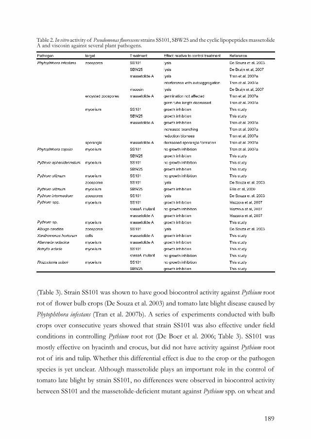
189
(Table 3). Strain SS101 was shown to have good biocontrol activity against Pythium root rot of flower bulb crops (De Souza et al. 2003) and tomato late blight disease caused by Phytophthora infestans (Tran et al. 2007b). A series of experiments conducted with bulb crops over consecutive years showed that strain SS101 was also effective under field conditions in controlling Pythium root rot (De Boer et al. 2006; Table 3). SS101 was mostly effective on hyacinth and crocus, but did not have activity against Pythium root rot of iris and tulip. Whether this differential effect is due to the crop or the pathogen species is yet unclear. Although massetolide plays an important role in the control of tomato late blight by strain SS101, no differences were observed in biocontrol activity between SS101 and the massetolide-deficient mutant against Pythium spp. on wheat and
Table 2. In vitro activity of Pseudomonas fluorescens strains SS101, SBW25 and the cyclic lipopeptides massetolide A and viscosin against several plant pathogens.
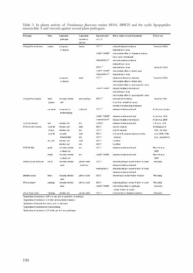
190
Table 3. In planta activity of Pseudomonas fluorescens strains SS101, SBW25 and the cyclic lipopeptides massetolide A and viscosin against several plant pathogens.
191
apple seedlings (Mazzola et al. 2007). The strong reduction in disease severity by both the wild type strain and the mutant indicates that mechanisms other than massetolide production play a major role in these host-pathogen systems. Next to these biocontrol activity assays, several other experiments were performed with strain SS101 to optimize the application strategy and to determine its behaviour in diverse soil and plant systems (Table 3). The most promising application strategy tested so far is biopriming, which consists of introducing the biocontrol strain during seed priming. Priming of seeds accelerates and improves uniformity of seed germination and seedling vigour. However, this procedure may also lead to increases in population densities of some seed-borne pathogens (Wright et al. 2003). When carrot seeds were bioprimed with strain SS101, the population density of strain SS101 remained stable (approximately 106 cells per seed) over a storage period of at least nine months. Moreover, the quality of seed germination and seedling vigour was not affected by the bacterial treatment and seedlings grown from the bioprimed seeds showed reduced disease severity caused by Xanthomonas hortorum pv. carotae and Pythium sp. (Table 3). Whether this is an effective strategy to control multiple diseases under field conditions remains to be tested. For final commercialization of biocontrol agents, several steps need to be taken, including large-scale production, fermentation, formulation, (eco)toxicology and safety assessments, registration, and efficacy trials under diverse conditions (Fravel, 2005). For strain SS101, three formulations were recently produced: a liquid formulation, an air-dried and a freeze-dried formulation. Each of these prototype formulations are currently being tested in efficacy trials for their activity against several plant pathogenic oomycetes and fungi.
REFERENCESBerti, A. D., Greve, N. J., Christensen, Q. H., and Thomas, M. G. 2007. Identification of a biosynthetic gene cluster and the six associated lipopeptides involved in swarming motility of Pseudomonas syringae pv. tomato DC3000. J. Bacteriol. 189:6312-6323.Bode, H. B., and Muller, R. 2005. The impact of bacterial genomics on natural product research. Angew. Chem. Int. Ed. Engl. 44:6828-46.Cosson, P., Zulianello, L., Join-Lambert, O., Faurisson, F., Gebbie, L., Benghezal, M., Van Delden, C., Curty, L. K., and Kohler, T. 2002. Pseudomonas aeruginosa virulence analyzed in a Dictyostelium discoideum host system. J. Bacteriol. 184:3027-33.
Cui, X., Harling, R., Mutch, P., and Darling, D. 2005. Identification of N-3-hydroxyoctanoyl-homoserine lactone production in Pseudomonas fluorescens 5064, pathogenic to broccoli, and controlling biosurfactant production by quorum sensing. Eur. J. Plant Pathol. 111:297-308.De Boer, M., Van Os, G. J., Bijman, V., and Raaijmakers, J. M. 2006. Biological control of soil-borne diseases in flowerbulb cultivation in the Netherlands. Multitrophic interaction is soil IOBC/wprs Bulletin 29:83-87.De Bruijn, I., de Kock, M. J., de Waard, P., van Beek, T. A., and Raaijmakers, J. M. 2008. Massetolide A biosynthesis in Pseudomonas fluorescens. J. Bacteriol. 190:2777-2789.
192
De Bruijn, I., de Kock, M. J. D., Yang, M., de Waard, P., van Beek, T. A., and Raaijmakers, J. M. 2007. Genome-based discovery, structure prediction and functional analysis of cyclic lipopeptide antibiotics in Pseudomonas species. Mol. Microbiol. 63:417-428.De Souza, J. T., De Boer, M., De Waard, P., Van Beek, T. A., and Raaijmakers, J. M. 2003. Biochemical, genetic, and zoosporicidal properties of cyclic lipopeptide surfactants produced by Pseudomonas fluorescens. Appl. Environ. Microbiol. 69:7161-72.Dubern, J.-F., Lagendijk, E. L., Lugtenberg, B. J. J., and Bloemberg, G. V. 2005. The heat shock genes dnaK, dnaJ, and grpE are involved in regulation of putisolvin biosynthesis in Pseudomonas putida PCL1445. J. Bacteriol. 187:5967-5976.Dubern, J. F., Coppoolse, E. R., Stiekema, W. J., and Bloemberg, G. V. 2008. Genetic and functional characterization of the gene cluster directing the biosynthesis of putisolvin I and II in Pseudomonas putida strain PCL1445. Microbiology 154:2070-83.Dubern, J. F., Lugtenberg, B. J., and Bloemberg, G. V. 2006. The ppuI-rsaL-ppuR quorum-sensing system regulates biofilm formation of Pseudomonas putida PCL1445 by controlling biosynthesis of the cyclic lipopeptides putisolvins I and II. J. Bacteriol. 188:2898-906.Ducros, V. M., Lewis, R. J., Verma, C. S., Dodson, E. J., Leonard, G., Turkenburg, J. P., Murshudov, G. N., Wilkinson, A. J., and Brannigan, J. A. 2001. Crystal structure of GerE, the ultimate transcriptional regulator of spore formation in Bacillus subtilis. J. Mol. Biol. 306:759-71.Ellis, R. J., Timms-Wilson, T. M., and Bailey, M. J. 2000. Identification of conserved traits in fluorescent pseudomonads with antifungal activity. Environ. Microbiol. 2:274-84.Feil, H., Feil, W. S., Chain, P., Larimer, F., Dibartolo, G., Copeland, A., Lykidis, A., Trong, S., Nolan, M., Goltsman, E., Thiel, J., Malfatti, S., Loper, J. E., Lapidus, A., Detter, J. C., Land, M., Richardson, P. M., Kyrpides, N. C., Ivanova, N., and Lindow, S. E. 2005. Comparison of the complete genome sequences of Pseudomonas syringae pv. syringae B728a and pv. tomato DC3000. Proc. Natl. Acad. Sci. USA 102:11064-9.Ficke, A., De Boer, M., Geerds, C. F., and Raaijmakers, J. M. Control of oomycete diseases in various crops: applicability and efficacy of Pseudomonas fluorescens SS101. Unpublished.Finking, R., and Marahiel, M. A. 2004. Biosynthesis of nonribosomal peptides. Annu. Rev. Microbiol. 58:453-488.Flynn, J. M., Neher, S. B., Kim, Y. I., Sauer, R. T., and Baker, T. A. 2003. Proteomic discovery of cellular substrates of the ClpXP protease reveals five classes of ClpX-recognition signals. Mol. Cell 11:671-83.Fravel, D. R. 2005. Commercialization and implementation of biocontrol. Annu. Rev.
Phytopathol. 43:337-59.Fuqua, W. C., Winans, S. C., and Greenberg, E. P. 1994. Quorum sensing in bacteria: the LuxR-LuxI family of cell density-responsive transcriptional regulators. J. Bacteriol. 176:269-75.Gross, H., Stockwell, V. O., Henkels, M. D., Nowak-Thompson, B., Loper, J. E., and Gerwick, W. H. 2007. The genomisotopic approach: a systematic method to isolate products of orphan biosynthetic gene clusters. Chem. Biol. 14:53-63.Guenzi, E., Galli, G., Grgurina, I., Gross, D. C., and Grandi, G. 1998. Characterization of the syringomycin synthetase gene cluster - A link between prokaryotic and eukaryotic peptide synthetases. J. Biol. Chem. 273:32857-32863.Haas, D., and Defago, G. 2005. Biological control of soil-borne pathogens by fluorescent pseudomonads. Nat. Rev. Microbiol. 3:307-19.Hildebrand, P. D., Braun, P. G., McRae, K. B., and Lu, X. 1998. Role of the biosurfactant viscosin in broccoli head rot caused by a pectolytic strain of Pseudomonas fluorescens. Can. J. Plant Pathol. 20:296-303.Kitten, T., Kinscherf, T. G., McEvoy, J. L., and Willis, D. K. 1998. A newly identified regulator is required for virulence and toxin production in Pseudomonas syringae. Mol. Microbiol. 28:917-29.Koch, B., Nielsen, T. H., Sorensen, D., Andersen, J. B., Christophersen, C., Molin, S., Givskov, M., Sorensen, J., and Nybroe, O. 2002. Lipopeptide production in Pseudomonas sp. strain DSS73 is regulated by components of sugar beet seed exudate via the Gac two-component regulatory system. Appl. Environ. Microbiol. 68:4509-16.Kruijt, M., Tran, H., and Raaijmakers, J. M. 2008. Functional, genetic and chemical characterization of biosurfactants produced by plant growth-promoting Pseudomonas putida 267. J. Appl. Microbiol. Accepted.Lapouge, K., Schubert, M., Allain, F. H., and Haas, D. 2008. Gac/Rsm signal transduction pathway of gamma-proteobacteria: from RNA recognition to regulation of social behaviour. Mol. Microbiol. 67:241-53.Lu, S. E., Scholz-Schroeder, B. K., and Gross, D. C. 2002. Characterization of the salA, syrF, and syrG regulatory genes located at the right border of the syringomycin gene cluster of Pseudomonas syringae pv. syringae. Mol. Plant-Microbe Interact. 15:43-53.Lu, S. E., Wang, N., Wang, J., Chen, Z. J., and Gross, D. C. 2005. Oligonucleotide microarray analysis of the salA regulon controlling phytotoxin production by Pseudomonas syringae pv. syringae. Mol. Plant-Microbe Interact. 18:324-333.Matz, C., Deines, P., Boenigk, J., Arndt, H., Eberl, L., Kjelleberg, S., and Jurgens, K. 2004. Impact of violacein-producing bacteria on survival and feeding of bacterivorous nanoflagellates. Appl. Environ. Microbiol. 70:1593-9.Matz, C., and Kjelleberg, S. 2005. Off the hook-how bacteria survive protozoan grazing. Trends Microbiol. 13:302-7.

193
Matz, C., Moreno, A. M., Alhede, M., Manefield, M., Hauser, A. R., Givskov, M., and Kjelleberg, S. 2008. Pseudomonas aeruginosa uses type III secretion system to kill biofilm-associated amoebae. ISME J. 2:843-52.Mazzola, M., Zhao, X., Cohen, M. F., and Raaijmakers, J. M. 2007. Cyclic lipopeptide surfactant production by Pseudomonas fluorescens SS101 is not required for suppression of complex Pythium spp. populations. Phytopathology 97:1348-1355.Nybroe, O., and Sorensen, J. 2004. Production of cyclic lipopeptides by fluorescent pseudomonads. Pages 147-172 in: Pseudomonas volume 3, Biosynthesis of Macromolecules and Molecular Metabolism, Ramos, J-L., ed., Kluwer Academic/Plenum Publishers, New York.Pedersen, A. L., Nybroe, O., Winding, A., Ekelund, F., and Bjornlund, L. 2008. Bacterial feeders, the nematode Caenorhabditis elegans and the flagellate Cercomonas longicauda, have different effects on outcome of competition among the Pseudomonas biocontrol strains CHA0 and DSS73. Microb. Ecol. DOI. 10.1007/s00248-008-9455-y.Ronn, R., McCaig, A. E., Griffiths, B. S., and Prosser, J. I. 2002. Impact of protozoan grazing on bacterial community structure in soil microcosms. Appl. Environ. Microbiol. 68:6094-6105.Scholz-Schroeder, B. K., Hutchison, M. L., Grgurina, I., and Gross, D. C. 2001a. The contribution of syringopeptin and syringomycin to virulence of Pseudomonas syringae pv. syringae strain B301D on the basis of sypA and syrB1 biosynthesis mutant analysis. Mol. Plant-Microbe Interact. 14:336-348.Scholz-Schroeder, B. K., Soule, J. D., and Gross, D. C. 2003. The sypA, sypB, and sypC synthetase genes encode twenty-two modules involved in the nonribosomal peptide synthesis of syringopeptin by Pseudomonas syringae pv. syringae B301D. Mol. Plant-Microbe Interact. 16:271-280.Scholz-Schroeder, B. K., Soule, J. D., Lu, S. E.,
Grgurina, I., and Gross, D. C. 2001b. A physical map of the syringomycin and syringopeptin gene clusters localized to an approximately 145-kb DNA region of Pseudomonas syringae pv. syringae strain B301D. Mol. Plant-Microbe Interact. 14:1426-1435.Tran, H. 2007a. Interactions between biosurfactant-producing Pseudomonas and Phytophthora species. Ph.D. thesis.Tran, H., Ficke, A., Asiimwe, T., Hofte, M., and Raaijmakers, J. M. 2007b. Role of the cyclic lipopeptide massetolide A in biological control of Phytophthora infestans and in colonization of tomato plants by Pseudomonas fluorescens. New Phytol. 175:731-42.Tran, H., Kruijt, M., and Raaijmakers, J. M. 2008. Diversity and activity of biosurfactant-producing Pseudomonas in the rhizosphere of black pepper in Vietnam. J. Appl. Microbiol. 104:839-51.Walsh, U. F., Morrissey, J. P., and O’Gara, F. 2001. Pseudomonas for biocontrol of phytopathogens: from functional genomics to commercial exploitation. Curr. Opin. Biotechnol. 12:289-95.Wang, N., Lu, S. E., Records, A. R., and Gross, D. C. 2006. Characterization of the transcriptional activators SalA and SyrF, which are required for syringomycin and syringopeptin production by Pseudomonas syringae pv. syringae. J. Bacteriol. 188:3290-3298.Wright, B., Rowse, H., and Whipps, J. M. 2003. Microbial population dynamics on seeds during drum and steeping priming. Plant and Soil 255:631-640.Yu, A. Y., and Houry, W. A. 2007. ClpP: a distinctive family of cylindrical energy-dependent serine proteases. FEBS Lett. 581:3749-57.Zhang, J. H., Quigley, N. B., and Gross, D. C. 1995. Analysis of the syrB and syrC genes of Pseudomonas syringae pv. syringae indicates that syringomycin is synthesized by a thiotemplate mechanism. J. Bacteriol. 177:4009-4020.

194
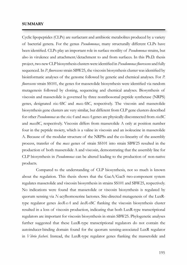
195
SUMMARY
Cyclic lipopeptides (CLPs) are surfactant and antibiotic metabolites produced by a variety of bacterial genera. For the genus Pseudomonas, many structurally different CLPs have been identifi ed. CLPs play an important role in surface motility of Pseudomonas strains, but also in virulence and attachment/detachment to and from surfaces. In this Ph.D. thesis project, two new CLP biosynthesis clusters were identifi ed in Pseudomonas fl uorescens and fully sequenced. In P. fl uorescens strain SBW25, the viscosin biosynthesis cluster was identifi ed by bioinformatic analyses of the genome followed by genetic and chemical analyses. For P. fl uorescens strain SS101, the genes for massetolide biosynthesis were identifi ed via random mutagenesis followed by cloning, sequencing and chemical analyses. Biosynthesis of viscosin and massetolide is governed by three nonribosomal peptide synthetase (NRPS) genes, designated viscABC and massABC, respectively. The viscosin and massetolide biosynthesis gene clusters are very similar, but different from CLP gene clusters described for other Pseudomonas as the viscA and massA genes are physically disconnected from viscBC and massBC, respectively. Viscosin differs from massetolide A only at position number four in the peptide moiety, which is a valine in viscosin and an isoleucine in massetolide A. Because of the modular structure of the NRPSs and the co-linearity of the assembly process, transfer of the mass genes of strain SS101 into strain SBW25 resulted in the production of both massetolide A and viscosin, demonstrating that the assembly line for CLP biosynthesis in Pseudomonas can be altered leading to the production of non-native products. Compared to the understanding of CLP biosynthesis, not so much is known about the regulation. This thesis shows that the GacA/GacS two-component system regulates massetolide and viscosin biosynthesis in strains SS101 and SBW25, respectively. No indications were found that massetolide or viscosin biosynthesis is regulated by quorum sensing via N-acylhomoserine lactones. Site-directed mutagenesis of the LuxR-type regulator genes luxR-vA and luxR-vBC fl anking the viscosin biosynthesis cluster resulted in a loss of viscosin production, indicating that both LuxR-type transcriptional regulators are important for viscosin biosynthesis in strain SBW25. Phylogenetic analyses further suggested that these LuxR-type transcriptional regulators do not contain the autoinducer-binding domain found for the quorum sensing-associated LuxR regulator in Vibrio fi scheri. Instead, the LuxR-type regulator genes fl anking the massetolide and
196
viscosin biosynthesis genes are closely related to the LuxR-type regulators identifi ed for syringomycin/ syringopeptin biosynthesis and appear to belong to a separate LuxR-type regulator subfamily, different from the autonomous effector domain protein GerE. Via random mutagenesis and subsequent screening for massetolide-defi cient mutants, also other regulator genes were identifi ed including clpP. ClpP is a serine protease that plays a crucial role in intracellular refolding and degradation of proteins, which is an essential process for the viability of cells. ClpP was shown to affect transcription of luxR-mA, thereby regulating transcription of the massetolide biosynthesis genes. Results further suggested that, at the transcriptional level, ClpP-mediated regulation of massetolide biosynthesis operates independently from regulation by the GacA/GacS two-component system. In conclusion, the results of this thesis led to the identifi cation of several genes and previously unknown pathways involved in regulation of CLP biosynthesis and highlighted the complexity of the signaling cascades underlying CLP biosynthesis in Pseudomonas. CLPs have diverse functions for the producing bacterial strains, including a role in motility, biofi lm formation, antimicrobial activity and virulence. Also in establishment and persistence in plant environments, CLPs were shown to confer a competitive advantage. A new function of CLPs, identifi ed in a collaboration with Mark Mazzola (USDA) and presented in this thesis, is their protective effects against predation by protozoa. In vitro assays showed that both massetolide and viscosin can lyse the trophozoites of Naeglaria americana and that wild type strains SS101 and SBW25 were substantially less sensitive to protozoan grazing than their CLP-defi cient mutants. Also in soil containing N. americana, population densities of wild type strains SS101 and SBW25 were signifi cantly higher compared to the massetolide and viscosin-defi cient mutants, showing that CLP production confers a competitive advantage in survival in complex environments. Moreover, transcription of the CLP-biosynthesis genes increased signifi cantly upon protozoan grazing, indicating that the Pseudomonas strains sense the protozoa and react by producing CLPs as defense compounds. Which signal triggers the induction of the CLP biosynthesis genes is not known yet and currently under investigation. Based on these results, we postulate that CLPs are an important component of the pre-ingestional defense mechanisms of bacteria against protozoan predation, not only due to their lytic effects on protozoa, but also because CLPs contribute to evasion of protozoan grazing via altered cell surface properties, swimming and swarming, and microcolony and biofi lm formation.

197

198
199
SAMENVATTING
Cyclische lipopeptiden (CLP’s) zijn oppervlakte-actieve en antibiotische metabolieten die worden geproduceerd door diverse bacteriële genera. Binnen het genus Pseudomonas zijn verscheidene CLP’s geïdentifi ceerd en deze spelen een belangrijke rol bij motiliteit, virulentie en aanhechting van de Pseudomonas bacterie aan oppervlakten. In dit proefschrift wordt de identifi catie beschreven van twee nieuwe biosynthese clusters voor CLP’s in Pseudomonas fl uorescens. In P. fl uorescens stam SBW25 werd het viscosine biosynthese cluster geïdentifi ceerd met behulp van bioinformatische analyses van de genoomsequentie, hetgeen vervolgens bevestigd werd met behulp van genetische en biochemische analyses. In P. fl uorescens stam SS101 werd het massetolide biosynthese cluster geïdentifi ceerd met behulp van Tn5 mutagenese, gevolgd door kloneren, genetische karakterisatie en chemische analyses. De CLP’s viscosine en massetolide worden in de bacteriële cel gesynthetiseerd door niet-ribosomale peptide synthetases (NRPS’s), genaamd viscABC respectievelijk massABC. De viscosine en massetolide biosynthese clusters zijn zeer vergelijkbaar, maar afwijkend van andere Pseudomonas CLP biosynthese clusters; viscA en massA zijn namelijk ontkoppeld van viscBC en massBC. Het verschil tussen viscosine en massetolide bevindt zich in het vierde aminozuur van het cyclische peptide; in viscosine is dit valine, terwijl dit in massetolide isoleucine is. Vanwege de modulaire structuur van de NRPS eiwitten en de co-lineariteit van het assemblage proces, bleek het mogelijk te zijn om de mass genen van stam SS101 in stam SBW25 tot expressie te brengen hetgeen resulteerde in de productie van zowel massetolide als viscosine. Hiermee werd aangetoond dat het assemblage proces van CLP’s in Pseudomonas gemodifi ceerd kan worden, resulterend in de productie van niet-natieve producten. In vergelijking met de uitgebreide kennis over de biosynthese van CLP’s in Pseudomonas, is er aanzienlijk minder bekend over de regulatie ervan. Dit proefschrift toont aan dat het twee-componenten regulatiesysteem GacA/GacS betrokken is bij de productie van viscosine en massetolide in P. fl uorescens stammen SBW25 respectievelijk SS101. Er zijn echter geen aanwijzingen gevonden voor celdichtheid-afhankelijke regulatie (‘quorum sensing’) middels N-homoserine lactonen bij de biosynthese van viscosine en massetolide. Gen-specifi eke mutagenese van luxR-vA en luxR-vBC, twee LuxR-type regulatie-genen fl ankerend aan het viscosine biosynthese gencluster, resulteerde in verlies van viscosine productie hetgeen aantoont dat beide transcriptionele
200
regulatoren belangrijk zijn voor viscosine biosynthese in stam SBW25. Fylogenetische analyse suggereert dat deze LuxR-type transcriptionele regulatoren geen zogenaamd autoinducer-binding domein bevatten, dat wel aanwezig is in de LuxR-type regulator die betrokken is bij celdichtheid-afhankelijke regulatie in Vibrio fischeri. Echter, de LuxR-type regulatoren die de massetolide en viscosine biosynthese clusters flankeren, lijken veel op de LuxR-type regulatoren die betrokken zijn bij de syringomycine/syringopeptine biosynthese en vormen een afzonderlijke LuxR-type regulator familie. Via Tn5 mutagenese en selectie van massetolide-deficiënte mutanten werden ook andere regulatie-genen ontdekt waaronder clpP. ClpP is een serine protease dat een cruciale rol speelt bij de intracellulaire vouwing en afbraak van eiwitten. ClpP heeft een effect op de transcriptie van luxR-mA en reguleert daarmee de transcriptie van de massetolide biosynthese genen. Onderzoek toonde verder aan dat, op transcriptioneel niveau, de ClpP-gemediëerde regulatie van massetolide biosynthese onafhankelijk is van het GacA/GacS twee-componenten regulatie systeem. Samengevat heeft dit proefschrift geresulteerd in de identificatie van een aantal voorheen onbekende regulatie-genen die betrokken zijn bij CLP biosynthese in Pseudomonas. Het onderzoek toonde tevens de complexiteit van de signaal-transductie cascades aan die van invloed zijn op CLP biosynthese in Pseudomonas. CLP’s hebben verscheidene functies voor de producerende bacteriën waaronder een rol in motiliteit, biofilm vorming, antimicriobiële activiteit en virulentie. Ook voor het vestigen en handhaven van de bacterie in plant-ecosystemen geeft de productie van CLP’s een competitief voordeel. Een nieuwe functie van CLP’s, geïdentificeerd in samenwerking met een Amerikaanse onderzoekgroep onder leiding van Dr. Mazzola (USDA), betreft de bescherming van Pseudomonas tegen predatie door protozoa. In vitro experimenten toonden aan dat zowel massetolide als viscosine de trophozoiten van de amoebe Naegleria americana kunnen lyseren; stammen SS101 en SBW25 waren aanzienlijk minder gevoelig voor predatie door de protozoa dan hun CLP-deficiënte mutanten. Ook in grond verrijkt met N. americana waren de populatiedichtheden van SS101 en SBW25 significant hoger dan die van de CLP-deficiënte mutanten, hetgeen suggereert dat CLP productie een competitief voordeel geeft bij overleving in een bodem-ecosysteem. Bovendien was ook transcriptie van de CLP biosynthese genen significant verhoogd in reactie op het begrazen door de protozoa, hetgeen aantoont dat de Pseudomonas stammen de protozoa kunnen ‘waarnemen’ en vervolgens CLP’s

201
gaan produceren ter verdediging. Welke signaalmoleculen betrokken zijn bij de inductie van de CLP biosynthese genen is nog niet bekend en dit wordt op dit moment verder uitgezocht. Gebaseerd op deze resultaten is de hypothese gerechtvaardigd dat CLP’s belangrijk zijn bij de afweer tegen predatie. Dit is niet alleen gebaseerd op hun lytische effecten op protozoa, maar ook omdat CLP’s bijdragen aan het ontsnappen van predatie door verandering van de bacteriële celoppervlakte, de verhoging van mobiliteit en hun rol bij biofi lm vorming.

202

203
ACKNOWLEDGEMENTS
First of all, I would like to thank the person who helped me the most in achieving this thesis. Jos, thank you for all your guidance. We discussed regularly the many options in my project, and by keeping in mind my preferences, and by thinking in terms of what is required for a manuscript, we achieved a great output. We had a very good collaboration, and fruitful conversations, also made possible because you also concern somebody’s personal life. Every day you make your round in the lab to ask how everything was going, and you always noticed when something was not going okay so well, even when I did not tell you. Not always you would ask about it further, I would tell you in the end anyway, even when it was something personal. We also had lots of fun and I enjoyed our afternoons/evenings in the pub, and want to thank you for being such a good supervisor; also by your enthusiasm, optimism and humor. You are a perfectionist, even more than I am… But that made us, as Dr. Peter Bakker said, a Golden team! I also want to thank my paranimphs and direct colleagues: Marco, thank you for teaching me many molecular techniques in the lab, but also you were a great colleague to chat with during the coffee break, but also to spend time with outside the working hours. Judith, I enjoyed that you started as my project-buddy, I think we made a good team and I had lots of fun chatting about anything. I would also want to thank the other members of the Molecular Ecology group, Corry for showing me how to do the bioassays, Lia for giving me lots of practical advice and also I want to thank Maarten, Ha, Cuong, Rodrigo, Ester, Andrews, Olesya and ChunXu, my MSc students Esther, Olivia, Rodanthi, Henco and Chrysa and all the other MSc students. We went regularly for drinks or dinners and because of the many nationalities in our group, lots of different cultural differences were discussed, which I thought were fun, informative and sometimes really surprising. In the lab we created a very pleasant working environment, quite organized and everybody helping each other and with good spirit. I also want to thank the other members of Phytopathology for the fun moments during the coffee breaks, labweekends or laboutings and the unforgettable girls dinners. Pierre and the other staff members: thank you for your help when needed and for creating a fantastic and successful laboratory! I would also like to thank my external supervisor Wilbert Bitter, who gave me nice suggestions for my thesis and possibilities for post-doc positions. Also, I would
204
like to acknowledge the people we closely collaborated with, including Teris van Beek, Frank Claasen and Pieter de Waard of the Laboratory of Organic Chemistry (WUR) and Wageningen NMR Centre. I would like to express my special thanks to Mark Mazzola (USDA-ARS, Wenatchee, WA, USA) for the close collaboration on the protozoa project and the very nice contribution to my thesis. I would also like to thank Mark Bailey, Tracey Timms-Wilson, and Andrew Spiers (NERC, Oxford University, Oxford, UK) for fruitful discussions and access to the genome sequence of strain SBW25; without this I would not have been able to get so many results. I am also very grateful to Christoph Keel for providing plasmids for complementation, and Teresa Sweat, Joyce Loper (USDA, Corvallis, OR) and Dimitri Mavrodi (USDA, Pullman, WA) for providing the plasmids, protocols and advice on the site-directed mutagenesis. I would also like to thank the members of the Genetics and Microbiology Department of Ecology at KU-LIFE in Copenhagen (Denmark), especially to Ole Nybroe for his supervision and guidance and Stefan Olssen for his help with the fluorescence microscopy. But also to Mette, Lisa, Dorte and all their colleagues for giving me a very good time in Copenhagen! Kijafa! I would also like to acknowledge the financers of the project, the Dutch Technology Foundation STW, and the committee members for very interesting discussions about the results and the potential applications.
I would like to thank my flatmates Wieteke, Bram, Ruben, Charel, Edo, Monique, Esther, Louis and all my friends, who helped me through rough times and with whom I had many good moments. Noor, thank you for being my climbing buddy! Evelien, thank you for having the nice walks around the Utrechtse Heuvelrug, or going to the movies, and always telling me not to be too hard on myself. Ronald, I will miss our sessions in De Vlaamsche Reus. Luckily we can still enjoy it for a couple of months! It was great that you also came to Wageningen for your Ph.D. I could always talk to you about things going on with my project, but also about very personal matters. You’re very direct, which can be quite confronting, but sometimes it was an eye-opener. And luckily I can do the same to you ;-). Andreia, even though we did not speak to each other so much because of our busy lives, the times we did were really good, special, fun, supportive and fantastic. You will always be a special friend, and: you never know who you will meet in the bus… Kat, due to your field works for your Ph.D., we rarely had contact, but the times we meet each other are like we’ve seen each other the day before.
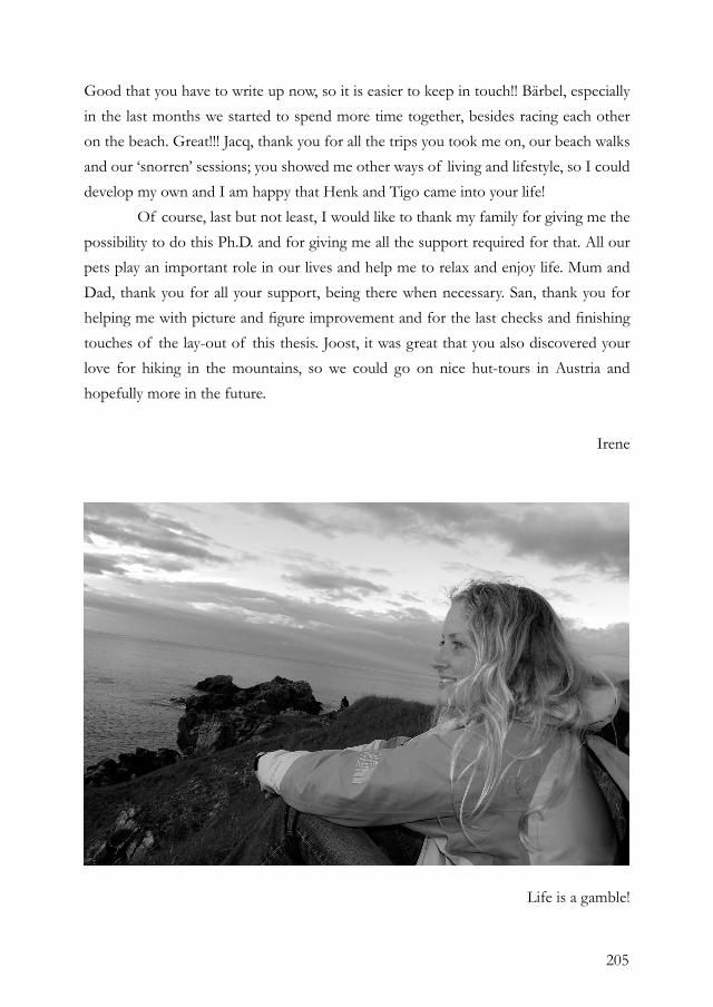
205
Good that you have to write up now, so it is easier to keep in touch!! Bärbel, especially in the last months we started to spend more time together, besides racing each other on the beach. Great!!! Jacq, thank you for all the trips you took me on, our beach walks and our ‘snorren’ sessions; you showed me other ways of living and lifestyle, so I could develop my own and I am happy that Henk and Tigo came into your life! Of course, last but not least, I would like to thank my family for giving me the possibility to do this Ph.D. and for giving me all the support required for that. All our pets play an important role in our lives and help me to relax and enjoy life. Mum and Dad, thank you for all your support, being there when necessary. San, thank you for helping me with picture and figure improvement and for the last checks and finishing touches of the lay-out of this thesis. Joost, it was great that you also discovered your love for hiking in the mountains, so we could go on nice hut-tours in Austria and hopefully more in the future.
Irene
Life is a gamble!

206
207
ABOUT THE AUTHOR
Irene de Bruijn was born on the 27th of April 1981 in Leiden, The Netherlands. After graduating high school (VWO) in 1999, she started her B.Sc. and M.Sc. Biology at Utrecht University, the Netherlands. During her M.Sc. thesis projects, she worked on the disease suppressive mechanisms of Pseudomonas fluorescens at Utrecht University (the Netherlands), under supervision of Dr. Peter Bakker and Prof. Kees van Loon. Her second M.Sc. thesis project was conducted at the Institute for Medical Sciences in Aberdeen (Scotland) under the supervision of Dr. Carol Munro, Prof. dr. Neil Gow and Prof. dr. Han Wösten, and focused on the regulation of chitin biosynthesis genes in the human pathogen Candida albicans. After successfully completing her M.Sc. training in 2004, she started with her Ph.D thesis project entitled ‘Biosynthesis and regulation of cyclic lipopeptides in Pseudomonas fluorescens’ at the laboratory of Phytopathology at Wageningen University (the Netherlands), under the supervision of Dr. Jos Raaijmakers and Prof. dr. ir. Pierre de Wit. In 2007, she was awarded the Kiemprijs by the Dutch Society of Microbiology (Nederlandse Vereniging van Microbiologie) for the best publication of a Ph.D student as a first author. From November 2008, she is working as a postdoc on the NGI-Ecogenomics project at the laboratory of Phytopathology (Wageningen University, the Netherlands) to identify new antibiotic compounds and genes.
PUBLICATIONSDe Bruijn, I. and Raaijmakers, J.M. Heterologous expression of regulatory and biosynthesis genes directing cyclic lipopeptide antibiotic production in Pseudomonas fluorescens. Submitted.Mazzola, M., De Bruijn, I., Cohen, M.F., and Raaijmakers, J.M. Molecular dialogues mediate regulation and activity of cyclic lipopeptide surfactants in bacterial defense against protozoan predation. Submitted.De Bruijn, I. and Raaijmakers, J.M. Regulation of cyclic lipopeptide biosynthesis in Pseudomonas fluorescens by the ClpP protease. J. Bacteriol. In press.Walker, L.A., Munro, C.A., de Bruijn, I., Lenardon, M.D., McKinnon, A., and Gow, N.A. 2008. Stimulation of chitin synthesis rescues Candida albicans from echinocandins. PLoS Pathog. 4(4):e1000040. De Bruijn, I., de Kock, M.J., de Waard, P., van Beek, T.A., and Raaijmakers, J.M. 2008. Massetolide A biosynthesis in Pseudomonas fluorescens. J. Bacteriol. 190:2777-89.Munro, C.A., Selvaggini, S., de Bruijn, I., Walker, L., Lenardon, M.D., Gerssen, B.,
Milne, S., Brown, A.J., and Gow, N.A. 2007. The PKC, HOG and Ca2+ signalling pathways co-ordinately regulate chitin synthesis in Candida albicans. Mol. Microbiol. 63:1399-413. De Bruijn, I., de Kock, M.J., Yang, M., de Waard, P., van Beek, T.A., and Raaijmakers, J.M. 2007. Genome-based discovery, structure prediction and functional analysis of cyclic lipopeptide antibiotics in Pseudomonas species. Mol. Microbiol. 63:417-28. Raaijmakers, J.M., de Bruijn, I., and de Kock, M.J. 2006. Cyclic lipopeptide production by plant-associated Pseudomonas spp.: diversity, activity, biosynthesis, and regulation. Mol. Plant-Microbe Interact. 19:699-710.Bakker, P.A.H.M., De Boer, M., de Bruijn, I., Zhang, K., Van der Sluis, I., and Van Loon, L.C. 2003. Dose response relationships for control of fusarium wilts by Pseudomonas fluorescens RS111. In: 6th International Workshop on Plant Growth Promoting Rhizobacteria (M.S. Reddy, M. Anandaraj, S.J. Eapen, Y.R. Sarma, J.W. Kloepper, eds.), pp. 462-465. Indian Institute of Spices Research, Calicut, India.

208
209

210

211
‘Our greatest glory is not in never falling, but in rising every time we fall’
Confucius
‘Als het niet kan zoals je het wilt.... ...dan wil het maar zoals het kan’
Moeke
212
This research was conducted in the laboratory of Phytopathology of Wageningen University and was financially supported by the Dutch Technology Foundation STW (06404). The printing of this thesis was financially supported by the J.E. Jurriaanse Stichting.
Cover: a selection of pictures of surface motility on soft agar by Pseudomonas fluorescens, collected during this research.
Reproduced by GVO B.V./Ponsen & Looijen, Ede