Embed Size (px)
Citation preview

REVIEWpublished: 19 March 2015
doi: 10.3389/fmicb.2015.00212
Frontiers in Microbiology | www.frontiersin.org 1 March 2015 | Volume 6 | Article 212
Edited by:
Pattanathu K. S. M. Rahman,
Teesside University, UK
Reviewed by:
Toru Matsui,
University of the Ryukyus, Japan
Wael Ismail,
Arabian Gulf University, Bahrain
*Correspondence:
Johannes H. Kügler,
Section II: Technical Biology, Institute
of Process Engineering in Life
Sciences, Karlsruhe Institute of
Technology, Engler-Bunte-Ring 1,
76131 Karlsruhe, Germany
Specialty section:
This article was submitted to
Microbiotechnology, Ecotoxicology
and Bioremediation, a section of the
journal Frontiers in Microbiology
Received: 23 January 2015
Accepted: 02 March 2015
Published: 19 March 2015
Citation:
Kügler JH, Le Roes-Hill M, Syldatk C
and Hausmann R (2015) Surfactants
tailored by the class Actinobacteria.
Front. Microbiol. 6:212.
doi: 10.3389/fmicb.2015.00212
Surfactants tailored by the classActinobacteria
Johannes H. Kügler 1*, Marilize Le Roes-Hill 2, Christoph Syldatk 1 and Rudolf Hausmann 3
1 Technical Biology, Institute of Process Engineering in Life Sciences, Karlsruhe Institute of Technology, Karlsruhe, Germany,2 Biocatalysis and Technical Biology Research Group, Institute of Biomedical and Microbial Biotechnology, Cape Peninsula
University of Technology, Bellville, South Africa, 3 Bioprocess Engineering, Institute of Food Science and Biotechnology,
University of Hohenheim, Stuttgart, Germany
Globally the change towards the establishment of a bio-based economy has resulted
in an increased need for bio-based applications. This, in turn, has served as a
driving force for the discovery and application of novel biosurfactants. The class
Actinobacteria represents a vast group of microorganisms with the ability to produce
a diverse range of secondary metabolites, including surfactants. Understanding the
extensive nature of the biosurfactants produced by actinobacterial strains can assist
in finding novel biosurfactants with new potential applications. This review therefore
presents a comprehensive overview of the knowledge available on actinobacterial
surfactants, the chemical structures that have been completely or partly elucidated,
as well as the identity of the biosurfactant-producing strains. Producer strains of
not yet elucidated compounds are discussed, as well as the original habitats of
all the producer strains, which seems to indicate that biosurfactant production
is environmentally driven. Methodology applied in the isolation, purification and
structural elucidation of the different types of surface active compounds, as well as
surfactant activity tests, are also discussed. Overall, actinobacterial surfactants can
be summarized to include the dominantly occurring trehalose-comprising surfactants,
other non-trehalose containing glycolipids, lipopeptides and the more rare actinobacterial
surfactants. The lack of structural information on a large proportion of actinobacterial
surfactants should be considered as a driving force to further explore the abundance
and diversity of these compounds. This would allow for a better understanding
of actinobacterial surface active compounds and their potential for biotechnological
application.
Keywords: biosurfactant, emulsifier, glycolipid, lipopeptide, trehalose lipid, Rhodococcus, rhamnolipid
Microbial Surfactants and their Applications
Microbially derived compounds that share hydrophilic and hydrophobic moieties, and thatare surface active, are commonly referred to as biosurfactants. Many have been detected anddescribed, and the majorityare molecules of low molecular weight. Within this group of lowmolecular weight microbial surfactants, the classes of lipopeptides or glycolipids, where fattyacid or hydroxy fatty acid chains are linked to either peptides or carbohydrates, have beenextensively studied (Hausmann and Syldatk, 2014). The combinations of different types ofhydrophilic and hydrophobic moieties within surfactants are innumerable and highly biodiverse.
Kügler et al. Surfactants tailored by the class Actinobacteria
Due to their amphiphillic structures, surfactants act as emulsify-ing agents, resulting in low surface tensions of interphases. Often,microorganisms produce them when growing on hydrophobiccarbon sources or when exposed to growth limiting conditions.It is hypothesized, that biosurfactants play a role in the uptakeof various hydrophobic carbon sources thus making nutrientsbioavailable, as well as the protection of bacteria from harshenvironmental conditions (Ristau and Wagner, 1983; Vollbrechtet al., 1998; Philp et al., 2002). Some biosurfactants show antimi-crobial effects and the distinction of secondary metabolites asantibiotics or biosurfactants is often not strict.
Biosurfactants, compared to chemically derived surfactants,are independent of mineral oil as a feedstock, they are read-ily biodegradable and can be produced at low temperatures.Furthermore, they are described to be less toxic, effective at lowconcentrations and show effects in bioremediation. Industrialinterest in biosurfactants is not solely based on the bio-acitivity ofthese molecules, but is also due to the broader ecological aware-ness linked to their application, which in turn is driven by sus-tainability initiatives and green agendas (Marchant and Banat,2012). Biosurfactants can be applied in various areas such as thenutrient-, cosmetic-, textile-, varnish-, pharmaceutical-, mining-,and oil recovery industries (Henkel et al., 2012; Marchant andBanat, 2012; Müller et al., 2012).
An example of an actinobacterial biosurfactant that hasalready entered the market and found industrial application, isthe lipopeptide antibiotic daptomycin. This antibiotic is used inthe treatment of diseases caused by Gram positive pathogensand has been marketed as Cubicin R© by Cubist Pharmaceuticals.
FIGURE 1 | Systematic classification of the class Actinobacteria
including subclasses and orders. Suborder, families and genera
examined for the production of biosurfactants and bioemulsifying
compounds are displayed in numbers. Thirty six surfactant-producing
genera are reported, all belonging to the largest order within the
Actinobacteria: Actinomycetales.
Other promising studies for the potential application of acti-nobacterial biosurfactants are in environmental applications suchas bioremediation: Oil spills were successfully dispersed by bio-surfactants produced by a Gordonia sp. (Saeki et al., 2009), aDietzia sp. (Wang et al., 2014) and a Rhodococcus sp. (Kuyukinaand Ivshina, 2010); and trehalose lipids were applied in micro-bial enhanced oil recovery and the cleaning of oil storage tanks(Franzetti et al., 2010). In medical applications, the productionof biosurfactants are generally considered safer than syntheticallyproduced compounds due to high enzymatic precision duringsynthesis. Antiproliferation activities of cancerogenic cells couldbe induced by application of various glycolipids (Isoda et al.,1997; Sudo et al., 2000). In cosmetic applications, the use of tre-halose lipids is favored above that of sodium dodecyl sulfate as itcauses less irritation (Marques et al., 2009).
Different types of biosurfactants or bioemulsifiers have beendescribed to be produced as secondary metabolites within theclass Actinobacteria, and to the best of our knowledge, allof the producing species belong to the order Actinomycetales(Figure 1). The following section of the review will focus onthe different types of actinobacterial biosurfactants reported inliterature as well as their key structural features and bio-activities.
Metabolite Production within the ClassActinobacteria
Over the past few decades, there has been an increased interestin the discovery of bioactive metabolites with novel bioactive
Frontiers in Microbiology | www.frontiersin.org 2 March 2015 | Volume 6 | Article 212

Kügler et al. Surfactants tailored by the class Actinobacteria
properties and their potential for application in medical- orindustrial-based processes. Microbial products are still consid-ered to be the most promising source for the discovery of novelchemicals or therapeutic agents (Berdy, 2005). In addition, vastmicrobial genetic resources remains untapped and can lead to thedevelopment of novel bioactive metabolites.
In contrast to primary metabolites, secondary metabolitesoften accumulate and have miscellaneous chemical composi-tions that are species-specific. These secondary metabolites oftenexhibit bioactivity and are therefore of great interest to vari-ous industries. The most dominant source of microbially derivedbioactive compounds is a group of bacteria known to have rela-tively large genomes and constitutes one of the main phyla withinthe Prokaryotes: The class Actinobacteria (Ludwig and Klenk,2001). The class Actinobacteria play important roles in the envi-ronment, e.g., nutrient cycling, but also include major plant, ani-mal and human pathogens (Embley and Stackebrandt, 1994), wellknown examples are the causative agents of leprosy and tuber-culosis. Baltz (2008) assumed 5–10% of their genome codingcapacity to be used for the production of secondary metabo-lites and indeed more than 35% of all known bioactive micro-bial metabolites and more than 63% of all known prokaryoticbioactive metabolites arise from actinobacteria (Bérdy, 2012).Most secondary metabolite producers described belong to fam-ilies of the Actinomycetales, but it is estimated that only ∼1% ofthem are culturable (Bérdy, 2012). Many of these actinobacterialsecondary metabolites exhibit antibacterial, antifungal, antitu-mor, anticancer and/or cytotoxic properties (Manivasagan et al.,2013). Antibiotics, with around 10,000 compounds described(Bérdy, 2012) is by far the largest group of metabolites isolatedfrom actinobacteria. Depending on their chemical nature, thehuge number of antibiotic compounds can roughly be classi-fied into peptides, aminoglycosides, polyketides, alkaloids, fattyacids, and terpenes (Manivasagan et al., 2013; Abdelmohsenet al., 2014). Besides antibiotics, other actinobacterial compoundsdescribed are bioactive compounds with pharmacological activity(pheromones, toxins, enzyme inhibitors, receptors and immuno-logical modulators), with agricultural activity (pesticides, herbi-cides and insecticides) and other industrially relevant properties(pigments and surfactants). Most compounds are derived frommembers of the genus Streptomyces, however, other so-called“rare” actinomycetes are increasingly playing a more importantrole in the production of biocompounds (Berdy, 2005; Kurtboke,2010).
To fully understand the taxonomic distribution of the acti-nobacterial strains identified to produce biosurfactants andbioemulsifying compounds, taxonomic data of the class Acti-nobacteria was evaluated. Information were retrieved from thetaxonomy browser of the National Center for BiotechnologyInformation1 considering 16S rRNA gene sequence based reclas-sifications according to Zhi et al. (2009) and Goodfellow andFiedler (2010). The order Thermoleophilales that has been reclas-sified into a new class (Euzéby, 2013) has been excluded and the
1National Center for Biotechnology Information (NCBI) Taxonomy
Browser. Available online at: http://www.ncbi.nlm.nih.gov/Taxonomy/
Browser/wwwtax.cgi?mode=Undef&id=201174&lvl=5&lin (accessed 01.07.2014 -
07.01.2015).
recently identified order Gaiellales has been included (Euzéby,2012). Overall, the class Actinobacteria contains five subclassesand nine orders with a total of 54 families (Figure 1). The largestorder, Actinomycetales, is divided into 14 suborders and con-tains by far the highest diversity within the class Actinobacteria.It is therefore not surprising that biosurfactants reported in lit-erature focuses on members of this order. The next few para-graphs will go into more detail around the different types ofbiosurfactants that have been identified to be produced by acti-nobacterial strains, their production, purification and structuralelucidation, as well as the clear influence of the environmentthe producer organism is found in and their ability to producebiosurfactants.
Trehalose-Comprising Glycolipids
The best described biosurfactants amongst the actinobacteriaare glucose-based glycolipids, most of which have a hydrophilicbackbone consisting of two α,α-1,1 glycosidic linked glucoseunits forming a trehalose moiety. Different types of trehalose-containing glycolipids and their producers have been exten-sively reviewed (Asselineau and Asselineau, 1978; Asselineau andLanéelle, 1998; Franzetti et al., 2010; Kuyukina and Ivshina, 2010;Shao, 2011; Khan et al., 2012). Those of the class Actinobacteriaaremainly foundwithin the generaRhodococcus,Mycobacterium,Nocardia, Arthrobacter and Corynebacterium, and less frequentlywithin the genera Tsukamurella, Brevibacterium, and Micrococ-cus (Tables 1, 2). Different structures of trehalose lipid com-prising amphiphilic molecules have been reported: Acyl chainswith glycosidic linkages to glucose or trehalose units have beenreported to vary in number of occurrence, length and type, aswell as the position (and number) of their linkage to the sugarrings and exhibit different cellular functions.
For the hydrophobic moiety of trehalose-comprising glycol-ipids, the structures of two main types of trehalose lipids havebeen elucidated: those carrying a mycolic fatty acid ester andthose carrying a fatty acid ester.
The smallest hydrophilic backbone in glycolipids constitutesglucose, the building block of the sugar dimer trehalose. Com-plete structures of acylglucoses carrying mycolic acid esters havebeen elucidated and reported to be produced by isolates belong-ing to the genera Corynebacterium andMycobacterium (Brennanet al., 1970) (Table 1), whereas acylglucoses carrying fatty acidesters have been described for Brevibacterium spp. (Okazaki et al.,1969) (Table 2).
Trehalose Lipid Mycolic Acid EstersMycolic acids are long-chain fatty acids and a major compo-nent of the cell wall in various actinobacteria. Species-dependent,its lengths varies from 22 to 92 carbon atoms; they possesslong β-hydroxy-α-branched acyl chains, including cyclopropanepatterns and oxygenic groups. The synthesis of mycolic acidsincludes condensation reactions, and they are also referred to aseumycolic acid, corynemycolic acid and nocardio-mycolic acid,depending on their presence in Mycobacterium spp., Corynebac-terium spp., and Nocardia spp., respectively (Asselineau andLanéelle, 1998).
Frontiers in Microbiology | www.frontiersin.org 3 March 2015 | Volume 6 | Article 212

Kügler et al. Surfactants tailored by the class Actinobacteria
TABLE 1 | Mycolic and corynemycolic containing trehalose lipids that are of actinobacterial origin.
Species Strain TL mycolic acid ester References
Arthrobacter paraffineus KY 4303 TL mycolic (C32–C36) Suzuki et al., 1969
Brevibacterium sp. KY 4304/4305 TL mycolic (C32–36) Suzuki et al., 1969
Brevibacterium vitarumen 12143 TL dimycolic (C28–C38) Lanéelle and Asselineau, 1977
Corynebacterium diphtheriae n.a. Glucose mycolic (C32) Brennan et al., 1970
Corynebacterium spp.
(fasciens, pseudodiphtheriae)
KY 3543
KY 3541
TL mycolic (C32–36) Suzuki et al., 1969
Corynebacterium matruchotii ATCC 14266 TL dimycolic (C28–C38) Datta and Takayama, 1993
Mycobacterium spp.
(smegmatis, tuberculosis)
BCG, n.a. Glucose mycolic (C32) Brennan et al., 1970
Mycobacterium spp.*
(bovis, fortuitum, kansaii, malmoense, phlei,
tuberculosis, smegmatis, szulgai, etc.)
Various TL mycolic, dimycolic, Reviewed in: Asselineau and Asselineau, 1978; Gautier
et al., 1992; Asselineau and Lanéelle, 1998; Vergne and
Daffé, 1998; Dembitsky, 2004; Ishikawa et al., 2009;
Shao, 2011
Nocardia spp. n.a. TL mycolic (C32–36) Suzuki et al., 1969
Rhodococcus spp.*
(erythropolis, opacus, ruber, etc.)
Various TL mycolic,dimycolic, Reviewed in: Asselineau and Asselineau, 1978; Lang
and Philp, 1998; Kuyukina and Ivshina, 2010; Shao,
2011; Khan et al., 2012
EXAMPLES OF MYCOLIC ACID CONTAINING TREHALOSE LIPIDS
1
Trehalose dimycolate produced by Mycobacterium tuberculosis
2
Trehalose dicorynemycolate produced by Rhodococcus erythropolis
*Several producing species are reported; TL, trehalose lipid; n.a., information not available.
Mycolic acid comprising trehalose lipids (Table 1) can be dis-tinguished into two different types, the trehalose mycolic lipidsand the trehalose corynemycolic lipids. These mycobacterial tre-halose mycolates or dimycolates are by far the most hydrophobicglycolipids. Linked to C6 (and C6′) of the sugar rings, they varyamong species in length and branching. They are shaped to formbilayers, implemented in the outer cell wall and usually not foundon the bacterial cell surface (Vergne and Daffé, 1998). Trehalosedimycolates (1, Table 1), also referred to as “cord factor,” servea particular function for the cell. They act as virulence factorsand have immuno-modulating activity (Shao, 2011). They mayfurther be important to maintain a hydrophobic cell wall of the
organism hence facilitating the uptake of hydrophobic carbonsources. The other type, trehalose lipids containing corynemy-colic acid also carry β-hydroxy-α-branched fatty acid moietiesand have been described to occur within the genus Rhodococcus(2, Table 1), carrying 30–56 carbon atoms and within the genusCorynebacterium, carrying 22–36 carbon atoms. They are alsodescribed to occur in mycobacteria (Brennan et al., 1970) andfound in trehalose lipids of Brevibacterium vitarumen (Lanéelleand Asselineau, 1977), Arthrobacter paraffineus and a Nocar-dia sp. (Suzuki et al., 1969). Corynemycolic acids are muchshorter than their mycobacterial counterparts: they lack func-tional groups and are often unsaturated. Within virulent strains
Frontiers in Microbiology | www.frontiersin.org 4 March 2015 | Volume 6 | Article 212

Kügler et al. Surfactants tailored by the class Actinobacteria
TABLE 2 | Trehalose lipid ester of actinobacterial origin.
Species Strain TL ester References
Arthrobacter sp. EK 1 TL tetraester (C12–C18) Passeri et al., 1990
Brevibacterium thiogenitalis No. 653 Glucose diester (C18) Okazaki et al., 1969
Micrococcus luteus BN56 TL tetraester (C9–C14) Tuleva et al., 2009
Mycobacterium spp.*
(africanum, bovis, fortuitum, tuberculosis, etc.)
Various TL ester Reviewed in: Vergne and Daffé, 1998;
Dembitsky, 2004; Shao, 2011
Mycobacterium tuberculosis H37Rv TL sulfolipid Goren, 1970; Gilleron et al., 2004
Nocardia farcinica BN26 TL succinic tetraester (C7-12) Christova et al., 2014
Rhodococcus spp.* (erythropolis, longus,
wratislavensis, etc.)
Various TL ester, TL succinic ester Reviewed in: Asselineau and Asselineau, 1978;
Lang and Philp, 1998; Kuyukina and Ivshina,
2010; Shao, 2011; Khan et al., 2012
Tsukamurella pulmonis PCM 2578T TL diester (C18–20/C4–5) Pasciak et al., 2010a
Tsukamurella spumae
Tsukamurella pseudospumae
DSM 44113,
DSM 44114
DSM 44117
TL diester (C16–18/C4–6) Kügler et al., 2014
Tsukamurella tyrosinosolvens DSM 44370 TL diester (C16–18/C2–6) Vollbrecht et al., 1998
EXAMPLES OF TREHALOSE LIPID ESTERS
3 4
Trehalose diester produced by Tsukamurella spumae Succinic trehalose tetraester produced by Nocardia farcinia
5
Diacetylated trehalose sulfolipid produced by Mycobacterium tuberculosis
*Several producing species are reported; TL, trehalose lipid.
ofmycobacteria, five different sulfonated forms of trehalose estershave been found, varying in their acylation pattern (Khan et al.,2012).
Trehalose Lipid EstersActinobacterial trehalose lipid esters are mainly acylated atC6/C6′ or at C2/C3 and are summarized in Table 2. The amountof hydrophobic chains linked to the trehalose unit varies fromone to four, forming trehalose mono-, di-, tri- and tetraesters,but also octaesters (Singer et al., 1990) (3, Table 2). The acylchains varies in lengths from C8 to C20, show an unsaturatedpattern or form short succinoyl acids, giving the trehaloselipid an anionic character (Lang and Philp, 1998; Tokumotoet al., 2009) (4, Table 2). They are reported to be linked tothe chain length present in hydrophobic carbon source fed to
the producing strain. These glycolipid-linked medium chainlength fatty acids are found within the following actinobacterialgenera: Arthrobacter, Brevibacterium, Caseobacter, Micrococcus,Mycobacterium, Nocardia, Rhodococcus, and Tsukamurella(Table 2).
An exception among the trehalose lipid esters described, is sul-folipid 1 (Goren, 1970) (5, Table 2), a sulfonated and acylatedtrehalose lipid carrying phtio- and hydroxyphtioceranic com-partments. They are known to contribute to the pathogenesis andvirulence of Mycobacterium tuberculosis, the causative agent oftuberculosis. Diacyltrehalose sulfate, the biosynthetic precursorfor sulfolipid 1, has recently been isolated from M. tuberculo-sis (Domenech et al., 2004) and has been used as a target forT-cell mediated recognization and elimination ofM. tuberculosisinfected cells (Gilleron et al., 2004).
Frontiers in Microbiology | www.frontiersin.org 5 March 2015 | Volume 6 | Article 212
Kügler et al. Surfactants tailored by the class Actinobacteria
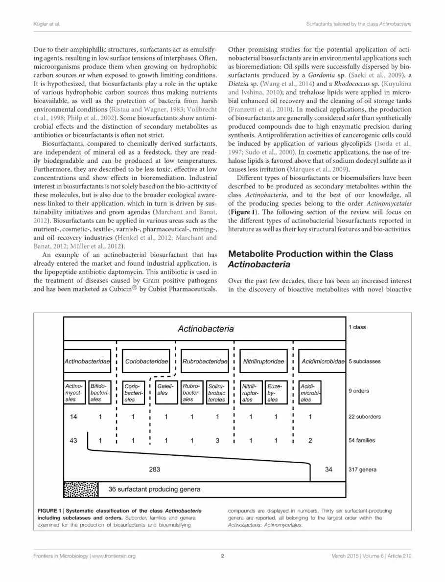
Oligosaccharide LipidsA glycosylated backbone of trehalose is found in oligosaccharidelipids (Table 3) carrying two to five sugar units. Trisaccharidelipids that have been reported for the class Actinobacteria all dif-fer with respect to the acylation pattern of the third glucose unit.One sugar of the 1-1′ linked di-glucose backbone is further linkedto a third sugar unit at C2 in the hydrophilic moeity of oligosac-charides produced byMycobacterium leprae (Brennan, 1989) andTsukamurella tyrosinosolvens (Vollbrecht et al., 1998). The thirdsugar unit is linked at C3 in a terrestrial actinomycete reportedby Esch et al. (1999) and at C4 in a Rhodococcus sp. (Konishiet al., 2014) (6, Table 3). They also differ with respect to theirhydrophobic nature. The latter two are acylated at all three sugar
units, both carrying a C6 fatty acid moiety at the third sugar unitand succinic acid at the first sugar unit. Something that is ratherexceptional is the acylation pattern at the trehalose backbone that,in its hydrophobic moieties, carries at each unit an acyloxyacylstructure in the O-ester linkage to the carbohydrate where the 3-hydroxy C8 or C10 fatty acid moiety is further acylated with a C6fatty acid (6, Table 3). The Tsukamurella sp. trisaccharide lipidsare acylated at two sugar units, each carrying two ordinary C8–C10 fatty acid units. Furthermore, a tetrasaccharide lipid formof this glycolipid has also been found to occur (Vollbrecht et al.,1998) (7, Table 3).
Non-trehalose based oligosaccharide lipids are found withinphenol-phtiocerol glycosides in various mycobacteria. These
TABLE 3 | Actinobacterial oligosaccharide lipids.
Species Strain Oligosaccharid lipids References
Mycobacterium spp.*
(avium, kansaii, leprae, linda, malmoense, smegmatis, szulgai,
tuberculosis)
Various oligosaccharide ester, phenolic
glycolipids
Reviewed in: Saadat and Ballou,
1983; Brennan, 1989;
Dembitsky, 2005b
Nocardia corynebacteroides SM1 Pentasaccharide succinic octaester
(C2–C8)
Powalla et al., 1989
Rhodococcus sp.
Rhodococcus fascians
NBRC 1097287
NBRC 12155
Trisaccharid succinic tetraester
(C8-O-C6/C6)
Konishi et al., 2014
Tsukamurella tyrosinosolvens DSM 44370 Tri/tetrasaccharide ester (C8-10) Vollbrecht et al., 1998
EXAMPLES OF OLIGOSACCHARIDE LIPIDS
6 7
Succinic trisaccharide lipid produced by Rhodococcus fascians Tetrasaccharide lipid produced by Tsukamurella tyrosinosolvens
8
Methylated dirhamnose/glucose phenol phtiocerol named phenolic glycolipid I of Mycobacterium leprae Brennan, 1989
*Several producing strains are reported.
Frontiers in Microbiology | www.frontiersin.org 6 March 2015 | Volume 6 | Article 212
Kügler et al. Surfactants tailored by the class Actinobacteria
oligosaccharide lipids, also termed phenolic glycolipids, containtri- and tetraglycosyl units composed of various methylated sug-ars that are mainly based on rhamnose and partly on fucose,glucose and arabinose (Brennan, 1989). The rarely described phe-nolic acylation pattern is bound to dimycocerosyl phtiocerol acylgroups. The phenolic glycolipid I ofM. leprae carries three myco-cerosyl acyl groups each in length of C30–C34 (Brennan, 1989)(8, Table 3).
In industrial and environmental processes the potential of tre-halose lipids could become valuable as they have shown interest-ing properties in several studies that focus on the remediationof hydrocarbon contaminated soils, the removal of suspendedsolids fromwastewater (Franzetti et al., 2010) and in enhanced oilrecovery (Christofi and Ivshina, 2002). However, most researchare centered around the bio-activity of trehalose lipid moleculesthat exhibit biomedical properties such as antimicrobial, antiviral(Azuma et al., 1987; Watanabe et al., 1999; Shao, 2011) and anti-tumor activities (Sudo et al., 2000; Franzetti et al., 2010; Gudiñaet al., 2013). Due to their functions in cell membrane interactionsthey can act as therapeutic agents (Zaragoza et al., 2009; Shao,2011) or have an impact on the pathogenesis of causative agentsof infections, such as those caused by pathogenic M. tuberculo-sis, Corynebacterium diphteriae, and the opportunistic pathogens,Mycobacterium avium, Mycobacterium intracellulare, Nocardiaasteroides, Corynebacterium matruchotii, and Corynebacteriumxerosis (Kuyukina and Ivshina, 2010). Trehalose lipids can beexcreted into the cultivation supernatant or can be produced asnon-covalently linked lipids bound to the cell wall or they canbe cell wall integrated thus posing limits to quantities producedby the organisms, a disadvantage for its potential exploitation inlarge scale production processes.
Non-Trehalose Glyolipids
Hexose-Comprising GlycolipidsBesides the trehalose-containing biosurfactants and its con-geners, several glycolipids have been elucidated that are producedby actinobacteria and share other hydrophilic moieties. By simplyvarying the carbon source in the growth media from n-alkanes toeither sucrose or fructose, the hydrophilic part of the surfactantproduced was reported to be switched from trehalose to fructoseby members of the genus Arthrobacter, Corynebacterium, Nocar-dia, Brevibacterium, andMycobacterium (Itoh and Suzuki, 1974)and sucrose in the case of the same genera exceptMycobacterium(Suzuki et al., 1974). Compounds for which structures have beenelucidated are listed in Table 4.
Besides the rhamnose-containing phenolic glycolipids men-tioned in the oligosaccharide lipid section, the occurrence ofother rhamnose-based lipids have recently been detected in adeep sea isolate identified as Dietzia maris (Wang et al., 2014)and has been identified as a C10:C10 di-rhamnolipid. This repre-sents a unique occurrence within the class Actinobacteria. Otherrhamnolipid producing actinobacteria are admittedly declaredas producing strains in literature, however the surface activecompounds produced have either not been elucidated or iden-tified as rhamnolipids with debatable structural characteriza-tions (Rhodoccocus fascians Gesheva et al., 2010, Renibacterium
salonariumChristova et al., 2004, and aNocardioides sp. Vasileva-Tonkova and Gesheva, 2005) (Table 11).
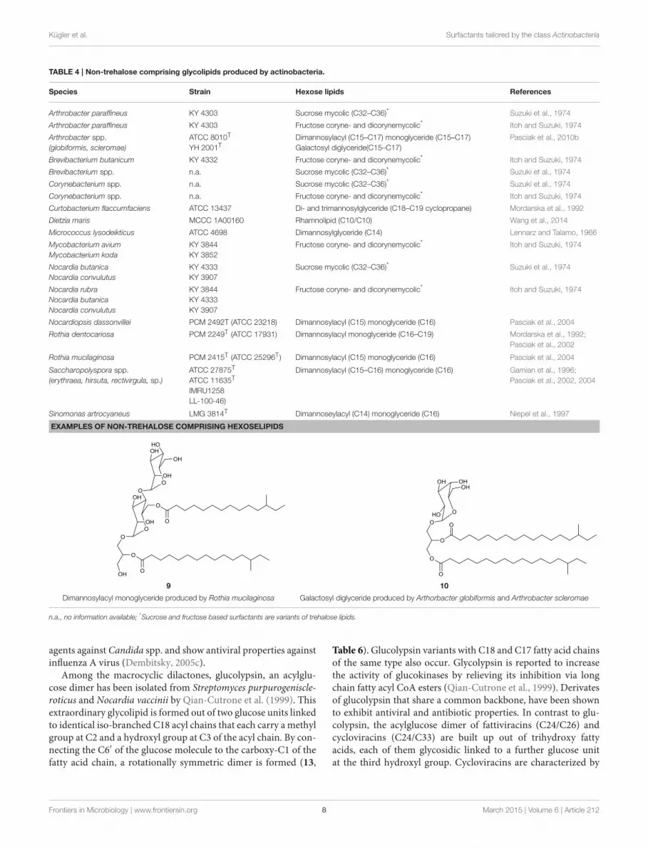
A different group of glycolipids are lipidic structures basedon dimannose. Typically they are linked via a glycerol unitto different numbers of fatty acid chains. They have beenreviewed in Shaw (1970) and structures have been identifiedfor compounds produced by species belonging to the actinobac-terial genera Micrococcus (Lennarz and Talamo, 1966), Curto-bacterium (Mordarska et al., 1992), Saccharopolyspora (Gamianet al., 1996), Rothia (Pasciak et al., 2002, 2004), Nocardiop-sis (Pasciak et al., 2004), Arthrobacter (Pasciak et al., 2010b)as well as the strain Sinomonas atrocyaneus (Niepel et al.,1997), formerly classified as Arthrobacter atrocyaneus. These di-mannose based glycolipids are composed of hydrophilic α-D-mannopyranose dimers linked with two C14–C16 iso or anteisofatty acid chains. One chain is directly esterified to the C6hydroxyl group of one sugar unit, while the second fatty acidchain is linked via a glycerol moiety to the C3 of the samesugar unit. The glycerol moiety is monoacylated at either theprimary or secondary methylene position (9, Table 4) and itsacylation site can be used to distinguish taxonomic proper-ties of the different producer strains. These compounds havebeen isolated intracellularly and they act as precursors and cellmembrane anchors for the synthesis of lipoarabinomannan, apolymeric surfactant and actinobacterial cell wall component(Pakkiri and Waechter, 2005) (see section on polymeric biosur-factants).
The coexistence of galactosyl diglycerides (10, Table 4) inArthrobacter scleromae and Arthrobacter globiformis (Pasciaket al., 2010b) have been described and can be used as a gly-comarker to distinguish these strains from the opportunisticpathogens, Rothia mucilaginosa and Rothia dentocariosa (Pasciaket al., 2002, 2004).
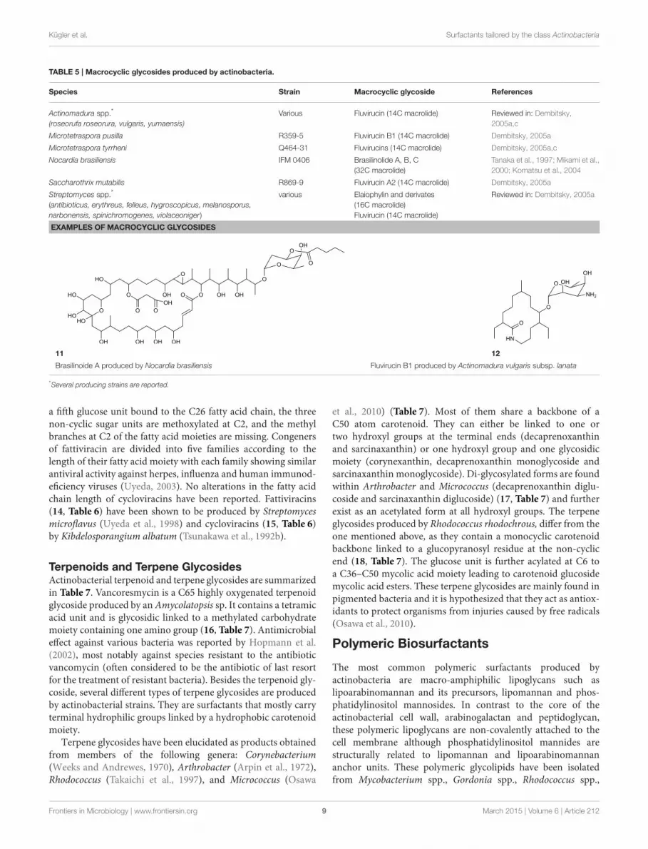
Macrocyclic GlycosidesAmong the biosurfactants produced by actinobacteria, macro-cyclic glycosides (Table 5) and macrocylcic dilactones (Table 6)can be distinguished and are often known to exhibit bio-activityagainst a range of organisms. The aliphatic macrolide antibiotic,brasilinolide, is produced by Nocardia brasiliensis and exhibitsboth antifungal and antibacterial activity. Three different vari-ants have been described by Tanaka et al. (1997), Mikami et al.(2000) and Komatsu et al. (2004). All consist of a C32-memberedmacrolide with a sugar moiety but differ with regards to the acy-lation site of a malonic acid ester side chain (11, Table 5). TheC16-membered dimeric macrolide elaiophylin and its variantshave been isolated from various Streptomyces spp. including highproducer strains. It exhibits bio-active properties against intesti-nal worms as well as antimicrobial, antitumor and immunosup-pressant activities. A putative 95 kbp biosynthetic gene cluster ofelaiophylin has been proposed (Haydock et al., 2004). Dembitsky(2005a,c) reviewed the different types of C14-membered lactamrings that are attached to an aminosugar (12, Table 5). Fluvirucinhas been isolated from various Actinomadura spp., Streptomycesspp., Microtetraspora spp., and Saccharotrix mutabilis. The dif-ferent fluvirucins share a common lactam ring unit but differin terms of glycosylation. All of them act as potent antifungal
Frontiers in Microbiology | www.frontiersin.org 7 March 2015 | Volume 6 | Article 212
Kügler et al. Surfactants tailored by the class Actinobacteria
TABLE 4 | Non-trehalose comprising glycolipids produced by actinobacteria.
Species Strain Hexose lipids References
Arthrobacter paraffineus KY 4303 Sucrose mycolic (C32–C36)* Suzuki et al., 1974
Arthrobacter paraffineus KY 4303 Fructose coryne- and dicorynemycolic* Itoh and Suzuki, 1974
Arthrobacter spp.
(globiformis, scleromae)
ATCC 8010T
YH 2001TDimannosylacyl (C15–C17) monoglyceride (C15–C17)
Galactosyl diglyceride(C15–C17)
Pasciak et al., 2010b
Brevibacterium butanicum KY 4332 Fructose coryne- and dicorynemycolic* Itoh and Suzuki, 1974
Brevibacterium spp. n.a. Sucrose mycolic (C32–C36)* Suzuki et al., 1974
Corynebacterium spp. n.a. Sucrose mycolic (C32–C36)* Suzuki et al., 1974
Corynebacterium spp. n.a. Fructose coryne- and dicorynemycolic* Itoh and Suzuki, 1974
Curtobacterium flaccumfaciens ATCC 13437 Di- and trimannosylglyceride (C18–C19 cyclopropane) Mordarska et al., 1992
Dietzia maris MCCC 1A00160 Rhamnolipid (C10/C10) Wang et al., 2014
Micrococcus lysodeikticus ATCC 4698 Dimannosylglyceride (C14) Lennarz and Talamo, 1966
Mycobacterium avium
Mycobacterium koda
KY 3844
KY 3852
Fructose coryne- and dicorynemycolic* Itoh and Suzuki, 1974
Nocardia butanica
Nocardia convulutus
KY 4333
KY 3907
Sucrose mycolic (C32–C36)* Suzuki et al., 1974
Nocardia rubra
Nocardia butanica
Nocardia convulutus
KY 3844
KY 4333
KY 3907
Fructose coryne- and dicorynemycolic* Itoh and Suzuki, 1974
Nocardiopsis dassonvillei PCM 2492T (ATCC 23218) Dimannosylacyl (C15) monoglyceride (C16) Pasciak et al., 2004
Rothia dentocariosa PCM 2249T (ATCC 17931) Dimannosylacyl monoglyceride (C16–C19) Mordarska et al., 1992;
Pasciak et al., 2002
Rothia mucilaginosa PCM 2415T (ATCC 25296T ) Dimannosylacyl (C15) monoglyceride (C16) Pasciak et al., 2004
Saccharopolyspora spp.
(erythraea, hirsuta, rectivirgula, sp.)
ATCC 27875T
ATCC 11635T
IMRU1258
LL-100-46)
Dimannosylacyl (C15–C16) monoglyceride (C16) Gamian et al., 1996;
Pasciak et al., 2002, 2004
Sinomonas artrocyaneus LMG 3814T Dimannoseylacyl (C14) monoglyceride (C16) Niepel et al., 1997
EXAMPLES OF NON-TREHALOSE COMPRISING HEXOSELIPIDS
9 10
Dimannosylacyl monoglyceride produced by Rothia mucilaginosa Galactosyl diglyceride produced by Arthorbacter globiformis and Arthrobacter scleromae
n.a., no information available; *Sucrose and fructose based surfactants are variants of trehalose lipids.
agents against Candida spp. and show antiviral properties againstinfluenza A virus (Dembitsky, 2005c).
Among the macrocyclic dilactones, glucolypsin, an acylglu-cose dimer has been isolated from Streptomyces purpurogeniscle-roticus and Nocardia vaccinii by Qian-Cutrone et al. (1999). Thisextraordinary glycolipid is formed out of two glucose units linkedto identical iso-branched C18 acyl chains that each carry a methylgroup at C2 and a hydroxyl group at C3 of the acyl chain. By con-necting the C6′ of the glucose molecule to the carboxy-C1 of thefatty acid chain, a rotationally symmetric dimer is formed (13,
Table 6). Glucolypsin variants with C18 and C17 fatty acid chainsof the same type also occur. Glycolypsin is reported to increasethe activity of glucokinases by relieving its inhibition via longchain fatty acyl CoA esters (Qian-Cutrone et al., 1999). Derivatesof glucolypsin that share a common backbone, have been shownto exhibit antiviral and antibiotic properties. In contrast to glu-colypsin, the acylglucose dimer of fattiviracins (C24/C26) andcycloviracins (C24/C33) are built up out of trihydroxy fattyacids, each of them glycosidic linked to a further glucose unitat the third hydroxyl group. Cycloviracins are characterized by
Frontiers in Microbiology | www.frontiersin.org 8 March 2015 | Volume 6 | Article 212
Kügler et al. Surfactants tailored by the class Actinobacteria
TABLE 5 | Macrocyclic glycosides produced by actinobacteria.
Species Strain Macrocyclic glycoside References
Actinomadura spp.*
(roseorufa roseorura, vulgaris, yumaensis)
Various Fluvirucin (14C macrolide) Reviewed in: Dembitsky,
2005a,c
Microtetraspora pusilla R359-5 Fluvirucin B1 (14C macrolide) Dembitsky, 2005a
Microtetraspora tyrrheni Q464-31 Fluvirucins (14C macrolide) Dembitsky, 2005a,c
Nocardia brasiliensis IFM 0406 Brasilinolide A, B, C
(32C macrolide)
Tanaka et al., 1997; Mikami et al.,
2000; Komatsu et al., 2004
Saccharothrix mutabilis R869-9 Fluvirucin A2 (14C macrolide) Dembitsky, 2005a
Streptomyces spp.*
(antibioticus, erythreus, felleus, hygroscopicus, melanosporus,
narbonensis, spinichromogenes, violaceoniger)
various Elaiophylin and derivates
(16C macrolide)
Fluvirucin (14C macrolide)
Reviewed in: Dembitsky, 2005a
EXAMPLES OF MACROCYCLIC GLYCOSIDES
11 12
Brasilinoide A produced by Nocardia brasiliensis Fluvirucin B1 produced by Actinomadura vulgaris subsp. lanata
*Several producing strains are reported.
a fifth glucose unit bound to the C26 fatty acid chain, the threenon-cyclic sugar units are methoxylated at C2, and the methylbranches at C2 of the fatty acid moieties are missing. Congenersof fattiviracin are divided into five families according to thelength of their fatty acid moiety with each family showing similarantiviral activity against herpes, influenza and human immunod-eficiency viruses (Uyeda, 2003). No alterations in the fatty acidchain length of cycloviracins have been reported. Fattiviracins(14, Table 6) have been shown to be produced by Streptomycesmicroflavus (Uyeda et al., 1998) and cycloviracins (15, Table 6)by Kibdelosporangium albatum (Tsunakawa et al., 1992b).
Terpenoids and Terpene GlycosidesActinobacterial terpenoid and terpene glycosides are summarizedin Table 7. Vancoresmycin is a C65 highly oxygenated terpenoidglycoside produced by an Amycolatopsis sp. It contains a tetramicacid unit and is glycosidic linked to a methylated carbohydratemoiety containing one amino group (16, Table 7). Antimicrobialeffect against various bacteria was reported by Hopmann et al.(2002), most notably against species resistant to the antibioticvancomycin (often considered to be the antibiotic of last resortfor the treatment of resistant bacteria). Besides the terpenoid gly-coside, several different types of terpene glycosides are producedby actinobacterial strains. They are surfactants that mostly carryterminal hydrophilic groups linked by a hydrophobic carotenoidmoiety.
Terpene glycosides have been elucidated as products obtainedfrom members of the following genera: Corynebacterium(Weeks and Andrewes, 1970), Arthrobacter (Arpin et al., 1972),Rhodococcus (Takaichi et al., 1997), and Micrococcus (Osawa
et al., 2010) (Table 7). Most of them share a backbone of aC50 atom carotenoid. They can either be linked to one ortwo hydroxyl groups at the terminal ends (decaprenoxanthinand sarcinaxanthin) or one hydroxyl group and one glycosidicmoiety (corynexanthin, decaprenoxanthin monoglycoside andsarcinaxanthin monoglycoside). Di-glycosylated forms are foundwithin Arthrobacter and Micrococcus (decaprenoxanthin diglu-coside and sarcinaxanthin diglucoside) (17, Table 7) and furtherexist as an acetylated form at all hydroxyl groups. The terpeneglycosides produced by Rhodococcus rhodochrous, differ from theone mentioned above, as they contain a monocyclic carotenoidbackbone linked to a glucopyranosyl residue at the non-cyclicend (18, Table 7). The glucose unit is further acylated at C6 toa C36–C50 mycolic acid moiety leading to carotenoid glucosidemycolic acid esters. These terpene glycosides are mainly found inpigmented bacteria and it is hypothesized that they act as antiox-idants to protect organisms from injuries caused by free radicals(Osawa et al., 2010).
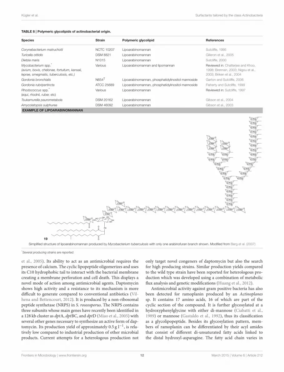
Polymeric Biosurfactants
The most common polymeric surfactants produced byactinobacteria are macro-amphiphilic lipoglycans such aslipoarabinomannan and its precursors, lipomannan and phos-phatidylinositol mannosides. In contrast to the core of theactinobacterial cell wall, arabinogalactan and peptidoglycan,these polymeric lipoglycans are non-covalently attached to thecell membrane although phosphatidylinositol mannides arestructurally related to lipomannan and lipoarabinomannananchor units. These polymeric glycolipids have been isolatedfrom Mycobacterium spp., Gordonia spp., Rhodococcus spp.,
Frontiers in Microbiology | www.frontiersin.org 9 March 2015 | Volume 6 | Article 212
Kügler et al. Surfactants tailored by the class Actinobacteria
TABLE 6 | Macrocyclic dilactones produced by actinobacteria.
Species Strain Macrocyclic dilactones References
Kibdelosporangium albatum ATCC 55061 Cycloviracin B1 and B2 (C23/C26) Tsunakawa et al., 1992a,b
Nocardia vaccinii WC65712 Glucolypsin A and B (C19/C19) Qian-Cutrone et al., 1999
Streptomyces microflavus No.2445 Fattiviracin a1 (C22–28/C22–24) Uyeda et al., 1998; Yokomizo et al., 1998
Streptomyces purpurogeniscleroticus WC71634 Glucolypsin A and B (C19/C19) Qian-Cutrone et al., 1999
EXAMPLES OF MACROCYCLIC DILACTONES
13
Glucolypsin A produced by Nocardia vaccinii and Streptomyces purpurogeniscleroticus
14
Fattiviracin produced by Streptomyces microflavus
15
Cycloviracin B1 produced by Kibdelosporangium albatum
Dietzia maris, Tsukamurella paurometabolus, Turicella otitidis,and Amycolatopsis sulphurea (Table 8). Except for A. sulphurea,all of these strains belong to the suborder Corynebacteridae thatare known to contain mycolic acids in their cell wall. It comprisesthe presence of mycolic acids and contain lipid rich cell envelopestructures (Sutcliffe, 1997) forming an extremely robust andimpermeable cell envelope (Berg et al., 2007). Lipoarabinoman-nans are well known to cause immunorepressive functions indiseases such as tuberculosis and leprosy that are caused by thepathogenic mycobacterial strains M. tuberculosis and M. leprae.However, non-pathogenic species have also been shown to pro-duce lipoarabinomannans and are reported to have an oppositeeffect thus stimulating pro-inflammatory responses (Briken et al.,2004). The mannan core of lipoarabinomannan and the numberof branching units is species dependent. Further differencesin its structure is traced back to capping motifs present at thenon-reducing termini of the arabinosyl side chains. Mannancaps are mainly present in pathogenic strains, whereas inositolphosphate caps are present in non-pathogenic mycobacteria
(Briken et al., 2004). Lipoarabinomannans show structuralsimilarity to its precursors lipomannan and phosphatidylinositolmannoside and consist of an α-1,6 linked mannan core withfrequent α-1,2 mannose branches leading to a mannan backboneof approximately 20–25 mannose residues substituted witharabinofuran residues that carry terminal extension motifs,which vary among the producer species (Berg et al., 2007). Thelipophilic part consists mainly of C16 glycerides that are linkedto the mannan core by a phosphate group (19, Table 8).
Lipopeptides
Cyclic and linear lipopeptides are produced by various actinobac-terial strains and are summarized in Table 9.
Cyclic LipopeptidesCyclic lipopeptides are the most common type of lipopeptidesand consist of a peptide chain of various types and numbersof amino acids circularized and linked to mainly one fatty acid
Frontiers in Microbiology | www.frontiersin.org 10 March 2015 | Volume 6 | Article 212

Kügler et al. Surfactants tailored by the class Actinobacteria
TABLE 7 | Terpenoid and terpene-containing biosurfactants produced by actinobacteria.
Species Strain Terpenoids and terpenes References
Amycolatopsis sp. DSM 12216 Vancoresmycin (65C terpenoid) Hopmann et al., 2002
Arthrobacter sp. M3 Corynexanthin mono- and diglycosides (C50 terpene) Arpin et al., 1972; Dembitsky, 2005b
Corynebacterium sp. CMB 8 Corynexanthin (C50 terpene) Weeks and Andrewes, 1970
Micrococcus yunnanensis AOY-1 Sarcinaxanthin, sarcinaxanthin mono- and diglucosides (C50 terpene) Osawa et al., 2010
Rhodococcus rhodochrous RNMS1 Carotenoid (C40 terpene) glycoside (C36–C50 mycolic) Takaichi et al., 1997
EXAMPLES OF TERPENE AND TERPENOID GLYCOSIDES
16
The terpenoidic glycoside vancoresmycin produced by Amycolatopsis sp.
17
Sarcinaxanthin diglycoside produced by Micrococcus yunnanensis
18
Carotenoid glycoside esterified with a rhodococcus type mycolic acid produced by Rhodococcus rhodochrous
chain. A surfactant often falsely cited to be produced by anactinobacterium but not of actinobacterial nature, is the elevenamino acid cyclic lipopeptide arthrofactin. It was initially pos-tulated to be produced by an Arthrobacter sp. (Morikawa et al.,1993) but later corrected to originate from a Pseudomonas strain(Roongsawang et al., 2003).
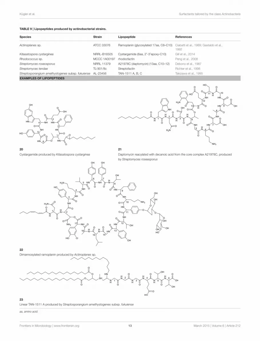
Cyclic lipopeptides that have been reported within the classActinobacteria are the six amino acid containing cystargamideproduced by Kitasatospora cystarginea (Gill et al., 2014) (20,Table 9), the thirteen amino acid containing daptomycin pro-duced by Streptomyces roseosporus (Debono et al., 1987) (21,Table 9) and the depsipeptide ramoplanin, containing 16 aminoacids, and which is produced by an Actinoplanes sp. (Ciabattiet al., 1989) (22, Table 9). All of them are cyclic due to an esterlinkage between the carboxyl terminus and a hydroxyl group ofeither a threonine or hydroxyl-asparagine.
In cystargamide, the smallest cyclic lipopeptide, anuncommon 2,3 epoxy fatty acid chain (C10) is linked tothe threonine amine. Besides proteinogenic amino acids,
cystargamide further contains rare 5′-hydroxy-trypthophan and4′-hydroxyphenylglycine (20, Table 9). No antimicrobial activityof cystargamide could be demonstrated (Gill et al., 2014).
An outstanding example of successful screening for a surfac-tant with bioactive properties are A21978C complexes, known asprecursors of daptomycin. They were structurally elucidated in1987 (Debono et al., 1987) and A21978C comprises thirteen dif-ferent amino acids, 10 of them in the cyclic part of the structureand three in the extension of the hydrophobic tail (21, Table 9).Three different lipophilic tails are known, C10 anteiso, C11 isobranched and C12 anteiso. The most bioactive form of A21978Cis daptomycin and has been generated by enzymatic deacyla-tion of the mixture of lipophilic tails and chemical reacylationwith a decanoyl fatty acid moiety. It was approved by the U.S.Food and Drug Association (FDA) in 2003 as the first antibioticof its kind, and commercialized as cubicin R©. It is active againstvarious gram positive bacteria including the methicilin-resistantpathogen Staphylococcus aureus, penicillin-resistant Streptococ-cus pneumoniae and vancomycin resistant enterococci (Miao
Frontiers in Microbiology | www.frontiersin.org 11 March 2015 | Volume 6 | Article 212
Kügler et al. Surfactants tailored by the class Actinobacteria
TABLE 8 | Polymeric glycolipids of actinobacterial origin.
Species Strain Polymeric glycolipid References
Corynebacterium matruchotii NCTC 10207 Lipoarabinomannan Sutcliffe, 1995
Turicella otitidis DSM 8821 Lipoarabinomannan Gilleron et al., 2005
Dietzia maris N1015 Lipoarabinomannan Sutcliffe, 2000
Mycobacterium spp.*
(avium, bovis, chelonae, fortuitum, kansaii,
leprae, smegmatis, tuberculosis, etc.)
Various Lipoarabinomannan and lipomannan Reviewed in: Chatterjee and Khoo,
1998; Brennan, 2003; Nigou et al.,
2003; Briken et al., 2004
Gordonia bronchialis N654T Lipoarabinomannan, phosphatidylinositol mannoside Garton and Sutcliffe, 2006
Gordonia rubripertincta ATCC 25689 Lipoarabinomannan, phosphatidylinositol mannoside Flaherty and Sutcliffe, 1999
Rhodococcus spp.*
(equi, rhodnii, ruber, etc)
Various Lipoarabinomannan Reviewed in: Sutcliffe, 1997
Tsukamurella paurometabola DSM 20162 Lipoarabinomannan Gibson et al., 2004
Amycolatopsis sulphurea DSM 46092 Lipoarabinomannan Gibson et al., 2003
EXAMPLE OF LIPOARABINOMANNAN
19
Simplified structure of lipoarabinomannan produced by Mycobacterium tuberculosis with only one arabinofuran branch shown. Modified from Berg et al. (2007)
*Several producing strains are reported.
et al., 2005). Its ability to act as an antimicrobial requires thepresence of calcium. The cyclic lipopeptide oligomerizes and usesits C10 hydrophobic tail to interact with the bacterial membranecreating a membrane perforation and cell death. This displays anovel mode of action among antimicrobial agents. Daptomycinshows high activity and a resistance to its mechanism is moredifficult to generate compared to conventional antibiotics (Vil-hena and Bettencourt, 2012). It is produced by a non-ribosomalpeptide synthetase (NRPS) in S. roseosporus. The NRPS containsthree subunits whose main genes have recently been identified ina 128 kb cluster as dptA, dptBC, and dptD (Miao et al., 2005) withseveral other genes necessary to synthesize an active form of dap-tomycin. Its production yield of approximately 0.5 g l−1, is rela-tively low compared to industrial production of other microbialproducts. Current attempts for a heterologous production not
only target novel congeners of daptomycin but also the searchfor high producing strains. Similar production yields comparedto the wild type strain have been reported for heterologous pro-duction which was developed using a combination of metabolicflux analysis and genetic modifications (Huang et al., 2012).
Antimicrobial activity against gram positive bacteria has alsobeen detected for ramoplanin produced by an Actinoplanessp. It contains 17 amino acids, 16 of which are part of thecyclic section of the compound. It is further glycosylated at ahydroxyphenylglycine with either di-mannose (Ciabatti et al.,1989) or mannose (Gastaldo et al., 1992), thus its classificationas a glycolipopeptide. Besides its glycosylation pattern, mem-bers of ramoplanin can be differentiated by their acyl amidesthat consist of different di-unsaturated fatty acids linked tothe distal hydroxyl-asparagine. The fatty acid chain varies in
Frontiers in Microbiology | www.frontiersin.org 12 March 2015 | Volume 6 | Article 212
Kügler et al. Surfactants tailored by the class Actinobacteria
TABLE 9 | Lipopeptides produced by actinobacterial strains.
Species Strain Lipopeptide References
Actinoplanes sp. ATCC 33076 Ramoplanin (glycosylated 17aa, C8–C10) Ciabatti et al., 1989; Gastaldo et al.,
1992
Kitasatospora cystarginea NRRL-B16505 Cystargamide (6aa, 2′-3′epoxy-C10) Gill et al., 2014
Rhodococcus sp. MCCC 1A00197 rhodocfactin Peng et al., 2008
Streptomyces roseosporus NRRL 11379 A21978C (daptomycin) (13aa, C10–12) Debono et al., 1987
Streptomyces tendae Tü 901/8c Streptofactin Richter et al., 1998
Streptosporangium amethystogenes subsp. fukuiense AL-23456 TAN-1511 A, B, C Takizawa et al., 1995
EXAMPLES OF LIPOPEPTIDES
20 21
Cystargamide produced by Kitasatospora cystarginea Daptomycin reacylated with decanoic acid from the core complex A21978C, produced
by Streptomyces roseasporus
22
Dimannosylated ramoplanin produced by Actinoplanes sp.
23
Linear TAN-1511 A produced by Streptosporangium amethystogenes subsp. fukuiense
aa, amino acid.
Frontiers in Microbiology | www.frontiersin.org 13 March 2015 | Volume 6 | Article 212
Kügler et al. Surfactants tailored by the class Actinobacteria
length between C8 and terminal branched C9 and C10 (22,Table 9).
A peptide-based surfactant produced by Streptomyces tendae,streptofactin, was found to contain hydrophobic amino acids, butlacked fatty acid chains (Richter et al., 1998).
Linear LipopeptidesLinear lipopeptides have been found in Streptosporangiumamethystogenes (Takizawa et al., 1995). They are reported toprotect against infections in patients with leucopenia caused bycancer therapies by stimulating bone marrow cells. Differentstructures of these compounds are described, all share a 4′-thioC7 fatty acid chain with two ester linked C16–C19 fatty acidchains and one amide linked C13–C15 fatty acid chain. Threeglycine amino acids are linked at the amide bond of the thio fattyacid with three to four proceeding amino acids varying in type(23, Table 9).
Other Actinobacterial Biosurfactants
Phenazine EsterPhenazines are a rare class of alkaloid esters. A marineStreptomyces sp. has been described to produce a phenazineester that contain the desoxy pyranose quinovose ester-ified at either C3 or C4 to the carboxyl end of thephenazine. This phenazine-quinovose ester has beenshown to exhibit antimicrobial activity. Several differ-ent types of the compound have been characterizedalso varying in hydroxylation and acetylation patternat the desoxyglucose unit (Pathirana et al., 1992) (24,Table 10).
Amide GlycosidesVarious surfactants with nucleoside fatty amide glycosidestructure are produced by actinobacteria. A group of amide glu-cosides is based on the uracil and disaccharide-containing tuni-camycin, a glycoprotein with antibacterial properties (Dembit-sky, 2005c). In this glycoprotein, two saturated or unsaturatedpartly branched fatty acid chains varying in length are linked viaan amide to the galactosamine/glucosamine disaccharide. Besidestunicamycin, produced by Streptomyces spp., the tunicamycin-based surfactants streptovirudin (containing dihydrouracil) andcorynetoxin (25, Table 10) have been reported. The latter is pro-duced by Corynebacterium rathayi, a pathogen of rye grass. Theorganism multiplies within the galls of sheep spreading the toxicmetabolite (Frahn et al., 1984). In addition, the inhibitors ofbacterial peptidoglycan synthesis, liposidomycin A, B, and C,have been reported to be produced by Streptomyces griseosporus.Liposidomycin A contains the so far uniquely described fatty acidcomposition of 3′-hydroxy-7,10-hexadecanoic acid (Dembitsky,2005c) (26, Table 10).
Not Yet Elucidated Surfactants and theirProducing Strains
Surface or emulsifying activity has been observed to occurfrom secondary metabolites of other members of the class
Actinobacteria. Table 11 gives an overview of strains that aredescribed to produce surface active compounds. Only some ofthe structures of these compounds have been partially eluci-dated.
Partly characterized surface active flocculants consisting oflipids, fatty acids and corynemycolic fatty acids of Corynebac-terium lepus have been described by Cooper et al. (1979b). Inaddition, eleven different glycolipids that consist of hexoses andpentoses linked to diverse fatty acid moieties that vary in lengthof C10–C18 have also been described.
Besides D. maris (see glycolipid section), three other puta-tive rhamnolipid-producing actinobacteria have been described.Vasileva-Tonkova and Gesheva (2005) and Gesheva et al. (2010)detected thin layer retention values equal to L-rhamnose afteracid hydrolysis of a biosurfactant produced by a Nocardioides sp.and Rhodococcus fascians. The putative rhamnolipid was not fur-ther examined in terms of the hydrophilic moiety or fatty acidcompositions. Christova et al. (2004) reported the productionof rhamnolipid by Renibacterium solmonarium in comparisonto commercial rhamnolipids in thin layer chromatography andinfrared spectroscopy. The infrared spectra showed homologiesto ester and carboxylic groups; thin layer chromatographic datawere not shown in the study. In all cases the detection of rham-nolipids were putative and further structural analyses remainsnecessary for confirmation.
Other surface active compounds were only putatively classi-fied based on the component analysis of the crude extract towardlipid, peptide and carbohydrate compositions. Based on this lim-ited information, it was concluded that the production of eitherglycolipids or lipopeptides took place (Table 11).
Mass spectroscopic analysis greatly assisted to partly charac-terize the putative wax esters produced by D. maris (Nakanoet al., 2011). In addition, Kiran et al. (2010a,b, 2014) described theproduction of furan-containing glycolipids in Brachybacteriumspp., Brevibacterium spp., and Nocardiopsis spp. By analyzinghydrophilic and hydrophobic moieties after acid hydrolization,database comparison of gas chromatography-mass spectroscopicplots were used. 1HNMR evaluation of compounds from the twolatter strains were described to approve the resulting structure,however relative data were not shown.
Similar results have been observed for surface active extractswith a majority of peptidic compounds in the hydrophilicpart in Brevibacterium aurum (Kiran et al., 2010c) wherefractions of the biosurfactant showed molecular weights ofC9–C29 methyl esters and a mass that putatively confers toa proline-leucine-glycine-glycine amino acid chain. However,mass spectroscopic database comparisons remains putative.Leucobacter komagate is described to produce surfactin or asurfactin-like lipopeptide. This was concluded from mass spec-troscopy, 1H NMR and infrared spectral data by Saimmai et al.(2012b), but the full elucidation of the structures could not beachieved.
The long list of non-elucidated actinobacterial surface activecompounds underlines the extraordinary potential of findingnovel biosurfactants in actinobacteria and displays the great needfor structure elucidation to allow for a better understanding ofthe novelty and biodiversity of the compounds produced.
Frontiers in Microbiology | www.frontiersin.org 14 March 2015 | Volume 6 | Article 212
Kügler et al. Surfactants tailored by the class Actinobacteria
TABLE 10 | Other biosurfactants produced by actinobacteria.
Species Strain Compound References
Streptomyces sp. CNB-253 Phenazine-quinovose Pathirana et al., 1992
Streptomyces spp.*
(griseoflavus, griseosporus, halstedii,
lysosuperficus, nursei, vinausdrappus)
various Fatty acid amide glycoside
(Tunicamycin, Streptovirudin, Liposidomycins)
Reviewed in: Dembitsky, 2005c
Corynebacterium rathayi n.a. Corynetoxin Frahn et al., 1984
EXAMPLES
24 25
Phenanzine-quinovose ester produced by Streptomyces sp. Corynetoxin produced by Corynebacterium rathayi
26
Liposidomycin A produced by Streptomyces sp.
*Several producing strains are reported.
Structural Elucidations of ActinobacterialSurfactants
Various factors have been shown to influence the production,extraction, purification and structure elucidation of novelbiosurfactants produced by actinobacterial strains. Due to theirphenotypic growth characteristics, distinct membrane compo-sitions and their function within the utilization of hydrocar-bons, the surfactants produced are often membrane integrated,membrane associated, extracellular or a mixture of the above,and is always dependent on their particular function within theproducing strains. Commonly the compounds produced exhibitantimicrobial properties, on the one hand proposing wide rang-ing applications, on the other resulting in opposing challengesduring the production process. Special considerations are nec-essary when aiming for the extraction of the compound in anadequate amount and purity for structural elucidation as well assurfactant characterization. This section gives an overview of themost common techniques used to achieve successful structuralelucidations.
DetectionNovel surfactant producing strains can be detected through theuse of screening assays that determine a surfactant’s activity either
from liquid culture (cell-free supernatant or culture broth) orfrom solid agar plates. Various detection methods have beendescribed, but they mostly focus on changes observed in surfacetension or the solubilization and emulsification of hydrocarbons.High throughput compatible assays can be distinct from moreprecise assays that need several milliliters of the compound to betested. The latter often are also applied to characterize the activityof a purified biosurfactant. Good reviews on screening techniqueshave been summarized by Walter et al. (2010) and Satpute et al.(2010).
ProductionThe manufacturing capacity of biosurfactants by a bacterial cul-ture is limited. Wild type producing strains of the best describedmicrobial surfactants, cultured with optimized process methodsin suitable media and culture vessels reach production quanti-ties of up to 422 g l−1 for sophorose lipids (Daniel et al., 1998),112 g l−1 for rhamnose lipids (Giani et al., 1996), 110 g l−1 for spi-culisporic acids (Tabuchi et al., 1977), 106 g l−1 for mannosylery-thritol lipids (Morita et al., 2008) and 3,6 g l−1 for surfactin (Yehet al., 2005). These are rare exceptions within the typical amountsproduced by microorganisms, which usually do not exceed mil-ligram amounts. The production level is strongly influenced bynon-favorable growth and production conditions due to a lack
Frontiers in Microbiology | www.frontiersin.org 15 March 2015 | Volume 6 | Article 212
Kügler et al. Surfactants tailored by the class Actinobacteria
TABLE 11 | Actinobacterial strains identified to produce surface active compounds for which no structures have been elucidated.
Species Strain Compound References
Actinopolyspora sp. A18 n.d. GLP Doshi et al., 2010
Amycolatopsis tucumanensis DSM 45259 n.d. (bioemulsifier) Colin et al., 2013
Brachybacterium paraconglomeratum MSA21 n.d. GL (putative furan lipid/C12) Kiran et al., 2014
Brevibacterium aureum MSA13 n.d. LP (putative brevifactin/C18) Kiran et al., 2010c
Brevibacterium casei MSA19 n.d. GL (putative furan lipid/C18) Kiran et al., 2010a
Corynebacterium hydrocarboclastus n.a. n.d. polymer Zajic et al., 1997
Corynebacterium lepus n.a. n.d. LP Cooper et al., 1979a
Corynebacterium lepus n.a. n.d. GL Cooper et al., 1979a
Corynebacterium lepus n.a. p.d. (lipid, fatty acid, mycolic acid) Cooper et al., 1979b
Corynebacterium xerosis n.a. n.d. LP Margaritis et al., 1979
Dietzia maris WR-3 p.d. (putative wax-ester) Nakano et al., 2011
Dietzia sp. S-JS-1 n.d. LP Liu et al., 2009
Frankia sp. CpI1 n.d. GL Tunlid et al., 1989
Gordonia amarae SC1 n.d. (extracellular with high molecular weight) Iwahori et al., 2001
Gordonia rubripertincta DSM 46038 n.d. Pizzul et al., 2006
Gordonia sp. ADP n.d. Pizzul et al., 2006
Gordonia sp. BS29 n.d. GL Franzetti et al., 2010
Gordonia sp. JE-1058 n.d. (extracellular) Saeki et al., 2009
Kocuria marina BS-15 n.d. LP Sarafin et al., 2014
Leucobacter komagatae 183 p.d. LP Saimmai et al., 2012b
Microlunatus sp. NA2 n.d. Saimmai et al., 2012a
Nocardia erythropolis ATCC 4277 n.d. GL, PL Macdonald et al., 1981
Nocardioides sp. A-8 n.d. GL (putative Rhamnolipid) Vasileva-Tonkova and Gesheva, 2005
Nocardiopsis alba MSA10 n.d. LP Gandhimathi et al., 2009
Nocardiopsis lucentensis MSA04 n.d. GL (putative furan lipid/C9) Kiran et al., 2010b
Oerskovia xanthineolytica CIP 104849 p.d. GL (hexose, pentose C10–C18) Arino et al., 1998
Pseudonocardia sp. BSNC30C n.d. Ruggeri et al., 2009
Renibacterium salmoninarum 27BN n.d. GL (putative Rhamnolipid) Christova et al., 2004
Rhodococcus fascians A-3 n.d. GL (putative Rhamnolipid) Gesheva et al., 2010
Streptomyces sp. n.a. n.d. GL Khopade et al., 2011
GL, Glycolipid; GLP, Glycolipiopeptide; LP, Lipopeptide; PL, Phospholipid; n.a., information not available; n.d., not determined; p.d., partly determined.
of knowledge about the organism used and compound producedwhen initially screening for novel surfactants or novel producerstrains.
With a few exceptions (Qian-Cutrone et al., 1999; Kügler et al.,2014), the average minimum volume for successful structure elu-cidation of an actinobacterial biosurfactant, is typically 20 l. Har-vesting of the surfactants is type dependent and either whole cellbroth (intracellular or membrane associated surfactants) or cellfree supernatant is used as a starting point.
GlycolipidsA typical method for the extraction of surfactants from culturebroth or supernatant is the use of two phase extractions. In a firststep, if appropriate, non-polar solvents (e.g., n-hexane) are usedto remove residual hydrocarbons from the cultivation broth. Ifextraction is carried out from whole cell broth or wet cell mass,glycolipids are either captured by direct cell extraction or by celltreatment (e.g., sonication) prior to the extraction.
In a second step, the surfactant is removed by repeated agi-tation with a medium polar solvent or solvent mixture. Mostcommonly, combinations of chloroform and methanol or polaraprotic solvents such as ethyl acetate or methyl-tert-butyl etherare used. A frequency solvent distribution for the extractionof glycolipids from “rare” actinobacteria is shown in Figure 2,comprising data of 47 two-phase extraction methods used toenrich surfactants produced from either cell-free supernatantor the culture broth. Depending on the chemical characteristicsof the glycolipid, an acidification step (pH2–pH3) with subse-quent incubation (4◦C) prior to the extraction process couldresult in enhanced product recoveries (Passeri et al., 1990; Kon-ishi et al., 2014). Often, after dehumification, further washingsteps are applied, either of a hydrophilic (e.g., ultrapure water)or a hydrophobic (e.g., n-hexane) nature. For the polymeric gly-colipid lipoarabinomannan and related structures, a hot-phenolwater method is almost exclusively used (Sutcliffe, 2000).
The glycolipids produced, mainly present in mixtures ofdifferent forms, need to be separated for structural analysis.
Frontiers in Microbiology | www.frontiersin.org 16 March 2015 | Volume 6 | Article 212

Kügler et al. Surfactants tailored by the class Actinobacteria
FIGURE 2 | Frequency distribution of solvents used for the enrichment
of surfactants by two-phase extraction from the culture broth or cell
free supernatant of 47 “rare” actinobacteria.
This procedure is usually performed by combinations of chro-matographic steps using either gradient columns or preparativemedium- and high pressure chromatography. In addition,preparative planar chromatographies are reported as an addi-tional purification step for the isolation of pure compounds(Powalla et al., 1989; Pasciak et al., 2002, 2004). Rarely appliedis the use of absorbers within the cultivation process. The num-ber one choice for chromatography is the use of hydrophobicityaffiliated separations with silicic acids as an absorbing material.In approximately 80% of structure reports from “rare” actinobac-teria, silicic acid is used with various elution gradients of non-polar and polar solvents. Separated compounds are often furtherpurified by repetitive silica chromatography using different gradi-ents or by subsequent (or preceding) steps with different columnmaterial. Therefore, either reverse-phase C18 chromatographyor cellulose-based ionic interaction chromatography are widelyused.
LipopeptidesThe diversity of different peptide-based surface active com-pounds produced by actinobacterial strains is much smaller thanthat of reported glycolipids. Depending on the lipopeptide pro-duced, two different approaches for the concentration of thesurfactants are used. Either the lipopeptide can be precipitatedfrom the liquid culture/supernatant by either using cold ace-tone, methanol, salt concentrations, acidic environments, or adirect extraction by medium polar solvents similar to those usedfor glycolipids have been reported. Besides the chromatographicpurification steps used for glycolipids, gel filtration has beensuccessfully used as an additional step (Takizawa et al., 1995).
Structural ElucidationOnce a compound is purified to a sufficient extent, componentanalysis, specific staining methods and mass spectroscopic exam-inations are widely used to get a first hint about the type of sur-factant produced. A more detailed schematic of the surfactant
can be deduced from mass spectroscopy fragmentation studies,often revealing mass abundances of separated hydrophilic andhydrophobic parts of the glycolipid. However, complete struc-ture examinations (of complete compounds or hydrolyzed com-ponents) rely on multi-dimensional nuclear magnetic resonancespectroscopy.
Natural Habitats ofBiosurfactant-Producing Actinobacteria
With the exception of a few strains, the great majority ofsurfactant-producing actinobacteria have been isolated fromthree different environments. These are: (1) Hydrocarbon con-taminated soils, (2) infections caused by the actinobacteriumitself, and (3) marine-derived samples. Obviously, this must notreflect the distribution of surfactant-producing actinobacteria innature, but it is clear that there is a link between the type ofenvironment and the ability of actinobacteria to produce biosur-factants and can be considered to be environmentally-driven.
Hydrocarbon Contaminated SoilThe formation of various actinobacterial surfactants is mainlyobserved during growth in a range of different hydrophobic car-bon sources such as n-paraffin, n-hexadecane or vegetable oils.Occurrences of surfactant-producing microorganisms seems tocorrelate to environments in which hydrophobic carbon sourcesare present, no matter if these are oil contaminated or oilenriched (Powalla et al., 1989; Arino et al., 1998; Christova et al.,2004, 2014; Pizzul et al., 2006; Liu et al., 2009; Ruggeri et al.,2009). Evoked by their hydrophobic cell wall due to incorpo-ration and association of various lipoglycosides, actinobacteriapreferably grow in hydrophobic droplets that are dispersed inthe aqueous phase when cultured in cultivation devices. The sur-factants produced facilitate the uptake of these difficult–to-accesscarbon sources by dispersing it into small droplets that can easilybe pre-digested by extracellular enzymes.
InfectionsA second feature of surfactants is the antimicrobial propertyexhibited by most of these compounds. Endowed with nutri-tional and growth advantages toward surrounding organisms,surfactant producers can become rampant, and are often lessaffected by substances present during its growth, e.g., antimi-crobial drugs. They have been found in patients that sufferfrom infections/diseases caused by human deficiency viruses(Guérardel et al., 2003), patients with lung infections and infec-tions of the oral cavity (Datta and Takayama, 1993; Sutcliffe, 1995;Tanaka et al., 1997). In addition, biosurfactant-producing acti-nobacterial strains have also been isolated from infected planttissue (Frahn et al., 1984).
Marine HabitatMany actinobacteria are specialists in survival and native to awide range of extreme environments. Surfactant-producing gen-era have been isolated from various marine-associated habitats(Passeri et al., 1990; Khopade et al., 2011; Nakano et al., 2011).
Frontiers in Microbiology | www.frontiersin.org 17 March 2015 | Volume 6 | Article 212

Kügler et al. Surfactants tailored by the class Actinobacteria
Several of these environments exhibit rather extreme condi-tions, amongst which are deep sea sediments or hydrothermalfields (Peng et al., 2008; Konishi et al., 2014; Wang et al., 2014),ornithogenic exposed soil (Vasileva-Tonkova and Gesheva, 2005)as well as actinobacteria isolated from sponges (Gandhimathiet al., 2009; Kiran et al., 2010a,b,c, 2014) and hard corals (Osawaet al., 2010). An antimicrobial effect of surfactants produced ina highly procaryotic populated sponge tissue is apparent. How-ever, the reason for the frequent occurrence of surfactant pro-ducers within the other marine habitats, still remains to beunderstood.
Summary and Conclusion
A wide range of unique and diverse surfactants produced by acti-nobacteria have been reported. Various glycolipids, lipopeptidesand other surfactant types are produced by numerous species,all belonging to the order Actinomycetales. Taking into accountthe fact that only a minority of actinobacteria is culturable andthe given list of surfactant producing strains without structurallyelucidated compounds (Table 11), the sheer magnitude of acti-nobacterial surfactants that still remain undetermined is evident.The ability of actinobacteria to produce biosurfactants seemsto be influenced by their natural habitat. From the three mainsources of surfactant producing actinobacteria it can be con-cluded that the compounds produced mainly serve for eithergaining access to hydrophobic carbon sources or as a bioactiveagent against competing strains.
In order to pave the way toward biotechnological applica-tions of actinobacterial surfactants, emphasis should be placedon (1) structural elucidation of described, but not identified bio-surfactants, (2) the identification of novel actinobacterial sur-factants by the implementation of next generation screeningmethods; (3) the production of sufficient amounts of surfactantsfor application based studies; and (4) production processes thatresult in high yields and that would cut down on the productioncosts.
(1) Actinobacterial strains with a surface active culture brothor supernatant often are declared as “novel” biosurfactantproducing strains, without elucidation of the surface activecompound(s) produced and a list of producing strains isgiven in this article whose surfactant structures remain tobe identified (Table 11). For a successful structural identifi-cation of the compound, sufficient quantities of the isolatedsurface active compound at an adequate purity is necessaryin order to apply the various analytical methods necessary.This aspect was reviewed in the structural elucidation ofactinobacterial compounds section. Quite a few of the stud-ies cited lacked sufficient strain information and furtherresearch can only be ensured if the strains reported havedesignated strain numbers and thus are available for otherresearchers to pursue the production of these potentiallynovel biosurfactants.
(2) Approaches for the identification of novel biosurfactantsmainly remain traditional by the detection of interesting
producing strains and subsequent isolation and charac-terization of the compound produced. To further expandthe variety of actinobaterial surfactants, alternative screen-ing methodologies that are already known to be used forthe detection of novel lead molecules in the pharmaceuti-cal industry could be applied. Genome-based informationtechnology to reveal pathways that can be implementedinto artificial surfactant synthesis cascades are currentlybeing investigated. These attempts would allow for accessto both undetected and cryptic pathways present in acti-nobacteria. By direct sequencing of metagenomic derivedDNA, enzyme information acquired could be expanded toinformation gained from non-culturable and slow growingspecies.
(3) Many of the surface active compounds produced byactinobacteria potentially show interesting properties asbiotechnological products or additives. Often, as is the casefor many of the compounds summarized in this article, anapplication based study is lacking. This is most probablydue to low availability of the product and can be tracedback to the use of low quantity producing strains. Focuson a novel actinobacterial surfactant, along with progress inthe development toward novel biotechnology-based prod-ucts, will only be made possible if enough substance forinitial studies on bioactivity or other interesting applica-tions can be acquired. If an adequate amount of sub-stance is not achievable by standard bioprocess engineeringattempts, metabolomic approaches and flux analysis couldlead the way. Furthermore, the identification of enzymesinvolved in the synthesis and their genetic regulation cangive an important input into the improvement of fermenta-tion processes. An implementation of the surfactant’s synthe-sis through adequate heterologous production strains couldlead to higher quantities of the different surfactants pro-duced. Potential applications of a novel compound is a guar-antee of success in white biotechnology and negates theefforts made with regards to its production, purification andelucidation.
(4) Currently, comparatively high production costs combinedwith low production yields restrict the development of com-pounds as valuable products, and are mainly limited to highpurity applications, e.g., the drug industry. Several examplesin the past have shown that once a potential application fora specific compound is foreseen, intensive research is set inmotion to facilitate production and purification processes,cutting costs, enhancing yields and, although research oftenlasts for decades, compounds might end in industrial scaleproduction and application.
One example of an actinobacterial surfactant that successfullyunderwent the process from detection to application is theantimicrobial agent daptomycin. It was initially produced semi-synthetically in a three step procedure, but later a direct synthesisof daptomycin was achieved by feeding toxic decanoic acid to acarbon-limited production culture (Huber et al., 1988). Produc-tion rates were further increased by 10–30% by using a mixtureof less toxic decanal and a solvent to solubilize the hydrophobic
Frontiers in Microbiology | www.frontiersin.org 18 March 2015 | Volume 6 | Article 212
Kügler et al. Surfactants tailored by the class Actinobacteria
carbon feed (Bertetti et al., 2012). Mutagenesis approaches (Yuet al., 2011; Li et al., 2013), genome shuffling (Yu et al., 2014) anddirected overexpression (Huang et al., 2012), have recently led tofurther increases in production yields. Other examples of successstories, are non-actinobacterial surfactants that have been pushedto application: sophorolipids, mannosyl erythritol lipids and thelipopeptide surfactin have found application in cosmetic indus-tries (Fracchia et al., 2014). Sophorolipids are even applied in lowcost cleaning products.
Actinobacteria clearly represents a unique and vast untappedresource for the discovery of novel and potentially useful bio-surfactants. The surfactants produced by members of the classActinobacteria are a highly interesting group of products thatcould be of great importance in the future in both the area ofbasic research and application-oriented industrial research.
Author Contributions
JK has designed, conceived, and written this review, it’s fig-ures and tables as well as acquired and interpreted the rel-evant data used. All authors have fruitfully discussed con-tent and structure of the review. In particular, ML has givensubstantial contributions related to actinobacteria and CS andRH have given substantial contributions related to biosurfac-tants.
Acknowledgments
We acknowledge support by Deutsche Forschungsgemeinschaftand Open Access Publishing Fund of Karlsruhe Institute ofTechnology, Germany.
References
Abdelmohsen, U. R., Bayer, K., andHentschel, U. (2014). Diversity, abundance and
natural products of marine sponge-associated actinomycetes. Nat. Prod. Rep.
31, 381–399. doi: 10.1039/c3np70111e
Arino, S., Marchal, R., and Vandecasteele, J.-P. (1998). Production of new extracel-
lular glycolipids by a strain of Cellulomonas cellulans (Oerskovia xanthineolyt-
ica) and their structural characterization. Can. J. Microbiol. 44, 238–243. doi:
10.1139/w97-156
Arpin, N., Liaaen-Jensen, S., and Trouilloud, M. (1972). Bacterial carotenoids.
XXXVIII. C 50-Carotenoids. 9. Isolation of decaprenoxanthin mono-and
diglucoside from an Arthrobacter sp. Acta Chem. Scand. 26, 2524–2526.
Asselineau, C., and Asselineau, J. (1978). Trehalose-containing glycolipids. Prog.
Chem. Fats Other Lipids 16, 59–99. doi: 10.1016/0079-6832(78)90037-X
Asselineau, J., and Lanéelle, G. (1998). Mycobacterial lipids: a historical perspec-
tive. Front. Biosci. 3, e164–e174.
Azuma, M., Suzutani, T., Sazaki, K., Yoshida, I., Sakuma, T., and Yoshida, T.
(1987). Role of interferon in the augmented resistance of trehalose-6, 6′-
dimycolate-treated mice to influenza virus infection. J. Gen. Virol. 68, 835–843.
doi: 10.1099/0022-1317-68-3-835
Baltz, R. H. (2008). Renaissance in antibacterial discovery from actinomycetes.
Curr. Opin. Pharmacol. 8, 557–563. doi: 10.1016/j.coph.2008.04.008
Berdy, J. (2005). Bioactive microbial metabolites. J. Antibiot. 58, 1–26. doi:
10.1038/ja.2005.1
Bérdy, J. (2012). Thoughts and facts about antibiotics: where we are now and where
we are heading. J. Antibiot. 65, 385–395. doi: 10.1038/ja.2012.27
Berg, S., Kaur, D., Jackson, M., and Brennan, P. J. (2007). The glycosyltrans-
ferases of Mycobacterium tuberculosis—roles in the synthesis of arabinogalac-
tan, lipoarabinomannan, and other glycoconjugates.Glycobiology 17, 35R–56R.
doi: 10.1093/glycob/cwm010
Bertetti, G., Malcangi, A., Muraca, R., Trione, G., and Rossi, A. (2012). Process for
the Production of Daptomycin. United states Patent 8, 313,922. Washington,
DC: U.S. Patent and Trademark Office.
Brennan, P. (2003). Structure, function, and biogenesis of the cell wall ofMycobac-
terium tuberculosis.Tuberculosis 83, 91–97. doi: 10.1016/S1472-9792(02)00089-
6
Brennan, P. J. (1989). Structure of mycobacteria: recent developments in defin-
ing cell wall carbohydrates and proteins. Rev. Infec. Dis. 11, S420–S430. doi:
10.1093/clinids/11.Supplement_2.S420
Brennan, P. J., Lehane, D. P., and Thomas, D. W. (1970). Acylglucoses of
the Corynebacteria and Mycobacteria. Eur. J. Biochem. 13, 117–123. doi:
10.1111/j.1432-1033.1970.tb00906.x
Briken, V., Porcelli, S. A., Besra, G. S., and Kremer, L. (2004). Mycobacterial
lipoarabinomannan and related lipoglycans: from biogenesis to modulation
of the immune response. Mol. Microbiol. 53, 391–403. doi: 10.1111/j.1365-
2958.2004.04183.x
Chatterjee, D., and Khoo, K.-H. (1998). Mycobacterial lipoarabinomannan: an
extraordinary lipoheteroglycan with profound physiological effects. Glycobiol-
ogy 8, 113–120. doi: 10.1093/glycob/8.2.113
Christofi, N., and Ivshina, I. (2002). Microbial surfactants and their use in field
studies of soil remediation. J. Appl. Microbiol. 93, 915–929. doi: 10.1046/j.1365-
2672.2002.01774.x
Christova, N., Lang, S., Wray, V., Kaloyanov, K., Konstantinov, S., and Stoineva,
I. (2014). Production, structural elucidation and in vitro antitumor activity
of trehalose lipid biosurfactant from Nocardia farcinica strain. J. Microbiol.
Biotechnol. doi: 10.4014/jmb.1406.06025. [Epub ahead of print].
Christova, N., Tuleva, B., Lalchev, Z., Jordanova, A., and Jordanov, B. (2004).
Rhamnolipid biosurfactants produced by Renibacterium salmoninarum 27BN
during growth on n-hexadecane. Z. Naturforsch C 59, 70–74. doi: 10.1515/znc-
2004-1-215
Ciabatti, R., Kettenring, J., Winters, G., Tuan, G., Zerilli, L., and Cavalleri, B.
(1989). Ramoplanin (A-16686), a new glycolipodepsipeptide antibiotic. III.
Structure elucidation. J. Antibiot. 42, 254–267. doi: 10.7164/antibiotics.42.254
Colin, V. L., Castro, M. F., Amoroso, M. J., and Villegas, L. B. (2013). Production of
bioemulsifiers by Amycolatopsis tucumanensis DSM 45259 and their potential
application in remediation technologies for soils contaminated with hexava-
lent chromium. J. Hazard. Mater. 261, 577–583. doi: 10.1016/j.jhazmat.2013.
08.005
Cooper, D. G., Zajic, J. E., and Gerson, D. F. (1979a). Production of surface-active
lipids by Corynebacterium lepus. Appl. Environ. Microbiol. 37, 4–10.
Cooper, D. G., Zajic, J. E., and Gracey, D. (1979b). Analysis of corynomycolic acids
and other fatty acids produced by Corynebacterium lepus grown on kerosene.
J. Bacteriol. 137, 795–801.
Daniel, H.-J., Reuss, M., and Syldatk, C. (1998). Production of sophorolipids in
high concentration from deproteinized whey and rapeseed oil in a two stage
fed batch process using Candida bombicola ATCC 22214 and Cryptococcus cur-
vatus ATCC 20509. Biotechnol. Lett. 20, 1153–1156. doi: 10.1023/A:1005332
605003
Datta, A. K., and Takayama, K. (1993). Isolation and purification of tre-
halose 6-mono-and 6,6′-di-corynomycolates from Corynebacterium matru-
chotii. Structural characterization by 1H NMR. Carbohyd. Res. 245, 151–158.
doi: 10.1016/0008-6215(93)80068-P
Debono, M., Barnhart, M., Carrell, C., Hoffmann, J., Occolowitz, J., Abbott, B.,
et al. (1987). A21978C, a complex of new acidic peptide antibiotics: isolation,
chemistry, andmass spectral structure elucidation. J. Antibiot. 40, 761–777. doi:
10.7164/antibiotics.40.761
Dembitsky, V. M. (2004). Astonishing diversity of natural surfactants: 1. Glyco-
sides of fatty acids and alcohols. Lipids 39, 933–953. doi: 10.1007/s11745-004-
1316-1
Dembitsky, V. M. (2005a). Astonishing diversity of natural surfactants: 2.
Polyether glycosidic ionophores and macrocyclic glycosides. Lipids 40,
219–248. doi: 10.1007/s11745-005-1378-0
Frontiers in Microbiology | www.frontiersin.org 19 March 2015 | Volume 6 | Article 212

Kügler et al. Surfactants tailored by the class Actinobacteria
Dembitsky, V. M. (2005b). Astonishing diversity of natural surfactants: 3.
Carotenoid glycosides and isoprenoid glycolipids. Lipids 40, 535–557. doi:
10.1007/s11745-005-1415-z
Dembitsky, V. M. (2005c). Astonishing diversity of natural surfactants: 4. Fatty
acid amide glycosides, their analogs and derivatives. Lipids 40, 641–660. doi:
10.1007/s11745-005-1427-8
Domenech, P., Reed, M. B., Dowd, C. S., Manca, C., Kaplan, G., and
Barry, C. E. (2004). The role of MmpL8 in sulfatide biogenesis and viru-
lence of Mycobacterium tuberculosis. J. Biol. Chem. 279, 21257–21265. doi:
10.1074/jbc.M400324200
Doshi, D. V., Maniyar, J. P., Bhuyan, S. S., and Mujumdar, S. S. (2010).
Studies on bioemulsifier production by Actinopolyspora sp. A 18 isolated
from garden soil. Indian J. Biotechnol. 9, 391–396. Available online at:
http://nopr.niscair.res.in/handle/123456789/10437
Embley, T., and Stackebrandt, E. (1994). The molecular phylogency and sys-
tematics of the actinomycetes. Annu. Rev. Microbiol. 48, 257–289. doi:
10.1146/annurev.mi.48.100194.001353
Esch, S.W., Morton, M. D., Williams, T. D., and Buller, C. S. (1999). A novel trisac-
charide glycolipid biosurfactant containing trehalose bears ester-linked hex-
anoate, succinate, and acyloxyacyl moieties: NMR and MS characterization of
the underivatized structure. Carbohyd. Res. 319, 112–123. doi: 10.1016/S0008-
6215(99)00122-6
Euzéby, J. (2012). Validation List No. 144 List of new names and new combinations
previously effectively, but not validly, published. Int. J. Syst. Evol. Microbiol. 62,
473–475. doi: 10.1099/ijs.0.041269-0
Euzéby, J. (2013). Validation List No. 151 List of new names and new combinations
previously effectively, but not validly, published. Int. J. Syst. Evol. Microbiol. 63,
1577–1580. doi: 10.1099/ijs.0.052571-0
Flaherty, C., and Sutcliffe, I. C. (1999). Identification of a lipoarabinomannan-like
lipoglycan in Gordonia rubropertincta. Syst. Appl. Microbiol. 22, 530–533. doi:
10.1016/S0723-2020(99)80005-8
Fracchia, L., Ceresa, C., Franzetti, A., Cavallo, M., Gandolfi, I., Van Hamme, J.,
et al. (2014). Industrial applications of biosurfactants. Biosurfactants: Produc-
tion and Utilization—Processes, Technologies, and Economics, eds N. Kosaric
and F. Vardar-Sukan (Boca Raton; London; New York: CRC Press; Taylor &
Francis Group), 159, 245. doi: 10.1201/b17599-15
Frahn, J., Edgar, J., Jones, A., Cockrum, P., Anderton, N., and Culvenor, C. (1984).
Structure of the corynetoxins, metabolites of Corynebacterium rathayi respon-
sible for toxicity of annual ryegrass (Lolium rigidum) pastures. Aust. J. Chem.
37, 165–182. doi: 10.1071/CH9840165
Franzetti, A., Gandolfi, I., Bestetti, G., Smyth, T. J., and Banat, I. M. (2010). Produc-
tion and applications of trehalose lipid biosurfactants. Eur. J. Lipid Sci. Technol.
112, 617–627. doi: 10.1002/ejlt.200900162
Gamian, A., Mordarska, H., Ekiel, I., Ulrich, J., Szponar, B. A., and Defaye, J.
(1996). Structural studies of themajor glycolipid from Saccharopolyspora genus.
Carbohyd. Res. 296, 55–67. doi: 10.1016/S0008-6215(96)00246-7
Gandhimathi, R., Kiran, G. S., Hema, T. A., Selvin, J., Raviji, T. R., and Shan-
mughapriya, S. (2009). Production and characterization of lipopeptide biosur-
factant by a sponge-associatedmarine actinomycetesNocardiopsis albaMSA10.
Bioprocess Biosyst. Eng. 32, 825–835. doi: 10.1007/s00449-009-0309-x
Garton, N. J., and Sutcliffe, I. C. (2006). Identification of a lipoarabinomannan-
like lipoglycan in the actinomycete Gordonia bronchialis. Arch. Microbiol. 184,
425–427. doi: 10.1007/s00203-005-0050-z
Gastaldo, L., Ciabatti, R., Assi, F., Restelli, E., Kettenring, J. K., Zerilli, L. F., et al.
(1992). Isolation, structure determination and biological activity of A-16686
factors A’ 1, A’ 2 and A’ 3 glycolipodepsipeptide antibiotics. J. Ind. Microbiol.
11, 13–18. doi: 10.1007/BF01583726
Gautier, N., Marín, L. M. L., Lanéelle, M.-A., and Daffé, M. (1992). Struc-
ture of mycoside F, a family of trehalose-containing glycolipids of Mycobac-
terium fortuitum. FEMS Microbiol. Lett. 98, 81–87. doi: 10.1111/j.1574-
6968.1992.tb05494.x
Gesheva, V., Stackebrandt, E., and Vasileva-Tonkova, E. (2010). Biosurfactant
production by halotolerant Rhodococcus fascians from Casey station, Wilkes
land, Antarctica. Curr. Microbiol. 61, 112–117. doi: 10.1007/s00284-010-
9584-7
Giani, C., Wullbrandt, D., Rothert, R., andMeiwes, J. (1996). Pseudomonas Aerugi-
nosa and its Use in a Process for the Biotechnological Preparation of L-rhamnose.
United states Patent 5,501,966. Washington, DC: U.S. Patent and Trademark
Office.
Gibson, K. J., Gilleron, M., Constant, P., Brando, T. R. S., Puzo, G., Besra, G. S.,
et al. (2004). Tsukamurella paurometabola lipoglycan, a new lipoarabinoman-
nan variant with pro-inflammatory activity. J. Biol. Chem. 279, 22973–22982.
doi: 10.1074/jbc.M310906200
Gibson, K. J., Gilleron, M., Constant, P., Puzo, G., Nigou, J., and Besra, G. S.
(2003). Identification of a novel mannose-capped lipoarabinomannan from
Amycolatopsis sulphurea. Biochem. J. 372, 821–829. doi: 10.1042/BJ20030197
Gill, K. A., Berrué, F., Arens, J. C., and Kerr, R. G. (2014). Isolation and struc-
ture elucidation of Cystargamide, a lipopeptide from Kitasatospora cystarginea.
J. Nat. Prod. 77, 1372–1376. doi: 10.1021/np500122s
Gilleron, M., Garton, N. J., Nigou, J. R. M., Brando, T. R. S., Puzo, G., and
Sutcliffe, I. C. (2005). Characterization of a truncated lipoarabinomannan
from the Actinomycete Turicella otitidis. J. Bacteriol. 187, 854–861. doi:
10.1128/JB.187.3.854-861.2005
Gilleron, M., Stenger, S., Mazorra, Z., Wittke, F., Mariotti, S., Böhmer, G., et al.
(2004). Diacylated sulfoglycolipids are novel mycobacterial antigens stimulat-
ing CD1-restricted T cells during infection with Mycobacterium tuberculosis.
J. Exp. Med. 199, 649–659. doi: 10.1084/jem.20031097
Goodfellow, M., and Fiedler, H.-P. (2010). A guide to successful bioprospect-
ing: informed by actinobacterial systematics. Antonie Van Leeuwenhoek 98,
119–142. doi: 10.1007/s10482-010-9460-2
Goren, M. B. (1970). Sulfolipid I of Mycobacterium tuberculosis, strain H37Rv
II. Structural studies. Biochim. Biophys. Acta 210, 127–138. doi: 10.1016/0005-
2760(70)90068-8
Gudiña, E. J., Rangarajan, V., Sen, R., and Rodrigues, L. R. (2013). Potential ther-
apeutic applications of biosurfactants. Trends Pharmacol. Sci. 34, 667–675. doi:
10.1016/j.tips.2013.10.002
Guérardel, Y., Maes, E., Briken, V., Chirat, F., Leroy, Y., Locht, C.,
et al. (2003). Lipomannan and lipoarabinomannan from a clinical iso-
late of Mycobacterium kansasii novel structural features and apoptosis-
inducing properties. J. Biol. Chem. 278, 36637–36651. doi: 10.1074/jbc.M30
5427200
Hausmann, R., and Syldatk, C. (2014). “Types and classification of microbial sur-
factants,” in Biosurfactants: Production andUtilization—Processes, Technologies,
and Economics, Vol. 159, eds N. Kosaric and F. Vardar-Sukan (Boca Raton;
London; New York: CRC Press; Taylor & Francis Group), 1.
Haydock, S. F., Mironenko, T., Ghoorahoo, H. I., and Leadlay, P. F. (2004). The
putative elaiophylin biosynthetic gene cluster in Streptomyces sp. DSM4137 is
adjacent to genes encoding adenosylcobalamin-dependent methylmalonyl CoA
mutase and to genes for synthesis of cobalamin. J. Biotechnol. 113, 55–68. doi:
10.1016/j.jbiotec.2004.03.022
Henkel, M., Müller, M. M., Kügler, J. H., Lovaglio, R. B., Contiero, J., Syldatk,
C., et al. (2012). Rhamnolipids as biosurfactants from renewable resources:
Concepts for next-generation rhamnolipid production. Process Biochem. 47,
1207–1219. doi: 10.1016/j.procbio.2012.04.018
Hopmann, C., Kurz, M., Brönstrup, M., Wink, J., and Lebeller, D. (2002). Isolation
and structure elucidation of vancoresmycin—a new antibiotic from Amyco-
latopsis sp. ST 101170. Tetrahedron. Lett. 43, 435–438. doi: 10.1016/S0040-
4039(01)02171-2
Huang, D., Wen, J., Wang, G., Yu, G., Jia, X., and Chen, Y. (2012). In silico
aided metabolic engineering of Streptomyces roseosporus for daptomycin yield
improvement. Appl. Microbiol. Biotechnol. 94, 637–649. doi: 10.1007/s00253-
011-3773-6
Huber, F., Pieper, R., and Tietz, A. (1988). The formation of daptomycin by sup-
plying decanoic acid to Streptomyces roseosporus cultures producing the antibi-
otic complex A21978C. J. Biotechnol. 7, 283–292. doi: 10.1016/0168-1656(88)
90040-5
Ishikawa, E., Ishikawa, T., Morita, Y. S., Toyonaga, K., Yamada, H., Takeuchi,
O., et al. (2009). Direct recognition of the mycobacterial glycolipid, tre-
halose dimycolate, by C-type lectin Mincle. J. Exp. Med. 206, 2879–2888. doi:
10.1084/jem.20091750
Isoda, H., Kitamoto, D., Shinmoto, H., Matsumura, M., and Nakahara, T.
(1997). Microbial extracellular glycolipid induction of differentiation and
inhibition of protein kinase C activity of human promyelocytic leukaemia
cell line HL60. Biosci. Biotechnol. Biochem. 61, 609–614. doi: 10.1271/
bbb.61.609
Itoh, S., and Suzuki, T. (1974). Fructose-lipids of Arthrobacter, Corynebacteria,
Nocardia and Mycobacteria grown on fructose. Agr. Biol. Chem. Tokyo 38,
1443–1449. doi: 10.1271/bbb1961.38.1443
Frontiers in Microbiology | www.frontiersin.org 20 March 2015 | Volume 6 | Article 212

Kügler et al. Surfactants tailored by the class Actinobacteria
Iwahori, K., Tokutomi, T., Miyata, N., and Fujita, M. (2001). Formation of sta-
ble foam by the cells and culture supernatant of Gordonia (Nocardia) amarae.
J. Biosci. Bioeng. 92, 77–79. doi: 10.1016/S1389-1723(01)80203-6
Khan, A. A., Stocker, B. L., and Timmer, M. S. (2012). Trehalose glycolipids—
synthesis and biological activities. Carbohyd. Res. 356, 25–36. doi:
10.1016/j.carres.2012.03.010
Khopade, A., Ren, B., Liu, X.-Y., Mahadik, K., Zhang, L., and Kokare, C. (2011).
Production and characterization of biosurfactant from marine Streptomyces
species B3. J. Colloid Interface Sci. 367, 311–318. doi: 10.1016/j.jcis.2011.
11.009
Kiran, G. S., Sabarathnam, B., Thajuddin, N., and Selvin, J. (2014). Produc-
tion of glycolipid biosurfactant from sponge-associated marine actinobac-
terium Brachybacterium paraconglomeratumMSA21. J. Surfactants Deterg. 17,
531–542. doi: 10.1007/s11743-014-1564-7
Kiran, G. S., Sabu, A., and Selvin, J. (2010a). Synthesis of silver nanoparticles by
glycolipid biosurfactant produced from marine Brevibacterium casei MSA19.
J. Biotechnol. 148, 221–225. doi: 10.1016/j.jbiotec.2010.06.012
Kiran, G. S., Thomas, T. A., and Selvin, J. (2010b). Production of a
new glycolipid biosurfactant from marine Nocardiopsis lucentensis MSA04
in solid-state cultivation. Colloids Surf. B Biointerfaces 78, 8–16. doi:
10.1016/j.colsurfb.2010.01.028
Kiran, G. S., Thomas, T. A., Selvin, J., Sabarathnam, B., and Lipton, A. P. (2010c).
Optimization and characterization of a new lipopeptide biosurfactant produced
by marine Brevibacterium aureum MSA13 in solid state culture. Bioresour.
Technol. 101, 2389–2396. doi: 10.1016/j.biortech.2009.11.023
Komatsu, K., Tsuda, M., Tanaka, Y., Mikami, Y., and Kobayashi, J. I. (2004).
Absolute stereochemistry of immunosuppressive macrolide brasilinolide A
and its new congener brasilinolide C. J. Org. Chem. 69, 1535–1541. doi:
10.1021/jo035773v
Konishi, M., Nishi, S., Fukuoka, T., Kitamoto, D., Watsuji, T.-O., Nagano, Y.,
et al. (2014). Deep-sea Rhodococcus sp. BS-15, lacking the phytopathogenic fas
genes, produces a novel glucotriose lipid biosurfactant. Mar. Biotechnol. (NY)
16, 484–493. doi: 10.1007/s10126-014-9568-x
Kügler, J. H., Muhle-Goll, C., Kühl, B., Kraft, A., Heinzler, R., Kirschhöfer, F.,
et al. (2014). Trehalose lipid biosurfactants produced by the actinomycetes
Tsukamurella spumae and T. pseudospumae. Appl. Microbiol. Biotechnol. 98,
8905–8915. doi: 10.1007/s00253-014-5972-4
Kurtboke, I. (2010). Biodiscovery from microbial resources: actinomycetes
leading the way. Microbiol. Aust. 31, 53–56. Available online at:
http://microbiology.publish.csiro.au/?paper=MA10053
Kuyukina, M. S., and Ivshina, I. B. (2010). “Rhodococcus biosurfactants: biosyn-
thesis, properties, and potential applications,” in Biology of Rhodococcus ed H.
M. Alvarez (Berlin, Heidelberg: Springer), 291–313.
Lanéelle, M.-A., and Asselineau, J. (1977). Glycolipids of Brevibacterium vitaru-
men. Biochim. Biophys. Acta 486, 205–208. doi: 10.1016/0005-2760(77)90085-6
Lang, S., and Philp, J. C. (1998). Surface-active lipids in rhodococci. Antonie Van
Leeuwenhoek 74, 59–70. doi: 10.1023/A:1001799711799
Lennarz, W., and Talamo, B. (1966). The chemical characterization and enzy-
matic synthesis of mannolipids inMicrococcus lysodeikticus. J. Biol. Chem. 241,
2707–2719.
Li, L., Ma, T., Liu, Q., Huang, Y., Hu, C., and Liao, G. (2013). Improve-
ment of daptomycin production in Streptomyces roseosporus through the
acquisition of pleuromutilin resistance. Biomed. Res. Int. 2013:479742. doi:
10.1155/2013/479742
Liu, J., Huang, X.-F., Lu, L.-J., Xu, J.-C., Wen, Y., Yang, D.-H., et al. (2009). Com-
parison between waste frying oil and paraffin as carbon source in the produc-
tion of biodemulsifier byDietzia sp. S-JS-1. Bioresour. Technol. 100, 6481–6487.
doi: 10.1016/j.biortech.2009.07.006
Ludwig, W., and Klenk, H.-P. (2001). “Overview: a phylogenetic backbone and
taxonomic framework for procaryotic systematics,” in Bergey’s Manual of Sys-
tematic Bacteriology, eds D. R. Boone, R. W. Castenholz, and G. M. Garrity
(New York, NY: Springer), 49–65.
Macdonald, C. R., Cooper, D. G., and Zajic, J. E. (1981). Surface-active lipids from
Nocardia erythropolis grown on hydrocarbons. Appl. Environ. Microbiol. 41,
117–123.
Manivasagan, P., Venkatesan, J., Sivakumar, K., and Kim, S.-K. (2013).Marine acti-
nobacterial metabolites: current status and future perspectives. Microbiol. Res.
168, 311–332. doi: 10.1016/j.micres.2013.02.002
Marchant, R., and Banat, I. M. (2012). Microbial biosurfactants: challenges and
opportunities for future exploitation. Trends Biotechnol. 30, 558–565. doi:
10.1016/j.tibtech.2012.07.003
Margaritis, A., Zajic, J., and Gerson, D. (1979). Production and surface-active
properties of microbial surfactants. Biotechnol. Bioeng. 21, 1151–1162. doi:
10.1002/bit.260210706
Marques, A., Pinazo, A., Farfan, M., Aranda, F., Teruel, J., Ortiz, A., et al. (2009).
The physicochemical properties and chemical composition of trehalose lipids
produced by Rhodococcus erythropolis 51T7. Chem. Phys. Lipids 158, 110–117.
doi: 10.1016/j.chemphyslip.2009.01.001
Miao, V., Coeffet-Legal, M.-F., Brian, P., Brost, R., Penn, J., Whiting, A., et al.
(2005). Daptomycin biosynthesis in Streptomyces roseosporus: cloning and anal-
ysis of the gene cluster and revision of peptide stereochemistry. Microbiology
151, 1507–1523. doi: 10.1099/mic.0.27757-0
Mikami, Y., Komaki, H., Imai, T., Yazawa, K., Nemoto, A., Tanaka, Y., et al. (2000).
A new antifungal macrolide component, brasilinolide B, produced by Nocardia
brasiliensis. J. Antibiot. 53:70. doi: 10.7164/antibiotics.53.70
Mordarska, H., Gamian, A., and Ekiel, I. (1992). Structural studies of major
glycolipids from Curtobacterium flaccumfaciens pathovar betae and Rothia
dentocariosa. Actinomycetes 3, 31–36.
Morikawa, M., Daido, H., Takao, T., Murata, S., Shimonishi, Y., and Imanaka, T.
(1993). A new lipopeptide biosurfactant produced by Arthrobacter sp. strain
MIS38. J. Bacteriol. 175, 6459–6466.
Morita, T., Konishi, M., Fukuoka, T., Imura, T., Sakai, H., and Kitamoto, D. (2008).
Efficient production of di- and tri-acylated mannosylerythritol lipids as glycol-
ipid biosurfactants by Pseudozyma parantarctica JCM 11752T. J. Oleo Sci. 57,
557–565. doi: 10.5650/jos.57.557
Müller, M. M., Kügler, J. H., Henkel, M., Gerlitzki, M., Hörmann, B., Pöhnlein,
M., et al. (2012). Rhamnolipids—next generation surfactants? J. Biotechnol. 162,
366–380. doi: 10.1016/j.jbiotec.2012.05.022
Nakano, M., Kihara, M., Iehata, S., Tanaka, R., Maeda, H., and Yoshikawa, T.
(2011). Wax ester-like compounds as biosurfactants produced by Dietzia maris
from n-alkane as a sole carbon source. J. Basic Microbiol. 51, 490–498. doi:
10.1002/jobm.201000420
Niepel, T., Meyer, H., Wray, V., and Abraham, W.-R. (1997). A new type of
glycolipid, 1-[α-mannopyranosyl-(1α-3)-(6-O-acyl-α-mannopyranosyl)]-3-O-
acylglycerol, from Arthrobacter atrocyaneus. Tetrahedron 53, 3593–3602. doi:
10.1016/S0040-4020(97)00079-3
Nigou, J. R. M., Gilleron, M., and Puzo, G. (2003). Lipoarabinomannans:
from structure to biosynthesis. Biochimie 85, 153–166. doi: 10.1016/S0300-
9084(03)00048-8
Okazaki, H., Sugino, H., Kanzaki, T., Fukuda, H., and Isobe, K. (1969). l-glutamic
acid fermentation: Part VI. structure of a sugar lipid produced by Brevibac-
terium thiogenitalis Part VII. Relation between biotin and oleic acid. Agr. Biol.
Chem. Tokyo 33, 764–780. doi: 10.1271/bbb1961.33.764
Osawa, A., Ishii, Y., Sasamura, N., Morita, M., Kasai, H., Maoka, T., et al.
(2010). Characterization and antioxidative activities of rare C50carotenoids-
Sarcinaxanthin, Sarcinaxanthin monoglucoside, and Sarcinaxanthin
diglucoside—obtained fromMicrococcus yunnanensis. J. Oleo Sci. 59, 653–659.
doi: 10.5650/jos.59.653
Pakkiri, L. S., andWaechter, C. J. (2005). Dimannosyldiacylglycerol serves as a lipid
anchor precursor in the assembly of the membrane-associated lipomannan in
Micrococcus luteus. Glycobiology 15, 291–302. doi: 10.1093/glycob/cwi003
Pasciak, M., Ekiel, I., Grzegorzewicz, A., Mordarska, H., and Gamian, A. (2002).
Structure of the major glycolipid from Rothia dentocariosa. Biochim. Biophys.
Act 1594, 199–205. doi: 10.1016/S0167-4838(01)00301-6
Pasciak, M., Holst, O., Lindner, B., Mierzchala, M., Grzegorzewicz, A., Mordarska,
H., et al. (2004). Structural and serological characterization of the major gly-
colipid from Rothia mucilaginosa. Biochim. Biophys. Act 1675, 54–61. doi:
10.1016/j.bbagen.2004.08.004
Pasciak, M., Kaczynski, Z., Lindner, B., Holst, O., and Gamian, A. (2010a).
Immunochemical studies of trehalose-containing major glycolipid
from Tsukamurella pulmonis. Carbohyd. Res. 345, 1570–1574. doi:
10.1016/j.carres.2010.04.026
Pasciak, M., Sanchez-Carballo, P., Duda-Madej, A., Lindner, B., Gamian, A.,
and Holst, O. (2010b). Structural characterization of the major glycolipids
from Arthrobacter globiformis and Arthrobacter scleromae. Carbohyd. Res. 345,
1497–1503. doi: 10.1016/j.carres.2010.03.014
Frontiers in Microbiology | www.frontiersin.org 21 March 2015 | Volume 6 | Article 212

Kügler et al. Surfactants tailored by the class Actinobacteria
Passeri, A., Lang, S., Wagner, F., and Wray, V. (1990). Marine biosurfactants, II.
Production and characterization of an anionic trehalose tetraester from the
marine bacterium Arthrobacter sp. EK 1. Z. Naturforsch 46, 204–209.
Pathirana, C., Jensen, P. R., Dwight, R., and Fenical, W. (1992). Rare phenazine L-
quinovose esters from a marine actinomycete. J. Org. Chem. 57, 740–742. doi:
10.1021/jo00028a060
Peng, F., Wang, Y., Sun, F., Liu, Z., Lai, Q., and Shao, Z. (2008). A novel lipopep-
tide produced by a Pacific Ocean deep-sea bacterium, Rhodococcus sp. TW53.
J. Appl. Microbiol. 105, 698–705. doi: 10.1111/j.1365-2672.2008.03816.x
Philp, J., Kuyukina, M., Ivshina, I., Dunbar, S., Christofi, N., Lang, S., et al. (2002).
Alkanotrophic Rhodococcus ruber as a biosurfactant producer. Appl. Microbiol.
Biotechnol. 59, 318–324. doi: 10.1007/s00253-002-1018-4
Pizzul, L., Pilar Castillo, M. D., and Stenström, J. (2006). Characterization
of selected actinomycetes degrading polyaromatic hydrocarbons in liquid
culture and spiked soil. World J. Microbiol. Bioechnol. 22, 745–752. doi:
10.1007/s11274-005-9100-6
Powalla, M., Lang, S., andWray, V. (1989). Penta-and disaccharide lipid formation
byNocardia corynebacteroides grown on n-alkanes. Appl. Microbiol. Biotechnol.
31, 473–479. doi: 10.1007/BF00270779
Qian-Cutrone, J., Ueki, T., Huang, S., Mookhtiar, K. A., Ezekiel, R., Kalinowski, S.
S., et al. (1999). Glucolipsin A and B, two new glucokinase activators produced
by Streptomyces purpurogeniscleroticus and Nocardia vaccinii. J. Antibiot. 52,
245–255. doi: 10.7164/antibiotics.52.245
Richter, M., Willey, J. M., Süßmuth, R., Jung, G., and Fiedler, H.-P. (1998).
Streptofactin, a novel biosurfactant with aerial mycelium inducing activity
from Streptomyces tendae Tü 901/8c. FEMS Microbiol. Lett. 163, 165–171. doi:
10.1111/j.1574-6968.1998.tb13041.x
Ristau, E., and Wagner, F. (1983). Formation of novel anionic trehalosetetraesters
from Rhodococcus erythropolis under growth limiting conditions. Biotechnol.
Lett. 5, 95–100. doi: 10.1007/BF00132166
Roongsawang, N., Hase, K.-I., Haruki,M., Imanaka, T.,Morikawa,M., andKanaya,
S. (2003). Cloning and characterization of the gene cluster encoding arthro-
factin synthetase from Pseudomonas sp. MIS38. Chem. Biol. 10, 869–880. doi:
10.1016/j.chembiol.2003.09.004
Ruggeri, C., Franzetti, A., Bestetti, G., Caredda, P., La Colla, P., Pintus, M.,
et al. (2009). Isolation and characterisation of surface active compound-
producing bacteria from hydrocarbon-contaminated environments. Int. Biode-
terior. Biodegradation 63, 936–942. doi: 10.1016/j.ibiod.2009.05.003
Saadat, S., and Ballou, C. (1983). Pyruvylated glycolipids from Mycobacterium
smegmatis. Structures of two oligosaccharide components. J. Biol. Chem. 258,
1813–1818.
Saeki, H., Sasaki, M., Komatsu, K., Miura, A., and Matsuda, H. (2009). Oil spill
remediation by using the remediation agent JE1058BS that contains a bio-
surfactant produced by Gordonia sp. strain JE-1058. Bioresour. Technol. 100,
572–577. doi: 10.1016/j.biortech.2008.06.046
Saimmai, A., Rukadee, O., Onlamool, T., Sobhon, V., and Maneerat, S.
(2012a). Characterization and phylogenetic analysis of microbial surface active
compound-producing bacteria.Appl. Biochem. Biotechnol. 168, 1003–1018. doi:
10.1007/s12010-012-9836-z
Saimmai, A., Sobhon, V., and Maneerat, S. (2012b). Production of biosurfactant
from a new and promising strain of Leucobacter komagatae 183.Ann.Microbiol.
62, 391–402. doi: 10.1007/s13213-011-0275-9
Sarafin, Y., Donio, M. B. S., Velmurugan, S., Michaelbabu, M., and Citarasu, T.
(2014). Kocuria marina BS-15 a biosurfactant producing halophilic bacteria
isolated from solar salt works in India. Saudi J. Biol. Sci. 21, 511–519. doi:
10.1016/j.sjbs.2014.01.001
Satpute, S. K., Banpurkar, A. G., Dhakephalkar, P. K., Banat, I. M., and
Chopade, B. A. (2010). Methods for investigating biosurfactants and bioemul-
sifiers: a review. Crit. Rev. Biotech. 30,127–144. doi: 10.3109/0738855090
3427280
Shao, Z. (2011). “Trehalolipids,” in Biosurfactants—From Genes to Applications, ed
G. Soberón-Chávez (Berlin; Heidelberg: Springer), 121–143.
Shaw, N. (1970). Bacterial glycolipids. Bacteriol. Rev. 34, 365.
Singer, M. V., Finnerty, W., and Tunelid, A. (1990). Physical and chemical prop-
erties of a biosurfactant synthesized by Rhodococcus species H13-A. Can. J.
Microbiol. 36, 746–750. doi: 10.1139/m90-128
Sudo, T., Zhao, X., Wakamatsu, Y., Shibahara, M., Nomura, N., Nakahara, T.,
et al. (2000). Induction of the differentiation of human HL-60 promyelocytic
leukemia cell line by succinoyl trehalose lipids.Cytotechnology 33, 259–264. doi:
10.1023/A:1008137817944
Sutcliffe, I. C. (1995). Identification of a lipoarabinomannan-like lipogly-
can in Corynebacterium matruchotii. Arch. Oral Biol. 40, 1119–1124. doi:
10.1016/0003-9969(95)00086-0
Sutcliffe, I. C. (1997). Macroamphiphilic cell envelope components of Rhodococ-
cus equi and closely related bacteria. Vet. Microbiol. 56, 287–299. doi:
10.1016/S0378-1135(97)00097-7
Sutcliffe, I. C. (2000). Characterisation of a lipomannan lipoglycan from the
mycolic acid containing actinomyceteDietzia maris.Antonie Van Leeuwenhoek
78, 195–201. doi: 10.1023/A:1026562610490
Suzuki, T., Tanaka, H., and Itoh, S. (1974). Sucrose lipids of Arthrobacteria,
Corynebacteria andNocardia grown on sucrose.Agric. Biol. Chem. 38, 557–563.
doi: 10.1271/bbb1961.38.557
Suzuki, T., Tanaka, K., Matsubara, I., and Kinoshita, S. (1969). Trehalose lipid and
α-branched-β-hydroxy fatty acid formed by bacteria grown on n-alkanes. Agr.
Biol. Chem. Tokyo 33, 1619–1627. doi: 10.1271/bbb1961.33.1619
Tabuchi, T., Nakamura, I., Higashi, E., and Kobayashi, H. (1977). Factors affect-
ing the production of the open-ring acid of spiculisporic acid by Penicillium
spiculisporum. J. Ferment Technol. 55, 43–49.
Takaichi, S., Tamura, Y., Azegami, K., Yamamoto, Y., and Ishidsu, J.-I.
(1997). Carotenoid glucoside mycolic acid esters from the nocardioform
actinomycetes, Rhodococcus rhodochrous. Phytochemistry 45, 505–508. doi:
10.1016/S0031-9422(97)00002-2
Takizawa, M., Hida, T., Horiguchi, T., Hiramoto, A., Harada, S., and Tanida, S.
(1995). TAN-1511 A, B and C, microbial lipopeptides with G-CSF and GM-CSF
inducing activity. J. Antibiot. 48, 579–588. doi: 10.7164/antibiotics.48.579
Tanaka, Y., Komaki, H., Yazawa, K., Mikami, Y., Nemoto, A., Tojyo, T., et al.
(1997). Brasilinolide A, a new macrolide antibiotic produced by Nocardia
brasiliensis: producing strain, isolation and biological activity. J. Antibiot. 50,
1036–1041. doi: 10.7164/antibiotics.50.1036
Tokumoto, Y., Nomura, N., Uchiyama, H., Imura, T., Morita, T., Fukuoka, T., et al.
(2009). Structural characterization and surface-active properties of a succinoyl
trehalose lipid produced by Rhodococcus sp. SD-74. J. Oleo Sci. 58, 97–102. doi:
10.5650/jos.58.97
Tsunakawa, M., Komiyama, N., Tenmyo, O., Tomita, K., Kawano, K., Kotake, C.,
et al. (1992a). New antiviral antibiotics, cycloviracins B1 and B2. I. Production,
isolation, physico-chemical properties and biological activity. J. Antibiot. 45,
1467–1471. doi: 10.7164/antibiotics.45.1467
Tsunakawa, M., Kotake, C., Yamasaki, T., Moriyama, T., Konishi, M., and Oki,
T. (1992b). New antiviral antibiotics, cycloviracins B1 and B2. II. Structure
determination. J. Antibiot. 45, 1472–1480. doi: 10.7164/antibiotics.45.1472
Tuleva, B., Christova, N., Cohen, R., Antonova, D., Todorov, T., and Stoineva,
I. (2009). Isolation and characterization of trehalose tetraester biosurfactants
from a soil strain Micrococcus luteus BN56. Process Biochem. 44, 135–141. doi:
10.1016/j.procbio.2008.09.016
Tunlid, A., Schultz, N. A., Benson, D. R., Steele, D. B., and White, D. C. (1989).
Differences in fatty acid composition between vegetative cells and N2-fixing
vesicles of Frankia sp. strain CpI1. Proc. Natl. Acad. Sci. U.S.A. 86, 3399–3403.
doi: 10.1073/pnas.86.9.3399
Uyeda, M. (2003). Fattiviracins, antiviral antibiotics produced by an actinomycete.
Actinomycetologica 17, 57–66. doi: 10.3209/saj.17_57
Uyeda, M., Yokomizo, K., Miyamoto, Y., and Habib, E. (1998). Fattiviracin A1,
a novel antiherpetic agent produced by Streptomyces microflavus Strain No.
2445. I. Taxonomy, fermentation, isolation, physico-chemical properties and
structure elucidation. J. Antibiot. 51, 823–828. doi: 10.7164/antibiotics.51.823
Vasileva-Tonkova, E., and Gesheva, V. (2005). Glycolipids produced by Antarctic
Nocardioides sp. during growth on n-paraffin. Process Biochem. 40, 2387–2391.
doi: 10.1016/j.procbio.2004.09.018
Vergne, I., and Daffé, M. (1998). Interaction of mycobacterial glycolipids with host
cells. Front. Biosci. 3, d865–d876.
Vilhena, C., and Bettencourt, A. (2012). Daptomycin: a review of properties, clin-
ical use, drug delivery and resistance. Mini Rev. Med. Chem. 12, 202–209. doi:
10.2174/1389557511209030202
Vollbrecht, E., Heckmann, R., Wray, V., Nimtz, M., and Lang, S. (1998). Pro-
duction and structure elucidation of di-and oligosaccharide lipids (biosurfac-
tants) from Tsukamurella sp. nov. Appl. Microbiol. Biotechnol. 50, 530–537. doi:
10.1007/s002530051330
Frontiers in Microbiology | www.frontiersin.org 22 March 2015 | Volume 6 | Article 212

Kügler et al. Surfactants tailored by the class Actinobacteria
Walter, V., Syldatk, C., and Hausmann, R. (2010). “Screening Concepts for the Iso-
lation of Biosurfactant ProducingMicroorganisms,” in Biosurfactants.Advances
in Experimental Medicine and Biology, Vol. 672, ed R. Sen (New York, NY:
Springer), 1–13.
Wang, W., Shao, Z., and Cai, B. (2014). Oil degradation and biosurfactant produc-
tion by the deep sea bacterium Dietzia maris As-13-3. Front. Microbiol. 5:711.
doi: 10.3389/fmicb.2014.00711
Watanabe, R., Yoo, Y. C., Hata, K., Mitobe, M., Koike, Y., Nishizawa,
M., et al. (1999). Inhibitory effect of trehalose dimycolate (TDM)
and its stereoisometric derivatives, trehalose dicorynomycolates
(TDCMs), with low toxicity on lung metastasis of tumour cells
in mice. Vaccine 17, 1484–1492. doi: 10.1016/S0264-410X(98)
00367-3
Weeks, O., and Andrewes, A. (1970). Structure of the glycosidic carotenoid
corynexanthin. Arch. Biochem. Biophys. 137, 284–286. doi: 10.1016/0003-
9861(70)90435-2
Yeh, M.-S., Wei, Y.-H., and Chang, J.-S. (2005). Enhanced production of Sur-
factin from Bacillus subtilis by addition of solid carriers. Biotechnol. Prog. 21,
1329–1334. doi: 10.1021/bp050040c
Yokomizo, K., Miyamoto, Y., Nagao, K., Kumagae, E., Habib, E., Suzuki, K.,
et al. (1998). Fattiviracin A1, a novel antiviral agent produced by Streptomyces
microflavus strain No. 2445. II. Biological properties. J. Antibiot. 51, 1035–1039.
doi: 10.7164/antibiotics.51.1035
Yu, G., Hu, Y., Hui, M., Chen, L., Wang, L., Liu, N., et al. (2014). Genome
shuffling of Streptomyces roseosporus for improving daptomycin produc-
tion. Appl. Biochem. Biotechnol. 172, 2661–2669. doi: 10.1007/s12010-013-
0687-z
Yu, G., Jia, X., Wen, J., Lu, W., Wang, G., Caiyin, Q., et al. (2011). Strain
Improvement of Streptomyces roseosporus for daptomycin production by ratio-
nal screening of He-Ne laser and NTG induced mutants and kinetic modeling.
Appl. Biochem. Biotechnol. 163, 729–743. doi: 10.1007/s12010-010-9078-x
Zajic, J. E., Gignard, H., and Gerson, D. F. (1977). Properties and biodegradation
of a bioemulsifier from Corynebacterium hydrocarboclastus. Biotechnol. Bioeng.
19, 1303–1320. doi: 10.1002/bit.260190905
Zaragoza, A., Aranda, F. J., Espuny, M. J., Teruel, J. A., Marqués, A., Manresa, A.,
et al. (2009). Mechanism of membrane permeabilization by a bacterial trehalose
lipid biosurfactant produced by Rhodococcus sp. Langmuir 25, 7892–7898. doi:
10.1021/la900480q
Zhi, X.-Y., Li, W.-J., and Stackebrandt, E. (2009). An update of the structure and
16S rRNA gene sequence-based definition of higher ranks of the class Acti-
nobacteria, with the proposal of two new suborders and four new families and
emended descriptions of the existing higher taxa. Int. J. Syst. Evol. Microbiol. 59,
589–608. doi: 10.1099/ijs.0.65780-0
Conflict of Interest Statement: The authors declare that the research was con-
ducted in the absence of any commercial or financial relationships that could be
construed as a potential conflict of interest.
Copyright © 2015 Kügler, Le Roes-Hill, Syldatk and Hausmann. This is an open-
access article distributed under the terms of the Creative Commons Attribution
License (CC BY). The use, distribution or reproduction in other forums is permitted,
provided the original author(s) or licensor are credited and that the original publica-
tion in this journal is cited, in accordance with accepted academic practice. No use,
distribution or reproduction is permitted which does not comply with these terms.
Frontiers in Microbiology | www.frontiersin.org 23 March 2015 | Volume 6 | Article 212