Embed Size (px)
Citation preview
ISSN: 2319-8753
International Journal of Innovative Research in Science,
Engineering and Technology
(An ISO 3297: 2007 Certified Organization)
Vol.3, Issue 1, January 2014
Copyright to IJIRSET www.ijirset.com 8472
Actinobacteria of Magnesite Mines Habitat:
Diversity, Siderophore Production and Its
Antagonistic Potential
Chandrasekaran Swaminathan1, Ramasamy Balagurunathan
2*
Assistant Professor and Head, Department of Microbiology, Vysya College, Salem – 636 103, Tamil Nadu, India1
Professor and Head, Actinobacterial Research Laboratory, Department of Microbiology, Periyar University,
Salem - 636 011, Tamil Nadu, India2
* Corresponding Author
Abstract: The magnesite mines habitats in the world can be considered as an inexhaustible resource for biotechnology
that has not been well exploited. In the present study, eight soil samples were collected from magnesite mines located
in Chalk Hills region of Salem, South India for the isolation of actinobacteria. Actinobacterial count of magnesite soil
ranged from 3.0 to 16 x 103 cfu/g of dry soil. Fifteen actinobacterial strains were selected from the total isolates based
on their cultural characteristics and were screened for siderophore production, antibacterial activity and enzyme activity.
All actinobacterial isolates produced siderophores in Iron free succinate agar medium (pH 8.0) as evidenced by positive
reaction in O-CAS assay. Among the actinobacterial isolates tested for antagonism, strain A-6 showed broad spectrum
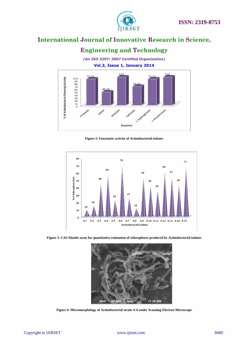
of activity against test bacterial pathogens. Screening for enzyme activity revealed that 100% of isolates showed
amylase and L-glutaminase activity, followed by 93.33% L-asparaginase and protease, 66.66% cellulase, and 46.66%
lipase activity. Detection of chemical nature of siderophore produced by all the actinobacterial strains revealed them as
carboxylates. Quantitative estimation of siderophores by CAS shuttle assay revealed the yield of 10% to 73%
siderophore units. Studies carried out in-vitro revealed that the antibacterial activity of crude siderophore obtained from
the potent actinobacterial strain A-6 was by way of chelation of iron resulting in its unavailability to the test bacterial
pathogens. Based on phenotypic characteristics and 16s rRNA gene sequence analysis the potent actinobacterial strain
A-6 has been identified as Streptomyces mutabilis. Phylogenetic tree was made by neighbor-joining method. The
present study is the first report on carboxylate type of siderophore production in actinobacteria isolated from Indian
magnesite mines. The in-vitro antagonistic action of siderophores of Streptomyces mutabilis has given a new focus of
attempts to exploit the siderophore systems for the treatment of human infections.
Key words: Magnesite mines, Siderophores, Antagonism, Streptomyces mutabilis
I. INTRODUCTION
Iron is an essential micronutrient for almost all organisms in the biosphere, including bacteria, fungi and plants.
It plays an essential role in respiration, nitrogen fixation, DNA and chlorophyll biosynthesis and other important
enzymatic systems [1]. Although iron is the second most abundant transition metal on earth, the solubility of iron is
very low at physiological pH as it forms insoluble stable complexes (10-17
M) of ferric oxyhydride (FeOOH), severely
limiting the bioavailability of iron [2]. To sequester and solubilise ferric iron many microorganisms utilize an efficient
system consisting of low molecular weight (< 1000 Da) compounds with high iron affinity, termed Siderophores [3,4,5].
Microbial siderophores are a structurally and architecturally diverse group of molecules and can be classified on the
basis of the chemical structure of the functional group that interacts with Fe (III) in to hydroxamates, catecholate,
carboxylates and mixed type [6, 7]. Besides microbial nutrition, siderophores also play a critical role in the expression
of virulence and development of biofilms by different microorganisms.
ISSN: 2319-8753
International Journal of Innovative Research in Science,
Engineering and Technology
(An ISO 3297: 2007 Certified Organization)
Vol.3, Issue 1, January 2014
Copyright to IJIRSET www.ijirset.com 8473
Siderophores and their substituted derivatives have a large number of applications in medical sciences. The
most important application is selective drug delivery, a Trojan horse strategy, to defeat drug resistant bacteria.
Microbial siderophores are also used in the treatment of malaria caused by Plasmodium falciparum, iron overload
diseases like hereditary haemochromatosis (HHC), sickle cell anaemia, thalassemia major and Friedreich’s Ataxia.
Furthermore, iron chelators such as dexrazoxane, O-trensox, desferriexochelins, desferrithiocin, tachpyridine, have
been found useful in cancer therapy [8].
Actinobacteria are a group of gram positive bacteria play a quite important role in natural ecological system
and they are also profile producers of siderophores, antibiotics, antitumor agents, therapeutic enzymes, enzyme
inhibitors, immune modifiers, antiparasitic agents, herbicides, insecticides and growth promoters [9]. Streptomyces is
the largest genus of Actinobacteria most commonly produce some type of trihydroxamate siderophore known as
desferrioxamine [10] but reports have described isolates capable of producing enterobactin [11] the characteristic
siderophore of Enterobacteriaceae, which was not excreted into the external environment but remained in the bacterial
biomass. Others have described the Streptomyces siderophores coelichelin [12] and griseobactin [13], while the novel
heterobactin siderophores have been found in Rhodococcus [14] and Nocardia [15].
Choice of natural materials like soil in researches is based on the assumption that samples from widely diverse
locations are more likely to yield novel microorganisms and therefore hopefully, novel metabolites as a result of the
geographical variation. The list of novel actinobacteria and products found in microbiologically poor explored areas of
India, China and Australia also suggests that a careful exploration of new habitats might continue to be useful
[16,17,18,19,20].
Presently, there is little documented information on the biodiversity of actinobacteria and its bioactive
potential in magnesite mines. With this view, the present study was focused to produce the siderophores by using
actinobacterium, isolated from magnesite mines habitat and to study the antagonistic potential of produced
siderophores.
II. MATERIALS AND METHODS
A. Sampling Strategies and Processing
About 500 gram of magnesite soil samples were collected during March 2010 at the depth of 20 cm from
eight stations of magnesite mines located in Chalk hills, Salem, South India (latitude 11.73º and longitude 78.13º) by
using sterile hand trowel. The distance of sampling stations from each other was 200 metre. The samples were
transported in sterile polyethylene bags to the laboratory within 1 hour of collection. Each sample was crushed,
thoroughly mixed and sieved through a 2 mm pore size mesh to get rid of large debris [21]. The sieved soils were used
for physico-chemical and microbiological analysis.
B. Physico-Chemical Analysis of Soil
Soil electrical conductivity (EC) and pH were determined using the saturation paste extract EC and saturation
paste pH methods [22]. Soil organic carbon was estimated using titration method of Walkley and Black [23]. Total
nitrogen content was determined following Kjeldahl method [24]. The available phosphorous content in soil samples
were estimated using chloro-stannous reduced molybdophosphoric blue colour method [25].
C. Pretreatment of Soil and Isolation of Actinobacteria
The magnesite soil samples were air dried for one week and kept at 45ºC for 1 hour to minimize bacterial
contaminants [26]. One gram of the soil sample was aseptically added to 9 ml of sterile buffer consisting of 0.5%
KH2PO4, 0.5% K2HPO4; pH 7.0 [27]. The suspension was vortexed and serially diluted up to 10-6
dilution. An aliquot
of 0.1 ml from suitable dilution was taken and spread on Starch casein agar medium supplemented with cyclohexamide
(100 mg/l) and nalidixic acid (20 mg/l) to inhibit the growth of fungi and bacteria respectively. The pH was adjusted to
8±0.2 prior to autoclaving. The agar plates were incubated at 25ºC to 28ºC under aerobic condition and the colonies
ISSN: 2319-8753
International Journal of Innovative Research in Science,
Engineering and Technology
(An ISO 3297: 2007 Certified Organization)
Vol.3, Issue 1, January 2014
Copyright to IJIRSET www.ijirset.com 8474
were observed for 30 days. Morphologically different actinobacteria were preserved both as slant culture in ISP-2
medium and as glycerol stock in 20% glycerol [28].
D. Screening of Actinobacteria for Siderophore Production by O-CAS Assay
Siderophores were detected by using the Overlaid Chrome Azurol Sulphonate (O-CAS) assay [29]. The
medium for a litre of overlay as follows: Chrome azurol S (CAS) 60.5 mg, hexadecyltrimethyl ammonium bromide
(HDTMA) 72.9 mg, Piperazine - 1,4-bis(2-ethanesulfonic acid) (PIPES) 30.24 g and 1mM FeCl3.6H2O in 10 mM HCl
10 ml. Agarose (0.9%) was used as gelling agent. 10 ml of CAS medium was applied over Iron free succinate agar
medium (grams/liter: K2HPO4 6.0; KH2PO4 3.0; MgSO4,7H2O 0.2; (NH4)2SO4 1.0; Succinic acid 4.0; Agar 20.0; pH
8±0.2) containing cultivated actinobacterial isolates to be tested for siderophore production. After a maximum period of
15 minutes, siderophore production was indicated by a change in colour from blue to purple/yellow/orange in the
overlaid medium.
E. Screening of Actinobacteria for Antibacterial and Enzymatic Activity
Actinobacterial isolates were grown on ISP-2 medium for 14 days. Agar cylinders (5 mm in diameter) were
then taken with hollow punch and deposited on the surface of Mueller-Hinton agar plates (Himedia, Mumbai)
previously seeded with the following bacterial pathogens: Staphylococcus aureus, Escherichia coli, Klebsiella
pneumoniae, Salmonella typhi, Salmonella paratyphi B, Shigella dysenteriae, Shigella sonnei and Proteus mirabilis.
Plates were incubated at 37ºC for 24 hours and the zone of inhibition around the plug were examined. Bacterial cultures
used in the present study were obtained from Gokulam hospitals (P) Ltd., Salem, India. Actinobacterial isolates were
also screened for protease, lipase, amylase, cellulase, L-asparaginase and L-glutaminase enzyme activity by spot
inoculation method [30].
F. Production of Siderophores
All the glass wares were soaked overnight in 6 M HCl to remove traces of iron present and rinsed 10 times
with distilled deionized water before use. Media used in the present study were also prepared with distilled deionized
water. Seed cultures were prepared by growing the actinobacterial isolates on Oat meal agar [31] at 28ºC for 14 days.
After incubation, the whole aerial mycelium were scrapped by a sterile inoculation loop and then suspended in 10 ml of
sterile distilled deionized water. Aliquots of 2.5 ml of the spore suspension were inoculated in to 500 ml Erlenmeyer
flasks containing 250ml of Iron free succinate media [32] consisting of grams/liter: K2HPO4, 6.0; KH2PO4, 3.0;
MgSO4.7H2O, 0.2; (NH4)2SO4, 1.0; Succinic acid, 4.0 and pH adjusted to 8.0 prior to sterilization. The flasks were then
incubated at 25ºC to 28ºC under aerobic condition on a rotary shaker (Remi, India) for 7 days. At the end of
fermentation, fermentation broths were centrifuged in a refrigerated centrifuge (Remi, India) at 6000 rpm and 4ºC for
15 minutes and the supernatant were subjected to qualitative and quantitative estimation of siderophores and detection
of chemical nature.
G. Estimation of Siderophores
Qualitatively siderophore production was detected by Chrome Azurol Sulphonate (CAS) assay [33]. The
change in colour from blue to orange after 2-4 minutes on addition of 1 ml of CAS solution to 1 ml of culture
supernatant indicated siderophore production. Quantitative estimation of siderophore was done by CAS shuttle assay
[34] in which 0.5 ml of cell free culture supernatant was mixed with 0.5 ml of CAS reagent and absorbance was
measured at 630 nm against a reference consisting of un inoculated Iron free succinate medium and 0.5 ml of CAS
reagent. Siderophore produced was calculated by using following formula,
% Siderophore units = [(Ar - As) / Ar] x 100
Where, Ar is the absorbance of reference and As is the absorbance of sample at 630 nm.
ISSN: 2319-8753
International Journal of Innovative Research in Science,
Engineering and Technology
(An ISO 3297: 2007 Certified Organization)
Vol.3, Issue 1, January 2014
Copyright to IJIRSET www.ijirset.com 8475
H. Determination of Functional Groups of siderophores
The chemical nature of siderophore produced was examined for catecholate nature by FeCl3 test [1] and
Arnow’s test [35] and hydroxamate nature by FeCl3 [1], while carboxylate nature was determined by
Spectrophotometric test [36].
I. Determination of Antagonistic Potential of Siderophore by Agar Well Diffusion Method
The cell free culture supernatant of iron free succinate medium inoculated with Actinobacterial strain A-6 was
subjected to bioassay against bacterial pathogens by agar well diffusion method. Wells of 8 mm diameter were punched
in Mueller-Hinton agar plates seeded with test organisms. Then, 100 µl of crude siderophore was added in these wells.
Control wells contained sterile saline. Plates were incubated at 4°C for three hours, followed by 24 hours of incubation
at 37°C. The same experiment was repeated using Mueller-Hinton agar incorporated with 10 to 200 mM FeCl3. The
zone of inhibition in millimetre diameter were read and taken as the activity against the test pathogens.
J. Taxonomy of Potential Actinobacterial Strain
The potential Actinobacterial strain A-6 was identified based on the following characteristics, viz. aerial mass
colour, melanoid pigment, reverse side pigment, spore chain morphology and assimilation of carbon sources, and also
by comparing the characteristics with the keys given by Nonomura [37]. The chromosomal DNA of actinobacterial
strain A-6 was extracted using GenElute Bacterial Genomic DNA kit (Sigma). The 16s rDNA fragment was amplified
by using PCR with Taq DNA polymerase and primers 27f (51 AGTTTGATCCTGGCTCAG 3
1) and 1492r
(51ACGGCTACCTTGTTACGACTT 3
1). The conditions for thermal cycling were as follows: denaturation of the
target DNA at 94°C for 4 minutes followed by 30 cycles at 94°C for 1 minute, primer annealing at 52°C for 1 minute
and primer extension at 72°C for 1 minute. At the end of the cycling, the reaction mixture was held at 72°C for 10
minutes and then cooled to 4°C. PCR amplification was detected by agarose gel electrophoresis and was visualized by
UV fluorescence after ethidium bromide staining [38].
The PCR product obtained was sequenced by an automated sequencer (Genetic Analyzer 3130, Applied
Biosystem, USA). The same primers as above were used for this purpose. The sequence was compared for similarity
with the reference species of bacteria contained in genomic database banks, using the NCBI BLAST available at
http://www.ncbi-nlm-nih.gov/. A phylogenetic tree was constructed with the Molecular Evolutionary Genetics Analysis
(MEGA) software version 5 using the Neighbor-Joining algorithm [39, 40]. Tree topologies were evaluated by
bootstrap analysis based on 1000 resamplings of the neighbour joining data set [41]. The evolutionary distances were
computed using the Maximum Composite Likelihood method [42].
III. RESULTS AND DISCUSSION
A. Physico- Chemical and Microbiological Characteristics of Magnesite Mines Soil
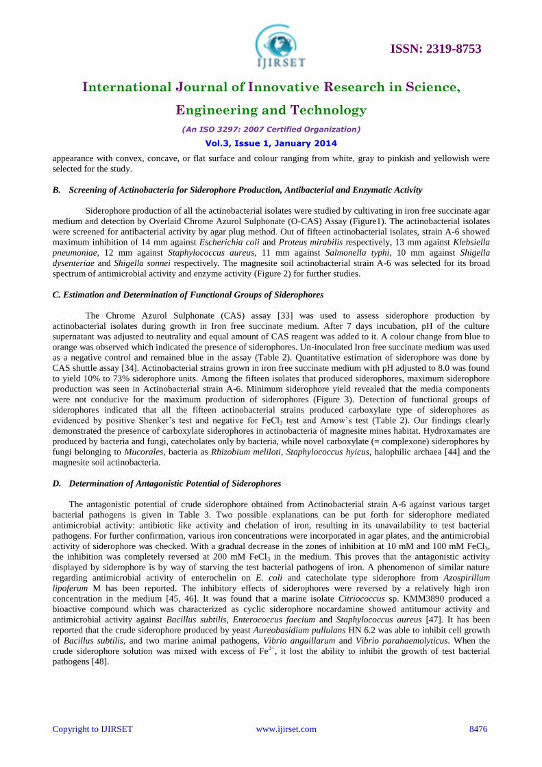
Physical, chemical and microbiological characteristics of magnesite mines soil have been represented in Table
1. Soil sample collected from station-1 was found to have sandy clay loam texture, whereas soil of the other stations is
of sandy loam texture. Soil EC, pH, N, P and K differed remarkably among eight different sampling stations. Physical
and chemical analysis of the soil clearly indicates that the nutrients, pH, temperature and humidity were different from
the normal ecosystem. Isolation of Actinobacteria has always been faced with difficulties in comparison to their
competitors like other bacteria and fungi [43]. However, the recovery rate of Actinobacteria was increased by pre-
treatment of the samples by air drying for 1 week and incubating at 45ºC for 1 hour. Use of Starch casein agar medium
supplemented with cyclohexamide (100 mg/l) and nalidixic acid (20 mg/l) was crucial in inhibiting contaminating
fungi and bacteria. Total actinobacterial count of magnesite soil was in the range of 3.0 - 16 x 103 cfu/g. The survival of
low number of actinobacteria in such harsh and challenging habitation might be due to their adaptation to the extreme
environment. A total of 15 different actinobacterial colonies having characteristic features such as leathery, powdery
ISSN: 2319-8753
International Journal of Innovative Research in Science,
Engineering and Technology
(An ISO 3297: 2007 Certified Organization)
Vol.3, Issue 1, January 2014
Copyright to IJIRSET www.ijirset.com 8476
appearance with convex, concave, or flat surface and colour ranging from white, gray to pinkish and yellowish were
selected for the study.
B. Screening of Actinobacteria for Siderophore Production, Antibacterial and Enzymatic Activity
Siderophore production of all the actinobacterial isolates were studied by cultivating in iron free succinate agar
medium and detection by Overlaid Chrome Azurol Sulphonate (O-CAS) Assay (Figure1). The actinobacterial isolates
were screened for antibacterial activity by agar plug method. Out of fifteen actinobacterial isolates, strain A-6 showed
maximum inhibition of 14 mm against Escherichia coli and Proteus mirabilis respectively, 13 mm against Klebsiella
pneumoniae, 12 mm against Staphylococcus aureus, 11 mm against Salmonella typhi, 10 mm against Shigella
dysenteriae and Shigella sonnei respectively. The magnesite soil actinobacterial strain A-6 was selected for its broad
spectrum of antimicrobial activity and enzyme activity (Figure 2) for further studies.
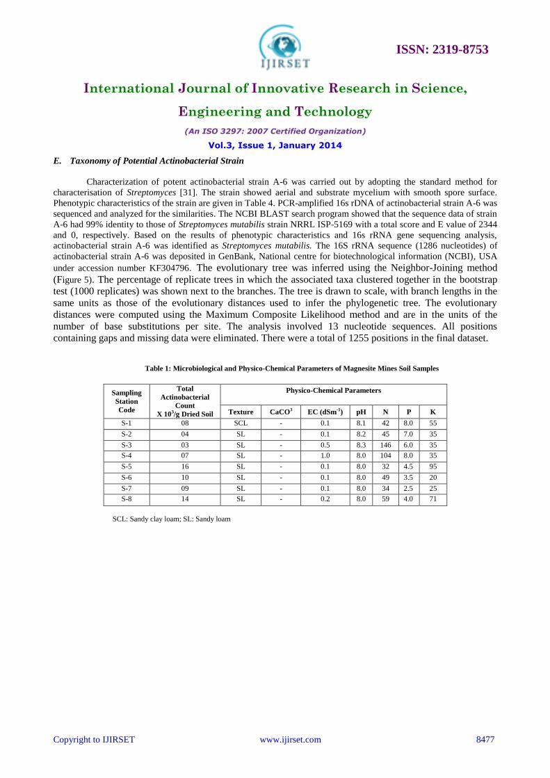
C. Estimation and Determination of Functional Groups of Siderophores
The Chrome Azurol Sulphonate (CAS) assay [33] was used to assess siderophore production by
actinobacterial isolates during growth in Iron free succinate medium. After 7 days incubation, pH of the culture
supernatant was adjusted to neutrality and equal amount of CAS reagent was added to it. A colour change from blue to
orange was observed which indicated the presence of siderophores. Un-inoculated Iron free succinate medium was used
as a negative control and remained blue in the assay (Table 2). Quantitative estimation of siderophore was done by
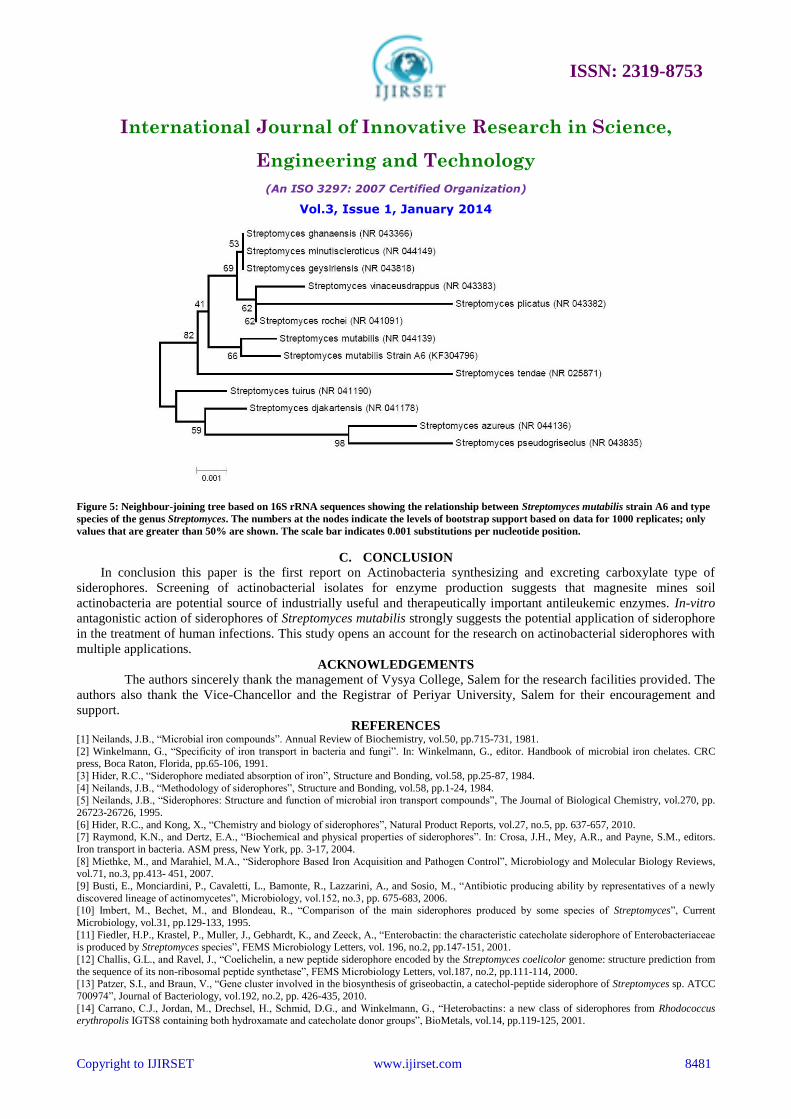
CAS shuttle assay [34]. Actinobacterial strains grown in iron free succinate medium with pH adjusted to 8.0 was found
to yield 10% to 73% siderophore units. Among the fifteen isolates that produced siderophores, maximum siderophore
production was seen in Actinobacterial strain A-6. Minimum siderophore yield revealed that the media components
were not conducive for the maximum production of siderophores (Figure 3). Detection of functional groups of
siderophores indicated that all the fifteen actinobacterial strains produced carboxylate type of siderophores as
evidenced by positive Shenker’s test and negative for FeCl3 test and Arnow’s test (Table 2). Our findings clearly
demonstrated the presence of carboxylate siderophores in actinobacteria of magnesite mines habitat. Hydroxamates are
produced by bacteria and fungi, catecholates only by bacteria, while novel carboxylate (= complexone) siderophores by
fungi belonging to Mucorales, bacteria as Rhizobium meliloti, Staphylococcus hyicus, halophilic archaea [44] and the
magnesite soil actinobacteria.
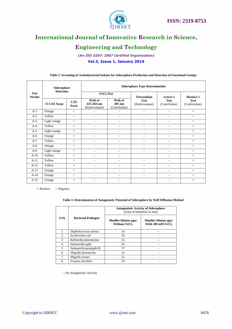
D. Determination of Antagonistic Potential of Siderophores
The antagonistic potential of crude siderophore obtained from Actinobacterial strain A-6 against various target
bacterial pathogens is given in Table 3. Two possible explanations can be put forth for siderophore mediated
antimicrobial activity: antibiotic like activity and chelation of iron, resulting in its unavailability to test bacterial
pathogens. For further confirmation, various iron concentrations were incorporated in agar plates, and the antimicrobial
activity of siderophore was checked. With a gradual decrease in the zones of inhibition at 10 mM and 100 mM FeCl3,
the inhibition was completely reversed at 200 mM FeCl3 in the medium. This proves that the antagonistic activity
displayed by siderophore is by way of starving the test bacterial pathogens of iron. A phenomenon of similar nature
regarding antimicrobial activity of enterochelin on E. coli and catecholate type siderophore from Azospirillum
lipoferum M has been reported. The inhibitory effects of siderophores were reversed by a relatively high iron
concentration in the medium [45, 46]. It was found that a marine isolate Citriococcus sp. KMM3890 produced a
bioactive compound which was characterized as cyclic siderophore nocardamine showed antitumour activity and
antimicrobial activity against Bacillus subtilis, Enterococcus faecium and Staphylococcus aureus [47]. It has been
reported that the crude siderophore produced by yeast Aureobasidium pullulans HN 6.2 was able to inhibit cell growth
of Bacillus subtilis, and two marine animal pathogens, Vibrio anguillarum and Vibrio parahaemolyticus. When the
crude siderophore solution was mixed with excess of Fe3+
, it lost the ability to inhibit the growth of test bacterial
pathogens [48].
ISSN: 2319-8753
International Journal of Innovative Research in Science,
Engineering and Technology
(An ISO 3297: 2007 Certified Organization)
Vol.3, Issue 1, January 2014
Copyright to IJIRSET www.ijirset.com 8477
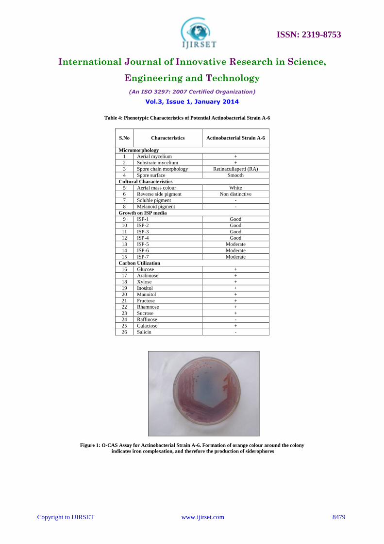
E. Taxonomy of Potential Actinobacterial Strain
Characterization of potent actinobacterial strain A-6 was carried out by adopting the standard method for
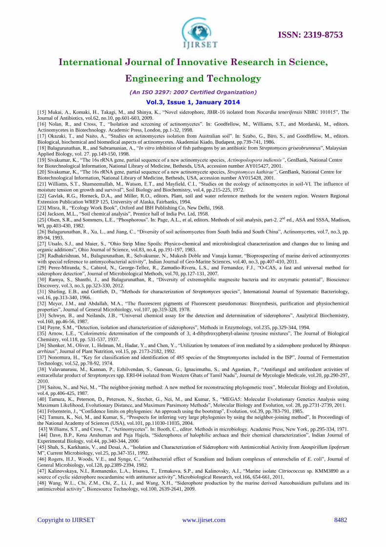
characterisation of Streptomyces [31]. The strain showed aerial and substrate mycelium with smooth spore surface.
Phenotypic characteristics of the strain are given in Table 4. PCR-amplified 16s rDNA of actinobacterial strain A-6 was
sequenced and analyzed for the similarities. The NCBI BLAST search program showed that the sequence data of strain
A-6 had 99% identity to those of Streptomyces mutabilis strain NRRL ISP-5169 with a total score and E value of 2344
and 0, respectively. Based on the results of phenotypic characteristics and 16s rRNA gene sequencing analysis,
actinobacterial strain A-6 was identified as Streptomyces mutabilis. The 16S rRNA sequence (1286 nucleotides) of
actinobacterial strain A-6 was deposited in GenBank, National centre for biotechnological information (NCBI), USA
under accession number KF304796. The evolutionary tree was inferred using the Neighbor-Joining method
(Figure 5). The percentage of replicate trees in which the associated taxa clustered together in the bootstrap
test (1000 replicates) was shown next to the branches. The tree is drawn to scale, with branch lengths in the
same units as those of the evolutionary distances used to infer the phylogenetic tree. The evolutionary
distances were computed using the Maximum Composite Likelihood method and are in the units of the
number of base substitutions per site. The analysis involved 13 nucleotide sequences. All positions
containing gaps and missing data were eliminated. There were a total of 1255 positions in the final dataset.
Table 1: Microbiological and Physico-Chemical Parameters of Magnesite Mines Soil Samples
Sampling
Station
Code
Total
Actinobacterial
Count
X 103/g Dried Soil
Physico-Chemical Parameters
Texture CaCO3 EC (dSm-1) pH N P K
S-1 08 SCL - 0.1 8.1 42 8.0 55
S-2 04 SL - 0.1 8.2 45 7.0 35
S-3 03 SL - 0.5 8.3 146 6.0 35
S-4 07 SL - 1.0 8.0 104 8.0 35
S-5 16 SL - 0.1 8.0 32 4.5 95
S-6 10 SL - 0.1 8.0 49 3.5 20
S-7 09 SL - 0.1 8.0 34 2.5 25
S-8 14 SL - 0.2 8.0 59 4.0 71
SCL: Sandy clay loam; SL: Sandy loam
ISSN: 2319-8753
International Journal of Innovative Research in Science,
Engineering and Technology
(An ISO 3297: 2007 Certified Organization)
Vol.3, Issue 1, January 2014
Copyright to IJIRSET www.ijirset.com 8478
Table 2: Screening of Actinobacterial Isolates for Siderophore Production and Detection of Functional Groups
Test
Strains
Siderophore
Detection
Siderophore Type Determination
FeCl3 Test Tetrazolium
Test
(Hydroxamate)
Arnow’s
Test
(Catecholate)
Shenker’s
Test
(Carboxylate) O-CAS Assay CAS
Assay
Peak at
425-450 nm
(Hydroxamate)
Peak at
495 nm
(Catecholate)
A-1 Orange + - - - - +
A-2 Yellow + - - - - +
A-3 Light orange + - - - - +
A-4 Yellow + - - - - +
A-5 Light orange + - - - - +
A-6 Orange + - - - - +
A-7 Yellow + - - - - +
A-8 Orange + - - - - +
A-9 Light orange + - - - - +
A-10 Yellow + - - - - +
A-11 Yellow + - - - - +
A-12 Yellow + - - - - +
A-13 Orange + - - - - +
A-14 Orange + - - - - +
A-15 Orange + - - - - +
+: Positive -: Negative
Table 3: Determination of Antagonistic Potential of Siderophore by Well Diffusion Method
S.No Bacterial Pathogen
Antagonistic Activity of Siderophore
(Zone of Inhibition in mm)
Mueller-Hinton agar
Without FeCl3
Mueller-Hinton agar
With 200 mM FeCl3
1 Staphylococcus aureus 14 -
2 Escherichia coli 19 -
3 Klebsiella pneumoniae 15 -
4 Salmonella typhi 16 -
5 Salmonella paratyphi B 17 -
6 Shigella dysenteriae 14 -
7 Shigella sonnei 15 -
8 Proteus mirabilis 18 -
- : No Antagonistic Activity
ISSN: 2319-8753
International Journal of Innovative Research in Science,
Engineering and Technology
(An ISO 3297: 2007 Certified Organization)
Vol.3, Issue 1, January 2014
Copyright to IJIRSET www.ijirset.com 8479
Table 4: Phenotypic Characteristics of Potential Actinobacterial Strain A-6
S.No Characteristics
Actinobacterial Strain A-6
Micromorphology
1 Aerial mycelium +
2 Substrate mycelium +
3 Spore chain morphology Retinaculiaperti (RA)
4 Spore surface Smooth
Cultural Characteristics
5 Aerial mass colour White
6 Reverse side pigment Non distinctive
7 Soluble pigment -
8 Melanoid pigment -
Growth on ISP media
9 ISP-1 Good
10 ISP-2 Good
11 ISP-3 Good
12 ISP-4 Good
13 ISP-5 Moderate
14 ISP-6 Moderate
15 ISP-7 Moderate
Carbon Utilization
16 Glucose +
17 Arabinose +
18 Xylose +
19 Inositol +
20 Mannitol +
21 Fructose +
22 Rhamnose +
23 Sucrose +
24 Raffinose -
25 Galactose +
26 Salicin -
Figure 1: O-CAS Assay for Actinobacterial Strain A-6. Formation of orange colour around the colony
indicates iron complexation, and therefore the production of siderophores
ISSN: 2319-8753
International Journal of Innovative Research in Science,
Engineering and Technology
(An ISO 3297: 2007 Certified Organization)
Vol.3, Issue 1, January 2014
Copyright to IJIRSET www.ijirset.com 8480
Figure 2: Enzymatic activity of Actinobacterial isolates
Figure 3: CAS-Shuttle assay for quantitative estimation of siderophores produced by Actinobacterial isolates
Figure 4: Micromorphology of Actinobacterial strain A-6 under Scanning Electron Microscope
ISSN: 2319-8753
International Journal of Innovative Research in Science,
Engineering and Technology
(An ISO 3297: 2007 Certified Organization)
Vol.3, Issue 1, January 2014
Copyright to IJIRSET www.ijirset.com 8481
Figure 5: Neighbour-joining tree based on 16S rRNA sequences showing the relationship between Streptomyces mutabilis strain A6 and type
species of the genus Streptomyces. The numbers at the nodes indicate the levels of bootstrap support based on data for 1000 replicates; only
values that are greater than 50% are shown. The scale bar indicates 0.001 substitutions per nucleotide position.
C. CONCLUSION
In conclusion this paper is the first report on Actinobacteria synthesizing and excreting carboxylate type of
siderophores. Screening of actinobacterial isolates for enzyme production suggests that magnesite mines soil
actinobacteria are potential source of industrially useful and therapeutically important antileukemic enzymes. In-vitro
antagonistic action of siderophores of Streptomyces mutabilis strongly suggests the potential application of siderophore
in the treatment of human infections. This study opens an account for the research on actinobacterial siderophores with
multiple applications.
ACKNOWLEDGEMENTS
The authors sincerely thank the management of Vysya College, Salem for the research facilities provided. The
authors also thank the Vice-Chancellor and the Registrar of Periyar University, Salem for their encouragement and
support.
REFERENCES [1] Neilands, J.B., “Microbial iron compounds”. Annual Review of Biochemistry, vol.50, pp.715-731, 1981.
[2] Winkelmann, G., “Specificity of iron transport in bacteria and fungi”. In: Winkelmann, G., editor. Handbook of microbial iron chelates. CRC press, Boca Raton, Florida, pp.65-106, 1991.
[3] Hider, R.C., “Siderophore mediated absorption of iron”, Structure and Bonding, vol.58, pp.25-87, 1984.
[4] Neilands, J.B., “Methodology of siderophores”, Structure and Bonding, vol.58, pp.1-24, 1984. [5] Neilands, J.B., “Siderophores: Structure and function of microbial iron transport compounds”, The Journal of Biological Chemistry, vol.270, pp.
26723-26726, 1995.
[6] Hider, R.C., and Kong, X., “Chemistry and biology of siderophores”, Natural Product Reports, vol.27, no.5, pp. 637-657, 2010. [7] Raymond, K.N., and Dertz, E.A., “Biochemical and physical properties of siderophores”. In: Crosa, J.H., Mey, A.R., and Payne, S.M., editors.
Iron transport in bacteria. ASM press, New York, pp. 3-17, 2004.
[8] Miethke, M., and Marahiel, M.A., “Siderophore Based Iron Acquisition and Pathogen Control”, Microbiology and Molecular Biology Reviews, vol.71, no.3, pp.413- 451, 2007.
[9] Busti, E., Monciardini, P., Cavaletti, L., Bamonte, R., Lazzarini, A., and Sosio, M., “Antibiotic producing ability by representatives of a newly
discovered lineage of actinomycetes”, Microbiology, vol.152, no.3, pp. 675-683, 2006. [10] Imbert, M., Bechet, M., and Blondeau, R., “Comparison of the main siderophores produced by some species of Streptomyces”, Current
Microbiology, vol.31, pp.129-133, 1995.
[11] Fiedler, H.P., Krastel, P., Muller, J., Gebhardt, K., and Zeeck, A., “Enterobactin: the characteristic catecholate siderophore of Enterobacteriaceae is produced by Streptomyces species”, FEMS Microbiology Letters, vol. 196, no.2, pp.147-151, 2001.
[12] Challis, G.L., and Ravel, J., “Coelichelin, a new peptide siderophore encoded by the Streptomyces coelicolor genome: structure prediction from
the sequence of its non-ribosomal peptide synthetase”, FEMS Microbiology Letters, vol.187, no.2, pp.111-114, 2000. [13] Patzer, S.I., and Braun, V., “Gene cluster involved in the biosynthesis of griseobactin, a catechol-peptide siderophore of Streptomyces sp. ATCC
700974”, Journal of Bacteriology, vol.192, no.2, pp. 426-435, 2010.
[14] Carrano, C.J., Jordan, M., Drechsel, H., Schmid, D.G., and Winkelmann, G., “Heterobactins: a new class of siderophores from Rhodococcus erythropolis IGTS8 containing both hydroxamate and catecholate donor groups”, BioMetals, vol.14, pp.119-125, 2001.
ISSN: 2319-8753
International Journal of Innovative Research in Science,
Engineering and Technology
(An ISO 3297: 2007 Certified Organization)
Vol.3, Issue 1, January 2014
Copyright to IJIRSET www.ijirset.com 8482
[15] Mukai, A., Komaki, H., Takagi, M., and Shinya, K., “Novel siderophore, JBIR-16 isolated from Nocardia tenerifensis NBRC 101015”, The Journal of Antibiotics, vol.62, no.10, pp.601-603, 2009.
[16] Nolan, R., and Cross, T., “Isolation and screening of actinomycetes”. In: Goodfellow, M., Williams, S.T., and Mordarski, M., editors.
Actinomycetes in Biotechnology. Academic Press, London, pp.1-32, 1998. [17] Okazaki, T., and Naito, A., “Studies on actinomycetes isolation from Australian soil”. In: Szabo, G., Biro, S., and Goodfellow, M., editors.
Biological, biochemical and biomedical aspects of actinomycetes. Akademiai Kiado, Budapest, pp.739-741, 1986.
[18] Balagurunathan, R., and Subramanian, A., “In vitro inhibition of fish pathogens by an antibiotic from Streptomyces griseobrunneus”, Malaysian
Applied Biology, vol. 27. pp.149-150, 1998.
[19] Sivakumar, K., “The 16s rRNA gene, partial sequence of a new actinomycete species, Actinopolospora indiensis”, GenBank, National Centre
for Biotechnological Information, National Library of Medicine, Bethesds, USA, accession number AY015427, 2001. [20] Sivakumar, K., “The 16s rRNA gene, partial sequence of a new actinomycete species, Streptomyces kathirae”, GenBank, National Centre for
Biotechnological Information, National Library of Medicine, Bethesds, USA, accession number AY015428, 2001.
[21] Williams, S.T., Shameemullah, M., Watson, E.T., and Mayfield, C.I., “Studies on the ecology of actinomycetes in soil-VI. The influence of moisture tension on growth and survival”, Soil Biology and Biochemistry, vol.4, pp.215-225, 1972.
[22] Gavlak, R.G., Horneck, D.A., and Miller, R.O., editors. Plant, soil and water reference methods for the western region. Western Regional
Extension Publication WREP 125, University of Alaska, Fairbanks, 1994. [23] Misra, R., “Ecology Work Book”, Oxford and IBH Publishing Co, New Delhi, 1968.
[24] Jackson, M.L., “Soil chemical analysis”, Prentice hall of India Pvt. Ltd, 1958.
[25] Olsen, S.R., and Sommers, L.E., “Phosphorous”. In: Page, A.L., et al, editors. Methods of soil analysis, part-2. 2nd ed., ASA and SSSA, Madison, WI, pp.403-430, 1982.
[26] Balagurunathan, R., Xu, L., and Jiang, C., “Diversity of soil actinomycetes from South India and South China”, Actinomycetes, vol.7, no.3, pp.
89-94, 1993. [27] Utsalo, S.J., and Maier, S., “Ohio Strip Mine Spoils: Physico-chemical and microbiological characterization and changes due to liming and
organic additions”, Ohio Journal of Science, vol.83, no.4, pp.191-197, 1983.
[28] Radhakrishnan, M., Balagurunathan, R., Selvakumar, N., Mukesh Doble and Vanaja kumar, “Bioprospecting of marine derived actinomycetes with special reference to antimycobacterial activity”, Indian Journal of Geo-Marine Sciences, vol.40, no.3, pp.407-410, 2011.
[29] Perez-Miranda, S., Cabirol, N., George-Tellez, R., Zamudio-Rivera, L.S., and Fernandez, F.J., “O-CAS, a fast and universal method for
siderophore detection”, Journal of Microbiological Methods, vol.70, pp.127-131, 2007. [30] Ramya, S., Shanthi, J., and Balagurunathan, R., “Diversity of extremophilic magnesite bacteria and its enzymatic potential”, Bioscience
Discovery, vol.3, no.3, pp.323-330, 2012.
[31] Shirling, E.B., and Gottlieb, D., “Methods for characterization of Streptomyces species”, International Journal of Systematic Bacteriology, vol.16, pp.313-340, 1966.
[32] Meyer, J.M., and Abdallah, M.A., “The fluorescent pigments of Fluorescent pseudomonas: Biosynthesis, purification and physiochemical
properties”, Journal of General Microbiology, vol.107, pp.319-328, 1978. [33] Schwyn, B., and Neilands, J.B., “Universal chemical assay for the detection and determination of siderophores”, Analytical Biochemistry,
vol.160, pp.46-56, 1987.
[34] Payne, S.M., “Detection, isolation and characterization of siderophores”, Methods in Enzymology, vol.235, pp.329-344, 1994. [35] Arnow, L.E., “Colorimetric determination of the compounds of 3, 4-dihydroxyphenyl-alanine tyrosine mixtures”, The Journal of Biological
Chemistry, vol.118, pp. 531-537, 1937. [36] Shenker, M., Oliver, I., Helman, M., Hadar, Y., and Chen, Y., “Utilization by tomatoes of iron mediated by a siderophore produced by Rhizopus
arrhizus”, Journal of Plant Nutrition, vol.15, pp. 2173-2182, 1992.
[37] Nonomura, H., “Key for classification and identification of 485 species of the Streptomycetes included in the ISP”, Journal of Fermentation Technology, vol.52, pp.78-92, 1974.
[38] Valavanarasu, M., Kannan, P., Ezhilvendan, S., Ganesan, G., Ignacimuthu, S., and Agastian, P., “Antifungal and antifeedant activities of
extracellular product of Streptomyces spp. ERI-04 isolated from Western Ghats of Tamil Nadu”, Journal de Mycologie Medicale, vol.20, pp.290-297, 2010.
[39] Saitou, N., and Nei, M., “The neighbor-joining method: A new method for reconstructing phylogenetic trees”, Molecular Biology and Evolution,
vol.4, pp.406-425, 1987. [40] Tamura, K., Peterson, D., Peterson, N., Stecher, G., Nei, M., and Kumar, S., “MEGA5: Molecular Evolutionary Genetics Analysis using
Maximum Likelihood, Evolutionary Distance, and Maximum Parsimony Methods”, Molecular Biology and Evolution, vol. 28, pp.2731-2739, 2011.
[41] Felsenstein, J., “Confidence limits on phylogenies: An approach using the bootstrap”, Evolution, vol.39, pp.783-791, 1985. [42] Tamura, K., Nei, M., and Kumar, S., “Prospects for inferring very large phylogenies by using the neighbor-joining method”, In Proceedings of
the National Academy of Sciences (USA), vol.101, pp.11030-11035, 2004.
[43] Williams, S.T., and Cross, T., “Actinomycetes”. In: Booth, C., editor. Methods in microbiology. Academic Press, New York, pp.295-334, 1971. [44] Dave, B.P., Kena Anshuman and Puja Hajela, “Siderophores of halophilic archaea and their chemical characterization”, Indian Journal of
Experimental Biology, vol.44, pp.340-344, 2006
[45] Shah, S., Karkhanis, V., and Desai, A., “Isolation and Characterization of Siderophore with Antimicrobial Activity from Azospirillum lipoferum M”, Current Microbiology, vol.25, pp.347-351, 1992.
[46] Rogers, H.J., Woods, V.E., and Synge, C., “Antibacterial effect of Scandium and Indium complexes of enterochelin of E. coli”, Journal of
General Microbiology, vol.128, pp.2389-2394, 1982. [47] Kalinovskaya, N.I., Romanenko, L.A., Irisawa, T., Ermakova, S.P., and Kalinovsky, A.I., “Marine isolate Citriococcus sp. KMM3890 as a
source of cyclic siderophore nocardamine with antitumor activity”, Microbiological Research, vol.166, 654-661, 2011.
[48] Wang, W.L., Chi, Z.M., Chi, Z., Li, J., and Wang, X.H., “Siderophore production by the marine derived Aureobasidium pullulans and its antimicrobial activity”, Bioresource Technology, vol.100, 2639-2641, 2009.

![Inhibition of Siderophore Biosynthesis by 2Triazole Substituted Analogues of 5′- O -[ N -(Salicyl)sulfamoyl]adenosine: Antibacterial Nucleosides Effective against Mycobacterium tuberculosis](https://img.dokumen.tips/doc/110x75/631e85df0ff042c6110c64c7/inhibition-of-siderophore-biosynthesis-by-2triazole-substituted-analogues-of-5-.jpg)


