Embed Size (px)
Citation preview
A Novel Entomopathogenic ActinobacteriaMediated Silver Nanoparticles Characterization andTheir Insecticidal, Antibacterial and CytotoxicActivitiesKrishnan Raguvaran
Periyar UniversityManickam Kalpana
Periyar UniversityThulasiraman Manimegalai
Periyar UniversityRengasamy Balakrishnan
Konkuk UniversityRajan Maheswaran ( [email protected] )
Periyar University https://orcid.org/0000-0002-6321-7173
Research Article
Keywords: Actinokineospora fastidiosa, silver nanoparticle, optimization, larvicidal, antimicrobial, non-target organisms
Posted Date: April 5th, 2021
DOI: https://doi.org/10.21203/rs.3.rs-316265/v1
License: This work is licensed under a Creative Commons Attribution 4.0 International License. Read Full License
1
A NOVEL ENTOMOPATHOGENIC ACTINOBACTERIA MEDIATED SILVER 1
NANOPARTICLES CHARACTERIZATION AND THEIR INSECTICIDAL, 2
ANTIBACTERIAL AND CYTOTOXIC ACTIVITIES 3
Krishnan Raguvaran1, Manickam Kalpana1, Thulasiraman Manimegalai1, Rengasamy 4
Balakrishnan2, Rajan Maheswaran 1 5
1. Entomology Laboratory, Department of Zoology, School of Life Sciences, Periyar 6
University, Periyar Palkalai Nagar, Salem – 636 011, Tamil Nadu, India. 7
2. Department of Applied Life Sciences and Integrated Bioscience, Graduate School, 8
Konkuk University, Chungju 27478, Republic of Korea. 9
Abstract 10
Nanomaterials were highly inspired in the field of nanotechnology especially bio 11
synthesized silver nanoparticles (AgNPs) have effectively attracted worldwide that helps to 12
improve in the field of medical science and human health. Actinabacteria mediated 13
nanoparticle were widely used to control insect pests, treatment of antibiotic resistant 14
bacteria, cancer and other diseases due to their potential pharmacological properties 15
compared with other chemical drugs. The present investigation an entomopathogenic 16
actinobacteria Actinokineospora fastidiosa was isolated and synthesis of AgNPs and tested 17
their mosquito larvicidal, antibacterial and anticancer activity. Moreover, the outcome of 18
biosynthesized AgNPs was characterized by UV-visible spectroscopy, X-ray Diffraction 19
(XRD), Fourier transforms infrared spectroscopy (FT-IR), Transmission electron microscope 20
(TEM), scanning electron microscope (SEM) with energy dispersive X-ray spectroscopy 21
Corresponding author
E-mail: [email protected] Tel. No. +919443323186
2
(EDX) and zeta potential analysis. The characterized AgNPs showed a potent larvicidal 22
activity against Aedes aegypti, Anopheles stephensi and Culex quinquefasciatus the obtained 23
LC50 values is 9.86 ppm, 8.50 ppm and 8.29 ppm respectively. However, at 25 ppm 24
concentration, AgNPs showed highest ovicidal activity (100 %, 100 % and 97.77 % against 25
A. aegypti, A. stephensi and C. quinquefasciatus respectively) and oviposition deterrence 26
activity was 100 %, 100 % and 98.66 % were achieved against A. aegypti, A. stephensi and 27
C. quinquefasciatus respectively. The enzymes of α, β esterase and Glutathione-S-transferase 28
level were increased due to detoxification process against AgNPs. The histopathological 29
results of AgNPs showed series damage in epithelial cells and brush border cell of mosquito 30
larval gut. In addition, actinobacterial mediated AgNPs was tested against different human 31
pathogenic bacteria. The obtained results showed biosynthesized AgNPs revealed remarkable 32
antibacterial activity against all tested pathogens. The lowest mortality was observed on non-33
target organisms of Artemia salina at highest concentration of AgNPs. Finally, in vitro 34
toxicity study showed that biosynthesized AgNPs potential anticancer activity and induced 35
reactive oxygen species (ROS) production in human cervical cancer (HeLa) cell line at an 36
inhibitory concentration (IC50) range 42.37 μg/mL. Therefore, it can be concluded that 37
biosynthesized AgNPs using a novel stains Actinokineospora fastidiosa potentially 38
influenced the vector mosquito and bacterial diseases management and pharmacological 39
applications in eco-friendly manner. 40
Keywords: Actinokineospora fastidiosa, silver nanoparticle, optimization, larvicidal, 41
antimicrobial, non-target organisms 42
1. Introduction 43
Recently, World health organization (WHO) reported that about 2100 million of 44
peoples are at risk of vector bore disease such as malaria, filariasis, Japanese encephalitis, 45
dengue fever, chikungunya, yellow fever and etc., which affects the current civilization in 46
3
terms of economic loss, morbidity and mortality (Ali and Venkatesalu 2020). The major 47
vector mosquitoes of A. aegypti, A. stephensi and C. quinquefasciatus were important carriers 48
of transmitting many deadly diseases and nuisance to public health (Balaraju et al. 2009) 49
(Sukumaran and Maheswaran 2020). A. stephensi is the most important vector of malaria 50
fever in India and other West Asian countries. Malaria alone affects 36% of the people in 51
worldwide i.e. 2020 million in 107 countries and territories of tropical and subtropical 52
regions (Ali and Venkatesalu 2020). In recent years 1.4 billion peoples in 73 countries were 53
life threatened by lymphatic filariasis, formerly named as elephantiasis caused by C. 54
quinquefasciatus. In other hand 120 million peoples are infected with about 40 million 55
disfigured and incapacitated by mosquito vector disease (Kalaimurugan et al. 2019). A. 56
aegypti is the major viral vector for Chikungunya, Dengue, Dengue hemorrhagic fever, 57
Yellow fever, Zika virus and etc. Nearly, 2500 million peoples were at risk of dengue viral 58
infection i.e., two-fifth of the world’s population and there may be 50 million cases were 59
reported in dengue fever in every year (Moulin et al. 2016). These are the thought-provoking 60
issues for public health in worldwide, and causes social and economic impact on worldwide 61
(Benelli and Mehlhorn 2016). Vector-borne diseases were tenacious due to resistant in 62
mosquitoes since the excessive application of chemical insecticides and pesticides (Enayati et 63
al. 2003) and lack of effective vaccines (Kumar et al. 2011). In modern years, frequent usage 64
of synthetic chemical insecticides for controlling vector mosquitoes has disrupted natural 65
systems and causes to resurgence in mosquito populations leading to major outbreaks of 66
mosquito borne diseases. Variety of control measures like physical, chemical and biological 67
methods were available to controlling mosquitoes. Bio control programs were an alternate 68
way to stabilize the life frightening insects in an ecofriendly approach. Hence, larvicides play 69
an important role in combating mosquitoes from their breeding sites (Maheswaran and 70
Ignacimuthu, 2013). 71
4
The wide range of biological resources were available for biosynthesis of 72
nanoparticles using bacteria, fungi and plants (Nadagouda et al. 2009). In this aspect 73
actinobacterium were more suitable for the production of nanometals and well known their 74
supreme ability for the production of many active molecules with several biological 75
applications (Manivasagan et al. 2013b). Streptomyces sp. was considered more significant 76
due to the approximate production of 50-55 % of antibiotics by itself (Manivasagan et al. 77
2014). Earlier reports of nanoparticle synthesis using Streptomyces viridogens 78
(Balagurunathan et al. 2011), Streptomyces naganishii (Shanmugasundaram et al. 2013), 79
Streptomyces hygroscopicus (Sadhasivam et al. 2010), Streptomyces sp. (Karthik et al. 2014), 80
Nocardia farcinica (Oza et al. 2012), Thermomonospora sp. (Ahmad et al. 2003b), 81
Nocardiopsis sp. (Manivasagan et al. 2013a) and Rhodococcus sp. (Ahmad et al. 2003a). 82
Rapid growth in the field of nanoparticles (NPs) research due to the development and 83
incorporation of nanocomposites having a wide range of applications into products and 84
various technologies (Moghimi et al. 2001) and also the AgNPs were most important vehicles 85
for drug delivery to small cell wall (Rastegari et al. 2019). Rising resistance in pathogenic 86
bacteria against certain antimicrobial agents was one of the major problems in the world. 87
Effective treatment of a disease demands the development of new potential source of novel 88
drugs (Kirtiwar et al. 2018). Green and chemically synthesized AgNPs could modulate the 89
antibacterial activity against pathogenic bacteria, antioxidant activity, DNA cleavage 90
mechanism and apoptosis. Hence, the current study was designed to evaluate newly isolated 91
actinobacteria mediated AgNPs were tested against eggs, larvae and adults of A. aegypti, A. 92
stephensi and C. quinquefasciatus. In addition, actinobacteria mediated synthesized AgNPs 93
was tested for their anti-cancer on human cervical cancer cell line (HeLa) and apoptotic 94
morphological analysis and non-target organisms. 95
2. MATERIALS AND METHODS 96
5
2.1. Mosquito rearing 97
The larvae of A. stephensi, A. aegypti and C. quinquefasciatus were collected from 98
paddy fields and stagnant water bodies of Sadayampatti village, Madurai, Tamil Nadu, India 99
to start the colony. The larvae were transferred in laboratory conditions at (RT: 26 ± 2 °C, 100
and 70–85 % relative humidity RH) and photoperiod of 14:10 h (light/dark). Around 200 101
larvae were reared under relaxed conditions (1 larva/ 5 mL) in each rearing tray (40 x 30 x 8 102
cm) with 1 L of tap water and water was restored every 2–3 days. The breeding tray was kept 103
closed with a muslin cloth to prevent the entering of foreign mosquitoes. Larvae were fed a 104
diet of Brewers yeast, dog biscuits and algae collected from ponds in a ratio of 3:1:1, 105
respectively (Maheswaran and Ignacimuthu, 2015a; Maheswaran and Ignacimuthu, 2015b). 106
Pupae were collected daily and transferred to glass beakers containing 500 mL of water that 107
placed in a mosquito cage (45×45×40 cm) for adult emergence. Adult mosquitoes were 108
maintained with 10 % sucrose and periodically blood-fed on chicken. After 3 days, ovitrap 109
was kept inside the mosquito cages for egg laying then eggs were collected and transferred to 110
enamel trays. After egg hatching, similar developmental stages of mosquito larvae (F1 111
Generation) were used for bioassays. Two developmental stages, larvae and adult females, 112
were continuously available for the experiments (Maheswaran and Ignacimuthu 2012). 113
2.2. Isolation and Identification of potential actinobacterium 114
Soil samples were collected from different place of Western Ghats of Tamil Nadu, 115
India. Samples were brought to the laboratory using ice cold pack and stored at 4 °C. 116
Isolation of actinobacterial strains from soil using starch casein agar with addition of Nystatin 117
(10 μg/mL) and Nalidixic acid (20 μg/mL) were used to avoid unnecessary microbial growth 118
on the culture plate. Serial dilution, spread plate and streak plate technique were used for 119
isolation and separation of different bacterial strains based on their morphology and pigment 120
6
production according to the Bergey’s systematic bacteriology manual (Deepika and 121
Kannabiran 2010). All the experimental chemicals were purchased form Himedia (Mumbai). 122
Each bacterial isolate was inoculated in 50 mL of starch casein broth medium. The 123
inoculated flasks were kept in a shaking incubator at 120 rpm and 28±2 °C for 7-10 days of 124
incubation period. After incubation period cultures were centrifuged at 10,000 rpm for 20 125
min, supernatants were filtered through sterile mesh line cloth (0.2 μm pore size). Preliminary 126
screening of larvicidal activity was evaluated by following the procedure of WHO with minor 127
variation (WHO 2005). Five batches of 20 number of C. quinquefasciatus larvae (IV instar) 128
were introduced in 199 mL of distilled water and 1.0 mL of desired actinobacterial cell free 129
filtrate. The larval mortality was calculated after 24 h of exposure period. The percentage of 130
mortality rate was calculated using Abbott’s formula (Abbott 1925). 131
2.3. Biosynthesis of AgNPs 132
In 250 mL Erlenmeyer flasks, 100 mL of 1 mM AgNO3 and 100 mL of cell free 133
filtrate of actinobacterial (CHI-10) strain were added then flasks were incubated in a rotary 134
shaker at 28±2 °C and 200 rpm for 120 h for synthesis of AgNPs (Abd-Elnaby et al. 2016). 135
The reduction of silver ions was continuously monitored visually based on colour change 136
from pale yellow to brown, which was confirmed the biosynthesis of AgNPs in the solution 137
mixer (Ramesh Kumar et al. 2014). Subsequently, the reduction of silver ions in the 138
inoculated flask which is primarily confirmed using UV–Vis spectrophotometer. After 139
preliminary confirmation of actinobacteria mediated synthesized AgNPs were purified and 140
separated by ultra-centrifugation then pellets were dried at 60 °C up to 24 h (hours) and 141
stored at 4 °C for further analysis. After centrifugation, the quantity of AgNPs was weighed 142
per 100 ml. The various optimization parameters such as pH (2.0, 4.0, 6.0 and 8.0), Metal 143
concentration ratio (1.0, 2.0, 3.0, 4.0 and 5.0 mM), Time (5, 10, 15, 20 and 25 h), Aqueous 144
cell-free filtrate (1.0, 2.0, 3.0 and 4.0 mL) and Temperature (10, 20, 30, 40, 50 and 60 °C) all 145
7
absorbance maxima reaction synthesized AgNPs were monitored and measured by UV–Vis 146
spectrophotometer at 420 nm. 147
2.4. UV- Vis spectrophotometer analysis 148
The bio reduction of silver ions was inspected by color change from pale yellow to 149
brown color. In additional that were confirmed by peaks obtained in a correspondence range 150
of spectrum value of actinobacteria mediated synthesized AgNPs solution by using UV–Vis 151
spectrophotometer (UV-1800) (Shimadzu, Japan) which, essential to validate the formation 152
of nano metal provides the surface resonance exists for the nanometals. An aliquot of the 153
tested solution was load in a cuvette (quartz) and monitored the scanning range 200 to 800 154
nm at room temperature (Singh et al. 2014). 155
2.5. X-Ray Diffraction (XRD) analysis 156
X-Ray Diffraction studies performed assist the crystalline phases present in a 157
nanomaterial and thereby reveal chemical composition information about nanometals 158
(Rigaku Ultima 4). The scan was measured by voltage at 40 kV and 30 mA with Cu-Kα 159
radiation in the 2Ɵ range from 10° to 80°. 160
2.6. Fourier Transform-Infrared Spectroscopy (FT-IR) analysis 161
The FT-IR analysis was to determine the bio transformed molecules found in the 162
extracellular from effective actinobacterium strain. The synthesized AgNPs suspension was 163
centrifuged at 10,000 rpm for 10 min and dried sample was subjected to analysis Perkin 164
Elmer one FT-IR spectrophotometer in the range from 4000 to 400 cm-1 at resolution of 165
4 cm-1. 166
2.7. TEM and SEM-EDX analysis 167
Transmission Electron Microscopy (TEM) is a significant characterization for 168
obtained assessable measures of particle, size distribution, and morphology. This action was 169
8
performed by casting a drop of AgNPs was transferred on to a carbon coated grid and 170
allowed to dry prior to measurements (Hitachi H-500) at voltage of 100kV. 171
Scanning Electron Microscopy (SEM) analysis achieved by interaction of electrons 172
with atoms in the synthesized NPs produces the surface topography and composition of the 173
AgNPs. The elemental composition of the NPs was obtained through Energy Dispersive X-174
Ray Spectroscopy (EDX) analysis conjunction with SEM (EDS- Jeol, JFSM 6380). The 175
potential stability of AgNPs was explored by using a Malvern Zetasizer Nano ZS, UK). 176
2.8. Larvicidal activity 177
Actinobacterium mediated synthesized AgNPs were tested for larvicidal activity 178
against fourth instar larvae of A. aegypti, A. stephensi and C. quinquefasciatus at 5, 10, 15, 179
20, 25 ppm concentrations by WHO (WHO 2005) method with some modifications. 0.05 % 180
of Dimethyl sulfoxide (DMSO) was used as negative control, Temephos (0.025 %) used as a 181
positive control. The Mortality and survival rate were calculated after 24 h exposure period. 182
Mortality percentage was calculated by Abbott formula (Abbott 1925). 183
184
2.9. Ovicidal Activity 185
The ovicidal activity was evaluated by the protocol of Shoukat et al., (2020). Thirty 186
freshly laid eggs of A. aegypti, A. stephensi and C. quinquefasciatus were treated with 187
synthesized AgNPs at 5, 10, 15, 20 and 25 ppm concentrations. Each experimental 188
concentration was replicated 5 times. DMSO (250 ppm) was used as a control. After 48 h of 189
treatment egg hatchability was recorded until 72 h. After treatment the eggs from each 190
concentration were separately transferred to distilled water cups for observing hatching 191
assessment under dissection microscope. The non-hatched eggs with unopened opercula were 192
9
counted in each experimental group then percentage ovicidal activity was determined by 193
using the following formula: 194
No. of hatched larvae% of egg mortality = X 100
Total No. of eggs
195
2.10. Oviposition deterrent activity 196
The effect of synthesized AgNPs on egg-laying of A. aegypti A. stephensi and 197
C. quinquefasciatus was tested the procedure of Prajapati et al., (2005). 25 gravid females 198
were fed on chicken blood (1–7 day old) and 50 males were instantaneously introduced in 199
oviposition cages (33x30x30 cm). 10 % of sucrose solution was provided for all times. The 200
cages contained plastic cups (100 mL) containing 5, 10, 15, 20 and 25 ppm of AgNPs and 201
250 ppm of DMSO used as control with five replicates. The numbers of eggs and egg rafts 202
were counted on the 7th day after treatment. The percentage of effective repellency was and 203
statistically analyzed by Tukey’s test using SPSS 21.0 software. 204
NC - NTER (%) X 100 (%)
NC 205
Where ER=percent effective repellency; NC=number of eggs in control; and 206
NT=number of eggs in treatment. 207
2.11. Extraction of crude enzyme 208
Different enzymatic assays were performed in in-vivo condition. After 24 h, of 209
experimental period survived larvae were subjected to crude enzyme extraction for determine 210
the level of α and β esterase and Glutathione-S-transferase (GST). The extraction method was 211
performed according to Maheswaran and Ignacimuthu, (2012) with minor modification. A 212
batches of 25 each larval species were homogenized individually in 1 mL of 0.1 M potassium 213
phosphate buffer (PBS) (pH 7.2) using a glass homogenizer immersed in the ice cold box. 214
10
Tissue homogenates were centrifuged at 4 °C and 12,000 rpm for 15 min. After that 215
supernatants were collected in new tubes and kept on -20 °C for to study the different enzyme 216
activities. 217
2.12. α and β esterase activity 218
For esterase activity, 20 μl of α-naphthyl acetate (1 mL of 30 mM α-naphthyl acetate in 219
acetone in 99 mL of 0.02 M PBS, pH 7.2) and 200 μg β-naphthyl acetate (prepared as for α-220
naphthyl acetate solution) were added into 200 μL of homogenate. The enzyme reaction 221
allows to running 2 minutes at 27±2 °C before the addition of 50 μL of fast blue stain solution 222
for to stop the enzymatic reaction. Absorbance value was measured at 570 nm (Matowo et al. 223
2010). 224
2.13. Glutathione-S-transferase activity 225
Glutathione-S-transferase enzyme assay was estimated following the method of Habig et 226
al., (1974). Five replicates of each treated group homogenate (100 µL) were placed in a 227
96 well plate, and then add 50 µL of 2 mM glutathione followed by add 50 µL of 1 mM 228
1-chloro-2, 4- dinitrobenzene (CDNB). The reaction mixture was incubated for 30 229
minutes at 27±2 °C. Enzyme activity was measured in a spectrophotometer at 340 nm. 230
2.14. Histopathology studies 231
After 24 h of experimental period treated and control larvae were dissected and 232
midgut was removed out then washed with 0.9 % of physiological saline and fixed with 10 % 233
formalin solution for histological studies. Tissues were embedded with paraffin wax; 234
transverse sections (5 μm) of paraffin embedded tissue were sectioned through microtome 235
(Leica-cryocut1800, Germany). The mounted glass slides were stained with hematoxylin and 236
11
eosin. Morphological changes were observed through light microscopy (Lawrence and Mayo: 237
NLCD-307) with the magnification at 10X (Shu et al. 2018). 238
2.15. Antimicrobial activity of the synthesized AgNPs 239
The actinobacterial mediated synthesized AgNPs was tested against Staphylococcus 240
aureus, Bacillus cereus, Bacillus subtilis, Pseudomonas aeruginosa, Escherichia coli and 241
Klebsiella pneumoniae by agar well diffusion method with 5, 10, 15 and 20 ppm 242
concentrations (Perez et al. 1990). Each concentration repeated three times. 20 ppm of 243
DMSO and Kanamycin was served as a negative and positive control. After 24 h zone of 244
inhibition was measured. 245
2.16. Cytotoxicity assay 246
The synthesized AgNPs using actinobacterium was evaluated against Human cervical 247
cancer cell line (HeLa) by MTT (3-(4, 5-dimethyl-thiazol2-yl)-2, 5-diphenyl tetrazolium 248
bromide) method (Palanivel et al. 2013). HeLa cells were attained from National Centre for 249
Cell Science, Pune, India and sustained as a monolayer culture in Dulbecco's Modified Eagle 250
Medium, complemented with 10 % Fetal Bovine Serum, with humidified atmosphere at 37 251
°C and 5 % CO2. Cells were counted on a Z2 Coulter Counter (Beckman Coulter, USA) and 252
seeded in Petri dishes and 96-well culture plates in a desired concentration. After 24 h 253
incubation, Cells were treated with 0, 2, 4, 8, 16, 32, 64, 128, 256, 512 μg/mL concentrations 254
of synthesized AgNPs with three replicates. Percentage of growth inhibition was calculated 255
by using the following formula. 256
257
2.17. Determination of apoptotic morphological changes 258
Acridine orange (AO) and ethidium bromide (EBr) stains were used to determine 259
apoptotic cells validations. The cells (3 x 10⁴/well) were cultured in 6-well plate treated with 260
12
AgNPs for 24 hrs. Then cells were fixed in methanol: glacial acetic acid (3:1) for 30 min at 4 261
ºC. Subsequently, cells were washed twice in PBS followed by stained with 1:1 ratio of 262
AO/EBr for 30 min at 37 ºC. After staining cells were washed with PBS twice and observed 263
under a floid cell imaging station (Invitrogen, USA). The number of cells showing features of 264
apoptosis was counted as a function of the total number of cells present in the field. 265
2.18. Toxicity of actinobacteria mediated AgNPs on non-target organism 266
The toxicity of synthesised AgNPs was tested on non-target organism of Brine shrimp 267
Artemia salina (L.). The brine shrimp cysts were procured from Sagar Aquarium, Gujarat, 268
India and the culture condition was maintained (Ragavendran et al. 2017). Briefly, encysted 269
A. salina was hydrated with distilled water at 4 °C for 12 h and then floating cysts were 270
separated into the conical flask. Subsequently, sinking cysts were collected using a buchner 271
funnel and washed with cold distilled water. Nearly, 2 g of A. salina cysts were added in 1.0 272
L of seawater in a glass jar at 25-28 °C. For successive hatching, culture condition was 273
maintained with pH 8 and constant lighting provided by A1500 lmx (daylight) fluorescent 274
lamps. Aeration was provided using air pump and after 36 h, hatching of A. salina was 275
noticed. The adult brine shrimp were exposed to AgNPs at varies concentration of 5, 4, 3, 2 276
and 1 ppm for a period of 24 h with three replications along with negative control (Apu et al. 277
2010)(Amutha et al. 2019). The mortality rate was calculated using standard formula, 278
279
3. Results and Discussion 280
3.1. Isolation and identification of Actinobacteria 281
Biosynthesized nanoparticles using actinobacteria have produced large number of 282
potential secondary metabolites, enzymes, proteins and it’s very easy to manipulate large 283
quantity as well as recovery of AgNPs (Sundaravadivelan and Padmanabhan 2014). Totally 284
13
50 actinobacterial strains were isolated from different soil samples collected from Western 285
Ghats of Tamil Nadu, India. Among them actinobacterial strain (CHI-10) showed potential 286
larvicidal activity against C. quinquefasciatus (86.66 %) compared with other strains (Table 287
1). Hence, present study was aimed to synthesis AgNPs using newly isolated 288
Actinokineospora fastidiosa (CHI-10) (MN337968) and evaluated their bio efficacy. 289
3.2. Biosynthesis of AgNPs production 290
The optical properties and bioreduction of synthesized Ag+ ions were visually 291
confirmed turn of color pale yellow to dark brown due to the reaction of cell free filtrate of A. 292
fastidiosa (CHI-10) was added into the AgNO3 solution. The pale yellow colour was 293
gradually changed to dark brown colour indicates the presence of nitrate reductase enzyme 294
that confirmed the formation of AgNPs (Yassin et al. 2017)(Manimegalai et al. 2020). AgNPs 295
have an absorbance spectrum exhibited a peak at 420nm due to the surface plasmon 296
resonance (Fig. 1). The dry weight of biosynthesized AgNPs was 58mg/100ml. No color 297
change was observed in culture supernatant without AgNO3. Our results were positively 298
correlates with earlier reports of Singh et al., (2015). They observed 420-450 nm using 299
Penicillium sp. by Maliszewska et al., (2009) on Streptomyces aegyptia mediated AgNPs 300
showed absorption peak at 415 nm (El-Naggar et al. 2014). 301
Different environmental conditions and physicochemical properties were involved in 302
size and shape of synthesized AgNPs, with respect to high yield. The actinobacteria mediated 303
AgNPs was optimized by following factors, wherein the optimum pH was found to be 8.0, 304
5 mM of AgNO3 concentration, 6 mL (cell free filtrate) + 44 mL of AgNO3, temperature of 305
60 °C and time at 24 h were noticed for the production of most favorable factors for synthesis 306
of AgNPs (Fig. 2). Similarly, Adiguzel et al., (2018) reported that AgNPs were synthesized 307
by using cell lysate of Streptomyces sp., the optimum of pH, AgNO3 and cell lysate 308
concentration was found to be pH 9.0, 1 mM AgNO3 and 2-fold diluted cell lysate. Our 309
14
observations corroborate with similar findings of biosynthesized AgNPs using Acinetobacter 310
calcoaceticus and optimized to produce AgNPs within 24 h. Furthermore, the achieved 311
monodisperse spherical nanoparticles of 8–12 nm were achieved with 0.7 mM AgNO3 at 70 312
°C (Singh et al. 2013). Those synthesized NPs possessed strong antibacterial activity. 313
3.3. XRD analysis A. fastidiosa mediated AgNPs 314
The attained XRD peaks of A. fastidiosa mediated AgNPs at 2θ values of 31.83°, 315
45.65°, 57.12° and 76.69° it could be attributed to (111), (200), (220) and (311), respectively 316
and the obtained plane was confirming the crystalline nature of the AgNPs (Fig. 3). The 317
average size of the AgNPs was determined using the Debye Scherrer’s equation 318
319
Where D is the crystalline size of NPs, k denotes the shape constant of geometric 320
factor (0.9 to 1), λ represents wavelength of the X-ray sources (0.1541 nm), β is the line 321
broadening at half maximum intensity and θ is the Braggs angle. The average particle size 322
was calculated to be 73.03 nm. The ability of the AgNPs over the range 5- 100 nm were 323
highly toxic to bacterial strains with lowering particle size (Agnihotri et al. 2014). The 324
spurious diffraction peaks were detected due to the presence of small particles in the medium 325
and values were positively correlated with Streptomyces grieseorubens mediated AgNPs 326
proved as antimicrobial agent against pathogenic microorganisms (Vidhyashree and Sudha 327
Lakshmi 2015). 328
3.4. FTIR analysis of A. fastidiosa mediated AgNPs 329
FTIR analysis used to detect biomolecule present in the filtrate and also 330
engrossment of biomolecule responsible for synthesis and stabilization of AgNPs 331
(Dhanasekaran and Thangaraj 2013). FT-IR analysis of A. fastidiosa mediated AgNPs 332
showed intense absorption bands at 3421.71 cm-1, 2957.37 cm-1, 2953.74 cm-1, 2853.74 cm-1, 333
1713.18 cm-1, 1638.65 cm-1, 1452.65 cm-1 and 1384.86 cm-1 (Fig. 4). The absorbance bands 334

15
situated between 3000–3600 cm-1 assigned to the stretching vibrations of hydroxyl groups 335
and amine groups where N-H was characterized by primary and secondary amines of amino 336
acids, peptides, proteins, etc. (Hamouda et al., 2019, Yana et al., 2013). The band in 1638.65 337
cm-1 shows amines and amides (C-N stretch and NH out plane) region which may involve in 338
stabilizing nanoparticles by proteins (Castro et al. 2013). The presence of amide linkage of 339
protein possessed the greater potential to join silver and subsequently forming protein 340
covering around AgNPs to prevent agglomeration of the medium it could be contributed 341
stabilization of the AgNPs (Shanmuganathan et al. 2018). Furthermore, the peak located at 342
1452.65 cm-1 is recognized to the vibration of proteins as being stabilizing agent via free 343
amine groups or cysteine groups (Adina et al. 2010). Our resultant bands were similar to that 344
preparation of nanoparticles using plant extracts (Marimuthu et al. 2012) and microorganism 345
(Naveen et al. 2010). 346
3.5. SEM-EDX and TEM analysis of A. fastidiosa mediated AgNPs 347
The surface morphology and topography of AgNPs were analyzed through the SEM 348
imaging technique. Synthesized nanoparticles were irregular in shape, as look like colloidal 349
form which means particles that can be uniformly dispersed within a solution (Fig. 5a). Green 350
synthesized AgNPs using Artemisia nilagirica possessed varied size and shape, interestingly 351
those nanoparticles showed significant mosquitocidal activity (Nalini et al. 2017). In the 352
present investigation elemental analysis was done by EDX, which possessed strong signals in 353
the silver region that confirmed formation of A. fastidiosa mediated AgNPs (Fig. 5b). Other 354
EDX peak O and Cu were corresponded to the X-ray emission from protein molecules 355
presence in the cell free filtrate that can bind with nanoparticles either through free amino or 356
cysteine residues (Elbeshehy et al. 2015). TEM image was confirmed mono disperse of 357
AgNPs that appeared as spherical in shape and size distribution with the range of 19–41 nm. 358
Similar size of synthesized nanoparticles (˂ 100 nm) exhibited antibacterial and cytotoxic 359
16
activity against colon adenocarcinoma cell line (Awad et al. 2016). The selected area electron 360
diffraction (SEAD) pattern of a mono dispersed nanoparticle proposed single crystalline of 361
nanoparticle (Fig. 6). The bio-compatibility and the bio-safety of the nanotechnology depend 362
on the unique properties such as particle size, shape and morphology (Dakhlaoui et al. 2009). 363
The biologically synthesized nanoparticles with size depended (45-95 nm) and effective H. 364
pylori strains and exhibits lesser toxicity on mammalian cells (Saravanan et al. 2017). 365
Correspondingly, our results were coincides with previous findings of mono dispersed 366
spherical nanoparticles with a size of 20 nm exhibited better growth inhibition on bacterial 367
strains and ability to scavenge DPPH radicals capability (Elemike et al. 2017). Different 368
particles size of synthesized AgNPs (10-40 nm) were effective against Staphylococcus aureus 369
and Escherichia coli and non-toxic on cell growth (Abdel-Mohsen et al. 2013). In this study 370
Zeta potential value -13.8 mV was observed on A. fastidiosa mediated AgNPs (Fig. 7) and 371
correlated with similar reports (Wypij et al. 2018)(Prakasham et al. 2012). Zeta potential 372
values should be more positive than +30 mV or more negative than −30 mV was considered 373
to be stable (Saeb et al. 2014). Zeta potential analysis as an important characteristic feature of 374
synthesis of AgNPs, since it gives saturation solubility and dissolution velocity, physical 375
stability, or even biological performances (Abdelmoteleb et al. 2018). 376
3.6. Larvicidal activity of A. fastidiosa mediated AgNPs 377
Larvicidal effect of A. fastidiosa mediated AgNPs against A. aegypti, A. stephensi and 378
C. quinquefasciatus. The highest percentage of mortality was found against the larvae of C. 379
quinquefasciatus (100.00 %) and A. stephensi (100.00 %) followed by A. aegpyti (98.66 %) 380
at 1000 ppm concentration. The obtained LC50 value was 8.29 ppm, 8.50 ppm and 9.86 ppm 381
against C. quinquefasciatus, A. stephensi and A. aegypti. The silver nitrate (AgNO3) alone 382
showed moderate larvicidal activity (Table 2). The influence of AgNO3 on larvicidal activity 383
overcome with coupling of silver ions coated with biomolecules becomes more 384
17
biocompatible (Rajput et al. 2020). Silver ions coated with bioorganic compounds that makes 385
them insight of pesticidal applications. Furthermore, uses of biomolecules involved in the 386
formation of nanoparticles which possess lethality on mosquito larvae and little or no effect 387
on non-target organisms (Pirtarighat et al. 2019). 388
Our results were corroborates with earlier report of synthesized AgNPS using 389
Euphorbia hirta against A. stephensi (Priyadarshini et al. 2012). Similar larvicidal activities 390
against C. quinquefasciatus and C. gelidus using Ficus racemosa mediated AgNPs with the 391
LC50 value of 67.72 mg/L and 63.70 mg/L against C. quinquefasciatus and C. gelidus 392
(Velayutham et al. 2013). The biologically synthesized AgNPs were highly toxic to A. 393
aegypti and A. stephensi and 100 % mortality was at 1.0 % of AgNPs (Nalini et al. 2017). 394
The actinobacteria of KA13-3 and KA25-A mediated AgNPs showed 100 % mortality against 395
C. quinquefasciatus (Rajesh et al. 2015). Similar agreement with the impact of Streptomyces 396
citreofluorescens mediated nanoparticles tested and obtained LC50 value was 122.6 μL/mL 397
and 60.0 μL/mL against A. stephensi and C. quinquefasciatus (Singh and Prakash 2012). The 398
actinobacterial strains showed most significant activity against A. aegypti and A. stephensi at 399
different concentrations (Balakrishnan et al. 2017). 400
The possible mechanism to implicates the larvicidal activity of AgNPs, due to silver 401
interact with sulfur ions it causes denaturation of the protein molecules, furthermore, 402
interaction of silver phosphorus complex in a DNA structure leads to the alteration of cellular 403
organelles and enzymes that causes to cell death (Choi et al. 2008). In other potential impact 404
of AgNPs that collapse cellular transport system in order to reduces cellular membrane 405
permeability and reduction in ATP synthesis which ends with cellular damage (Sap-Lam et 406
al. 2010). The present findings showed that AgNPs could bring a very promising target tool 407
which can be used for vector mosquito management. 408
3.7. Ovicidal activity 409
18
The results of ovicidal activity on eggs of A. aegypti, C. quinquefasciatus and 410
A. stephensi treated against AgNPs (Table 3). The LC50 values of 6.95, 6.74, and 9.14 ppm on 411
the eggs of A. aegypti, A. stephensi and C. quinquefasciatus, respectively. All the eggs were 412
hatched in control group. Luz et al., (2007) investigated that ovicidal activity of 21 fungal 413
strains was treated on the eggs of A. aegypti. They observed that high numbers of ovicidal 414
activity was obtained on eggs (≥70 %) during 25 days of exposure. Sun et al., (2017) tested 415
ovicidal and insecticidal activities of pyriproxyfen against Plutella xylostella, Myzus persicae 416
and Helicoverpa armigera. They found that moderate to high insecticidal activity against 417
P. xylostella and M. persicae and highest ovicidal activity at 600 μg/mL concentration against 418
H. armigera; especially isolated compounds of 5j, 5o, 5p, 5q, and 5s were inflicted 100 % 419
ovicidal activity. (Benelli and Govindarajan 2017) Benelli and Govindarajan examined NPs 420
synthesized from biological by products that showed high toxicity on eggs and larvae of A. 421
stephensi, A. aegypti and C. quinquefasciatus with the LC50 values was 12.45, 13.58 and 422
14.79 μg/mL respectively. No egg hatchability was noted post-treatment of 40, 50 and 60 423
μg/mL concentrations. The ovicidal action was associated with the ability of biomolecules 424
were interacted with precipitate proteins. Especially in eggs, a coating of active bio molecule-425
protein complexes were bound on the eggshells and prevent hatching (Borges et al. 2019). 426
3.8. Oviposition deterrent activity 427
The potential oviposition deterrent activity of 100.00 %, 100.00 % and 98.66 % was 428
observed against females of A. aegypti, A. stephensi and C. quinquefasciatus at 25 ppm 429
concentration of AgNPs (Table 3). Cent percent egg laid was observed in control group. 430
Similarly, Arjunan et al., (2012) noticed that bio synthesized AgNPs treated against 431
A. aegypti, C. quinquefasciatus and A. stephensi was achieved 36 % of eggs laid at 0.1 ppm 432
concentration. Karthik et al., (2011) noticed that extracts of marine actinobacterial strains 433
LK-1 and LK-3 showed high oviposition repellence against females of C. tritaeniorhynchus 434
19
and C. gelidus. Our results were positively corroborates with the findings of El-Gendy and 435
Shaalan, (2012) studied that extraction of essential oils obtained from Matricaria recutita, 436
Sesamum indicum, Simmondsia chinensis and Zingiber officinalis treated against C. pipiens. 437
They obtained various degrees of oviposition repellency ranging from 48.73-100 % on all the 438
tested oils. Swathi et al., (2010) examined ethanolic extract from Pongamia pinnata, Coleus 439
forskohlii, and Datura stramonium reduced egg laying capacity against A. aegypti, 440
C. quinquefasciatus at 0.1% concentration. Eden et al., (2020)studied active component of 441
geraniol and citronellol isolated from Cymbopogon winterianus showed 78 % and 77.33 % 442
oviposition repellency against A. aegypti. Reegan et al., (2015) reported that hexane extract 443
of Limonia acidissima showed 100 % oviposition deterrent effect against C. quinquefasciatus 444
and A. aegypti adult females at 500 ppm concentrations. In addition methanol extract of 445
Bryopsis pennata showed strongest oviposition deterrent effect of 100.00% was achieved 446
against A. aegypti and A. albopictus (Yu et al. 2015). The extract of A. indica exhibited 447
highest oviposition deterrent index (ODI) 71.48% against C. quinquefasciatus at both 0.4 and 448
0.5% concentrations (Fatima et al. 2011). 449
3.9. α, β esterase and Glutathione-S-transferase activity 450
In insects, usually, external stress factors that leads to resistance are morphological, 451
physiological, biochemical and behavioral. In this scenario important to find and designated 452
the metabolic variations in the mosquito resistance (Montella et al. 2012). The enzymatic 453
activity of α esterase level was increased (0.49±0.060, 0.59±0.009 and 0.51±0.057 μg napthol 454
produced/min/mg larval protein) and β esterase level was also increased (0.52±0.095, 455
0.57±0.062 and 0.49±0.172 μg napthol produced/min/mg larval protein) at 25 ppm 456
concentration of AgNPs against larvae of A. aegypti, A. stephensi and C. quinquefasciatus, 457
respectively. Subsequently GST level was increased in A. aegypti, A. stephensi and C. 458
quinquefasciatus (0.71±0.98, 0.73±0.09 and 0.69±1.15 μmol/min/mg larval protein) 459
20
compared to control group (Table 4). The α- and β-esterase were important detoxification 460
enzyme in the insect biology to defend itself against various poisonous allelochemicals and 461
pesticides. In the present investigation α-and β-esterase enzyme level were quantitatively 462
increased in treated group due to the inhibition of enzyme activity on fourth instar larvae A. 463
aegypti, A. stephensi and C. quinquefasciatus. The results indicated that A. fastidiosa 464
mediated synthesized AgNPs proved the ability for inhibition of detoxifying enzyme activity 465
on target organisms without developing any resistance. When increase the total amount of 466
esterase activity indicates regulatory genes modification or regulatory loci joint with 467
structural genes, which leads to change the enzyme synthesis in the organism which causes 468
cell death (Stankovic and Kostic 2017). GST is a key biomarker enzyme to perceive whether 469
organism developed a resistance or susceptibility upon exposed to the certain insecticides. 470
Therefore, GST plays a major significant role in the detoxification process. Naturally 471
established insecticides were involved in the inhibition of these enzymes, may reactant in 472
metabolic disparity, impairment of growth and induction of mortality. Fouad et al., (2018) 473
reported that green synthesis of AgNPs using Cassia fistula were effective against the larvae 474
and pupae of A. albopictus and C. pipiens. The synthesized AgNPs caused normal 475
detoxifications enzymes of Acetylcholinesterase and α- and β-carboxylesterase activity on the 476
larvae of A. albopictus and C. pipiens compared to control larval groups. Parthiban et al., 477
(2019) evaluated biochemical parameters on A. aegypti treated with AgNPs synthesized by 478
using Annona reticulata. The enzymatic activity of acetylcholinesterase, α-and β-479
carboxylesterase and GST enzymes were significantly reduced in the treated larvae of A. 480
aegypti compared to control group. 481
3.10. Histology studies 482
The results of AgNPs treated larval midgut tissue of A. aegypti, A. stephensi and C. 483
quinquefasciatus showed disrupted cell and organelle integrity. Additionally, epithelial cells 484
21
were damaged and appendages of the cell arrangement appeared in vacuolization with 485
destroyed intercellular membranes also noticed. Subsequently, degeneration of nuclei, brush 486
border cells and microvilli were seriously damaged with consequence of nanoparticles 487
exposed larvae as compared to control (Fig. 8). Our result accordance well with previous 488
findings of synthesized AgNPs using Aquilaria sinensis against Aedes albopictus and 489
observed severe cell damage in epithelial and brush border cells (Ga’al et al. 2018). The 490
toxicity and histopathological study of AgNPs using Matricharia chamomella showed 491
significant tissue damage on midgut of C. quinquefasciatus (Almehmadi 2011) . 492
3.11. Antibacterial activity of A. fastidiosa mediated AgNPs 493
Antibacterial activity of A. fastidiosa mediated AgNPs was investigated against S. 494
aureus, B. cereus, B. subtilis, P. aeruginosa, E. coli and K. pneumoniae by agar well 495
diffusion method. The present investigation A. fastidiosa mediated AgNPs showed acceptable 496
anti-bacterial activity against all tested pathogens. The Zone of inhibition was obtained from 497
K. pneumoniae (21.1 mm) followed by B. subtilis (20.3 mm), S. aureus (14.6 mm), B. cereus 498
(16.0 mm), E. coli (12.3 mm) P. aeruginosa (10.3 mm) at 20 ppm concentration (Fig. 9 and, 499
Table. 5). Similar results of antibacterial activity of biosynthesized AgNPs using acidophilic 500
actinobacteria toward gram positive and gram negative bacteria was documented (Buszewski 501
et al. 2018). Biologically synthesized AgNPs using actinobacteria inhibits the growth of P. 502
aeruginosa (10 mm) followed by S. aureus, B. subtilis and P. mirabilis (all 8 mm) (Railean-503
Plugaru et al. 2016). The active mechanism of inhibitory action of synthesized AgNPs on 504
microorganisms through electrostatic attraction between the positive exciting nanoparticles 505
and negative exciting cell membrane of the microorganism (Hamouda et al. 2001). Based on 506
the results, our study has demonstrated that AgNPs synthesized using A. fastidiosa have 507
potential anti-bacterial activity by inhibiting microbial growth. 508
22
Anti-bacterial activity of AgNPs mainly affects the cellular biomolecules like proteins 509
and enzymes which important to produce ATP that becomes inactivated form (Yamanaka et 510
al. 2005). In addition, AgNPs pass through the inner membrane by inhibiting the enzymatic 511
respiratory system of the microbes and generate over production of ROS which arrest the 512
DNA replication or destroyed the cell membrane to end with cellular apoptosis (Yamanaka et 513
al. 2005)(Palza 2015). Biologically synthesized AgNPs released silver ions inside the 514
bacterial cells and enhancing their bactericidal activity on pathogenic bacteria (Morones et al. 515
2005) (Park et al. 2011). 516
3.12. Effect of AgNPs on cytotoxic and Apoptotic morphological changes 517
The MTT assay of A. fastidiosa mediated AgNPs showed significant cytotoxicity on 518
HeLa cell line. MTT assay decreased by living cells and the resultant formazan product was 519
proportional to the cell viability. The cell viability of biosynthesized AgNPs treatment (2, 4, 520
8, 16, 32, 64, 128, 256, 512 μg/mL for 24 h) on HeLa cells that induced a dose-dependent 521
cytotoxicity with IC50 value of 42.37 μg/mL, whereas Paclitaxel shows IC50 value of 32.76 522
μg/mL (Fig. 10 and 11). Our results suggested that cytotoxicity was greatly increased using 523
biosynthesized AgNPs treated HeLa cells due to its potential anti-carcinogenic effect. These 524
results were well consistent with previous findings of AgNPs synthesized using Phoenix 525
dactylifera (Oves et al. 2018). They observed dose-dependent inhibition of cell proliferation 526
was achieved at 29.6 μg/mL concentration. The cytotoxicity of synthesized nanoparticles 527
using Streptomyces sp. was treated against A549 adenocarcinoma lung cancer cell line that 528
showed 83.23% activity at 100 μl with IC50 value of 50 μl (Saravana Kumar et al. 2015). The 529
relevant mechanism of apoptosis induced by AgNPs may occur due to mitochondria 530
determines the viable and non-viable of the cells by playing a central role in apoptotic cell 531
death signaling and controlling the cellular energy metabolism, contribution of excessive 532
ROS generation, and release of apoptotic factors into the cytosol (Al-sheddi et al. 2018). 533
23
Moreover, our results also agree with Wypij et al., (2018) synthesized AgNPs from 534
Streptomyces calidiresistens IF 11 and IF 17 strains exhibited a potential cytotoxicity against 535
HeLa cell line at 28.5 and 53.8 μg/mL−1, respectively. Similarly, Streptomyces sp. mediated 536
biosynthesized NPs was reported as an anticancer agent, when employed against human 537
osteosarcoma cell line (Shanmugasundaram and Balagurunathan 2017). Based on our results, 538
current study suggested that biosynthesized AgNPs proved direct and dose dependent 539
apoptotic action on HeLa cells. Biosynthesized AgNPs induce apoptosis in HeLa cells was 540
also determined by the dual staining method using AO/EtBr. This result provided supportive 541
microscopic evidence of apoptotic property of AgNPs. AO/EtBr staining categorizes viable 542
cells seen uniform bright green nuclei and non-viable cells with orange to red nuclei after 543
EtBr intercalating with DNA. Imbalance between deoxyribonuclease (DNase) and enzymes 544
righty responsible to maintain DNA integrity as consequences in chromatin condensation and 545
cell death was occur during apoptosis (Buttacavoli et al. 2018) (Nakkala et al. 2018). The 546
results obtained from AO/EtBr staining were accessible in control cells fluoresced brightly 547
with green nuclei and exhibited normal cell morphology (Fig. 12). In addition, at 42.37 548
μg/mL AgNPs exposure revealed an orange luminescent with apoptotic body formation when 549
compared to control cells. Following AgNPs exposure, cells yield to apoptosis indicating its 550
anti-carcinogenic effect. In a recent study, synthesized AgNPs from the Streptomyces rochei 551
showed similar dose dependent apoptotic action (Abd-Elnaby et al. 2016)(Hamed et al. 552
2020). 553
3.10. Effect of biosynthesized AgNPs on non-targeted organisms 554
The results of brine shrimp toxicity were expressed in Table 6 and Fig 13. The A. 555
fastidiosa mediated synthesized AgNPs caused mortality of 13.0 % at highest concentration 556
of 5 ppm concentration followed by 10.0 % and 6.0 % at 4 and 3 ppm concentration. No 557
mortality was observed at 2 and 1 ppm concentrations. When treated with AgNo3 56.6 % 558

24
mortality was observed at 5 ppm concentration followed 43.3, 30.0, 26.6, and 16.0 % at 4, 3, 559
2 and 1 ppm concentrations, respectively. Fate and behavior of AgNPs and Ag+ ions were 560
dependent on the test media and should be considered a crucial factor in evaluating NP 561
toxicity (Lekamge et al. 2018). It was observed that the increase in mortality rate was dose 562
dependent manner. The biologically synthesized AgNPs using Oscillatoria sp. was tested and 563
observed dose dependent effect against brine shrimp (Adebayo-Tayo et al. 2019). The present 564
findings were corroborated well with previous report of AgNPs concentration were increased, 565
subsequently it increases the mortality rate and DNA damage in Artemia nauplii (Arulvasu et 566
al. 2014). Based on this study we suggest understanding the prospective impacts of these A. 567
fastidiosa mediated nanomaterials can preserve the aquatic environment while also advancing 568
medical and environmental technology. 569
4. Conclusion 570
Nano medicines research an important new prospect to develop unique techniques and 571
used to treat against several human diseases. Combining nanoparticle with antibiotics not 572
only reduces the toxicity of both agents towards human cells by minimizing the requirement 573
for high dosages. In the present piece of work, we have successfully synthesized silver 574
nanomaterial using newly isolated novel strain Actinokineospora fastidiosa and demonstrated 575
their potential applications against important pathogens and cancer cell line. In this study, 576
adopted a nanoparticles preparation method and synthesis was very easy, rapid and 577
environmentally friendly without any involvement of energy consuming steps. 578
Biosynthesized nano materials were applied for several biological activities like anticancer 579
activity on cervical cancer cell line (HeLa), antimicrobial activity treated against E. coli, P. 580
aeruginosa, B. cereus and S. aureus. Biosynthesized AgNPs possess strong larvicidal activity 581
treated agonist A. aegypti, A. stephensi and C. quinquefasciatus. The biochemical parameters 582
of α, β esterase and Glutathione-S-transferase enzymes were significantly increased due to 583
25
detoxifying the effect of AgNPs. Overall, our findings we conclude that biosynthesized 584
AgNPs were biocompatible and an effectively influenced on pest management and it showed 585
bio efficacy of microbial infections and cancer treatment in eco friendly manner. This 586
biosynthesized AgNPs could also serve as an advantage for the treatment of cancer especially 587
cervical cancer. 588
Credit author statement 589
Krishnan Raguvaran: Investigation, Writing - original draft, Manickam Kalpana & 590
Thulasiraman Manimegalai: Investigation, Writing - review & editing, Rengasamy 591
Balakrishnan: Cytotoxicity investigation. Rajan Maheswaran: Conceptualization, 592
Supervision. 593
Ethical approval 594
Not Applicable 595
Competing interests 596
The authors declare that they have no competing interests. 597
Consent to participate 598
Not Applicable 599
Consent to publish 600
We confirm that if the paper is accepted, it could be published in this prestigious journal. 601
Availability of Date and materials 602
The datasets used or analysed during the study are available from the corresponding author 603
on reasonable request. 604
Acknowledgements 605
The author (Dr. Rajan Maheswaran/Principal Investigator/DST-SERB/Project File 606
No. EEQ/2018/000557) is thankful to the Science and Engineering Research Board (SERB), 607
Department of Science and Technology (DST), New Delhi, India for providing financial 608
support. 609
26
Reference 610
611
Abbott W (1925) A method of computing the effectiveness of an insecticide. J Econ Entomol 612
18:256–267. https://doi.org/10.1002/1522-2675(200204)85 613
Abd-Elnaby H, Abo-Elala G, Abdel-Raouf U, et al (2016) Antibacterial and anticancer 614
activity of marine Streptomyces parvus: Optimization and application. Biotechnol 615
Biotechnol Equip 30:180–191. https://doi.org/10.1080/13102818.2015.1086280 616
Abdel-Mohsen AM, Hrdina R, Burgert L, et al (2013) Antibacterial activity and cell viability 617
of hyaluronan fiber with silver nanoparticles. Carbohydr Polym 92:1177–1187. 618
https://doi.org/10.1016/j.carbpol.2012.08.098 619
Abdelmoteleb A, Valdez-Salas B, Carrillo-Beltran M, et al (2018) Green Synthesis of Silver 620
Nanoparticles Using Pluchea sericea a Native Plants from Baja California, Mexico and 621
their Potential Application as Antimicrobials. Iran J Sci Technol Trans A Sci 42:457–622
463. https://doi.org/10.1007/s40995-016-0019-6 623
Adebayo-Tayo B, Salaam A, Ajibade A (2019) Green synthesis of silver nanoparticle using 624
Oscillatoria sp. extract, its antibacterial, antibiofilm potential and cytotoxicity activity. 625
Heliyon 5:e02502. https://doi.org/10.1016/j.heliyon.2019.e02502 626
Adiguzel MC, Sigirci BD, Celik B, et al (2018) Phenotypic and genotypic examination of 627
antimicrobial resistance in thermophilic Campylobacter species isolated from poultry in 628
Turkey. J Vet Res 62:463–468. https://doi.org/10.2478/jvetres-2018-0071 629
Adina C, Florinela F, Abdelmoumen T, Carmen S (2010) Application of FTIR spectroscopy 630
for a rapid determination of some hydrolytic enzymes activity on sea buckthorn 631
substrate. Rom Biotechnol Lett 15:5738–5744 632
Agnihotri S, Mukherji S, Mukherji S (2014) Size-controlled silver nanoparticles synthesized 633
over the range 5-100 nm using the same protocol and their antibacterial efficacy. RSC 634
27
Adv 4:3974–3983. https://doi.org/10.1039/c3ra44507k 635
Ahmad A, Senapati S, Khan MI, et al (2003a) Intracellular synthesis of gold nanoparticles by 636
a novel alkalotolerant actinomycete, Rhodococcus species. Nanotechnology 14:824–637
828. https://doi.org/10.1088/0957-4484/14/7/323 638
Ahmad A, Senapati S, Khan MI, et al (2003b) Extracellular Biosynthesis of Monodisperse 639
Gold Nanoparticles by a Novel Extremophilic Actinomycete , Thermomonospora sp . 640
Langmuir 19:3550–3553 641
Al-sheddi ES, Farshori NN, Al-oqail MM, et al (2018) Anticancer Potential of Green 642
Synthesized Silver Nanoparticles Using Extract of Nepeta deflersiana against Human 643
Cervical Cancer Cells ( HeLA ). Bioinorg Chem Appl Vol 1–12 644
Ali SI, Venkatesalu V (2020) Larvicidal Activity of Neolamarckia cadamba Against the 645
Anopheles stephensi, Aedes aegypti and Culex quinquefasciatus. Proc Zool Soc 73:227–646
234. https://doi.org/10.1007/s12595-020-00323-9 647
Almehmadi R (2011) Larvicidal, Histopathological and Ultra-structure Studies of Matricharia 648
Chammella Extracts Aganist the Rift Valley Fever mosquito Culex quinquefasciatus 649
(Culicidae:Diptera). J Entomol 8:63–72 650
Amutha V, Deepak P, Kamaraj C, et al (2019) Mosquito-Larvicidal Potential of Metal and 651
Oxide nanoparticles Synthesized from Aqueous Extract of the Seagrass , Cymodocea 652
serrulata. J Clust Sci 7:. https://doi.org/10.1007/s10876-019-01542-7 653
Apu AS, Muhit MA, Tareq SM, et al (2010) Antimicrobial activity and brine shrimp lethality 654
bioassay of the leaves extract of Dillenia indica linn. J Young Pharm 2:50–53. 655
https://doi.org/10.4103/0975-1483.62213 656
Arjunan NK, Murugan K, Rejeeth C, et al (2012) Green synthesis of silver nanoparticles for 657
the control of mosquito vectors of malaria, filariasis, and dengue. Vector-Borne 658
Zoonotic Dis 12:262–268. https://doi.org/10.1089/vbz.2011.0661 659
28
Arulvasu C, Jennifer SM, Prabhu D, Chandhirasekar D (2014) Toxicity effect of silver 660
nanoparticles in brine shrimp artemia. Sci World J 2014:. 661
https://doi.org/10.1155/2014/256919 662
Awad MM, Oxnard GR, Jackman DM, et al (2016) MET exon 14 mutations in Non-small-663
cell lung cancer are associated with advanced age and stage-dependent MET genomic 664
amplification and c-Met overexpression. J Clin Oncol 34:721–730. 665
https://doi.org/10.1200/JCO.2015.63.4600 666
Balagurunathan R, Radhakrishnan M, Babu Rajendran R, Velmurugan D (2011) Biosynthesis 667
of gold nanoparticles by actinomycete streptomyces viridogens strain HM10. Indian J 668
Biochem Biophys 48:331–335 669
Balakrishnan S, Santhanam P, Srinivasan M (2017) Larvicidal potency of marine 670
actinobacteria isolated from mangrove environment against Aedes aegypti and 671
Anopheles stephensi. J Parasit Dis 41:387–394. https://doi.org/10.1007/s12639-016-672
0812-3 673
Balaraju K, Maheswaran R, Agastian P, Ignacimuthu S (2009) Egg hatchability and larvicidal 674
activity of Swertia chirata Buch. - Hams. ex Wall. against Aedes aegypti L. and Culex 675
quinquefasciatus Say. Indian J Sci Technol 2:46–49. 676
https://doi.org/10.17485/ijst/2009/v2i12/29558 677
Benelli G, Govindarajan M (2017) Green-Synthesized Mosquito Oviposition Attractants and 678
Ovicides: Towards a Nanoparticle-Based “Lure and Kill” Approach? J Clust Sci 679
28:287–308. https://doi.org/10.1007/s10876-016-1088-6 680
Benelli G, Mehlhorn H (2016) Declining malaria, rising of dengue and Zika virus: insights 681
for mosquito vector control. Parasitol Res 115:1747–1754. 682
https://doi.org/10.1007/s00436-016-4971-z 683
Borges DGL, Echeverria JT, De Oliveira TL, et al (2019) Discovery of potential ovicidal 684
29
natural products using metabolomics. PLoS One 14:. 685
https://doi.org/10.1371/journal.pone.0211237 686
Buszewski B, Railean-Plugaru V, Pomastowski P, et al (2018) Antimicrobial activity of 687
biosilver nanoparticles produced by a novel Streptacidiphilus durhamensis strain. J 688
Microbiol Immunol Infect 51:45–54. https://doi.org/10.1016/j.jmii.2016.03.002 689
Buttacavoli M, Albanese NN, Di Cara G, et al (2018) Anticancer activity of biogenerated 690
silver nanoparticles: An integrated proteomic investigation. Oncotarget 9:9685–9705. 691
https://doi.org/10.18632/oncotarget.23859 692
Castro L, Blázquez ML, Muñoz JA, et al (2013) Biological synthesis of metallic 693
nanoparticles using algae. IET Nanobiotechnology 7:109–116. 694
https://doi.org/10.1049/iet-nbt.2012.0041 695
Choi O, Deng KK, Kim NJ, et al (2008) The inhibitory effects of silver nanoparticles, silver 696
ions, and silver chloride colloids on microbial growth. Water Res 42:3066–3074. 697
https://doi.org/10.1016/j.watres.2008.02.021 698
Dakhlaoui A, Jendoubi M, Smiri LS, et al (2009) Synthesis, characterization and optical 699
properties of ZnO nanoparticles with controlled size and morphology. J Cryst Growth 700
311:3989–3996. https://doi.org/10.1016/j.jcrysgro.2009.06.028 701
Deepika L, Kannabiran K (2010) Antagonistic activity of Streptomyces VITDDK1 spp. 702
(GU223091) isolated from the coastal region of Tamil Nadu, India. Pharmacologyonline 703
1:17–29 704
Dhanasekaran D, Thangaraj R (2013) E valuation of causing C ulex larvicidal activity of 705
biogenic nanoparticles against filariasis mosquito vector. Asian Pacific J Trop Dis 706
3:174–179. https://doi.org/10.1016/S2222-1808(13)60035-3 707
Eden WT, Alighiri D, Supardi KI, Cahyono E (2020) The Mosquito Repellent Activity of the 708
Active Component of Air Freshener Gel from Java Citronella Oil (Cymbopogon 709
30
winterianus). J Parasitol Res 2020:. https://doi.org/10.1155/2020/9053741 710
El-Gendy N, Shaalan E (2012) Oviposition Deterrent Activity of Some Volatile Oils against 711
the Filaria Mosquito Vector Culex pipiens. J Entomol 9:435–441 712
El-Naggar NEA, Abdelwahed NAM, Darwesh OMM (2014) Fabrication of biogenic 713
antimicrobial silver nanoparticles by Streptomyces aegyptia NEAE 102 as eco-friendly 714
nanofactory. J Microbiol Biotechnol 24:453–464. 715
https://doi.org/10.4014/jmb.1310.10095 716
Elbeshehy EKF, Elazzazy AM, Aggelis G (2015) Silver nanoparticles synthesis mediated by 717
new isolates of Bacillus spp., nanoparticle characterization and their activity against 718
Bean Yellow Mosaic Virus and human pathogens. Front Microbiol 6:1–13. 719
https://doi.org/10.3389/fmicb.2015.00453 720
Elemike EE, Fayemi OE, Ekennia AC, et al (2017) Silver nanoparticles mediated by costus 721
afer leaf extract: Synthesis, antibacterial, antioxidant and electrochemical properties. 722
Molecules 22:. https://doi.org/10.3390/molecules22050701 723
Enayati AA, Vatandoost H, Ladonni H, et al (2003) Molecular evidence for a kdr-like 724
pyrethroid resistance mechanism in the malaria vector mosquito Anopheles stephensi. 725
Med Vet Entomol 17:138–144 726
Fatima W, Shahid A, Imran M, et al (2011) Leptin deficiency and leptin gene mutations in 727
obese children from Pakistan. Int J Pediatr Obes 6:419–427. 728
https://doi.org/10.3109/17477166.2011.608431 729
Fouad H, Hongjie L, Hosni D, et al (2018) Controlling Aedes albopictus and Culex pipiens 730
pallens using silver nanoparticles synthesized from aqueous extract of Cassia fistula fruit 731
pulp and its mode of action. Artif Cells, Nanomedicine Biotechnol 46:558–567. 732
https://doi.org/10.1080/21691401.2017.1329739 733
Ga’al H, Fouad H, Mao G, et al (2018) Larvicidal and pupicidal evaluation of silver 734
31
nanoparticles synthesized using Aquilaria sinensis and Pogostemon cablin essential oils 735
against dengue and zika viruses vector Aedes albopictus mosquito and its 736
histopathological analysis. Artif Cells, Nanomedicine Biotechnol 46:1171–1179. 737
https://doi.org/10.1080/21691401.2017.1365723 738
Habig W, Pabst M, Jakoby W (1974) Glutathione S-transferases. The first enzymatic step in 739
mercapturic acid formation. J Biol Chem 249:7130–9. 740
https://doi.org/10.2307/j.ctv18b5cjk.40 741
Hamed AA, Kabary H, Khedr M, Emam AN (2020) Antibiofilm, antimicrobial and cytotoxic 742
activity of extracellular green-synthesized silver nanoparticles by two marine-derived 743
actinomycete. RSC Adv 10:10361–10367. https://doi.org/10.1039/c9ra11021f 744
Hamouda RA, Yousuf WE, Abdeen EE, Mohamed A (2019) Research Article Biological and 745
Chemical Synthesis of Silver Nanoparticles : Characterization , MIC and Antibacterial 746
Activity against Pathogenic Bacteria. 11:1–12 747
Hamouda T, Myc A, Donovan B, et al (2001) A novel surfactant nanoemulsion with a unique 748
non-irritant topical antimicrobial activity against bacteria, enveloped viruses and fungi. 749
Microbiol Res 156:1–7. https://doi.org/10.1078/0944-5013-00069 750
Kalaimurugan D, Vivekanandhan P, Sivasankar P, et al (2019) Larvicidal Activity of Silver 751
Nanoparticles Synthesized by Pseudomonas fluorescens YPS3 Isolated from the Eastern 752
Ghats of India. J Clust Sci 30:225–233. https://doi.org/10.1007/s10876-018-1478-z 753
Karthik L, Gaurav K, Rao KVB, et al (2011) Larvicidal, repellent, and ovicidal activity of 754
marine actinobacteria extracts against Culex tritaeniorhynchus and Culex gelidus. 755
Parasitol Res 108:1447–1455. https://doi.org/10.1007/s00436-010-2193-3 756
Karthik L, Kumar G, Kirthi AV, et al (2014) Streptomyces sp. LK3 mediated synthesis of 757
silver nanoparticles and its biomedical application. Bioprocess Biosyst Eng 37:261–267. 758
https://doi.org/10.1007/s00449-013-0994-3 759
32
Kirtiwar S, Gharpure S, Balaprasad A (2018) Effect of Nutrient Media on Antibacterial 760
Activity of Silver Nanoparticles Synthesized Using Neolamarckia cadamba . J Nanosci 761
Nanotechnol 19:1923–1933. https://doi.org/10.1166/jnn.2019.16117 762
Kumar K, Sharma AK, Kumar S, et al (2011) Multiple insecticide resistance/susceptibility 763
status of Culex quinquefasciatus, principal vector of bancroftian filariasis from filaria 764
endemic areas of northern India. Asian Pac J Trop Med 4:426–429. 765
https://doi.org/10.1016/S1995-7645(11)60119-3 766
Lekamge S, Miranda AF, Abraham A, et al (2018) The toxicity of silver nanoparticles 767
(AgNPs) to three freshwater invertebrates with different life strategies: Hydra vulgaris, 768
Daphnia carinata, and Paratya australiensis. Front Environ Sci 6:1–13. 769
https://doi.org/10.3389/fenvs.2018.00152 770
Luz C, Tai MHH, Santos AH, et al (2007) Ovicidal activity of entomopathogenic 771
hyphomycetes on Aedes aegypti (Diptera: Culicidae) under laboratory conditions. J Med 772
Entomol 44:799–804. https://doi.org/10.1603/0022-773
2585(2007)44[799:OAOEHO]2.0.CO;2 774
Maheswaran R, Ignacimuthu S (2013) Bioefficacy of essential oil from Polygonum 775
hydropiper L. against mosquitoes, Anopheles stephensi and Culex quinquefasciatus. 776
Ecotoxicol Environ Saf 97:26–31. https://doi.org/10.1016/j.ecoenv.2013.06.028 777
Maheswaran R, Ignacimuthu S (2015a) Effect of confertifolin from Polygonum hydropiper L. 778
against dengue vector mosquitoes Aedes aegypti L. Environ Sci Pollut Res 22:8280–779
8287. https://doi.org/10.1007/s11356-014-3936-y 780
Maheswaran R, Ignacimuthu S (2015b) A novel biopesticide PONNEEM to control human 781
vector mosquitoes Anopheles stephensi L. and Culex quinquefasciatus Say. Environ Sci 782
Pollut Res 22:13153–13166. https://doi.org/10.1007/s11356-015-4586-4 783
Maheswaran R, Ignacimuthu S (2012) A novel herbal formulation against dengue vector 784
33
mosquitoes Aedes aegypti and Aedes albopictus. Parasitol Res 110:1801–1813. 785
https://doi.org/10.1007/s00436-011-2702-z 786
Maliszewska I, Szewczyk K, Waszak K (2009) Biological synthesis of silver nanoparticles. J 787
Phys Conf Ser 146:. https://doi.org/10.1088/1742-6596/146/1/012025 788
Manimegalai T, Raguvaran K, Kalpana M, Maheswaran R (2020) Green synthesis of silver 789
nanoparticle using Leonotis nepetifolia and their toxicity against vector mosquitoes of 790
Aedes aegypti and Culex quinquefasciatus and agricultural pests of Spodoptera litura 791
and Helicoverpa armigera. Environ Sci Pollut Res. https://doi.org/10.1007/s11356-020-792
10127-1 793
Manivasagan P, Venkatesan J, Senthilkumar K, et al (2013a) Biosynthesis, antimicrobial and 794
cytotoxic effect of silver nanoparticles using a novel Nocardiopsis sp. MBRC-1. Biomed 795
Res Int 2013:. https://doi.org/10.1155/2013/287638 796
Manivasagan P, Venkatesan J, Sivakumar K, Kim SK (2013b) Marine actinobacterial 797
metabolites: Current status and future perspectives. Microbiol Res 168:311–332. 798
https://doi.org/10.1016/j.micres.2013.02.002 799
Manivasagan P, Venkatesan J, Sivakumar K, Kim SK (2014) Pharmaceutically active 800
secondary metabolites of marine actinobacteria. Microbiol Res 169:262–278. 801
https://doi.org/10.1016/j.micres.2013.07.014 802
Marimuthu J, Essakimuthu P, Janakiraman N, et al (2012) Phytochemical characterization of 803
brown seaweed Sargassum wightii. Asian Pacific J Trop Dis 2:. 804
https://doi.org/10.1016/S2222-1808(12)60134-0 805
Matowo J, Kulkarni MA, Mosha FW, et al (2010) Biochemical basis of permethrin resistance 806
in Anopheles arabiensis from Lower Moshi, north-eastern Tanzania. Malar J 9:1–9. 807
https://doi.org/10.1186/1475-2875-9-193 808
Moghimi SM, Hunter AC, Murray JC (2001) Long-Circulating and Target-Specific 809
34
Nanoparticles: Theory to Practice. Pharmacol Rev 53:283 LP – 318 810
Montella IR, Schama R, Valle D (2012) The classification of esterases: An important gene 811
family involved in insecticide resistance - A review. Mem Inst Oswaldo Cruz 107:437–812
449. https://doi.org/10.1590/S0074-02762012000400001 813
Morones JR, Elechiguerra JL, Camacho A, et al (2005) The bactericidal effect of silver 814
nanoparticles. https://doi.org/10.1088/0957-4484/16/10/059 815
Moulin E, Selby K, Cherpillod P, et al (2016) Simultaneous outbreaks of dengue, 816
chikungunya and Zika virus infections: Diagnosis challenge in a returning traveller with 817
nonspecific febrile illness. New Microbes New Infect 11:6–7. 818
https://doi.org/10.1016/j.nmni.2016.02.003 819
Nadagouda MN, Hoag G, Collins J, Varma RS (2009) Green synthesis of Au nanostructures 820
at room temperature using biodegradable plant surfactants. Cryst Growth Des 9:4979–821
4983. https://doi.org/10.1021/cg9007685 822
Nakkala JR, Mata R, Raja K, et al (2018) Green synthesized silver nanoparticles: Catalytic 823
dye degradation, in vitro anticancer activity and in vivo toxicity in rats. Mater Sci Eng C 824
91:372–381. https://doi.org/10.1016/j.msec.2018.05.048 825
Nalini M, Lena M, Sumathi P, Sundaravadivelan C (2017) Effect of phyto-synthesized silver 826
nanoparticles on developmental stages of malaria vector, Anopheles stephensi and 827
dengue vector, Aedes aegypti . Egypt J Basic Appl Sci 4:212–218. 828
https://doi.org/10.1016/j.ejbas.2017.04.005 829
Naveen HK, Kumar G, Rao BK (2010) Extracellular biosynthesis of silver nanoparticles 830
using the filamentous fungus Penicillium sp. Arch Appl Sci Res 2:161–167 831
Oves M, Aslam M, Rauf MA, et al (2018) Antimicrobial and anticancer activities of silver 832
nanoparticles synthesized from the root hair extract of Phoenix dactylifera. Mater Sci 833
Eng C 89:429–443. https://doi.org/10.1016/j.msec.2018.03.035 834
35
Oza G, Pandey S, Gupta A, et al (2012) Biosynthetic Reduction of Gold Ions to Gold 835
Nanoparticles by Nocardia farcinica. J Microbiol Biotech 2:511–515 836
Palanivel K, Kanimozhi V, Ros Á, Verrucarin ÁTÁ (2013) Verrucarin A , a protein synthesis 837
inhibitor , induces growth inhibition and apoptosis in breast cancer cell lines. Biotechnol 838
Lett 35:1395–1403. https://doi.org/10.1007/s10529-013-1238-y 839
Palza H (2015) Antimicrobial polymers with metal nanoparticles. Int J Mol Sci 16:2099–840
2116. https://doi.org/10.3390/ijms16012099 841
Park MVDZ, Neigh AM, Vermeulen JP, et al (2011) Biomaterials The effect of particle size 842
on the cytotoxicity , in fl ammation , developmental toxicity and genotoxicity of silver 843
nanoparticles. Biomaterials 32:9810–9817. 844
https://doi.org/10.1016/j.biomaterials.2011.08.085 845
Parthiban E, Manivannan N, Ramanibai R, Mathivanan N (2019) Green synthesis of silver-846
nanoparticles from Annona reticulata leaves aqueous extract and its mosquito larvicidal 847
and anti-microbial activity on human pathogens. Biotechnol Reports 21:e00297. 848
https://doi.org/10.1016/j.btre.2018.e00297 849
Perez C, Paul M, Bazerque P (1990) An Antibiotic assay by the agar well diffusion method. 850
Acta Bio Med Exp 15:113–11 851
Pirtarighat S, Ghannadnia M, Baghshahi S (2019) Green synthesis of silver nanoparticles 852
using the plant extract of Salvia spinosa grown in vitro and their antibacterial activity 853
assessment. J Nanostructure Chem 9:1–9. https://doi.org/10.1007/s40097-018-0291-4 854
Prajapati V, Tripathi AK, Aggarwal KK, Khanuja SPS (2005) Insecticidal, repellent and 855
oviposition-deterrent activity of selected essential oils against Anopheles stephensi, 856
Aedes aegypti and Culex quinquefasciatus. Bioresour Technol 96:1749–1757. 857
https://doi.org/10.1016/j.biortech.2005.01.007 858
Prakasham R, Buddana S, Yannam S, Guntuku G (2012) Characterization of Silver 859
36
Nanoparticles Synthesized by Using Marine I solate Streptomyces albidoflavus. J 860
Microbiol Biotechnol 22:614–621 861
Priyadarshini KA, Murugan K, Panneerselvam C, et al (2012) Biolarvicidal and pupicidal 862
potential of silver nanoparticles synthesized using Euphorbia hirta against Anopheles 863
stephensi Liston (Diptera: Culicidae). Parasitol Res 111:997–1006. 864
https://doi.org/10.1007/s00436-012-2924-8 865
Ragavendran C, Mariappan T, Natarajan D (2017) Larvicidal, histopathological efficacy of 866
Penicillium daleae against larvae of Culex quinquefasciatus and Aedes aegypti plus 867
biotoxicity on Artemia nauplii a non-target aquatic organism. Front Pharmacol 8:1–14. 868
https://doi.org/10.3389/fphar.2017.00773 869
Railean-Plugaru V, Pomastowski P, Wypij M, et al (2016) Study of silver nanoparticles 870
synthesized by acidophilic strain of Actinobacteria isolated from the of Picea sitchensis 871
forest soil. J Appl Microbiol 120:1250–1263. https://doi.org/10.1111/jam.13093 872
Rajesh K, Dhanasekaran D, Tyagi BK (2015) Mosquito survey and larvicidal activity of 873
actinobacterial isolates against Culex larvae (Diptera: Culicidae). J Saudi Soc Agric Sci 874
14:116–122. https://doi.org/10.1016/j.jssas.2013.08.001 875
Rajput S, Kumar D, Agrawal V (2020) Green synthesis of silver nanoparticles using Indian 876
Belladonna extract and their potential antioxidant, anti-inflammatory, anticancer and 877
larvicidal activities. Plant Cell Rep 39:921–939. https://doi.org/10.1007/s00299-020-878
02539-7 879
Ramesh Kumar K, Nattuthurai, Gopinath P, Mariappan T (2014) Biosynthesis of Silver 880
Nanoparticles from Morinda tinctoria Leaf Extract and their Larvicidal Activity against 881
Aedes aegypti Linnaeus 1762. J Nanomedicine Nanotechnol 5:242–246. 882
https://doi.org/10.4172/2157-7439.1000242 883
Rastegari A, Mottaghitalab F, Dinarvand R, et al (2019) Inhibiting hepatic gluconeogenesis 884
37
by chitosan lactate nanoparticles containing CRTC2 siRNA targeted by poly(ethylene 885
glycol)-glycyrrhetinic acid. Drug Deliv Transl Res 9:694–706. 886
https://doi.org/10.1007/s13346-019-00618-1 887
Reegan AD, Gandhi MR, Paulraj MG, Ignacimuthu S (2015) Ovicidal and Oviposition 888
Deterrent Activities of Medicinal Plant Extracts Against Aedes aegypti L. and Culex 889
quinquefasciatus Say Mosquitoes (Diptera: Culicidae). Osong Public Heal Res Perspect 890
6:64–69. https://doi.org/10.1016/j.phrp.2014.08.009 891
Sadhasivam S, Shanmugam P, Yun KS (2010) Biosynthesis of silver nanoparticles by 892
Streptomyces hygroscopicus and antimicrobial activity against medically important 893
pathogenic microorganisms. Colloids Surfaces B Biointerfaces 81:358–362. 894
https://doi.org/10.1016/j.colsurfb.2010.07.036 895
Saeb ATM, Alshammari AS, Al-Brahim H, Al-Rubeaan KA (2014) Production of silver 896
nanoparticles with strong and stable antimicrobial activity against highly pathogenic and 897
multidrug resistant bacteria. Sci World J 2014:. https://doi.org/10.1155/2014/704708 898
Sap-Lam N, Homklinchan C, Larpudomlert R, et al (2010) UV irradiation-induced silver 899
nanoparticles as mosquito larvicides. J. Appl. Sci. 10:3132–3136 900
Saravana Kumar P, Balachandran C, Duraipandiyan V, et al (2015) Extracellular biosynthesis 901
of silver nanoparticle using Streptomyces sp. 09 PBT 005 and its antibacterial and 902
cytotoxic properties. Appl Nanosci 5:169–180. https://doi.org/10.1007/s13204-014-903
0304-7 904
Saravanan C, Rajesh R, Kaviarasan T, et al (2017) Synthesis of silver nanoparticles using 905
bacterial exopolysaccharide and its application for degradation of azo-dyes. Biotechnol 906
Reports 15:33–40. https://doi.org/10.1016/j.btre.2017.02.006 907
Shanmuganathan R, MubarakAli D, Prabakar D, et al (2018) An enhancement of 908
antimicrobial efficacy of biogenic and ceftriaxone-conjugated silver nanoparticles: green 909
38
approach. Environ Sci Pollut Res 25:10362–10370. https://doi.org/10.1007/s11356-017-910
9367-9 911
Shanmugasundaram T, Balagurunathan R (2017) Bio-medically active zinc oxide 912
nanoparticles synthesized by using extremophilic actinobacterium, Streptomyces sp. 913
(MA30) and its characterization. Artif Cells, Nanomedicine Biotechnol 45:1521–1529. 914
https://doi.org/10.1080/21691401.2016.1260577 915
Shanmugasundaram T, Radhakrishnan M, Gopikrishnan V, et al (2013) A study of the 916
bactericidal, anti-biofouling, cytotoxic and antioxidant properties of actinobacterially 917
synthesised silver nanoparticles. Colloids Surfaces B Biointerfaces 111:680–687. 918
https://doi.org/10.1016/j.colsurfb.2013.06.045 919
Shoukat RF, Shakeel M, Rizvi SAH, et al (2020) Larvicidal, ovicidal, synergistic, and 920
repellent activities of sophora alopecuroides and its dominant constituents against aedes 921
albopictus. Insects 11:. https://doi.org/10.3390/insects11040246 922
Shu B, Zhang J, Cui G, et al (2018) Azadirachtin affects the growth of Spodoptera litura 923
Fabricius by inducing apoptosis in larval midgut. Front Physiol 9:1–12. 924
https://doi.org/10.3389/fphys.2018.00137 925
Singh BN, Rawat AKS, Khan W, et al (2014) Biosynthesis of stable antioxidant ZnO 926
nanoparticles by Pseudomonas aeruginosa Rhamnolipids. PLoS One 9:. 927
https://doi.org/10.1371/journal.pone.0106937 928
Singh G, Prakash S (2012) Lethal effect of Streptomyces citreofluorescens against larvae of 929
malaria, filaria and dengue vectors. Asian Pac J Trop Med 5:594–597. 930
https://doi.org/10.1016/S1995-7645(12)60123-0 931
Singh R, Shedbalkar UU, Wadhwani SA, Chopade BA (2015) Bacteriagenic silver 932
nanoparticles: synthesis, mechanism, and applications. Appl Microbiol Biotechnol 933
99:4579–4593. https://doi.org/10.1007/s00253-015-6622-1 934
39
Singh R, Wagh P, Wadhwani S, et al (2013) Synthesis, optimization, and characterization of 935
silver nanoparticles from Acinetobacter calcoaceticus and their enhanced antibacterial 936
activity when combined with antibiotics. Int J Nanomedicine 8:4277–4290. 937
https://doi.org/10.2147/IJN.S48913 938
Stankovic S, Kostic M (2017) Role of Carboxylesterases (ALiE) Regarding Resistance to 939
Insecticides: Case Study of Colorado Potato Beetle (Leptinotarsa decemlineata Say). 940
Insect Physiol Ecol. https://doi.org/10.5772/66254 941
Sukumaran S, Maheswaran R (2020) Effect of Gut Microbes from Eyprepocnemis alacris 942
alacris (Serv. 1838) against Culex quinquefasciatus Say - An Ecofriendly Approach. 943
Adv Zool Bot 8:199–208. https://doi.org/10.13189/azb.2020.080316 944
Sun GS, Xu X, Jin SH, et al (2017) Ovicidal and insecticidal activities of pyriproxyfen 945
derivatives with an oxime ester group. Molecules 22:1–11. 946
https://doi.org/10.3390/molecules22060958 947
Sundaravadivelan C, Padmanabhan MN (2014) Effect of mycosynthesized silver 948
nanoparticles from filtrate of Trichoderma harzianum against larvae and pupa of dengue 949
vector Aedes aegypti L. Environ Sci Pollut Res 21:4624–4633. 950
https://doi.org/10.1007/s11356-013-2358-6 951
Swathi S, Murugananthan G, Ghosh SK (2010) Oviposition deterrent activity from the 952
ethanolic extract of Pongamia pinnata, Coleus forskohlii, and Datura stramonium leaves 953
against Aedes aegypti and Culex quinquefaciatus. Pharmacogn Mag 6:320–322. 954
https://doi.org/10.4103/0973-1296.71796 955
Velayutham K, Rahuman AA, Rajakumar G, et al (2013) Larvicidal activity of green 956
synthesized silver nanoparticles using bark aqueous extract of Ficus racemosa against 957
Culex quinquefasciatus and Culex gelidus. Asian Pac J Trop Med 6:95–101. 958
https://doi.org/10.1016/S1995-7645(13)60002-4 959
40
Vidhyashree N, Sudha Lakshmi Y. (2015) Screening, Isolation, Identification, 960
Characterisation and Applications of Silver Nanoparticles Synthesized From Marine 961
Actinomycetes (Streptomyces Grieseorubens). World J Pharm Res 4:1801–1820 962
WHO (2005) Guidelines for laboratory and field testing of mosquito larvicides. World Heal 963
Organ 1–41. https://doi.org/Ref: WHO/CDS/WHOPES/GCDPP/2005.11 964
Wypij M, Świecimska M, Czarnecka J, et al (2018) Antimicrobial and cytotoxic activity of 965
silver nanoparticles synthesized from two haloalkaliphilic actinobacterial strains alone 966
and in combination with antibiotics. J Appl Microbiol 124:1411–1424. 967
https://doi.org/10.1111/jam.13723 968
Yamanaka M, Hara K, Kudo J (2005) Bactericidal actions of a silver ion solution on 969
Escherichia coli, studied by energy-filtering transmission electron microscopy and 970
proteomic analysis. Appl Environ Microbiol 71:7589–7593. 971
https://doi.org/10.1128/AEM.71.11.7589-7593.2005 972
Yana JK, Cai PF, Cao XQ, et al (2013) Green synthesis of silver nanoparticles using 4-973
acetamido-TEMPO-oxidized curdlan. Carbohydr Polym 97:391–397. 974
https://doi.org/10.1016/j.carbpol.2013.05.049 975
Yassin AK, Gong J, Kelly P, et al (2017) Antimicrobial resistance in clinical Escherichia coli 976
isolates from poultry and livestock, China. PLoS One 12:1–8. 977
https://doi.org/10.1371/journal.pone.0185326 978
Yu KX, Wong CL, Ahmad R, Jantan I (2015) Mosquitocidal and oviposition repellent 979
activities of the extracts of seaweed Bryopsis pennata on Aedes aegypti and Aedes 980
albopictus. Molecules 20:14082–14102. https://doi.org/10.3390/molecules200814082 981
982
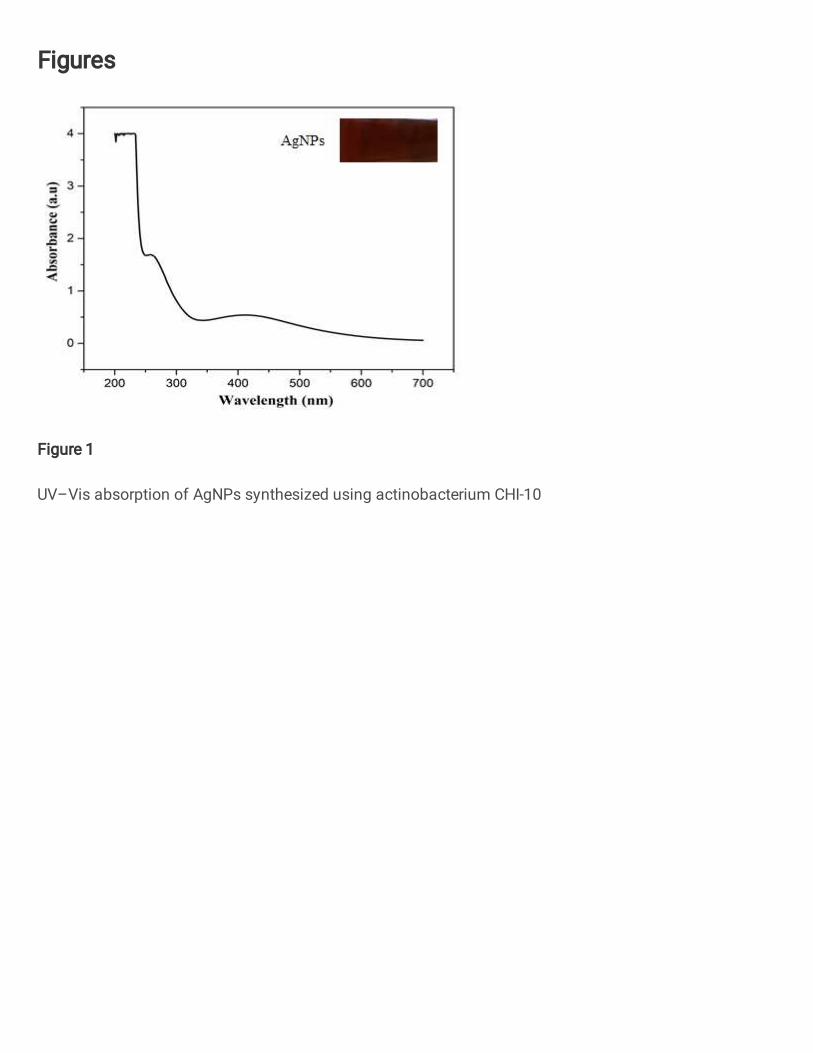
Figures
Figure 1
UV–Vis absorption of AgNPs synthesized using actinobacterium CHI-10
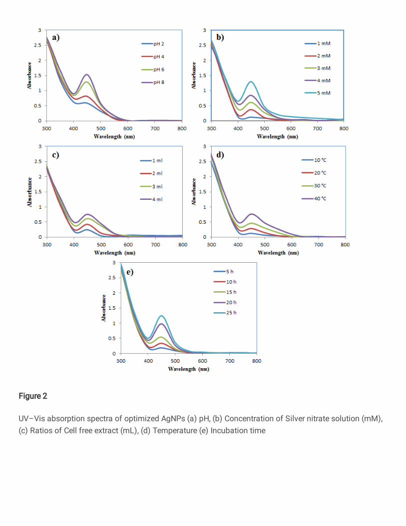
Figure 2
UV–Vis absorption spectra of optimized AgNPs (a) pH, (b) Concentration of Silver nitrate solution (mM),(c) Ratios of Cell free extract (mL), (d) Temperature (e) Incubation time
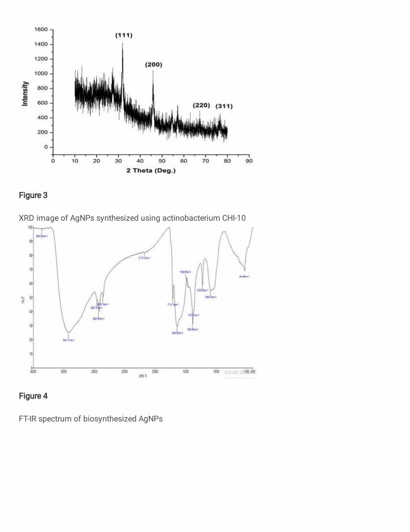
Figure 3
XRD image of AgNPs synthesized using actinobacterium CHI-10
Figure 4
FT-IR spectrum of biosynthesized AgNPs
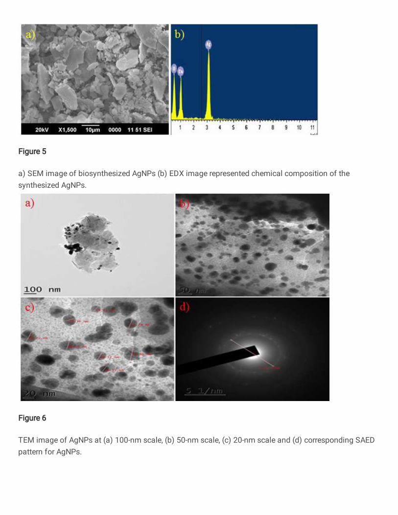
Figure 5
a) SEM image of biosynthesized AgNPs (b) EDX image represented chemical composition of thesynthesized AgNPs.
Figure 6
TEM image of AgNPs at (a) 100-nm scale, (b) 50-nm scale, (c) 20-nm scale and (d) corresponding SAEDpattern for AgNPs.
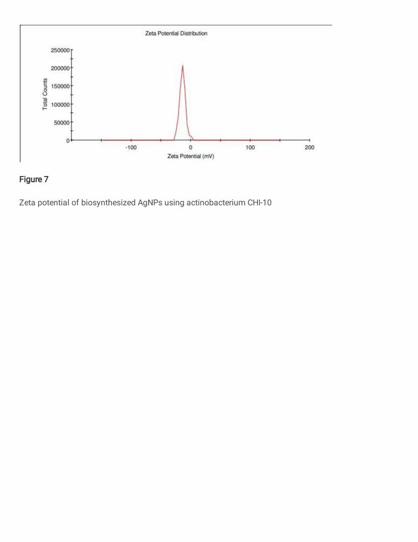
Figure 7
Zeta potential of biosynthesized AgNPs using actinobacterium CHI-10
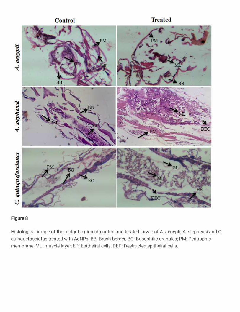
Figure 8
Histological image of the midgut region of control and treated larvae of A. aegypti, A. stephensi and C.quinquefasciatus treated with AgNPs. BB: Brush border; BG: Basophilic granules; PM: Peritrophicmembrane; ML: muscle layer; EP: Epithelial cells; DEP: Destructed epithelial cells.
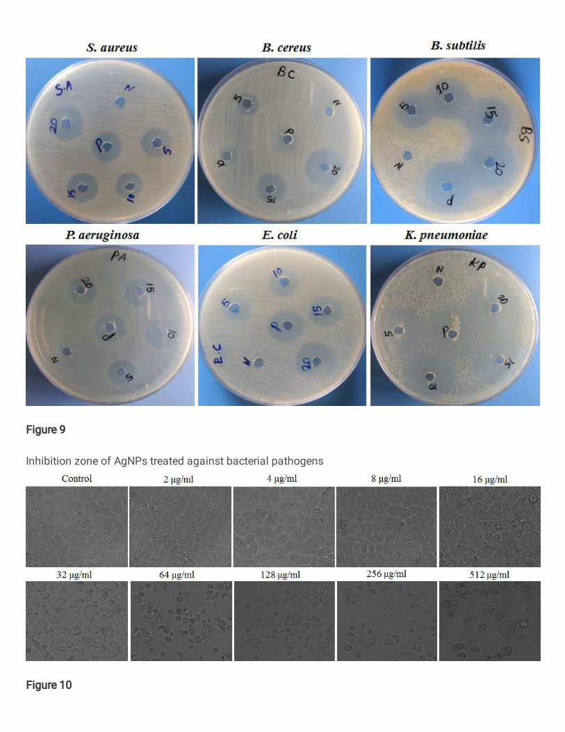
Figure 9
Inhibition zone of AgNPs treated against bacterial pathogens
Figure 10
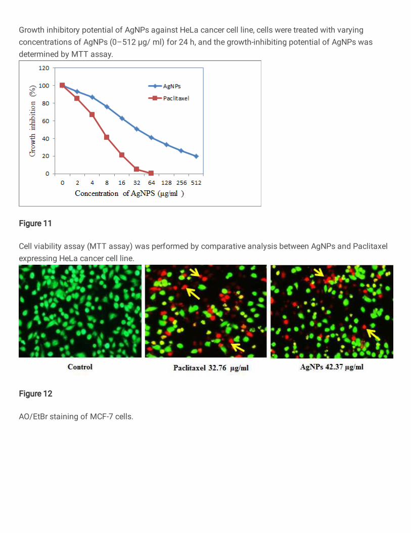
Growth inhibitory potential of AgNPs against HeLa cancer cell line, cells were treated with varyingconcentrations of AgNPs (0–512 μg/ ml) for 24 h, and the growth-inhibiting potential of AgNPs wasdetermined by MTT assay.
Figure 11
Cell viability assay (MTT assay) was performed by comparative analysis between AgNPs and Paclitaxelexpressing HeLa cancer cell line.
Figure 12
AO/EtBr staining of MCF-7 cells.
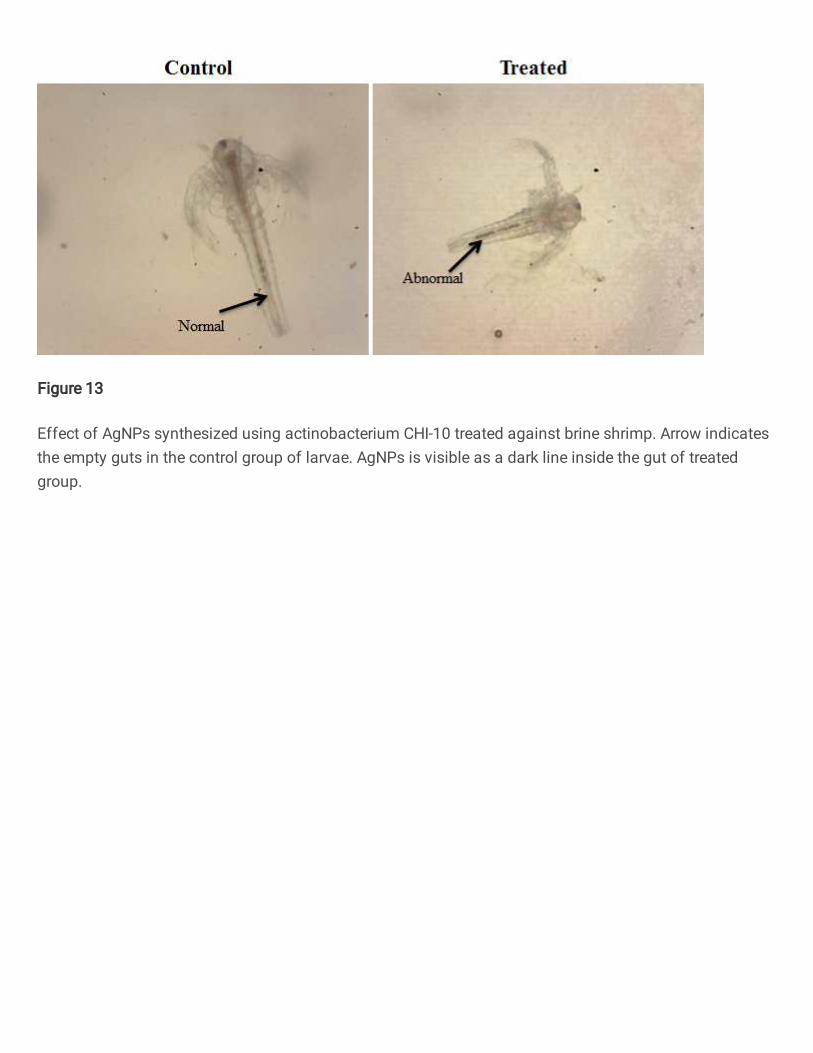
Figure 13
Effect of AgNPs synthesized using actinobacterium CHI-10 treated against brine shrimp. Arrow indicatesthe empty guts in the control group of larvae. AgNPs is visible as a dark line inside the gut of treatedgroup.