Embed Size (px)
Citation preview
G
J
Pe
LVMa
b
c
d
a
ARR2AA
KDTHR
1
opt
N
0d
ARTICLE IN PRESSModel
VAC-11340; No. of Pages 10
Vaccine xxx (2011) xxx–xxx
Contents lists available at ScienceDirect
Vaccine
journa l homepage: www.e lsev ier .com/ locate /vacc ine
reclinical evaluation of HIV-1 therapeutic ex vivo dendritic cell vaccinesxpressing consensus Gag antigens and conserved Gag epitopes
iguo Niua,1, James M. Terminia, Saravana K. Kanagavelua, Sachin Guptaa, Morgane M. Rollandc,iraj Kulkarnid, George N. Pavlakisd, Barbara K. Felberd, James I. Mullinsc,argaret A. Fischlb, Geoffrey W. Stonea,∗
Department of Microbiology & Immunology, Miller School of Medicine, University of Miami, 1580 NW 10th Ave, Miami, FL, United StatesDepartment of Medicine, Miller School of Medicine, University of Miami, Miami, FL, United StatesDepartment of Microbiology and Department of Medicine, University of Washington, Seattle, WA, United StatesNational Cancer Institute-Frederick, Frederick, MD, United States
r t i c l e i n f o
rticle history:eceived 7 November 2010eceived in revised form2 December 2010ccepted 24 December 2010vailable online xxx
eywords:endritic cellherapeutic vaccinationIVNA
a b s t r a c t
Background: Dendritic cell (DC) therapy is a promising technology for the treatment of HIV infected indi-viduals. HIV-1 Gag- and Nef RNA-loaded DC have previously been shown to induce immune responsesex vivo following coculture with autologous lymphocytes. However, polyfunctionality and memoryresponses following coculture have not been evaluated. In addition, little is known regarding whetherspecific HIV-1 proteome components, such as highly conserved regions of the HIV-1, could enhanceclinical responses following DC therapy.Methodology and principal findings: To determine the breadth of the immune responses to antigen loadedDC, we analyzed polyfunctional T cell response ex vivo to Gag RNA loaded DC. Blood samples were used togenerate monocyte derived DC, which were then matured and cocultured with autologous lymphocytes.We found that cytokine-matured DC loaded with Gag RNA was able to induce Gag-specific IFN-� and IL-2 responses after a 12-day coculture. We characterized these responses by polyfunctional intracellularcytokine staining and evaluation of T cell memory phenotypes. Central memory CD8+ T cells were inducedex vivo after DC coculture from each of 3 patients, and the effector memory pool was increased by DCcoculture from 2 patients. We also observed a decrease in the terminal effector and intermediate CD8+T cell pool and an increase in the naïve/other population. There was a reduction in terminal effector andintermediate CD4+ T cells, and a corresponding increase in naïve/other CD4+ T cells. Finally, we evaluatedconserved regions of Gag as a novel DC therapy immunogen and found that a conserved element (CE) p24
Gag antigen elicited IFN-� and IL-2 responses comparable to those induced by a full-length Gag antigen.Conclusions: We showed that RNA-loaded DC therapy induced a polyfunctional T cell response ex vivo,supporting the use of such DC-therapy for HIV infection. However, the central and effector memory phe-notypes of T cells did not appear to be enhanced during coculture with Gag RNA-loaded DC. Furthermore,comparable antigen-specific responses were induced in HIV infected individuals using full-length Gag orof thnts o
only conserved elementsonto the conserved eleme
. Introduction
Please cite this article in press as: Niu L, et al. Preclinical evaluation of HIVGag antigens and conserved Gag epitopes. Vaccine (2011), doi:10.1016/j.va
Dendritic cell therapy remains one of the more promising meth-ds to induce an immune response against antigens in canceratients [1–3], Induction of robust polyfunctional T cells is essen-ial for successful immune based therapies in HIV infection. DC are
∗ Corresponding author. Tel.: +1 305 243 2189; fax: +1 305 243 7211.E-mail address: [email protected] (G.W. Stone).
1 Current address: University of North Carolina School of Medicine, Chapel Hill,C, United States.
264-410X/$ – see front matter © 2011 Elsevier Ltd. All rights reserved.oi:10.1016/j.vaccine.2010.12.131
e Gag p24 protein. This indicates that immune responses can be focusedf Gag in the absence of other Gag components.
© 2011 Elsevier Ltd. All rights reserved.
highly effective antigen-presenting cells that play a central rolein pathogen-specific immune activity and the innate and adap-tive immune response. There is continued interest in developingdendritic cell therapy programs for the treatment of HIV infectedindividuals. One approach is the use of mRNA-loaded dendritic cells[3–8]. In previous studies, mRNA has been used to encode eitherconsensus HIV antigens able to induce responses in a variety of HIV
-1 therapeutic ex vivo dendritic cell vaccines expressing consensusccine.2010.12.131
infected patients [6], or HIV antigen sequences derived from eachpatient individually [5].
Prior to Phase I studies for HIV dendritic cell therapy, it isimportant to preclinically evaluate the response generated bymRNA-loaded DC from HIV infected patients. While most studies
ING
J
2 ine xx
hyb
nnmitaciwB[
alsttcwt
2
2
erUsMpIi
2
cc0taPfpcsti2wapictfldad
ARTICLEModel
VAC-11340; No. of Pages 10
L. Niu et al. / Vacc
ave tested DC function by IFN-� ELISPOT, a more detailed anal-sis of T cell polyfunctionality and memory responses would beeneficial.
Another key issue for HIV DC therapy optimization is theature of the HIV antigen. Recent studies suggest that recombi-ant antigens composed of only highly conserved HIV epitopesay be superior to full-length HIV antigens [9–12]. A major issue
s the role of non-conserved ‘decoy’ epitopes encoded withinhe full-length antigens. These non-conserved epitopes, whichre typically the dominant immune response in HIV patients,an be mutated without a fitness cost to the virus [13], lead-ng to immune evasion. In contrast, CTL responses associated
ith viral control, such as those elicited in individuals carrying*27 or B*57 HLA alleles often target conserved regions of HIV-114–16].
Here we demonstrated the preclinical efficacy of a HIV DC ther-py protocol using RNA-loaded DC from a cohort of HIV patientsocated in Miami, FL. We also evaluated RNA encoding either con-erved elements of the HIV-1M group Gag p24 or a full-length Gago compare induction of Gag responses. Overall these studies showhat HIV patient samples can be induced to respond to dendriticell therapy, and supports future HIV DC therapy clinical trials, asell as the use of vaccines designed to direct responses exclusively
o conserved elements of viral proteins.
. Materials and methods
.1. PBMC samples
All samples were collected in sodium Heparin unless oth-rwise noted. For HIV infected patient samples, patients wereecruited from the University of Miami AIDS Clinical Researchnit. A total of 23 patient samples were evaluated. All patients
igned informed consent in accordance with the University ofiami Institutional Review Board. Uninfected human blood sam-
les (buffy coat) were purchased from Continental Services Groupnc., Miami, FL and were negative for HIV, HBV, and HCVnfection.
.2. Dendritic cell preparation and cryopreservation
Whole blood from buffy coat or from HIV+ patients wasentrifuged at 400 × g for 10 min, and plasma was removed,entrifuged at 800 × g for 20 min, then filtered with Centrex.2 �m filters (Whatman) and stored in −80◦C. The cell frac-ion was then centrifuged over ficoll at 2100 rpm for 25 mint room temperature. The monocyte layer was re-suspended inBS, centrifuged sequentially at 1800 rpm for 10 min, 1100 rpmor 10 min, then 1000 rpm for 10 min with aspiration and resus-ension between each spin. PBMC were finally resuspended inomplete media: RPMI1640 (Hyclone) containing 5% human ABerum (Lonza) or autologous plasma, PenStrep (1×), and glu-amine. To isolate monocytes, 8 × 10⁷ HIV PBMC were placedn a T175 flask in 20 ml of complete media, and cultured forh at 37 ◦C in 5% CO2. Unattached cells were removed witharm media three times by pipette. Unattached cells (defined
s peripheral blood lymphocytes, PBL) were pelleted and resus-ended in 80% FBS + 10% Glucose + 10% DMSO. PBL were frozen
n liquid nitrogen until use. The adherent monocyte cells wereultured in 20 ml of media containing 500 U/ml IL-4 (R&D sys-
Please cite this article in press as: Niu L, et al. Preclinical evaluation of HIVGag antigens and conserved Gag epitopes. Vaccine (2011), doi:10.1016/j.va
ems) and 1000 U/ml GM-CSF (BERLEX Inc.). After 6 days of culture,asks containing immature DC were placed at 4 ◦C for 30 min toetach DC, then collected by gentle pipetting to isolate unattachednd loosely attached cells. Any remaining adherent cells wereiscarded.
PRESSx (2011) xxx–xxx
2.3. HIV antigen mRNA preparation and DC transfection
HIV-1 p24 CE construct will be described in detail in Mothe et al.(submitted). This artificial sequence contains a string of 7 highlyconserved regions of p24 Gag linked together into a single cod-ing sequence. This sequence contains >40 known HLA epitopes.HIV-1 sequences encoding codon optimized Clade B consensus Gag,Clade B consensus Nef (kindly provided by Daniel Kavanagh), andp24 CE (Mothe et al., submitted) were cloned into the pGEM4zplasmid containing a T7 promoter and a 64 bp poly A tail (vec-tor pGEM4Z/A64). The construct pGEM4Z/A64-GFP containing theGFP gene (a gift from Eli Gilboa) was used as a control in all exper-iments. The mMachine mMassage RNA in vitro transcription kit(Ambion) was used to generate mRNA from all constructs. Day6 DC were washed, resuspended in OPTI-MEM medium (GIBCO)and transfected with 10 �g RNA. A total of 1 × 10⁶ immature DC in100 �l OPTI-MEM were placed in a 0.4 �m Gene Pulser cuvette (Bio-Rad) for each electroporation. Electroporation was performed on aGenePulser X cell (Rio-Rad) with exponential decay at 300 V and150 �F. The transfected DC were transferred to 6-well plates with3 ml complete media containing Mimic cytokine mix (5 ng/ml TNF�(R&D), 5 ng/ml IL-1b (R&D), 750 ng/ml IL-6 (R&D)) + PGE2 (Sigma)1 ug/ml). Cells were cultured overnight at 37 ◦C in 5% CO2. Thesupernatant of Gag RNA transfected DC was collected and testedfor p24 level as a transfection efficiency indicator using the AllianceHIV-1 p24 ELISA kit (Perkin Elmer). Nef RNA transfection efficiencywas tested by reverse transcription PCR, and GFP RNA transfectionefficiency confirmed by flow cytometry.
2.4. Coculture of RNA transfected DC with autologous peripheralblood lymphocytes
Transfected DC were cultured at 37 ◦C in 5% CO2 for 18–36 h,then placed at 4 ◦C for 30 min to detach cells. DC were then stainedwith CD14 FITC, CD80 PE, CD86 APC, CD83 PerCP, CD11c PE cy7,CCR7 PE, HLA-DR APC cy7 (all from BD, Becton Dickinson) as matu-ration markers. For HIV patient-derived DC, cells were also stainedwith the following alternative antibody panel (all from BectonDickinson): CD86 FITC, HLA-DR PE, CD83 APC, CCR7 PE cy7, CD40APC in one tube and CD14 FITC, CD80 PE, and CD11c PE cy7 in asecond tube. Data was acquired on an LSRII fluorometer (BD) usingDiva software. For coculture, RNA transfected DC (2 × 10⁴/well)were mixed with 2 × 10⁵ autologous PBL/well in round bottom96-well plates, and cultured in 37 ◦C in 5% CO2 for 12 days. EitherIL-2 (Chiron) at 20 U/ml, or IL-7 (R&D Systems) at 5 U/ml was addedon day 3.
2.5. ELISPOT assays
ELISPOT plates (Millipore) were pre-coated with IFN-�, IL-2,or granzyme B capture antibody using human ELISPOT sets (BDBiosciences). After blocking with complete media for 1 h at roomtemperature, 3 × 10⁴ to 1 × 10⁵ PBL were added from 12-day cocul-tures. One of the following set of peptides were then added: 5 �g/mlHIV consensus clade B Gag 15-mer peptides overlapping by 11amino acids (NIH AIDS research and reference reagent program),5 �g/ml Nef HIV consensus clade B 15-mer peptides overlappingby 11 amino acids (NIH AIDS research and reference reagent pro-gram), 5 �g/ml CE Gag 10-mer peptide pool overlapping by 9 aminoacids, 5 �g/ml Ova peptide (amino acids 323–339, Genscript), or
-1 therapeutic ex vivo dendritic cell vaccines expressing consensusccine.2010.12.131
5 �g/ml c-myc peptide (Sigma) as negative control. For positivecontrols, 5 ng/ml PMA (Sigma) and 500 ng/ml ionomycin (Sigma)was added. Plates were incubated at 37 ◦C in 5% CO2 overnight,washed, read and then analyzed with Immunospot CTL analyzersoftware (Cellular Technologies).
ING
J
ine xx
2s
PcboB5sFbwCpiwswwR2D
wC(a
2
F
3
3a
vACdimHcl
ipastefpitPo
a
ARTICLEModel
VAC-11340; No. of Pages 10
L. Niu et al. / Vacc
.6. Intracellular staining and T cells memory marker surfacetaining
After 12-day coculture, at least 1 × 10⁶ PBL or thawed patientsBL (day 0) were placed into FACS tubes with 1 ml complete mediaontaining 1 ul anti-CD49 and anti-CD28, 20 �l CD107a FITC anti-ody plus either 5 �g/ml Gag peptide pool, 5 �g/ml ova peptide,r PMA/iono. Unless otherwise noted, all antibodies were fromD Biosciences. Tubes were incubated at 37 ◦C in 5% CO2, forh. A total of 10 �g/ml BFA (Sigma) and 0.7 ul monensin (eBio-
ciences) was added after 2 hours. Cells were washed twice withACS buffer (1% human serum + 0.05% sodium azide in PBS), thenlocked with 10% human serum in PBS for 10 min at RT. Cells wereashed with FACS buffer twice, then stained with antibodies toD8 APC-cy7, CD4 PerCP, CD3 Amcyan, and an exclusion mix: CD14acblue/CD19 pacblue/live-dead violet (Invitrogen). Tubes were
ncubated at room temperature in the dark for 20 min and thenashed twice with FACS buffer. 300 �l Cytofix/Cytoperm (BD Bio-
ciences) was added and incubated for 15 min at 4 ◦C. Cells wereashed twice with PermWash buffer (BD Biosciences) then stainedith IFN-� PE-Cy7, IL-2 PE, and TNF� APC antibodies for 30 min atT. Cells were washed twice with PermWash buffer then fixed with% buffered formalin. Data was acquired with a BD FACS Aria usingiva software.
For T cell memory marker staining, after 12-day coculture, PBLere stained with CD4 FITC, CD3 PerCP, CCR7 PE, CD45RO PE-cy7,D8 APC, and CD27 APC-H7 antibodies, as well as Live-dead aquaInvitrogen). Data was acquired with a BD FACS Aria machine andnalyzed using FlowJo Software.
.7. Statistics
For two-group comparison, we used unpaired Student’s T-test.or multiple samples the Wilcoxon matched pair test was used.
. Results
.1. Ex vivo derived dendritic cells induce a de novo response tontigen
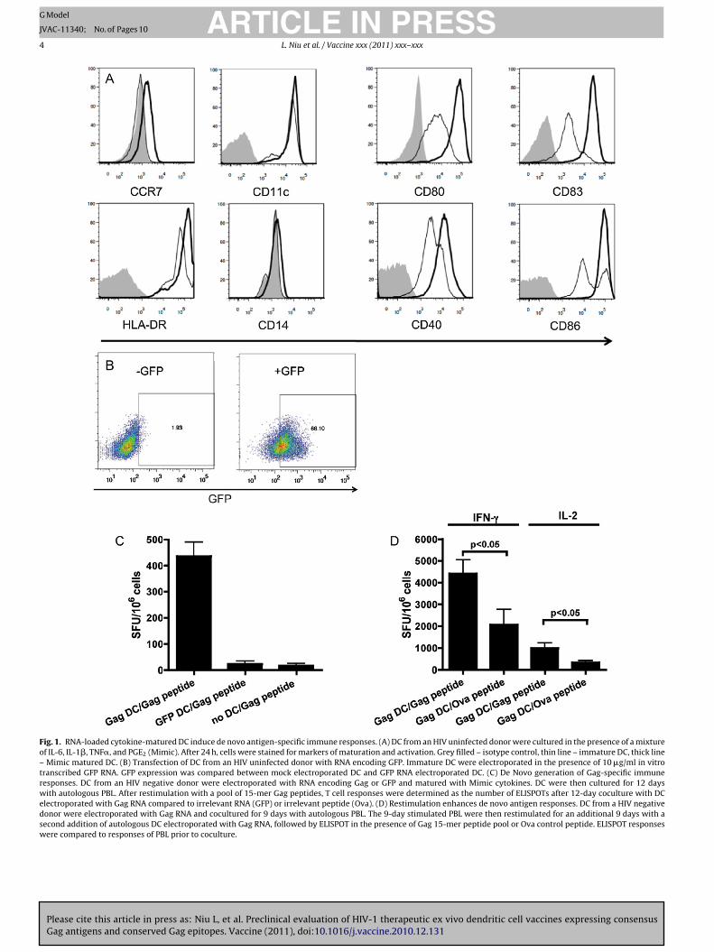
Ex vivo derived DC were stained for various markers of acti-ation and maturation both before and after cytokine maturation.s shown in Fig. 1A, both immature and mature DC cultures wereD11c+ and CD14−, indicating monocytes had fully developed intoendritic cells. Cytokine maturation with the Mimic cytokine mix
nduced the upregulation of chemokine receptor CCR7, activationarkers CD80 and CD86, and maturation markers CD83, CD40, andLA-DR (Fig. 1A). Fig. 1B showed GFP expression was induced whenells were electroporated in the presence of GFP RNA, but not fol-owing mock electroporation.
Mature DC transfected with antigen RNA would be expected tonduce T cell responses after ex vivo culture with autologous lym-hocytes, and it has been previously shown that lymphocytes fromn antigen-naïve donor are able to induce measurable antigen-pecific responses with sufficient costimulation by mature DC. Toest this, DC from HIV uninfected donors were electroporated withither Gag or GFP RNA, matured, and cultured with autologous PBLor 12 days. Cells were then stimulated with overlapping 15-mereptides to Gag. As shown in Fig. 1C, a significant (p < 0.05) increase
n spot forming units (SFUs) was observed only when PBL were cul-
Please cite this article in press as: Niu L, et al. Preclinical evaluation of HIVGag antigens and conserved Gag epitopes. Vaccine (2011), doi:10.1016/j.va
ured for 12 days with Gag RNA transfected DC (at a 10:1 ratio ofBL to DC), but not in the presence of DC transfected with GFP RNA,r PBL cultured in the absence of DC.
The quality of DC activation is also related to its ability to inducerecall response. To confirm that the de novo immune response
PRESSx (2011) xxx–xxx 3
we observed could be restimulated using this ex vivo model, DCwere loaded with Gag RNA and matured, followed by a 9-daycoculture with autologous PBL. Half the 9-day culture was testedby IFN-� and IL-2 ELISPOT and did not induce an antigen-specificresponses (data not shown). The remaining PBL culture was againmixed at a 10:1 ratio with autologous Gag RNA loaded matureDC, and restimulated in a second 9-day incubation. This secondincubation induced a significant (p < 0.05) increase in both IFN-� and IL-2 ELISPOT responses (Fig. 1D) after restimulation withGag peptide pool compared to restimulation with control Ovapeptide.
3.2. Ex vivo dendritic cells from HIV patients can enhance HIVantigen specific responses
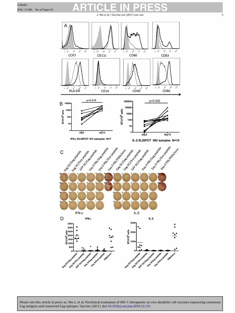
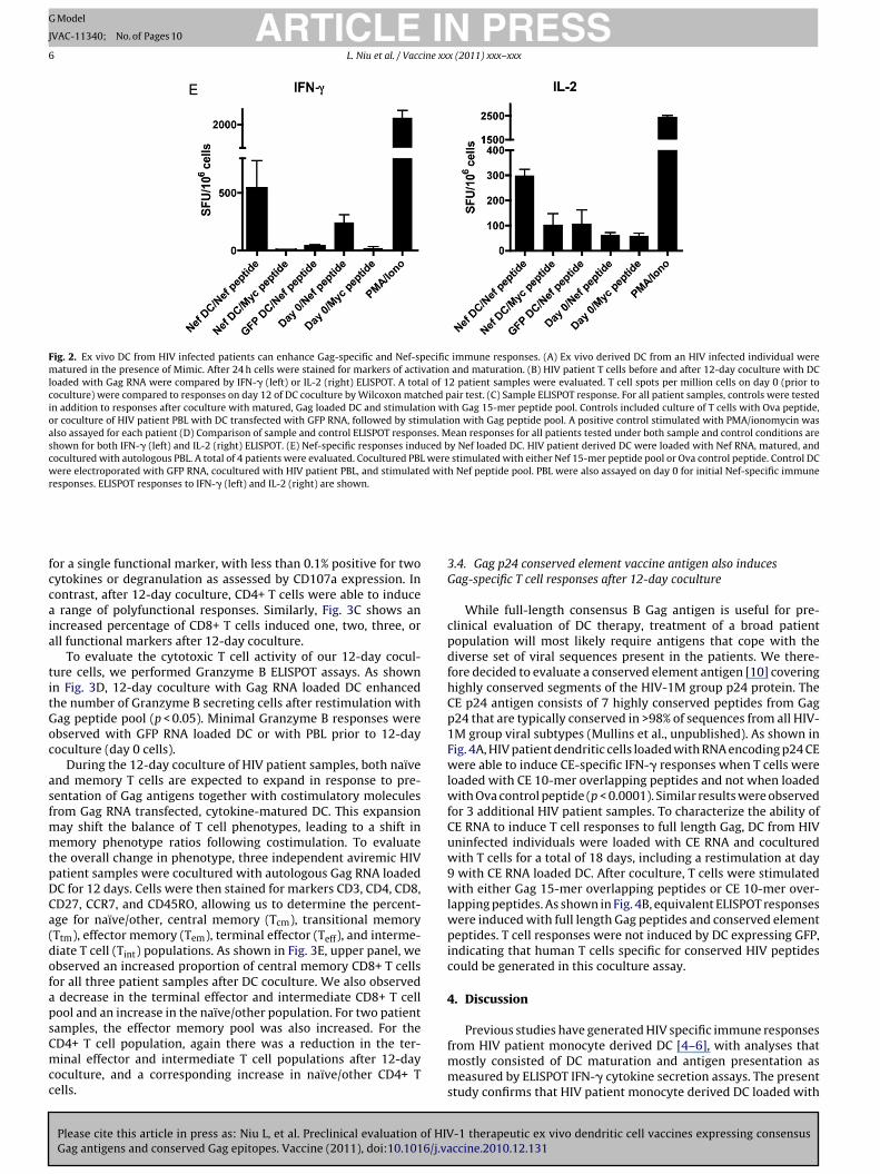
Immune dysregulation during chronic HIV infection has beenshown to disrupt dendritic cell generation and HIV-specific T cellresponses. DC derived from HIV patient monocytes were testedto assess whether they could enhance antigen-specific responsesfrom the patient’s autologous lymphocyte population. Immatureand Mimic-matured DC derived from an HIV patient blood samplewere compared by flow cytometry (Fig. 2). As shown in Fig. 2A, mat-uration induced the upregulation of CD40, CD80, CD86, and CD83 ina manner comparable to DC maturation of cells from HIV negativevolunteers (compare to Fig. 1A). To determine whether HIV-1 Gagantigen-specific responses could be enhanced, we performed a DC-T cell coculture with autologous PBL for 12 days, followed by Gagpeptide pool stimulation and ELISPOT assay. As shown in Fig. 2B, leftpanel, a significant increase (p = 0.016) in IFN-� secreting cells wasobserved in a set of 7 patient samples by Wilcoxon matched pairtest. Similarly, IL-2 ELISPOT responses were significantly enhanced(p = 0.002) after coculture with Gag RNA loaded DC in a set of 10patient samples. To control for non-specific DC activation and DC-independent stimulation during the 12-day coculture, a number ofcontrol conditions were tested in parallel. Fig. 2C shows a represen-tative ELISPOT panel while Fig. 2D shows the overall responses ofour patient samples under the various assay conditions. DC loadedwith Gag RNA, cocultured with autologous PBL, and restimulatedwith Ova peptide induced only background levels of IFN-� and IL-2ELISPOT responses. Similarly, PBL cocultured for 12 days with DCloaded with GFP RNA and then restimulated with Gag peptide poolresulted in significantly lower levels of ELISPOT responses (Fig. 2D).PBL from HIV-1 infected individuals prior to coculture were unableto induce significant Gag-specific ELISPOT responses. This likelyreflects the fact that most patients had been on antiretroviral ther-apy (ART) for >1 year with viral load <50 copies/ml at the time ofblood donation. Any memory T cells present were probably unableto generate cytokines after the brief overnight peptide stimulationemployed.
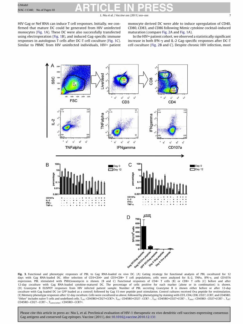
Similar studies were performed with Nef RNA loaded DC. Asshown in Fig. 2E, only Nef loaded DC cocultured with PBL for 12days and restimulated with Nef peptide pool were able to induce asignificant number (p < 0.05) of IFN-� and IL-2 ELISPOTs.
3.3. Polyfunctional CD4+ and CD8+ T cells are induced by GagRNA loaded DC
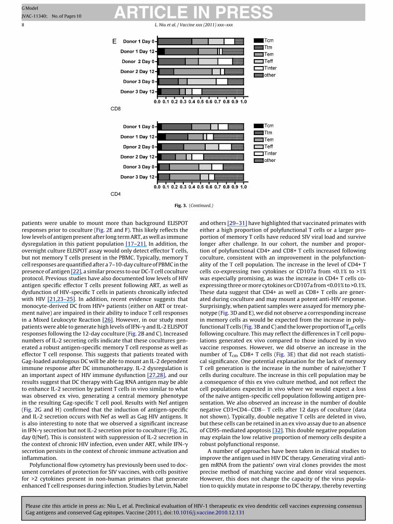
The ELISPOT responses illustrated in Fig. 2 indicate a general Tcell activation after DC coculture. To determine the specific role ofCD4+ and CD8+ T cells in DC induced immune activation of HIVpatients, multiparameter flow cytometry was performed on three
-1 therapeutic ex vivo dendritic cell vaccines expressing consensusccine.2010.12.131
patient samples after a 12-day coculture (Fig. 3A). We selected theCD3+ live cell lymphocyte population and analyzed CD4+ or CD8+T cells for their expression of IL-2, TNF�, IFN-�, and CD107a. Func-tional CD4 responses were plotted as a percentage of the total CD4+population (Fig. 3B). Prior to coculture, <1% of cells were positive
Please cite this article in press as: Niu L, et al. Preclinical evaluation of HIV-1 therapeutic ex vivo dendritic cell vaccines expressing consensusGag antigens and conserved Gag epitopes. Vaccine (2011), doi:10.1016/j.vaccine.2010.12.131
ARTICLE IN PRESSG Model
JVAC-11340; No. of Pages 10
4 L. Niu et al. / Vaccine xxx (2011) xxx–xxx
Fig. 1. RNA-loaded cytokine-matured DC induce de novo antigen-specific immune responses. (A) DC from an HIV uninfected donor were cultured in the presence of a mixtureof IL-6, IL-1�, TNF�, and PGE2 (Mimic). After 24 h, cells were stained for markers of maturation and activation. Grey filled – isotype control, thin line – immature DC, thick line– Mimic matured DC. (B) Transfection of DC from an HIV uninfected donor with RNA encoding GFP. Immature DC were electroporated in the presence of 10 �g/ml in vitrotranscribed GFP RNA. GFP expression was compared between mock electroporated DC and GFP RNA electroporated DC. (C) De Novo generation of Gag-specific immuneresponses. DC from an HIV negative donor were electroporated with RNA encoding Gag or GFP and matured with Mimic cytokines. DC were then cultured for 12 dayswith autologous PBL. After restimulation with a pool of 15-mer Gag peptides, T cell responses were determined as the number of ELISPOTs after 12-day coculture with DCelectroporated with Gag RNA compared to irrelevant RNA (GFP) or irrelevant peptide (Ova). (D) Restimulation enhances de novo antigen responses. DC from a HIV negativedonor were electroporated with Gag RNA and cocultured for 9 days with autologous PBL. The 9-day stimulated PBL were then restimulated for an additional 9 days with asecond addition of autologous DC electroporated with Gag RNA, followed by ELISPOT in the presence of Gag 15-mer peptide pool or Ova control peptide. ELISPOT responseswere compared to responses of PBL prior to coculture.
Please cite this article in press as: Niu L, et al. Preclinical evaluation of HIV-1 therapeutic ex vivo dendritic cell vaccines expressing consensusGag antigens and conserved Gag epitopes. Vaccine (2011), doi:10.1016/j.vaccine.2010.12.131
ARTICLE IN PRESSG Model
JVAC-11340; No. of Pages 10
L. Niu et al. / Vaccine xxx (2011) xxx–xxx 5
ARTICLE IN PRESSG Model
JVAC-11340; No. of Pages 10
6 L. Niu et al. / Vaccine xxx (2011) xxx–xxx
Fig. 2. Ex vivo DC from HIV infected patients can enhance Gag-specific and Nef-specific immune responses. (A) Ex vivo derived DC from an HIV infected individual werematured in the presence of Mimic. After 24 h cells were stained for markers of activation and maturation. (B) HIV patient T cells before and after 12-day coculture with DCloaded with Gag RNA were compared by IFN-� (left) or IL-2 (right) ELISPOT. A total of 12 patient samples were evaluated. T cell spots per million cells on day 0 (prior tococulture) were compared to responses on day 12 of DC coculture by Wilcoxon matched pair test. (C) Sample ELISPOT response. For all patient samples, controls were testedin addition to responses after coculture with matured, Gag loaded DC and stimulation with Gag 15-mer peptide pool. Controls included culture of T cells with Ova peptide,or coculture of HIV patient PBL with DC transfected with GFP RNA, followed by stimulation with Gag peptide pool. A positive control stimulated with PMA/ionomycin wasalso assayed for each patient (D) Comparison of sample and control ELISPOT responses. Mean responses for all patients tested under both sample and control conditions areshown for both IFN-� (left) and IL-2 (right) ELISPOT. (E) Nef-specific responses induced by Nef loaded DC. HIV patient derived DC were loaded with Nef RNA, matured, andcocultured with autologous PBL. A total of 4 patients were evaluated. Cocultured PBL were stimulated with either Nef 15-mer peptide pool or Ova control peptide. Control DCwere electroporated with GFP RNA, cocultured with HIV patient PBL, and stimulated with Nef peptide pool. PBL were also assayed on day 0 for initial Nef-specific immuneresponses. ELISPOT responses to IFN-� (left) and IL-2 (right) are shown.
fccaia
titGoc
asfmmtpDCa(dofapsCmcc
or a single functional marker, with less than 0.1% positive for twoytokines or degranulation as assessed by CD107a expression. Inontrast, after 12-day coculture, CD4+ T cells were able to inducerange of polyfunctional responses. Similarly, Fig. 3C shows an
ncreased percentage of CD8+ T cells induced one, two, three, orll functional markers after 12-day coculture.
To evaluate the cytotoxic T cell activity of our 12-day cocul-ure cells, we performed Granzyme B ELISPOT assays. As shownn Fig. 3D, 12-day coculture with Gag RNA loaded DC enhancedhe number of Granzyme B secreting cells after restimulation withag peptide pool (p < 0.05). Minimal Granzyme B responses werebserved with GFP RNA loaded DC or with PBL prior to 12-dayoculture (day 0 cells).
During the 12-day coculture of HIV patient samples, both naïvend memory T cells are expected to expand in response to pre-entation of Gag antigens together with costimulatory moleculesrom Gag RNA transfected, cytokine-matured DC. This expansion
ay shift the balance of T cell phenotypes, leading to a shift inemory phenotype ratios following costimulation. To evaluate
he overall change in phenotype, three independent aviremic HIVatient samples were cocultured with autologous Gag RNA loadedC for 12 days. Cells were then stained for markers CD3, CD4, CD8,D27, CCR7, and CD45RO, allowing us to determine the percent-ge for naïve/other, central memory (Tcm), transitional memoryTtm), effector memory (Tem), terminal effector (Teff), and interme-iate T cell (Tint) populations. As shown in Fig. 3E, upper panel, webserved an increased proportion of central memory CD8+ T cellsor all three patient samples after DC coculture. We also observed
decrease in the terminal effector and intermediate CD8+ T cellool and an increase in the naïve/other population. For two patient
Please cite this article in press as: Niu L, et al. Preclinical evaluation of HIVGag antigens and conserved Gag epitopes. Vaccine (2011), doi:10.1016/j.va
amples, the effector memory pool was also increased. For theD4+ T cell population, again there was a reduction in the ter-inal effector and intermediate T cell populations after 12-day
oculture, and a corresponding increase in naïve/other CD4+ Tells.
3.4. Gag p24 conserved element vaccine antigen also inducesGag-specific T cell responses after 12-day coculture
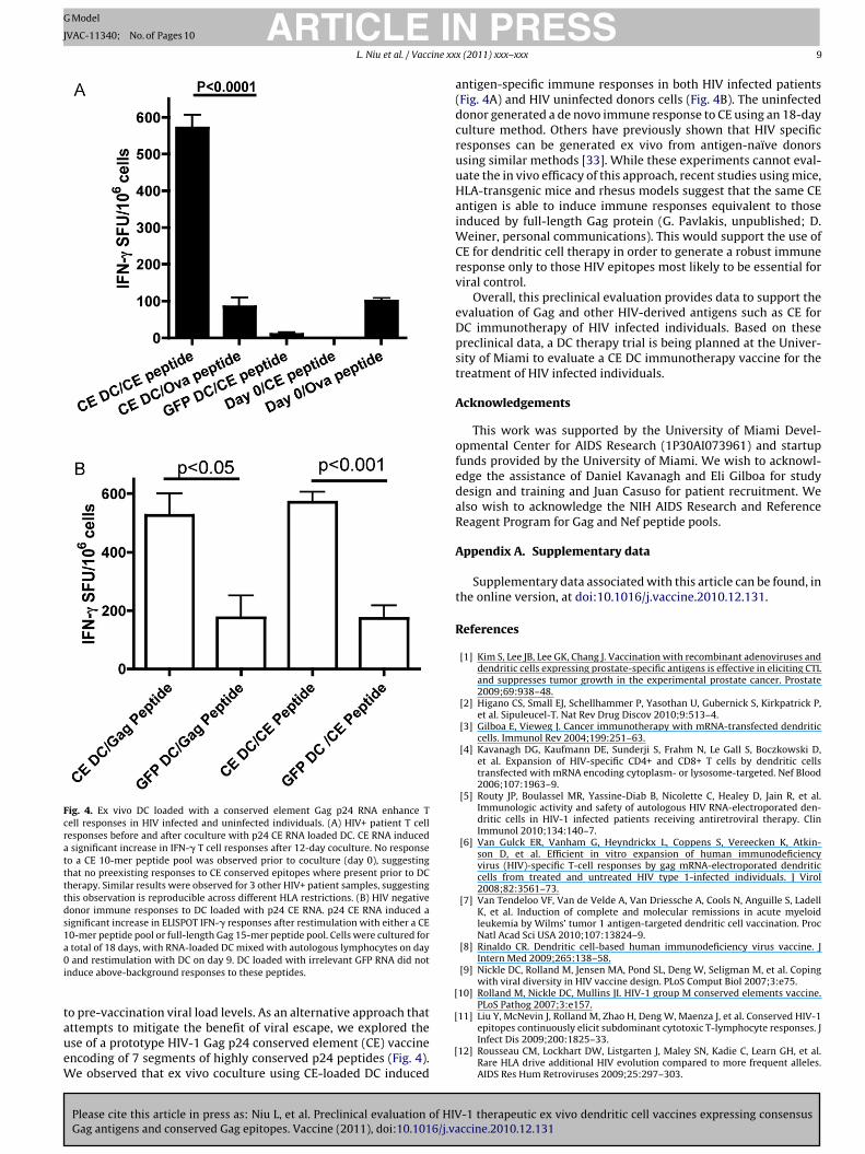
While full-length consensus B Gag antigen is useful for pre-clinical evaluation of DC therapy, treatment of a broad patientpopulation will most likely require antigens that cope with thediverse set of viral sequences present in the patients. We there-fore decided to evaluate a conserved element antigen [10] coveringhighly conserved segments of the HIV-1M group p24 protein. TheCE p24 antigen consists of 7 highly conserved peptides from Gagp24 that are typically conserved in >98% of sequences from all HIV-1M group viral subtypes (Mullins et al., unpublished). As shown inFig. 4A, HIV patient dendritic cells loaded with RNA encoding p24 CEwere able to induce CE-specific IFN-� responses when T cells wereloaded with CE 10-mer overlapping peptides and not when loadedwith Ova control peptide (p < 0.0001). Similar results were observedfor 3 additional HIV patient samples. To characterize the ability ofCE RNA to induce T cell responses to full length Gag, DC from HIVuninfected individuals were loaded with CE RNA and coculturedwith T cells for a total of 18 days, including a restimulation at day9 with CE RNA loaded DC. After coculture, T cells were stimulatedwith either Gag 15-mer overlapping peptides or CE 10-mer over-lapping peptides. As shown in Fig. 4B, equivalent ELISPOT responseswere induced with full length Gag peptides and conserved elementpeptides. T cell responses were not induced by DC expressing GFP,indicating that human T cells specific for conserved HIV peptidescould be generated in this coculture assay.
4. Discussion
-1 therapeutic ex vivo dendritic cell vaccines expressing consensusccine.2010.12.131
Previous studies have generated HIV specific immune responsesfrom HIV patient monocyte derived DC [4–6], with analyses thatmostly consisted of DC maturation and antigen presentation asmeasured by ELISPOT IFN-� cytokine secretion assays. The presentstudy confirms that HIV patient monocyte derived DC loaded with
ING
J
ine xx
HfimurS
Fde1(c(“C
ARTICLEModel
VAC-11340; No. of Pages 10
L. Niu et al. / Vacc
IV Gag or Nef RNA can induce T cell responses. Initially, we con-rmed that mature DC could be generated from HIV uninfected
Please cite this article in press as: Niu L, et al. Preclinical evaluation of HIVGag antigens and conserved Gag epitopes. Vaccine (2011), doi:10.1016/j.va
onocytes (Fig. 1A). These DC were also successfully transfectedsing electroporation (Fig. 1B), and induced Gag-specific immuneesponses in autologous T cells after DC-T cell coculture (Fig. 1C).imilar to PBMC from HIV uninfected individuals, HIV+ patient
ig. 3. Functional and phenotypic responses of PBL to Gag RNA-loaded ex vivoays with Gag RNA-loaded DC. After selection of CD3+CD4+ and CD3+CD8+ Txpression. PBL stimulated with PMA/ionomycin is shown. (B and C) Function2-day coculture with Gag RNA-loaded cytokine-matured DC. The percentageD) Granzyme B ELISPOT responses from HIV infected patient sample. Numbeoculture with Gag loaded DC (or GFP loaded as a control) followed by Gag 15-mer peE) Memory phenotype responses after 12-day coculture. Cells were cocultured as above, fOther” includes naïve T cells and undefined cells. Tcm: CD45RO+CD27+CCR7+, Tem: CD45D45RO−CD27−CCR7−, Tnaïve/other: CD45RO−CCR7+.
PRESSx (2011) xxx–xxx 7
monocyte derived DC were able to induce upregulation of CD40,CD80, CD83, and CD86 following Mimic cytokine cocktail-induced
-1 therapeutic ex vivo dendritic cell vaccines expressing consensusccine.2010.12.131
maturation (compare Fig. 2A and Fig. 1A).In the HIV+ patient cohort, we observed a statistically significant
increase in both IFN-� and IL-2 Gag-specific responses after DC-Tcell coculture (Fig. 2B and C). Despite chronic HIV infection, most
DC. (A) Gating strategy for functional analysis of PBL cocultured for 12cell populations, cells were analyzed for IL-2, TNF�, IFN-�, and CD107A
al responses of CD4+ T cells (B) or CD8+ T cells (C) before and afterof cells positive for each marker (alone or in combination) is shown.
r of PBL secreting Granzyme B is shown either before or after 12-dayptide pool stimulation. Control cultures received Ova peptide for restimulation.ollowed by phenotyping by staining with CD3, CD4, CD8, CD27, CCR7, and CD45RO.RO+CD27−CCR7−, Ttm: CD45RO+CD27+CCR7−, Tinter: CD45RO−CD27+CCR7−, Teff:
ARTICLE IN PRESSG Model
JVAC-11340; No. of Pages 10
8 L. Niu et al. / Vaccine xxx (2011) xxx–xxx
(Cont
prldobcppadwmmiprneeGiartwi(aiidtsi
ufe
Fig. 3.
atients were unable to mount more than background ELISPOTesponses prior to coculture (Fig. 2E and F). This likely reflects theow levels of antigen present after long term ART, as well as immuneysregulation in this patient population [17–21]. In addition, thevernight culture ELISPOT assay would only detect effector T cells,ut not memory T cells present in the PBMC. Typically, memory Tell responses are quantified after a 7–10-day culture of PBMC in theresence of antigen [22], a similar process to our DC-T cell coculturerotocol. Previous studies have also documented low levels of HIVntigen specific effector T cells present following ART, as well asysfunction of HIV-specific T cells in patients chronically infectedith HIV [21,23–25]. In addition, recent evidence suggests thatonocyte-derived DC from HIV+ patients (either on ART or treat-ent naïve) are impaired in their ability to induce T cell responses
n a Mixed Leukocyte Reaction [26]. However, in our study mostatients were able to generate high levels of IFN-� and IL-2 ELISPOTesponses following the 12-day coculture (Fig. 2B and C). Increasedumbers of IL-2 secreting cells indicate that these cocultures gen-rated a robust antigen-specific memory T cell response as well asffector T cell response. This suggests that patients treated withag-loaded autologous DC will be able to mount an IL-2 dependent
mmune response after DC immunotherapy. IL-2 dysregulation isn important aspect of HIV immune dysfunction [27,28], and ouresults suggest that DC therapy with Gag RNA antigen may be ableo enhance IL-2 secretion by patient T cells in vivo similar to whatas observed ex vivo, generating a central memory phenotype
n the resulting Gag-specific T cell pool. Results with Nef antigenFig. 2G and H) confirmed that the induction of antigen-specificnd IL-2 secretion occurs with Nef as well as Gag HIV antigens. Its also interesting to note that we observed a significant increasen IFN-� secretion but not IL-2 secretion prior to coculture (Fig. 2G,ay 0/Nef). This is consistent with suppression of IL-2 secretion inhe context of chronic HIV infection, even under ART, while IFN-�ecretion persists in the context of chronic immune activation and
Please cite this article in press as: Niu L, et al. Preclinical evaluation of HIVGag antigens and conserved Gag epitopes. Vaccine (2011), doi:10.1016/j.va
nflammation.Polyfunctional flow cytometry has previously been used to doc-
ment correlates of protection for SIV vaccines, with cells positiveor >2 cytokines present in non-human primates that generatenhanced T cell responses during infection. Studies by Letvin, Nabel
inued.)
and others [29–31] have highlighted that vaccinated primates witheither a high proportion of polyfunctional T cells or a larger pro-portion of memory T cells have reduced SIV viral load and survivelonger after challenge. In our cohort, the number and propor-tion of polyfunctional CD4+ and CD8+ T cells increased followingcoculture, consistent with an improvement in the polyfunction-ality of the T cell population. The increase in the level of CD4+ Tcells co-expressing two cytokines or CD107a from <0.1% to >1%was especially promising, as was the increase in CD4+ T cells co-expressing three or more cytokines or CD107a from <0.01% to >0.1%.These data suggest that CD4+ as well as CD8+ T cells are gener-ated during coculture and may mount a potent anti-HIV response.Surprisingly, when patient samples were assayed for memory phe-notype (Fig. 3D and E), we did not observe a corresponding increasein memory cells as would be expected from the increase in poly-functional T cells (Fig. 3B and C) and the lower proportion of Teff cellsfollowing coculture. This may reflect the differences in T cell popu-lations generated ex vivo compared to those induced by in vivovaccine responses. However, we did observe an increase in thenumber of Tcm CD8+ T cells (Fig. 3E) that did not reach statisti-cal significance. One potential explanation for the lack of memoryT cell generation is the increase in the number of naïve/other Tcells during coculture. The increase in this cell population may bea consequence of this ex vivo culture method, and not reflect thecell populations expected in vivo where we would expect a lossof the naïve antigen-specific cell population following antigen pre-sentation. We also observed an increase in the number of doublenegative CD3+CD4−CD8− T cells after 12 days of coculture (datanot shown). Typically, double negative T cells are deleted in vivo,but these cells can be retained in an ex vivo assay due to an absenceof CD95-mediated apoptosis [32]. This double negative populationmay explain the low relative proportion of memory cells despite arobust polyfunctional response.
A number of approaches have been taken in clinical studies to
-1 therapeutic ex vivo dendritic cell vaccines expressing consensusccine.2010.12.131
improve the antigen used in HIV DC therapy. Generating viral anti-gen mRNA from the patients’ own viral clones provides the mostprecise method of matching vaccine and donor viral sequences.However, this does not change the capacity of the virus popula-tion to quickly mutate in response to DC therapy, thereby reverting
ARTICLE ING Model
JVAC-11340; No. of Pages 10
L. Niu et al. / Vaccine xx
Fig. 4. Ex vivo DC loaded with a conserved element Gag p24 RNA enhance Tcell responses in HIV infected and uninfected individuals. (A) HIV+ patient T cellresponses before and after coculture with p24 CE RNA loaded DC. CE RNA induceda significant increase in IFN-� T cell responses after 12-day coculture. No responseto a CE 10-mer peptide pool was observed prior to coculture (day 0), suggestingthat no preexisting responses to CE conserved epitopes where present prior to DCtherapy. Similar results were observed for 3 other HIV+ patient samples, suggestingthis observation is reproducible across different HLA restrictions. (B) HIV negativedonor immune responses to DC loaded with p24 CE RNA. p24 CE RNA induced asignificant increase in ELISPOT IFN-� responses after restimulation with either a CE1a0i
taueW
[
0-mer peptide pool or full-length Gag 15-mer peptide pool. Cells were cultured fortotal of 18 days, with RNA-loaded DC mixed with autologous lymphocytes on dayand restimulation with DC on day 9. DC loaded with irrelevant GFP RNA did not
nduce above-background responses to these peptides.
o pre-vaccination viral load levels. As an alternative approach that
Please cite this article in press as: Niu L, et al. Preclinical evaluation of HIVGag antigens and conserved Gag epitopes. Vaccine (2011), doi:10.1016/j.va
ttempts to mitigate the benefit of viral escape, we explored these of a prototype HIV-1 Gag p24 conserved element (CE) vaccinencoding of 7 segments of highly conserved p24 peptides (Fig. 4).e observed that ex vivo coculture using CE-loaded DC induced
[
[
PRESSx (2011) xxx–xxx 9
antigen-specific immune responses in both HIV infected patients(Fig. 4A) and HIV uninfected donors cells (Fig. 4B). The uninfecteddonor generated a de novo immune response to CE using an 18-dayculture method. Others have previously shown that HIV specificresponses can be generated ex vivo from antigen-naïve donorsusing similar methods [33]. While these experiments cannot eval-uate the in vivo efficacy of this approach, recent studies using mice,HLA-transgenic mice and rhesus models suggest that the same CEantigen is able to induce immune responses equivalent to thoseinduced by full-length Gag protein (G. Pavlakis, unpublished; D.Weiner, personal communications). This would support the use ofCE for dendritic cell therapy in order to generate a robust immuneresponse only to those HIV epitopes most likely to be essential forviral control.
Overall, this preclinical evaluation provides data to support theevaluation of Gag and other HIV-derived antigens such as CE forDC immunotherapy of HIV infected individuals. Based on thesepreclinical data, a DC therapy trial is being planned at the Univer-sity of Miami to evaluate a CE DC immunotherapy vaccine for thetreatment of HIV infected individuals.
Acknowledgements
This work was supported by the University of Miami Devel-opmental Center for AIDS Research (1P30AI073961) and startupfunds provided by the University of Miami. We wish to acknowl-edge the assistance of Daniel Kavanagh and Eli Gilboa for studydesign and training and Juan Casuso for patient recruitment. Wealso wish to acknowledge the NIH AIDS Research and ReferenceReagent Program for Gag and Nef peptide pools.
Appendix A. Supplementary data
Supplementary data associated with this article can be found, inthe online version, at doi:10.1016/j.vaccine.2010.12.131.
References
[1] Kim S, Lee JB, Lee GK, Chang J. Vaccination with recombinant adenoviruses anddendritic cells expressing prostate-specific antigens is effective in eliciting CTLand suppresses tumor growth in the experimental prostate cancer. Prostate2009;69:938–48.
[2] Higano CS, Small EJ, Schellhammer P, Yasothan U, Gubernick S, Kirkpatrick P,et al. Sipuleucel-T. Nat Rev Drug Discov 2010;9:513–4.
[3] Gilboa E, Vieweg J. Cancer immunotherapy with mRNA-transfected dendriticcells. Immunol Rev 2004;199:251–63.
[4] Kavanagh DG, Kaufmann DE, Sunderji S, Frahm N, Le Gall S, Boczkowski D,et al. Expansion of HIV-specific CD4+ and CD8+ T cells by dendritic cellstransfected with mRNA encoding cytoplasm- or lysosome-targeted. Nef Blood2006;107:1963–9.
[5] Routy JP, Boulassel MR, Yassine-Diab B, Nicolette C, Healey D, Jain R, et al.Immunologic activity and safety of autologous HIV RNA-electroporated den-dritic cells in HIV-1 infected patients receiving antiretroviral therapy. ClinImmunol 2010;134:140–7.
[6] Van Gulck ER, Vanham G, Heyndrickx L, Coppens S, Vereecken K, Atkin-son D, et al. Efficient in vitro expansion of human immunodeficiencyvirus (HIV)-specific T-cell responses by gag mRNA-electroporated dendriticcells from treated and untreated HIV type 1-infected individuals. J Virol2008;82:3561–73.
[7] Van Tendeloo VF, Van de Velde A, Van Driessche A, Cools N, Anguille S, LadellK, et al. Induction of complete and molecular remissions in acute myeloidleukemia by Wilms’ tumor 1 antigen-targeted dendritic cell vaccination. ProcNatl Acad Sci USA 2010;107:13824–9.
[8] Rinaldo CR. Dendritic cell-based human immunodeficiency virus vaccine. JIntern Med 2009;265:138–58.
[9] Nickle DC, Rolland M, Jensen MA, Pond SL, Deng W, Seligman M, et al. Copingwith viral diversity in HIV vaccine design. PLoS Comput Biol 2007;3:e75.
10] Rolland M, Nickle DC, Mullins JI. HIV-1 group M conserved elements vaccine.PLoS Pathog 2007;3:e157.
-1 therapeutic ex vivo dendritic cell vaccines expressing consensusccine.2010.12.131
11] Liu Y, McNevin J, Rolland M, Zhao H, Deng W, Maenza J, et al. Conserved HIV-1epitopes continuously elicit subdominant cytotoxic T-lymphocyte responses. JInfect Dis 2009;200:1825–33.
12] Rousseau CM, Lockhart DW, Listgarten J, Maley SN, Kadie C, Learn GH, et al.Rare HLA drive additional HIV evolution compared to more frequent alleles.AIDS Res Hum Retroviruses 2009;25:297–303.
ING
J
1 ine xx
[
[
[
[
[
[
[
[
[
[
[
[
[
[
[
[
[
[
[
ARTICLEModel
VAC-11340; No. of Pages 10
0 L. Niu et al. / Vacc
13] Rolland M, Carlson JM, Manocheewa S, Swain JV, Lanxon-Cookson E, Deng W,et al. Amino-acid co-variation in HIV-1 Gag subtype C: HLA-mediated selectionpressure and compensatory dynamics. PLoS One 2010;5:pii:e12463.
14] Leslie AJ, Pfafferott KJ, Chetty P, Draenert R, Addo MM, Feeney M, et al. HIVevolution: CTL escape mutation and reversion after transmission. Nat Med2004;10:282–9.
15] Altfeld M, Addo MM, Rosenberg ES, Hecht FM, Lee PK, Vogel M, et al. Influence ofHLA-B57 on clinical presentation and viral control during acute HIV-1 infection.Aids 2003;17:2581–91.
16] Hendel H, Caillat-Zucman S, Lebuanec H, Carrington M, O’Brien S, Andrieu JM,et al. New class I and II HLA alleles strongly associated with opposite patternsof progression to AIDS. J Immunol 1999;162:6942–6.
17] Sabado RL, O’Brien M, Subedi A, Qin L, Hu N, Taylor E, et al. Evidence of dysreg-ulation of dendritic cells in primary HIV infection. Blood 2010;116:3839–52.
18] Chougnet C. Role of CD40 ligand dysregulation in HIV-associated dysfunctionof antigen-presenting cells. J Leukoc Biol 2003;74:702–9.
19] Boasso A, Shearer GM. Chronic innate immune activation as a cause of HIV-1immunopathogenesis. Clin Immunol 2008;126:235–42.
20] Kaufmann DE, Kavanagh DG, Pereyra F, Zaunders JJ, Mackey EW, Miura T,et al. Upregulation of CTLA-4 by HIV-specific CD4+ T cells correlates with dis-ease progression and defines a reversible immune dysfunction. Nat Immunol2007;8:1246–54.
21] Paiardini M, Frank I, Pandrea I, Apetrei C, Silvestri G. Mucosal immune dysfunc-tion in AIDS pathogenesis. Aids Rev 2008;10:36–46.
22] Calarota SA, Foli A, Maserati R, Baldanti F, Paolucci S, Young MA, et al.
Please cite this article in press as: Niu L, et al. Preclinical evaluation of HIVGag antigens and conserved Gag epitopes. Vaccine (2011), doi:10.1016/j.va
HIV-1-specific T cell precursors with high proliferative capacity correlatewith low viremia and high CD4 counts in untreated individuals. J Immunol2008;180:5907–15.
23] Rehr M, Cahenzli J, Haas A, Price DA, Gostick E, Huber M, et al. Emergence ofpolyfunctional CD8+ T cells after prolonged suppression of human immunode-ficiency virus replication by antiretroviral therapy. J Virol 2008;82:3391–404.
[
[
PRESSx (2011) xxx–xxx
24] Trautmann L, Janbazian L, Chomont N, Said EA, Gimmig S, Bessette B, et al.Upregulation of PD-1 expression on HIV-specific CD8+ T cells leads to reversibleimmune dysfunction. Nat Med 2006;12:1198–202.
25] Zhang D, Shankar P, Xu Z, Harnisch B, Chen G, Lange C, et al. Most antiviral CD8T cells during chronic viral infection do not express high levels of perforin andare not directly cytotoxic. Blood 2003;101:226–35.
26] Buisson S, Benlahrech A, Gazzard B, Gotch F, Kelleher P, Patterson S. Monocyte-derived dendritic cells from HIV type 1-infected individuals show reducedability to stimulate T cells and have altered production of interleukin (IL)-12and IL-10. J Infect Dis 2009;199:1862–71.
27] Erikstrup C, Kronborg G, Lohse N, Rye Ostrowski S, Gerstoft J, Ullum H. T-cell dysfunction in HIV-1-infected patients with impaired recovery of CD4cells despite suppression of viral replication. J Acquir Immune Defic Syndr2010;53:303–10.
28] Fuse S, Tsai CY, Molloy MJ, Allie SR, Zhang W, Yagita H, et al. Recall responsesby helpless memory CD8+ T cells are restricted by the up-regulation of PD-1. JImmunol 2009;182:4244–54.
29] Letvin NL, Mascola JR, Sun Y, Gorgone DA, Buzby AP, Xu L, et al. Preserved CD4+central memory T cells and survival in vaccinated SIV-challenged monkeys.Science 2006;312:1530–3.
30] Mattapallil JJ, Douek DC, Buckler-White A, Montefiori D, Letvin NL, Nabel GJ,et al. Vaccination preserves CD4 memory T cells during acute simian immun-odeficiency virus challenge. J Exp Med 2006;203:1533–41.
31] Precopio ML, Betts MR, Parrino J, Price DA, Gostick E, Ambrozak DR, et al.Immunization with vaccinia virus induces polyfunctional and phenotypically
-1 therapeutic ex vivo dendritic cell vaccines expressing consensusccine.2010.12.131
distinctive CD8(+) T cell responses. J Exp Med 2007;204:1405–16.32] Mehal WZ, Crispe IN. TCR ligation on CD8+ T cells creates double-negative cells
in vivo. J Immunol 1998;161:1686–93.33] Subramanya S, Armant M, Salkowitz JR, Nyakeriga AM, Haridas V, Hasan M,
et al. Enhanced induction of HIV-specific cytotoxic T lymphocytes by dendriticcell-targeted delivery of SOCS-1 siRNA. Mol Ther 2010;18:2028–37.