Embed Size (px)
Citation preview
Potent Inhibition of HIV-1 Replication by a Tat MutantLuke W. Meredith1,4.¤, Haran Sivakumaran1,3., Lee Major2, Andreas Suhrbier2,4, David Harrich1,4*
1 Division of Infectious Diseases, Queensland Institute of Medical Research, Herston, Queensland, Australia, 2 Division of Immunology, Queensland Institute of Medical
Research, Herston, Queensland, Australia, 3 School of Population Health, The University of Queensland, Herston, Queensland, Australia, 4 Griffith Medical Research
College, Brisbane, Queensland, Australia
Abstract
Herein we describe a mutant of the two-exon HIV-1 Tat protein, termed Nullbasic, that potently inhibits multiple steps ofthe HIV-1 replication cycle. Nullbasic was created by replacing the entire arginine-rich basic domain of wild type Tat withglycine/alanine residues. Like similarly mutated one-exon Tat mutants, Nullbasic exhibited transdominant negative effectson Tat-dependent transactivation. However, unlike previously reported mutants, we discovered that Nullbasic also stronglysuppressed the expression of unspliced and singly-spliced viral mRNA, an activity likely caused by redistribution and thusfunctional inhibition of HIV-1 Rev. Furthermore, HIV-1 virion particles produced by cells expressing Nullbasic had severelyreduced infectivity, a defect attributable to a reduced ability of the virions to undergo reverse transcription. Combination ofthese inhibitory effects on transactivation, Rev-dependent mRNA transport and reverse transcription meant that permissivecells constitutively expressing Nullbasic were highly resistant to a spreading infection by HIV-1. Nullbasic and its activitiesthus provide potential insights into the development of potent antiviral therapeutics that target multiple stages of HIV-1infection.
Citation: Meredith LW, Sivakumaran H, Major L, Suhrbier A, Harrich D (2009) Potent Inhibition of HIV-1 Replication by a Tat Mutant. PLoS ONE 4(11): e7769.doi:10.1371/journal.pone.0007769
Editor: Linqi Zhang, Tsinghua University, China
Received July 2, 2009; Accepted October 18, 2009; Published November 10, 2009
Copyright: � 2009 Meredith et al. This is an open-access article distributed under the terms of the Creative Commons Attribution License, which permitsunrestricted use, distribution, and reproduction in any medium, provided the original author and source are credited.
Funding: This work was supported by grants from the Australian National Health and Medical Research Council (NHMRC; ID No. 389826) and the AustralianCentre for HIV and Hepatitis Virology to DH, and an Australian Postgraduate Award to HS. The funders had no role in study design, data collection and analysis,decision to publish, or preparation of the manuscript.
Competing Interests: The authors have declared that no competing interests exist.
* E-mail: [email protected]
¤ Current address: HCV Research Group, School of Immunity and Infection, University of Birmingham, Edgbaston, Birmingham, United Kingdom
. These authors contributed equally to this work.
Introduction
Tat is an essential HIV-1 regulatory protein whose best
described role is to promote high levels of viral gene expression
via interactions with the HIV-1 transactivation response element
(TAR) RNA [1,2]. Full-length Tat is encoded by two exons
comprising 101 amino acids (varying between 99 and 104 residues)
and represents the most abundant form of Tat from patient-
derived HIV-1. The first exon is organized into two major
domains: the activation domain, which interacts with numerous
cellular proteins including cyclin T1, and the basic domain, which
is primarily comprised of arginine and lysine residues. The basic
domain (amino acids 49–57) is required for many of Tat’s activities
including nuclear localization [3,4] and TAR binding [5]. The
basic domain has also been reported to facilitate other Tat
activities such as membrane transduction [6], assisting HIV-1
reverse transcription [7] and augmenting integrin receptor
binding [8].
A transdominant negative mutant is typically an altered form of
a protein that can inhibit the normal function of its wild type
counterpart. Engineered Tat proteins with altered basic domains
possess transdominant negative phenotypes against wild type Tat.
However, most studies of Tat transdominance have used one-exon
tat mutants encoding truncated proteins of 72 amino acids or less.
For example, Tat truncated at residue 53 can suppress
transactivation initiated by wild type Tat [9]. This is despite the
mutant localizing mainly to the cytoplasm of the cell, in contrast to
wild type Tat, which localizes to the nucleus. One exon tat mutants
with a deleted basic domain or where the basic domain has been
substituted with neutrally-charged amino acids also recapitulate
the transdominant negative effects on transactivation [10,11].
Localization of Tat mutants to the nucleus, via fusion of the Tat
nuclear localization signal to their carboxy termini, results in
retention of the transdominant negative phenotype [10]. More-
over, mutations in the activation domain of the Tat mutant, which
normally suppress the transactivation function of wild type Tat,
can suppress transdominance [10].
The mechanism of transdominance of Tat basic-domain
mutants is unclear. While several hypotheses have been proposed,
studies suggest a model by which Tat mutants sequester one or
more cofactors required for Tat-mediated transactivation [10,12].
Sequestration of cofactors by transdominant Tat mutants may be
mediated by an intact activation domain, but due to the disrupted
basic domain, Tat mutants may be incapable of recruiting the
cofactors (e.g., cyclin T1) to the site of transactivation [5,13].
Here we describe for the first time the transdominant effects of a
two-exon, 101 amino-acid Tat mutant called Nullbasic, which has
the entire basic domain (amino acids 49–57) replaced with
glycine/alanine residues. Surprisingly, the Nullbasic Tat mutant
potently inhibited three different steps in HIV-1 replication. As
expected, Nullbasic inhibited Tat-mediated transactivation. Un-
expectedly, Nullbasic also potently inhibited Rev-mediated
transport of HIV-1 mRNA. Furthermore, HIV-1 produced by
cells expressing Nullbasic had greatly reduced infectivity due to a
PLoS ONE | www.plosone.org 1 November 2009 | Volume 4 | Issue 11 | e7769
potent decrease in its ability to undergo reverse transcription.
These combined activities meant that a cell line expressing
Nullbasic was almost completely protected from high-dose HIV-1
challenge. These studies potentially highlight hitherto unrecog-
nised activities of Tat and may open new avenues for therapeutic
intervention.
Results
Nullbasic Is a Transdominant Tat MutantTo investigate the molecular effects of transdominant Tat
mutants, a novel mutant termed Nullbasic was created. Unlike
previous studies, we mutated the full length, 101 amino-acid form
of Tat since we noted that two-exon Tat is expressed at greater
levels that one-exon Tat in most cell lines (unpublished
observations), and that two-exon Tat is the primary form
expressed by HIV-1 clinical isolates [14]. Nullbasic was engi-
neered from the BH10 clone of Tat (101 amino acid variant) by
replacing the basic domain with a glycine/alanine sequence and
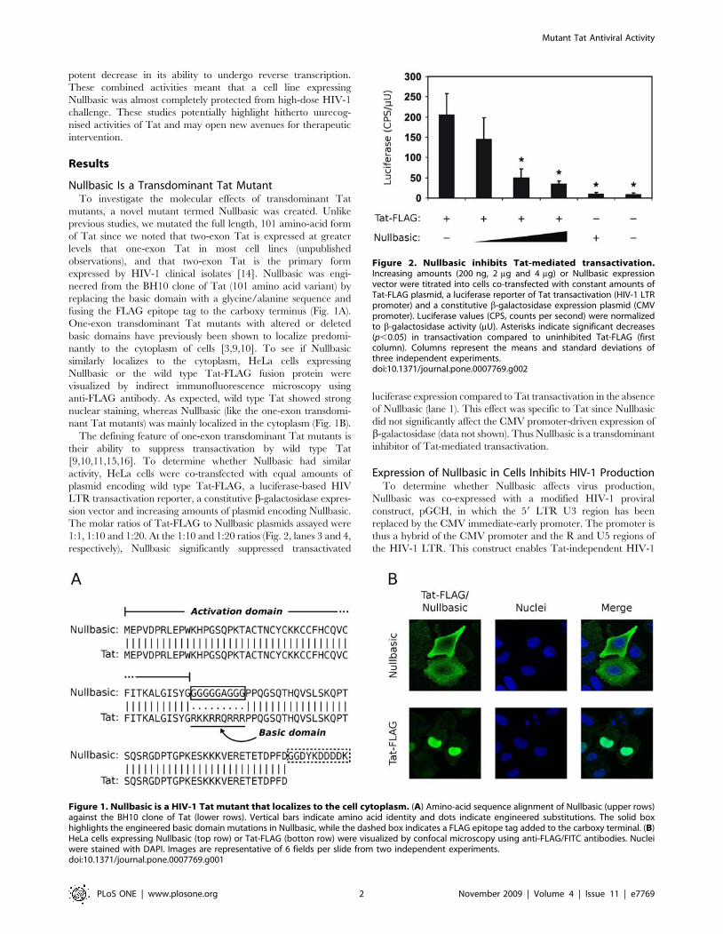
fusing the FLAG epitope tag to the carboxy terminus (Fig. 1A).
One-exon transdominant Tat mutants with altered or deleted
basic domains have previously been shown to localize predomi-
nantly to the cytoplasm of cells [3,9,10]. To see if Nullbasic
similarly localizes to the cytoplasm, HeLa cells expressing
Nullbasic or the wild type Tat-FLAG fusion protein were
visualized by indirect immunofluorescence microscopy using
anti-FLAG antibody. As expected, wild type Tat showed strong
nuclear staining, whereas Nullbasic (like the one-exon transdomi-
nant Tat mutants) was mainly localized in the cytoplasm (Fig. 1B).
The defining feature of one-exon transdominant Tat mutants is
their ability to suppress transactivation by wild type Tat
[9,10,11,15,16]. To determine whether Nullbasic had similar
activity, HeLa cells were co-transfected with equal amounts of
plasmid encoding wild type Tat-FLAG, a luciferase-based HIV
LTR transactivation reporter, a constitutive b-galactosidase expres-
sion vector and increasing amounts of plasmid encoding Nullbasic.
The molar ratios of Tat-FLAG to Nullbasic plasmids assayed were
1:1, 1:10 and 1:20. At the 1:10 and 1:20 ratios (Fig. 2, lanes 3 and 4,
respectively), Nullbasic significantly suppressed transactivated
luciferase expression compared to Tat transactivation in the absence
of Nullbasic (lane 1). This effect was specific to Tat since Nullbasic
did not significantly affect the CMV promoter-driven expression of
b-galactosidase (data not shown). Thus Nullbasic is a transdominant
inhibitor of Tat-mediated transactivation.
Expression of Nullbasic in Cells Inhibits HIV-1 ProductionTo determine whether Nullbasic affects virus production,
Nullbasic was co-expressed with a modified HIV-1 proviral
construct, pGCH, in which the 59 LTR U3 region has been
replaced by the CMV immediate-early promoter. The promoter is
thus a hybrid of the CMV promoter and the R and U5 regions of
the HIV-1 LTR. This construct enables Tat-independent HIV-1
Figure 1. Nullbasic is a HIV-1 Tat mutant that localizes to the cell cytoplasm. (A) Amino-acid sequence alignment of Nullbasic (upper rows)against the BH10 clone of Tat (lower rows). Vertical bars indicate amino acid identity and dots indicate engineered substitutions. The solid boxhighlights the engineered basic domain mutations in Nullbasic, while the dashed box indicates a FLAG epitope tag added to the carboxy terminal. (B)HeLa cells expressing Nullbasic (top row) or Tat-FLAG (botton row) were visualized by confocal microscopy using anti-FLAG/FITC antibodies. Nucleiwere stained with DAPI. Images are representative of 6 fields per slide from two independent experiments.doi:10.1371/journal.pone.0007769.g001
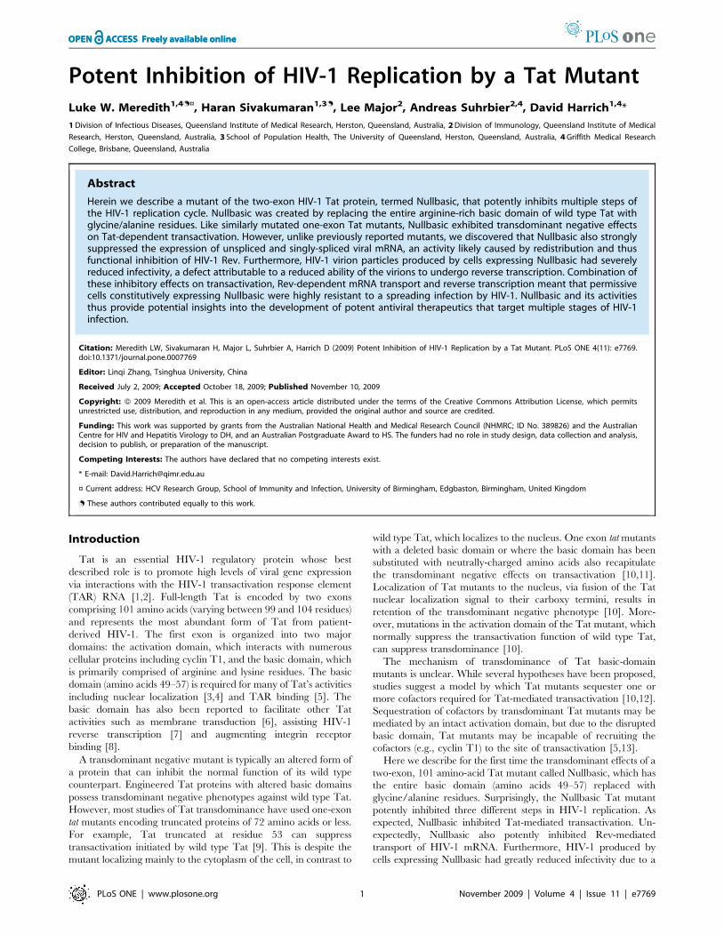
Figure 2. Nullbasic inhibits Tat-mediated transactivation.Increasing amounts (200 ng, 2 mg and 4 mg) or Nullbasic expressionvector were titrated into cells co-transfected with constant amounts ofTat-FLAG plasmid, a luciferase reporter of Tat transactivation (HIV-1 LTRpromoter) and a constitutive b-galactosidase expression plasmid (CMVpromoter). Luciferase values (CPS, counts per second) were normalizedto b-galactosidase activity (mU). Asterisks indicate significant decreases(p,0.05) in transactivation compared to uninhibited Tat-FLAG (firstcolumn). Columns represent the means and standard deviations ofthree independent experiments.doi:10.1371/journal.pone.0007769.g002
Mutant Tat Antiviral Activity
PLoS ONE | www.plosone.org 2 November 2009 | Volume 4 | Issue 11 | e7769
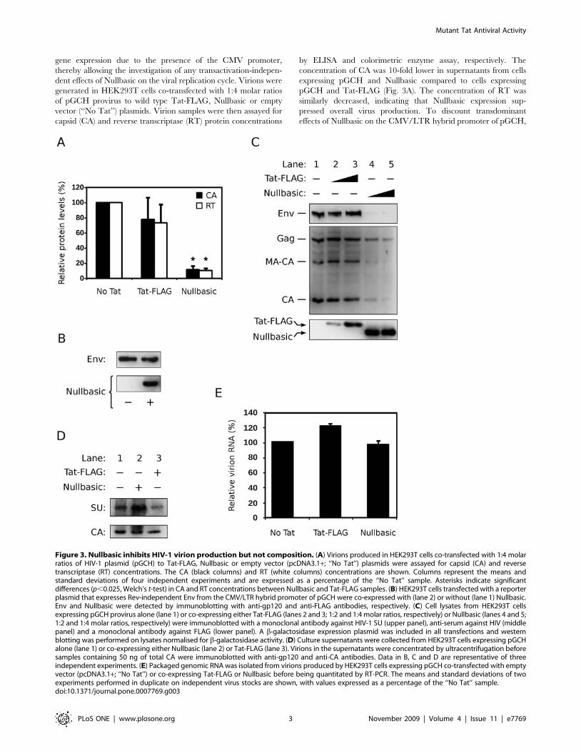
gene expression due to the presence of the CMV promoter,
thereby allowing the investigation of any transactivation-indepen-
dent effects of Nullbasic on the viral replication cycle. Virions were
generated in HEK293T cells co-transfected with 1:4 molar ratios
of pGCH provirus to wild type Tat-FLAG, Nullbasic or empty
vector (‘‘No Tat’’) plasmids. Virion samples were then assayed for
capsid (CA) and reverse transcriptase (RT) protein concentrations
by ELISA and colorimetric enzyme assay, respectively. The
concentration of CA was 10-fold lower in supernatants from cells
expressing pGCH and Nullbasic compared to cells expressing
pGCH and Tat-FLAG (Fig. 3A). The concentration of RT was
similarly decreased, indicating that Nullbasic expression sup-
pressed overall virus production. To discount transdominant
effects of Nullbasic on the CMV/LTR hybrid promoter of pGCH,
Figure 3. Nullbasic inhibits HIV-1 virion production but not composition. (A) Virions produced in HEK293T cells co-transfected with 1:4 molarratios of HIV-1 plasmid (pGCH) to Tat-FLAG, Nullbasic or empty vector (pcDNA3.1+; ‘‘No Tat’’) plasmids were assayed for capsid (CA) and reversetranscriptase (RT) concentrations. The CA (black columns) and RT (white columns) concentrations are shown. Columns represent the means andstandard deviations of four independent experiments and are expressed as a percentage of the ‘‘No Tat’’ sample. Asterisks indicate significantdifferences (p,0.025, Welch’s t-test) in CA and RT concentrations between Nullbasic and Tat-FLAG samples. (B) HEK293T cells transfected with a reporterplasmid that expresses Rev-independent Env from the CMV/LTR hybrid promoter of pGCH were co-expressed with (lane 2) or without (lane 1) Nullbasic.Env and Nullbasic were detected by immunoblotting with anti-gp120 and anti-FLAG antibodies, respectively. (C) Cell lysates from HEK293T cellsexpressing pGCH provirus alone (lane 1) or co-expressing either Tat-FLAG (lanes 2 and 3; 1:2 and 1:4 molar ratios, respectively) or Nullbasic (lanes 4 and 5;1:2 and 1:4 molar ratios, respectively) were immunoblotted with a monoclonal antibody against HIV-1 SU (upper panel), anti-serum against HIV (middlepanel) and a monoclonal antibody against FLAG (lower panel). A b-galactosidase expression plasmid was included in all transfections and westernblotting was performed on lysates normalised for b-galactosidase activity. (D) Culture supernatants were collected from HEK293T cells expressing pGCHalone (lane 1) or co-expressing either Nullbasic (lane 2) or Tat-FLAG (lane 3). Virions in the supernatants were concentrated by ultracentrifugation beforesamples containing 50 ng of total CA were immunoblotted with anti-gp120 and anti-CA antibodies. Data in B, C and D are representative of threeindependent experiments. (E) Packaged genomic RNA was isolated from virions produced by HEK293T cells expressing pGCH co-transfected with emptyvector (pcDNA3.1+; ‘‘No Tat’’) or co-expressing Tat-FLAG or Nullbasic before being quantitated by RT-PCR. The means and standard deviations of twoexperiments performed in duplicate on independent virus stocks are shown, with values expressed as a percentage of the ‘‘No Tat’’ sample.doi:10.1371/journal.pone.0007769.g003
Mutant Tat Antiviral Activity
PLoS ONE | www.plosone.org 3 November 2009 | Volume 4 | Issue 11 | e7769
a reporter construct expressing Rev-independent HIV-1 envelope
(Env) [17] from the CMV/LTR promoter was co-expressed with
Nullbasic in HEK293T cells. Nullbasic had no effect on the
expression of Env from this reporter (Fig. 3B), indicating that
Nullbasic does not significantly affect expression from the CMV/
LTR promoter. Taken together, these experiments suggest that
Nullbasic substantially reduces virion production by a mechanism
independent of the inhibition of Tat transactivation.
To further examine the production of HIV-1 viral proteins,
western blotting was performed on b-galactosidase-equalized
lysates of cells co-expressing pGCH provirus and either Nullbasic
or Tat-FLAG. Figure 3C shows that provirus-expressed Gag levels
were reduced in the presence of Nullbasic (lanes 4 and 5)
compared to Tat-FLAG (lanes 2 and 3). Gag-related proteolytic
products, specifically HIV-1 p41gag (MA-CA) and CA, were
proportionally reduced. This is consistent with the reduction in
virion CA and RT protein levels observed in the supernatants of
cells co-expressing Nullbasic (Fig. 3A). Expression of HIV-1 Env in
the lysates was also reduced in the presence of Nullbasic (Fig. 3C).
The decrease in Env levels by Nullbasic appeared to be greater
than the decrease in Gag levels, but this is likely due to differences
in antibody sensitivities since subsequent western blot analyses of
purified virions showed CA and envelope surface antigen (SU)
levels to be proportional when HIV-1 was co-expressed with either
Nullbasic or Tat-FLAG (Fig. 3D). Finally, co-expression of
Nullbasic did not significantly affect packaging of viral genomic
RNA into HIV-1 virions compared to co-expression of Tat-FLAG
(Fig. 3E). These experiments demonstrate that Nullbasic potently
inhibits HIV-1 gene expression, leading to decreased production
of the viral structural proteins without altering the relative protein
or RNA compositions of virions.
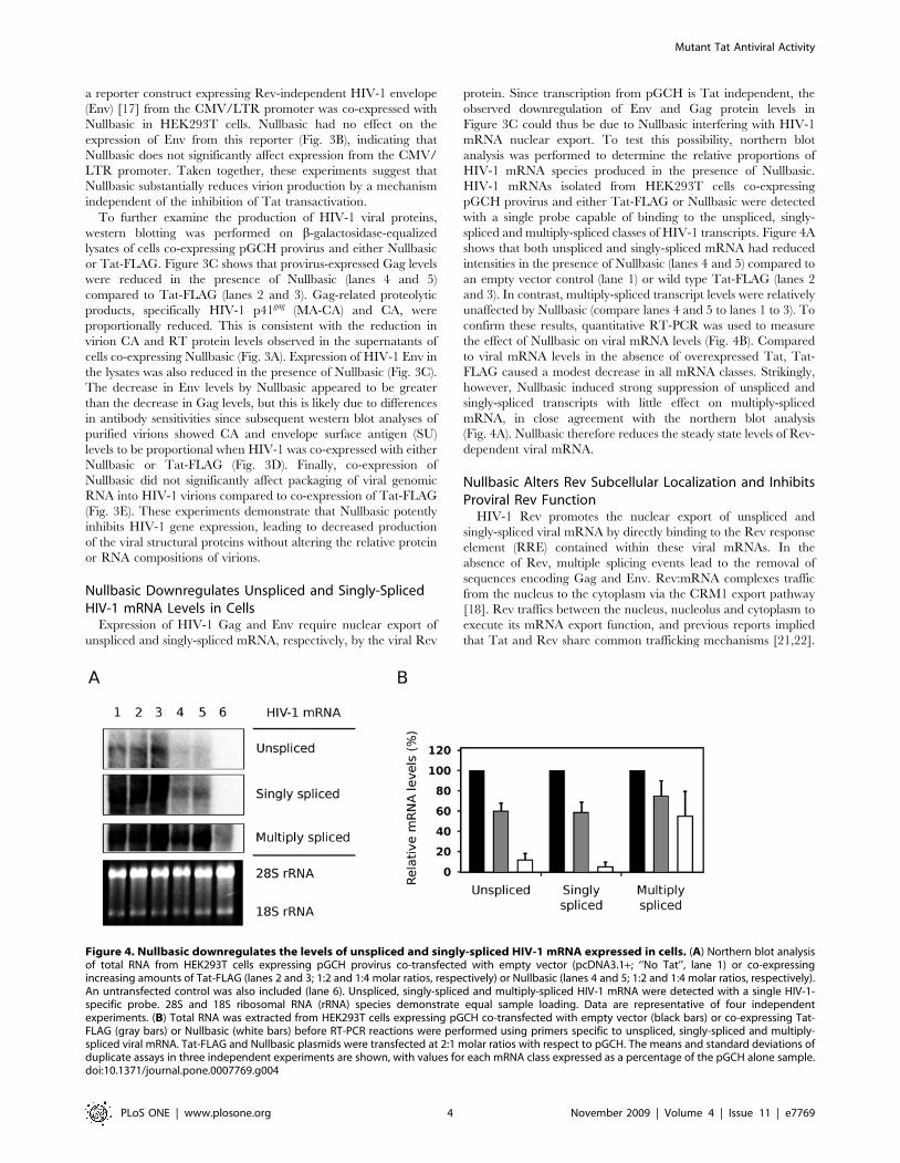
Nullbasic Downregulates Unspliced and Singly-SplicedHIV-1 mRNA Levels in Cells
Expression of HIV-1 Gag and Env require nuclear export of
unspliced and singly-spliced mRNA, respectively, by the viral Rev
protein. Since transcription from pGCH is Tat independent, the
observed downregulation of Env and Gag protein levels in
Figure 3C could thus be due to Nullbasic interfering with HIV-1
mRNA nuclear export. To test this possibility, northern blot
analysis was performed to determine the relative proportions of
HIV-1 mRNA species produced in the presence of Nullbasic.
HIV-1 mRNAs isolated from HEK293T cells co-expressing
pGCH provirus and either Tat-FLAG or Nullbasic were detected
with a single probe capable of binding to the unspliced, singly-
spliced and multiply-spliced classes of HIV-1 transcripts. Figure 4A
shows that both unspliced and singly-spliced mRNA had reduced
intensities in the presence of Nullbasic (lanes 4 and 5) compared to
an empty vector control (lane 1) or wild type Tat-FLAG (lanes 2
and 3). In contrast, multiply-spliced transcript levels were relatively
unaffected by Nullbasic (compare lanes 4 and 5 to lanes 1 to 3). To
confirm these results, quantitative RT-PCR was used to measure
the effect of Nullbasic on viral mRNA levels (Fig. 4B). Compared
to viral mRNA levels in the absence of overexpressed Tat, Tat-
FLAG caused a modest decrease in all mRNA classes. Strikingly,
however, Nullbasic induced strong suppression of unspliced and
singly-spliced transcripts with little effect on multiply-spliced
mRNA, in close agreement with the northern blot analysis
(Fig. 4A). Nullbasic therefore reduces the steady state levels of Rev-
dependent viral mRNA.
Nullbasic Alters Rev Subcellular Localization and InhibitsProviral Rev Function
HIV-1 Rev promotes the nuclear export of unspliced and
singly-spliced viral mRNA by directly binding to the Rev response
element (RRE) contained within these viral mRNAs. In the
absence of Rev, multiple splicing events lead to the removal of
sequences encoding Gag and Env. Rev:mRNA complexes traffic
from the nucleus to the cytoplasm via the CRM1 export pathway
[18]. Rev traffics between the nucleus, nucleolus and cytoplasm to
execute its mRNA export function, and previous reports implied
that Tat and Rev share common trafficking mechanisms [21,22].
Figure 4. Nullbasic downregulates the levels of unspliced and singly-spliced HIV-1 mRNA expressed in cells. (A) Northern blot analysisof total RNA from HEK293T cells expressing pGCH provirus co-transfected with empty vector (pcDNA3.1+; ‘‘No Tat’’, lane 1) or co-expressingincreasing amounts of Tat-FLAG (lanes 2 and 3; 1:2 and 1:4 molar ratios, respectively) or Nullbasic (lanes 4 and 5; 1:2 and 1:4 molar ratios, respectively).An untransfected control was also included (lane 6). Unspliced, singly-spliced and multiply-spliced HIV-1 mRNA were detected with a single HIV-1-specific probe. 28S and 18S ribosomal RNA (rRNA) species demonstrate equal sample loading. Data are representative of four independentexperiments. (B) Total RNA was extracted from HEK293T cells expressing pGCH co-transfected with empty vector (black bars) or co-expressing Tat-FLAG (gray bars) or Nullbasic (white bars) before RT-PCR reactions were performed using primers specific to unspliced, singly-spliced and multiply-spliced viral mRNA. Tat-FLAG and Nullbasic plasmids were transfected at 2:1 molar ratios with respect to pGCH. The means and standard deviations ofduplicate assays in three independent experiments are shown, with values for each mRNA class expressed as a percentage of the pGCH alone sample.doi:10.1371/journal.pone.0007769.g004
Mutant Tat Antiviral Activity
PLoS ONE | www.plosone.org 4 November 2009 | Volume 4 | Issue 11 | e7769
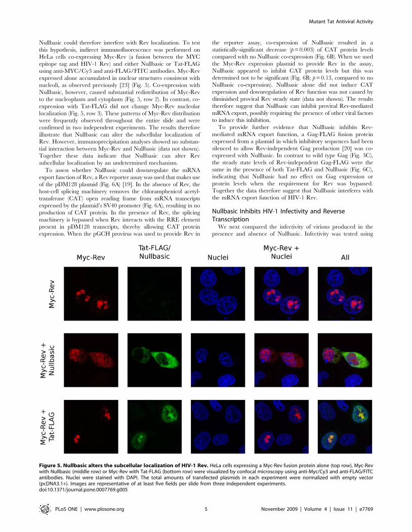
Nullbasic could therefore interfere with Rev localization. To test
this hypothesis, indirect immunofluorescence was performed on
HeLa cells co-expressing Myc-Rev (a fusion between the MYC
epitope tag and HIV-1 Rev) and either Nullbasic or Tat-FLAG
using anti-MYC/Cy3 and anti-FLAG/FITC antibodies. Myc-Rev
expressed alone accumulated in nuclear structures consistent with
nucleoli, as observed previously [23] (Fig. 5). Co-expression with
Nullbasic, however, caused substantial redistribution of Myc-Rev
to the nucleoplasm and cytoplasm (Fig. 5, row 2). In contrast, co-
expression with Tat-FLAG did not change Myc-Rev nucleolar
localization (Fig. 5, row 3). These patterns of Myc-Rev distribution
were frequently observed throughout the entire slide and were
confirmed in two independent experiments. The results therefore
illustrate that Nullbasic can alter the subcellular localization of
Rev. However, immunoprecipitation analyses showed no substan-
tial interaction between Myc-Rev and Nullbasic (data not shown).
Together these data indicate that Nullbasic can alter Rev
subcellular localization by an undetermined mechanism.
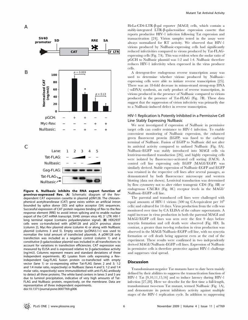
To assess whether Nullbasic could downregulate the mRNA
export function of Rev, a Rev reporter assay was used that makes use
of the pDM128 plasmid (Fig. 6A) [19]. In the absence of Rev, the
host-cell splicing machinery removes the chloramphenicol acetyl-
transferase (CAT) open reading frame from mRNA transcripts
expressed by the plasmid’s SV40 promoter (Fig. 6A), resulting in no
production of CAT protein. In the presence of Rev, the splicing
machinery is bypassed when Rev interacts with the RRE element
present in pDM128 transcripts, thereby allowing CAT protein
expression. When the pGCH provirus was used to provide Rev in
the reporter assay, co-expression of Nullbasic resulted in a
statistically-significant decrease (p = 0.003) of CAT protein levels
compared with no Nullbasic co-expression (Fig. 6B). When we used
the Myc-Rev expression plasmid to provide Rev in the assay,
Nullbasic appeared to inhibit CAT protein levels but this was
determined not to be significant (Fig. 6B; p = 0.13, compared to no
Nullbasic co-expression). Nullbasic alone did not induce CAT
expression and downregulation of Rev function was not caused by
diminished proviral Rev steady state (data not shown). The results
therefore suggest that Nullbasic can inhibit proviral Rev-mediated
mRNA export, possibly requiring the presence of other viral factors
to induce this inhibition.
To provide further evidence that Nullbasic inhibits Rev-
mediated mRNA export function, a Gag-FLAG fusion protein
expressed from a plasmid in which inhibitory sequences had been
silenced to allow Rev-independent Gag production [20] was co-
expressed with Nullbasic. In contrast to wild type Gag (Fig. 3C),
the steady state levels of Rev-independent Gag-FLAG were the
same in the presence of both Tat-FLAG and Nullbasic (Fig. 6C),
indicating that Nullbasic had no effect on Gag expression or
protein levels when the requirement for Rev was bypassed.
Together the data therefore suggest that Nullbasic interferes with
the mRNA export function of HIV-1 Rev.
Nullbasic Inhibits HIV-1 Infectivity and ReverseTranscription
We next compared the infectivity of virions produced in the
presence and absence of Nullbasic. Infectivity was tested using
Figure 5. Nullbasic alters the subcellular localization of HIV-1 Rev. HeLa cells expressing a Myc-Rev fusion protein alone (top row), Myc-Revwith Nullbasic (middle row) or Myc-Rev with Tat-FLAG (bottom row) were visualized by confocal microscopy using anti-Myc/Cy3 and anti-FLAG/FITCantibodies. Nuclei were stained with DAPI. The total amounts of transfected plasmids in each experiment were normalized with empty vector(pcDNA3.1+). Images are representative of at least five fields per slide from three independent experiments.doi:10.1371/journal.pone.0007769.g005
Mutant Tat Antiviral Activity
PLoS ONE | www.plosone.org 5 November 2009 | Volume 4 | Issue 11 | e7769
HeLa-CD4-LTR-b-gal reporter (MAGI) cells, which contain a
stably-integrated LTR-b-galactosidase expression cassette that
reports productive HIV-1 infection following Tat expression and
transactivation [24]. Virion samples tested in the assay were
always normalized for RT activity. We observed that HIV-1
virions produced by Nullbasic-expressing cells had significantly
reduced infectivities compared to virons produced by Tat-FLAG-
expressing cells (Fig. 7A). This was evident when the molar ratio of
pGCH to Nullbasic plasmid was 1:2 and 1:4. Nullbasic therefore
reduces HIV-1 infectivity when expressed in the virus producer
cells.
A detergent-free endogenous reverse transcription assay was
used to determine whether virions produced by Nullbasic-
expressing cells were able to initiate reverse transcription [25].
There was an 18-fold decrease in minus-strand strong-stop DNA
(–ssDNA) synthesis, an early product of reverse transcription, in
virions produced in the presence of Nullbasic compared to virions
produced in the presence of Tat-FLAG (Fig. 7B). These data
suggest that the suppression of virion infectivity was primarily due
to a Nullbasic-induced defect in reverse transcription.
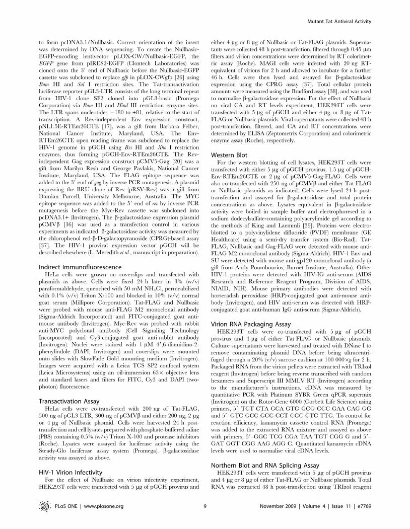
HIV-1 Replication Is Potently Inhibited in a Permissive CellLine Stably Expressing Nullbasic
We next investigated if expression of Nullbasic in permissive
target cells can confer resistance to HIV-1 infection. To enable
convenient monitoring of Nullbasic expression, the enhanced
green fluorescent protein (EGFP) was fused to the carboxy
terminal of Nullbasic. Fusion of EGFP to Nullbasic did not alter
its antiviral activity compared to unfused Nullbasic (Fig. 8A).
Nullbasic-EGFP was stably introduced into MAGI cells via
lentivirus-mediated transduction [26], and highly expressing cells
were isolated by fluorescence-activated cell sorting (FACS). A
control cell line expressing only EGFP (MAGI/EGFP) was
similarly derived. Stable expression of Nullbasic-EGFP and EGFP
was retained in the respective cell lines after several passages, as
demonstrated by both fluorescence microscopy and western
blotting (data not shown). Lentiviral transduction was determined
by flow cytometry not to alter either transgenic CD4 (Fig. 8B) or
endogenous CXCR4 (Fig. 8C) receptor levels in the MAGI/
Nullbasic-EGFP cell line.
The parental and transduced cell lines were challenged with
equal amounts of HIV-1 virions (500 ng CA-equivalent per 106
cells) and cultured for 14 days. Virus production from the cells was
monitored over time by CA ELISA of the culture supernatants. A
rapid increase in virus production in both the parental MAGI and
MAGI/EGFP cell lines was seen over the first 9 days before
syncytia formation and cell death were observed (Fig. 8D). In
contrast, a greater than two-log reduction in virus production was
observed in the MAGI/Nullbasic-EGFP cell line, with no syncytia
formation or cell death being apparent even at the end of the
experiment. These results were confirmed in two independently
derived MAGI/Nullbasic-EGFP cell lines. Expression of Nullbasic
in permissive cells is therefore protective against HIV-1 challenge
and suppresses viral spread.
Discussion
Transdominant-negative Tat mutants have to date been mainly
defined by their abilities to suppress the transactivation function of
HIV-1 Tat [9,10,11,15,16] and to induce latency during HIV-1
infection [27,28]. Here we describe for the first time a full-length,
transdominant two-exon Tat mutant, termed Nullbasic (Fig. 1A),
and demonstrate its potent inhibitory activity against multiple
stages of the HIV-1 replication cycle. In addition to suppressing
Figure 6. Nullbasic inhibits the RNA export function ofprovirus-expressed Rev. (A) Schematic diagram of the Rev-dependent CAT expression cassette in plasmid pDM128. The chloram-phenicol acetyltransferase (CAT) gene exists within an artificial intronbounded by splice donor (SD) and splice acceptor (SA) sequences.Successful expression of CAT protein requires binding of Rev to the Revresponse element (RRE) to avoid intron splicing and to enable nuclearexport of the CAT mRNA transcript. SV40: simian virus 40; 39 LTR: HIV-1long terminal repeat (contains polyadenylation signal). (B) HEK293Tcells were co-transfected with pDM128 and pGCH provirus alone(column 2), Myc-Rev plasmid alone (column 4) or along with Nullbasicplasmid (columns 3 and 5). Empty vector (pcDNA3.1+) was used tonormalize the total amount of transfected plasmids. A pDM128 onlytransfection was included as a negative control (column 1) and aconstitutive b-galactosidase plasmid was included in all transfections toaccount for variations in transfection efficiencies. CAT expression wasmeasured by ELISA and is expressed relative to b-galactosidase activity(ng/mU). Columns represent means and standard deviations of threeindependent experiments. (C) Lysates from cells expressing a Rev-independent Gag-FLAG fusion protein co-transfected with emptyvector (lane 1) or co-expressing either Tat-FLAG (lanes 2 and 3; 1:2and 1:4 molar ratio, respectively) or Nullbasic (lanes 4 and 5; 1:2 and 1:4molar ratio, respectively) were immunoblotted with anti-FLAG antibodyto detect all three proteins. The white band centers in lanes 3 and 5 aredue to luminol precipitation, indicative of very high amounts of Tat-FLAG and Nullbasic protein, respectively, on the membrane. Data arerepresentative of three independent experiments.doi:10.1371/journal.pone.0007769.g006
Mutant Tat Antiviral Activity
PLoS ONE | www.plosone.org 6 November 2009 | Volume 4 | Issue 11 | e7769
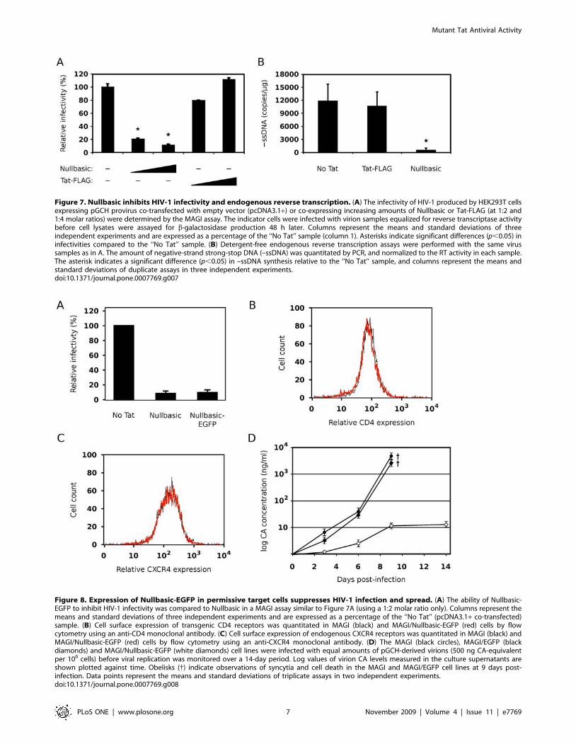
Figure 7. Nullbasic inhibits HIV-1 infectivity and endogenous reverse transcription. (A) The infectivity of HIV-1 produced by HEK293T cellsexpressing pGCH provirus co-transfected with empty vector (pcDNA3.1+) or co-expressing increasing amounts of Nullbasic or Tat-FLAG (at 1:2 and1:4 molar ratios) were determined by the MAGI assay. The indicator cells were infected with virion samples equalized for reverse transcriptase activitybefore cell lysates were assayed for b-galactosidase production 48 h later. Columns represent the means and standard deviations of threeindependent experiments and are expressed as a percentage of the ‘‘No Tat’’ sample (column 1). Asterisks indicate significant differences (p,0.05) ininfectivities compared to the ‘‘No Tat’’ sample. (B) Detergent-free endogenous reverse transcription assays were performed with the same virussamples as in A. The amount of negative-strand strong-stop DNA (–ssDNA) was quantitated by PCR, and normalized to the RT activity in each sample.The asterisk indicates a significant difference (p,0.05) in –ssDNA synthesis relative to the ‘‘No Tat’’ sample, and columns represent the means andstandard deviations of duplicate assays in three independent experiments.doi:10.1371/journal.pone.0007769.g007
Figure 8. Expression of Nullbasic-EGFP in permissive target cells suppresses HIV-1 infection and spread. (A) The ability of Nullbasic-EGFP to inhibit HIV-1 infectivity was compared to Nullbasic in a MAGI assay similar to Figure 7A (using a 1:2 molar ratio only). Columns represent themeans and standard deviations of three independent experiments and are expressed as a percentage of the ‘‘No Tat’’ (pcDNA3.1+ co-transfected)sample. (B) Cell surface expression of transgenic CD4 receptors was quantitated in MAGI (black) and MAGI/Nullbasic-EGFP (red) cells by flowcytometry using an anti-CD4 monoclonal antibody. (C) Cell surface expression of endogenous CXCR4 receptors was quantitated in MAGI (black) andMAGI/Nullbasic-EGFP (red) cells by flow cytometry using an anti-CXCR4 monoclonal antibody. (D) The MAGI (black circles), MAGI/EGFP (blackdiamonds) and MAGI/Nullbasic-EGFP (white diamonds) cell lines were infected with equal amounts of pGCH-derived virions (500 ng CA-equivalentper 106 cells) before viral replication was monitored over a 14-day period. Log values of virion CA levels measured in the culture supernatants areshown plotted against time. Obelisks ({) indicate observations of syncytia and cell death in the MAGI and MAGI/EGFP cell lines at 9 days post-infection. Data points represent the means and standard deviations of triplicate assays in two independent experiments.doi:10.1371/journal.pone.0007769.g008
Mutant Tat Antiviral Activity
PLoS ONE | www.plosone.org 7 November 2009 | Volume 4 | Issue 11 | e7769

Tat transactivation (Fig. 2), Nullbasic reduced HIV-1 virion
production by suppressing Rev-dependant RNA export (Figs. 3
and 4) and virus produced in the presence of Nullbasic was
severely defective for reverse transcription (Fig. 7B). As a result of
these multiple inhibitory activities, expression of Nullbasic in
permissive cells conferred strong resistance against high-dose
HIV-1 challenge and the reduction by greater than two orders of
magnitude of a spreading viral infection (Fig. 8). To our
knowledge, this is the first demonstration of a transdominant
Tat mutant that targets multiple, distinct steps in the HIV-1
replication cycle: proviral gene transcription, Rev-dependent
mRNA transport and reverse transcription.
Nullbasic downregulated the expression of Gag and Env from a
HIV-1 proviral plasmid that expressed viral mRNA from a CMV
promoter (Fig. 3C). Two different assays, quantitation of viral
mRNA (Fig. 4) and a functional assay (Fig. 6), pointed to a defect
in Rev mRNA export function. Nullbasic appeared to disrupt Rev
distribution (Fig. 5) and function leading to decreased steady state
levels of unspliced and singly-spliced viral mRNA, resulting in the
observed downregulation of Gag and Env protein levels,
respectively. Interestingly, Nullbasic did not significantly affect
the mRNA export function of ectopically-expressed Rev (Fig. 6B,
Myc-Rev), suggesting that a complex interaction between
Nullbasic, Rev and other HIV-1 factors is required to inhibit
viral mRNA export. Furthermore, the data imply that Nullbasic
inhibits Rev function via an indirect mechanism. Further
investigation is required to determine which viral or cellular
factor intermediates between Nullbasic and Rev to enable viral
mRNA export inhibition.
Rev normally binds the Rev response element (RRE), an RNA
structure located within HIV-1 env, to facilitate export of unspliced
and singly-spliced mRNA from the nucleus. A dominant-negative
Rev mutant called M10 has been described that was shown to
inhibit wild type Rev function [29]. The M10 mutant protein is
dominant negative because it retains the ability to bind the HIV-1
RRE but is unable to promote export of the viral mRNA from the
nucleus, thereby inhibiting HIV-1 replication [29,30]. Our data
suggest that Nullbasic and M10 inhibit Rev by different
mechanisms. Confocal microscopy experiments indicated that
Nullbasic can disrupt Rev subcellular localization (Fig. 5). The
nucleolar localization of Rev appears to be important for its
function [23,31], so the Nullbasic-induced redistribution of Rev
from nucleolus to nucleoplasm and cytoplasm is likely to be
necessary, but is not sufficient, to inhibit Rev function. There are
reports that Tat and Rev share common nuclear trafficking
pathways involving importin b [22] and B23 (nucleophosmin)
[21,23], so it is possible that Nullbasic may directly interfere with
Rev trafficking. Tat trafficking, however, remains controversial,
with conflicting reports that Tat nuclear accumulation requires
active, factor-dependent pathways [32] or passive, factor-inde-
pendent mechanisms [33]. The lack of a demonstrable interaction
between Nullbasic and Myc-Rev in immunoprecipitation exper-
iments (unpublished observations) suggests that Nullbasic inter-
feres with Rev trafficking by an indirect mechanism. Conceivably,
Nullbasic may sequester cellular factors (such as importins)
normally required for Rev nucleolar targeting. Whatever the
mechanism, disruption of Rev trafficking and therefore HIV-1
mRNA export function represents a major antiviral activity of
Nullbasic.
The third major activity of Nullbasic was abrogation of
intravirion reverse transcription activity (Fig. 7B). Co-expression
of Nullbasic in virus producer cells did not alter the RNA content
of virions (Fig. 3E), indicating that Nullbasic does not affect HIV-1
genomic RNA packaging. Tat is usually only present in virions at
very low concentrations [34]. However, increased nonspecific
inclusion of Nullbasic into virions may occur due to the high
cytoplasmic concentrations of Nullbasic (Fig. 1B). Once in the
virion, Nullbasic may have a dominant negative effect on Tat-
mediated enhancement of reverse transcription [25]. Alternatively,
Nullbasic may negatively affect nucleocapsid activity, which was
recently reported to precisely regulate reverse transcription [35].
Nullbasic might also bind other crucial intravirion factors and
thereby indirectly disrupt the regulation of reverse transcription.
Whatever the mechanism, virions produced by cells expressing
Nullbasic have low infectivity most likely due to a reverse
transcription defect.
The potential therapeutic value of Nullbasic is illustrated by
experiments showing that Nullbasic expression protected cells
from high-dose HIV-1 infection (500 ng CA-equivalent virions per
106 cells) by greater than two orders of magnitude compared to
control cells (Fig. 8D). Furthermore, Nullbasic-expressing cells
were protected against HIV-1-induced syncytia formation and cell
death, indicating that Nullbasic expression protected cells against a
spreading infection. All three of the previously described antiviral
activities of Nullbasic (inhibition of transactivation, Rev function
and reverse transcription) likely combined to suppress this
spreading infection.
In conclusion, we demonstrate the potent antiviral activity of a
transdominant Tat mutant and show that multiple steps in the
HIV-1 replication cycle are targeted. In addition to negative
effects on viral gene expression, we report for the first time that
Nullbasic also inhibits Rev-dependent viral mRNA transport and
intravirion reverse transcription. Moreover, these inhibitory effects
combined potently to reduce HIV infection, illustrating Nullbasic
and its activities as potential avenues for the development of new
therapeutic interventions. Identification of the cellular or viral
factors that interact with Nullbasic to induce Rev and reverse
transcription inhibition may also reveal novel aspects of HIV-1
replication.
Materials and Methods
Cell Culture and TransfectionsHeLa and HEK293T cells were cultured in RPMI 1640
medium supplemented with 100 U/ml penicillin, 100 mg/ml
streptomycin and 10% (v/v) newborn bovine serum (Invitrogen
Corporation). HeLa-CD4-LTR-b-gal (MAGI) cells [24] were
obtained from Michael Emerman through the NIH AIDS
Research and Reference Reagent Program, Division of AIDS,
NIAID, NIH. The cells were maintained in the same medium as
above but supplemented with 0.2 mg/ml G418 and 0.1 mg/ml
hygromycin B. All cells were incubated at 37uC under a
humidified atmosphere of 5% CO2 in air. Transfections were
performed with Lipofectamine 2000 (Invitrogen) or FuGENE 6
(Roche Diagnostics Corporation) transfection reagents according
to the manufacturers’ instructions. Transfections were performed
in 6-cm dishes for reporter assays and western blotting, and 10-cm
dishes for HIV-1 virion production.
PlasmidsThe plasmid expressing the two-exon, 101 amino-acid, BH10
clone of Tat fused to the FLAG epitope (pcDNA3.1/Tat-FLAG)
was a gift from Monsef Benkirane, Institut de Genetique
Humaine, France. Nullbasic was created by firstly removing the
basic domain sequence (corresponding to amino acids 49–57 in
Tat) in pcDNA3.1/Tat-FLAG by inverse PCR before comple-
mentary oligonucleotides encoding the amino acid sequence, Gly–
Gly–Gly–Gly–Gly–Ala–Gly–Gly–Gly were annealed and ligated
Mutant Tat Antiviral Activity
PLoS ONE | www.plosone.org 8 November 2009 | Volume 4 | Issue 11 | e7769
to form pcDNA3.1/Nullbasic. Correct orientation of the insert
was determined by DNA sequencing. To create the Nullbasic-
EGFP-encoding lentivector pLOX-CW/Nullbasic-EGFP, the
EGFP gene from pIRES2-EGFP (Clontech Laboratories) was
cloned onto the 39 end of Nullbasic before the Nullbasic-EGFP
cassette was subcloned to replace gfp in pLOX-CWgfp [26] using
Bam HI and Sal I restriction sites. The Tat-transactivation
luciferase reporter pGL3-LTR consists of the long terminal repeat
from HIV-1 clone SF2 cloned into pGL3-basic (Promega
Corporation) via Bam HI and Hind III restriction enzyme sites.
The LTR spans nucleotides 2180 to +81, relative to the start of
transcription. A Rev-independent Env expression construct,
pNL1.5E-RTEm26CTE [17], was a gift from Barbara Felber,
National Cancer Institute, Maryland, USA. The Env-
RTEm26CTE open reading frame was subcloned to replace the
HIV-1 genome in pGCH using Bss HI and Xho I restriction
enzymes, thus forming pGCH-Env-RTEm26CTE. The Rev-
independent Gag expression construct pCMV5-Gag [20] was a
gift from Marilyn Resh and George Pavlakis, National Cancer
Institute, Maryland, USA. The FLAG epitope sequence was
added to the 39 end of gag by inverse PCR mutagenesis. A plasmid
expressing the BRU clone of Rev (pRSV-Rev) was a gift from
Damian Purcell, University Melbourne, Australia. The MYC
epitope sequence was added to the 59 end of rev by inverse PCR
mutagenesis before the Myc-Rev cassette was subcloned into
pcDNA3.1+ (Invitrogen). The b-galactosidase expression plasmid
pCMVb [36] was used as a transfection control in various
experiments as indicated. b-galactosidase activity was measured by
the chlorophenol red-b-D-galactopyranoside (CPRG)-based assay
[37]. The HIV-1 proviral expression vector pGCH will be
described elsewhere (L. Meredith et al., manuscript in preparation).
Indirect ImmunofluorescenceHeLa cells were grown on coverslips and transfected with
plasmids as above. Cells were fixed 24 h later in 3% (w/v)
paraformaldehyde, quenched with 50 mM NH4Cl, permeabilized
with 0.1% (v/v) Triton X-100 and blocked in 10% (v/v) normal
goat serum (Millipore Corporation). Tat-FLAG and Nullbasic
were probed with mouse anti-FLAG M2 monoclonal antibody
(Sigma-Aldrich Incorporated) and FITC-conjugated goat anti-
mouse antibody (Invitrogen). Myc-Rev was probed with rabbit
anti-MYC polyclonal antibody (Cell Signaling Technology
Incorporated) and Cy3-conjugated goat anti-rabbit antibody
(Invitrogen). Nuclei were stained with 1 mM 49,6-diamidino-2-
phenylindole (DAPI; Invitrogen) and coverslips were mounted
onto slides with SlowFade Gold mounting medium (Invitrogen).
Images were acquired with a Leica TCS SP2 confocal system
(Leica Microsystems) using an oil-immersion 636 objective lens
and standard lasers and filters for FITC, Cy3 and DAPI (two-
photon) fluorescence.
Transactivation AssayHeLa cells were co-transfected with 200 ng of Tat-FLAG,
500 ng of pGL3-LTR, 300 ng of pCMVb and either 200 ng, 2 mg
or 4 mg of Nullbasic plasmid. Cells were harvested 24 h post-
transfection and cell lysates prepared with phosphate-buffered saline
(PBS) containing 0.5% (w/v) Triton X-100 and protease inhibitors
(Roche). Lysates were assayed for luciferase activity using the
Steady-Glo luciferase assay system (Promega). b-galactosidase
activity was assayed as above.
HIV-1 Virion InfectivityFor the effect of Nullbasic on virion infectivity experiment,
HEK293T cells were transfected with 5 mg of pGCH provirus and
either 4 mg or 8 mg of Nullbasic or Tat-FLAG plasmids. Superna-
tants were collected 48 h post-transfection, filtered through 0.45 mm
filters and virion concentrations were determined by RT colorimet-
ric assay (Roche). MAGI cells were infected with 20 ng RT-
equivalent of virions for 2 h and allowed to incubate for a further
46 h. Cells were then lysed and assayed for b-galactosidase
expression using the CPRG assay [37]. Total cellular protein
amounts were measured using the Bradford assay [38], and was used
to normalise b-galactosidase expression. For the effect of Nullbasic
on viral CA and RT levels experiment, HEK293T cells were
transfected with 5 mg of pGCH and either 4 mg or 8 mg of Tat-
FLAG or Nullbasic plasmids. Viral supernatants were collected 48 h
post-transfection, filtered, and CA and RT concentrations were
determined by ELISA (Zeptometrix Corporation) and colorimetric
enzyme assay (Roche), respectively.
Western BlotFor the western blotting of cell lysates, HEK293T cells were
transfected with either 5 mg of pGCH provirus, 1.5 mg of pGCH-
Env-RTEm26CTE or 2 mg of pCMV5-Gag-FLAG. Cells were
also co-transfected with 250 ng of pCMVb and either Tat-FLAG
or Nullbasic plasmids as indicated. Cells were lysed 24 h post-
transfection and assayed for b-galactosidase and total protein
concentrations as above. Lysates equivalent in b-galactosidase
activity were boiled in sample buffer and electrophoresed in a
sodium dodecylsulfate-containing polyacrylimide gel according to
the methods of King and Laemmli [39]. Proteins were electro-
blotted to a polyvinylidene difluoride (PVDF) membrane (GE
Healthcare) using a semi-dry transfer system (Bio-Rad). Tat-
FLAG, Nullbasic and Gag-FLAG were detected with mouse anti-
FLAG M2 monoclonal antibody (Sigma-Aldrich). HIV-1 Env and
SU were detected with mouse anti-gp120 monoclonal antibody (a
gift from Andy Poumbourios, Burnet Institute, Australia). Other
HIV-1 proteins were detected with HIV-IG anti-serum (AIDS
Research and Reference Reagent Program, Division of AIDS,
NIAID, NIH). Mouse primary antibodies were detected with
horseradish peroxidase (HRP)-conjugated goat anti-mouse anti-
body (Invitrogen), and HIV anti-serum was detected with HRP-
conjugated goat anti-human IgG anti-serum (Sigma-Aldrich).
Virion RNA Packaging AssayHEK293T cells were co-transfected with 5 mg of pGCH
provirus and 4 mg of either Tat-FLAG or Nullbasic plasmids.
Culture supernatants were harvested and treated with DNase I to
remove contaminating plasmid DNA before being ultracentri-
fuged through a 20% (v/v) sucrose cushion at 100 0006g for 2 h.
Packaged RNA from the virion pellets were extracted with TRIzol
reagent (Invitrogen) before being reverse transcribed with random
hexamers and Superscript III MMLV RT (Invitrogen) according
to the manufacturer’s instructions. cDNA was measured by
quantitative PCR with Platinum SYBR Green qPCR supermix
(Invitrogen) on the Rotor-Gene 6000 (Corbett Life Science) using
primers, 59–TCT CTA GCA GTG GCG CCC GAA CAG GG
and 59–GTC GCC GCC CCT CGC CTC TTG. To control for
reaction efficiency, kanamycin cassette control RNA (Promega)
was added to the extracted RNA mixture and assayed as above
with primers, 59–GGC TCG CGA TAA TGT CGG G and 59–
GAT GGT CGG AAG AGG C. Quantitated kanamycin cDNA
levels were used to normalise viral cDNA levels.
Northern Blot and RNA Splicing AssayHEK293T cells were transfected with 5 mg of pGCH provirus
and 4 mg or 8 mg of either Tat-FLAG or Nullbasic plasmids. Total
RNA was extracted 48 h post-transfection using TRIzol reagent
Mutant Tat Antiviral Activity
PLoS ONE | www.plosone.org 9 November 2009 | Volume 4 | Issue 11 | e7769
(Invitrogen) according to the manufacturer’s instructions. Twenty
micrograms of RNA samples were electrophoresed in a 1% (w/v)
agarose gel containing 0.6 M formaldehyde and either stained
with ethidium bromide to visualize ribosomal RNA, or blotted to a
nitrocellulose membrane using a TurboBlotter transfer system
(Schleicher and Schuell). RNAs were cross-linked to the
membrane with ultraviolet light and heat, and HIV-1 mRNA
species were detected with a 32P-labelled probe corresponding
to the Bam HI–Xho I fragment in the 39 LTR of HIV-1.
Hybridizations were visualized with a Typhoon 8600 imager (GE
Healthcare). For the RNA splicing assay, total RNA obtained for
the northern analysis was used as a template for quantitative RT-
PCR as described above. The primers used to detect unspliced,
singly-spliced and multiply-spliced viral mRNA have been
previously described [40]. Kanamycin cassette control RNA, as
described above, was included in the assay to normalise for
reaction efficiency.
Rev Reporter AssayHEK293T cells were transfected with either 1 mg of pGCH or
20 ng of pcDNA3.1/Myc-Rev, along with 100 ng of pDM128,
100 ng of pCMVb and 1.5 mg of either Nullbasic or empty vector
(pcDNA3.1+) plasmids. Cells were harvested and lysed 24 h post-
transfection before CAT expression was assayed by ELISA
(Roche) according to the manufacturer’s instructions. b-galacto-
sidase activity was assayed as above.
Establishment of the MAGI/Nullbasic-EGFP Cell LinePseudotyped lentivirus particles were generated by co-transfect-
ing HEK293T cells with pLOX-CW/Nullbasic-EGFP or pLOX-
CWgfp along with pCMVDR8.91 [41] and pHEF-VSV-G (a gift
from Sabine Piller, Westmead Millennium Institute, Australia).
MAGI cells were transduced with lentivirus particles in the
presence of hexadimethrine bromide (8 mg/ml; Sigma-Aldrich) for
48 h before transduction was confirmed by fluorescence micros-
copy. Highly expressing cells were isolated by FACS using a
MoFlo cell sorter (Beckman Coulter Incorporated), and expression
of Nullbasic was confirmed by western blot analysis.
Flow Cytometry of Cell-Surface Receptor LevelsMAGI/Nullbasic-EGFP and nontransduced MAGI cells were
incubated with mouse anti-CD4 or mouse anti-CXCR4 mono-
clonal antibodies (R&D Systems) followed by Cy5-conjugated goat
anti-mouse antibody (Invitrogen). Receptor levels were quantitated
by measuring Cy5 fluorescence using a FACScalibur flow
cytometer (Becton Dickinson), counting 105 cells per sample.
Detergent-Free Endogenous Reverse Transcription AssayHIV-1 virions from HEK293T cells co-transfected with 5 mg of
pGCH provirus and 4 mg of either Tat-FLAG or Nullbasic
plasmids were assayed for endogenous (intravirion) reverse tran-
scription as previously described [42]. Virions were normalized
for equivalent RT activity before assay. The primers used to
quantitate minus-strand strong-stop DNA were 59–GGG TCT
CTC TGG TTG ACC AGA and 59–ACA CAA CAG ACG
GGC ACA CAC.
Viral Replication KineticsMAGI/Nullbasic-EGFP, MAGI/EGFP and nontransduced
MAGI cells were infected with high doses (500 ng CA-equivalent)
of pGCH-derived HIV-1 for 2 h. Non-adsorbed virions were
removed by washing cells with PBS before infected cells were
incubated for a 14-day period. Culture supernatants were
periodically sampled for virion production by CA ELISA in
triplicate.
Statistical AnalysesHartley’s Fmax test was used to determine variance homosce-
dasticity between data sets. Student’s t-test was used to evaluate
null hypotheses for homoscedastic data, while Welch’s t-test was
used for heteroscedastic data. The underlying distributions were
two tailed for all tests and significant difference was defined as
p,0.05.
Acknowledgments
We kindly thank Ann Apolloni for her initial investigations of Nullbasic and
for performing experiments that led to this study.
Author Contributions
Conceived and designed the experiments: LWM HS DH. Performed the
experiments: LWM HS. Analyzed the data: LWM HS AS DH.
Contributed reagents/materials/analysis tools: LM AS. Wrote the paper:
LWM HS AS DH.
References
1. Dayton AI, Sodroski JG, Rosen CA, Goh WC, Haseltine WA (1986) The trans-
activator gene of the human T cell lymphotropic virus type III is required forreplication. Cell 44: 941–947.
2. Hauber J, Perkins A, Heimer EP, Cullen BR (1987) Trans-activation of human
immunodeficiency virus gene expression is mediated by nuclear events. ProcNatl Acad Sci U S A 84: 6364–6368.
3. Hauber J, Malim MH, Cullen BR (1989) Mutational analysis of the conservedbasic domain of human immunodeficiency virus tat protein. J Virol 63:
1181–1187.
4. Ruben S, Perkins A, Purcell R, Joung K, Sia R, et al. (1989) Structural andfunctional characterization of human immunodeficiency virus tat protein. J Virol
63: 1–8.
5. Berkhout B, Silverman RH, Jeang KT (1989) Tat trans-activates the human
immunodeficiency virus through a nascent RNA target. Cell 59: 273–282.
6. Vives E, Brodin P, Lebleu B (1997) A truncated HIV-1 Tat protein basic domainrapidly translocates through the plasma membrane and accumulates in the cell
nucleus. J Biol Chem 272: 16010–16017.
7. Apolloni A, Hooker CW, Mak J, Harrich D (2003) Human immunodeficiency
virus type 1 protease regulation of tat activity is essential for efficient reverse
transcription and replication. J Virol 77: 9912–9921.
8. Barillari G, Gendelman R, Gallo RC, Ensoli B (1993) The Tat protein of human
immunodeficiency virus type 1, a growth factor for AIDS Kaposi sarcoma andcytokine-activated vascular cells, induces adhesion of the same cell types by using
integrin receptors recognizing the RGD amino acid sequence. Proc Natl Acad
Sci U S A 90: 7941–7945.
9. Pearson L, Garcia J, Wu F, Modesti N, Nelson J, et al. (1990) A transdominant
tat mutant that inhibits tat-induced gene expression from the humanimmunodeficiency virus long terminal repeat. Proc Natl Acad Sci U S A 87:
5079–5083.
10. Orsini MJ, Debouck CM (1996) Inhibition of human immunodeficiency virustype 1 and type 2 Tat function by transdominant Tat protein localized to both
the nucleus and cytoplasm. J Virol 70: 8055–8063.
11. Ulich C, Harrich D, Estes P, Gaynor RB (1996) Inhibition of human
immunodeficiency virus type 1 replication is enhanced by a combination of
transdominant Tat and Rev proteins. J Virol 70: 4871–4876.
12. Modesti N, Garcia J, Debouck C, Peterlin M, Gaynor R (1991) Trans-dominant
Tat mutants with alterations in the basic domain inhibit HIV-1 gene expression.New Biol 3: 759–768.
13. Wei P, Garber ME, Fang SM, Fischer WH, Jones KA (1998) A novel CDK9-
associated C-type cyclin interacts directly with HIV-1 Tat and mediates its high-affinity, loop-specific binding to TAR RNA. Cell 92: 451–462.
14. Jeang KT, Xiao H, Rich EA (1999) Multifaceted activities of the HIV-1transactivator of transcription, Tat. J Biol Chem 274: 28837–28840.
15. Echetebu CO, Rhim H, Herrmann CH, Rice AP (1994) Construction and
characterization of a potent HIV-2 Tat transdominant mutant protein. J AcquirImmune Defic Syndr 7: 655–664.
16. Rossi C, Balboni PG, Betti M, Marconi PC, Bozzini R, et al. (1997) Inhibition ofHIV-1 replication by a Tat transdominant negative mutant in human peripheral
blood lymphocytes from healthy donors and HIV-1-infected patients. Gene
Ther 4: 1261–1269.
Mutant Tat Antiviral Activity
PLoS ONE | www.plosone.org 10 November 2009 | Volume 4 | Issue 11 | e7769
17. Smulevitch S, Bear J, Alicea C, Rosati M, Jalah R, et al. (2006) RTE and CTE
mRNA export elements synergistically increase expression of unstable, Rev-dependent HIV and SIV mRNAs. Retrovirology 3: 6.
18. Felber BK, Zolotukhin AS, Pavlakis GN (2007) Posttranscriptional control of
HIV-1 and other retroviruses and its practical applications. Adv Pharmacol 55:161–197.
19. Hope TJ, McDonald D, Huang XJ, Low J, Parslow TG (1990) Mutationalanalysis of the human immunodeficiency virus type 1 Rev transactivator:
essential residues near the amino terminus. J Virol 64: 5360–5366.
20. Tritel M, Resh MD (2000) Kinetic analysis of human immunodeficiency virustype 1 assembly reveals the presence of sequential intermediates. J Virol 74:
5845–5855.21. Li YP (1997) Protein B23 is an important human factor for the nucleolar
localization of the human immunodeficiency virus protein Tat. J Virol 71:4098–4102.
22. Truant R, Cullen BR (1999) The arginine-rich domains present in human
immunodeficiency virus type 1 Tat and Rev function as direct importin beta-dependent nuclear localization signals. Mol Cell Biol 19: 1210–1217.
23. Dundr M, Leno GH, Hammarskjold ML, Rekosh D, Helga-Maria C, et al.(1995) The roles of nucleolar structure and function in the subcellular location of
the HIV-1 Rev protein. J Cell Sci 108: 2811–2823.
24. Kimpton J, Emerman M (1992) Detection of replication-competent andpseudotyped human immunodeficiency virus with a sensitive cell line on the
basis of activation of an integrated beta-galactosidase gene. J Virol 66:2232–2239.
25. Apolloni A, Meredith LW, Suhrbier A, Kiernan R, Harrich D (2007) The HIV-1 Tat protein stimulates reverse transcription in vitro. Curr HIV Res 5:
473–483.
26. Salmon P, Oberholzer J, Occhiodoro T, Morel P, Lou J, et al. (2000) Reversibleimmortalization of human primary cells by lentivector-mediated transfer of
specific genes. Mol Ther 2: 404–414.27. Balboni PG, Bozzini R, Zucchini S, Marconi PC, Grossi MP, et al. (1993)
Inhibition of human immunodeficiency virus reactivation from latency by a tat
transdominant negative mutant. J Med Virol 41: 289–295.28. Caputo A, Grossi MP, Bozzini R, Rossi C, Betti M, et al. (1996) Inhibition of
HIV-1 replication and reactivation from latency by tat transdominant negativemutants in the cysteine rich region. Gene Ther 3: 235–245.
29. Malim MH, McCarn DF, Tiley LS, Cullen BR (1991) Mutational definition ofthe human immunodeficiency virus type 1 Rev activation domain. J Virol 65:
4248–4254.
30. Stauber R, Gaitanaris GA, Pavlakis GN (1995) Analysis of trafficking of Rev and
transdominant Rev proteins in living cells using green fluorescent protein
fusions: transdominant Rev blocks the export of Rev from the nucleus to the
cytoplasm. Virology 213: 439–449.
31. Cochrane AW, Perkins A, Rosen CA (1990) Identification of sequences
important in the nucleolar localization of human immunodeficiency virus Rev:
relevance of nucleolar localization to function. J Virol 64: 881–885.
32. Efthymiadis A, Briggs LJ, Jans DA (1998) The HIV-1 Tat nuclear localization
sequence confers novel nuclear import properties. J Biol Chem 273: 1623–1628.
33. Cardarelli F, Serresi M, Bizzarri R, Giacca M, Beltram F (2007) In vivo study of
HIV-1 Tat arginine-rich motif unveils its transport properties. Mol Ther 15:
1313–1322.
34. Chertova E, Chertov O, Coren LV, Roser JD, Trubey CM, et al. (2006)
Proteomic and biochemical analysis of purified human immunodeficiency virus
type 1 produced from infected monocyte-derived macrophages. J Virol 80:
9039–9052.
35. Houzet L, Morichaud Z, Didierlaurent L, Muriaux D, Darlix JL, et al. (2008)
Nucleocapsid mutations turn HIV-1 into a DNA-containing virus. Nucleic Acids
Res 36: 2311–2319.
36. MacGregor GR, Caskey CT (1989) Construction of plasmids that express E. coli
beta-galactosidase in mammalian cells. Nucleic Acids Res 17: 2365.
37. Eustice DC, Feldman PA, Colberg-Poley AM, Buckery RM, Neubauer RH
(1991) A sensitive method for the detection of beta-galactosidase in transfected
mammalian cells. Biotechniques 11: 739–740, 742–733.
38. Bradford MM (1976) A rapid and sensitive method for the quantitation of
microgram quantities of protein utilizing the principle of protein-dye binding.
Anal Biochem 72: 248–254.
39. King J, Laemmli UK (1971) Polypeptides of the tail fibres of bacteriophage T4.
J Mol Biol 62: 465–477.
40. Arrigo SJ, Weitsman S, Zack JA, Chen IS (1990) Characterization and
expression of novel singly spliced RNA species of human immunodeficiency
virus type 1. J Virol 64: 4585–4588.
41. Zufferey R, Nagy D, Mandel RJ, Naldini L, Trono D (1997) Multiply attenuated
lentiviral vector achieves efficient gene delivery in vivo. Nat Biotechnol 15:
871–875.
42. Warrilow D, Meredith L, Davis A, Burrell C, Li P, et al. (2008) Cell factors
stimulate human immunodeficiency virus type 1 reverse transcription in vitro.
J Virol 82: 1425–1437.
Mutant Tat Antiviral Activity
PLoS ONE | www.plosone.org 11 November 2009 | Volume 4 | Issue 11 | e7769