Embed Size (px)
Citation preview
Long-term Posterior Capsule OpacificationReduction with Square-EdgePolymethylmethacrylate Intraocular Lens
Randomized Controlled Study
Aravind Haripriya, MD,1 David F. Chang, MD,2 Balakrishnan Vijayakumar, MSc,1 Agrawal Niraj, MD,1
Madhu Shekhar, MD,1 Singh Tanpreet, MD,1 Srinivasan Aravind, MD1
Purpose: To objectively assess the long-term posterior capsule opacification (PCO) and neodymium-dopedyttrium aluminium garnet (Nd:YAG) capsulotomy rate of a square-edge (SE) polymethylmethacrylate (PMMA)intraocular lens (IOL) modification in comparison with a round-edge (RE) PMMA IOL or an SE hydrophobic acrylicIOL (SE-Acrylic).
Design: Prospective, randomized, controlled fellow eye clinical study.Participants: Ninety-four patients scheduled for bilateral phacoemulsification had an SE-PMMA IOL
implanted in 1 eye. An RE-PMMA IOL was implanted in the fellow eye in 46 patients (group A), and an SE-AcrylicIOL was implanted in the fellow eye in 48 patients (group B). Randomization was used to determine groupassignment and which IOL was implanted in the first eye to undergo surgery.
Methods: Evaluation of Posterior Capsule Opacification (EPCO) image analysis software was used toobjectively grade PCO density from standardized, high-resolution retroillumination photographs obtainedannually for the first 5 postoperative years and at year 9.
Main Outcome Measures: The PCO scores and Nd:YAG capsulotomy rate.Results: Nine-year follow-up was achieved by 72% from group A and 63% from group B. In group A, the
mean PCO score was significantly lower in the SE-PMMA IOL eyes compared with the contralateral RE-PMMAeyes at all follow-up visits (P < 0.05). In group B, the mean PCO score was statistically lower in the SE-PMMA IOLeyes compared with the contralateral SE-Acrylic IOL eyes at all but the 1- and 3-year follow-up visits. Nine-yearNd:YAG capsulotomy rates were 2% for SE-PMMA IOLs versus 37% for RE-PMMA IOLs in group A (P < 0.001),and 4% for SE-PMMA IOLs versus 10% for SE-Acrylic IOLs in group B (P ¼ 0.435). The RE-PMMA PCO rate didnot plateau and continued to increase throughout the 9-year study period.
Conclusions: This prospective, 9-year fellow eye comparison study suggests that an inexpensive PMMA IOLdesign modificationda squared optic edgedcould significantly reduce the burden of vision-impairing secondarymembrane in developing countries. Ophthalmology 2016;-:1e9 ª 2016 by the American Academy of Ophthal-mology
Sutureless, manual small-incision cataract surgery (M-SICS) is widely used in developing countries, where itachieves excellent outcomes at low cost and with lowcomplication rates for mature cataracts.1e7 In conjunctionwith the larger M-SICS incision, polymethylmethacrylate(PMMA) intraocular lenses (IOLs) are typically usedto reduce cost.8 However, multiple studies have consistentlydocumented a higher rate of posterior capsular opacification(PCO) with PMMA IOLs compared with foldablehydrophobic acrylic IOLs.9e18 Posterior capsular opacifi-cation is a significant cause of visual loss in the developingworld, where many patients receive PMMA IOLs andhave limited access to postoperative care and
ª 2016 by the American Academy of OphthalmologyPublished by Elsevier Inc.
neodymium-doped yttrium aluminium garnet (Nd:YAG)capsulotomy.19
The elegant bilateral rabbit eye experiments byNishi et al20e22 comparing different IOL designs andmaterials demonstrated that a sharp, truncated posterioroptic edge was the key factor in preventing lensepithelial cell (LEC) migration behind the IOL. Anexperimental sharp-edged PMMA optic effectivelyblocked LEC migration in the rabbit model described byNishi and Nishi,21 suggesting that the roundedoptic edge, rather than the material, was responsible forthe higher clinical rate of PCO observed with PMMAIOLs.
1http://dx.doi.org/10.1016/j.ophtha.2016.11.010ISSN 0161-6420/16
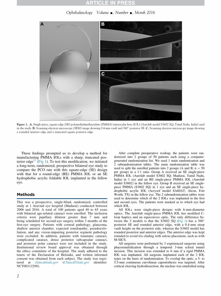
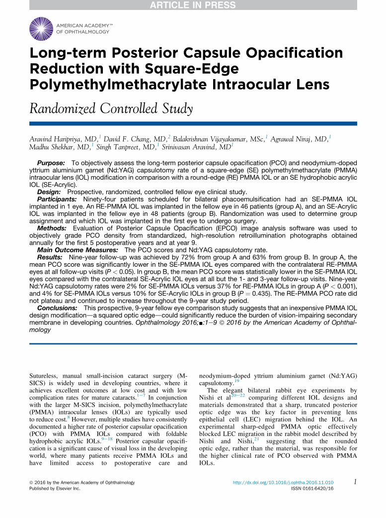
Figure 1. A, Single-piece, square-edge (SE) polymethylmethacrylate (PMMA) intraocular lens (IOL) (Aurolab model S3602 SQ; Tamil Nadu, India) usedin the study. B, Scanning electron microscopy (SEM) image showing 0.4-mm vault and 360� posterior SE. C, Scanning electron microscopy image showinga rounded anterior edge and a truncated square posterior edge.
Ophthalmology Volume -, Number -, Month 2016
These findings prompted us to develop a method formanufacturing PMMA IOLs with a sharp, truncated pos-terior edge21 (Fig 1). To test this modification, we initiateda long-term, randomized, prospective bilateral eye study tocompare the PCO rate with this square-edge (SE) designwith that for a round-edge (RE) PMMA IOL or an SEhydrophobic acrylic foldable IOL implanted in the felloweye.
Methods
This was a prospective, single-blind, randomized, controlledstudy at 1 Aravind eye hospital (Madurai) conducted between2006 and 2016. A total of 100 patients aged 40 to 65 yearswith bilateral age-related cataract were enrolled. The inclusioncriteria were pupillary dilation greater than 7 mm andbeing scheduled for second-eye surgery within 3 months of thefirst-eye surgery. Patients with corneal pathology, glaucoma,shallow anterior chamber, expected zonulopathy, pseudoexfo-liation, and any vision-impairing posterior segment pathologywere excluded. In addition, patients with traumatic cataract,complicated cataract, dense posterior subcapsular cataract,and posterior polar cataract were not included in the study.Institutional review board approval was obtained throughthe ethics committee of the hospital. The study adhered to thetenets of the Declaration of Helsinki, and written informedconsent was obtained from each subject. The study was regis-tered at clinicaltrials.gov (ClinicalTrials.gov identifier:NCT00312299).
2
After complete preoperative workup, the patients were ran-domized into 2 groups of 50 patients each using a computer-generated randomization list. We used 1 main randomization and2 subrandomization tables. The main randomization table wasused to split the enrolled patients into 2 groups (A and B; n ¼ 50per group) in a 1:1 ratio. Group A received an SE single-piecePMMA IOL (Aurolab model S3602 SQ; Madurai, Tamil Nadu,India) in 1 eye and an RE single-piece PMMA IOL (Aurolabmodel S3602) in the fellow eye. Group B received an SE single-piece PMMA (S3602 SQ) in 1 eye and an SE single-piece hy-drophobic acrylic IOL (Acrysof model SA60AT; Alcon, FortWorth, TX) in the fellow eye. The 2 subrandomization tables wereused to determine which of the 2 IOLs was implanted in the firstand second eyes. The patients were masked as to which eye hadwhich IOL.
All IOLs were single-piece designs with 6-mm-diameteroptics. The Aurolab single-piece PMMA IOL has modified C-loop haptics and an equiconvex optic. The only difference be-tween the 2 models is that the S3602 SQ (Fig 1) has a 360�posterior SE and rounded anterior edge, with a 0.4-mm steepvault height on the posterior side, whereas the S3602 model hasrounded posterior and anterior edges. The anterior edge was keptrounded to avoid iris chafing with sulcus placement, such as withM-SICS.
All surgeries were performed by 3 experienced surgeons usingphacoemulsification through a temporal 3-mm scleral tunnelincision. This incision was extended to 6 mm if a rigid PMMAIOL was implanted. All surgeons implanted each of the 3 IOLtypes on the basis of randomization. To overlap the optic, a 5- to5.5-mm continuous curvilinear capsulorhexis was targeted. Aftercortical cleaving hydrodissection, the nucleus was emulsified using
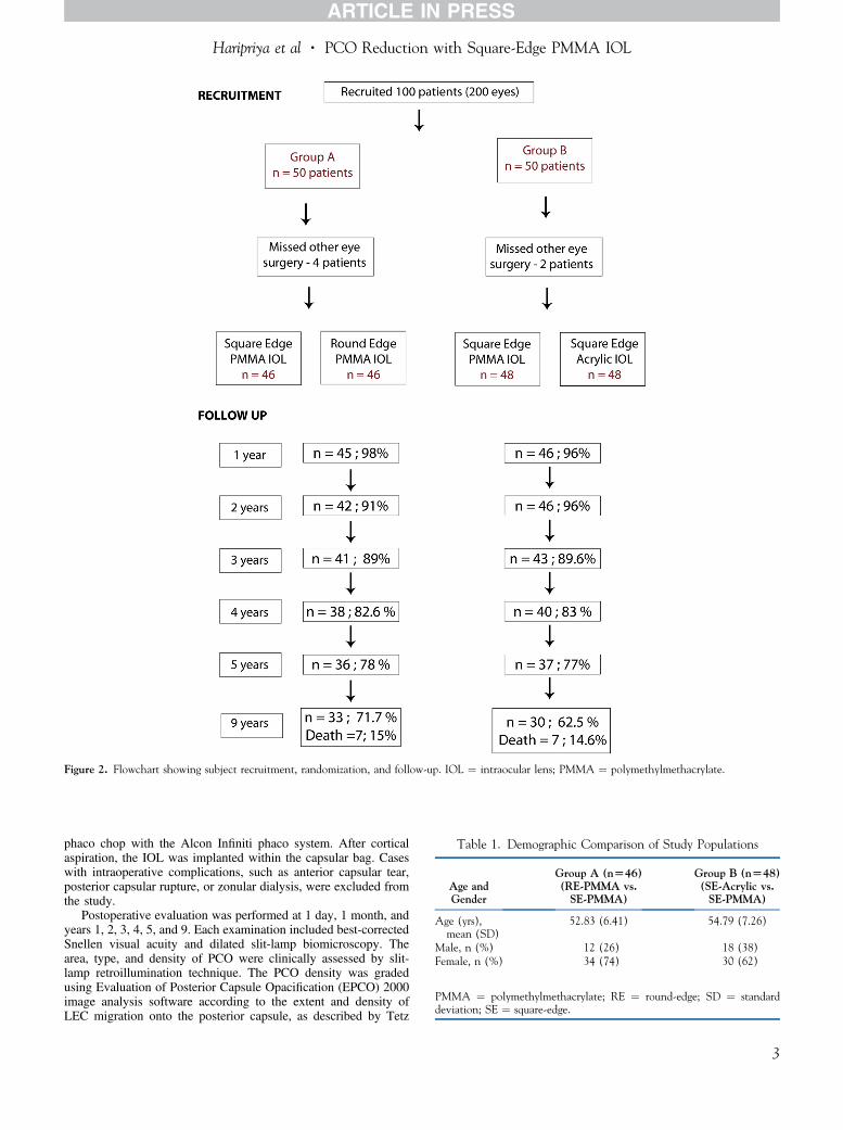
Figure 2. Flowchart showing subject recruitment, randomization, and follow-up. IOL ¼ intraocular lens; PMMA ¼ polymethylmethacrylate.
Table 1. Demographic Comparison of Study Populations
Age andGender
Group A (n[46)(RE-PMMA vs.SE-PMMA)
Group B (n[48)(SE-Acrylic vs.SE-PMMA)
Age (yrs),mean (SD)
52.83 (6.41) 54.79 (7.26)
Male, n (%) 12 (26) 18 (38)Female, n (%) 34 (74) 30 (62)
PMMA ¼ polymethylmethacrylate; RE ¼ round-edge; SD ¼ standarddeviation; SE ¼ square-edge.
Haripriya et al � PCO Reduction with Square-Edge PMMA IOL
phaco chop with the Alcon Infiniti phaco system. After corticalaspiration, the IOL was implanted within the capsular bag. Caseswith intraoperative complications, such as anterior capsular tear,posterior capsular rupture, or zonular dialysis, were excluded fromthe study.
Postoperative evaluation was performed at 1 day, 1 month, andyears 1, 2, 3, 4, 5, and 9. Each examination included best-correctedSnellen visual acuity and dilated slit-lamp biomicroscopy. Thearea, type, and density of PCO were clinically assessed by slit-lamp retroillumination technique. The PCO density was gradedusing Evaluation of Posterior Capsule Opacification (EPCO) 2000image analysis software according to the extent and density ofLEC migration onto the posterior capsule, as described by Tetz
3
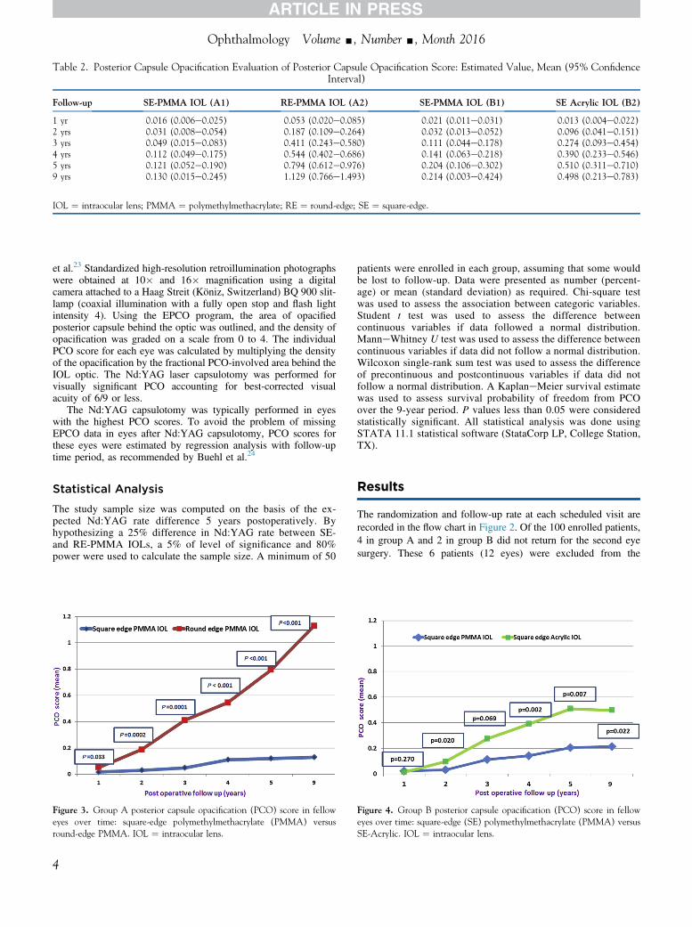
Table 2. Posterior Capsule Opacification Evaluation of Posterior Capsule Opacification Score: Estimated Value, Mean (95% ConfidenceInterval)
Follow-up SE-PMMA IOL (A1) RE-PMMA IOL (A2) SE-PMMA IOL (B1) SE Acrylic IOL (B2)
1 yr 0.016 (0.006e0.025) 0.053 (0.020e0.085) 0.021 (0.011e0.031) 0.013 (0.004e0.022)2 yrs 0.031 (0.008e0.054) 0.187 (0.109e0.264) 0.032 (0.013e0.052) 0.096 (0.041e0.151)3 yrs 0.049 (0.015e0.083) 0.411 (0.243e0.580) 0.111 (0.044e0.178) 0.274 (0.093e0.454)4 yrs 0.112 (0.049e0.175) 0.544 (0.402e0.686) 0.141 (0.063e0.218) 0.390 (0.233e0.546)5 yrs 0.121 (0.052e0.190) 0.794 (0.612e0.976) 0.204 (0.106e0.302) 0.510 (0.311e0.710)9 yrs 0.130 (0.015e0.245) 1.129 (0.766e1.493) 0.214 (0.003e0.424) 0.498 (0.213e0.783)
IOL ¼ intraocular lens; PMMA ¼ polymethylmethacrylate; RE ¼ round-edge; SE ¼ square-edge.
Ophthalmology Volume -, Number -, Month 2016
et al.23 Standardized high-resolution retroillumination photographswere obtained at 10� and 16� magnification using a digitalcamera attached to a Haag Streit (Köniz, Switzerland) BQ 900 slit-lamp (coaxial illumination with a fully open stop and flash lightintensity 4). Using the EPCO program, the area of opacifiedposterior capsule behind the optic was outlined, and the density ofopacification was graded on a scale from 0 to 4. The individualPCO score for each eye was calculated by multiplying the densityof the opacification by the fractional PCO-involved area behind theIOL optic. The Nd:YAG laser capsulotomy was performed forvisually significant PCO accounting for best-corrected visualacuity of 6/9 or less.
The Nd:YAG capsulotomy was typically performed in eyeswith the highest PCO scores. To avoid the problem of missingEPCO data in eyes after Nd:YAG capsulotomy, PCO scores forthese eyes were estimated by regression analysis with follow-uptime period, as recommended by Buehl et al.24
Statistical Analysis
The study sample size was computed on the basis of the ex-pected Nd:YAG rate difference 5 years postoperatively. Byhypothesizing a 25% difference in Nd:YAG rate between SE-and RE-PMMA IOLs, a 5% of level of significance and 80%power were used to calculate the sample size. A minimum of 50
Figure 3. Group A posterior capsule opacification (PCO) score in felloweyes over time: square-edge polymethylmethacrylate (PMMA) versusround-edge PMMA. IOL ¼ intraocular lens.
4
patients were enrolled in each group, assuming that some wouldbe lost to follow-up. Data were presented as number (percent-age) or mean (standard deviation) as required. Chi-square testwas used to assess the association between categoric variables.Student t test was used to assess the difference betweencontinuous variables if data followed a normal distribution.ManneWhitney U test was used to assess the difference betweencontinuous variables if data did not follow a normal distribution.Wilcoxon single-rank sum test was used to assess the differenceof precontinuous and postcontinuous variables if data did notfollow a normal distribution. A KaplaneMeier survival estimatewas used to assess survival probability of freedom from PCOover the 9-year period. P values less than 0.05 were consideredstatistically significant. All statistical analysis was done usingSTATA 11.1 statistical software (StataCorp LP, College Station,TX).
Results
The randomization and follow-up rate at each scheduled visit arerecorded in the flow chart in Figure 2. Of the 100 enrolled patients,4 in group A and 2 in group B did not return for the second eyesurgery. These 6 patients (12 eyes) were excluded from the
Figure 4. Group B posterior capsule opacification (PCO) score in felloweyes over time: square-edge (SE) polymethylmethacrylate (PMMA) versusSE-Acrylic. IOL ¼ intraocular lens.
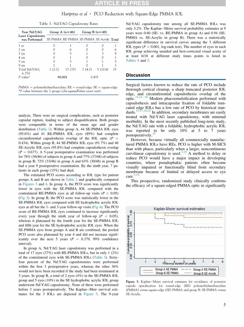
Table 3. Nd:YAG Capsulotomy Rates
Year Nd:YAGLaser Capsulotomywas Performed
Group A (n[46) Group B (n[48)
TotalSE-PMMA RE-PMMA SE-PMMA SE-Acrylic
1 yr 0 1 0 0 12 yrs 0 6 0 0 63 yrs 0 5 1 3 94 yrs 0 1 0 0 15 yrs 0 3 1 1 59 yrs 1 1 0 1 3Total Nd:YAG,n (%)
1 (2.2) 17 (37) 2 (4.2) 5 (10.4) 25
P value* <0.001 0.435
PMMA ¼ polymethylmethacrylate; RE ¼ round-edge; SE ¼ square-edge.*P value between the 2 groups (chi-square/Fisher exact test).
Figure 5. KaplaneMeier survival estimates for avoidance of posteriorcapsule opacification for round-edge (RE) polymethylmethacrylate(PMMA) versus square-edge (SE) PMMA and group B: SE-PMMA versusSE-Acrylic.
Haripriya et al � PCO Reduction with Square-Edge PMMA IOL
analysis. There were no surgical complications, such as posteriorcapsular rupture, leading to subject disqualification. Both groupswere comparable in terms of the mean age and genderdistribution (Table 1). Within group A, 44 SE-PMMA IOL eyes(95.6%) and 41 RE-PMMA IOL eyes (89%) had completecircumferential capsulorhexis overlap of the IOL optic (P ¼0.434). Within group B, 44 SE-PMMA IOL eyes (91.7%) and 46SE-Acrylic IOL eyes (95.8%) had complete capsulorhexis overlap(P ¼ 0.677). A 5-year postoperative examination was performedfor 78% (36/46) of subjects in group A and 77% (37/48) of subjectsin group B; 72% (33/46) in group A and 63% (30/48) in group Bhad a year 9 postoperative examination. By the ninth year, 7 pa-tients in each group (15%) had died.
The estimated PCO scores according to IOL type for patientgroups A and B are shown in Table 2 and graphically comparedin Figures 3 and 4. In group A, the PCO score was significantlylower in eyes with the SE-PMMA IOL compared with thecontralateral RE-PMMA eyes at all follow-up visits (P < 0.05)(Fig 3). In group B, the PCO score was statistically lower in theSE-PMMA IOL eyes compared with SE hydrophobic acrylic IOLeyes at all but the 1- and 3-year follow-up visits (Fig 4). The PCOscore of RE-PMMA IOL eyes continued to increase significantlyevery year through the ninth year of follow-up (P < 0.05),whereas it plateaued by the fourth year for the SE-PMMA IOLand fifth year for the SE hydrophobic acrylic IOL eyes. When theSE-PMMA eyes from groups A and B are combined, the pooledPCO score also plateaued by year 4 and did not increase signif-icantly over the next 5 years (P ¼ 0.379, 99% confidenceinterval).
In group A, Nd:YAG laser capsulotomy was performed in atotal of 17 eyes (37%) with RE-PMMA IOLs, but in only 1 (2%)of the contralateral eyes with SE-PMMA IOLs (Table 3). Sixty-four percent of the Nd:YAG capsulotomies were performedwithin the first 3 postoperative years, whereas the other 36%would not have been recorded if the study had been terminated at3 years. In group B, a total of 2 eyes (4%) in the SE-PMMA IOLgroup and 5 eyes (10%) in the SE hydrophobic acrylic IOL groupunderwent Nd:YAG capsulotomy. None of these were performedbefore 3 years postoperatively. The KaplaneMeir survival esti-mates for the 3 IOLs are depicted in Figure 5. The 9-year
Nd:YAG capsulotomy rate among all SE-PMMA IOLs wasonly 3.2%. The KaplaneMeier survival probability estimates at 9years were 0.60 (SE- vs. RE-PMMA in group A) and 0.94 (SE-PMMA vs. SE-Acrylic in group B). There was a statisticallysignificant difference in survival curves among the 3 differentIOL types (P < 0.001, log-rank test). The number of eyes in eachIOL group achieving unaided and best-corrected visual acuity ofat least 6/18 at different study times points is listed inTables 4 and 5.
Discussion
Surgical factors known to reduce the rate of PCO includethorough cortical cleanup, a sharp truncated posterior IOLedge, and circumferential capsulorhexis overlap of theoptic.12,15e18 Modern phacoemulsification performed withcapsulorhexis and intracapsular fixation of foldable trun-cated edge IOLs has a low rate of PCO by historical stan-dards.15,17,18,25 In addition, secondary membranes are easilytreated with Nd:YAG laser capsulotomy, with minimalmorbidity. In the most recently published long-term study,the Nd:YAG rate with a foldable, hydrophobic acrylic IOLwas reported to be only 10% at 5 to 7 yearspostoperatively.25
However, because virtually all commercially manufac-tured PMMA IOLs have REs, PCO is higher with M-SICSthan with phaco, particularly when a larger, noncontinuouscurvilinear capsulotomy is used.3,19 A method to delay orreduce PCO would have a major impact in developingcountries, where pseudophakic patients often becomevisually impaired or functionally blind from secondarymembrane because of limited or delayed access to eyecare.3,19
This prospective, randomized study clinically confirmsthe efficacy of a square-edged PMMA optic in significantly
5
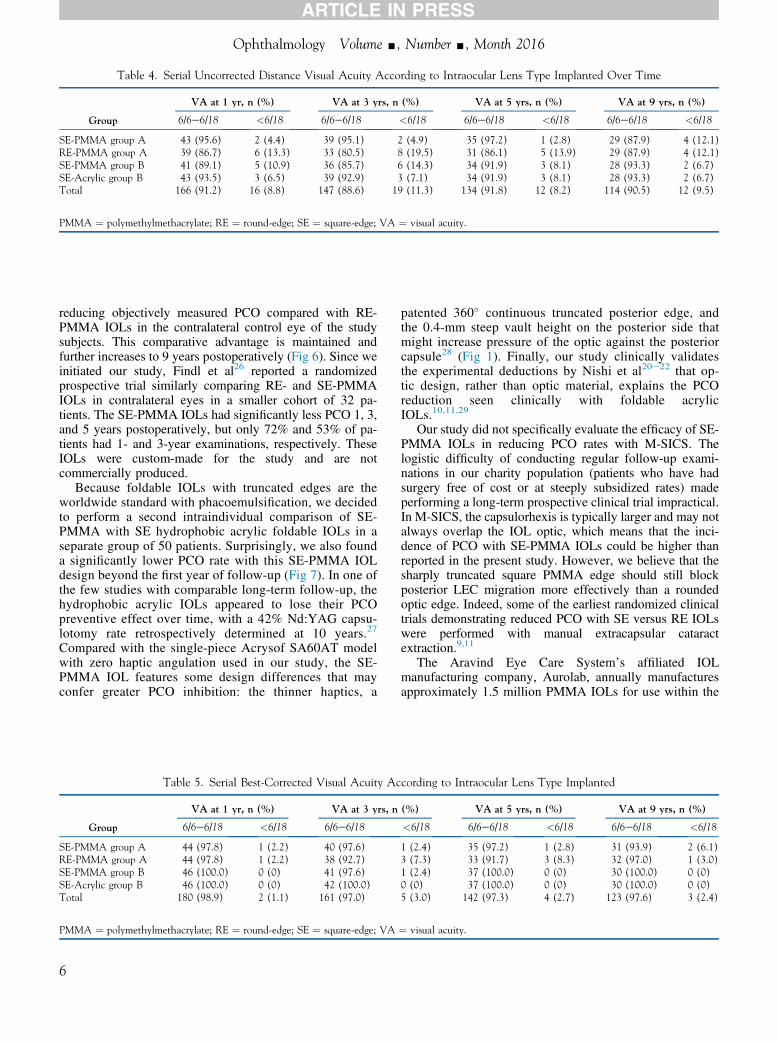
Table 4. Serial Uncorrected Distance Visual Acuity According to Intraocular Lens Type Implanted Over Time
Group
VA at 1 yr, n (%) VA at 3 yrs, n (%) VA at 5 yrs, n (%) VA at 9 yrs, n (%)
6/6e6/18 <6/18 6/6e6/18 <6/18 6/6e6/18 <6/18 6/6e6/18 <6/18
SE-PMMA group A 43 (95.6) 2 (4.4) 39 (95.1) 2 (4.9) 35 (97.2) 1 (2.8) 29 (87.9) 4 (12.1)RE-PMMA group A 39 (86.7) 6 (13.3) 33 (80.5) 8 (19.5) 31 (86.1) 5 (13.9) 29 (87.9) 4 (12.1)SE-PMMA group B 41 (89.1) 5 (10.9) 36 (85.7) 6 (14.3) 34 (91.9) 3 (8.1) 28 (93.3) 2 (6.7)SE-Acrylic group B 43 (93.5) 3 (6.5) 39 (92.9) 3 (7.1) 34 (91.9) 3 (8.1) 28 (93.3) 2 (6.7)Total 166 (91.2) 16 (8.8) 147 (88.6) 19 (11.3) 134 (91.8) 12 (8.2) 114 (90.5) 12 (9.5)
PMMA ¼ polymethylmethacrylate; RE ¼ round-edge; SE ¼ square-edge; VA ¼ visual acuity.
Ophthalmology Volume -, Number -, Month 2016
reducing objectively measured PCO compared with RE-PMMA IOLs in the contralateral control eye of the studysubjects. This comparative advantage is maintained andfurther increases to 9 years postoperatively (Fig 6). Since weinitiated our study, Findl et al26 reported a randomizedprospective trial similarly comparing RE- and SE-PMMAIOLs in contralateral eyes in a smaller cohort of 32 pa-tients. The SE-PMMA IOLs had significantly less PCO 1, 3,and 5 years postoperatively, but only 72% and 53% of pa-tients had 1- and 3-year examinations, respectively. TheseIOLs were custom-made for the study and are notcommercially produced.
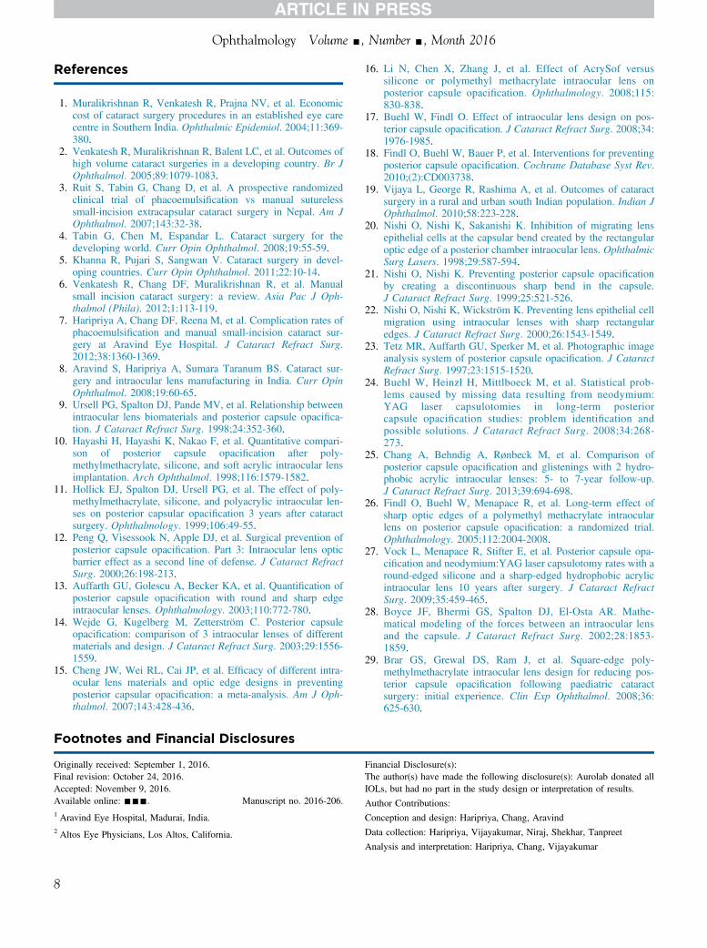
Because foldable IOLs with truncated edges are theworldwide standard with phacoemulsification, we decidedto perform a second intraindividual comparison of SE-PMMA with SE hydrophobic acrylic foldable IOLs in aseparate group of 50 patients. Surprisingly, we also founda significantly lower PCO rate with this SE-PMMA IOLdesign beyond the first year of follow-up (Fig 7). In one ofthe few studies with comparable long-term follow-up, thehydrophobic acrylic IOLs appeared to lose their PCOpreventive effect over time, with a 42% Nd:YAG capsu-lotomy rate retrospectively determined at 10 years.27
Compared with the single-piece Acrysof SA60AT modelwith zero haptic angulation used in our study, the SE-PMMA IOL features some design differences that mayconfer greater PCO inhibition: the thinner haptics, a
Table 5. Serial Best-Corrected Visual Acuity Ac
Group
VA at 1 yr, n (%) VA at 3 yrs, n
6/6e6/18 <6/18 6/6e6/18
SE-PMMA group A 44 (97.8) 1 (2.2) 40 (97.6)RE-PMMA group A 44 (97.8) 1 (2.2) 38 (92.7)SE-PMMA group B 46 (100.0) 0 (0) 41 (97.6)SE-Acrylic group B 46 (100.0) 0 (0) 42 (100.0)Total 180 (98.9) 2 (1.1) 161 (97.0)
PMMA ¼ polymethylmethacrylate; RE ¼ round-edge; SE ¼ square-edge; VA
6
patented 360� continuous truncated posterior edge, andthe 0.4-mm steep vault height on the posterior side thatmight increase pressure of the optic against the posteriorcapsule28 (Fig 1). Finally, our study clinically validatesthe experimental deductions by Nishi et al20e22 that op-tic design, rather than optic material, explains the PCOreduction seen clinically with foldable acrylicIOLs.10,11,29
Our study did not specifically evaluate the efficacy of SE-PMMA IOLs in reducing PCO rates with M-SICS. Thelogistic difficulty of conducting regular follow-up exami-nations in our charity population (patients who have hadsurgery free of cost or at steeply subsidized rates) madeperforming a long-term prospective clinical trial impractical.In M-SICS, the capsulorhexis is typically larger and may notalways overlap the IOL optic, which means that the inci-dence of PCO with SE-PMMA IOLs could be higher thanreported in the present study. However, we believe that thesharply truncated square PMMA edge should still blockposterior LEC migration more effectively than a roundedoptic edge. Indeed, some of the earliest randomized clinicaltrials demonstrating reduced PCO with SE versus RE IOLswere performed with manual extracapsular cataractextraction.9,11
The Aravind Eye Care System’s affiliated IOLmanufacturing company, Aurolab, annually manufacturesapproximately 1.5 million PMMA IOLs for use within the
cording to Intraocular Lens Type Implanted
(%) VA at 5 yrs, n (%) VA at 9 yrs, n (%)
<6/18 6/6e6/18 <6/18 6/6e6/18 <6/18
1 (2.4) 35 (97.2) 1 (2.8) 31 (93.9) 2 (6.1)3 (7.3) 33 (91.7) 3 (8.3) 32 (97.0) 1 (3.0)1 (2.4) 37 (100.0) 0 (0) 30 (100.0) 0 (0)0 (0) 37 (100.0) 0 (0) 30 (100.0) 0 (0)5 (3.0) 142 (97.3) 4 (2.7) 123 (97.6) 3 (2.4)
¼ visual acuity.
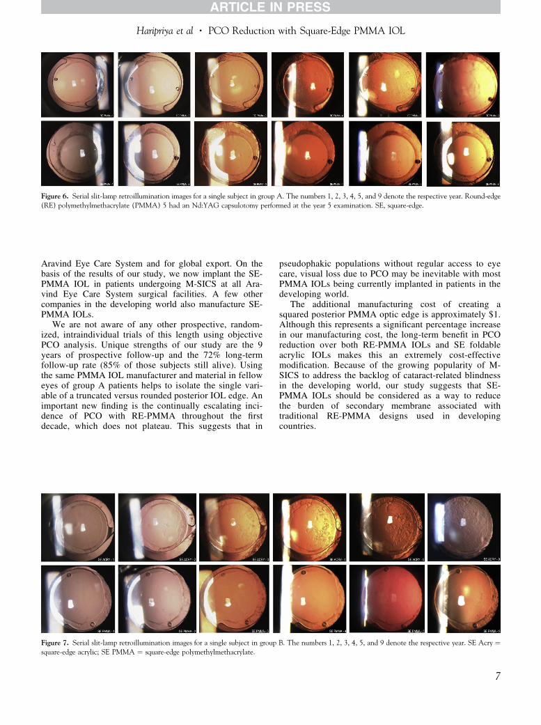
Figure 6. Serial slit-lamp retroillumination images for a single subject in group A. The numbers 1, 2, 3, 4, 5, and 9 denote the respective year. Round-edge(RE) polymethylmethacrylate (PMMA) 5 had an Nd:YAG capsulotomy performed at the year 5 examination. SE, square-edge.
Haripriya et al � PCO Reduction with Square-Edge PMMA IOL
Aravind Eye Care System and for global export. On thebasis of the results of our study, we now implant the SE-PMMA IOL in patients undergoing M-SICS at all Ara-vind Eye Care System surgical facilities. A few othercompanies in the developing world also manufacture SE-PMMA IOLs.
We are not aware of any other prospective, random-ized, intraindividual trials of this length using objectivePCO analysis. Unique strengths of our study are the 9years of prospective follow-up and the 72% long-termfollow-up rate (85% of those subjects still alive). Usingthe same PMMA IOL manufacturer and material in felloweyes of group A patients helps to isolate the single vari-able of a truncated versus rounded posterior IOL edge. Animportant new finding is the continually escalating inci-dence of PCO with RE-PMMA throughout the firstdecade, which does not plateau. This suggests that in
Figure 7. Serial slit-lamp retroillumination images for a single subject in groupsquare-edge acrylic; SE PMMA ¼ square-edge polymethylmethacrylate.
pseudophakic populations without regular access to eyecare, visual loss due to PCO may be inevitable with mostPMMA IOLs being currently implanted in patients in thedeveloping world.
The additional manufacturing cost of creating asquared posterior PMMA optic edge is approximately $1.Although this represents a significant percentage increasein our manufacturing cost, the long-term benefit in PCOreduction over both RE-PMMA IOLs and SE foldableacrylic IOLs makes this an extremely cost-effectivemodification. Because of the growing popularity of M-SICS to address the backlog of cataract-related blindnessin the developing world, our study suggests that SE-PMMA IOLs should be considered as a way to reducethe burden of secondary membrane associated withtraditional RE-PMMA designs used in developingcountries.
B. The numbers 1, 2, 3, 4, 5, and 9 denote the respective year. SE Acry ¼
7
Ophthalmology Volume -, Number -, Month 2016
References
1. Muralikrishnan R, Venkatesh R, Prajna NV, et al. Economiccost of cataract surgery procedures in an established eye carecentre in Southern India. Ophthalmic Epidemiol. 2004;11:369-380.
2. Venkatesh R, Muralikrishnan R, Balent LC, et al. Outcomes ofhigh volume cataract surgeries in a developing country. Br JOphthalmol. 2005;89:1079-1083.
3. Ruit S, Tabin G, Chang D, et al. A prospective randomizedclinical trial of phacoemulsification vs manual suturelesssmall-incision extracapsular cataract surgery in Nepal. Am JOphthalmol. 2007;143:32-38.
4. Tabin G, Chen M, Espandar L. Cataract surgery for thedeveloping world. Curr Opin Ophthalmol. 2008;19:55-59.
5. Khanna R, Pujari S, Sangwan V. Cataract surgery in devel-oping countries. Curr Opin Ophthalmol. 2011;22:10-14.
6. Venkatesh R, Chang DF, Muralikrishnan R, et al. Manualsmall incision cataract surgery: a review. Asia Pac J Oph-thalmol (Phila). 2012;1:113-119.
7. Haripriya A, Chang DF, Reena M, et al. Complication rates ofphacoemulsification and manual small-incision cataract sur-gery at Aravind Eye Hospital. J Cataract Refract Surg.2012;38:1360-1369.
8. Aravind S, Haripriya A, Sumara Taranum BS. Cataract sur-gery and intraocular lens manufacturing in India. Curr OpinOphthalmol. 2008;19:60-65.
9. Ursell PG, Spalton DJ, Pande MV, et al. Relationship betweenintraocular lens biomaterials and posterior capsule opacifica-tion. J Cataract Refract Surg. 1998;24:352-360.
10. Hayashi H, Hayashi K, Nakao F, et al. Quantitative compari-son of posterior capsule opacification after poly-methylmethacrylate, silicone, and soft acrylic intraocular lensimplantation. Arch Ophthalmol. 1998;116:1579-1582.
11. Hollick EJ, Spalton DJ, Ursell PG, et al. The effect of poly-methylmethacrylate, silicone, and polyacrylic intraocular len-ses on posterior capsular opacification 3 years after cataractsurgery. Ophthalmology. 1999;106:49-55.
12. Peng Q, Visessook N, Apple DJ, et al. Surgical prevention ofposterior capsule opacification. Part 3: Intraocular lens opticbarrier effect as a second line of defense. J Cataract RefractSurg. 2000;26:198-213.
13. Auffarth GU, Golescu A, Becker KA, et al. Quantification ofposterior capsule opacification with round and sharp edgeintraocular lenses. Ophthalmology. 2003;110:772-780.
14. Wejde G, Kugelberg M, Zetterström C. Posterior capsuleopacification: comparison of 3 intraocular lenses of differentmaterials and design. J Cataract Refract Surg. 2003;29:1556-1559.
15. Cheng JW, Wei RL, Cai JP, et al. Efficacy of different intra-ocular lens materials and optic edge designs in preventingposterior capsular opacification: a meta-analysis. Am J Oph-thalmol. 2007;143:428-436.
8
16. Li N, Chen X, Zhang J, et al. Effect of AcrySof versussilicone or polymethyl methacrylate intraocular lens onposterior capsule opacification. Ophthalmology. 2008;115:830-838.
17. Buehl W, Findl O. Effect of intraocular lens design on pos-terior capsule opacification. J Cataract Refract Surg. 2008;34:1976-1985.
18. Findl O, Buehl W, Bauer P, et al. Interventions for preventingposterior capsule opacification. Cochrane Database Syst Rev.2010;(2):CD003738.
19. Vijaya L, George R, Rashima A, et al. Outcomes of cataractsurgery in a rural and urban south Indian population. Indian JOphthalmol. 2010;58:223-228.
20. Nishi O, Nishi K, Sakanishi K. Inhibition of migrating lensepithelial cells at the capsular bend created by the rectangularoptic edge of a posterior chamber intraocular lens. OphthalmicSurg Lasers. 1998;29:587-594.
21. Nishi O, Nishi K. Preventing posterior capsule opacificationby creating a discontinuous sharp bend in the capsule.J Cataract Refract Surg. 1999;25:521-526.
22. Nishi O, Nishi K, Wickström K. Preventing lens epithelial cellmigration using intraocular lenses with sharp rectangularedges. J Cataract Refract Surg. 2000;26:1543-1549.
23. Tetz MR, Auffarth GU, Sperker M, et al. Photographic imageanalysis system of posterior capsule opacification. J CataractRefract Surg. 1997;23:1515-1520.
24. Buehl W, Heinzl H, Mittlboeck M, et al. Statistical prob-lems caused by missing data resulting from neodymium:YAG laser capsulotomies in long-term posteriorcapsule opacification studies: problem identification andpossible solutions. J Cataract Refract Surg. 2008;34:268-273.
25. Chang A, Behndig A, Rønbeck M, et al. Comparison ofposterior capsule opacification and glistenings with 2 hydro-phobic acrylic intraocular lenses: 5- to 7-year follow-up.J Cataract Refract Surg. 2013;39:694-698.
26. Findl O, Buehl W, Menapace R, et al. Long-term effect ofsharp optic edges of a polymethyl methacrylate intraocularlens on posterior capsule opacification: a randomized trial.Ophthalmology. 2005;112:2004-2008.
27. Vock L, Menapace R, Stifter E, et al. Posterior capsule opa-cification and neodymium:YAG laser capsulotomy rates with around-edged silicone and a sharp-edged hydrophobic acrylicintraocular lens 10 years after surgery. J Cataract RefractSurg. 2009;35:459-465.
28. Boyce JF, Bhermi GS, Spalton DJ, El-Osta AR. Mathe-matical modeling of the forces between an intraocular lensand the capsule. J Cataract Refract Surg. 2002;28:1853-1859.
29. Brar GS, Grewal DS, Ram J, et al. Square-edge poly-methylmethacrylate intraocular lens design for reducing pos-terior capsule opacification following paediatric cataractsurgery: initial experience. Clin Exp Ophthalmol. 2008;36:625-630.
Footnotes and Financial Disclosures
Originally received: September 1, 2016.Final revision: October 24, 2016.Accepted: November 9, 2016.Available online: ---. Manuscript no. 2016-206.1 Aravind Eye Hospital, Madurai, India.2 Altos Eye Physicians, Los Altos, California.
Financial Disclosure(s):The author(s) have made the following disclosure(s): Aurolab donated allIOLs, but had no part in the study design or interpretation of results.
Author Contributions:
Conception and design: Haripriya, Chang, Aravind
Data collection: Haripriya, Vijayakumar, Niraj, Shekhar, Tanpreet
Analysis and interpretation: Haripriya, Chang, Vijayakumar

Haripriya et al � PCO Reduction with Square-Edge PMMA IOL
Obtained funding: Not applicable
Overall responsibility: Haripriya, Chang, Aravind
Abbreviations and Acronyms:EPCO ¼ Evaluation of Posterior Capsule Opacification; IOL ¼ intraocularlens; LEC ¼ lens epithelial cell; M-SICS ¼ manual small-incision cataractsurgery; Nd:YAG ¼ neodymium-doped yttrium aluminium garnet;
PCO ¼ posterior capsule opacification; PMMA ¼ polymethylmethacrylate;RE¼ round-edge; SE¼ square-edge.
Correspondence:Aravind Haripriya, MD, Aravind Eye Hospital, 1, Anna Nagar, Madurai625020, India. E-mail: [email protected].
9


