Embed Size (px)
Citation preview
Fall 2021 Education Series- Forward Thinking
About Posterior Stroke
Posterior Circulation Strokes:
Identification, Recognition and Hyperacute Workup
Aristeidis H. Katsanos, MD, PhDClinical Fellow, McMaster University &
Investigator, Population Health Research Institute,Hamilton, ON, Canada
www.phri.ca
Disclosures-Acknowledgements
• Drs. Katsanos serves as the PI for the blooD prEssure management in sTroke following
EndovasCular Treatment (DETECT) trial, funded by the New Investigator Fund from the
Hamilton Health Sciences, and co-PI for the TRanscranial doppler Ultrasound after
Endovascular Stroke Treatment (TRUEST) study, funded by the Division of Neurology
Innovation Fund 2020, McMaster University.
• Serves as co-Chair for the OPTIMISE Research Committee.
• Has been tricked by symptoms suggestive posterior circulation stroke several
times during his training.
www.phri.ca
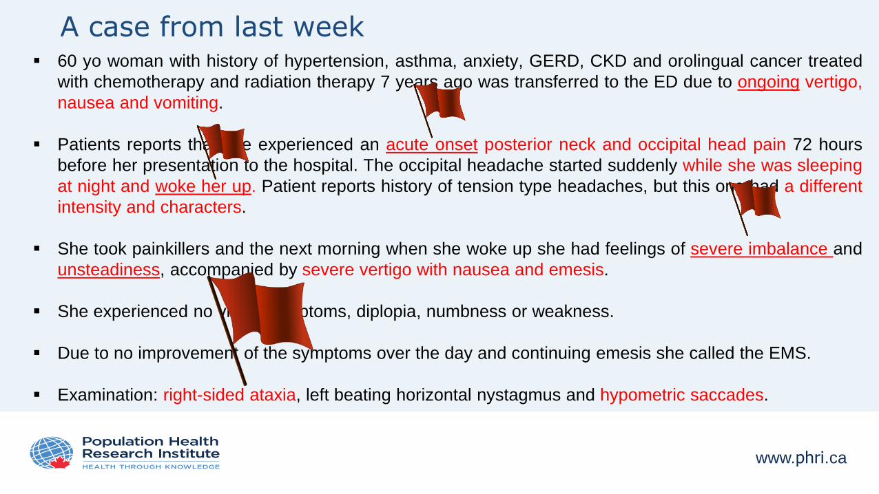
A case from last week 60 yo woman with history of hypertension, asthma, anxiety, GERD, CKD and orolingual cancer treated
with chemotherapy and radiation therapy 7 years ago was transferred to the ED due to ongoing vertigo,
nausea and vomiting.
Patients reports that she experienced an acute onset posterior neck and occipital head pain 72 hours
before her presentation to the hospital. The occipital headache started suddenly while she was sleeping
at night and woke her up. Patient reports history of tension type headaches, but this one had a different
intensity and characters.
She took painkillers and the next morning when she woke up she had feelings of severe imbalance and
unsteadiness, accompanied by severe vertigo with nausea and emesis.
She experienced no visual symptoms, diplopia, numbness or weakness.
Due to no improvement of the symptoms over the day and continuing emesis she called the EMS.
Examination: right-sided ataxia, left beating horizontal nystagmus and hypometric saccades.
www.phri.ca
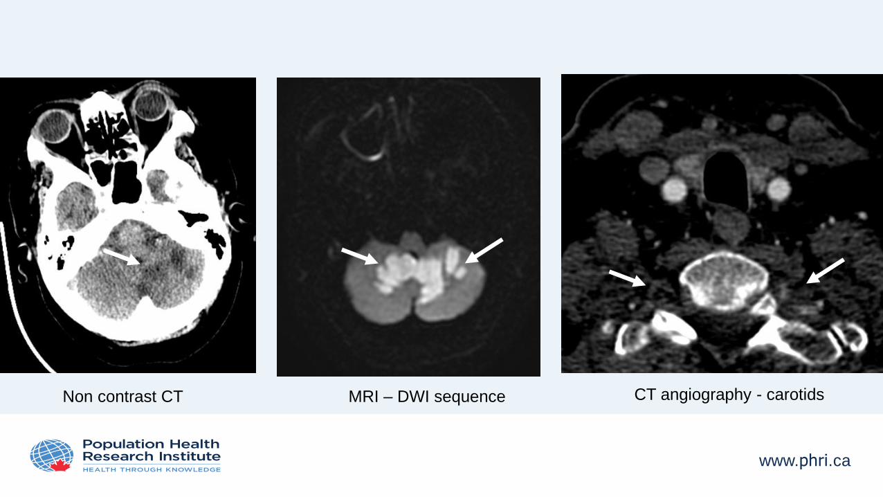
Non contrast CT MRI – DWI sequence CT angiography - carotids
www.phri.ca
Presentation outline
• Challenges in the diagnosis of posterior circulation stroke
• Anatomy of posterior circulation & mechanisms of posterior circulation ischemia
• Common posterior circulation stroke syndromes
• Diagnosis of posterior circulation stroke/TIA in the emergency setting
• Conclusions
www.phri.ca
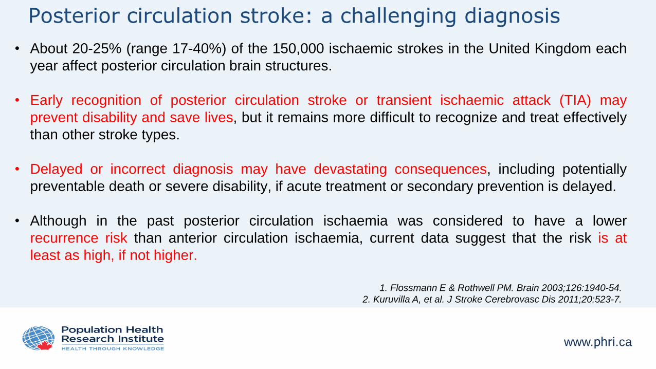
Posterior circulation stroke: a challenging diagnosis
• About 20-25% (range 17-40%) of the 150,000 ischaemic strokes in the United Kingdom each
year affect posterior circulation brain structures.
• Early recognition of posterior circulation stroke or transient ischaemic attack (TIA) may
prevent disability and save lives, but it remains more difficult to recognize and treat effectively
than other stroke types.
• Delayed or incorrect diagnosis may have devastating consequences, including potentially
preventable death or severe disability, if acute treatment or secondary prevention is delayed.
• Although in the past posterior circulation ischaemia was considered to have a lower
recurrence risk than anterior circulation ischaemia, current data suggest that the risk is at
least as high, if not higher.
1. Flossmann E & Rothwell PM. Brain 2003;126:1940-54.
2. Kuruvilla A, et al. J Stroke Cerebrovasc Dis 2011;20:523-7.
www.phri.ca
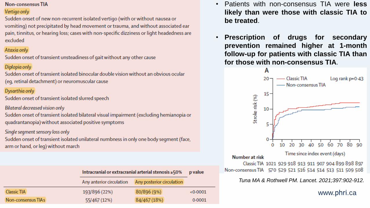
• Patients with non-consensus TIA were less
likely than were those with classic TIA to
be treated.
• Prescription of drugs for secondary
prevention remained higher at 1-month
follow-up for patients with classic TIA than
for those with non-consensus TIA.
Tuna MA & Rothwell PM. Lancet. 2021;397:902-912.
www.phri.ca
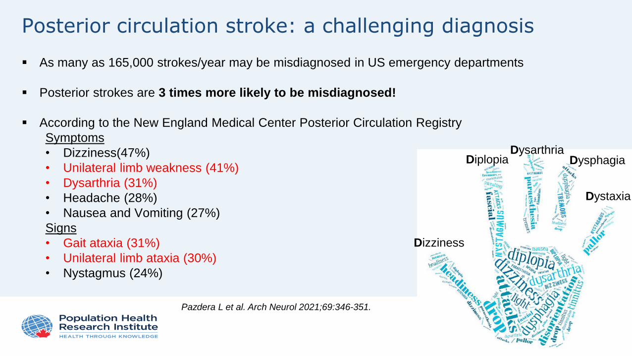
Posterior circulation stroke: a challenging diagnosis
As many as 165,000 strokes/year may be misdiagnosed in US emergency departments
Posterior strokes are 3 times more likely to be misdiagnosed!
According to the New England Medical Center Posterior Circulation Registry
Symptoms
• Dizziness(47%)
• Unilateral limb weakness (41%)
• Dysarthria (31%)
• Headache (28%)
• Nausea and Vomiting (27%)
Signs
• Gait ataxia (31%)
• Unilateral limb ataxia (30%)
• Nystagmus (24%)
Pazdera L et al. Arch Neurol 2021;69:346-351.
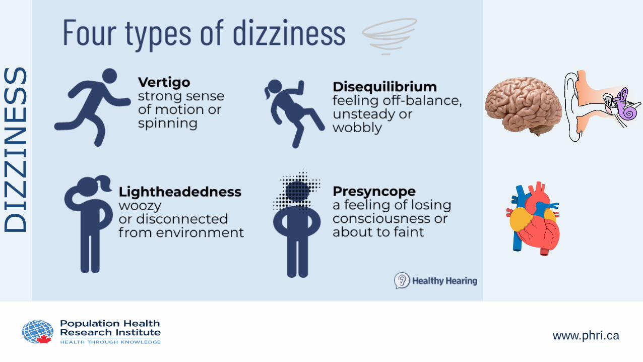
Dizziness
DiplopiaDysarthria
Dysphagia
Dystaxia
www.phri.ca
DIZ
ZIN
ESS
www.phri.ca
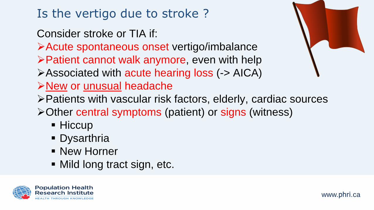
Is the vertigo due to stroke ?
Consider stroke or TIA if:
Acute spontaneous onset vertigo/imbalance
Patient cannot walk anymore, even with help
Associated with acute hearing loss (-> AICA)
New or unusual headache
Patients with vascular risk factors, elderly, cardiac sources
Other central symptoms (patient) or signs (witness)
Hiccup
Dysarthria
New Horner
Mild long tract sign, etc.
www.phri.ca
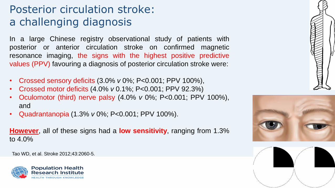
Posterior circulation stroke: a challenging diagnosis
In a large Chinese registry observational study of patients with
posterior or anterior circulation stroke on confirmed magnetic
resonance imaging, the signs with the highest positive predictive
values (PPV) favouring a diagnosis of posterior circulation stroke were:
• Crossed sensory deficits (3.0% v 0%; P<0.001; PPV 100%),
• Crossed motor deficits (4.0% v 0.1%; P<0.001; PPV 92.3%)
• Oculomotor (third) nerve palsy (4.0% v 0%; P<0.001; PPV 100%),
and
• Quadrantanopia (1.3% v 0%; P<0.001; PPV 100%).
However, all of these signs had a low sensitivity, ranging from 1.3%
to 4.0%
Tao WD, et al. Stroke 2012;43:2060-5.
www.phri.ca
Presentation outline
• Challenges in the diagnosis of posterior circulation stroke
• Anatomy of posterior circulation & mechanisms of posterior circulation ischemia
• Common posterior circulation stroke syndromes
• Diagnosis of posterior circulation stroke/TIA in the emergency setting
• Conclusions
www.phri.ca
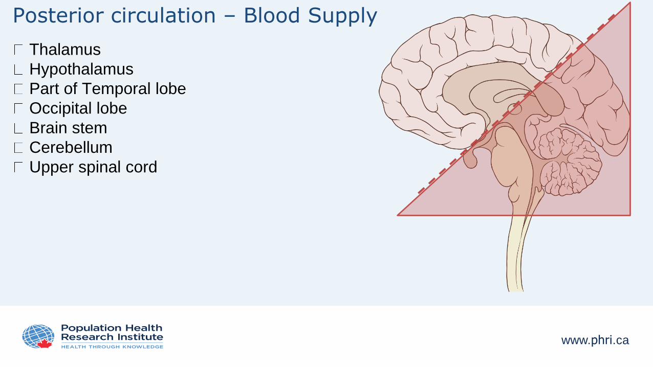
Posterior circulation – Blood Supply
Thalamus
Hypothalamus
Part of Temporal lobe
Occipital lobe
Brain stem
Cerebellum
Upper spinal cord
www.phri.ca
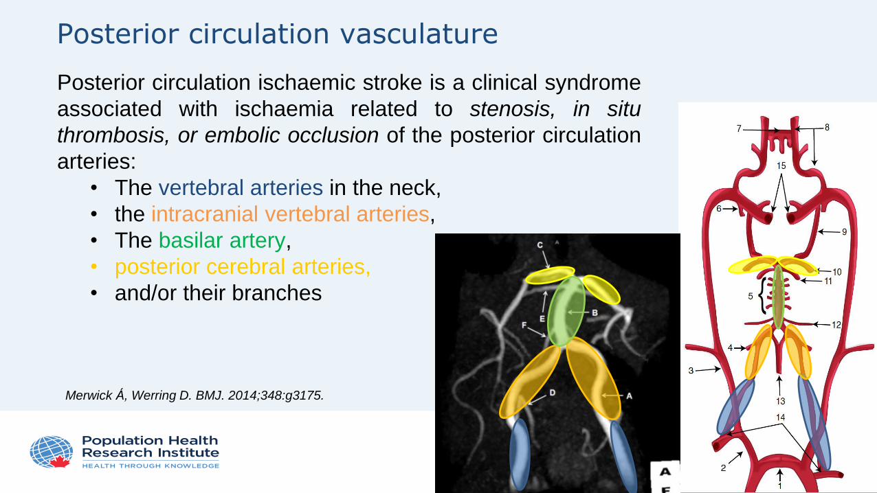
Posterior circulation vasculature
Posterior circulation ischaemic stroke is a clinical syndrome
associated with ischaemia related to stenosis, in situ
thrombosis, or embolic occlusion of the posterior circulation
arteries:
• The vertebral arteries in the neck,
• the intracranial vertebral arteries,
• The basilar artery,
• posterior cerebral arteries,
• and/or their branches
Merwick Á, Werring D. BMJ. 2014;348:g3175.
www.phri.ca
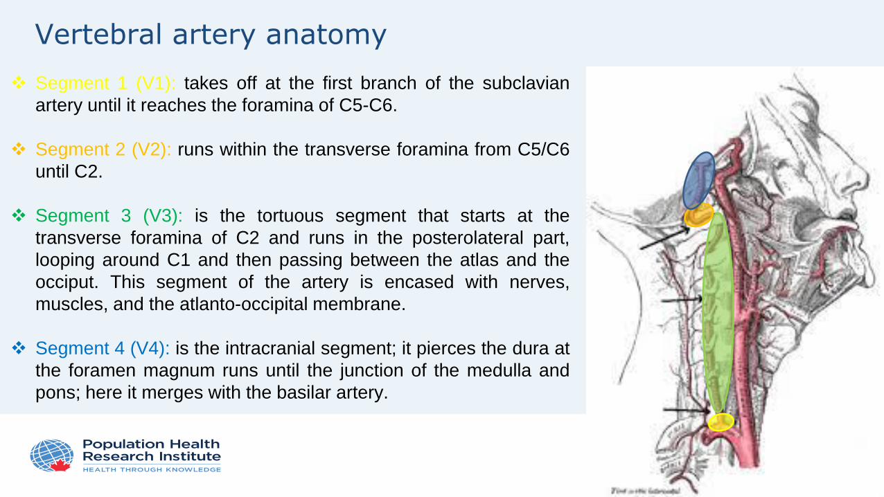
Vertebral artery anatomy
Segment 1 (V1): takes off at the first branch of the subclavian
artery until it reaches the foramina of C5-C6.
Segment 2 (V2): runs within the transverse foramina from C5/C6
until C2.
Segment 3 (V3): is the tortuous segment that starts at the
transverse foramina of C2 and runs in the posterolateral part,
looping around C1 and then passing between the atlas and the
occiput. This segment of the artery is encased with nerves,
muscles, and the atlanto-occipital membrane.
Segment 4 (V4): is the intracranial segment; it pierces the dura at
the foramen magnum runs until the junction of the medulla and
pons; here it merges with the basilar artery.
www.phri.ca
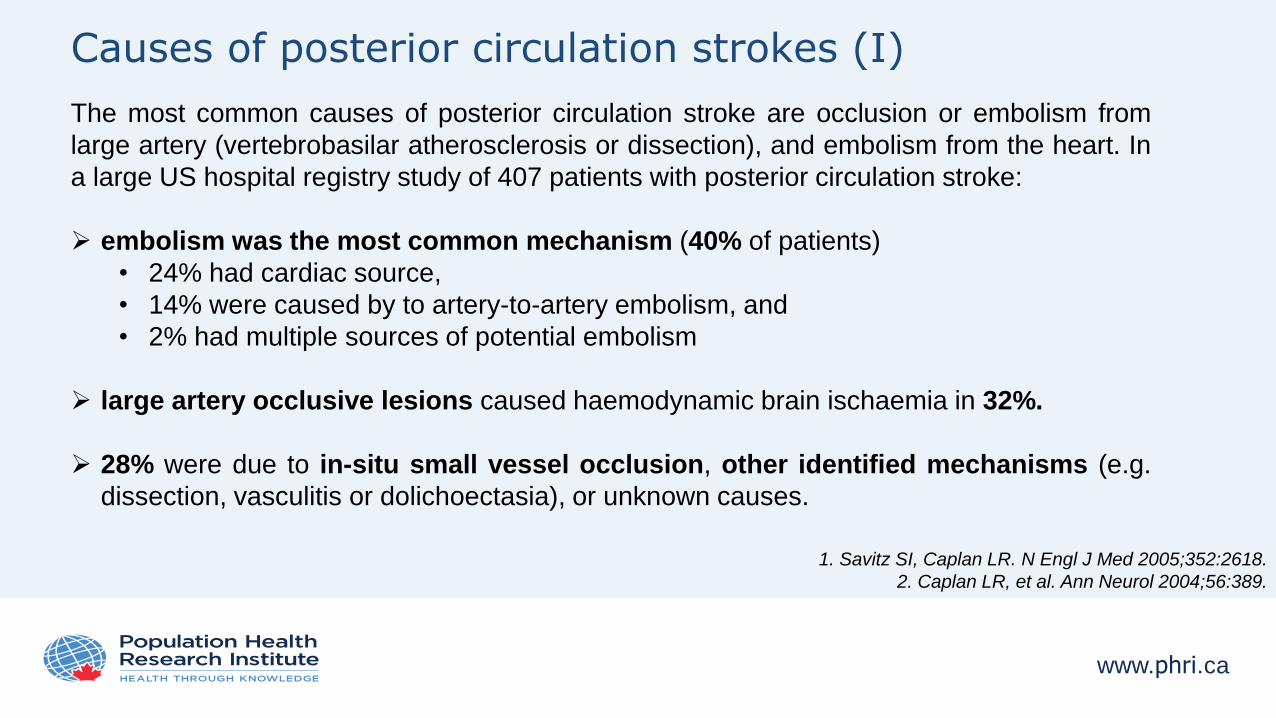
Causes of posterior circulation strokes (I)
The most common causes of posterior circulation stroke are occlusion or embolism from
large artery (vertebrobasilar atherosclerosis or dissection), and embolism from the heart. In
a large US hospital registry study of 407 patients with posterior circulation stroke:
embolism was the most common mechanism (40% of patients)
• 24% had cardiac source,
• 14% were caused by to artery-to-artery embolism, and
• 2% had multiple sources of potential embolism
large artery occlusive lesions caused haemodynamic brain ischaemia in 32%.
28% were due to in-situ small vessel occlusion, other identified mechanisms (e.g.
dissection, vasculitis or dolichoectasia), or unknown causes.
1. Savitz SI, Caplan LR. N Engl J Med 2005;352:2618.
2. Caplan LR, et al. Ann Neurol 2004;56:389.
www.phri.ca
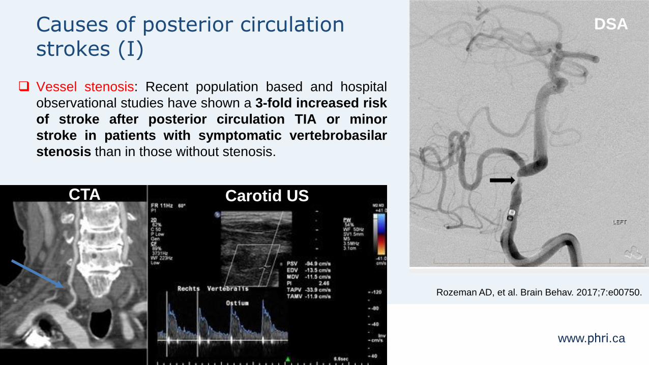
Causes of posterior circulation strokes (I)
Vessel stenosis: Recent population based and hospital
observational studies have shown a 3-fold increased risk
of stroke after posterior circulation TIA or minor
stroke in patients with symptomatic vertebrobasilar
stenosis than in those without stenosis.
DSA
CTA Carotid US
Rozeman AD, et al. Brain Behav. 2017;7:e00750.
www.phri.ca
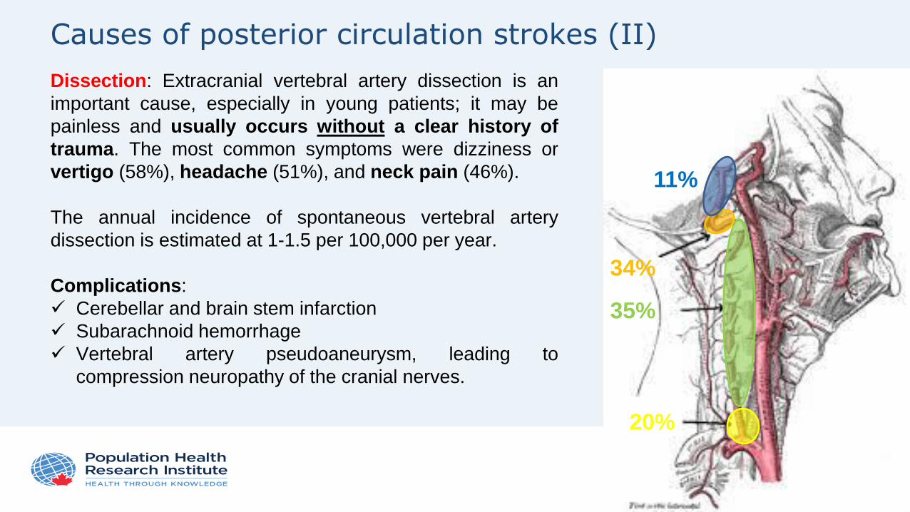
Causes of posterior circulation strokes (II)
Dissection: Extracranial vertebral artery dissection is an
important cause, especially in young patients; it may be
painless and usually occurs without a clear history of
trauma. The most common symptoms were dizziness or
vertigo (58%), headache (51%), and neck pain (46%).
The annual incidence of spontaneous vertebral artery
dissection is estimated at 1-1.5 per 100,000 per year.
Complications:
Cerebellar and brain stem infarction
Subarachnoid hemorrhage
Vertebral artery pseudoaneurysm, leading to
compression neuropathy of the cranial nerves.
11%
34%
35%
20%
www.phri.ca
Causes of posterior circulation strokes – dissection (III)
It is estimated that vertebral artery dissection is the cause of approximately 2% of all ischemic
strokes. However, in middle-aged and younger patients (30 to 45 years of age), it is believed to be
as high as 10% to 25%.
From a study of 169 patients with spontaneous vertebral artery dissection (sVAD) in Switzerland:
mean age, 43±9; median, 43; range, 21–69 years
15% were found to have with bilateral sVAD
Median time interval from symptom onset to diagnosis was 4 days (range, 2 hours to 88 days)
8% had asymptomatic sVAD
Presenting clinical symptoms were:
• ischemic stroke in 114 (67%),
• TIA in 17 (10%),
• occipital head and/or neck pain alone in 21 (12%) patients,
• SAH without ischemia in three (2%) patients, and
• sensorimotor cervical radiculopathy C5/C6 in one patient (1%)
Arnold M, et al. Stroke. 2006;37:2499-503.
www.phri.ca
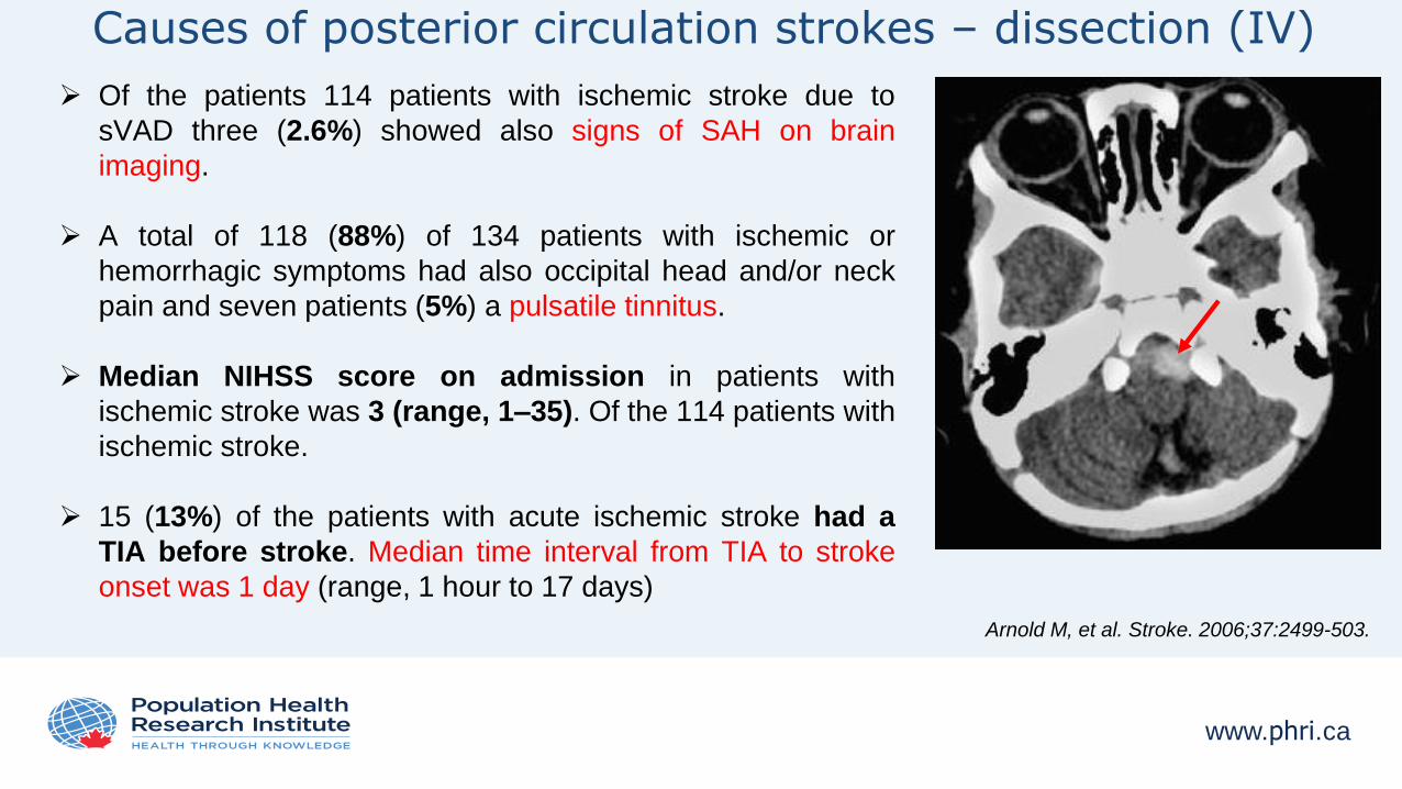
Causes of posterior circulation strokes – dissection (IV)
Of the patients 114 patients with ischemic stroke due to
sVAD three (2.6%) showed also signs of SAH on brain
imaging.
A total of 118 (88%) of 134 patients with ischemic or
hemorrhagic symptoms had also occipital head and/or neck
pain and seven patients (5%) a pulsatile tinnitus.
Median NIHSS score on admission in patients with
ischemic stroke was 3 (range, 1–35). Of the 114 patients with
ischemic stroke.
15 (13%) of the patients with acute ischemic stroke had a
TIA before stroke. Median time interval from TIA to stroke
onset was 1 day (range, 1 hour to 17 days)
Arnold M, et al. Stroke. 2006;37:2499-503.
www.phri.ca
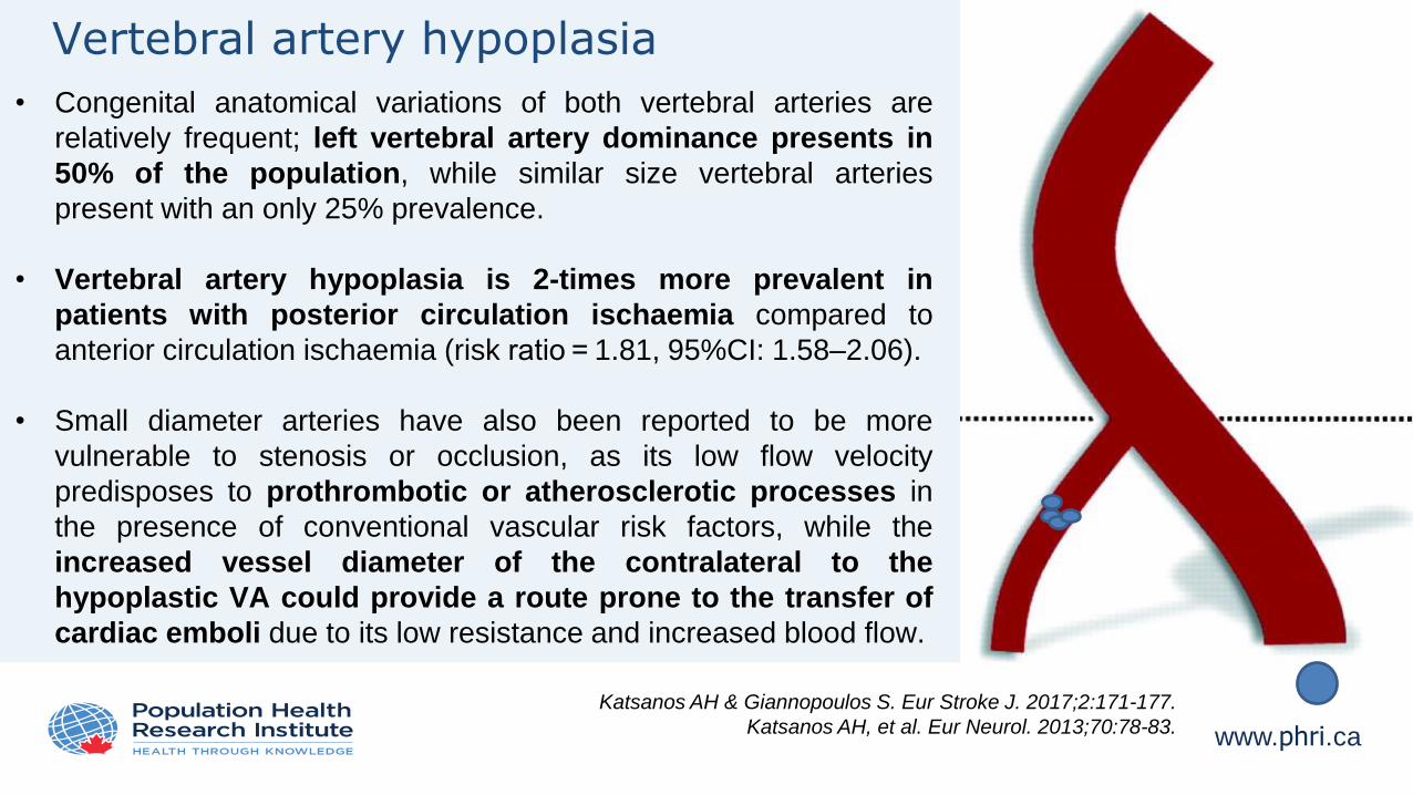
Vertebral artery hypoplasia
• Congenital anatomical variations of both vertebral arteries are
relatively frequent; left vertebral artery dominance presents in
50% of the population, while similar size vertebral arteries
present with an only 25% prevalence.
• Vertebral artery hypoplasia is 2-times more prevalent in
patients with posterior circulation ischaemia compared to
anterior circulation ischaemia (risk ratio = 1.81, 95%CI: 1.58–2.06).
• Small diameter arteries have also been reported to be more
vulnerable to stenosis or occlusion, as its low flow velocity
predisposes to prothrombotic or atherosclerotic processes in
the presence of conventional vascular risk factors, while the
increased vessel diameter of the contralateral to the
hypoplastic VA could provide a route prone to the transfer of
cardiac emboli due to its low resistance and increased blood flow.
Katsanos AH & Giannopoulos S. Eur Stroke J. 2017;2:171-177.
Katsanos AH, et al. Eur Neurol. 2013;70:78-83.
www.phri.ca
Presentation outline
• Challenges in the diagnosis of posterior circulation stroke
• Anatomy of posterior circulation & mechanisms of posterior circulation ischemia
• Common posterior circulation stroke syndromes
• Diagnosis of posterior circulation stroke/TIA in the emergency setting
• Conclusions
www.phri.ca
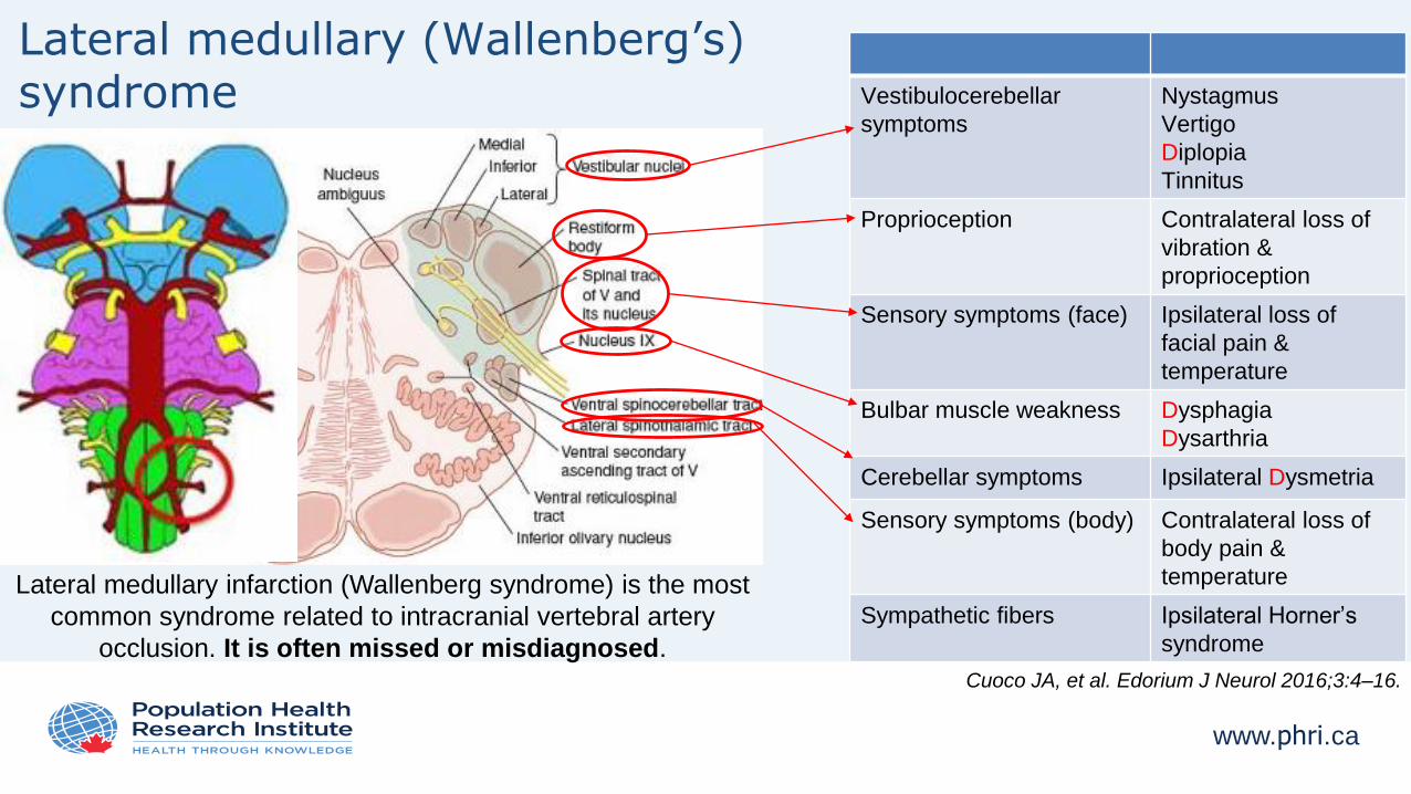
Lateral medullary (Wallenberg’s) syndrome Vestibulocerebellar
symptoms
Nystagmus
Vertigo
Diplopia
Tinnitus
Proprioception Contralateral loss of
vibration &
proprioception
Sensory symptoms (face) Ipsilateral loss of
facial pain &
temperature
Bulbar muscle weakness Dysphagia
Dysarthria
Cerebellar symptoms Ipsilateral Dysmetria
Sensory symptoms (body) Contralateral loss of
body pain &
temperature
Sympathetic fibers Ipsilateral Horner’s
syndrome
Lateral medullary infarction (Wallenberg syndrome) is the most
common syndrome related to intracranial vertebral artery
occlusion. It is often missed or misdiagnosed.Cuoco JA, et al. Edorium J Neurol 2016;3:4–16.
www.phri.ca
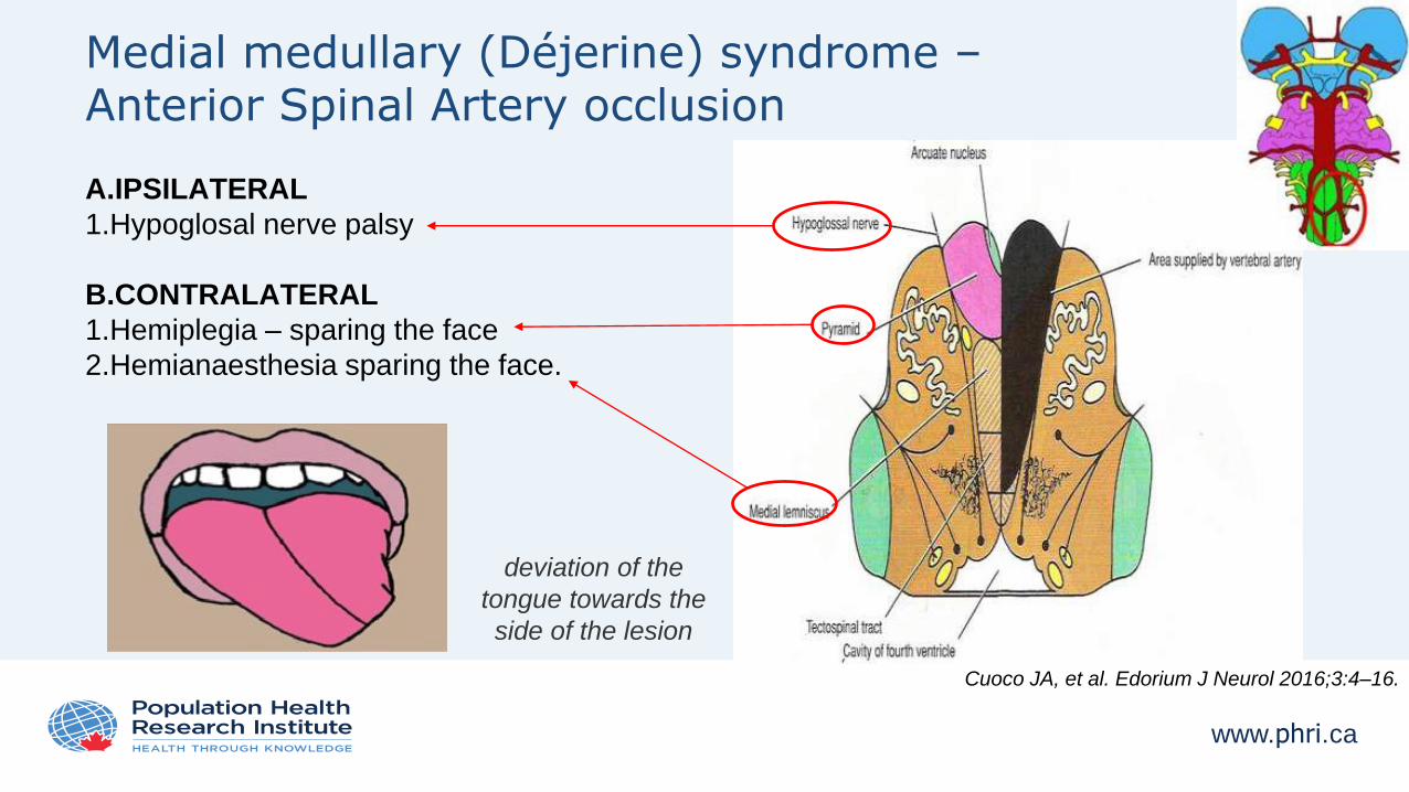
Medial medullary (Déjerine) syndrome –Anterior Spinal Artery occlusion
A.IPSILATERAL
1.Hypoglosal nerve palsy
B.CONTRALATERAL
1.Hemiplegia – sparing the face
2.Hemianaesthesia sparing the face.
deviation of the
tongue towards the
side of the lesion
Cuoco JA, et al. Edorium J Neurol 2016;3:4–16.
www.phri.ca
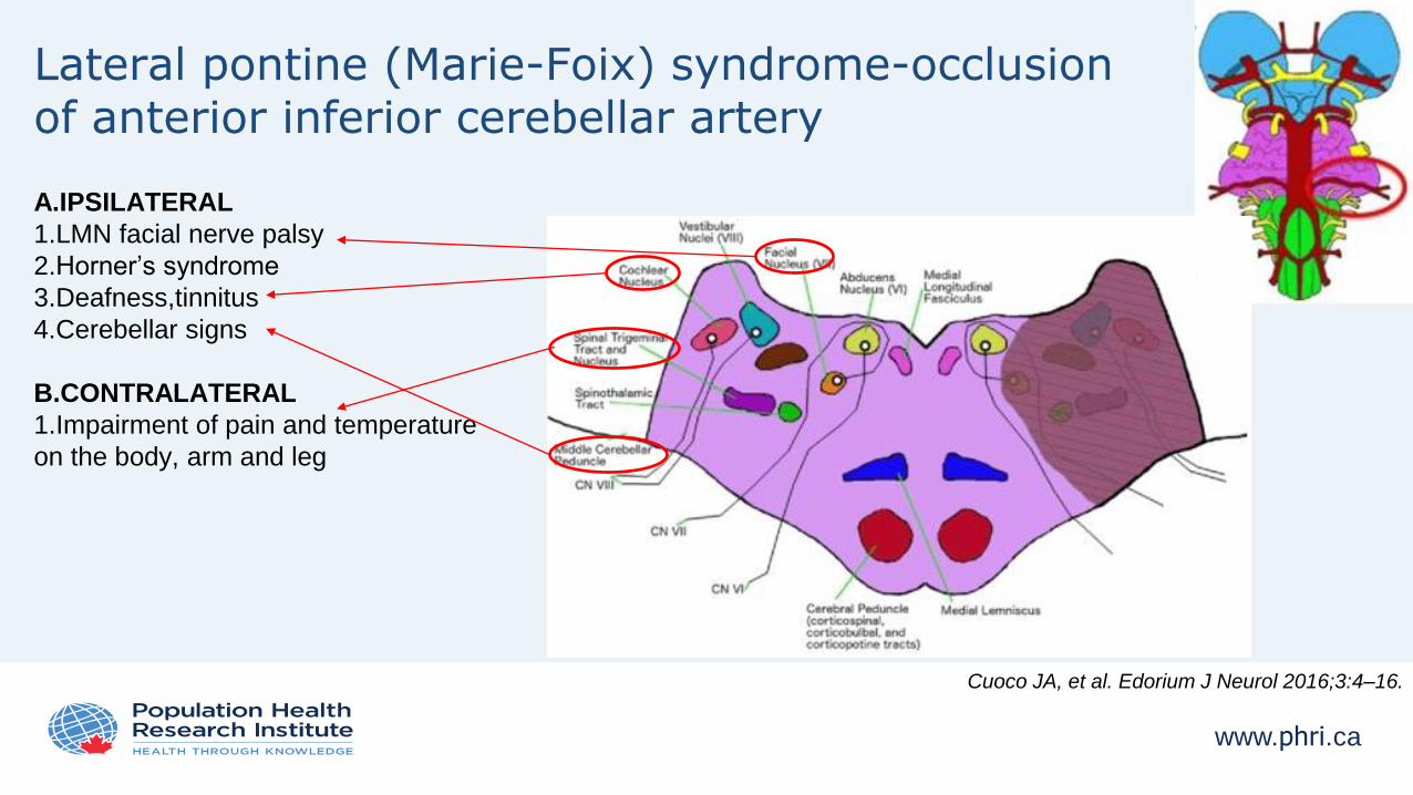
Lateral pontine (Marie-Foix) syndrome-occlusion of anterior inferior cerebellar artery
A.IPSILATERAL
1.LMN facial nerve palsy
2.Horner’s syndrome
3.Deafness,tinnitus
4.Cerebellar signs
B.CONTRALATERAL
1.Impairment of pain and temperature
on the body, arm and leg
Cuoco JA, et al. Edorium J Neurol 2016;3:4–16.
www.phri.ca
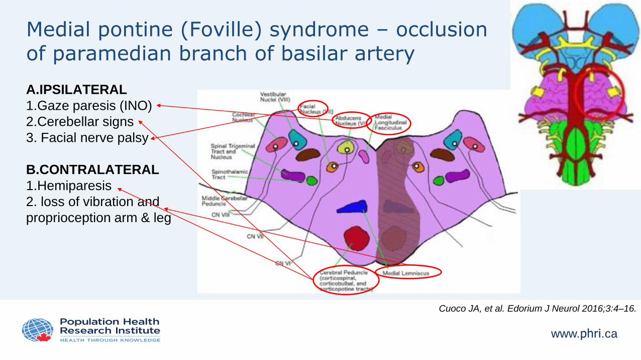
Medial pontine (Foville) syndrome – occlusionof paramedian branch of basilar artery
A.IPSILATERAL
1.Gaze paresis (INO)
2.Cerebellar signs
3. Facial nerve palsy
B.CONTRALATERAL
1.Hemiparesis
2. loss of vibration and
proprioception arm & leg
Cuoco JA, et al. Edorium J Neurol 2016;3:4–16.
www.phri.ca
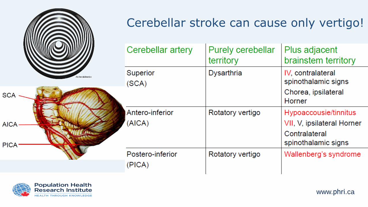
Cerebellar stroke can cause only vertigo!
www.phri.ca
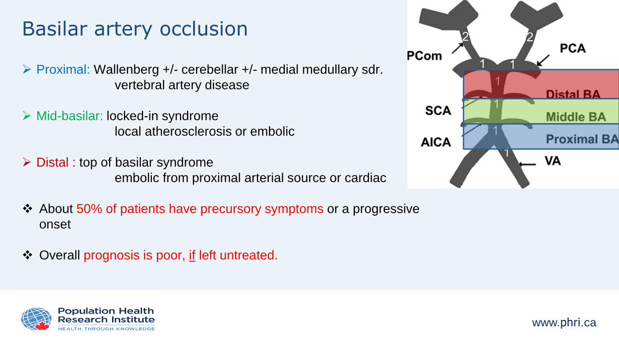
Basilar artery occlusion
Proximal: Wallenberg +/- cerebellar +/- medial medullary sdr.
vertebral artery disease
Mid-basilar: locked-in syndrome
local atherosclerosis or embolic
Distal : top of basilar syndrome
embolic from proximal arterial source or cardiac
About 50% of patients have precursory symptoms or a progressive
onset
Overall prognosis is poor, if left untreated.
www.phri.ca
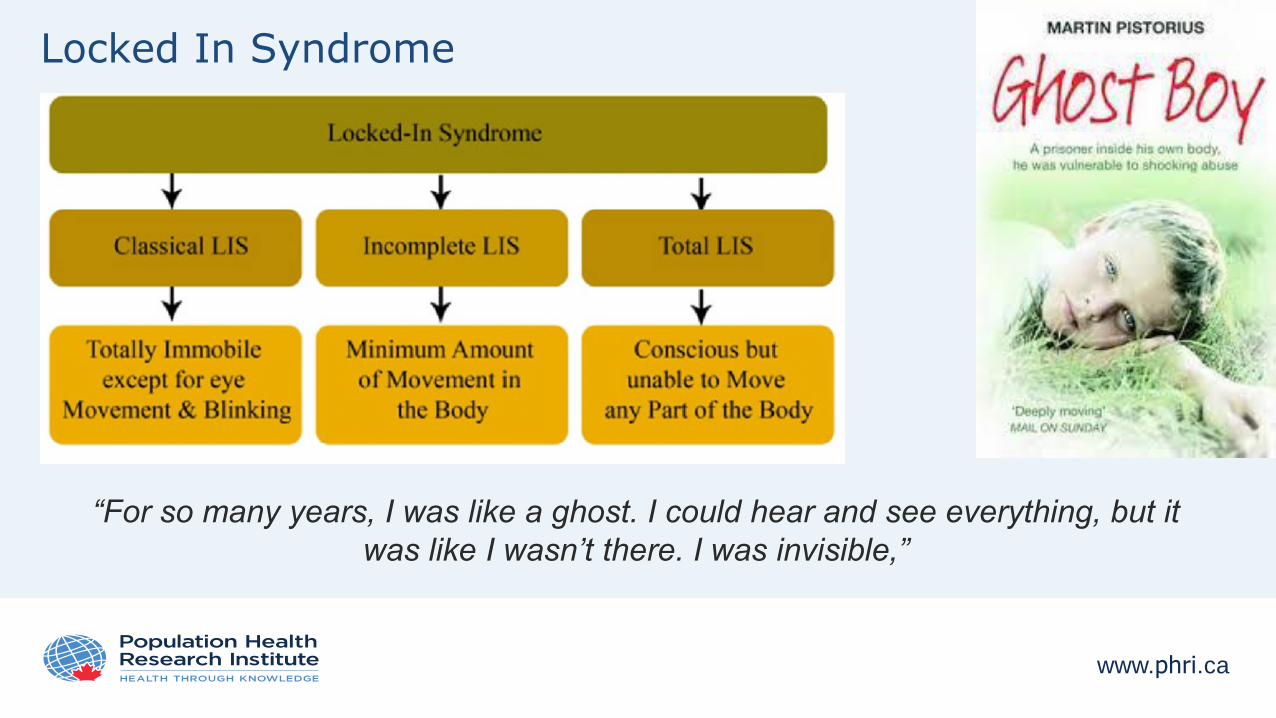
Locked In Syndrome
“For so many years, I was like a ghost. I could hear and see everything, but it
was like I wasn’t there. I was invisible,”
www.phri.ca
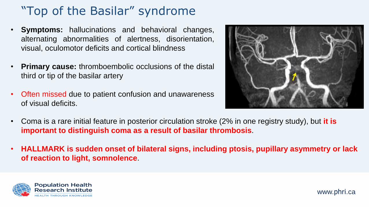
“Top of the Basilar” syndrome
• Symptoms: hallucinations and behavioral changes,
alternating abnormalities of alertness, disorientation,
visual, oculomotor deficits and cortical blindness
• Primary cause: thromboembolic occlusions of the distal
third or tip of the basilar artery
• Often missed due to patient confusion and unawareness
of visual deficits.
• Coma is a rare initial feature in posterior circulation stroke (2% in one registry study), but it is
important to distinguish coma as a result of basilar thrombosis.
• HALLMARK is sudden onset of bilateral signs, including ptosis, pupillary asymmetry or lack
of reaction to light, somnolence.
www.phri.ca
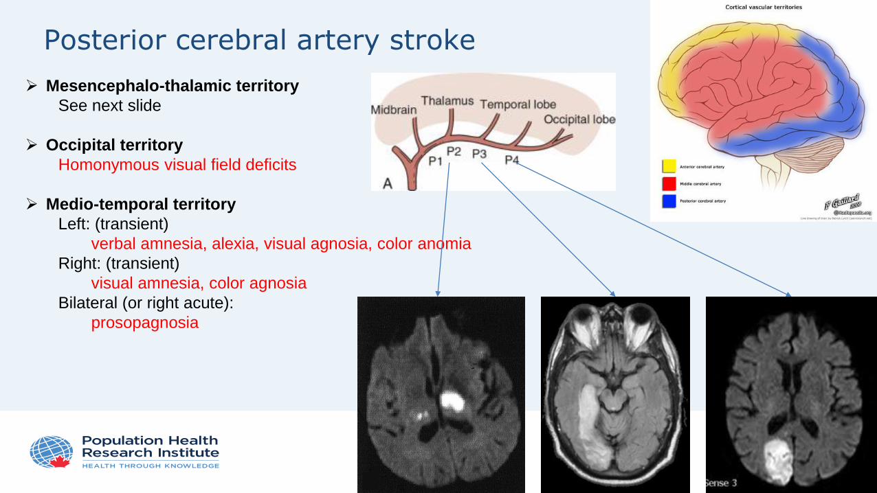
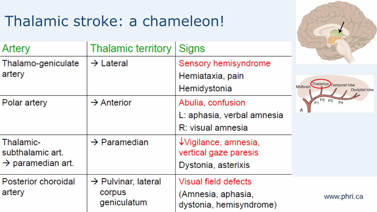
Posterior cerebral artery stroke
Mesencephalo-thalamic territory
See next slide
Occipital territory
Homonymous visual field deficits
Medio-temporal territory
Left: (transient)
verbal amnesia, alexia, visual agnosia, color anomia
Right: (transient)
visual amnesia, color agnosia
Bilateral (or right acute):
prosopagnosia
www.phri.ca
Thalamic stroke: a chameleon!
www.phri.ca
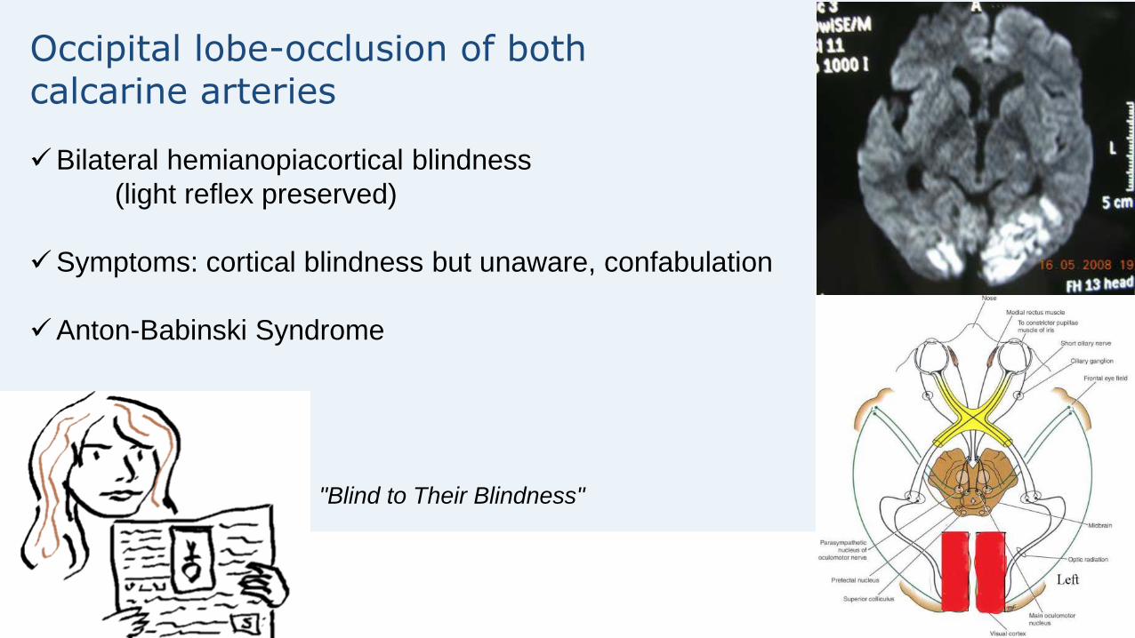
Occipital lobe-occlusion of bothcalcarine arteries
Bilateral hemianopiacortical blindness
(light reflex preserved)
Symptoms: cortical blindness but unaware, confabulation
Anton-Babinski Syndrome
"Blind to Their Blindness"
www.phri.ca
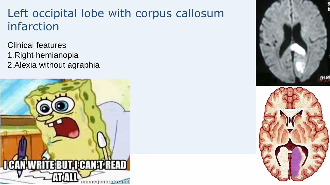
Left occipital lobe with corpus callosum infarction
Clinical features
1.Right hemianopia
2.Alexia without agraphia
www.phri.ca
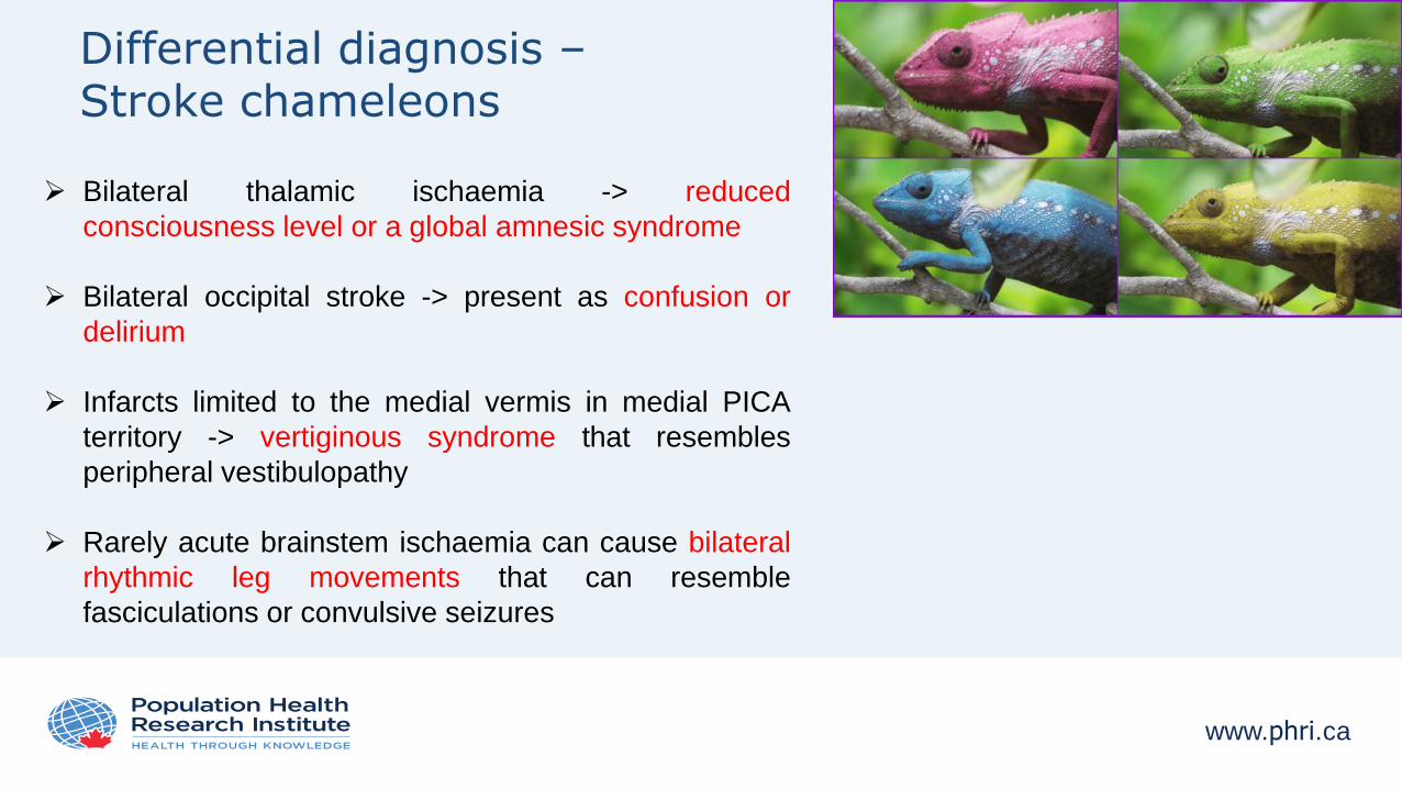
Differential diagnosis –Stroke chameleons
Bilateral thalamic ischaemia -> reduced
consciousness level or a global amnesic syndrome
Bilateral occipital stroke -> present as confusion or
delirium
Infarcts limited to the medial vermis in medial PICA
territory -> vertiginous syndrome that resembles
peripheral vestibulopathy
Rarely acute brainstem ischaemia can cause bilateral
rhythmic leg movements that can resemble
fasciculations or convulsive seizures
www.phri.ca
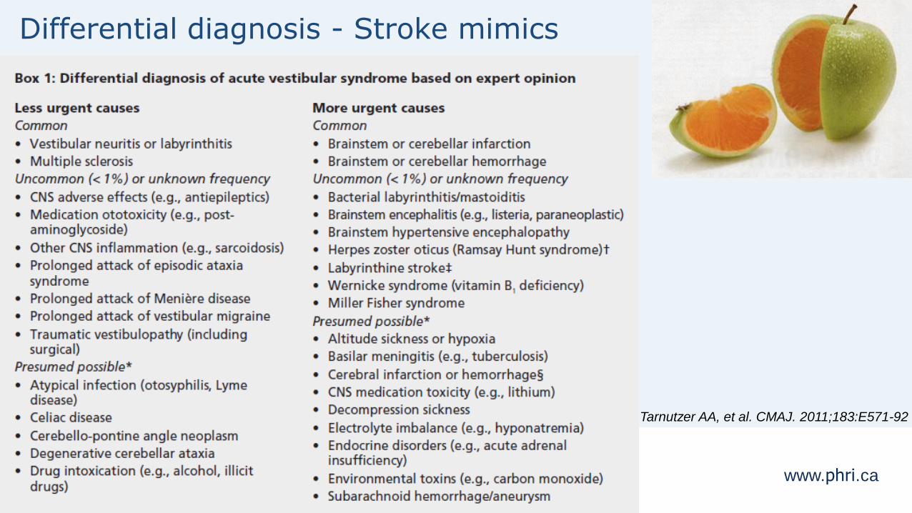
Differential diagnosis - Stroke mimics
Acute peripheral vestibular dysfunction (isolated vertigo with no
other brainstem symptoms or signs)
tumor (imaging can confirm the diagnosis)
Basilar migraine (aura features including vertigo and
diplopia, as well as severe occipital headache)
Toxic or metabolic disturbances (hypoglycaemia, central
pontine myelinolysis, and post-infectious disorders)
Miller Fisher syndrome (ophthalmoplegia, ataxia, areflexia)
Posterior reversible encephalopathy (PRES, visual
disturbance, seizures, and other focal symptoms)
Neuroinflammatory or chronic infectious disorders (sarcoidosis,
Behçet’s disease, Whipple’s disease)
Infection of the medulla, pons, and cerebellum
(rhomboencephalitis)
Tarnutzer AA, et al. CMAJ. 2011;183:E571-92
www.phri.ca
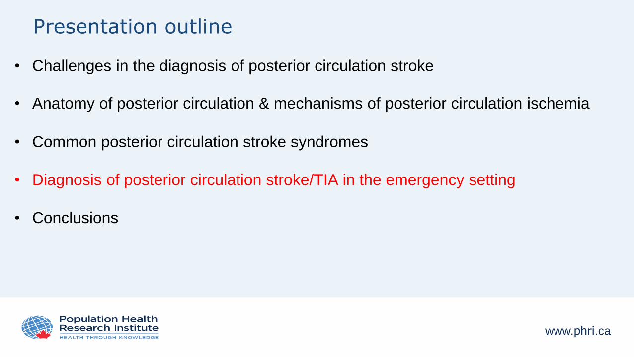
Presentation outline
• Challenges in the diagnosis of posterior circulation stroke
• Anatomy of posterior circulation & mechanisms of posterior circulation ischemia
• Common posterior circulation stroke syndromes
• Diagnosis of posterior circulation stroke/TIA in the emergency setting
• Conclusions
www.phri.ca
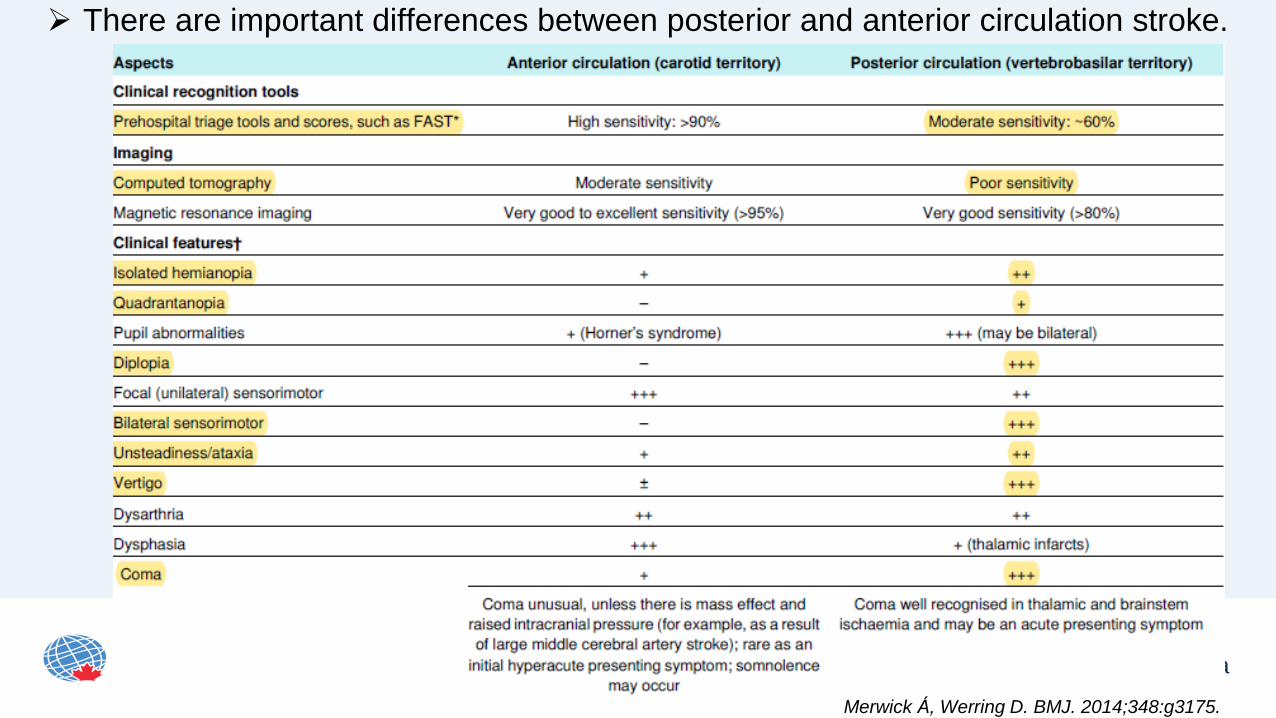
There are important differences between posterior and anterior circulation stroke.
Merwick Á, Werring D. BMJ. 2014;348:g3175.
www.phri.ca
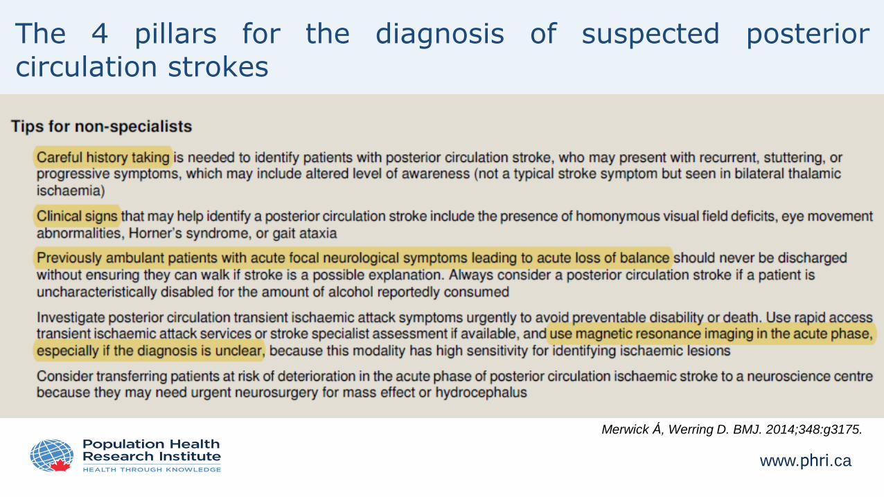
The 4 pillars for the diagnosis of suspected posteriorcirculation strokes
Merwick Á, Werring D. BMJ. 2014;348:g3175.
www.phri.ca
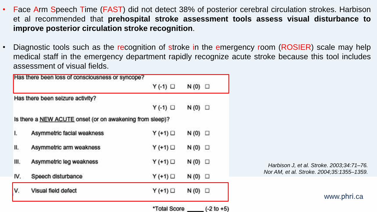
• Face Arm Speech Time (FAST) did not detect 38% of posterior cerebral circulation strokes. Harbison
et al recommended that prehospital stroke assessment tools assess visual disturbance to
improve posterior circulation stroke recognition.
• Diagnostic tools such as the recognition of stroke in the emergency room (ROSIER) scale may help
medical staff in the emergency department rapidly recognize acute stroke because this tool includes
assessment of visual fields.
Harbison J, et al. Stroke. 2003;34:71–76.
Nor AM, et al. Stroke. 2004;35:1355–1359.
www.phri.ca
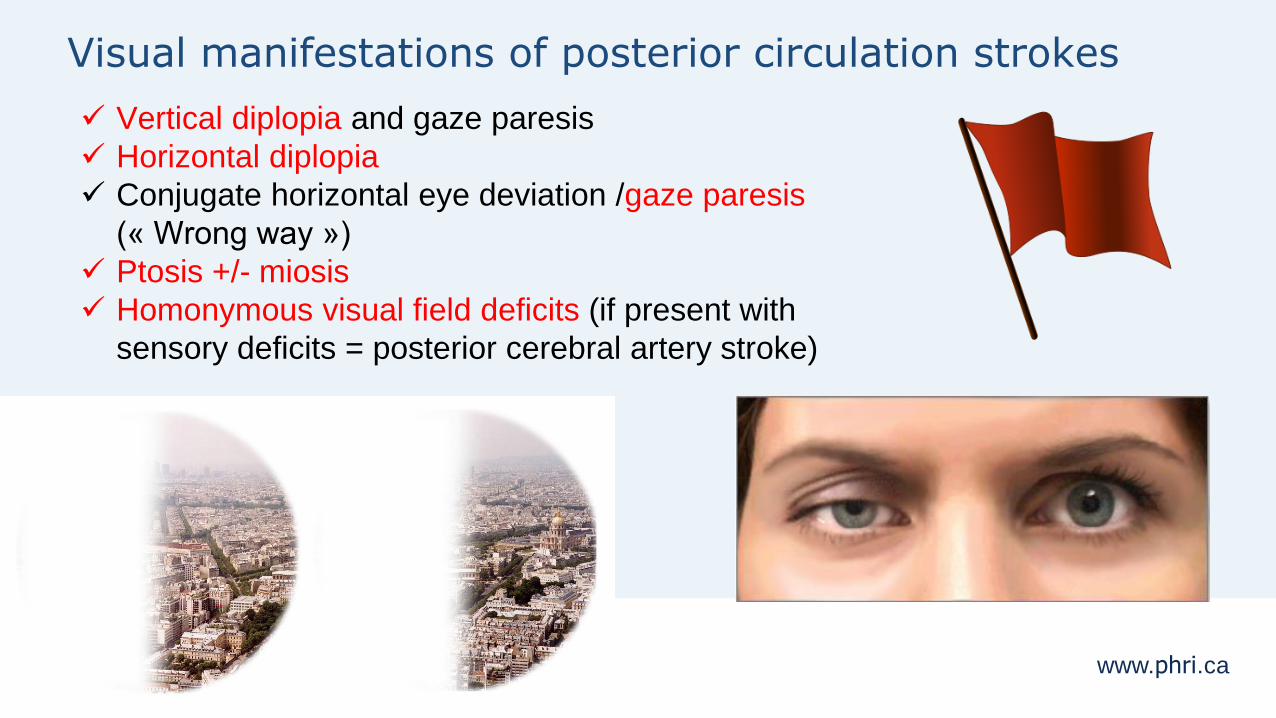
Visual manifestations of posterior circulation strokes
Vertical diplopia and gaze paresis
Horizontal diplopia
Conjugate horizontal eye deviation /gaze paresis
(« Wrong way »)
Ptosis +/- miosis
Homonymous visual field deficits (if present with
sensory deficits = posterior cerebral artery stroke)
www.phri.ca
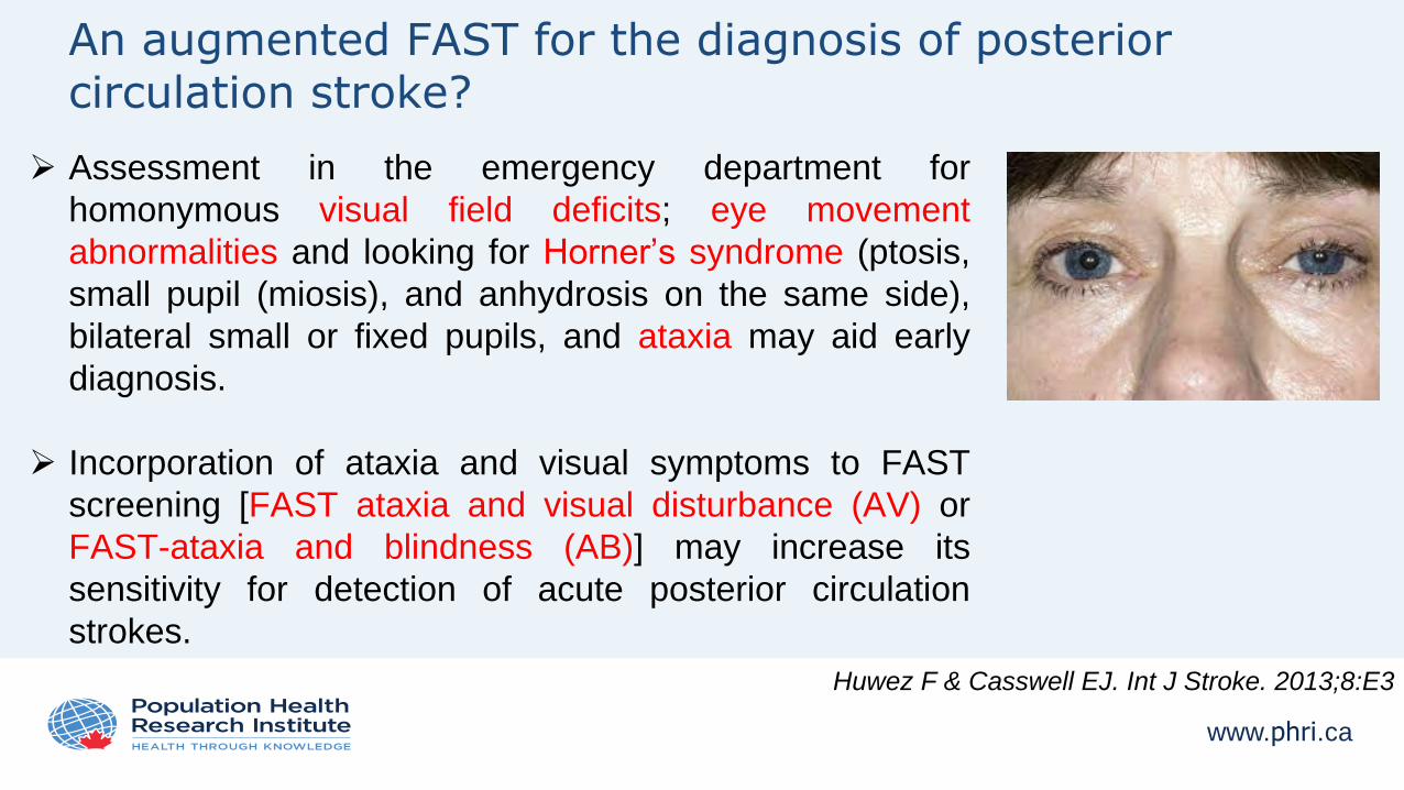
An augmented FAST for the diagnosis of posterior circulation stroke?
Assessment in the emergency department for
homonymous visual field deficits; eye movement
abnormalities and looking for Horner’s syndrome (ptosis,
small pupil (miosis), and anhydrosis on the same side),
bilateral small or fixed pupils, and ataxia may aid early
diagnosis.
Incorporation of ataxia and visual symptoms to FAST
screening [FAST ataxia and visual disturbance (AV) or
FAST-ataxia and blindness (AB)] may increase its
sensitivity for detection of acute posterior circulation
strokes.
Huwez F & Casswell EJ. Int J Stroke. 2013;8:E3
www.phri.ca
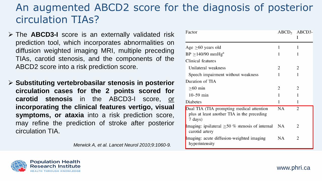
An augmented ABCD2 score for the diagnosis of posteriorcirculation TIAs?
The ABCD3-I score is an externally validated risk
prediction tool, which incorporates abnormalities on
diffusion weighted imaging MRI, multiple preceding
TIAs, carotid stenosis, and the components of the
ABCD2 score into a risk prediction score.
Substituting vertebrobasilar stenosis in posterior
circulation cases for the 2 points scored for
carotid stenosis in the ABCD3-I score, or
incorporating the clinical features vertigo, visual
symptoms, or ataxia into a risk prediction score,
may refine the prediction of stroke after posterior
circulation TIA.
Merwick A, et al. Lancet Neurol 2010;9:1060-9.
www.phri.ca
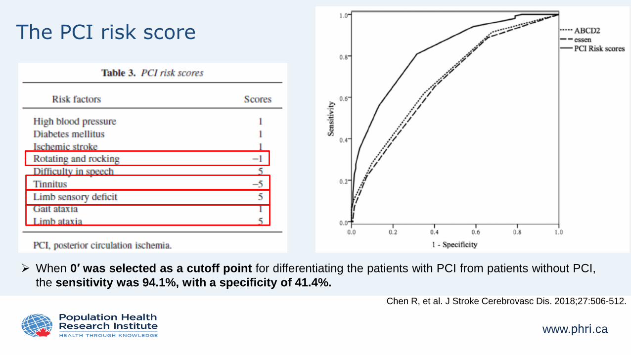
The PCI risk score
When 0′ was selected as a cutoff point for differentiating the patients with PCI from patients without PCI,
the sensitivity was 94.1%, with a specificity of 41.4%.
Chen R, et al. J Stroke Cerebrovasc Dis. 2018;27:506-512.
www.phri.ca
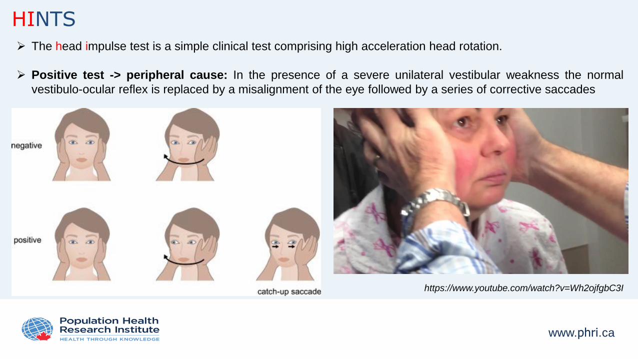
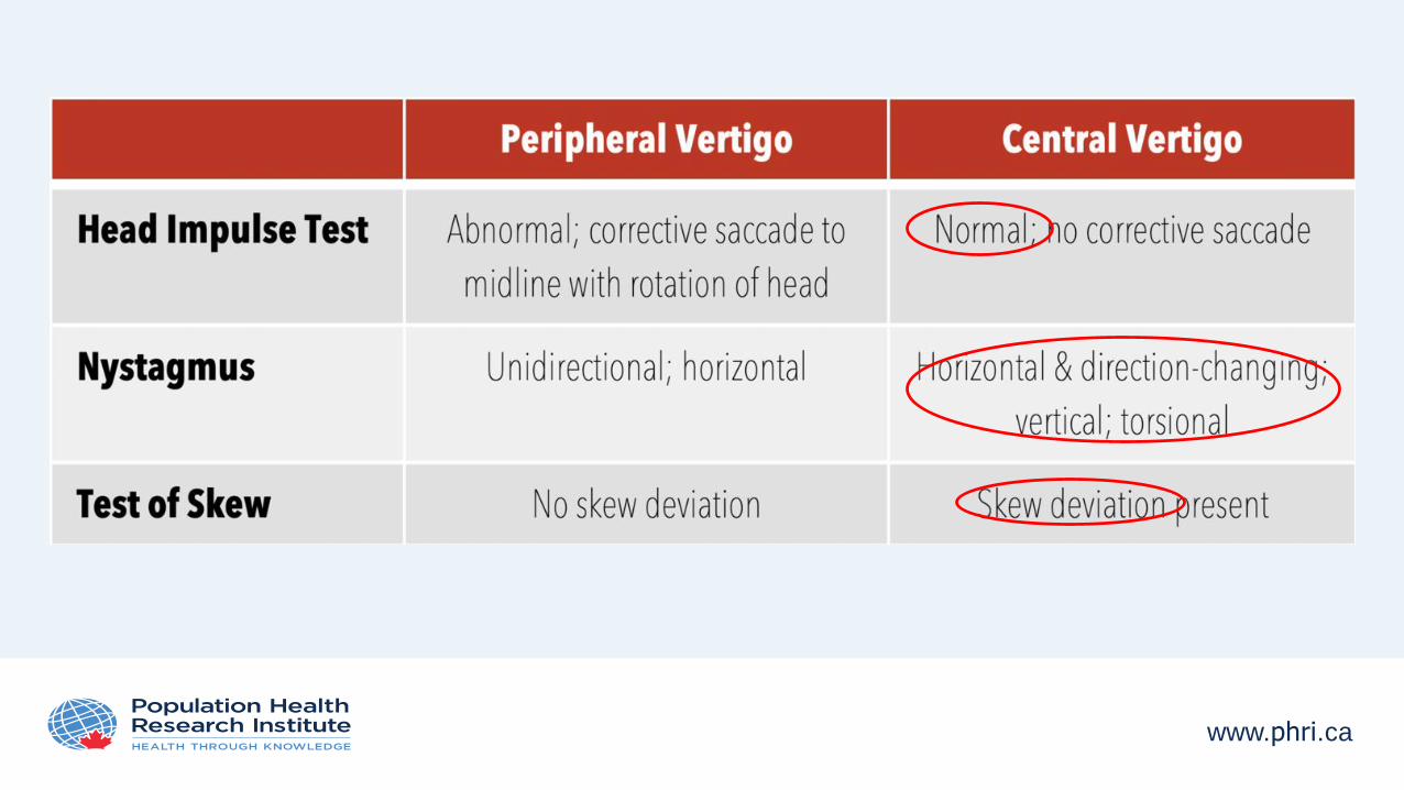
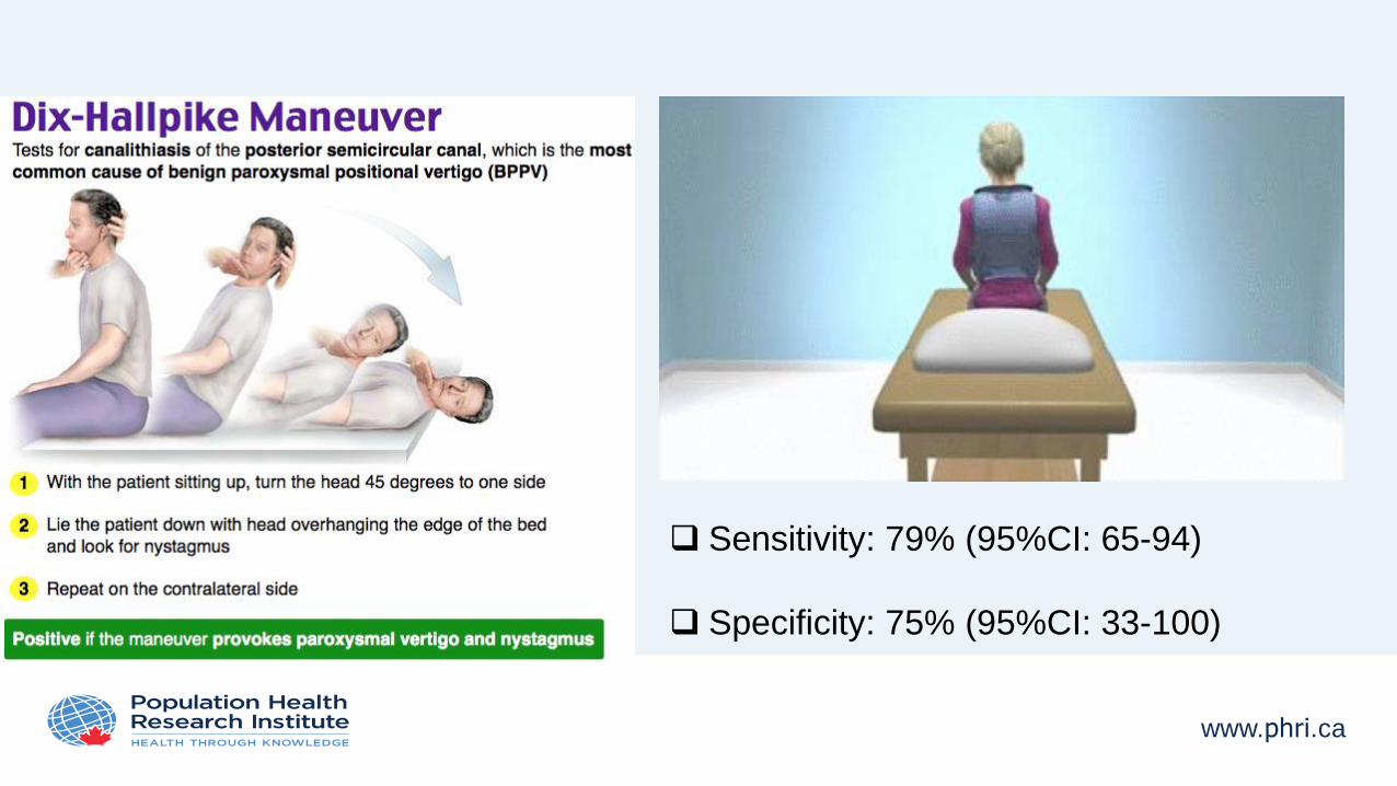
HINTS
https://www.youtube.com/watch?v=Wh2ojfgbC3I
The head impulse test is a simple clinical test comprising high acceleration head rotation.
Positive test -> peripheral cause: In the presence of a severe unilateral vestibular weakness the normal
vestibulo-ocular reflex is replaced by a misalignment of the eye followed by a series of corrective saccades
www.phri.ca
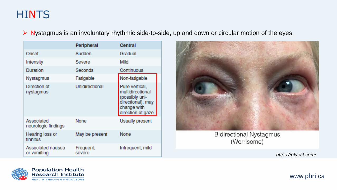
HINTS
https://gfycat.com/
Nystagmus is an involuntary rhythmic side-to-side, up and down or circular motion of the eyes
www.phri.ca
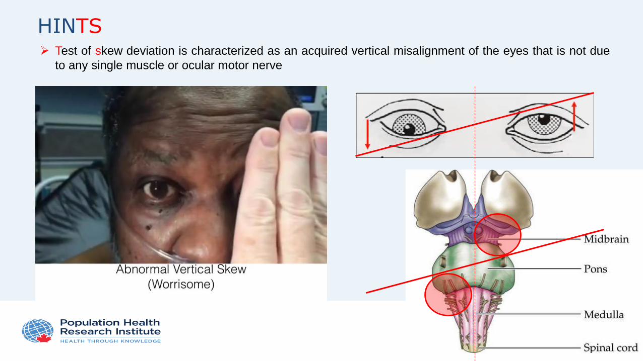
HINTS
https://gfycat.com/
Test of skew deviation is characterized as an acquired vertical misalignment of the eyes that is not due
to any single muscle or ocular motor nerve
www.phri.ca
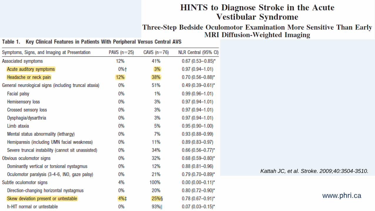
www.phri.ca
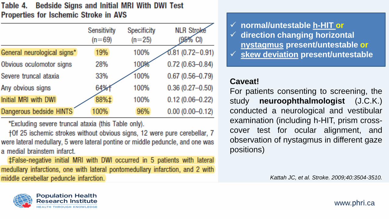
Kattah JC, et al. Stroke. 2009;40:3504-3510.
www.phri.ca
normal/untestable h-HIT or
direction changing horizontal
nystagmus present/untestable or
skew deviation present/untestable
Caveat!
For patients consenting to screening, the
study neuroophthalmologist (J.C.K.)
conducted a neurological and vestibular
examination (including h-HIT, prism cross-
cover test for ocular alignment, and
observation of nystagmus in different gaze
positions)
Kattah JC, et al. Stroke. 2009;40:3504-3510.
www.phri.ca
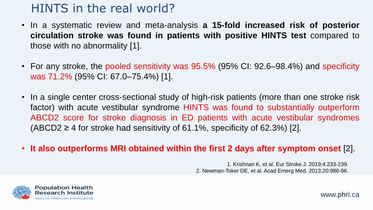
HINTS in the real world?
• In a systematic review and meta-analysis a 15-fold increased risk of posterior
circulation stroke was found in patients with positive HINTS test compared to
those with no abnormality [1].
• For any stroke, the pooled sensitivity was 95.5% (95% CI: 92.6–98.4%) and specificity
was 71.2% (95% CI: 67.0–75.4%) [1].
• In a single center cross-sectional study of high-risk patients (more than one stroke risk
factor) with acute vestibular syndrome HINTS was found to substantially outperform
ABCD2 score for stroke diagnosis in ED patients with acute vestibular syndromes
(ABCD2 ≥ 4 for stroke had sensitivity of 61.1%, specificity of 62.3%) [2].
• It also outperforms MRI obtained within the first 2 days after symptom onset [2].
1. Krishnan K, et al. Eur Stroke J. 2019;4:233-239.
2. Newman-Toker DE, et al. Acad Emerg Med. 2013;20:986-96.

www.phri.ca
HINTS exam only if nystagmus is present
www.phri.ca
Sensitivity: 79% (95%CI: 65-94)
Specificity: 75% (95%CI: 33-100)
www.phri.ca
Imaging for suspected acute stroke in posterior circulation
• MRI with diffusion weighted imaging is the brain imaging modality of choice for suspected posterior
circulation stroke. It can help diagnose disorders that mimic stroke and TIA, can help verify vascular
territory, and diffusion weighted imaging abnormalities independently predict early stroke risk after
TIA.
• MRI is far more sensitive than CT in the diagnosis of acute ischaemic stroke for all vascular
territories, with study results indicating 80-95% sensitivity in the first 24 hours when diffusion
weighted imaging is used, versus 16% sensitivity with CT.
• Sensitivity may be lower in the posterior circulation and false negatives can occur with early
MRI—a 19% false negative rate was reported in one single centre case series of 31 patients with
• vertebrobasilar stroke.
• MRI or magnetic resonance angiography with dedicated fat saturated sequences may help
identify vertebral dissection.
1. Edlow JA, et al. Lancet Neurol 2008;7:951-64.
2. Merwick A, et al. Lancet Neurol 2010;9:1060-9.
www.phri.ca
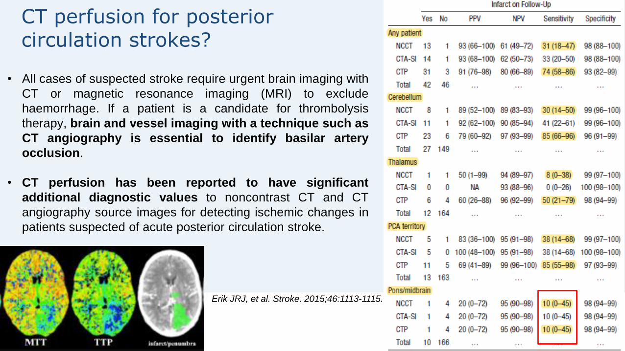
CT perfusion for posterior circulation strokes?
• All cases of suspected stroke require urgent brain imaging with
CT or magnetic resonance imaging (MRI) to exclude
haemorrhage. If a patient is a candidate for thrombolysis
therapy, brain and vessel imaging with a technique such as
CT angiography is essential to identify basilar artery
occlusion.
• CT perfusion has been reported to have significant
additional diagnostic values to noncontrast CT and CT
angiography source images for detecting ischemic changes in
patients suspected of acute posterior circulation stroke.
Erik JRJ, et al. Stroke. 2015;46:1113-1115.
www.phri.ca
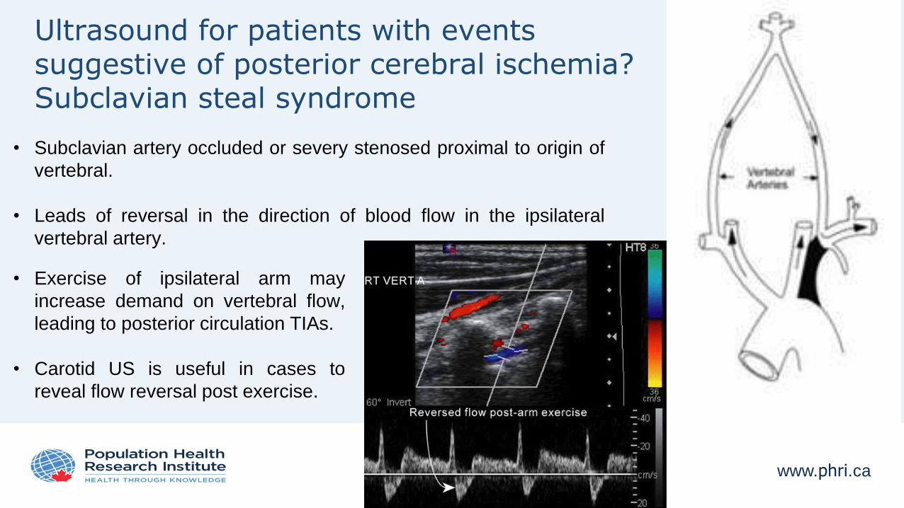
Ultrasound for patients with events suggestive of posterior cerebral ischemia? Subclavian steal syndrome
• Subclavian artery occluded or severy stenosed proximal to origin of
vertebral.
• Leads of reversal in the direction of blood flow in the ipsilateral
vertebral artery.
• Exercise of ipsilateral arm may
increase demand on vertebral flow,
leading to posterior circulation TIAs.
• Carotid US is useful in cases to
reveal flow reversal post exercise.
www.phri.ca
Presentation outline
• Challenges in the diagnosis of posterior circulation stroke
• Anatomy of posterior circulation & mechanisms of posterior circulation ischemia
• Common posterior circulation stroke syndromes
• Diagnosis of posterior circulation stroke/TIA in the emergency setting
• Conclusions
www.phri.ca
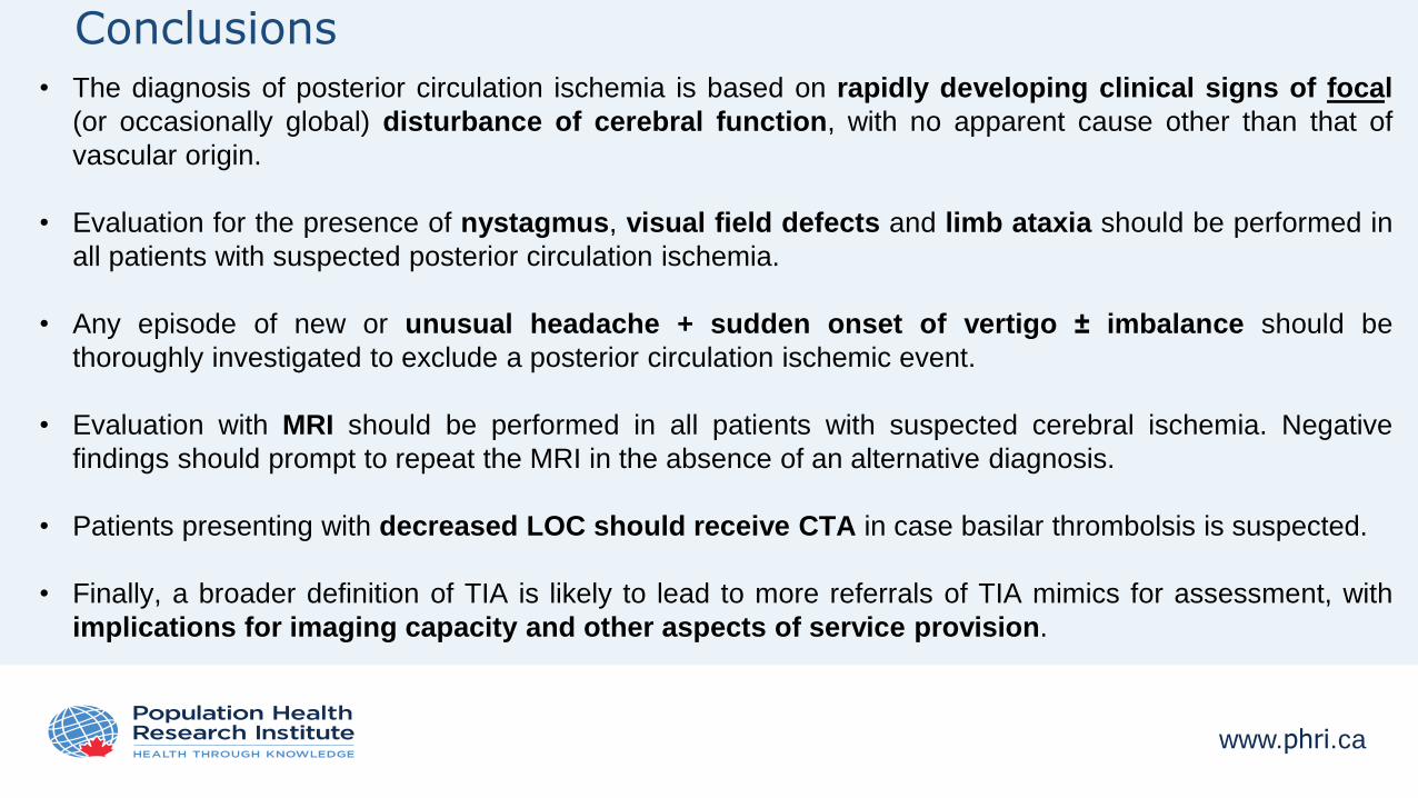
Conclusions• The diagnosis of posterior circulation ischemia is based on rapidly developing clinical signs of focal
(or occasionally global) disturbance of cerebral function, with no apparent cause other than that of
vascular origin.
• Evaluation for the presence of nystagmus, visual field defects and limb ataxia should be performed in
all patients with suspected posterior circulation ischemia.
• Any episode of new or unusual headache + sudden onset of vertigo ± imbalance should be
thoroughly investigated to exclude a posterior circulation ischemic event.
• Evaluation with MRI should be performed in all patients with suspected cerebral ischemia. Negative
findings should prompt to repeat the MRI in the absence of an alternative diagnosis.
• Patients presenting with decreased LOC should receive CTA in case basilar thrombolsis is suspected.
• Finally, a broader definition of TIA is likely to lead to more referrals of TIA mimics for assessment, with
implications for imaging capacity and other aspects of service provision.
www.phri.ca
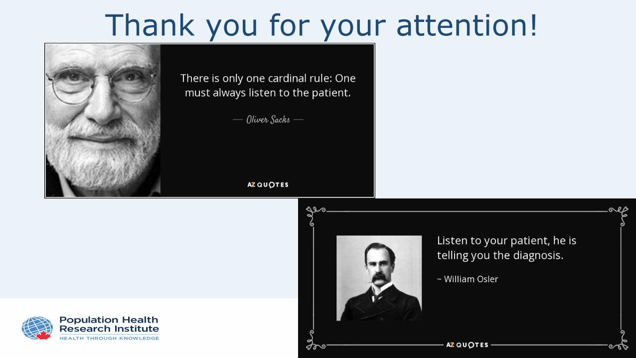
Thank you for your attention!


