Embed Size (px)
Citation preview

First Fatality Associated with ElephantEndotheliotropic Herpesvirus 5 in anAsian Elephant: Pathological Findingsand Complete Viral Genome SequenceGavin S. Wilkie1, Andrew J. Davison1, Karen Kerr1, Mark F. Stidworthy2, Sharon Redrobe3,Falko Steinbach4,5, Akbar Dastjerdi4 & Daniela Denk2
1MRC–University ofGlasgowCentre for Virus Research,GlasgowG115JR, United Kingdom, 2International Zoo VeterinaryGroup,Station House, Keighley BD21 4NQ, United Kingdom, 3Twycross Zoo – East Midland Zoological Society, Atherstone CV9 3PX,United Kingdom, 4Virology Department, Animal Health and Veterinary Laboratories Agency Weybridge, Addlestone KT15 3NB,United Kingdom, 5School of Veterinary Medicine, University of Surrey, Guildford GU2 7TE, United Kingdom.
Infections of Asian elephants (Elephas maximus) with elephant endotheliotropic herpesvirus (EEHV) cancause a rapid, highly lethal, hemorrhagic disease, which primarily affects juvenile animals up to the age offour years. So far, the majority of deaths have been attributed to infections with genotype EEHV1 or, morerarely, EEHV3 and EEHV4. Here, we report the pathological characteristics of the first fatality linked toEEHV5 infection, and describe the complete viral DNA sequence. Gross post-mortem and histologicalfindings were indistinguishable from lethal cases previously attributed to other EEHV genotypes, and thepresence of characteristic herpesviral inclusions in capillary endothelial cells at several sites was consistentwith the diagnosis of acute EEHV infection. Molecular analysis confirmed the presence of EEHV5DNA andwas followed by sequencing of the viral genome directly from post-mortem material. The genome is180,800 bp in size and contains 120 predicted protein-coding genes, five of which are fragmented andpresumably nonfunctional. The seven families of paralogous genes recognized in EEHV1 are alsorepresented in EEHV5. The overall degree of divergence (37%) between the EEHV5 and EEHV1 genomes,and phylogenetic analysis of eight conserved genes, support the proposed classification of EEHV5 into a newspecies (Elephantid herpesvirus 5).
Since the first reported death attributed to a herpesvirus in an Asian elephant (Elephas maximus) in 19881,elephant endotheliotropic herpesvirus (EEHV) infection has emerged as a serious threat to captive and wildAsian elephant populations. To date, seven distinct genotypes, named EEHV1 to EEHV7, have been
identified in Asian elephants and African elephants (Loxodonta africana). The spectrum of associated diseaseranges from subclinical infection to rapidly fatal hemorrhagic disease2. The genotype most often involved infatalities is EEHV1, and the complete genome sequences of the two subgenotypes (EEHV1A and EEHV1B) wereresolved recently3,4. Two further genotypes, EEHV3 and EEHV4, are known to cause sporadic fatalities in youngAsian elephants, and EEHV2 has been attributed to a similar disease in African elephants5–8. The tropism ofEEHVs for endothelium results in widespread capillary damage and subsequent lethal pericardial, thoracic andabdominal effusions and variably extensive hemorrhages in a variety of tissues7–9.
EEHV5 was first detected in 2007 in whole blood samples from a clinically healthy, 59 year-old, wild-bornAsian elephant7. This genotype has since been associated with a small number of subclinical and clinical infectionsin captive Asian elephants10. Clinical findings in an EEHV5-positive herd included bilaterally swollen temporalglands, oral mucosal hyperaemia, vesicles on the tongue, and generalized lethargy in an otherwise healthy adultand a one year-old calf. DNA sequence analysis revealed the presence of EEHV5 subgenotypes (EEHV5A andEEHV5B) within the herd, but no fatalities were recorded10.
Initial DNA sequence characterization of EEHV1A and EEHV1B5,11,12 was instrumental in classifying theviruses into the species Elephantid herpesvirus 1 in the genus Proboscivirus, subfamily Betaherpesvirinae, familyHerpesviridae, order Herpesvirales13. Derivation of the complete genome sequences of two EEHV1A strains(EEHV1A/Kimba and EEHV1A/Raman; GenBank accession numbers KC618527 and KC462165, respectively)
OPEN
SUBJECT AREAS:
HERPES VIRUS
INFECTION
Received9 June 2014
Accepted28 July 2014
Published9 September 2014
Correspondence and
requests for materials
should be addressed to
F.S. (falko.steinbach@
ahvla.gsi.gov.uk)
SCIENTIFIC REPORTS | 4 : 6299 | DOI: 10.1038/srep06299 1
and one EEHV1B strain (EEHV1B/Emelia; KC462164) recently pro-vided substantial insights into the genetic compositions of theseviruses, which are characterized by numerous genes lacking counter-parts in other herpesviruses3,4. In this report, we describe detailedpathological findings and a complete viral genome sequence from thefirst case of EEHV5-associated fatality in an Asian elephant.
MethodsCase history.The affected animal was a juvenile Asian elephant, Vijay (=; barn name;official name Ganesh), which was born at Twycross Zoo, UK, within a herd of fouradult, female Asian elephants on 6 August 2009 (http://www.elephant.se/database2.php?elephant_id54826). This animal had a history of retarded growth and ill-health,and developed an oral lesion during December 2010 and a mucocutaneous perianallesion duringMarch 2011, both of which resolved spontaneously over a period of 2–3weeks. In April 2011, lethargy followed by rapidly progressive hemorrhages ofmucosal membranes, edema, and lingual cyanosis led to the presumptive clinicaldiagnosis of EEHV infection. Treatment followed protocols for extensive antiviraland supportive therapy, including drainage of the pericardial effusion underultrasonographic guidance. Despite all efforts over a period of 6 days, the calf’s healthdeteriorated, and the decision was made to euthanize the animal. All treatments andclinical decision making in this case were conducted in accordance with theVeterinary Surgeons Act under the direct supervision of a RCVS Specialist andwithinthe ethical policies of the zoo.
Pathology. A comprehensive post-mortem procedure following elephant necropsyprotocols14 was carried out within 24 hours. For histological examination, tongue,heart, lung, spleen, liver, kidneys, stomach, and all segments of intestine, urinarybladder, skeletal muscle, thyroid, adrenal and salivary glands, skin, oral mucosa and asection of trunk, pharyngeal lymphoid tissue, lymph nodes, trigeminal nerve, brain(including the pituitary gland), medulla, and bone marrow were processed routinely,and paraffin-embedded sections were stained with hematoxylin and eosin.
Viral diagnosis.DNAwas extracted from heart, tongue, trunk mucosa, whole blood,and serum samples by using a DNeasy blood and tissue kit (Qiagen, Crawley, UK). Itwas tested for EEHV1A and EEHV1B by using a published PCR method15, and forEEHV2 to EEHV6 by using previously unpublished PCR methods, with all assaystargeting regions of the DNA polymerase gene (U38). Semi-nested PCRs for EEHV2/EEHV5/EEHV6 were performed by using forward primer 59-GTT ACS RYB GTYACSACTAACAC-39with reverse primers 59-GCTATMGSYARACACGGRAACAT-39 in the first round and 59-ACR GCR TTR CAY GTD AGT TTC AG-39 in thesecond round. Semi-nested PCRs for EEHV3/EEHV4 were carried out by usingforward primer 59-CAT CCAGGCCTACAACCTCTG-39with reverse primers 59-CAG TCG TTG AAG GTG TCG CAG-39 and 59-AGA CGG CGT TGC AGG TGAGCT-39 in the first and second rounds, respectively. The predicted PCR product sizesfor EEHV2/EEHV5/EEHV6 and EEHV3/EEHV4 were 204 and 281 bp, respectively.The tissues were also tested by using nested pan-EEHV and specific EEHV5 PCRs7,performing the first round PCRs only.
The PCRs were performed by using a fast cycling PCR kit (Qiagen), the reactionsconsisting of 10 ml 23 fast cycling PCRmaster mix, 1 ml each of forward and reverseprimers at 20 pmol/ml, 2 ml template DNA, andwater to a total volume of 20 ml. PCRswere initiated at 95uC for 5 min, followed by 35 cycles of 95uC for 15 s, 55uC for 30 s,and 72uC for 1 min. The products were detected by agarose gel electrophoresis,purified by using a MinElute PCR purification kit (Qiagen), and sequenced by usingSanger technology.
DNA sequencing. An aliquot of DNA (240 ng) from heart tissue was shearedacoustically to an average size of 525 bp in a volume of 50 ml, using a Covaris S220sonicator (Covaris Inc., Woburn, MA, USA). Fragment size was measured on anAgilent 2200 TapeStation (Agilent, Santa Clara, CA, USA). A KAPA librarypreparation kit (KAPA Biosystems, Wilmington, MA, USA) was used to prepare thesheared DNA fragments for Illumina sequencing, with the followingmodifications ofthe standard protocol. Sheared DNA was end-repaired directly (i.e., withoutisolation), and end-repaired DNA was purified by using 0.8 volumes of AMPureXPbeads (Beckman Coulter, Indianapolis, IN, USA) in order to remove fragmentssmaller than the anticipated read length (250 bp), and eluted in 30 ml TE (10 mMTris-HCl, pH 8.5), retaining the beads in the tube during all subsequent steps in orderto minimize sample loss16. A 2051 molar ratio of NEBnext Illumina adaptor (NewEngland Biolabs, Ipswich, MA, USA) was ligated to the DNA fragments. In order toreduce PCR amplification bias and improve fidelity, the adaptor-ligated DNA wasamplified by three cycles on an ABI 7500 real-time PCR cycler (Life Technologies,Grand Island, NY, USA) using a KAPA real-time library amplification kit.
A MiSeq DNA sequencer running v2 chemistry (Illumina, San Diego, CA, USA)was used to generate 15,054,115 paired-end, 250 nucleotide (nt) reads from the Vijay/EEHV5 library. The data were processed for adapter removal, quality-filtered,trimmed, and subjected to read pair validation by using Trim Galore v. 0.2.2 (http://www.bioinformatics.babraham.ac.uk/projects/trim_galore), yielding 15,002,189paired-end reads. In order to identify host data, BWA v. 0.6.2-r12617 was used ingapped, paired-end mode to map reads to the African elephant genome (EnsemblLoxafr3.0 v. 69.3), since the Asian elephant genome was not available. Unmappedread pairs were extracted by using Samtools v. 0.1.1818, and contigs were assembled de
novo by using ABySS19. Scaffold_builder20 was used to produce a draft sequence byordering the contigs against the EEHV1A/Raman genome sequence. Additionalcontigs were incorporated, and gaps were closed by using Megamerger21, GapFiller v.1–1122, and custom Perl scripts. The integrity of the resulting single, circular contigwas verified by aligning against the original dataset using BWA and visualizing usingTablet v. 1.13.08.0523.
Identification of genome termini. The termini of the EEHV5 genome wereidentified essentially as described for EEHV1A/Raman and EEHV1B/Emelia4, usingcomponents described in the KAPA library preparation kit (KAPA Biosystems) andMarathon cDNA amplification kit (Clontech Laboratories, Palo Alto, CA, USA)according to themanufacturers’ instructions. Briefly, aliquots of DNA extracted fromheart tissue were either blunt-ended or left untreated. The former DNAwas ligated toa partially double-stranded DNA adaptor that was blunt at the end designed to ligateto the genome termini, and the latter DNA to a corresponding adaptor that containedan additional unpaired, redundant 39-nucleotide (i.e., a mixture of G, A, T, and C).First-round PCR was then carried out on the two ligation products by using anEEHV5-specific primer mapping close to the anticipated left or right genometerminus, plus an adaptor-specific primer (AP1). The locations of the EEHV5 terminiwere predicted from sequence similarities to those of EEHV1A. The EEHV5-specificprimers were 59-TCC GGG AGG GTA TAC GTC ACG GTG-39 (left terminus) and59-CAG AGA TGA TTG ACG TCT ACA CGG-39 (right terminus). Although PCRproducts were not detectable as visible bands by agarose gel electrophoresis, theregions containing DNA molecules of the anticipated sizes (approximately 200 and180 bp, respectively) were purified. Nested PCR was carried out on each product byusing EEHV5-specific primers 59-TTA CTA TGA CGT CAT GAC CGG AAG-39(left terminus) or 59-GCCCACCCGCCAGCGGTAATGACT-39 (right terminus),plus a second adaptor-specific primer (AP2). The PCR products (approximately 160and 110 bp, respectively) were detected by agarose gel electrophoresis, purified byusing a Geneclean turbo kit (MP Biomedicals, Solon, OH, USA), and cloned intoplasmids by using the pGEM-T vector system (Promega Corporation, Madison, WI,USA) according to the manufacturer’s instructions. The inserts in 12 plasmids foreach of the four products were sequenced by using Sanger technology with universalprimers, and the termini were defined as being located at the positions in the circularsequence represented by the majority of clones.
Sequence comparisons. Overall percentages of sequence identity between EEHVgenomes were assessed using Stretcher (http://emboss.bioinformatics.nl). Similarityamong imputed amino acid sequences was investigated by using Needle (http://emboss.bioinformatics.nl). Phylogenetic trees were constructed by using Neighbor-Joining and Poisson model implemented in the MEGA v. 5.03 software24 withconfidence values calculated from 2000 replicates, having obtained sequences fromNCBI RefSeq (http://www.ncbi.nlm.nih.gov/genomes/GenomesGroup.cgi?taxid510292). Matrix sequence comparison plots were computed by usingCompare (window5 20, stringency5 16) and Dotplot in the GCG package (http://www.accelrys.com).
Database accession. The genome sequence of EEHV5/Vijay was deposited in NCBIGenBank under accession number KF921519.
ResultsPathological findings. On gross post-mortem examination, Vijaypresented in good body condition. No skin lesions were identified oncareful examination. The tongue was enlarged and cyanotic, withmultifocal (petechial to ecchymosal) mucosal and intraparen-chymal hemorrhages. The head, in particular the eyelids, wasswollen, with moderate to marked subcutaneous edema accom-panied by few disseminated subcutaneous petechial hemorrhages.Petechial to ecchymotic hemorrhages were in addition detectedwithin the conjunctiva, brain (including meninges), esophagus,trachea, stomach, intestine and mesentery, liver, urinary bladder,and adrenal glands. Extensive atrial and ventricular hemorrhageswere observed within the heart, comprising severe, confluent,irregular areas of subepicardial and subendocardial bleeding extend-ing into themyocardium and papillarymusculatures25. Severe edemaand multifocal subserosal hemorrhages, typical of EEHV-inducedlesions, were observed in the dorsal mediastinum (Fig. 1a). Freeaccumulations of approximately 500–700 ml of clear, watery fluidwere present within the abdominal cavity, accompanying markedexpansion of the intestinal wall, mesentery, and omentum byedema. The kidneys were diffusely congested, with mottled light-to dark-red cut surfaces and retained cortico-medullary distinc-tion. The uniformly dark-red lungs exhibited multifocal subpleuralhemorrhages. Generalized diffuse reddening and mild enlargementwere noted in examined visceral and subcutaneous lymph nodes.
www.nature.com/scientificreports
SCIENTIFIC REPORTS | 4 : 6299 | DOI: 10.1038/srep06299 2

On histological examination, varying degrees of multifocal, acutehemorrhage with underlying vascular changes, including edema ofthe vascular wall, mild endothelial cell hypertrophy, and occasionalfibrinoid necrosis with thrombus formation, were present in severalsections of the heart, skeletal muscle, trigeminal nerve, kidney,adrenal gland, tongue, stomach, lung, intestine, trunk, liver, oralmucosa, lymph node, pituitary, urinary bladder, cerebral medulla,and cortex. Accompanying edema was most prominent withinintestinal sections and lymphoid tissues. Moderate cellular infiltratescomprising neutrophils, fewer plasma cells, and other lymphocyteswere noted in the heart, tongue and liver, the latter of which demon-strated scattered pyknotic cells compatible with hepatocellulardegeneration and necrosis. Characteristic single, variably sized, eosi-nophilic or amphophilic, smudged, intranuclear inclusion bodiesconsistent with herpesviral inclusions were detected within capillaryendothelial cells of the heart, adrenal gland, lung, liver, and oralmucosa (Fig. 1b). Erythrophagocytosis and mild hemosiderosis(confirmed by Perl’s Prussian blue stain) were observed in lymphnodes and liver, respectively.
Preliminary molecular investigation. EEHV DNA was detected inheart, tongue, trunk mucosa, whole blood, and serum in both roundsof semi-nested EEHV2/EEHV5/EEHV6 PCR (data not shown).DNA sequencing revealed that the target region was identical to
that of the index EEHV5 case (EEHV5/#NAP28; GenBankaccession HM060764), as were the sequences obtained using pan-EEHV and specific EEHV5 primers7. All samples proved negative forthe other EEHV genotypes.
Genome structure and composition. The EEHV5/Vijay genome is180,800 bp in size, and consists of a unique sequence (U, 175,872 bp)flanked by a terminal direct repeat (TR, 2464 bp), in the arrangementTR-U-TR. The genome sizes and structures of EEHV1A/Raman andEEHV1B/Emelia are similar4. Like the genomes of EEHV1A/Ramanand EEHV1B/Emelia, the EEHV5/Vijay genome contains single,unpaired C and G residues at the right and left 39-termini,respectively. Sequence reads representing the junction between thetermini amounted to approximately 10% of the mean number fromthe TR-U and U-TR junctions. These may have been generated fromgenomes that were circular, concatemeric, or contained additionalcopies of TR at the termini.As calculated for TR-U (i.e. counting TR only once), 33,005
(0.11%) of the 30,004,378 processed reads for EEHV5/Vijaymatchedthe genome sequence, and coverage of the EEHV5 sequence wascomplete, with an average of 41 reads/nt (range 5–155). In theassembly of read data against the finished genome, there was noevidence for significant sequence variation, which indicated thatVijay had been infected by a single EEHV5 strain. The EEHV5/Vijay genotype was confirmed from comparisons with data availablein GenBank for three EEHV5 strains, in each case consisting ofseveral sequences scattered across the left half of the genome. Theaggregate levels of identity for the 14,614 bp (in nine sections) fromthe index EEHV5 case (EEHV5/#NAP28) and the 22,143 bp (in 16sections) from a case annotated as subgenotype EEHV5A (EEHV5A/#NAP50) are very high, at 99.6 and 99.2%, respectively. The level forthe 24,190 bp (in 13 sections) from a case annotated as subgenotypeEEHV5B (EEHV5B/#NAP58TW) is lower, at 94.1%. These resultsindicate that EEHV5/Vijay is an EEHV5A strain.The locations of open reading frames (ORFs) in EEHV5/Vijay that
encode functional proteins, and fragmented ORFs that used toencode functional proteins, were predicted by using the criteriaemployed for EEHV1A/Raman and EEHV1B/Emelia, and the namesof ORFs were assigned according to the system described and jus-tified previously for these viruses4. In this system, most EEHV ORFsthat are conserved in the subfamily Betaherpesvirinae are namedafter their orthologs in human herpesviruses 6A and 6B26,27, andare denoted by the prefix U. ORFs that lack orthologs in other generain the subfamily are denoted by the prefix EE. The genome ofEEHV5/Vijay contains 120 ORFs (Fig. 2). The seven families ofparalogous ORFs recognized in EEHV1A/Raman and EEHV1B/Emelia are represented in EEHV5/Vijay: the EE3, deoxyuridine tri-phosphatase-related protein (DURP), U4, seven transmembranedomain (7TM), EE20, OX-2, and EE50 families. There are five frag-mentedORFs: EE44A, EE49A, and EE61 by frameshifts inG:C tracts,EE62B by a frameshift in a A:T tract, and EE49C by several frame-shifts of other types. As indicated in Table 1, fragmented ORFs havebeen noted previously in EEHV1A/Raman and EEHV1B/Emelia4,and, from our analysis, are also evident in EEHV1A/Kimba. It shouldbe noted that ORFs fragmented by frameshifts in G:C or A:T tractsare also found in members of the genus Cytomegalovirus, and, inprinciple, can revert to functionality via selection of length variants28.For example, in EEHV5/Vijay, the majority of reads containing theG:C tracts in EE44A and EE61 (13/17 and 11/22, respectively) con-tains a tract length that results in frameshifting (11 and 9 nt, respect-ively, as in the consensus sequence). However, aminority of reads (4/17 and 10/17, respectively) in these ORFs contains a tract length thatdoes not result in frameshifting (10 and 10 nt, respectively), thusallowing the cognate protein to be synthesized. This analysis bearsthe caveat that tract length variation might result from the PCR-
Figure 1 | Pathology of EEHV5 infection in Vijay. (a) Severe edema and
multifocal subserosal hemorrhages in the dorsal mediastinum. (b)
Hematoxylin and eosin staining of a liver section. The hepatic architecture
is disrupted by hemorrhage and edema, with multifocal fibrin deposits
(arrow) and diffuse mixed cellular infiltrates. The central vessel displays
denuded endothelial cells, congestion, and mild granulocytostasis. Inset:
Herpetic intranuclear inclusion body.
www.nature.com/scientificreports
SCIENTIFIC REPORTS | 4 : 6299 | DOI: 10.1038/srep06299 3

based processes used during sequence library preparation, and notnecessarily from genuine variation in the viral genome.
Genome comparisons. Four complete EEHV genomes have now beensequenced: those of EEHV1A/Raman, EEHV1A/Kimba, EEHV1B/
Emelia, and EEHV5/Vijay. They are largely colinear, and have over-all percentages of DNA sequence identity as follows: EEHV1A/Ramanto EEHV1A/Kimba, 96.7; EEHV1A/Raman to EEHV1B/Emelia, 92.7;EEHV1A/Kimba to EEHV1B/Emelia, 92.1; EEHV1A/Raman toEEHV5/Vijay, 63.1; and EEHV1B/Emelia to EEHV5/Vijay, 63.0.
Figure 2 | Map of the EEHV5/Vijay genome.The copies of TR flankingU are shown in a thicker format. ORFs are shown by coloured arrows, with names
below. Fragmented ORFs are shown in square brackets, and are depicted as intact. Introns are indicated by narrow white bars. Colour shading
indicates core ORFs (present in the ancestor of the subfamilies Alphaherpesvirinae, Betaherpesvirinae, and Gammaherpesvirinae), betagamma ORFs
(present in the subfamilies Betaherpesvirinae andGammaherpesvirinae), betaORFs (present only in the subfamilyBetaherpesvirinae), andORFs belonging
to paralogous families. As well as being members of paralogous families, U54 and U4 are beta ORFs. A putative origin of DNA replication (ori) and a
tandem reiteration (R1) are marked.
www.nature.com/scientificreports
SCIENTIFIC REPORTS | 4 : 6299 | DOI: 10.1038/srep06299 4
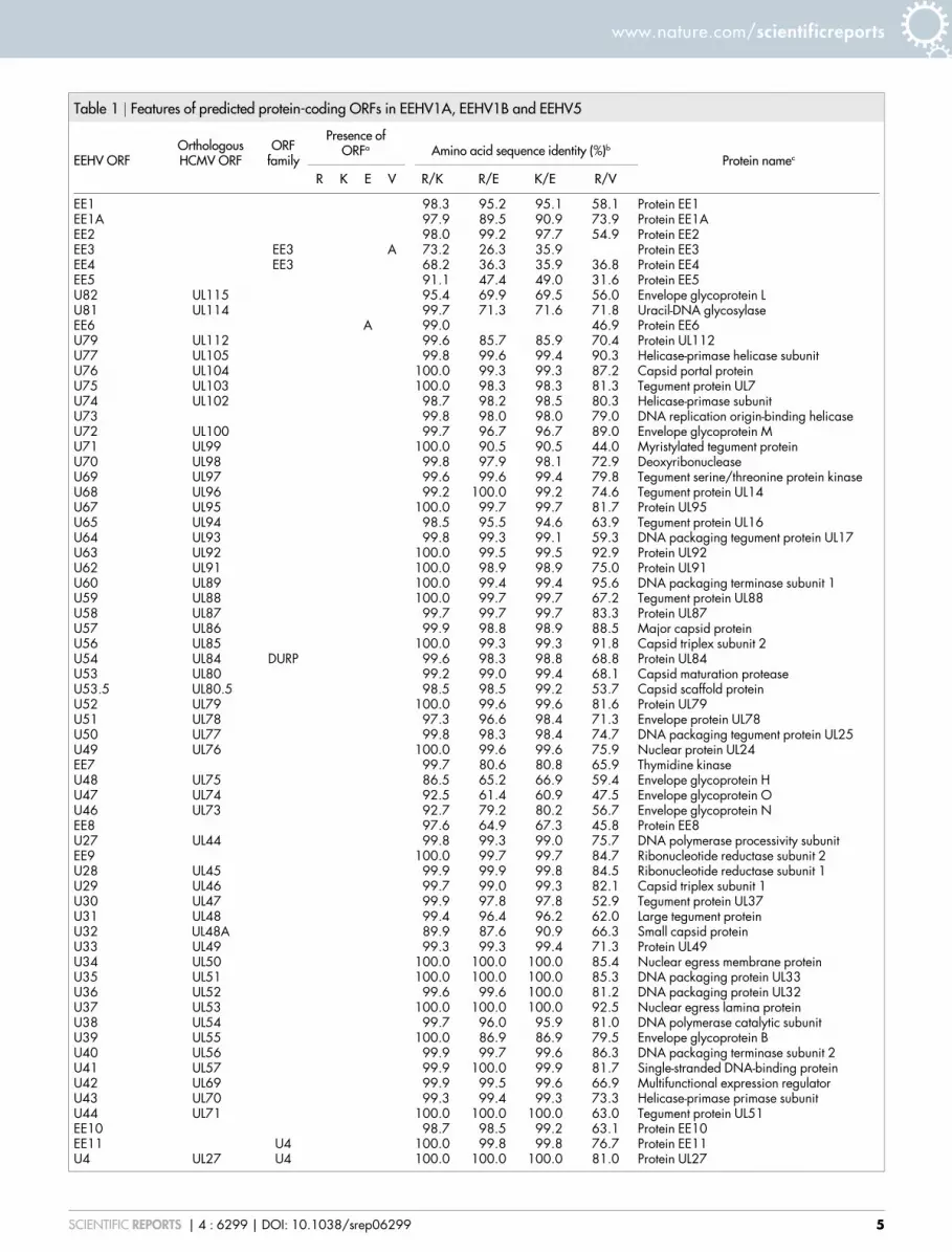
Table 1 | Features of predicted protein-coding ORFs in EEHV1A, EEHV1B and EEHV5
EEHV ORFOrthologousHCMV ORF
ORFfamily
Presence ofORFa Amino acid sequence identity (%)b
Protein namec
R K E V R/K R/E K/E R/V
EE1 98.3 95.2 95.1 58.1 Protein EE1EE1A 97.9 89.5 90.9 73.9 Protein EE1AEE2 98.0 99.2 97.7 54.9 Protein EE2EE3 EE3 A 73.2 26.3 35.9 Protein EE3EE4 EE3 68.2 36.3 35.9 36.8 Protein EE4EE5 91.1 47.4 49.0 31.6 Protein EE5U82 UL115 95.4 69.9 69.5 56.0 Envelope glycoprotein LU81 UL114 99.7 71.3 71.6 71.8 Uracil-DNA glycosylaseEE6 A 99.0 46.9 Protein EE6U79 UL112 99.6 85.7 85.9 70.4 Protein UL112U77 UL105 99.8 99.6 99.4 90.3 Helicase-primase helicase subunitU76 UL104 100.0 99.3 99.3 87.2 Capsid portal proteinU75 UL103 100.0 98.3 98.3 81.3 Tegument protein UL7U74 UL102 98.7 98.2 98.5 80.3 Helicase-primase subunitU73 99.8 98.0 98.0 79.0 DNA replication origin-binding helicaseU72 UL100 99.7 96.7 96.7 89.0 Envelope glycoprotein MU71 UL99 100.0 90.5 90.5 44.0 Myristylated tegument proteinU70 UL98 99.8 97.9 98.1 72.9 DeoxyribonucleaseU69 UL97 99.6 99.6 99.4 79.8 Tegument serine/threonine protein kinaseU68 UL96 99.2 100.0 99.2 74.6 Tegument protein UL14U67 UL95 100.0 99.7 99.7 81.7 Protein UL95U65 UL94 98.5 95.5 94.6 63.9 Tegument protein UL16U64 UL93 99.8 99.3 99.1 59.3 DNA packaging tegument protein UL17U63 UL92 100.0 99.5 99.5 92.9 Protein UL92U62 UL91 100.0 98.9 98.9 75.0 Protein UL91U60 UL89 100.0 99.4 99.4 95.6 DNA packaging terminase subunit 1U59 UL88 100.0 99.7 99.7 67.2 Tegument protein UL88U58 UL87 99.7 99.7 99.7 83.3 Protein UL87U57 UL86 99.9 98.8 98.9 88.5 Major capsid proteinU56 UL85 100.0 99.3 99.3 91.8 Capsid triplex subunit 2U54 UL84 DURP 99.6 98.3 98.8 68.8 Protein UL84U53 UL80 99.2 99.0 99.4 68.1 Capsid maturation proteaseU53.5 UL80.5 98.5 98.5 99.2 53.7 Capsid scaffold proteinU52 UL79 100.0 99.6 99.6 81.6 Protein UL79U51 UL78 97.3 96.6 98.4 71.3 Envelope protein UL78U50 UL77 99.8 98.3 98.4 74.7 DNA packaging tegument protein UL25U49 UL76 100.0 99.6 99.6 75.9 Nuclear protein UL24EE7 99.7 80.6 80.8 65.9 Thymidine kinaseU48 UL75 86.5 65.2 66.9 59.4 Envelope glycoprotein HU47 UL74 92.5 61.4 60.9 47.5 Envelope glycoprotein OU46 UL73 92.7 79.2 80.2 56.7 Envelope glycoprotein NEE8 97.6 64.9 67.3 45.8 Protein EE8U27 UL44 99.8 99.3 99.0 75.7 DNA polymerase processivity subunitEE9 100.0 99.7 99.7 84.7 Ribonucleotide reductase subunit 2U28 UL45 99.9 99.9 99.8 84.5 Ribonucleotide reductase subunit 1U29 UL46 99.7 99.0 99.3 82.1 Capsid triplex subunit 1U30 UL47 99.9 97.8 97.8 52.9 Tegument protein UL37U31 UL48 99.4 96.4 96.2 62.0 Large tegument proteinU32 UL48A 89.9 87.6 90.9 66.3 Small capsid proteinU33 UL49 99.3 99.3 99.4 71.3 Protein UL49U34 UL50 100.0 100.0 100.0 85.4 Nuclear egress membrane proteinU35 UL51 100.0 100.0 100.0 85.3 DNA packaging protein UL33U36 UL52 99.6 99.6 100.0 81.2 DNA packaging protein UL32U37 UL53 100.0 100.0 100.0 92.5 Nuclear egress lamina proteinU38 UL54 99.7 96.0 95.9 81.0 DNA polymerase catalytic subunitU39 UL55 100.0 86.9 86.9 79.5 Envelope glycoprotein BU40 UL56 99.9 99.7 99.6 86.3 DNA packaging terminase subunit 2U41 UL57 99.9 100.0 99.9 81.7 Single-stranded DNA-binding proteinU42 UL69 99.9 99.5 99.6 66.9 Multifunctional expression regulatorU43 UL70 99.3 99.4 99.3 73.3 Helicase-primase primase subunitU44 UL71 100.0 100.0 100.0 63.0 Tegument protein UL51EE10 98.7 98.5 99.2 63.1 Protein EE10EE11 U4 100.0 99.8 99.8 76.7 Protein EE11U4 UL27 U4 100.0 100.0 100.0 81.0 Protein UL27
www.nature.com/scientificreports
SCIENTIFIC REPORTS | 4 : 6299 | DOI: 10.1038/srep06299 5

Table 1 | Continued
EEHV ORFOrthologousHCMV ORF
ORFfamily
Presence ofORFa Amino acid sequence identity (%)b
Protein namec
R K E V R/K R/E K/E R/V
U11 UL32 96.2 96.7 98.9 33.1 Tegument protein pp150U12 UL33 99.7 99.8 99.5 68.4 Envelope glycoprotein UL33UL34 UL34 100.0 99.4 99.4 73.9 Protein UL34U14 UL35 91.9 94.0 88.9 49.2 Tegument protein UL35EE12 92.4 91.1 94.9 34.2 Protein EE12EE13 98.8 99.5 98.5 61.6 Protein EE13EE14 97.9 100.0 97.9 44.8 Protein EE14EE15 91.6 89.3 90.5 49.7 Protein EE15EE16 65.2 56.9 54.9 38.1 Protein EE16EE17 96.9 60.7 60.7 39.9 Protein EE17EE18 7TM F 99.1 98.7 98.7 75.8 Membrane protein EE18EE19 7TM 99.6 99.6 100.0 69.7 Membrane protein EE19EE20 EE20 100.0 100.0 100.0 71.1 Membrane protein EE20EE21 7TM 98.2 98.5 98.9 65.2 Membrane protein EE21EE22 OX-2 F 85.6 74.4 73.6 26.8 Membrane protein EE22E22A OX-2 A A A Membrane protein EE22AEE23 OX-2 78.1 100.0 78.1 30.9 Protein EE23EE24 93.2 100.0 93.2 41.9 Protein EE24EE25 98.9 98.9 100.0 58.9 Protein EE25EE26 7TM 98.3 92.9 92.2 52.8 Membrane protein EE26EE27 100.0 100.0 100.0 50.0 Protein EE27EE28 7TM 99.6 99.6 100.0 77.6 Membrane protein EE28EE29 DURP 100.0 100.0 100.0 71.7 Protein EE29EE30 96.2 98.1 96.2 27.2 Protein EE30EE30A 93.3 92.0 98.7 56.0 Protein EE30AEE31 7TM 98.0 98.0 99.2 51.4 Membrane protein EE31EE32 EE20 93.9 93.5 97.2 50.8 Protein EE32EE32A 93.4 100.0 93.4 47.4 Protein EE32AEE33 7TM 100.0 100.0 100.0 71.5 Membrane protein EE33EE34 7TM 99.4 99.4 99.4 67.1 Membrane protein EE34EE35 7TM 99.2 99.2 100.0 57.2 Membrane protein EE35EE36 7TM 100.0 99.2 99.2 69.1 Membrane protein EE36EE37 7TM 99.6 100.0 99.6 58.8 Membrane protein EE37EE38 7TM 100.0 100.0 100.0 64.1 Membrane protein EE38EE39 7TM 97.8 99.3 98.6 41.1 Membrane protein EE39EE40 7TM F 96.7 97.1 97.1 47.4 Membrane protein EE40EE41 7TM 99.6 98.9 99.3 39.0 Membrane protein EE41EE42 7TM 99.2 99.6 99.6 49.6 Membrane protein EE42EE43 7TM 99.2 99.6 98.8 65.8 Membrane protein EE43EE44 F F F A 76.3 70.7 88.2 Protein EE44EE44A EE50 A A A F Membrane protein EE44AEE45 7TM 97.0 91.2 93.5 54.7 Membrane protein EE45EE46 99.5 99.3 98.9 74.7 Beta-1,3-galactosyl-O-glycosyl-glycoprotein
beta-1,6-N-acetylglucosaminyltransferaseEE47 7TM 77.1 95.3 77.1 39.4 Membrane protein EE47EE48 7TM 97.9 99.3 97.9 35.9 Membrane protein EE48EE49 7TM 94.2 94.6 93.2 39.7 Membrane protein EE49EE49A EE50 A A A F Membrane protein EE49AEE49B EE50 A A A Membrane protein EE49BEE49C EE50 A A A F Membrane protein EE49CEE49D EE50 A A A Membrane protein EE49DEE50 EE50 58.9 100.0 58.9 33.3 Membrane protein EE50EE51 OX-2 93.9 96.6 95.9 70.7 Membrane protein EE51EE52 EE50 F 85.1 85.1 87.0 40.7 Membrane protein EE52EE53 EE50 F A A 42.5 Membrane protein EE53EE54 EE50 A A A Membrane protein EE54EE55 EE50 A A F A Membrane protein EE55EE56 EE50 A A A Membrane protein EE56EE57 EE50 F F F A 100.0 72.6 72.6 Membrane protein EE57EE58 EE50 F A 100.0 45.5 45.5 Membrane protein EE58EE59 7TM F F A Membrane protein EE59EE60 EE50 F F A A 100.0 Membrane protein EE60EE61 EE50 F F A F 100.0 32.4 Membrane protein EE61
www.nature.com/scientificreports
SCIENTIFIC REPORTS | 4 : 6299 | DOI: 10.1038/srep06299 6

Further investigation of similarities among the genomes was carriedout via pairwise alignment of imputed amino acid sequences(Table 1), and via phylogenetic analysis of core genes in membersof the subfamily Betaherpesvirinae (Fig. 3). As noted for EEHV1A/Raman and EEHV1B/Emelia4, divergence is particularly marked inthe region near the right terminus containing the EE50 family (Fig. 4),where many EEHV5 ORFs share less than 40% identity with theirEEHV1 counterparts, and duplications, transpositions, and losses mayhave occurred that make the assignment of orthologs problematic(e.g., the apparent insertion of EE49A–EE49D in EEHV5/Vijay).The EE50 family is remarkable both in its apparently rapid rate ofevolution and in its inclusion of fragmented ORFs (Fig. 2 and Table 1).Comparisons among ORFs in the four genomes led to the follow-
ing adjustments to the EEHV1A/Raman and EEHV1B/EmeliaGenBank annotations. EE1A was added because it is conserved toa greater degree than the overlapping region of EE1. Indeed, it ispossible either that EE1 starts downstream from EE1A, or thatEE1A is located in an unidentified intron in EE1, thus eliminatingthe overlap of protein-coding regions. EE30A and EE32A were alsoadded. The location of the first exon of U12 was corrected to thatreported previously3, and a second exon was added to EE20 andEE32, that in the latter also having been proposed previously3. EE1,U73, U53, U52, U30, EE16 (EEHV1A/Raman only), EE19, and EE31were truncated at their 59-ends, and EE57 was extended at its 59-end.Several other minor improvements were also made to the ORFdescriptions. It is notable that, as well as some ORFs being presentonly in EEHV-1 or EEHV-5 (e.g., EE3 and EE22A, respectively),some ORFs in EEHV-1B/Emilia and EEHV-5/Vijay lack counter-parts in EEHV-1A (e.g., EE6), probably having been lost duringevolution of the latter.
DiscussionA series of recent publications2–4,8,10,15,29 has demonstrated the wide-spread geographic distribution, prevalence, and occurrence ofEEHVs and enhanced our understanding of the molecular charac-teristics of these novel viruses via the complete genome sequencing ofEEHV1A and EEHV1B. To date, EEHV5 has been associated withnon-fatal clinical infection only10. However, our study demonstratesthat EEHV5must be considered with other EEHVs as a cause of fatalinfection.Vijay was reported to have suffered from ill-health of undeter-
mined cause over a prolonged period prior to death. It is unclearwhether, and to what extent, this may have contributed to thedevelopment of fatal disease. Of interest in this context is the occur-rence of a mucocutaneous perianal lesion. Mucosal lesions related toEEHV1 infection have been described in Asian elephants30, and adistinctive mucosal lesion in the trunk of an Asian elephant in thesame group was attributed to elephant gammaherpesvirus 531.Unfortunately, further analysis of Vijay’s lesion was not carried out
at the time. As a result, the underlying cause remains speculative, co-infection with multiple EEHVs and elephant gammaherpesvirusesbeing a possibility (7), and young age is probably a factor. However,PCR testing of a range of other tissues from Vijay failed to reveal thepresence of genotypes of EEHV other than EEHV5, and searches ofthe read data obtained from heart tissue using a selection of the mostextensive sequences available in GenBank for genotypes of EEHVother than EEHV5, and also for elephant gammaherpesvirus geno-types, were uniformly negative. Most elephants with fatal EEHV-
Table 1 | Continued
EEHV ORFOrthologousHCMV ORF
ORFfamily
Presence ofORFa Amino acid sequence identity (%)b
Protein namec
R K E V R/K R/E K/E R/V
EE62 7TM F A 100.0 45.9 Membrane protein EE62EE62A EE50 A A A Membrane protein EE62AEE62B 7TM A A A Membrane protein EE62BEE63 99.7 57.8 57.6 50.3 Alpha-(1,3)-fucosyltransferase
aR, EEHV1A/Raman; K, EEHV1B/Kimba; E, EEHV1B/Emelia; V, EEHV5/Vijay. A, ORF absent; F, ORF fragmented; blank, intact ORF present.bConceptually repaired versions of fragmentedORFswere used. Blanks indicate that calculations could not bemade because one or both of theORFs are absent from the cognate genomes, or, in the case ofEE59 in EEHV1A/Raman and EEHV1A/Kimba, too greatly fragmented to be repaired.cConserved proteins are named according to the nomenclature employed in NCBI Reference Sequence files (http://www.ncbi.nlm.nih.gov/genomes/GenomesGroup.cgi?taxid510292). The names arederived from various herpesvirus systems and not solely from that used for any particular herpesvirus.
Figure 3 | Neighbour-joining phylogenetic tree of the concatenatedamino acid sequences of core genes (U38, U39, U40, U41, U57, U60, U77,and U81) from members or probable members of the subfamilyBetaherpesvirinae, with recognized assignments of viruses to generaindicated. The tree is rooted at the midpoint, a position that has been
shown previously to be statistically robust for this subfamily28. Confidence
values are displayed as fractions. HCMV, human cytomegalovirus; CCMV,
chimpanzee cytomegalovirus; GMCMV, green monkey cytomegalovirus;
RhCMV, rhesus cytomegalovirus; OMCMV, owl monkey
cytomegalovirus; SMCMV, squirrel monkey cytomegalovirus; GPCMV,
guinea pig cytomegalovirus; MSHV,Miniopterus schreibersii herpesvirus;
TuHV, tupaiid herpesvirus 1; MCMV, murine cytomegalovirus; RCMVE,
rat cytomegalovirus England; RCMV, rat cytomegalovirus; HHV6A,
human herpesvirus 6A; HHV6B, human herpesvirus 6B; HHV7, human
herpesvirus 7; and PCMV, porcine cytomegalovirus. The scale bar shows nt
differences/nt.
www.nature.com/scientificreports
SCIENTIFIC REPORTS | 4 : 6299 | DOI: 10.1038/srep06299 7

related disease are juveniles and die within 24 hours of the onset ofclinical signs5,9. Novel treatment approaches, such as the drainage ofpericardial fluid, may have prolonged survival in this case. It is reas-onable to assume that the detection of increased inflammatory infil-trates, in particular in the heart, liver, and tongue, which were not arecorded feature in previous fatality cases, are a reflection of pro-longed survival.Pathological findings in fatal cases associated with recognized
EEHV genotypes are typically similar and comprise extensive edema,effusions, and hemorrhages. However, while the cardiovascular sys-tem appears the primary site in EEHV1 cases, fatal infection withEEHV3 has been related to renal medullary hemorrhage, retinaldamage, and tropism for larger veins6. Recent cases of EEHV4-assoc-iated fatality in Asia have demonstrated hemorrhages in mostorgans, including the gastrointestinal, respiratory, and cardiovascu-lar systems8. Widespread hemorrhages and edema were also evidentin Vijay. In addition, this is the first reported detection of viralinclusion bodies in the adrenal gland and oral mucosa, with addi-tional inclusions in the heart, lung, and liver. Taken together, thesefindings suggest a less selective organ tropism for EEHV3, EEHV4and EEHV5, which might reflect higher virulence of the strainsinvolved6,8.There is ample evidence that EEHVs are longstanding viruses of
Asian elephants2,10,15,29,30, and surveys into the shedding of EEHVs incaptive collections indicate that almost all animals within herds maycarry and intermittently shed these viruses in the absence of overt
signs10. These observations, together with the EEHV5-induced fat-ality reported here, have implications for herd management, wheremonitoring of viral shedding via PCR has become an option throughthe availability of sequence data and highly sensitive qPCR tests32.Thus far, however, routine investigations, in particular those intocaptive populations, remain focused on the detection of EEHV1and tend to neglect other genotypes. Consequently, it is uncertainwhether the sparse reporting of fatalities associated with other geno-types is a true reflection of an overall lower prevalence of theseviruses, or whether it is due to the appearance of virulent variants.The enhanced sequence comparisons possible as a result of the
current study confirm that, among EEHVs as a whole, the EEHV1Astrains are the most closely related to each other, with EEHV1B/Emelia somewhat more distant and EEHV5/Vijay much more dis-tant. There is no indication that recombination has occurred betweenEEHV5 and EEHV1. In contrast, there are regions of the genome inwhich EEHV1A/Raman and EEHV1B/Emelia are more closelyrelated to each other than is either to EEHV1A/Kimba (for example,in the amino acid sequences of EE23, EE24, EE47, and EE50;Table 1). This evidence for recombination between the EEHV1 sub-genotypes is in accord with their description as being partially chi-meric3. The apparent occurrence of recombination would renderdivisions of EEHV genotypes into subgenotypes (e.g., EEHV1Aand EEHV1B, and EEHV5A and EEHV5B) a potentially problematicexercise, particularly if PCR assays based on different genes form thebasis of these distinctions.
Figure 4 | Matrix sequence comparison plot of the regions near the right termini of the EEHV5/Vijay and EEHV1A/Raman genomes thatencompass the EE50 gene family. The layout of ORFs in each sequence is illustrated, with shading indicating gene families provided in the key.
FragmentedORFs are shown in square brackets, and are depicted as intact. The position of TR is also shown. Detectable sequence similarity is indicated by
diagonal lines.
www.nature.com/scientificreports
SCIENTIFIC REPORTS | 4 : 6299 | DOI: 10.1038/srep06299 8

Among the subfamilies of the family Herpesviridae, the genusProboscivirus clearly has its closest genetic affinities with the subfam-ily Betaherpesvirinae. Since this genus forms the earliest evolutionarybranch in this subfamily, it is rather divergent from the other genera.Thus, eight ORFs (U79, U54, U51, U4, U11, U12, UL34, and U14)have apparent orthologs only in the subfamilyBetaherpesvirinae, andtwo ORFs (EE7 and EE9) have orthologs only in the subfamiliesAlphaherpesvirinae and Gammaherpesvirinae (Fig. 1). The phylo-genetic data (Fig. 3) support the classification of EEHV5 as thefounding member of a new species (Elephantid herpesvirus 5) inthe genus Proboscivirus, as has been mooted on the basis of shortsequences7. In addition, the current subfamily assignment of thegenus Proboscivirus seems to be satisfactory, and we do not thinkthat there is a strong case for removing it from its current taxonom-ical position and assigning it to a new subfamily2.The EEHVs remain an important threat to a charismatic and
endangered species of major prominence, particularly juvenile ani-mals. However, the conditions under which EEHVs cause fatalhemorrhagic disease remain unclear. Recent reports demonstratinga much higher than expected prevalence of EEHVs, includingEEHV1, in healthy wild and captive Asian elephants, and evidenceshowing that wild African elephants carry EEHV2, EEHV3, EEHV6,and EEHV7 in skin and lung nodules, make it likely that EEHVs arenatural viruses of Asian and African elephants2,10,15,29,30. This casereport, including determination of the viral genome sequencedirectly from the animal concerned, adds significantly to our know-ledge of this fascinating group of viruses, and will facilitate furtherresearch into the role of EEHV5 in fatal infections and the develop-ment of tests for, and vaccines against, EEHVs.
1. Ossent, P. et al. Acute and fatal herpesvirus infection in a young Asian elephant(Elephas maximus). Vet. Pathol. 27, 131–133 (1990).
2. Hayward, G. S. Conservation: clarifying the risk from herpesvirus to captive Asianelephants. Vet. Rec. 170, 202–203 (2012).
3. Ling, P. D. et al. Complete genome sequence of elephant endotheliotropicherpesvirus 1A.Genome Announc. 1, e0010613. doi: 10.1128/genomeA.00106-13(2013).
4. Wilkie, G. S. et al. Complete genome sequences of elephant endotheliotropicherpesviruses 1A and 1B determined directly from fatal cases. J. Virol. 87,6700–6712 (2013).
5. Richman, L. K. et al. 1999. Novel endotheliotropic herpesviruses fatal for Asianand African elephants. Science 283, 1171–1176 (2013).
6. Garner, M. M. et al. Clinico-pathologic features of fatal disease attributed to newvariants of endotheliotropic herpesviruses in two Asian elephants (Elephasmaximus). Vet. Pathol. 46, 97–104 (2009).
7. Latimer, E., Zong, J.-C., Heaggans, S. Y., Richman, L. K. & Hayward, G. S.Detection and evaluation of novel herpesviruses in routine and pathologicalsamples from Asian and African elephants: identification of two newprobosciviruses (EEHV5 and EEHV6) and two new gammaherpesviruses(EGHV3B and EGHV5). Vet. Microbiol. 147, 28–41 (2011).
8. Sripiboon, S., Tankaew, P., Lungka, G. & Thitaram, C. The occurrence of elephantendotheliotropic herpesvirus in captive Asian elephants (Elephas maximus): firstcase of EEHV4 in Asia. J. Zoo. Wildl. Med. 44, 100–104 (2013).
9. Richman, L. K. et al. Clinical and pathological findings of a newly recognizeddisease of elephants caused by endotheliotropic herpesviruses. J. Wildl. Dis. 36,1–12 (2000).
10. Atkins, L. et al. Elephant endotheliotropic herpesvirus 5, a newly recognizedelephant herpesvirus associated with clinical and subclinical infections in captiveAsian elephants (Elephas maximus). J. Zoo. Wildl. Med. 44, 136–143 (2013).
11. Fickel, J. et al. A variant of the endotheliotropic herpesvirus in Asian elephants(Elephas maximus) in European zoos. Vet. Microbiol. 82, 103–109 (2001).
12. Ehlers, B. et al. Endotheliotropic elephant herpesvirus, the first betaherpesviruswith a thymidine kinase gene. J. Gen. Virol. 87, 2781–2789 (2006).
13. Pellett, P. E. et al. [Herpesviridae] Virus taxonomy, ninth report of theInternational Committee on Taxonomy of Viruses [King, A. M. Q., Adams, M. J.,Carstens, E. B. & Lefkowitz, E. J. (ed)] [111–122] (Elsevier Academic Press,London, UK 2011).
14. Keet, D. F. & Bengis, R. G. [A guide to post-mortem procedure and a review ofpathological processes identified in the elephant] Post-mortem Procedures ForWildlife Veterinarians And Field Biologists [Woodford, M. H. (ed)] [36–47](Office International des Epizooties, Care for the Wild, and the VeterinarySpecialist Group/Species Survival Commission of theWorld Conservation Union(IUCN), Paris, France 2000).
15. Hardman, K. et al. Detection of elephant endotheliotropic herpesvirus type 1 inasymptomatic elephants using TaqMan real-time PCR. Vet. Rec. 170, 205 (2012).
16. Fisher, S. et al. A scalable, fully automated process for construction of sequence-ready human exome targeted capture libraries.Genome Biol. 12, R1. doi: 10.1186/gb-2011-12-1-r1 (2011).
17. Li, H. & Durbin, R. Fast and accurate long-read alignment with Burrows-Wheelertransform. Bioinformatics 26, 589–595 (2010).
18. Li, H. et al. The Sequence Alignment/Map format and SAMtools. Bioinformatics25, 2078–2079 (2009).
19. Simpson, J. T. et al. ABySS: a parallel assembler for short read sequence data.Genome Res. 19, 1117–1123 (2009).
20. Silva, G. G. Z. et al. Combining de novo and reference-guided assembly withscaffold_builder. Source Code Biol. Med. 8, 23. doi: 10.1186/1751-0473-8-23(2013).
21. Rice, P., Longden, I. & Bleasby, A. EMBOSS: the European Molecular BiologyOpen Software Suite. Trends Genet. 16, 276–277 (2000).
22. Boetzer, M. & Pirovano, W. Toward almost closed genomes with GapFiller.Genome Biol. 13, R56. doi: 10.1186/gb-2012-13-6-r56 (2012)
23. Milne, I. et al. Tablet –next generation sequence assembly visualization.Bioinformatics 26, 401–402 (2010)
24. Tamura, K. et al. MEGA5: molecular evolutionary genetics analysis usingmaximum likelihood, evolutionary distance, and maximum parsimony methods.Mol. Biol. Evol. 28, 2731–2739 (2011).
25. Denk, D. et al. Fatal elephant endotheliotropic herpesvirus type 5 infection in acaptive Asian elephant. Vet. Rec. 171, 380–381 (2012).
26. Gompels, U. A. et al. The DNA sequence of human herpesvirus-6: structure,coding content, and genome evolution. Virology 209, 29–51 (1995).
27. Dominguez, G. et al. Human herpesvirus 6B genome sequence: coding contentand comparison with human herpesvirus 6A. J. Virol. 73, 8040–8052 (1999).
28. Davison, A. J. et al. [Comparative genomics of primate cytomegaloviruses].Cytomegaloviruses: From Molecular Pathogenesis To Intervention, vol.1.[Reddehase, M. J. (ed)] [1–22] (Caister Academic Press, Norwich, UK 2013).
29. Zachariah, A. et al. Fatal herpesvirus hemorrhagic disease in wild and orphanasian elephants in southern India. J. Wildl. Dis. 49, 381–393 (2012).
30. Schaftenaar, W., Reid, C., Martina, B., Fickel, J. & Osterhaus, A. D. Nonfatalclinical presentation of elephant endotheliotropic herpes virus discovered in agroup of captive Asian elephants (Elephas maximus). J Zoo. Wildl. Med. 41,626–632 (2010).
31. Masters, N. J., Stidworthy, M. F., Everest, D. J., Dastjerdi, A. & Baulmer, S.Detection of EGHV-5 in a self-limiting papilloma-like lesion in the trunk of anAsian elephant (Elephas maximus). Vet. Rec. 169, 209 (2011).
32. Stanton, J. J., Nofs, S. A., Peng, R., Hayward, G. S. & Ling, P. D. Development andvalidation of quantitative real-time polymerase chain reaction assays to detectelephant endotheliotropic herpesviruses-2, 3, 4, 5, and 6. J. Virol. Methods 186,73–77 (2012).
AcknowledgmentsThis work was supported by Twycross Zoo – East Midland Zoological Society, the UK
Biotechnology and Biological Sciences Research Council (BB/J004243/1, BB/J004235/1,
and BB/J004324/1), the UK Medical Research Council, and the Zoological Society of
London (ZSL).We are grateful to Erin Latimer (National Elephant Herpesvirus Laboratory,
Smithsonian Conservation Biology Institute, Washington DC, USA) and Gary Hayward
(Johns Hopkins School of Medicine, Baltimore, MD, USA) for their assistance during the
investigation of this case.We thankKatharina Seilern-Moy for preparingDNA fromVijay’s
post-mortem tissues, and Wai Kwong Lee and Andrew Carswell (BHF Glasgow
Cardiovascular Research Centre, University of Glasgow, Glasgow, UK) for providing
Sanger DNA sequencing services used in identifying the EEHV5/Vijay genome termini.
Author contributionsS.R. coordinated the clinical care and provided the clinical data of the case. D.D., M.F.S. and
A.D. carried out the post-mortem and initial virus characterization. G.S.W., K.K., A.D. and
A.J.D. carried out the full genome analysis. A.J.D. and D.D. drafted the manuscript. F.S.
coordinated the project and manuscript compilation. All authors reviewed the manuscript
and provided specific input.
Additional informationCompeting financial interests: The authors declare no competing financial interests.
How to cite this article: Wilkie, G.S. et al. First Fatality Associated with Elephant
Endotheliotropic Herpesvirus 5 in an Asian Elephant: Pathological Findings and Complete
Viral Genome Sequence. Sci. Rep. 4, 6299; DOI:10.1038/srep06299 (2014).
This work is licensed under a Creative Commons Attribution-NonCommercial-
NoDerivs 4.0 International License. The images or other third party material in
this article are included in the article’s Creative Commons license, unless indicated
otherwise in the credit line; if the material is not included under the Creative
Commons license, users will need to obtain permission from the license holder
in order to reproduce the material. To view a copy of this license, visit http://
creativecommons.org/licenses/by-nc-nd/4.0/
www.nature.com/scientificreports
SCIENTIFIC REPORTS | 4 : 6299 | DOI: 10.1038/srep06299 9