Embed Size (px)
Citation preview
J. Anat.
(2006)
208
, pp59–72
© 2006 The Authors Journal compilation © 2006 Anatomical Society of Great Britain and Ireland
Blackwell Publishing Ltd
The elephant knee joint: morphological and biomechanical considerations
G. E. Weissengruber,
1
F. K. Fuss,
2
G. Egger,
3
G. Stanek,
1
K. M. Hittmair
4
and G. Forstenpointner
1
1
Anatomy, and
3
Histology and Embryology, Department of Pathobiology, and
4
Radiology Clinic, University of Veterinary Medicine, Vienna, Austria
2
School of Chemical and Biomedical Engineering, Division of Bioengineering, and Biomedical Engineering Research Centre, Nanyang Technological University, Singapore
Abstract
Elephant limbs display unique morphological features which are related mainly to supporting the enormous body
weight of the animal. In elephants, the knee joint plays important roles in weight bearing and locomotion, but
anatomical data are sparse and lacking in functional analyses. In addition, the knee joint is affected frequently by
arthrosis. Here we examined structures of the knee joint by means of standard anatomical techniques in eight African
(
Loxodonta africana
) and three Asian elephants (
Elephas maximus
). Furthermore, we performed radiography in
five African and two Asian elephants and magnetic resonance imaging (MRI) in one African elephant. Macerated
bones of 11 individuals (four African, seven Asian elephants) were measured with a pair of callipers to give
standardized measurements of the articular parts. In one Asian and three African elephants, kinematic and functional
analyses were carried out using a digitizer and according to the helical axis concept. Some peculiarities of healthy and
arthrotic knee joints of elephants were compared with human knees. In contrast to those of other quadruped mammals,
the knee joint of elephants displays an extended resting position. The femorotibial joint of elephants shows a high
grade of congruency and the menisci are extremely narrow and thin. The four-bar mechanism of the cruciate ligaments
exists also in the elephant. The main motion of the knee joint is extension–flexion with a range of motion of 142
°
.
In elephants, arthrotic alterations of the knee joint can lead to injury or loss of the cranial (anterior) cruciate ligament.
Key words
Elephas maximus
;
Loxodonta africana
; cruciate ligaments; gonarthrosis; Helical axis; kinematic analysis;
stifle joint.
Introduction
The knee joint of elephants, the largest land animal,
plays a decisive role in transmitting thrust to the trunk
(Schwerda, 2003). Although data on elephant osteology
(Eales, 1929; Mariappa, 1955; Smuts & Bezuidenhout,
1994), myology (Weissengruber & Forstenpointner, 2004)
as well as on limb posture and movements (Hutchinson
et al. 2003; Schwerda, 2003) are available, anatomical
and functional knowledge of hard and soft tissues
forming the knee joint is sparse.
In standing elephants, the angle between femur and
tibia, which is close to 180
°
, differs to the half-bent posture
in most mammals. A similar ‘extended’ knee posture
occurs only in the bipedal humans. In addition, the kine-
matic patterns of the graviportal hindlimb in elephants
are more similar to those in humans than to those in
cursorial quadrupeds (Schwerda, 2003). In a previous study
(Sonnenschein, 1951), it was presumed that this posture
of the knee corresponds to plantigrady (or even semi-
plantigrady in elephants). Unlike those in in most other
mammals (including humans), the articular surfaces of
both tibial condyles are clearly concave in elephants
(Smuts & Bezuidenhout, 1994). Beside other peculiarities
in proboscidean limb design (e.g. prolongation of the
femur instead of metatarsals or the occurrence of a sole
cushion), the remarkable structure of the elephant knee
joint seems to stand in close relationship with particular
weight-bearing and locomotion patterns (Schwerda, 2003).
Correspondence
Dr Gerald E. Weissengruber, Anatomy, Department of Pathobiology, University of Veterinary Medicine Vienna, Veterinaerplatz 1, 1210 Vienna, Austria. T: +43 125077 2505; F: +43 125077 2590; E: [email protected]
Accepted for publication
7 October 2005
Elephant knee joint, G. E. Weissengruber et al.
© 2006 The AuthorsJournal compilation © 2006 Anatomical Society of Great Britain and Ireland
60
In elephants of greater age, the knee joint is affected
frequently by osteoarthritis, degenerative joint disease
or arthrosis (Ruthe, 1961; Salzert, 1972; Hittmair &
Vielgrader, 2000; Forstenpointner et al. 2001; Hittmair
et al. 2001). Owing to the sparse anatomical knowledge
regarding this joint, a clear diagnosis of knee joint
disorders in living elephants is not currently feasible
(Hittmair & Vielgrader, 2000; Hittmair et al. 2001).
The aims of this study were to give a detailed ana-
tomical overview of all structures forming the elephant
knee joint, to analyse the morphology and function of
the cruciate ligaments, to assess the kinematics of the
knee by means of the helical axis concept, to determine
how gonarthrosis affects the cruciate ligaments in ele-
phants, and to compare the morphological and bio-
mechanical properties of the elephant knee joint with
the human knee.
Materials and methods
Specimens and preparation
The structures of the knee joint were examined in ten
Asian elephants (
Elephas maximus
) and 12 African ele-
phants (
Loxodonta africana
) either in fresh specimens,
after formalin fixation or in macerated skeletons. The
ages of the examined animals range from less than 1 year
to more than 40 years, but in some cases, because of the
origin of the specimens, data on age remained unclear
or inaccurate. Therefore, we designated individuals
with fused epiphyses as ‘adults’ and those with partly
fused epiphyses as ‘subadults’. For the present study,
African [four juvenile, eight (sub)adult] and Asian [all
(sub)adult] elephants of both sexes were examined
using gross anatomical methods [four juvenile and four
(sub)adult African elephants, three (sub)adult Asian
elephants], radiography [three juvenile and two (sub)adult
African elephants, two (sub)adult Asian elephants] and
magnetic resonance imaging (MRI; one adult African
elephant). The macerated bones of 11 individuals from
the Museum of Natural History Vienna and the Univer-
sity of Veterinary Medicine Vienna were measured with
a pair of callipers to give the breadth of the articular
surfaces and of the Fossa intercondylaris femoris (for
data on species see Table 1). The specimens of the present
study come from the Kruger National Park, South Africa,
the Department of Anatomy and Physiology, Faculty of
Veterinary Science, University of Pretoria, South Africa,
the Museum of Natural History Vienna, Austria, the
Vienna Zoo Schoenbrunn, Austria, the Safaripark
Gaenserndorf, Austria, a private Austrian circus, and the
Institute of Anatomy of the University of Veterinary
Medicine Vienna, Austria. The animals were either shot
during the regular elephant culling programme (South
Africa) or euthanized by veterinarians due to various
medical reasons (Austria).
During the dissections of arthrotic
Elephas
(one indi-
vidual, one specimen) and
Loxodonta
(two individuals,
four specimens) knee joints it was noticed that the
anterior cruciate ligament (ACL) was missing. The knee
structures and shapes of arthrotic knees were compared
with non-arthrotic specimens used for the morphological
and kinematic studies in order to understand the reason
for the missing ACL. MR images of the left stifle joint
specimen in one African elephant was acquired with
a 0.23-T unit (Picker/Marconi) and surface body coil.
Slices were generated with proton-density-weighted
sequences (TR, 1600 ms; TE, 22 ms; 8-mm slice thickness)
(Fig. 6g, dorsal oblique plane), T1-weighted spin-echo
pulse sequences (TR, 380 ms; TE 18 ms; 5-mm slice
thickness), and T2-weighted sequences (TR, 1600 ms,
TE 100 ms; 8-mm slice thickness).
Kinematic analysis
The kinematic analysis was carried out according to the
helical axis concept (Fuss, 1993a, 1994, 2001, 2002; Fuss
et al. 1997). In spatial kinematics, a rigid body rotates
about and translates along an instantaneous helical
axis. During motion, the helical axis moves along a
regular surface, the so-called helical axis surface. The
helical axes were generated for the mobile tibia with
respect to the fixed femur and vice versa in four knee
joints, one of
Elephas maximus
(fresh) and three of
Loxodonta africana
(embalmed). The unloaded knee
joint was moved manually through extension and flexion.
The relative bone positions during extension and flexion
of the knees were tracked with a three-dimensional (3D)
electromagnetic digitizer (Polhemus 3Space®, Isotrak®
M100, McDonnell Douglas, Colchester, VT, USA), con-
nected to an Apple workstation (Macintosh IIci). The
sensor and the magnetic source were fixed to femur
and tibia, respectively. The data provided by the digitizer
were three point coordinates and three Euler angles.
The software Tarsós
β
2.0 (Fuss, 1994, 2001) recorded the
data and, upon calculation, displayed the helical axes.
The axis data were imported into ACAD14 (Autodesk,
San Rafael, CA, USA) via script-file for imaging purposes.
Elephant knee joint, G. E. Weissengruber et al.
© 2006 The Authors Journal compilation © 2006 Anatomical Society of Great Britain and Ireland
61
Each motion was recorded three times for repeatability
check. The coordinate system of the moving tibia is:
horizontal, z-axis – leftward, tibial shaft axis (
y
-axis) – in
proximal direction,
x
-axis – backward (towards the
caudal side of the tibia). The positive motions (positive
angular velocity vectors) consequently are: flexion,
internal rotation and abduction. For the motion analysis,
the helical axes were recorded with respect to the tibial
coordinate system. The rotations (angular displacements,
θ
z
,
θ
y
,
θ
x
) about the three main axes were calculated
according to Fuss (2001).
The velocity data of all tibial motions with respect
to the helical angle were fitted by three different
eighth-order polynomial functions. The three resulting
polynomial functions delivered the individual mean
velocities. The main motion is extension–flexion. Any
other motions (external/internal rotation, ab/adduction)
are compulsory motions and not additional, separate
degrees of freedom. Such compulsory motions are
typical for the knee joint, i.e. the so-called screw-home
motion (automatic rotation).
The function of the cruciate ligaments was analysed
according to the method described in previous publica-
tions (Fuss, 1989, 1991a,b, 1993b). The relative positions
of the tibia with respect to the femur were recorded by
a 3D electromagnetic digitizer (Polhemus 3Space®,
Isotrak® M100, McDonnell Douglas), connected to an
Apple workstation (Macintosh IIci), and the software
Hyperspace™ Modeler 5.0 (Mira® Imaging Inc., Salt
Lake City, UT, USA). This was performed in ten posi-
tions, whereby three points on both femur and tibia
served as reference points for defining the positions.
After the kinematic analysis, the knees were dissected
and the fibre arrangement of the cruciate ligaments
was determined, i.e. which points in the femoral and
tibial footprints (attachment areas) of the cruciate
ligaments are connected by fibres. After dissection, the
three reference points on both femur and tibia were
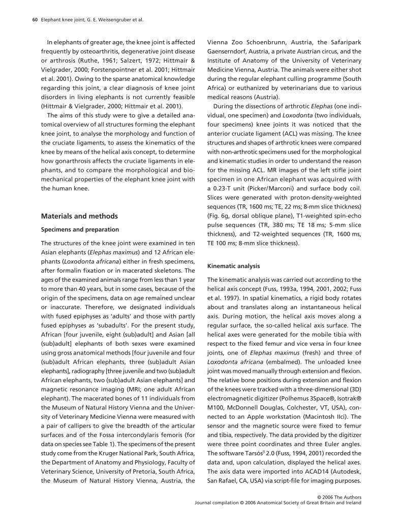
Table 1 Measurements of femur and tibia (macerated bones) in Elephas maximus (Em) and Loxodonta africana (La) (in mm)
Individual Sex SideEpiphyses(F: distal, T: proximal)
F T
Bd Bm Bl Bi Bp Bm Bl
Em 1 Female right fused 158 79 71 10.5left fused 160 86 81
Em 2 Male right partly fused 161 153 86 71left partly fused 166 84 73 9.5 160 94 78
Em 3 Male right partly fused 188 92 87 12.5 183 100 95Em 4 ? right F: fused, T: partly fused 172 93 75 9 171 97 84
left F: fused, T: partly fused 179 95 78 7.5 171 97 82Em 5 Male right not fused 158.5 77 78 11.5 154 84 82
left not fused 157 75.5 75.5 13 152 81 83Em 6 Female right not fused 147.5 66.5 62.5 19.5 148 80 79Em 7 ? right not fused 132 57 58 19 124 66 65
left not fused 133 56 56 21 125 64 65La 1 Female right fused 163 82 75 5 154 88 76(arthrotic left fused 165 85 79 0 159 85 82joints)La 2 Female right fused 160 91 64 9.5 157 90 73
left fused 159 90 61 12.5 154 90 74La 3 Male right not fused 221 116 100 10.5 211 122 105
left not fused 221 120 97 12.5 210 114 109La 4 Male right not fused 62 22 140 85 74
F, femur; T, tibia. Femur: Bd, distal breadth of femur according to Von den Driesch (1976) (from the medial to the lateral edge of the articular surfaces of the condyli; the measuring points lie close to the area of insertion of the medial collateral ligament or to the area of origin of the M. popliteus, respectively); Bm, breadth of the medial condyle of the femur (all femoral measurements are co-planar with Bd); Bl, breadth of the lateral condyle; Bi, breadth of the Fossa intercondylaris between the medial edge of the articular surface of the lateral condyle and the lateral edge of the articular surface of the medial condyle. Tibia: Bp, proximal breadth of the tibia according to Von den Driesch (1976); Bm, breadth of the medial condyle; Bl, breadth of the lateral condyle (measurements of the separate tibial condyles exclude the breadth of the Eminentia intercondylaris but are co-planar with Bp). No measurements indicated = bone is damaged or missing.
Elephant knee joint, G. E. Weissengruber et al.
© 2006 The AuthorsJournal compilation © 2006 Anatomical Society of Great Britain and Ireland
62
digitized again, together with the contours and several
points in the footprints of the cruciate ligaments. Based
on the reference points, the footprints were connected
in HyperSpace Modeller 5.0 to the ten positions recorded
previously. The distances between corresponding points
(connected by fibres) were measured in HyperSpace
Modeller 5.0. The behaviour of the distance between
two corresponding attachment points defines the
function of a ligament fibre (Fuss, 1989). The borders
between functionally different areas within the foot-
prints were determined according to Fuss (1991a).
Anatomical nomenclature
Anatomical names except the abbreviations for the
cruciate ligaments and where indicated follow NAV
(1994). The anterior (ACL) and posterior cruciate liga-
ment (PCL) were named in accordance with the
Termi-
nologia Anatomica
for humans, but correspond to the
Ligamentum (Lig.) cruciatum craniale (= ACL) or caudale
(= PCL), respectively, in quadrupeds.
Results
Morphology
Measurements of the articular surfaces and the Fossa
intercondylaris are given in Table 1. Although insigni-
ficant differences between the species in the external
morphology of the femur and the tibia were noticed
(but not studied in detail here), soft tissues and articular
surfaces revealed great similarity.
The knee joint consists of the Articulatio (Art.) femo-
rotibialis and the Art. femoropatellaris. It is covered
laterally by the Fascia lata and the distal end of the
Musculus (M.) biceps femoris. Medially, a thick aponeu-
rotic/fascial layer and the Mm. gracilis, semimembran-
osus and semitendinosus cover the knee. The heads of
the M. gastrocnemius and the M. flexor digitorum
superficialis are situated caudally of the knee joint. The
tendon of origin of the M. popliteus arises laterally on
the Condylus lateralis femoris and courses medio-
distally over caudal parts of the lateral meniscus. Passing
the caudal aspect of the lateral condyle of the tibia and
the Caput fibulae, a synovial recess of the femorotibial
joint lies below the M. popliteus (Fig. 1). The Mm. vastus
medialis, intermedius and lateralis insert on the patella,
whereas the distal tendon of the M. rectus femoris
runs cranially of the other parts of the M. quadriceps
femoris and inserts via a separate distal tendon on the
Tuberositas tibiae. A Lig. patellae is present, whereas
distinct Retinacula patellae are not discernible. The Lig.
collaterale mediale and the smaller laterale are embed-
ded within the thick fascial layers surrounding the
knee. The Lig. collaterale laterale inserts on the Caput
fibulae and the thicker mediale on the Condylus
medialis tibiae.
The lateral condyle of the femur is more slender than
the medial condyle but projects further distally. The
articular surfaces of the femoral condyles are continu-
ous with the articular surface of the Trochlea femoris
(Fig. 2). Each condyle of the femur and the proximal
part of the tibia have an epiphyseal ossification centre
(Fig. 3). The mineralized condyles of the femur first
fuse with each other and later in life with the diaphysis.
Owing to the marked concavity of the tibial condyles
and the small menisci (see below), the articular surfaces
of the tibia and the femur show a high degree of
congruency (see also Fig. 6g).
The joint capsule of the knee joint encloses both the
Art. femorotibialis and the Art. femoropatellaris. The
capsule is tight with the exception of a recess proximal
of the femoral trochlea and attaches also to the outer
edges of the menisci. The Corpus adiposum infrapatellare
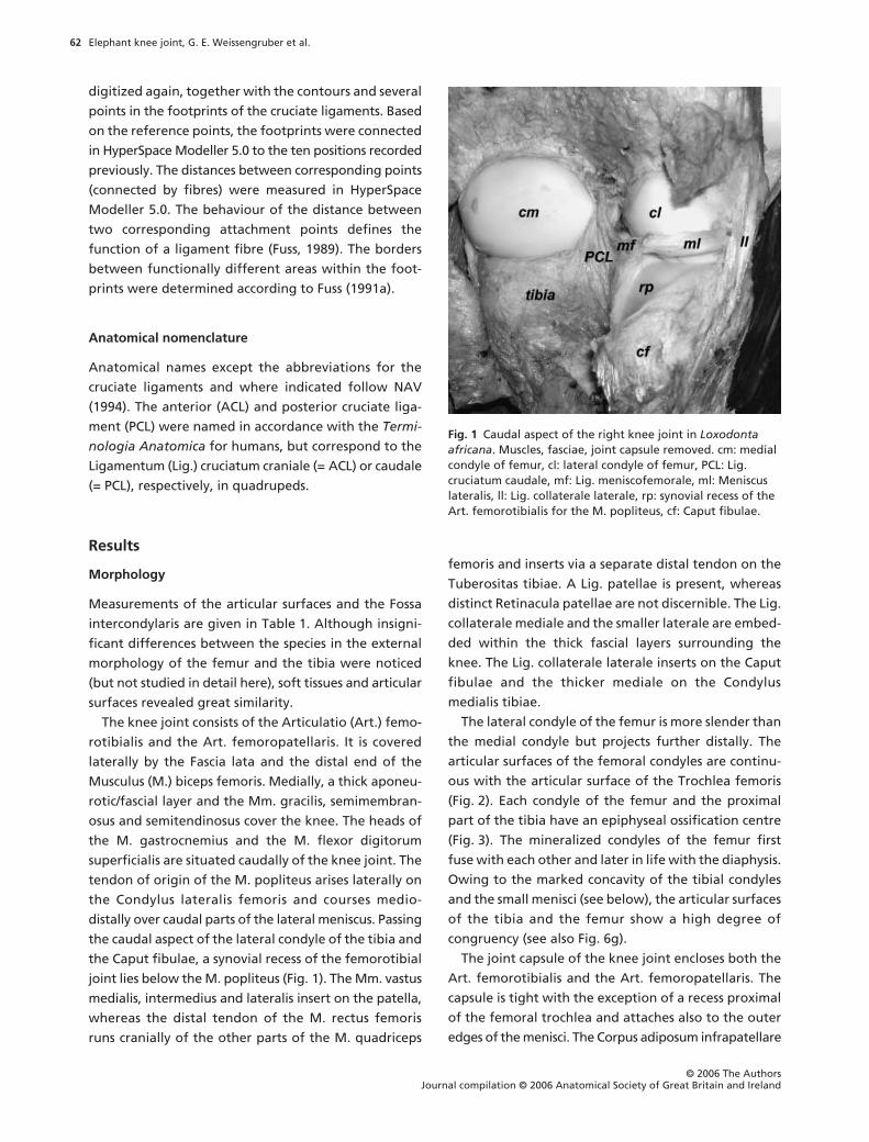
Fig. 1 Caudal aspect of the right knee joint in Loxodonta africana. Muscles, fasciae, joint capsule removed. cm: medial condyle of femur, cl: lateral condyle of femur, PCL: Lig. cruciatum caudale, mf: Lig. meniscofemorale, ml: Meniscus lateralis, ll: Lig. collaterale laterale, rp: synovial recess of the Art. femorotibialis for the M. popliteus, cf: Caput fibulae.
Elephant knee joint, G. E. Weissengruber et al.
© 2006 The Authors Journal compilation © 2006 Anatomical Society of Great Britain and Ireland
63
is large and a Plica synovialis infrapatellaris and Plicae
alares (
Terminologia Anatomica
; both terms not cited in
the
Nomina Anatomica Veterinaria
) are present (Fig. 4).
The Meniscus lateralis and especially the Meniscus
medialis are very narrow (lateral meniscus: breadth up
to approximately 18 mm; medial meniscus:
c
. 9 mm) and
on their medial or lateral edges, respectively, connected
with the joint capsule. Both menisci are C-shaped and
triangular in cross-section. The menisci are thicker on
the outer border close to the joint capsule (lateral
meniscus:
c
. 8 mm, medial meniscus:
c
. 5 mm) and thin
out towards the longitudinal limb axis (Fig. 2). Espe-
cially the caudal part of the lateral meniscus resembles
rather a fold of the joint capsule or a thin disc than a
typical meniscus. Caudolaterally, the lateral meniscus
forms a shallow groove for the M. popliteus (Fig. 1).
Both menisci are attached cranially to the tibia (Fig. 2).
The tibial attachment of the medial meniscus lies close
to that of the cranial cruciate ligament (ACL). A Lig.
meniscofemorale connecting the caudal part of the
lateral meniscus with the inner surface of the medial
femoral condyle is present (Fig. 1). The Lig. menis-
cofemorale fuses partly with the caudal cruciate liga-
ment (PCL) and the joint capsule and resembles a
ligamentous prolongation of the lateral meniscus towards
the medial femoral condyle. Caudomedially, the medial
meniscus merges with the joint capsule.
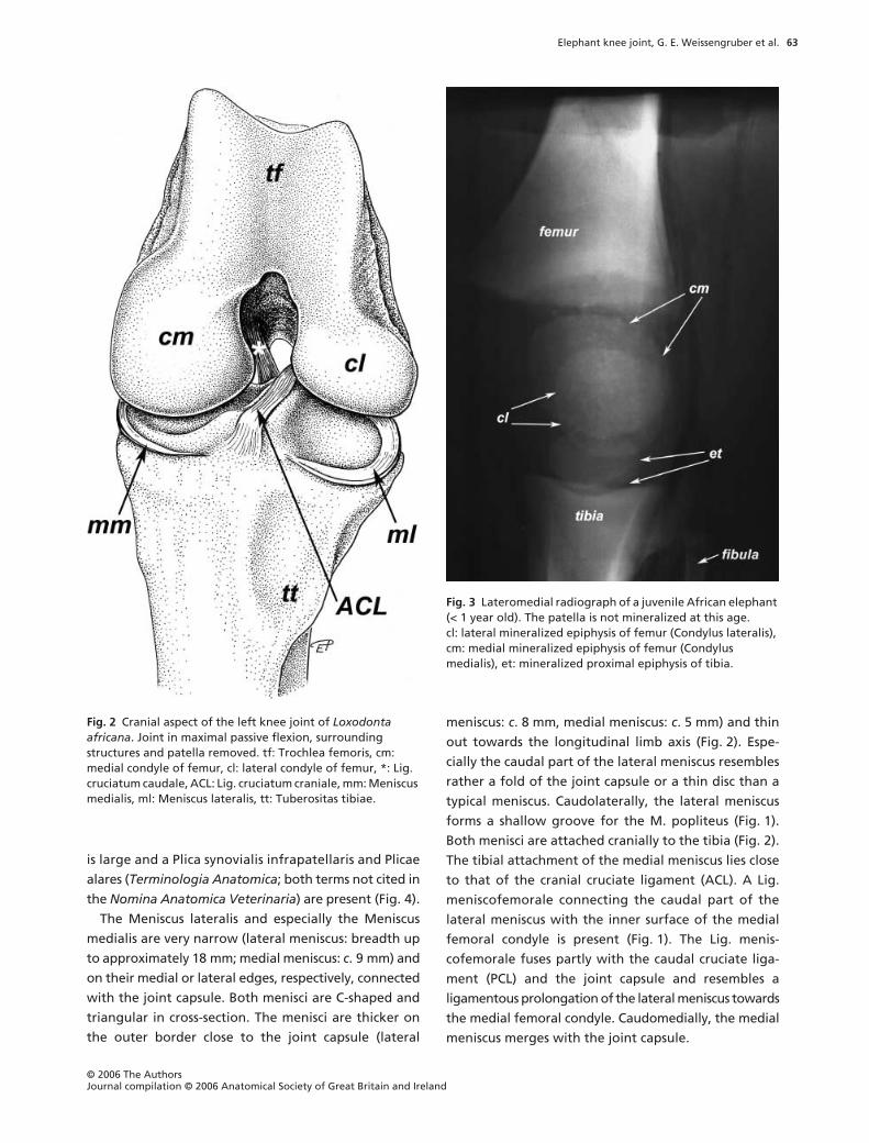
Fig. 2 Cranial aspect of the left knee joint of Loxodonta africana. Joint in maximal passive flexion, surrounding structures and patella removed. tf: Trochlea femoris, cm: medial condyle of femur, cl: lateral condyle of femur, *: Lig. cruciatum caudale, ACL: Lig. cruciatum craniale, mm: Meniscus medialis, ml: Meniscus lateralis, tt: Tuberositas tibiae.
Fig. 3 Lateromedial radiograph of a juvenile African elephant (< 1 year old). The patella is not mineralized at this age. cl: lateral mineralized epiphysis of femur (Condylus lateralis), cm: medial mineralized epiphysis of femur (Condylus medialis), et: mineralized proximal epiphysis of tibia.
Elephant knee joint, G. E. Weissengruber et al.
© 2006 The AuthorsJournal compilation © 2006 Anatomical Society of Great Britain and Ireland
64
The Ligamentum cruciatum craniale (ACL) is flat-
tened, its cross-section elliptical or approximately trian-
gular and it courses sagittally between the vault of the
Fossa intercondylaris femoris and the proximal surface
of the flattened Eminentia intercondylaris tibiae (Figs 2
and 4). The ACL attaches caudally to the medial surface
of the Condylus lateralis femoris and inserts on an
impression at the craniolateral surface (corresponding
to the Tuberculum intercondylare laterale) of the
Eminentia intercondylaris. A fold of the synovial mem-
brane attaches proximally to the ACL. The Ligamentum
cruciatum caudale (PCL) courses from the caudal end of
the lateral surface of the Condylus medialis femoris to
the Area intercondylaris caudalis (Figs 1 and 2). Caud-
ally, the PCL fuses with the joint capsule. The tendon
of origin of the M. popliteus passes caudally over the
Meniscus lateralis and a recess of the joint capsule lies
between this muscle and the Condylus lateralis tibiae
or the Caput fibulae, respectively (Fig. 1).
Pathological anatomy of arthrotic joints
Osteophytes or osteochondrophytes can appear in
several positions such as at the borders of the articular
surfaces (Fig. 5). The distal aspect of the femur shows a
notch, which serves the purpose of receiving the cranial
ridge of the tibial intercondylar eminence in extension
(arrows in Fig. 6). This notch is continuous with the
caudal intercondylar fossa. The cranial part of the
intercondylar fossa near the transition region to the
notch (arrowheads in Figs 5 and 6) can be narrow even
in healthy joints but allows the passage of the ACL. In
arthrotic elephant knees, the articular surfaces of the
medial and lateral femoral condyles expand towards
each other, therefore narrowing the intercondylar fossa
continuously (see Table 1, individual La 1). This process
compresses the ACL, which finally becomes worn out,
once the passage is completely used up and closed.
Additional osteophytic activity in the notch can injure
the ACL.
In healthy joints the proximal edge of the inter-
condylar eminence is proximally flattened but it is sharp
in arthrotic joints (Fig. 6e,g; below the arrowheads).
Additionally, in healthy joints the intercondylar eminence
is more flattened in younger or juvenile individuals
than in older animals.
Kinematic analysis
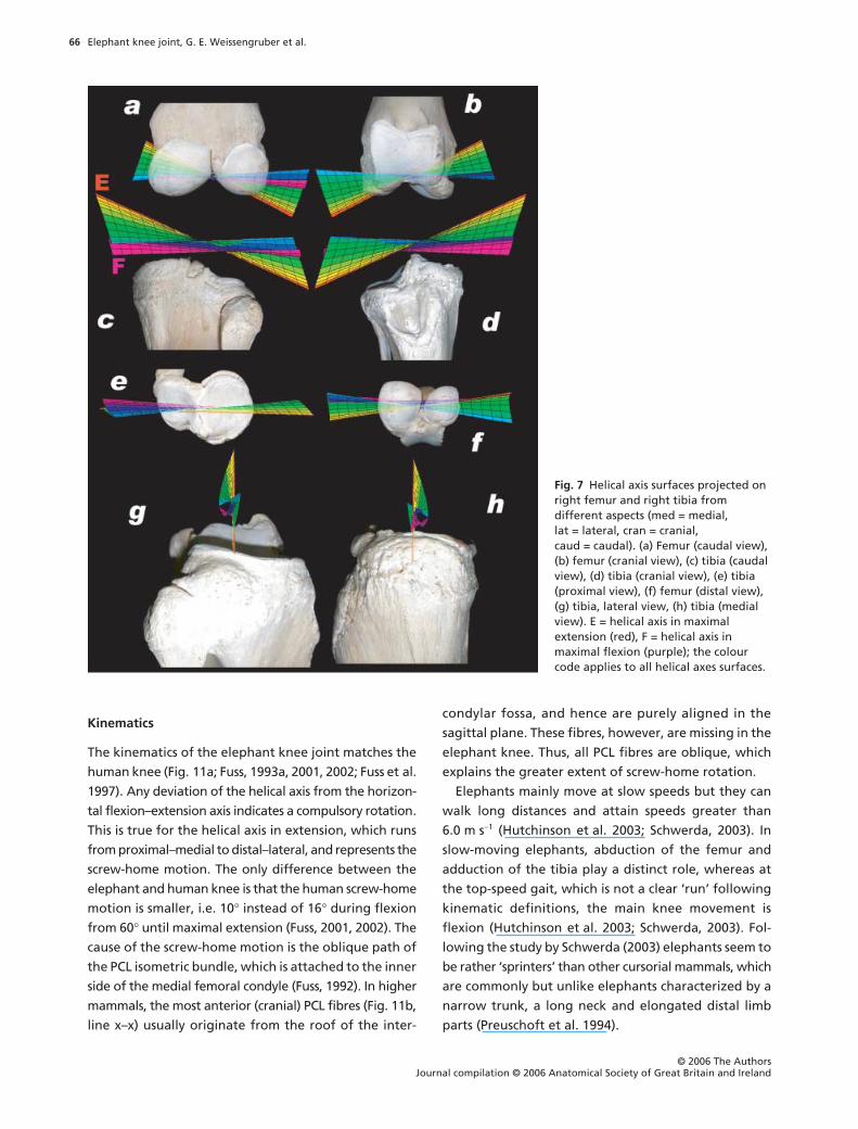
The helical axes surfaces are shown in Fig. 7 from dif-
ferent aspects. Between mid- and maximal flexion, the
extension–flexion axis is almost horizontal, which indi-
cates that the motion is a pure extension–flexion without
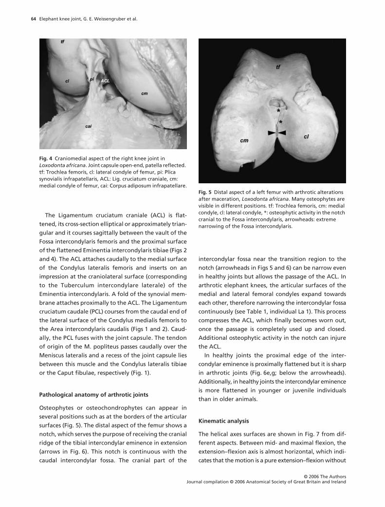
Fig. 4 Craniomedial aspect of the right knee joint in Loxodonta africana. Joint capsule open-end, patella reflected. tf: Trochlea femoris, cl: lateral condyle of femur, pi: Plica synovialis infrapatellaris, ACL: Lig. cruciatum craniale, cm: medial condyle of femur, cai: Corpus adiposum infrapatellare.
Fig. 5 Distal aspect of a left femur with arthrotic alterations after maceration, Loxodonta africana. Many osteophytes are visible in different positions. tf: Trochlea femoris, cm: medial condyle, cl: lateral condyle, *: osteophytic activity in the notch cranial to the Fossa intercondylaris, arrowheads: extreme narrowing of the Fossa intercondylaris.
Elephant knee joint, G. E. Weissengruber et al.
© 2006 The Authors Journal compilation © 2006 Anatomical Society of Great Britain and Ireland
65
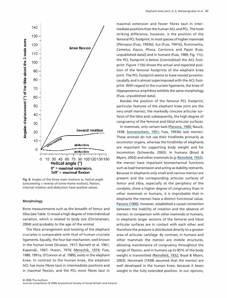
any significant additional automatic motions (rotations,
ab/adduction). Between a knee flexion angle of 60
°
and
0
°
(maximal extension), the helical axis becomes more
and more inclined. Its direction is from medial–proximal
to lateral–distal. This inclination indicates an automatic,
compulsory rotation, namely a combination of exten-
sion and outward rotation of the tibia, or flexion and
inward rotation, which corresponds to the so-called
screw-home motion of the knee. The mean automatic
rotation is 16
°
between a knee flexion angle of 0
°
and
60
°
, and 14
°
between 0
°
and 42
°
(Fig. 8).
Functional analysis of the cruciate ligaments
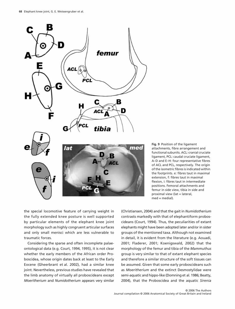
The fibre arrangement of the cruciate ligaments is
shown in Fig. 9. The cruciate ligament fibres are slightly
twisted about the ligament axis. Each cruciate ligament
contains a guiding or isometric fibre bundle (Fig. 9), as
well as flexion- and extension-constraining fibres and
fibres taut in intermediate positions (Fig. 9). The isometric
fibres constitute the side links of a four-bar linkage
(Fig. 10), whereby femur and tibia serve as mutual
coupler and ground link.
Discussion
Although the locomotory system of humans and ele-
phant are different, heavy and semi-plantigrade ele-
phants have more column-like limbs compared with
even-toed (including hippopotamuses) and odd-toed
animals (including rhinoceroses), as the latter show a
higher degree of flexion in the knee joint. Therefore,
elephants and plantigrade humans are more comparable
owing to the extended knee and evident similarities in
muscular architecture (Weissengruber & Forstenpointner,
2004) as well as locomotion patterns (Schwerda, 2003).
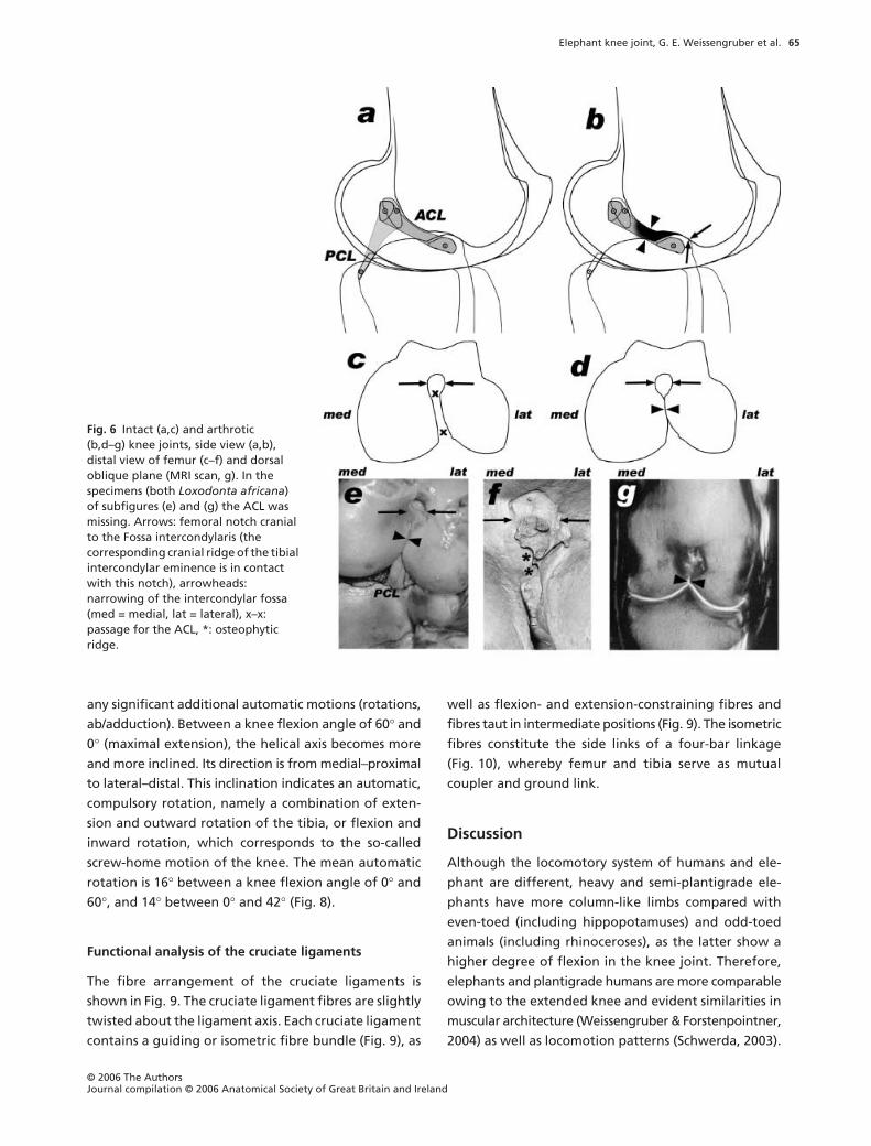
Fig. 6 Intact (a,c) and arthrotic(b,d–g) knee joints, side view (a,b), distal view of femur (c–f) and dorsal oblique plane (MRI scan, g). In the specimens (both Loxodonta africana) of subfigures (e) and (g) the ACL was missing. Arrows: femoral notch cranial to the Fossa intercondylaris (the corresponding cranial ridge of the tibial intercondylar eminence is in contact with this notch), arrowheads: narrowing of the intercondylar fossa (med = medial, lat = lateral), x–x: passage for the ACL, *: osteophytic ridge.
Elephant knee joint, G. E. Weissengruber et al.
© 2006 The AuthorsJournal compilation © 2006 Anatomical Society of Great Britain and Ireland
66
Kinematics
The kinematics of the elephant knee joint matches the
human knee (Fig. 11a; Fuss, 1993a, 2001, 2002; Fuss et al.
1997). Any deviation of the helical axis from the horizon-
tal flexion–extension axis indicates a compulsory rotation.
This is true for the helical axis in extension, which runs
from proximal–medial to distal–lateral, and represents the
screw-home motion. The only difference between the
elephant and human knee is that the human screw-home
motion is smaller, i.e. 10
°
instead of 16
°
during flexion
from 60
°
until maximal extension (Fuss, 2001, 2002). The
cause of the screw-home motion is the oblique path of
the PCL isometric bundle, which is attached to the inner
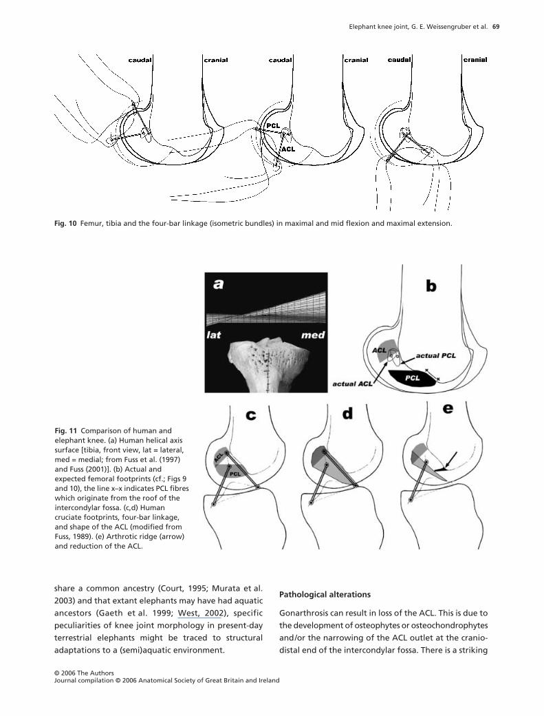
side of the medial femoral condyle (Fuss, 1992). In higher
mammals, the most anterior (cranial) PCL fibres (Fig. 11b,
line x–x) usually originate from the roof of the inter-
condylar fossa, and hence are purely aligned in the
sagittal plane. These fibres, however, are missing in the
elephant knee. Thus, all PCL fibres are oblique, which
explains the greater extent of screw-home rotation.
Elephants mainly move at slow speeds but they can
walk long distances and attain speeds greater than
6.0 m s
−
1
(Hutchinson et al. 2003; Schwerda, 2003). In
slow-moving elephants, abduction of the femur and
adduction of the tibia play a distinct role, whereas at
the top-speed gait, which is not a clear ‘run’ following
kinematic definitions, the main knee movement is
flexion (Hutchinson et al. 2003; Schwerda, 2003). Fol-
lowing the study by Schwerda (2003) elephants seem to
be rather ‘sprinters’ than other cursorial mammals, which
are commonly but unlike elephants characterized by a
narrow trunk, a long neck and elongated distal limb
parts (Preuschoft et al. 1994).
Fig. 7 Helical axis surfaces projected on right femur and right tibia from different aspects (med = medial, lat = lateral, cran = cranial, caud = caudal). (a) Femur (caudal view), (b) femur (cranial view), (c) tibia (caudal view), (d) tibia (cranial view), (e) tibia (proximal view), (f) femur (distal view), (g) tibia, lateral view, (h) tibia (medial view). E = helical axis in maximal extension (red), F = helical axis in maximal flexion (purple); the colour code applies to all helical axes surfaces.
Elephant knee joint, G. E. Weissengruber et al.
© 2006 The Authors Journal compilation © 2006 Anatomical Society of Great Britain and Ireland
67
Morphology
Bone measurements such as the breadth of femur and
tibia (see Table 1) reveal a high degree of interindividual
variation, which is related to body size (Christiansen,
2004) and probably to the age of the animal.
The fibre arrangement and twisting of the elephant
cruciates is comparable with that of human cruciate
ligaments. Equally, the four-bar mechanism, well known
in the human knee (Strasser, 1917; Barnett et al. 1961;
Kapandji, 1967; Huson, 1974; Menschik, 1974; Fuss,
1989, 1991a; O’Connor et al. 1989), exists in the elephant
knee. In contrast to the human knee, the elephant
ACL has more fibres taut in intermediate positions and
in maximal flexion, and the PCL more fibres taut in
maximal extension and fewer fibres taut in inter-
mediate positions than the human ACL and PCL. The most
striking difference, however, is the position of the
femoral PCL footprint. In most species of higher mammals
[
Pteropus
(Fuss, 1993b), S
us
(Fuss, 1991b), Ruminantia,
Camelus
,
Equus
,
Phoca
, Carnivora and
Papio
(Fuss,
unpublished data)] and in humans (Fuss, 1989; Fig. 11c),
the PCL footprint is below (craniodistal) the ACL foot-
print. Figure 11(b) shows the actual and expected posi-
tion of the femoral footprints of the elephant knee
joint. The PCL footprint seems to have moved proximo-
caudally and is almost superimposed with the ACL foot-
print. With regard to the cruciate ligaments, the knee of
Hippopotamus amphibius
exhibits the same morphology
(Fuss, unpublished data).
Besides the position of the femoral PCL footprint,
particular features of the elephant knee joint are the
very small menisci, the markedly concave articular sur-
faces of the tibia and, subsequently, the high degree of
congruency of the femoral and tibial articular surfaces.
In mammals, only certain bats (Parsons, 1900; Nauck,
1938; Sonnenschein, 1951; Fuss, 1993b) lack menisci.
These animals do not use their hindlimbs primarily as
locomotor organs, whereas the hindlimbs of elephants
are important for supporting body weight and for
locomotion (Schwerda, 2003). In humans (Boyd &
Myers, 2003) and other mammals (e.g. Reinsfeld, 1932),
the menisci have important biomechanical functions
such as load transmission and acting as stability restraints.
Because in elephants only small and narrow menisci are
present and the corresponding articular surfaces of
femur and tibia, especially at the periphery of the
condyles, show a higher degree of congruency than in
other mammals or humans, it is improbable that in
elephants the menisci have a distinct functional value.
Parsons (1900), however, established a causal connection
between the inability of rotation and the absence of
menisci. In comparison with other mammals or humans,
in elephants larger sections of the femoral and tibial
articular surfaces are in contact with each other and
therefore the pressure is distributed directly to a greater
area of articular cartilage. By contrast, in humans and
other mammals the menisci are mobile structures,
allowing maintenance of congruency throughout the
range of flexion, and in humans up to 85% of the body
weight is transmitted (Reinsfeld, 1932; Boyd & Myers,
2003). Herzmark (1938) assumed that the menisci are
well developed in the human knee, because it bears
weight in the fully extended position. In our opinion,
Fig. 8 Angles of the three main motions vs. helical angle (unscrewing = reverse of screw-home motion); flexion, internal rotation and abduction have positive values.
Elephant knee joint, G. E. Weissengruber et al.
© 2006 The AuthorsJournal compilation © 2006 Anatomical Society of Great Britain and Ireland
68
the special locomotive feature of carrying weight in
the fully extended knee posture is well supported
by particular elements of the elephant knee joint
morphology such as highly congruent articular surfaces
and only small menisci which are less vulnerable to
traumatic forces.
Considering the sparse and often incomplete palae-
ontological data (e.g. Court, 1994, 1995), it is not clear
whether the early members of the African order Pro-
boscidea, whose origin dates back at least to the Early
Eocene (Gheerbrant et al. 2002), had a similar knee
joint. Nevertheless, previous studies have revealed that
the limb anatomy of virtually all proboscideans except
Moeritherium
and
Numidotherium
appears very similar
(Christiansen, 2004) and that the gait in
Numidotherium
contrasts markedly with that of elephantiform probos-
cideans (Court, 1994). Thus, the peculiarities of extant
elephants might have been adopted later and/or in sister
groups of the mentioned taxa. Although not examined
in detail, it is evident from the literature (e.g. Aouadi,
2001; Fladerer, 2001; Koenigswald, 2002) that the
morphology of the femur and tibia of the
Mammuthus
group is very similar to that of extant elephant species
and therefore a similar structure of the soft tissues can
be assumed. Given that some early proboscideans such
as
Moeritherium
and the extinct Desmostylidae were
semi-aquatic and hippo-like (Domning et al. 1986; Beatty,
2004), that the Proboscidea and the aquatic
Sirenia
Fig. 9 Position of the ligament attachments, fibre arrangement and functional subunits. ACL: cranial cruciate ligament, PCL: caudal cruciate ligament, A–D and E–H: four representative fibres of ACL and PCL, respectively. The origin of the isometric fibres is indicated within the footprints. e: fibres taut in maximal extension, f: fibres taut in maximal flexion, i: fibres taut in intermediate positions. Femoral attachments and femur in side view, tibia in side and proximal view (lat = lateral, med = medial).
Elephant knee joint, G. E. Weissengruber et al.
© 2006 The Authors Journal compilation © 2006 Anatomical Society of Great Britain and Ireland
69
share a common ancestry (Court, 1995; Murata et al.
2003) and that extant elephants may have had aquatic
ancestors (Gaeth et al. 1999; West, 2002), specific
peculiarities of knee joint morphology in present-day
terrestrial elephants might be traced to structural
adaptations to a (semi)aquatic environment.
Pathological alterations
Gonarthrosis can result in loss of the ACL. This is due to
the development of osteophytes or osteochondrophytes
and/or the narrowing of the ACL outlet at the cranio-
distal end of the intercondylar fossa. There is a striking
Fig. 10 Femur, tibia and the four-bar linkage (isometric bundles) in maximal and mid flexion and maximal extension.
Fig. 11 Comparison of human and elephant knee. (a) Human helical axis surface [tibia, front view, lat = lateral, med = medial; from Fuss et al. (1997) and Fuss (2001)]. (b) Actual and expected femoral footprints (cf.; Figs 9 and 10), the line x–x indicates PCL fibres which originate from the roof of the intercondylar fossa. (c,d) Human cruciate footprints, four-bar linkage, and shape of the ACL (modified from Fuss, 1989). (e) Arthrotic ridge (arrow) and reduction of the ACL.
Elephant knee joint, G. E. Weissengruber et al.
© 2006 The AuthorsJournal compilation © 2006 Anatomical Society of Great Britain and Ireland
70
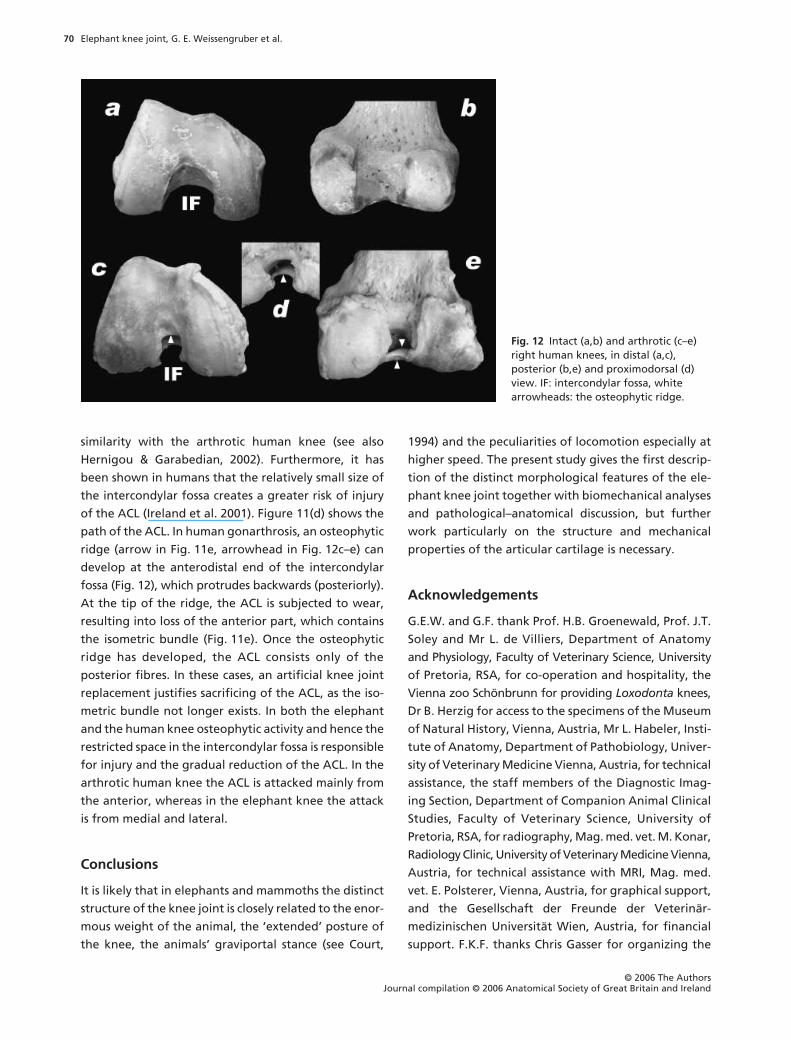
similarity with the arthrotic human knee (see also
Hernigou & Garabedian, 2002). Furthermore, it has
been shown in humans that the relatively small size of
the intercondylar fossa creates a greater risk of injury
of the ACL (Ireland et al. 2001). Figure 11(d) shows the
path of the ACL. In human gonarthrosis, an osteophytic
ridge (arrow in Fig. 11e, arrowhead in Fig. 12c–e) can
develop at the anterodistal end of the intercondylar
fossa (Fig. 12), which protrudes backwards (posteriorly).
At the tip of the ridge, the ACL is subjected to wear,
resulting into loss of the anterior part, which contains
the isometric bundle (Fig. 11e). Once the osteophytic
ridge has developed, the ACL consists only of the
posterior fibres. In these cases, an artificial knee joint
replacement justifies sacrificing of the ACL, as the iso-
metric bundle not longer exists. In both the elephant
and the human knee osteophytic activity and hence the
restricted space in the intercondylar fossa is responsible
for injury and the gradual reduction of the ACL. In the
arthrotic human knee the ACL is attacked mainly from
the anterior, whereas in the elephant knee the attack
is from medial and lateral.
Conclusions
It is likely that in elephants and mammoths the distinct
structure of the knee joint is closely related to the enor-
mous weight of the animal, the ‘extended’ posture of
the knee, the animals’ graviportal stance (see Court,
1994) and the peculiarities of locomotion especially at
higher speed. The present study gives the first descrip-
tion of the distinct morphological features of the ele-
phant knee joint together with biomechanical analyses
and pathological–anatomical discussion, but further
work particularly on the structure and mechanical
properties of the articular cartilage is necessary.
Acknowledgements
G.E.W. and G.F. thank Prof. H.B. Groenewald, Prof. J.T.
Soley and Mr L. de Villiers, Department of Anatomy
and Physiology, Faculty of Veterinary Science, University
of Pretoria, RSA, for co-operation and hospitality, the
Vienna zoo Schönbrunn for providing
Loxodonta
knees,
Dr B. Herzig for access to the specimens of the Museum
of Natural History, Vienna, Austria, Mr L. Habeler, Insti-
tute of Anatomy, Department of Pathobiology, Univer-
sity of Veterinary Medicine Vienna, Austria, for technical
assistance, the staff members of the Diagnostic Imag-
ing Section, Department of Companion Animal Clinical
Studies, Faculty of Veterinary Science, University of
Pretoria, RSA, for radiography, Mag. med. vet. M. Konar,
Radiology Clinic, University of Veterinary Medicine Vienna,
Austria, for technical assistance with MRI, Mag. med.
vet. E. Polsterer, Vienna, Austria, for graphical support,
and the Gesellschaft der Freunde der Veterinär-
medizinischen Universität Wien, Austria, for financial
support. F.K.F. thanks Chris Gasser for organizing the
Fig. 12 Intact (a,b) and arthrotic (c–e) right human knees, in distal (a,c), posterior (b,e) and proximodorsal (d) view. IF: intercondylar fossa, white arrowheads: the osteophytic ridge.
Elephant knee joint, G. E. Weissengruber et al.
© 2006 The Authors Journal compilation © 2006 Anatomical Society of Great Britain and Ireland
71
Loxodonta
knee joints from South Africa, Dr V. De
Vos, Manager Scientific Services, National Parks Board,
Skukuza, RSA, for providing the
Loxodonta
knees from
Kruger National Park, Mr G.W. Haupt, Nestlé South
Africa, Randburg, RSA, for organizing and shipping of
the specimens, Dir. A. Ross, Nestlé South Africa, and
V. Pres. R. Gasser, Nestlé S.A., Vevey, Switzerland, for
the support by Nestlé S.A. in this matter, Dr med. vet.
M. Antolini, Vienna, Austria, for providing one
Elephas
knee, Dir. Dr F. Weiss-Spitzenberger for arranging the
dissection of two
Elephas
knee joints at the Museum of
Natural History, Vienna, Austria, and the Austrian
Science Foundation (Fonds zur Förderung der wissen-
schaftlichen Forschung) for financial support (project
no. P7914-MED).
References
Aouadi N
(2001) New data on the diversity of elephants(Mammalia, Proboscidea) in the Early and early MiddlePleistocene of France. In
Proceedings of the 1st Inter-national Congress ‘The World of Elephants’
(eds Cavarretta G,Gioia P, Mussi M, Palombo MR) Rome, Consiglio Nationalsdelle Ricerche, 81–84.
Barnett CH, Davies DV, MacConaill MA
(1961)
Synovial Joints
.London: Longmans.
Beatty BL (2004) Evidence for suction feeding in the Desmo-stylidae (Desmostylia, Mammalia). J Morphol 260, 276–277.
Boyd KT, Myers BT (2003) Meniscus preservation; rationale,repair techniques and results. Knee 10, 1–11.
Christiansen P (2004) Body size in proboscideans, with noteson elephant metabolism. Zool J Linn Soc 140, 523–549.
Court N (1994) Limb posture and gait in Numidotheriumkoholense, a primitive proboscidean from the Eocene ofAlgeria. Zool J Linn Soc 111, 297–338.
Court N (1995) A new species of Numidotherium (Mammalia:Proboscidea) from the Eocene of Libya and the earlyphylogeny of the Proboscidea. J Vert Paleont 15, 650–671.
Domning DP, Ray CE, McKenna MC (1986) Two new Oligocenedesmostylians and a discussion of tethyterian systematics.Smithson Contrib Paleobiol 59, 1–56.
Eales NB (1929) Anatomy of a foetal african elephant, Elephasafricanus (Loxodonta africana). Part III. The contents of thethorax and abdomen, and the skeleton. Trans Roy Soc Edin56, 203–246.
Fladerer F (2001) Die Faunenreste vom jungpaläolithischenLagerplatz Krems-Wachtberg, Ausgrabung 1930. Wien:Österreichische Akademie der Wissenschaften.
Forstenpointner G, Weissengruber G, Kübber-Heiss A,Hittmair K, Konar M (2001) Morphological features of thestifle joint of the African elephant (Loxodonta africana,Blumenbach 1797). J Morph 248, 230.
Fuss FK (1989) Anatomy of the cruciate ligaments and theirfunction in extension and flexion of the human knee joint.Am J Anat 184, 165–176.
Fuss FK (1991a) The restraining function of the cruciate liga-ments on hyperextension and hyperflexion of the humanknee joint. Anat Rec 230, 283–289.
Fuss FK (1991b) Anatomy and function of the cruciate ligamentsof the domestic pig (Sus scrofa domestica): a comparisonwith human cruciates. J Anat 178, 11–20.
Fuss FK (1992) Principles and mechanisms of automatic rota-tion during terminal extension in the human knee joint. JAnat 180, 297–304.
Fuss FK (1993a) Helical axis surface of the knee joint. In Pro-ceedings of the XIVth Congress of the International Societyfor Biomechanics (eds Bouisset S, Métral S, Monod H) Paris,International Society of Biomechanics, 438–439.
Fuss FK (1993b) The knee joint of the flying fox (Pteropusrufus). Eur J Morph 31, 129–137.
Fuss FK (1994) A new method of clinical assessment of shoulderkinematics by means of the parameters of helical axes. EurJ Phys Med Rehabil 4, 125–130.
Fuss FK, Hamel G, Duval N, DeGuise J, Yahia LH (1997) Alteredknee kinematics after posterior cruciate ligament ruptureand replacement by means of a ligament prosthesis. In Pro-ceedings of the 1st International Symposium on AdvancedBiomaterials (SIBA 1997) (ed Yahia LH) Montréal: ÉcolePolytechnique, 136.
Fuss FK (2001) Helical axes – kinematic analysis, interpreta-tion, and application. In IFMBE Proceedings (Proceedings ofthe International Federation for Medical and BiologicalEngineering), vol. 1, Part II (eds Magjarevic R, Tonkovic S,Bilas V, Lackovic I), pp. 620–623. Zagreb: Croatian Medicaland Biological Engineering Society.
Fuss FK (2002) Kinematics of the knee after isometric single-bundle PCL reconstruction. Proceedings of the 2002 Com-bined Orthopaedic Meeting (25th Singapore OrthopaedicAssociation Meeting, 22nd Asean Orthopaedic AssociationMeeting, 5th Combined Meeting of Spinal and PediatricSections APOA, 7th Meeting of Knee and Sports MedicineSection APOA, 3rd Meeting of Asia Pacific Orthopaedic Societyfor Sports Medicine), Singapore: National University ofSingapore, CDROM edition.
Gaeth AP, Short RV, Renfree MB (1999) The developing renal,reproductive, and respiratory systems of the African elephantsuggest an aquatic ancestry. Proc Natl Acad Sci USA 96,5555–5558.
Gheerbrant E, Sudre J, Cappetta H, Iarochène M, AmaghzazM, Bouya B (2002) A new large mammal from the Ypresianof Morocco: evidence of surprising diversity of early probos-cideans. Acta Palaeontol Pol 47, 493–506.
Hernigou P, Garabedian JM (2002) Intercondylar notch widthand the risk for anterior cruciate ligament rupture in theosteoarthrotic knee: evaluation by plain radiography andCT scan. Knee 9, 313–316.
Herzmark MH (1938) The evolution of the knee joint. J BoneJoint Surg 20, 77–84.
Hittmair KM, Vielgrader HD (2000) Radiographic diagnosis oflameness in African elephants (Loxodonta africana). VetRadiol Ultrasound 41, 511–515.
Hittmair K, Vielgrader H, Konar M, Weissengruber G, Forsten-pointner G (2001) Diagnostic imaging of the limbs of Africanelephants (Loxodonta africana). Vet Radiol Ultrasound 42,175.
Elephant knee joint, G. E. Weissengruber et al.
© 2006 The AuthorsJournal compilation © 2006 Anatomical Society of Great Britain and Ireland
72
Huson A (1974) Biomechanik des Kniegelenks. Orthopäde 3,119–126.
Hutchinson JR, Famini D, Lair R, Kram R (2003) Are fast movingelephants really running? Nature 422, 493–494.
Ireland ML, Ballantyne BT, Little K, McClay IS (2001) A radio-graphic analysis of the relationship between the size and shapeof the intercondylar notch and anterior cruciate ligamentinjury. Knee Surg Sports Traumatol Arthrosc 9, 200–205.
Kapandji IA (1967) Cited in Funktionelle Anatomie derGelenke, Vol. 2 (ed. Kapandji IA). Stuttgart: Enke.
Koenigswald Wv (2002) Lebendige Eiszeit. Klima und TierweltIm Wandel. Darmstadt: Wissenschaftliche Buchgesellschaft.
Mariappa D (1955) The Anatomy of a foetal Indian Elephant.Part III. The bones and joints of the hind-limb. Indian Vet J32, 322–329.
Menschik A (1974) Mechanik des Kniegelenks, Teil 1. Z Orthop112, 481–495.
Murata Y, Nikaido M, Sasaki T, et al. (2003) Afrotherian phylo-geny as inferred from complete mitochondrial genomes.Mol Phylogenet Evol 28, 253–260.
Nauck ET (1938) Extremtitätenskelett der Tetrapoden. InHandbuch der Vergleichenden Anatomie der Wirbeltiere, 5.Band (eds Bolk L, Göppert E, Kallius E, Lubosch W), pp. 71–248. Berlin: Urban & Schwarzenberg.
NAV (1994) Nomina Anatomica Veterinaria, 4th edn. Zürich:World Ass. Vet. Anat.
O’Connor JJ, Shercliff TL, Biden E, Goodfellow JW (1989) Thegeometry of the knee in the sagittal plane. Proc Inst MechEng [H] 203, 223–233.
Parsons FG (1900) The joints of mammals compared with thoseof man. Part II. Joints of the hind limb. J Anat 34, 301–323.
Preuschoft H, Wille H, Christian A, Recknagel S (1994)Körpergestalt und Lokomotion bei großen Säugetieren.Verh Dtsch Zool Ges 87, 147–163.
Reinsfeld R (1932) Die Mechanik des Kniegelenkes vom Rinde.Z Anat Entwicklungsgeschichte 97, 487–508.
Ruthe H (1961) Fußleiden der Elefanten. Wissenschaftliche ZHumboldt-Universität Berlin, Mathematisch-Naturwissen-schaftliche Reihe 10, 471–516.
Salzert W (1972) Elefanten. Ihre Pathologie und denTiergärtner interessierende physiologische Daten. Thesis,Tierärztliche Hochschule Hannover.
Schwerda D (2003) Analyse Kinematischer Parameter derLokomotion Von Loxodonta Africana (Proboscidea:Elephantidae). Diplomarbeit, Institut für spezielle Zoologieund Evolutionsbiologie mit Phyletischem Museum derFriedrich-Schiller-Universität Jena.
Smuts MMS, Bezuidenhout AJ (1994) Osteology of the pelvic limbof the African elephant. Onderstepoort J Vet Res 61, 51–66.
Sonnenschein A (1951) Die Evolution des Kniegelenkes inner-halb der Wirbeltierreihe. Acta Anat 13, 289–328.
Strasser H (1917) Lehrbuch der Muskel und Gelenkmechanik,Vol. 3. Berlin: Strasser.
Terminologia Anatomica (1998) Stuttgart: Thieme.Von den Driesch A (1976) A guide to the measurement of
animal bones from archaeological sites. Peabody Mus Bull 1.Weissengruber GE, Forstenpointner G (2004) Musculature of
the crus and pes of the African elephant (Loxodonta africana):insight into semiplantigrade limb architecture. Anat Embryol208, 451–461.
West JB (2002) Why doesn’t the elephant have a pleuralspace? News Physiol Sci 17, 47–50.