Embed Size (px)
Citation preview
BioMed CentralJournal of Cardiothoracic Surgery
ss
Open AcceCase reportExtracorporeal membrane oxygenator as a bridge to successful surgical repair of bronchopleural fistula following bilateral sequential lung transplantation: a case report and review of literatureNouman U Khan*†, Mohamed Al-Aloul†, Noman Khasati†, Ali Machaal†, Colm T Leonard† and Nizar Yonan†Address: Department of Cardiothoracic Transplant, Wythenshawe Hospital, Manchester, M23 9LT, UK
Email: Nouman U Khan* - [email protected]; Mohamed Al-Aloul - [email protected]; Noman Khasati - [email protected]; Ali Machaal - [email protected]; Colm T Leonard - [email protected]; Nizar Yonan - [email protected]
* Corresponding author †Equal contributors
AbstractBackground: Lung transplantation (LTx) is widely accepted as a therapeutic option for end-stagerespiratory failure in cystic fibrosis. However, airway complications remain a major cause ofmorbidity and mortality in these patients, serious airway complications like bronchopleural fistula(BPF) are rare, and their management is very difficult.
Case presentation: A 47-year-old man with end-stage respiratory failure due to cystic fibrosisunderwent bilateral sequential lung transplantation. Severe post-operative bleeding occurred dueto dense intrapleural adhesions of the native lungs. He was re-explored and packed leading tosatisfactory haemostasis. He developed a bronchopleural fistula on the 14th post-operative day. Thefistula was successfully repaired using pericardial and intercostal vascular flaps with veno-venousextracorporeal membrane oxygenator (VV-ECMO) support. Subsequently his recovery wasuneventful.
Conclusion: The combination of pedicled intercostal and pericardial flaps provide adequatevascular tissue for sealing a large BPF following LTx. Veno-venous ECMO allows a feasible bridgeto recovery.
BackgroundAirway complications remain a major cause of morbidityand mortality after lung transplantation [1]. While theoverall incidence of such complications can be as high as15%, bronchopleural fistulas (BPF) are fortunately veryuncommon. This is, however, a disastrous event after lungtransplants. Most cases occur early after transplantation,
are extremely difficult to treat and are associated with ahigh mortality. We report successful surgical repair ofbronchopleural fistula in the donor bronchus employinga Levitronix Centrimag® [Waltham, Massachusetts]-basedveno-venous extracorporeal membrane oxygenator sup-port with 6 days bridge to recovery.
Published: 5 June 2007
Journal of Cardiothoracic Surgery 2007, 2:28 doi:10.1186/1749-8090-2-28
Received: 2 April 2007Accepted: 5 June 2007
This article is available from: http://www.cardiothoracicsurgery.org/content/2/1/28
© 2007 Khan et al; licensee BioMed Central Ltd. This is an Open Access article distributed under the terms of the Creative Commons Attribution License (http://creativecommons.org/licenses/by/2.0), which permits unrestricted use, distribution, and reproduction in any medium, provided the original work is properly cited.
Page 1 of 6(page number not for citation purposes)
Journal of Cardiothoracic Surgery 2007, 2:28 http://www.cardiothoracicsurgery.org/content/2/1/28
Case presentationA 47-year-old man with end-stage respiratory failure dueto cystic fibrosis underwent bilateral sequential lungtransplantation. The donor was a 48-year-old male whodied of sub-arachnoid haemorrhage and was ventilatedfor 24 hours. The pulmonary evaluation was normal, witha pO2 of 59.7 kPa on FiO2 of 1, normal chest X-ray andbronchoscopic appearance. The retrieval was performedwith the standard technique, using both ante-grade andretro-grade flush of low potassium dextran solution (Per-fadex®). The transplantation was carried out through aclamshell incision. Explantation of the native lungs wasprotracted and difficult due to dense intra-pleural adhe-sions. Implantation was performed with the standardtechnique. The right lung was implanted first, using con-tinuous 3/0 polypropylene sutures for the bronchial anas-tomosis (end-end-technique), continuous 4/0polypropylene sutures for the pulmonary vein to left atrialanastomosis, and continuous 5/0 polypropylene suturesfor the pulmonary arterial anastomosis. According to thestandard technique, the donor bronchus was left as shortas possible, and the sutures were applied almost level withthe upper lobe division. The left lung was implanted in asimilar fashion. The overall ischemia time was 6 hoursand 8 minutes for the right lung, and just over 8 hours forthe left. The patient had a persistent left-sided superiorvena cava, which was carefully dissected away from thepulmonary vessels. Despite measures to reduce bleeding,including the use of intra-operative veno-arterial extra-corporeal membrane oxygenation (ECMO) instead of car-diopulmonary bypass, generous use of blood productreplacement therapy and full dose of aprotinin, he bledexcessively from the pleural bed. Because of persistentbleeding, the chest was packed at the end of operationwith skin closure. He was given routine immunosuppres-sive therapy including induction with rabbit anti-thymo-cyte globulin (RATG) and intravenous methylprednisolone (three doses of 125 mg each at 8 hourlyintervals, followed by a tapering regimen from 1 mg/kg/day) according to our standard unit protocol.
During the immediate post-operative period, he was re-explored twice due to bleeding. After correction of theabnormal coagulation with fresh frozen plasma, cryopre-cipitate and platelet transfusions, he was also given 3.6 mgof recombinant activated factor VII. Eventually, the bleed-ing settled and the chest was closed on the 4th post-opera-tive day. Subsequent surveillance bronchoscopies showedslough at the bronchial anastomoses with thick purulentsecretions in the distal airways bilaterally. A percutaneoustracheostomy was performed on the 9th post-operativeday due to prolonged requirement for ventilatory supportand to aid in bronchial toilet, and the patient weaned suc-cessfully from mechanical ventilation. The bronchoalveo-
lar aspirate revealed Pseudomonas aeruginosa and Candidaparapsilosis, which were treated appropriately.
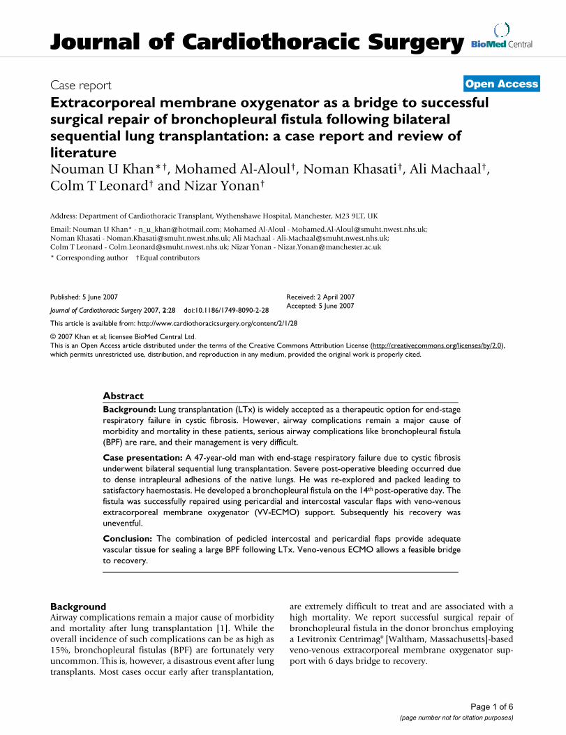
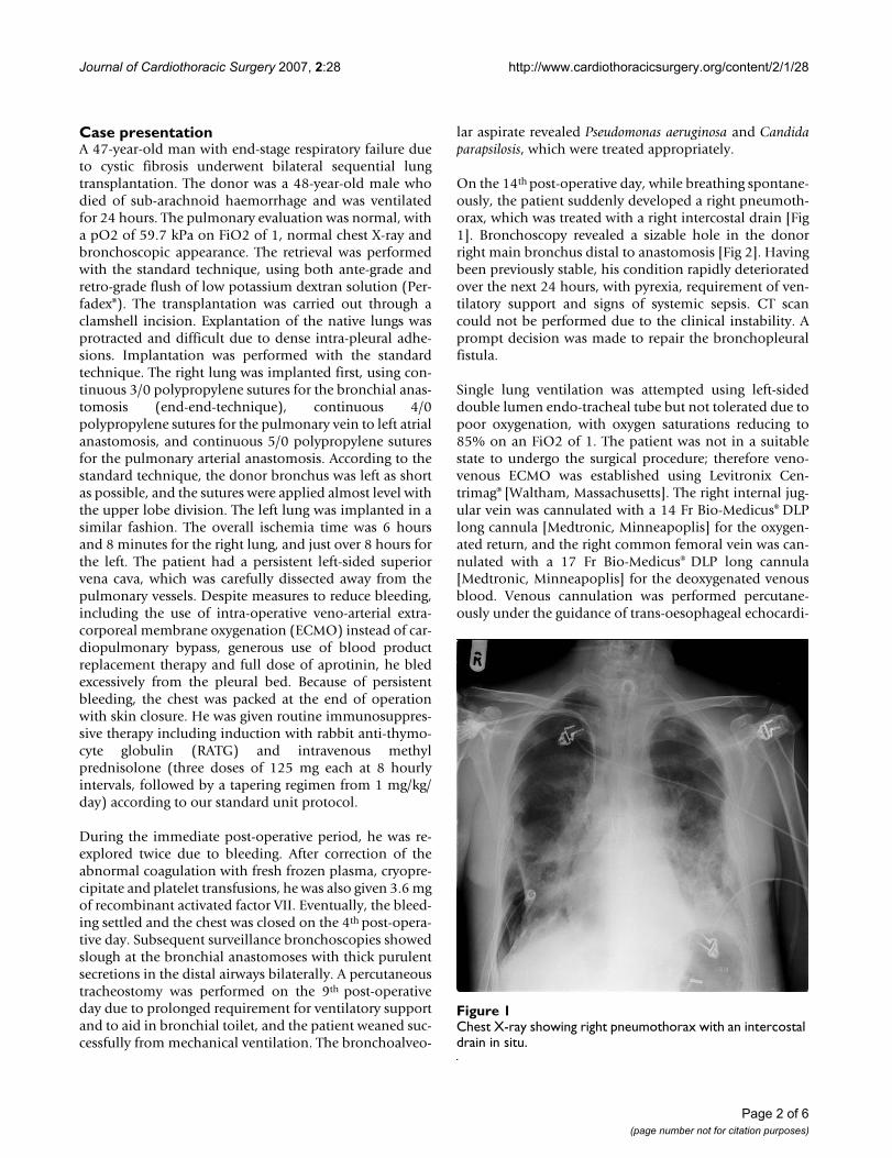
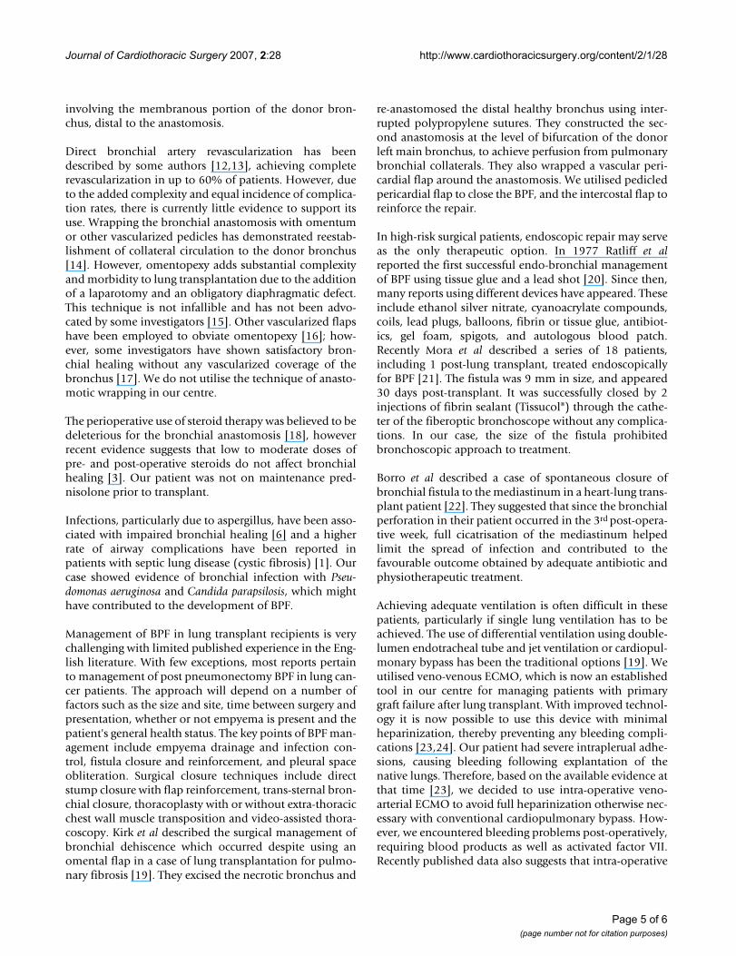
On the 14th post-operative day, while breathing spontane-ously, the patient suddenly developed a right pneumoth-orax, which was treated with a right intercostal drain [Fig1]. Bronchoscopy revealed a sizable hole in the donorright main bronchus distal to anastomosis [Fig 2]. Havingbeen previously stable, his condition rapidly deterioratedover the next 24 hours, with pyrexia, requirement of ven-tilatory support and signs of systemic sepsis. CT scancould not be performed due to the clinical instability. Aprompt decision was made to repair the bronchopleuralfistula.
Single lung ventilation was attempted using left-sideddouble lumen endo-tracheal tube but not tolerated due topoor oxygenation, with oxygen saturations reducing to85% on an FiO2 of 1. The patient was not in a suitablestate to undergo the surgical procedure; therefore veno-venous ECMO was established using Levitronix Cen-trimag® [Waltham, Massachusetts]. The right internal jug-ular vein was cannulated with a 14 Fr Bio-Medicus® DLPlong cannula [Medtronic, Minneapoplis] for the oxygen-ated return, and the right common femoral vein was can-nulated with a 17 Fr Bio-Medicus® DLP long cannula[Medtronic, Minneapoplis] for the deoxygenated venousblood. Venous cannulation was performed percutane-ously under the guidance of trans-oesophageal echocardi-
Chest X-ray showing right pneumothorax with an intercostal drain in situFigure 1Chest X-ray showing right pneumothorax with an intercostal drain in situ.
Page 2 of 6(page number not for citation purposes)
Journal of Cardiothoracic Surgery 2007, 2:28 http://www.cardiothoracicsurgery.org/content/2/1/28
ography to ensure adequate positioning of the cannulae.A single intravenous dose of 5000 units of heparin wasgiven before the start of ECMO. We utilized a HILITE®
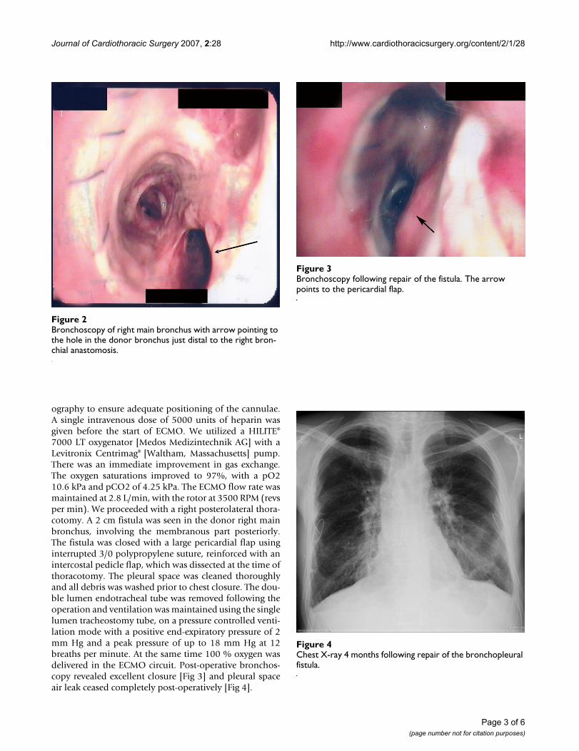
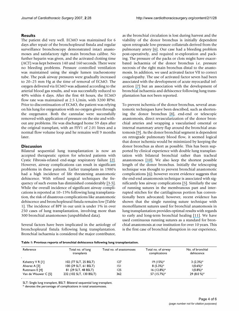
7000 LT oxygenator [Medos Medizintechnik AG] with aLevitronix Centrimag® [Waltham, Massachusetts] pump.There was an immediate improvement in gas exchange.The oxygen saturations improved to 97%, with a pO210.6 kPa and pCO2 of 4.25 kPa. The ECMO flow rate wasmaintained at 2.8 L/min, with the rotor at 3500 RPM (revsper min). We proceeded with a right posterolateral thora-cotomy. A 2 cm fistula was seen in the donor right mainbronchus, involving the membranous part posteriorly.The fistula was closed with a large pericardial flap usinginterrupted 3/0 polypropylene suture, reinforced with anintercostal pedicle flap, which was dissected at the time ofthoracotomy. The pleural space was cleaned thoroughlyand all debris was washed prior to chest closure. The dou-ble lumen endotracheal tube was removed following theoperation and ventilation was maintained using the singlelumen tracheostomy tube, on a pressure controlled venti-lation mode with a positive end-expiratory pressure of 2mm Hg and a peak pressure of up to 18 mm Hg at 12breaths per minute. At the same time 100 % oxygen wasdelivered in the ECMO circuit. Post-operative bronchos-copy revealed excellent closure [Fig 3] and pleural spaceair leak ceased completely post-operatively [Fig 4].
Chest X-ray 4 months following repair of the bronchopleural fistulaFigure 4Chest X-ray 4 months following repair of the bronchopleural fistula.
Bronchoscopy of right main bronchus with arrow pointing to the hole in the donor bronchus just distal to the right bron-chial anastomosisFigure 2Bronchoscopy of right main bronchus with arrow pointing to the hole in the donor bronchus just distal to the right bron-chial anastomosis.
Bronchoscopy following repair of the fistulaFigure 3Bronchoscopy following repair of the fistula. The arrow points to the pericardial flap.
Page 3 of 6(page number not for citation purposes)
Journal of Cardiothoracic Surgery 2007, 2:28 http://www.cardiothoracicsurgery.org/content/2/1/28
ResultsThe patient did very well. ECMO was maintained for 6days after repair of the bronchopleural fistula and regularsurveillance bronchoscopy demonstrated intact anasto-moses and satisfactory right main bronchus calibre. Nofurther heparin was given, and the activated clotting time(ACT) was kept between 140 and 160 seconds. There wereno bleeding problems. Pressure controlled ventilationwas maintained using the single lumen tracheostomytube. The peak airway pressures were gradually increasedto 20–25 mm Hg at the time of removal of ECMO. Theoxygen delivered via ECMO was adjusted according to thearterial blood gas results, and was successfully reduced to40% within 4 days. After the first 48 hours, the ECMOflow rate was maintained at 2.5 L/min, with 3200 RPM.Prior to discontinuation of ECMO, the patient was relyingon his lung for oxygenation with no oxygen given throughthe oxygenator. Both the cannulae were successfullyremoved with application of pressure on the site and with-out any problems. He was discharged home 59 days afterthe original transplant, with an FEV1 of 2.01 litres and anormal flow volume loop and he remains well 9 monthslater.
DiscussionBilateral sequential lung transplantation is now anaccepted therapeutic option for selected patients withCystic Fibrosis-related end-stage respiratory failure [2].However, airway complications can result in significantproblems in these patients. Earlier transplants in 1980'shad a high incidence of life threatening anastomoticdehiscence. With refined surgical techniques the fre-quency of such events has diminished considerably [3-5].While the overall incidence of significant airway compli-cations is reported at 10–15% following lung transplanta-tion, the risk of disastrous complications like anastomoticdehiscence and bronchopleural fistula remains low [Table1]. The incidence of BPF in our unit is under 1% in over250 cases of lung transplantation, involving more than300 bronchial anastomoses (unpublished data).
Several factors have been implicated in the aetiology ofbronchopleural fistula following lung transplantation.Bronchial ischaemia is considered the major contributor,
as the bronchial circulation is lost during harvest and theviability of the donor bronchus is initially dependentupon retrograde low-pressure collaterals derived from thepulmonary artery [6]. Our case had a bleeding problempost-operatively, and required re-exploration and pack-ing. The pressure of the packs or clots might have exacer-bated ischaemia of the donor bronchus i.e. pressurenecrosis of the right main bronchus distal to the anasto-mosis. In addition, we used activated factor VII to correctcoagulopathy. The use of activated factor seven had beenassociated with the development of acute myocardial inf-arction [7] but an association with the development ofbronchial ischaemia and dehiscence following lung trans-plantation has not been reported.
To prevent ischemia of the donor bronchus, several anas-tomotic techniques have been described, such as shorten-ing the donor bronchus [8], end-end or telescopicanastomosis, direct revascularization of the donor bron-chial arteries and wrapping a vascularised omental orinternal mammary artery flap around the bronchial anas-tomosis [9]. As the donor bronchial segment is dependenton retrograde pulmonary blood flow, it seemed logicalthat donor ischemia would be minimized by keeping thedonor bronchus as short as possible. This has been sup-ported by clinical experience with double lung transplan-tation with bilateral bronchial rather than trachealanastomoses [10]. We also keep the shortest possiblelength of the donor bronchus. Initially the telescopingtechnique was thought to prevent bronchial anastomoticcomplications [6]; however recent evidence suggests thatthe end-end anastomosis technique is associated with sig-nificantly less airway complications [5]. Similarly the useof running sutures in the membranous part and inter-rupted stitches for the cartilaginous portion has conven-tionally been advocated; however, recent evidence hasshown that the single running suture technique withmonofilament sutures used for bronchial anastomosis inlung transplantation provides optimal results with regardsto early and long-term bronchial healing [11]. We haveused continuous running sutures as a standard for bron-chial anastomosis at our institution for over 10 years. Thisis the first case of bronchial disruption in our experience,
Table 1: Previous reports of bronchial dehiscence following lung transplantation.
Reference Total no. of lung transplants
Total no. of anastomoses Total no. of airway complications
No. of bronchial dehiscence
Kshettry V R [1] 102 (77 SLT, 25 BSLT) 127 19 (15%)* 3 (2.3%)*Alvarez A [3] 100 (29 SLT, 61 BSLT) 151 8 (5.3%)* 1(0.6%)*Ruttmann E [4] 81 (29 SLT, 48 BSLT) 125 16 (12.8%)* 1(0.8%)*Van de Wauwer C [5] 232 (102 SLT, 130 BSLT) 362 57 (15.7%)* 29 (8.0 %)*
SLT: Single lung transplant, BSLT: Bilateral sequential lung transplant. * denotes the percentage of complications in total anastomoses.
Page 4 of 6(page number not for citation purposes)
Journal of Cardiothoracic Surgery 2007, 2:28 http://www.cardiothoracicsurgery.org/content/2/1/28
involving the membranous portion of the donor bron-chus, distal to the anastomosis.
Direct bronchial artery revascularization has beendescribed by some authors [12,13], achieving completerevascularization in up to 60% of patients. However, dueto the added complexity and equal incidence of complica-tion rates, there is currently little evidence to support itsuse. Wrapping the bronchial anastomosis with omentumor other vascularized pedicles has demonstrated reestab-lishment of collateral circulation to the donor bronchus[14]. However, omentopexy adds substantial complexityand morbidity to lung transplantation due to the additionof a laparotomy and an obligatory diaphragmatic defect.This technique is not infallible and has not been advo-cated by some investigators [15]. Other vascularized flapshave been employed to obviate omentopexy [16]; how-ever, some investigators have shown satisfactory bron-chial healing without any vascularized coverage of thebronchus [17]. We do not utilise the technique of anasto-motic wrapping in our centre.
The perioperative use of steroid therapy was believed to bedeleterious for the bronchial anastomosis [18], howeverrecent evidence suggests that low to moderate doses ofpre- and post-operative steroids do not affect bronchialhealing [3]. Our patient was not on maintenance pred-nisolone prior to transplant.
Infections, particularly due to aspergillus, have been asso-ciated with impaired bronchial healing [6] and a higherrate of airway complications have been reported inpatients with septic lung disease (cystic fibrosis) [1]. Ourcase showed evidence of bronchial infection with Pseu-domonas aeruginosa and Candida parapsilosis, which mighthave contributed to the development of BPF.
Management of BPF in lung transplant recipients is verychallenging with limited published experience in the Eng-lish literature. With few exceptions, most reports pertainto management of post pneumonectomy BPF in lung can-cer patients. The approach will depend on a number offactors such as the size and site, time between surgery andpresentation, whether or not empyema is present and thepatient's general health status. The key points of BPF man-agement include empyema drainage and infection con-trol, fistula closure and reinforcement, and pleural spaceobliteration. Surgical closure techniques include directstump closure with flap reinforcement, trans-sternal bron-chial closure, thoracoplasty with or without extra-thoracicchest wall muscle transposition and video-assisted thora-coscopy. Kirk et al described the surgical management ofbronchial dehiscence which occurred despite using anomental flap in a case of lung transplantation for pulmo-nary fibrosis [19]. They excised the necrotic bronchus and
re-anastomosed the distal healthy bronchus using inter-rupted polypropylene sutures. They constructed the sec-ond anastomosis at the level of bifurcation of the donorleft main bronchus, to achieve perfusion from pulmonarybronchial collaterals. They also wrapped a vascular peri-cardial flap around the anastomosis. We utilised pedicledpericardial flap to close the BPF, and the intercostal flap toreinforce the repair.
In high-risk surgical patients, endoscopic repair may serveas the only therapeutic option. In 1977 Ratliff et alreported the first successful endo-bronchial managementof BPF using tissue glue and a lead shot [20]. Since then,many reports using different devices have appeared. Theseinclude ethanol silver nitrate, cyanoacrylate compounds,coils, lead plugs, balloons, fibrin or tissue glue, antibiot-ics, gel foam, spigots, and autologous blood patch.Recently Mora et al described a series of 18 patients,including 1 post-lung transplant, treated endoscopicallyfor BPF [21]. The fistula was 9 mm in size, and appeared30 days post-transplant. It was successfully closed by 2injections of fibrin sealant (Tissucol®) through the cathe-ter of the fiberoptic bronchoscope without any complica-tions. In our case, the size of the fistula prohibitedbronchoscopic approach to treatment.
Borro et al described a case of spontaneous closure ofbronchial fistula to the mediastinum in a heart-lung trans-plant patient [22]. They suggested that since the bronchialperforation in their patient occurred in the 3rd post-opera-tive week, full cicatrisation of the mediastinum helpedlimit the spread of infection and contributed to thefavourable outcome obtained by adequate antibiotic andphysiotherapeutic treatment.
Achieving adequate ventilation is often difficult in thesepatients, particularly if single lung ventilation has to beachieved. The use of differential ventilation using double-lumen endotracheal tube and jet ventilation or cardiopul-monary bypass has been the traditional options [19]. Weutilised veno-venous ECMO, which is now an establishedtool in our centre for managing patients with primarygraft failure after lung transplant. With improved technol-ogy it is now possible to use this device with minimalheparinization, thereby preventing any bleeding compli-cations [23,24]. Our patient had severe intraplerual adhe-sions, causing bleeding following explantation of thenative lungs. Therefore, based on the available evidence atthat time [23], we decided to use intra-operative veno-arterial ECMO to avoid full heparinization otherwise nec-essary with conventional cardiopulmonary bypass. How-ever, we encountered bleeding problems post-operatively,requiring blood products as well as activated factor VII.Recently published data also suggests that intra-operative
Page 5 of 6(page number not for citation purposes)
Journal of Cardiothoracic Surgery 2007, 2:28 http://www.cardiothoracicsurgery.org/content/2/1/28
use of ECMO may be associated with increased risk ofbleeding [25].
During the first 24 hours following the repair of BPF, thepatient required full ECMO support with 100% oxygendelivery as the aim was to rest the lungs and reduce thepressure on the repair. Afterwards, the ECMO require-ment diminished. We, however, used ECMO for 6 days aswe believed that healing of the repaired bronchus wouldbe sufficient around that time. During this period we car-ried out frequent flexible bronchoscopic assessments. Atday 5, the anastomosis appeared to be healing well.
To our knowledge this is the first reported case for usingECMO as a bridge for successful surgical repair of BPF fol-lowing lung transplantation utilising pericardial andintercostal flaps. We have used the Levitronix Centrimagdevice, which was maintained without any anticoagula-tion for six days after repair of the fistula (ACT was keptbetween 140–160 seconds). There were no clots in thesystem on full examination when it was finally removed.
ConclusionIn summary, the combination of an intercostal and apedicled pericardial flap provides adequate robust vascu-larised tissue for sealing a large BP fistula following lungtransplantation. Veno-venous ECMO allows a feasiblebridge to recovery.
Competing interestsThe author(s) declare that they have no competing inter-ests.
Authors' contributionsNY performed the operation. NUK assisted in the opera-tion and took care of the patient in the ward along withNK and AM. NK and AM were also involved in re-explora-tion. MA and CL performed bronchoscopies and wereinvolved with immunosuppression, ward care and the fol-low up.
All authors read and approved the final manuscript.
References1. Kshettry VR, Kroshus TJ, Hertz MI, Hunter DW, Shumway SJ, Bolman
RM 3rd: Early and late airway complications after lung trans-plantation: incidence and management. Ann Thorac Surg 1997,63(6):1576-1583.
2. Egan TM, Detterbeck FC, Mill MR, Paradowski LJ, Lackner RP, OgdenWD, Yankaskas JR, Westerman JH, Thompson JT, Weiner MA, et al.:Improved results of lung transplantation for patients withcystic fibrosis. J Thorac Cardiovasc Surg 1995, 109(2):224-34; discus-sion 234-5.
3. Alvarez A, Algar J, Santos F, Lama R, Aranda JL, Baamonde C, Lopez-Pujol J, Salvatierra A: Airway complications after lung transplan-tation: a review of 151 anastomoses. Eur J Cardiothorac Surg 2001,19(4):381-387.
4. Ruttmann E, Ulmer H, Marchese M, Dunst K, Geltner C, Margreiter R,Laufer G, Mueller LC: Evaluation of factors damaging the bron-chial wall in lung transplantation. J Heart Lung Transplant 2005,24(3):275-281.
5. Van De Wauwer C, Van Raemdonck D, Verleden GM, Dupont L, DeLeyn P, Coosemans W, Nafteux P, Lerut T: Risk factors for airwaycomplications within the first year after lung transplantation.Eur J Cardiothorac Surg 2007, 31(4):703-710.
6. Shennib H, Massard G: Airway complications in lung transplan-tation. Ann Thorac Surg 1994, 57(2):506-511.
7. Peerlinck K, Vermylen J: Acute myocardial infarction followingadministration of recombinant activated factor VII (NovoSeven) in a patient with haemophilia A and inhibitor. ThrombHaemost 1999, 82(6):1775-1776.
8. Pinsker KL, Koerner SK, Kamholz SL, Hagstrom JW, Veith FJ: Effectof donor bronchial length on healing: a canine model to eval-uate bronchial anastomotic problems in lung transplantation.J Thorac Cardiovasc Surg 1979, 77(5):669-673.
9. Dubois P, Choiniere L, Cooper JD: Bronchial omentopexy incanine lung allotransplantation. Ann Thorac Surg 1984,38(3):211-214.
10. Noirclerc MJ, Metras D, Vaillant A, Dumon JF, Zimmermann JM, Caa-mano A, Orsoni PC: Bilateral bronchial anastomosis in doublelung and heart-lung transplantations. Eur J Cardiothorac Surg1990, 4(6):314-317.
11. Aigner C, Jaksch P, Seebacher G, Neuhauser P, Marta G, Wisser W,Klepetko W: Single running suture--the new standard tech-nique for bronchial anastomoses in lung transplantation. EurJ Cardiothorac Surg 2003, 23(4):488-493.
12. Couraud L, Baudet E, Martigne C, Roques X, Velly JF, Laborde N, Dub-rez J, Clerc F, Dromer C, Vallieres E: Bronchial revascularizationin double-lung transplantation: a series of 8 patients. Bor-deaux Lung and Heart-Lung Transplant Group. Ann Thorac Surg1992, 53(1):88-94.
13. Pettersson G, Norgaard MA, Arendrup H, Brandenhof P, Helvind M,Joyce F, Stentoft P, Olesen PS, Thiis JJ, Efsen F, Mortensen SA, Svend-sen UG: Direct bronchial artery revascularization and en blocdouble lung transplantation--surgical techniques and earlyoutcome. J Heart Lung Transplant 1997, 16(3):320-333.
14. Fell SC, Mollenkopf FP, Montefusco CM, Mitsudo S, Kamholz SL, Gold-smith J, Veith FJ: Revascularization of ischemic bronchial anas-tomoses by an intercostal pedicle flap. J Thorac Cardiovasc Surg1985, 90(2):172-178.
15. Khaghani A, Tadjkarimi S, al-Kattan K, Banner N, Daly R, Theodoro-poulos S, Madden B, Yacoub M: Wrapping the anastomosis withomentum or an internal mammary artery pedicle does notimprove bronchial healing after single lung transplantation:results of a randomized clinical trial. J Heart Lung Transplant1994, 13(5):767-773.
16. Taghavi S, Marta GM, Lang G, Seebacher G, Winkler G, Schmid K,Klepetko W: Bronchial stump coverage with a pedicled peri-cardial flap: an effective method for prevention of postpneu-monectomy bronchopleural fistula. Ann Thorac Surg 2005,79(1):284-288.
17. Auteri JS, Jeevanandam V, Sanchez JA, Marboe CC, Kirby TJ, Smith CR:Normal bronchial healing without bronchial wrapping incanine lung transplantation. Ann Thorac Surg 1992, 53(1):80-3;discussion 83-4.
18. Lima O, Cooper JD, Peters WJ, Ayabe H, Townsend E, Luk SC, Gold-berg M: Effects of methylprednisolone and azathioprine onbronchial healing following lung autotransplantation. J ThoracCardiovasc Surg 1981, 82(2):211-215.
19. Kirk AJ, Conacher ID, Corris PA, Ashcroft T, Dark JH: Successfulsurgical management of bronchial dehiscence after single-lung transplantation. Ann Thorac Surg 1990, 49(1):147-149.
20. Ratliff JL, Hill JD, Tucker H, Fallat R: Endobronchial control ofbronchopleural fistulae. Chest 1977, 71(1):98-99.
21. Mora G, de Pablo A, Garcia-Gallo CL, Laporta R, Ussetti P, Gamez P,Cordoba M, Varela A, Ferreiro MJ: [Is endoscopic treatment ofbronchopleural fistula useful?]. Arch Bronconeumol 2006,42(8):394-398.
22. Borro JM, Ramos F, Vicente R, Sanchis F, Morales P, Caffarena JM:Bronchial fistula to the mediastinum in a heart-lung trans-plant patient. Eur J Cardiothorac Surg 1992, 6(12):674-5; discussion676.
23. Ko WJ, Chen YS, Lee YC: Replacing cardiopulmonary bypasswith extracorporeal membrane oxygenation in lung trans-plantation operations. Artif Organs 2001, 25(8):607-612.
24. Oto T, Rosenfeldt F, Rowland M, Pick A, Rabinov M, Preovolos A, SnellG, Williams T, Esmore D: Extracorporeal membrane oxygena-tion after lung transplantation: evolving technique improvesoutcomes. Ann Thorac Surg 2004, 78(4):1230-1235.
25. Bittner HB, Binner C, Lehmann S, Kuntze T, Rastan A, Mohr FW:Replacing cardiopulmonary bypass with extracorporealmembrane oxygenation in lung transplantation operations.Eur J Cardiothorac Surg 2007, 31(3):462-7; discussion 467.
Page 6 of 6(page number not for citation purposes)


![[Pancreatic fistula following duodenocephalopancreatectomy with Wirsung occlusion]](https://img.dokumen.tips/doc/110x75/63527b8430053fbe620bfb86/pancreatic-fistula-following-duodenocephalopancreatectomy-with-wirsung-occlusion.jpg)


