Embed Size (px)
Citation preview
�����������������
Citation: Mosenko, V.; Jurevicius, S.;
Šileikis, A. Enterocutaneous Fistula:
Open Repair after Unsuccessful
Stenting—A Case Report. Medicina
2022, 58, 223. https://doi.org/
10.3390/medicina58020223
Received: 6 December 2021
Accepted: 26 January 2022
Published: 2 February 2022
Publisher’s Note: MDPI stays neutral
with regard to jurisdictional claims in
published maps and institutional affil-
iations.
Copyright: © 2022 by the authors.
Licensee MDPI, Basel, Switzerland.
This article is an open access article
distributed under the terms and
conditions of the Creative Commons
Attribution (CC BY) license (https://
creativecommons.org/licenses/by/
4.0/).
medicina
Case Report
Enterocutaneous Fistula: Open Repair after UnsuccessfulStenting—A Case ReportValerija Mosenko * , Saulius Jurevicius and Audrius Šileikis
Medical Faculty, Vilnius University, LT-03101 Vilnius, Lithuania; [email protected] (S.J.);[email protected] (A.Š.)* Correspondence: [email protected]; Tel.: +370-6225-9645
Abstract: Enterocutaneous fistula (ECF) is an abnormal connection between the gastrointestinal tractand the skin; by some estimates, it represents 88.2% of all fistulae. It can either develop spontaneouslydue to underlying malignancy, inflammatory bowel disease, radiation exposure, or, more commonly,as a complication of gastrointestinal surgery. A 75-year-old woman was treated for a small bowelenterocutaneous fistula that developed after laparoscopic cholecystectomy using a HANAROSTENTself-expanding metal stent (SEMS) to cover the fistula. Seven months later, the patient was discharged.For the following 2 years, the patient refused the reconstructive surgery until stent obstructionoccurred. After optimizing the patient’s nutritional status, laparotomy and small bowel resectionwere performed successfully. The use of SEMS in fistulas of the lower gastrointestinal tract is a heavilydebated and fairly under-researched topic, especially in the context of enterocutaneous fistulas. Nointernational guidelines officially recommend using SEMS in the small bowel ECF.
Keywords: enterocutaneous fistula; self-expanding metal stents; gastrointestinal surgery
1. Introduction
Enterocutaneous fistula (ECF) is an abnormal connection between the gastrointestinaltract and the skin [1]; by some estimates, it represents 88.2% of all fistulae [2]. It caneither develop spontaneously due to underlying malignancy, inflammatory bowel disease,radiation exposure, or, more commonly, as a complication of gastrointestinal surgery [3].Small enteric fistulas (<1 cm) as well as long fistulas (>2 cm) are more likely to closespontaneously [4,5]. The main danger of ECFs are wound infection and sepsis as wellas considerable loss of intestinal fluids, which may cause malnutrition, dehydration, andelectrolyte imbalance [6]. The use of SEMS in fistulas of the lower gastrointestinal tract is aheavily debated and fairly under-researched topic. In this article, we present the case ofusing SEMS in ECF as a “bridge” to definitive surgery.
2. Case Report
A 75-year-old woman presented to the ER with right upper quadrant pain, nausea,and vomiting due to acute cholecystitis. The history was significant for an open righthemicolectomy that was performed 6 years before this admission due to colon adenocarci-noma. Threee days after the laparoscopic cholecystectomy, the inflammation markers werefound to be elevated (WBC 11.36 × 10⁹/L, CRP 213 mg/L) and a diffuse peritonitis wasidentified. That same day, an exploratory laparotomy was performed. During the surgery,the small bowel was found to be perforated from suspected thermal injury (about 80 cmfrom plica duodenojejunalis), which was reconstructed successfully by primary bowelsuture. Seven days after the surgery, a small intestinal eventration with a renewed bowelperforation was identified and a conservative treatment was initiated. Despite conservativetreatment, the small intestinal fistula increased and eventually its ends separated, resultingin two open intestinal lobes. More than a month after the total parenteral feeding was
Medicina 2022, 58, 223. https://doi.org/10.3390/medicina58020223 https://www.mdpi.com/journal/medicina
Medicina 2022, 58, 223 2 of 6
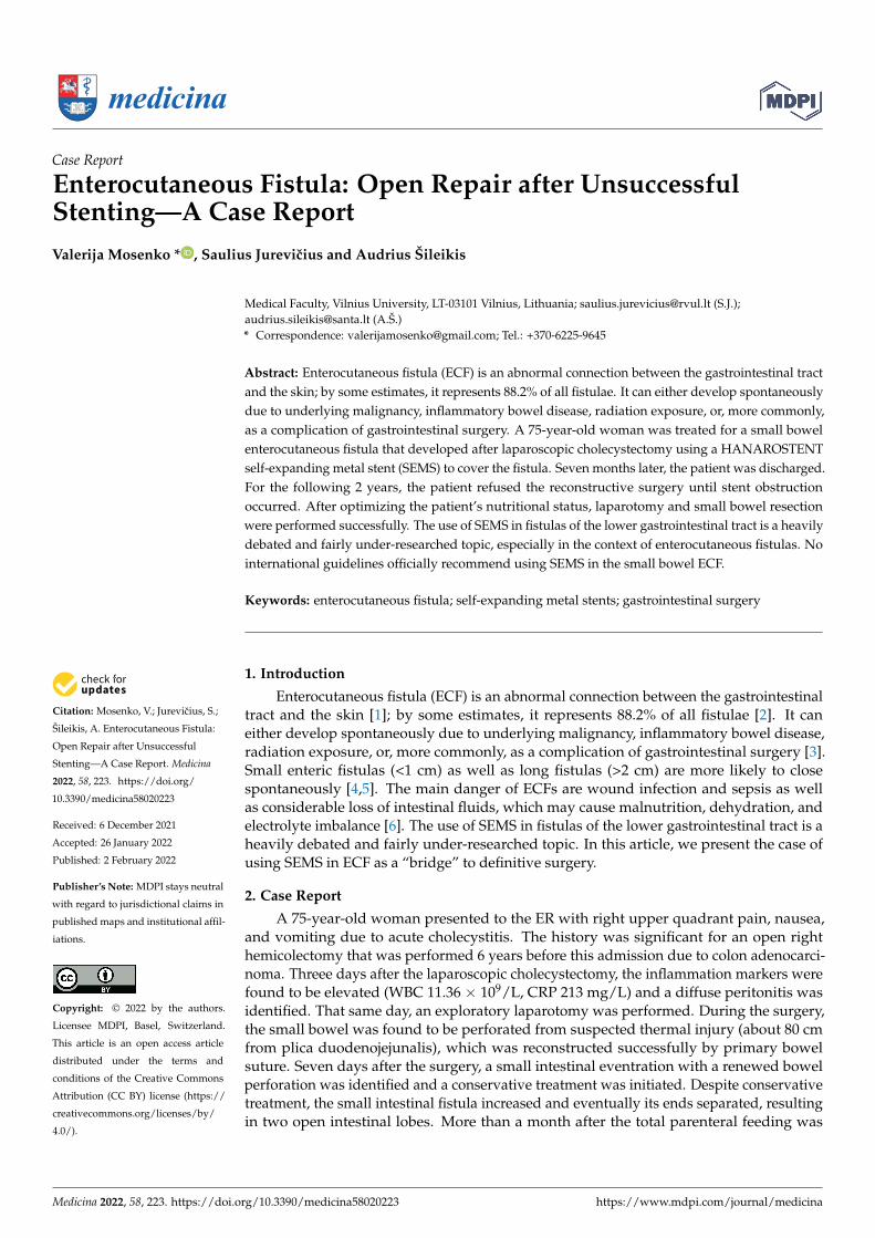
initiated, the HANAROSTENT TLD-20110-230 (a single-use duodenal pyloric partiallycovered self-expanding metal stent) was inserted at both ends of the small intestine andfixed with single sutures to restore intestinal integrity. Two weeks after the implantation,the bowel fistula reoccurred; however, the output was noted to be comparably lower. Themetallic stent was left in place for a longer time despite manufacturer’s recommendations,to act as a metal carcass to facilitate the healing of the external fistula. Seven monthslater, the patient was discharged after the total closure of the fistula (verified by upperGI and small bowel series), while being able to tolerate a regular feeding regimen. Thefollow-up appointment was scheduled to remove the stent, but unfortunately, the patientdid not show up for the appointment. For the following 2 years, the patient was beingintermittently hospitalized to a different hospital, due to mechanical bowel obstructions,which were treated conservatively due to the patient refusing surgery. Two years after theinitial discharge from the primary hospital, the patient was admitted into our care due toacute bowel obstruction. The metabolic panel showed signs of chronic malnutrition withsignificant hypoproteinemia (29.7 g/L) and hypoalbuminemia (56.7 g/L), with a BMI of20.76 kg/m2. Abdominal CT (Figure 1) showed a small bowel stent with fibrous/tumor-likechanges around it (signs of small bowel obstruction).
Medicina 2022, 58, x FOR PEER REVIEW 2 of 6
servative treatment, the small intestinal fistula increased and eventually its ends sepa-rated, resulting in two open intestinal lobes. More than a month after the total parenteral feeding was initiated, the HANAROSTENT TLD-20110-230 (a single-use duodenal pylo-ric partially covered self-expanding metal stent) was inserted at both ends of the small intestine and fixed with single sutures to restore intestinal integrity. Two weeks after the implantation, the bowel fistula reoccurred; however, the output was noted to be compa-rably lower. The metallic stent was left in place for a longer time despite manufacturer’s recommendations, to act as a metal carcass to facilitate the healing of the external fistula. Seven months later, the patient was discharged after the total closure of the fistula (veri-fied by upper GI and small bowel series), while being able to tolerate a regular feeding regimen. The follow-up appointment was scheduled to remove the stent, but unfortu-nately, the patient did not show up for the appointment. For the following 2 years, the patient was being intermittently hospitalized to a different hospital, due to mechanical bowel obstructions, which were treated conservatively due to the patient refusing sur-gery. Two years after the initial discharge from the primary hospital, the patient was ad-mitted into our care due to acute bowel obstruction. The metabolic panel showed signs of chronic malnutrition with significant hypoproteinemia (29.7 g/L) and hypoalbuminemia (56.7 g/L), with a BMI of 20.76 kg/m2. Abdominal CT (Figure 1) showed a small bowel stent with fibrous/tumor-like changes around it (signs of small bowel obstruction).
Figure 1. A small bowel stent with fibrous/tumor-like changes around it (red arrow).
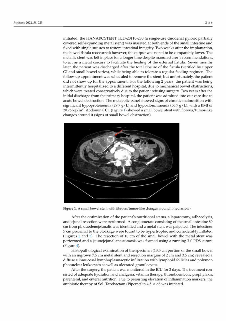
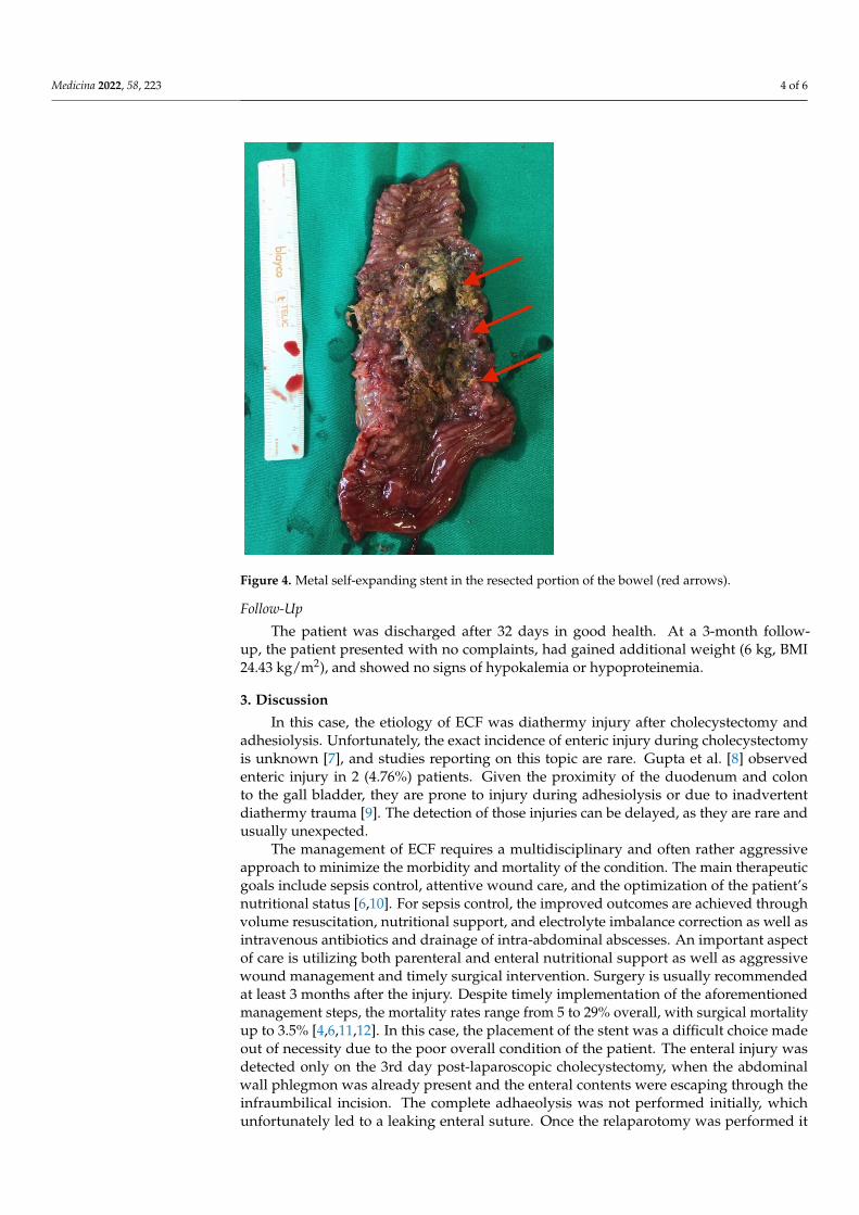
After the optimization of the patient’s nutritional status, a laparotomy, adhaeolysis, and jejunal resection were performed. A conglomerate consisting of the small intestine 80 cm from pl. duodenojejunalis was identified and a metal stent was palpated. The intes-tines 5 cm proximal to the blockage were found to be hypertrophic and considerably in-flated (Figures 2 and 3). The resection of 10 cm of the small bowel with the metal stent was performed and a jejunojejunal anastomosis was formed using a running 3-0 PDS suture (Figure 4).
Figure 1. A small bowel stent with fibrous/tumor-like changes around it (red arrow).
After the optimization of the patient’s nutritional status, a laparotomy, adhaeolysis,and jejunal resection were performed. A conglomerate consisting of the small intestine 80cm from pl. duodenojejunalis was identified and a metal stent was palpated. The intestines5 cm proximal to the blockage were found to be hypertrophic and considerably inflated(Figures 2 and 3). The resection of 10 cm of the small bowel with the metal stent wasperformed and a jejunojejunal anastomosis was formed using a running 3-0 PDS suture(Figure 4).
Histopathological examination of the specimen (13.5 cm portion of the small bowelwith an ingrown 7.5 cm metal stent and resection margins of 2 cm and 3.5 cm) revealed adiffuse submucosal lymphoplasmacytic infiltration with lymphoid follicles and polymor-phonuclear leukocytes as well as ulcerated granulocytes.
After the surgery, the patient was monitored in the ICU for 2 days. The treatment con-sisted of adequate hydration and analgesia, vitamin therapy, thromboembolic prophylaxis,parenteral, and enteral nutrition. Due to persisting elevation of inflammation markers, theantibiotic therapy of Sol. Tazobactam/Piperacilin 4.5 × q8 was initiated.
Medicina 2022, 58, 223 3 of 6Medicina 2022, 58, x FOR PEER REVIEW 3 of 6
Figure 2. Enterocutaneous fistula in situ (blue arrow).
Figure 3. Enterocutaneous fistula seen in the mobilized small bowel (blue arrow).
Figure 2. Enterocutaneous fistula in situ (blue arrow).
Medicina 2022, 58, x FOR PEER REVIEW 3 of 6
Figure 2. Enterocutaneous fistula in situ (blue arrow).
Figure 3. Enterocutaneous fistula seen in the mobilized small bowel (blue arrow).
Figure 3. Enterocutaneous fistula seen in the mobilized small bowel (blue arrow).
Medicina 2022, 58, 223 4 of 6Medicina 2022, 58, x FOR PEER REVIEW 4 of 6
Figure 4. Metal self-expanding stent in the resected portion of the bowel (red arrows).
Histopathological examination of the specimen (13.5 cm portion of the small bowel with an ingrown 7.5 cm metal stent and resection margins of 2 cm and 3.5 cm) revealed a diffuse submucosal lymphoplasmacytic infiltration with lymphoid follicles and polymor-phonuclear leukocytes as well as ulcerated granulocytes.
After the surgery, the patient was monitored in the ICU for 2 days. The treatment consisted of adequate hydration and analgesia, vitamin therapy, thromboembolic prophy-laxis, parenteral, and enteral nutrition. Due to persisting elevation of inflammation mark-ers, the antibiotic therapy of Sol. Tazobactam/Piperacilin 4.5 × q8 was initiated.
Follow-up The patient was discharged after 32 days in good health. At a 3-month follow-up, the
patient presented with no complaints, had gained additional weight (6 kg, BMI 24.43 kg/m2), and showed no signs of hypokalemia or hypoproteinemia.
3. Discussion In this case, the etiology of ECF was diathermy injury after cholecystectomy and ad-
hesiolysis. Unfortunately, the exact incidence of enteric injury during cholecystectomy is unknown [7], and studies reporting on this topic are rare. Gupta et al. [8] observed enteric injury in 2 (4.76%) patients. Given the proximity of the duodenum and colon to the gall bladder, they are prone to injury during adhesiolysis or due to inadvertent diathermy trauma [9]. The detection of those injuries can be delayed, as they are rare and usually unexpected.
The management of ECF requires a multidisciplinary and often rather aggressive ap-proach to minimize the morbidity and mortality of the condition. The main therapeutic goals include sepsis control, attentive wound care, and the optimization of the patient’s nutritional status [6,10]. For sepsis control, the improved outcomes are achieved through volume resuscitation, nutritional support, and electrolyte imbalance correction as well as intravenous antibiotics and drainage of intra-abdominal abscesses. An important aspect of care is utilizing both parenteral and enteral nutritional support as well as aggressive
Figure 4. Metal self-expanding stent in the resected portion of the bowel (red arrows).
Follow-Up
The patient was discharged after 32 days in good health. At a 3-month follow-up, the patient presented with no complaints, had gained additional weight (6 kg, BMI24.43 kg/m2), and showed no signs of hypokalemia or hypoproteinemia.
3. Discussion
In this case, the etiology of ECF was diathermy injury after cholecystectomy andadhesiolysis. Unfortunately, the exact incidence of enteric injury during cholecystectomyis unknown [7], and studies reporting on this topic are rare. Gupta et al. [8] observedenteric injury in 2 (4.76%) patients. Given the proximity of the duodenum and colonto the gall bladder, they are prone to injury during adhesiolysis or due to inadvertentdiathermy trauma [9]. The detection of those injuries can be delayed, as they are rare andusually unexpected.
The management of ECF requires a multidisciplinary and often rather aggressiveapproach to minimize the morbidity and mortality of the condition. The main therapeuticgoals include sepsis control, attentive wound care, and the optimization of the patient’snutritional status [6,10]. For sepsis control, the improved outcomes are achieved throughvolume resuscitation, nutritional support, and electrolyte imbalance correction as well asintravenous antibiotics and drainage of intra-abdominal abscesses. An important aspectof care is utilizing both parenteral and enteral nutritional support as well as aggressivewound management and timely surgical intervention. Surgery is usually recommendedat least 3 months after the injury. Despite timely implementation of the aforementionedmanagement steps, the mortality rates range from 5 to 29% overall, with surgical mortalityup to 3.5% [4,6,11,12]. In this case, the placement of the stent was a difficult choice madeout of necessity due to the poor overall condition of the patient. The enteral injury wasdetected only on the 3rd day post-laparoscopic cholecystectomy, when the abdominalwall phlegmon was already present and the enteral contents were escaping through theinfraumbilical incision. The complete adhaeolysis was not performed initially, whichunfortunately led to a leaking enteral suture. Once the relaparotomy was performed it
Medicina 2022, 58, 223 5 of 6
became apparent that the fistula was situated just behind the aponeurosis and subsequentlymigrated into the hypodermal layer. After the surgery, a total parenteral nutrition wasinitiated and continued for 6 weeks, before the decision to implant SEMS to cover thefistula was made, as there were no signs of spontaneous closure present and the patient’scondition remained critical. Although the primary team considered other managementstrategies, none of them were suitable for this patient. T-tube drainage and stoma formationwere excluded due to them leaving the “high” fistula open, which has been proven tobe detrimental to patients because of the high-volume output of the intestinal contentsand subsequent malnourishment. The resection of the fistula and anastomosis may havebeen beneficial; however, the primary surgical team as well as the patient were hopingto avoid the resection of the intestine for as long as possible. The long term TPN wasadministered for 7 months. Due to the concern of the foreign body in the intestinal tract, itwas recommended that the patient come back for follow up; however, the patient did notadhere to the recommendations and was intermittently treated conservatively for bowelobstruction every 6 months.
The placement of the stent in this patient provided as a temporary relief, providingan opportunity to optimize the patient’s nutritional status and prepare them for surgery,which was unfortunately postponed due to the patient’s concerns and refusal to adhere torecommendations.
The use of SEMS in fistulas of the lower gastrointestinal tract is a heavily debatedand fairly under-researched topic, especially in the context of enterocutaneous fistulas.No international guidelines officially recommend using SEMS in the small bowel ECF;therefore, no comparison of outcomes can be achieved.
4. Conclusions
Enterocutaneous fistula remains a dreaded surgical complication with high mortalityand morbidity rates, despite the considerable advances in ECF management. The use ofSEMS in fistulas of the lower gastrointestinal tract is a heavily debated and fairly under-researched topic, especially in the context of enterocutaneous fistulas. The controversialdecisions made in the management of the patient in the described case report highlightthe benefits and limitations of using it as a “bridging” therapy towards a definitive fistulatreatment. More data needs to be published to evaluate the efficacy of this treatmentstrategy in small bowel fistulas.
Author Contributions: Conceptualization, V.M. and A.Š.; methodology, V.M. and A.Š.; software,V.M.; validation, V.M., A.Š. and S.J.; formal analysis, V.M., A.Š. and S.J.; investigation, V.M., A.Š. andS.J.; resources, A.Š. and S.J.; data curation, A.Š. and S.J.; writing—original draft preparation, V.M..;writing—review and editing, V.M., A.Š. and S.J.; visualization, V.M.; supervision, A.Š. and S.J.; projectadministration, A.Š.; funding acquisition, n/a. All authors have read and agreed to the publishedversion of the manuscript.
Funding: This research received no external funding.
Institutional Review Board Statement: Not applicable.
Informed Consent Statement: Informed consent was obtained from the patients.
Conflicts of Interest: The authors declare no conflict of interest.
References1. Kumpf, V.J.; de Aguilar-Nascimento, J.E.; Graf Diaz-Pizarro, J.I.; Hall, A.M.; McKeever, L.; Steiger, E.; Winkler, M.F.; Compher,
C.W. ASPEN-FELANPE Clinical Guidelines: Nutrition Support of Adult Patients With Enterocutaneous Fistula. JPEN J. ParenterEnter. Nutr. 2017, 41, 104–112. [CrossRef] [PubMed]
2. Owen, R.M.; Love, T.P.; Perez, S.D.; Srinivasan, J.K.; Sharma, J.; Pollock, J.D.; Galloway, J.R. Definitive surgical treatment ofenterocutaneous fistula: Outcomes of a 23-year experience. JAMA Surg. 2013, 148, 118–126. [CrossRef] [PubMed]
3. Orangio, G. Enterocutaneous Fistula: Medical and Surgical Management Including Patients with Crohn’s Disease. Clin. ColonRectal Surg. 2010, 23, 169–175. [CrossRef] [PubMed]
4. Evenson, A.; Fischer, J. Current Management of Enterocutaneous Fistula. J. Gastrointest. Surg. 2006, 10, 455–464. [CrossRef]
Medicina 2022, 58, 223 6 of 6
5. Majercik, S.; Kinikini, M.; White, T. Enteroatmospheric Fistula: From Soup to Nuts. Nutr. Clin. Pract. 2012, 27, 507–512. [CrossRef][PubMed]
6. Da Lloyd, J.; Gabe, S.M.; Windsor, A.C.J. Nutrition and management of enterocutaneous fistula. Br. J. Surg. 2006, 93, 1045–1055.[CrossRef] [PubMed]
7. Machado, N.O. Duodenal injury post laparoscopic cholecystectomy: Incidence, mechanism, management and outcome. World J.Gastrointest Surg. 2016, 8, 335–344. [CrossRef] [PubMed]
8. Gupta, V.; Gupta, A.; Yadav, T.D.; Mittal, B.R.; Kochhar, R. Post-cholecystectomy acute injury: What can go wrong? Ann.Hepatobiliary Pancreat Surg. 2019, 23, 138–144. [CrossRef] [PubMed]
9. Alexander, H.C.; Bartlett, A.S.; Wells, C.I.; Hannam, J.A.; Moore, M.R.; Poole, G.H.; Merry, A.F. Reporting of complications afterlaparoscopic cholecystectomy: A systematic review. HPB 2018, 20, 786–794. [CrossRef] [PubMed]
10. Klek, S.; Forbes, A.; Gabe, S.; Holst, M.; Wanten, G.; Irtun, Ø.; Shaffer, J. Management of acute intestinal failure: A position paperfrom the European Society for Clinical Nutrition and Metabolism (ESPEN) Special Interest Group. Clin. Nutr. 2016, 35, 1209–1218.[CrossRef] [PubMed]
11. Wercka, J.; Cagol, P.P.; Melo, A.L.P.; de Locks, G.F.; Franzon, O.; Kruel, N.F. Epidemiology and outcome of patients withpostoperative abdominal fistula. Rev. Col. Bras. Cir. 2016, 43, 117–123. [CrossRef] [PubMed]
12. De Vries, F.E.E.; Atema, J.J.; van Ruler, O.; Vaizey, C.J.; Serlie, M.J.; Boermeester, M.A. A Systematic Review and Meta-analysis ofTiming and Outcome of Intestinal Failure Surgery in Patients with Enteric Fistula. World J. Surg. 2018, 42, 695–706. [CrossRef][PubMed]


![[Pancreatic fistula following duodenocephalopancreatectomy with Wirsung occlusion]](https://img.dokumen.tips/doc/110x75/63527b8430053fbe620bfb86/pancreatic-fistula-following-duodenocephalopancreatectomy-with-wirsung-occlusion.jpg)