Embed Size (px)
Citation preview
original article
Gut and Liver, Vol. 4, No. 4, December 2010, pp. 508-513
Correspondence to: Hwoon-Yong JungDivision of Gastroenterology, Department of Internal Medicine, Asan Medical Center, University of Ulsan College of Medicine, 388-1 Pungnap 2-dong, Songpa-gu, Seoul 138-736, KoreaTel: +82-2-3010-3197, Fax: +82-2-476-0824, E-mail: [email protected]
Received on April 29, 2010. Accepted on July 17, 2010.DOI: 10.5009/gnl.2010.4.4.508
Benign Bronchoesophageal Fistula in Adults: Endoscopic Closure as Primary Treatment
Ji Yong Ahn, Hwoon-Yong Jung, Ji Young Choi, Mi-Young Kim, Jeong Hoon Lee, Kwi-Sook Choi, Do Hoon Kim, Kee Don Choi, Ho June Song, Gin Hyug Lee, and Jin-Ho KimDivision of Gastroenterology, Department of Internal Medicine, Asan Medical Center, University of Ulsan College of Medicine, Seoul, Korea
Background/Aims: Benign bronchoesophageal fistula (BEF) is a rare condition that is usually treated surgi-cally; however, less invasive endoscopy procedures have been attempted to overcome the disadvantages of surgery. The aim of this study was thus to de-termine the results of endoscopic management as a primary treatment in patients with BEF. Methods: We retrospectively analyzed data from 368 patients with BEF who were treated at a tertiary care, academic medical center between January 2000 and August 2009. Results: Benign causes were found for only 18 of the 368 patients. Of these, seven were treated en-doscopically and the others by surgery or other methods. The first endoscopy procedures failed in all seven patients, with second trials of endoscopy per-formed in four patients at a median of 8 days (range, 3 to 11 days) after the first procedure. The second endoscopic procedure was successful in two out of four patients; one patient showed no recurrence of the fistula, whereas the second patient experienced a recurrence after 24 months. All patients underwent successful surgical procedures after the failure of en-doscopic treatment, with no further recurrences. Conclusions: Although we observed a low rate of success for primary endoscopic treatment of benign BEF, the invasive nature of surgery suggests the need for a prospective study with a large number of patients to evaluate the efficacy of less invasive pro-cedures such as endoscopic treatment. (Gut Liver 2010;4:508-513)
Key Words: Esophageal fistula; Endoscopy; Fibrin glue
INTRODUCTION
Fistulas, although rare, may develop between the esophageal lumen and other mediastinal structures. These bronchoesophageal fistulas (BEFs) may be due to con-genital or acquired causes. Most fistulas in adults are ac-quired because it is rare for a congenital fistula to remain asymptomatic until adulthood. Most acquired fistulas are caused by malignancies, either by a metastatic deposit or via direct invasion of a tumor. Fistulas may also be due to benign causes, including infections, such as tuber-culosis and histoplasmosis, trauma, and other causes.1-5 Owing to their nonspecific symptoms and rarity, however, benign BEF may be present for a long time without being diagnosed and properly treated, resulting in significant morbidity and death. Most patients with fistula are treated surgically, with removal of the fistula and any permanently damaged lung segments. The standard operative procedure involves a thoracotomy, during which the fistula is exposed and div-ided and the defects in the bronchus and the esophagus are repaired by interposition of viable tissue between the suture lines.3,6
Several reports have described attempts to repair fistu-las by endoscopic methods, thus avoiding the complica-tions and sequelae of surgery.4,7-11 These methods include the application of fibrin glue, sodium hydroxide and ace-tic acid, which have been used to close fistulas in both adults and children.7,8,10,11 Being biocompatible, fibrin glue can be repeatedly applied and also does not inhibit any subsequent surgical intervention. Other reports have de-
Ahn JY, et al: Benign Bronchoesophageal Fistula in Adults 509
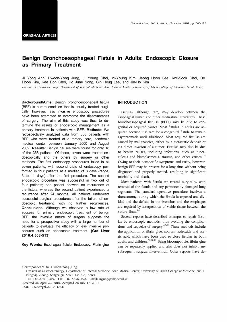
Fig. 1. (A) Fistula opening located 31 cm from the upper incisor. (B) Injection of fibrin glue into the area of the fistula opening.(C) Application of two hemoclips to the site of the fistula opening after the injection of fibrin glue.
Table 1. Baseline Characteristics of Benign Bronchoesophageal Fistula Patients
No. 18Median age, yr (range) 58.5 (28-86)Sex, M:F 5:13Underlying cause Tuberculosis infection 12 (67) Post operation 2 (11) Idiopathic 4 (22)Symptom Cough 11 (61) Sputum 9 (50) Hemoptysis 7 (39) Fever 5 (28) Chest pain 3 (17) Dyspnea 2 (11) None 1 (6) Location of fistula Right 11 (61) Left 3 (17) Subcarina 4 (22)
Data represent number of patients (% of total population).
scribed the use of hemoclips.4,9 To our knowledge, how-ever, there have been few reports about the success rate of endoscopic treatment for the primary treatment of be-nign BEF in adults. We therefore report here our experi-ence in the primary endoscopic management of patients with benign BEF.
MATERIALS AND METHODS
1. Patients
We examined the medical records of patients who un-derwent BEF between January 2000 and August 2009 at the Asan Medical Center. Of the 368 patients who under-went BEF, only 18 (4.8%) did so for benign BEF. Of these, 7 patients were treated endoscopically and 11 were treated surgically or by other methods. With the approval of our institutional review board, the charts of these pa-tients were retrospectively reviewed. Patients were excluded from the study if they were younger than 20 years old, if they had been diagnosed with a tracheoesophageal fistula caused by an endo-tracheal or tracheostomy tube, if they had a current his-tory of malignancy related to the fistula or a history of congenital fistula, or had previously been treated for fistu-las by bronchofibroscopy or stent insertion.
2. Endoscopic technique
These procedures were performed by expert endoscopists with the patient under conscious sedation with intravenous administration of midazolam. Following administration of local pharyngeal anesthesia, the endoscope was inserted in-to the esophagus until it reached the opening site of the fistula. Fibrin glue (Beriplast; Aventis Behring Ltd., Marburg, Germany), consisting of a solution of fibrinogen (one vial of fibrinogen concentrate and one vial of aproti-nin solution) and thrombin (one vial of human thrombin
and one vial of calcium chloride), was injected into the opening site and/or the metal hemoclips (HX-600-090L; Olympus Optical Co., Ltd, Tokyo, Japan) (Fig. 1).
3. Statistical analysis
Quantitative variables with a normal distribution are expressed as mean±standard deviation, whereas quantita-tive variables with skewed distributions are expressed as median (range). The χ2 test, or Fisher’s exact test if nec-essary, was used to compare relative frequencies of cate-gorical variables and the Student’s t-test or the Mann- Whitney U-test was used to compare continuous varia-bles. A p-value of <0.05 was considered significant. All statistical analyses were performed using the SPSS pro-gram, version 12.0 (SPSS Inc, Chicago, IL, USA).
510 Gut and Liver, Vol. 4, No. 4, December 2010
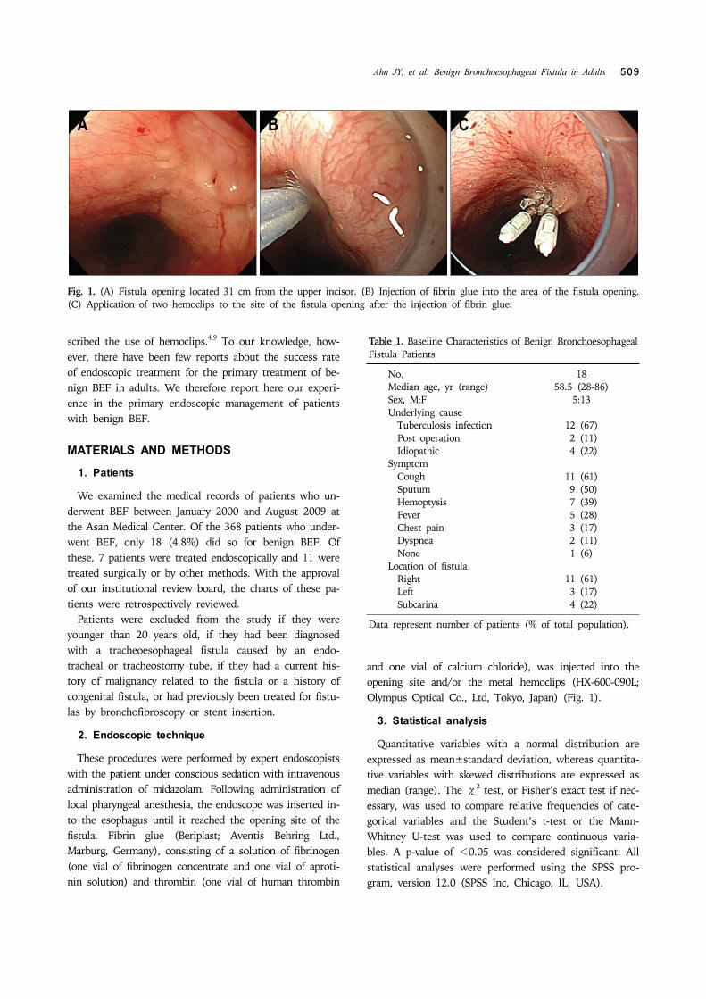
Fig. 2. Esophagography showing the communication between the right lower lobar bronchus with the esophageal cleft in themiddle of the esophagus, a finding compatible with a bronchoesophageal fistula.
Table 2. Comparison of the Sensitivities of Computed Tomography, Endoscopy, and Bronchoscopy in Detecting Benign Bronchoesophageal Fistula
CT Endoscopy Bronchoscopy
No. of patients 18 17 14True positive, n 16 16 7False negative, n 2 1 7Sensitivity, % 89 94 50
CT, computed tomography.
Table 3. Outcomes Following Endoscopic Treatment of Bronchoesophageal Fistula
Underlying cause
No. of trial1st/2nd injected fibrin glue, cc
1st/2nd no. of hemoclip
Duration, day*
Outcome Duration†
Patient 1 Infection 2 3/3 0 11 Incomplete success 2 yrPatient 2 Infection 1 2 0 - Failure 10 daysPatient 3 Infection 2 2/1 0 9 Complete success -Patient 4 Infection 1 0 2 - Failure 14 daysPatient 5 Infection 1 1 1 - Failure 15 daysPatient 6 Operation 2 3/0 0/2 3 Failure 36 daysPatient 7 Idiopathic 2 1/1 2/2 7 Failure 31 days
*Duration from first to second treatment; †
Duration from endoscopic failure to operation.
RESULTS
Over a 10-year treatment period, 18 patients (13 wom-en, 5 men), of median age 58.5 years (range, 28 to 86 years) were included in this study (Table 1). All 18 pa-tients underwent esophagography, computed tomography (CT), endoscopy, and bronchoscopy for the diagnosis of benign BEF. Of these patients, 7 (6 women, 1 man), of median age 67 years (range, 50 to 86 years), underwent primary endoscopic treatment, whereas 9 underwent pri-mary surgical treatment. Of the remaining two patients, one was treated radiologically by fibrin glue injection and one was observed while receiving treatment with anti-tu-berculosis medication.
1. Etiology
Of the 18 BEFs, 12 were caused by infection, 2 were
postoperative complications and 4 were idiopathic. The 12 patients with BEFs due to infection had pulmonary tu-berculosis, with BEF developing after cure of tuberculosis in 8 patients. Four patients, however, were diagnosed with pulmonary tuberculosis owing to BEF. The median duration from the diagnosis of tuberculous infection to the onset of fistula symptoms in the 8 patients who de-veloped BEF after cure of tuberculosis was 14.5 years (range, 5 to 30 years). Of the two patients who devel-oped BEF after surgery, one had a lung tumor that had been removed by pneumonectomy, with no recurrence over 8 years, and the other had a history of pneumo-thorax, which had previously been treated by chest tube insertion 35 years previously. In the remaining four pa-tients, we found no etiology of fistula (Table 1).
2. Symptoms
Eleven patients presented with a cough that lasted from 4 days to more than 8 years. Seven patients reported a mild hemoptysis that lasted from 20 days to more than 1 year prior to diagnosis. Among the various symptoms were cough, sputum, hemoptysis, fever, chest pain, and dyspnea, which were single or combined (Table 1). In the 12 patients with BEF resulting from tuberculosis in-fection, the median symptom duration was 30 days (range, 4 days to 2 years). In three patients with idio-pathic BEF, symptoms lasted from 20 days to 5 years.
Ahn JY, et al: Benign Bronchoesophageal Fistula in Adults 511
Table 4. Outcomes Following Treatment of Bronchoesopha-geal Fistula
Median follow-up period, mo (range) 37 (8-62)Median duration period from diagnosis to 14.5 (9-30) primary treatment, days (range)Success of primary endoscopic treatment 2/7 (29)Success of primary surgical treatment 9/9 (100)Success of other treatment 2/2 (100)Success of secondary surgical treatment 6/6 (100)Recurrence after surgical treatment 0/13 (0)
Data represent number of patients (% of total population).
The fourth patient with idiopathic BEF had no symptoms; in this patient, BEF was detected during a routine examination.
3. Diagnosis
The confirmative diagnosis was established by esoph-agography in all patients (Fig. 2). In contrast, CT, endos-copy, and bronchoscopy failed to reveal BEFs in 2, 1 and 7 patients, respectively, making the sensitivity of CT, en-doscopy, and bronchoscopy 89%, 94%, and 50%, re-spectively (Table 2). The mean fistula opening size in the 7 patients who underwent primary endoscopic treatment was 4.5 mm (range, 2 mm to 6 mm), with no significant difference in size between the patients treated endoscopi-cally and surgically.
4. Management and outcomes
The first endoscopic treatment failed in all seven pa-tients who underwent a primary endoscopic procedure. Four of these patients underwent a second endoscopic procedure, at a median 8 days (range, 3 to 11 days) after the first procedure. The second endoscopic procedure was successful in two of these four patients. One patient (no. 3) has shown no recurrence of fistula during 52 months of follow-up, whereas the second (no. 1) underwent sur-gery after 24 months because, although symptoms had disappeared, the fistula was still present (Table 3). The remaining three patients underwent surgery after endo-scopic treatment failure, with no further recurrences at a median 52 months (range, 12 months to 62 months) of follow-up. Treatment outcomes are summarized in Table 4.
DISCUSSION
BEF is a rare complication resulting from various dis-eases, with most BEFs in adults resulting from a locally advanced esophageal or bronchogenic malignancy. In con-trast, benign BEFs can remain undiagnosed for years, ow-ing to the combination of their rarity and their non-
specific symptoms. A recent review of BEFs at Massachu-setts General Hospital found that, over more than 40 years, only 228 cases were seen,3 with benign causes re-sponsible for only 6%. Of the latter, 30% developed after esophageal surgery. Another study reported that media-stinal granulomatous inflammation caused by tuberculosis could account for 43% of benign BEFs.6 We observed that tuberculosis was the most common etiology of benign BEF, but at a higher rate (67%) than seen in other stud-ies, which may be related to the high incidence of tuber-culosis in Korea. Most patients with benign BEF present with non-specific physical findings. For example, the ma-jority of patients with benign and congenital BEF have been found to present with a chronic cough lasting from 5 weeks to more than 30 years.3,6,12 We found that 61% of our patients presented with cough, and 11-50% pre-sented with other symptoms, including sputum, hemopt-ysis, fever, chest pain, and/or dyspnea. Most symptoms were not severe and the duration from symptom develop-ment to presentation varied from 4 days to 8 years. A diagnosis of BEF should be suspected in any patient presenting with a persistent unexplained cough or failing to improve after established medical therapy. Although various diagnostic modalities are available and comple-mentary to each other, barium esophagography is re-garded as a more sensitive and definitive diagnostic tool than endoscopic procedures.6,13,14 We also used esoph-agography as a confirmative diagnostic tool and we found that the sensitivities of CT, endoscopy, and bronchoscopy in diagnosing benign BEF were similar to previously re-ported findings.6 Once a BEF is confirmed, early surgical intervention is considered definitive, resulting in successful outcomes. Although early and definitive surgical repair has yielded promising results,5,6,15 the in-hospital mortality rate has been reported to be as high as 15.4%,3 and the surgery it-self is invasive. This has led to the development of endo-scopic and even medical treatment methods.4,7-11 For ex-ample, while covered esophageal-wall-stent placement has been described for malignant fistulas associated with stricture,16,17 esophageal stent insertion would probably not have been suitable for benign BEFs because the latter have no strictures to hold the stents in place. To date, there have been few reports about the results of primary endoscopic treatment for benign BEFs in adults. We found that the success rate was only 29%, with the two patients successfully treated requiring two endoscopic treatments each because the first treatment failed in both. The low success rate of endoscopic treatment may be as-sociated with the length and width of the fistula tract. If it is straight and too short, the fibrin glue can be washed
512 Gut and Liver, Vol. 4, No. 4, December 2010
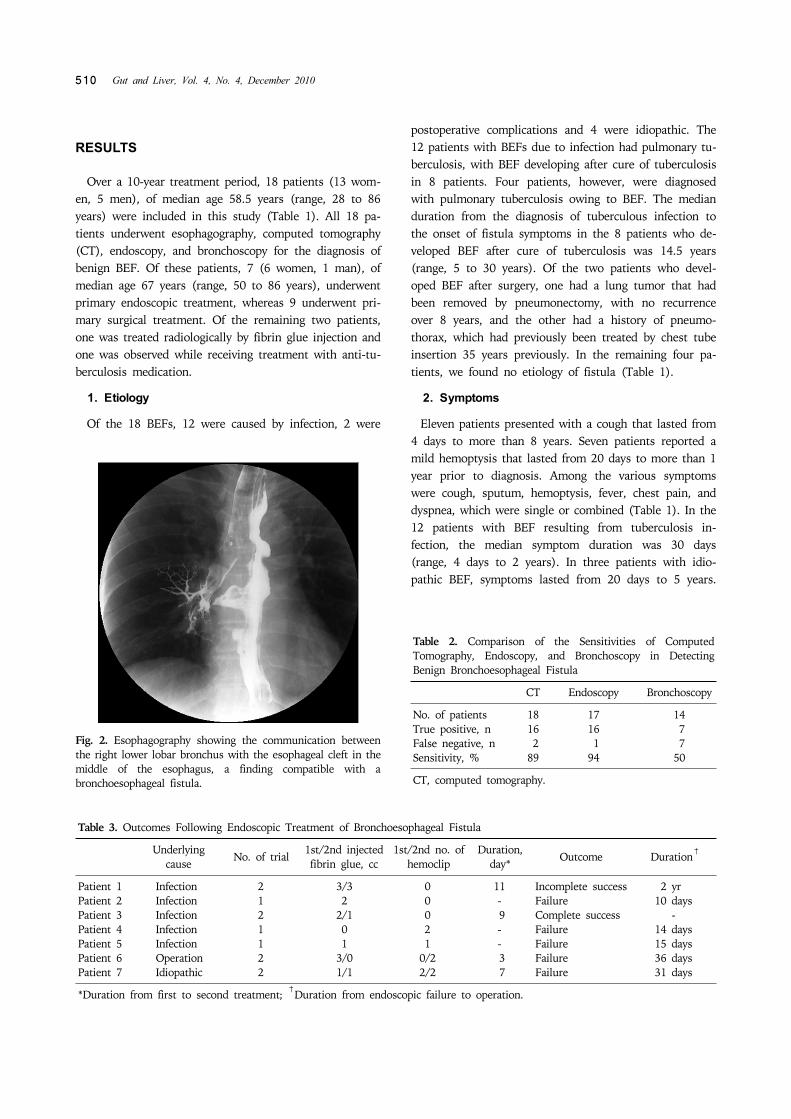
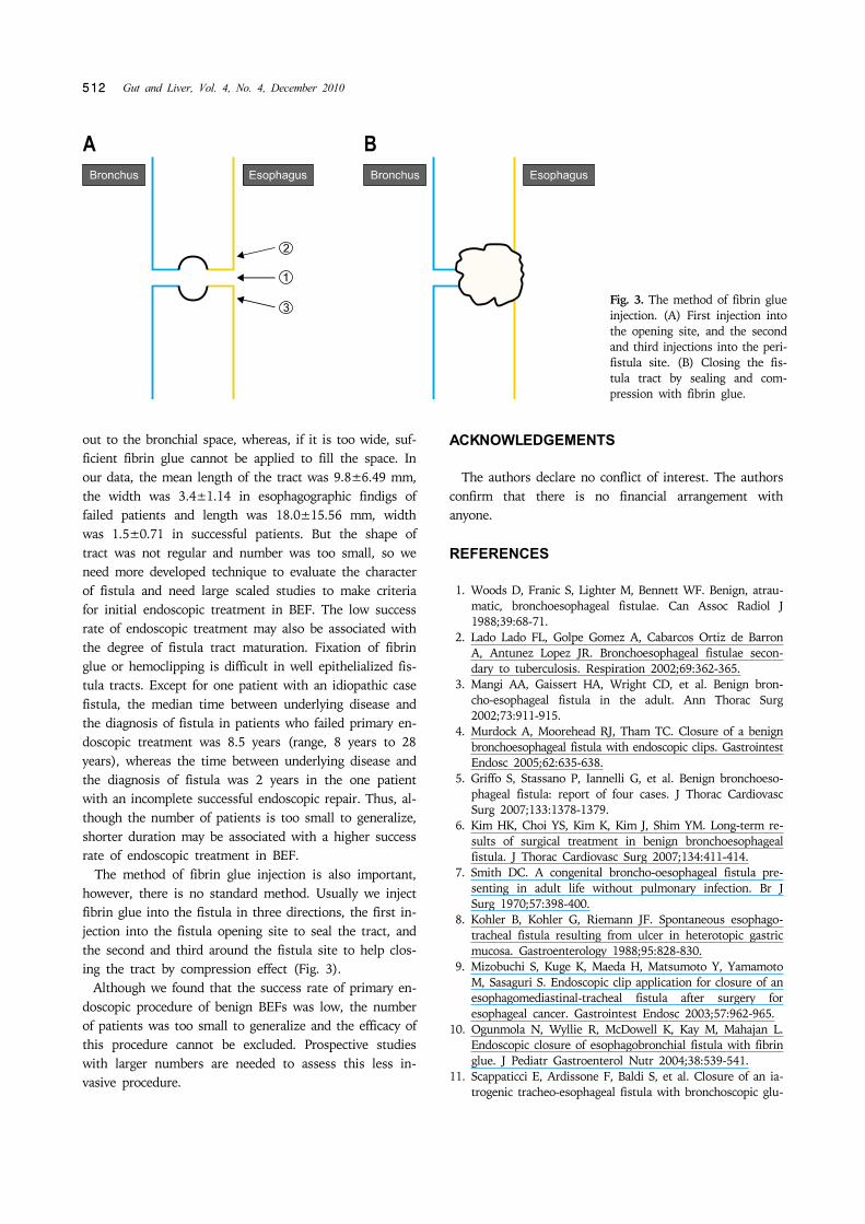
Fig. 3. The method of fibrin glue injection. (A) First injection into the opening site, and the second and third injections into the peri-fistula site. (B) Closing the fis-tula tract by sealing and com-pression with fibrin glue.
out to the bronchial space, whereas, if it is too wide, suf-ficient fibrin glue cannot be applied to fill the space. In our data, the mean length of the tract was 9.8±6.49 mm, the width was 3.4±1.14 in esophagographic findigs of failed patients and length was 18.0±15.56 mm, width was 1.5±0.71 in successful patients. But the shape of tract was not regular and number was too small, so we need more developed technique to evaluate the character of fistula and need large scaled studies to make criteria for initial endoscopic treatment in BEF. The low success rate of endoscopic treatment may also be associated with the degree of fistula tract maturation. Fixation of fibrin glue or hemoclipping is difficult in well epithelialized fis-tula tracts. Except for one patient with an idiopathic case fistula, the median time between underlying disease and the diagnosis of fistula in patients who failed primary en-doscopic treatment was 8.5 years (range, 8 years to 28 years), whereas the time between underlying disease and the diagnosis of fistula was 2 years in the one patient with an incomplete successful endoscopic repair. Thus, al-though the number of patients is too small to generalize, shorter duration may be associated with a higher success rate of endoscopic treatment in BEF. The method of fibrin glue injection is also important, however, there is no standard method. Usually we inject fibrin glue into the fistula in three directions, the first in-jection into the fistula opening site to seal the tract, and the second and third around the fistula site to help clos-ing the tract by compression effect (Fig. 3). Although we found that the success rate of primary en-doscopic procedure of benign BEFs was low, the number of patients was too small to generalize and the efficacy of this procedure cannot be excluded. Prospective studies with larger numbers are needed to assess this less in-vasive procedure.
ACKNOWLEDGEMENTS
The authors declare no conflict of interest. The authors confirm that there is no financial arrangement with anyone.
REFERENCES
1. Woods D, Franic S, Lighter M, Bennett WF. Benign, atrau-matic, bronchoesophageal fistulae. Can Assoc Radiol J 1988;39:68-71.
2. Lado Lado FL, Golpe Gomez A, Cabarcos Ortiz de Barron A, Antunez Lopez JR. Bronchoesophageal fistulae secon-dary to tuberculosis. Respiration 2002;69:362-365.
3. Mangi AA, Gaissert HA, Wright CD, et al. Benign bron-cho-esophageal fistula in the adult. Ann Thorac Surg 2002;73:911-915.
4. Murdock A, Moorehead RJ, Tham TC. Closure of a benign bronchoesophageal fistula with endoscopic clips. Gastrointest Endosc 2005;62:635-638.
5. Griffo S, Stassano P, Iannelli G, et al. Benign bronchoeso-phageal fistula: report of four cases. J Thorac Cardiovasc Surg 2007;133:1378-1379.
6. Kim HK, Choi YS, Kim K, Kim J, Shim YM. Long-term re-sults of surgical treatment in benign bronchoesophageal fistula. J Thorac Cardiovasc Surg 2007;134:411-414.
7. Smith DC. A congenital broncho-oesophageal fistula pre-senting in adult life without pulmonary infection. Br J Surg 1970;57:398-400.
8. Kohler B, Kohler G, Riemann JF. Spontaneous esophago-tracheal fistula resulting from ulcer in heterotopic gastric mucosa. Gastroenterology 1988;95:828-830.
9. Mizobuchi S, Kuge K, Maeda H, Matsumoto Y, Yamamoto M, Sasaguri S. Endoscopic clip application for closure of an esophagomediastinal-tracheal fistula after surgery for esophageal cancer. Gastrointest Endosc 2003;57:962-965.
10. Ogunmola N, Wyllie R, McDowell K, Kay M, Mahajan L. Endoscopic closure of esophagobronchial fistula with fibrin glue. J Pediatr Gastroenterol Nutr 2004;38:539-541.
11. Scappaticci E, Ardissone F, Baldi S, et al. Closure of an ia-trogenic tracheo-esophageal fistula with bronchoscopic glu-
Ahn JY, et al: Benign Bronchoesophageal Fistula in Adults 513
ing in a mechanically ventilated adult patient. Ann Thorac Surg 2004;77:328-329.
12. Kim JH, Park KH, Sung SW, Rho JR. Congenital bronchoe-sophageal fistulas in adult patients. Ann Thorac Surg 1995;60:151-155.
13. Ramo OJ, Salo JA, Mattila SP. Congenital bronchoesopha-geal fistula in the adult. Ann Thorac Surg 1995;59:887- 889.
14. Cherveniakov A, Tzekov C, Grigorov GE, Cherveniakov P. Acquired benign esophago-airway fistulas. Eur J Cardio-thorac Surg 1996;10:713-716.
15. Van Natta TL, Parekh KR, Reed CG, Shebrain SA, Omari BO. Benign esophagobronchial fistula with and without esophageal obstruction: two ends of the surgical spectrum. Ann Thorac Surg 2008;85:322-325.
16. Raijman I. Endoscopic management of esophagorespiratory fistulas: expanding our options with expandable stents. Am J Gastroenterol 1998;93:496-499.
17. Dumonceau JM, Cremer M, Lalmand B, Deviere J. Eso-phageal fistula sealing: choice of stent, practical manage-ment, and cost. Gastrointest Endosc 1999;49:70-78.


![[I Brazilian consensus of endoscopic ultrasonography]](https://img.dokumen.tips/doc/110x75/634ac5bce2b881b8bf0189bc/i-brazilian-consensus-of-endoscopic-ultrasonography.jpg)


