Embed Size (px)
Citation preview

www.elsevier.com/locate/ejctsEuropean Journal of Cardio-thoracic Surgery 32 (2007) 274—280
New ultrasonic radiation reduces cerebral emboli duringextracorporeal circulation§,§§
Loes D.C. Sauren a,*, Mark la Meir a, Meindert Palmen a, Ervin Severdija a,Frederik H. van der Veen a, Werner H. Mess b, Jos G. Maessen a
aCardiovascular Research Institute Maastricht, Department of Cardiothoracic Surgery, Academic Hospital Maastricht, The NetherlandsbCardiovascular Research Institute Maastricht, Department of Clinical Neurophysiology, Academic Hospital Maastricht, The Netherlands
Received 11 October 2006; received in revised form 15 February 2007; accepted 16 February 2007; Available online 12 April 2007
Abstract
Objective: Cardiac surgery is associated with intraoperative cerebral emboli, which can result in postoperative neurological complications. Anew ultrasonic transducer (EmBlockerTM) can be positioned on the ascending aorta and activation of the EmBlockerTM is expected to divert embolito the descending aorta, thereby decreasing emboli in the cerebral arteries. In this preliminary animal study, safety and efficiency of thistechnology were examined.Methods: In 14 pigs (�70 kg), a median sternotomy was performed and the EmBlockerTM was positioned on the aortaascendens at the level of the bifurcation of the aorta and the innominate artery. In one animal temperature measurements were performed.During these measurements, the EmBlockerTM was activated for four periods of 120 s of high power (1.5 W/cm2) and for four periods of 600 s oflow power (0.5 W/cm2). In the safety study (n = 6), the EmBlockerTM was activated twice the expected clinical duration (eight periods of 120 s ofhigh power and, subsequently, one period of 20 min of low power). Tissue samples (control and sonicated) were collected after 1 week forhistopathological evaluation (aorta, trachea, esophagus, vagus nerves). In the efficiency study (n = 7), extracorporeal circulation was installed.Emboli (air and solid (1200, size 500 mm—750 mm)) were introduced in the proximal ascending aorta and the EmBlockerTM was alternatelyactivated with high power for solid emboli injections and low power for air emboli injections. Transcranial Doppler (TCD) was used to analysemiddle cerebral artery blood flow for occurrence of embolic signals, which were manually counted offline. Results: Histopathology revealed nodifference between control and sonicated tissue. There is a rise in temperature during EmBlockerTM activation, but in all measured tissues it waswithin limits; less then 42 8C for 2 min in the aorta wall directly under the EmBlockerTM. Use of the EmBlockerTM significantly reduced emboli inthe cerebral arteries in an animal model; air emboli with 65% (left) and 69% (right) and solid emboli with 49% (left) and 50% (right). Conclusions:The new ultrasound technology can safely be applied and is capable of reducing emboli in the cerebral arteries during extracorporeal circulation.Use of the EmBlockerTM in cardiac surgery bears the potential to lower the risk of postoperative neurological complications. Clinical feasibilitystudies are in progress.# 2007 European Association for Cardio-Thoracic Surgery. Published by Elsevier B.V. All rights reserved.
Keywords: Cerebral emboli; Transcranial Doppler; Cardiac surgery; Ultrasound
1. Introduction
Several interventions in cardiac surgery are associatedwith the occurrence of intraoperative cerebral emboli [1—4].Transcranial Doppler (TCD) is a non-invasive method whichpermits intraoperative visualisation of these cerebralemboli. Using TCD, different causes of cerebral emboli havealready been identified such as: cannulation, cardioplegianeedle incision, cross-clamp placement, start and stop of
§ Presented at the joint 20th Annual Meeting of the European Association forCardio-thoracic Surgery and the 14th Annual Meeting of the European Societyof Thoracic Surgeons, Stockholm, Sweden, September 10—13, 2006.§§ We thank Neurosonix for financial support.* Corresponding author. Address: Department of Cardiothoracic Surgery,
Academic Hospital Maastricht, P. Debyelaan 25, 6229HX Maastricht, TheNetherlands. Tel.: +31 43 3875070; fax: +31 43 3875993.
1010-7940/$ — see front matter # 2007 European Association for Cardio-Thoracic Sdoi:10.1016/j.ejcts.2007.02.033
cardiopulmonary bypass (CPB) and de-clamping [2]. Thecomposition of the emboli can be either gaseous or solid (fator calcified plaque) and is mostly related to the kind ofintervention. Irrespective of the type of emboli composition,a correlation between the amount of emboli and neurologicalcomplications has been found [2,4]. The occurrence ofneurological complications after cardiac surgery varies from2% to 8% for stroke and 5% to 43% for cognitive decline [1—5].By reducing the number of cerebral emboli, the risk ofneurological complications will most likely be reduced. Inthis study, a new ultrasonic device is examined that has thepotential to reduce the number of cerebral emboli. Anultrasonic transducer (EmBlockerTM) is positioned on thedistal ascending aorta and by activating the ultrasonic poweremboli are expected to divert into to the aorta descendensand thereby reducing the emboli entering the innominate
urgery. Published by Elsevier B.V. All rights reserved.
L.D.C. Sauren et al. / European Journal of Cardio-thoracic Surgery 32 (2007) 274—280 275
artery and left common carotid artery. The aim of thispreliminary animal study was to investigate the safety andthe efficiency of this new ultrasonic technology.
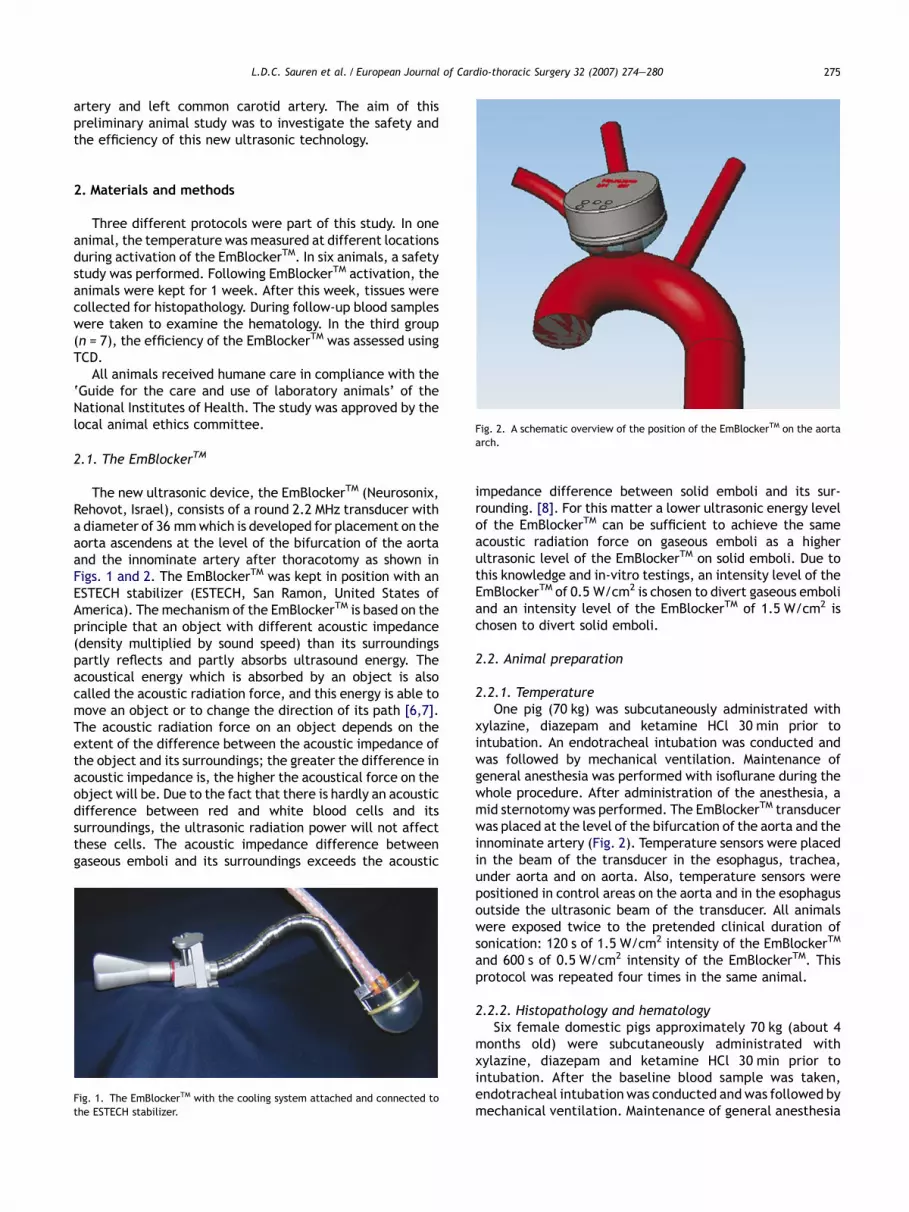
Fig. 2. A schematic overview of the position of the EmBlockerTM on the aortaarch.
2. Materials and methods
Three different protocols were part of this study. In oneanimal, the temperature was measured at different locationsduring activation of the EmBlockerTM. In six animals, a safetystudy was performed. Following EmBlockerTM activation, theanimals were kept for 1 week. After this week, tissues werecollected for histopathology. During follow-up blood sampleswere taken to examine the hematology. In the third group(n = 7), the efficiency of the EmBlockerTM was assessed usingTCD.
All animals received humane care in compliance with the‘Guide for the care and use of laboratory animals’ of theNational Institutes of Health. The study was approved by thelocal animal ethics committee.
2.1. The EmBlockerTM
The new ultrasonic device, the EmBlockerTM (Neurosonix,Rehovot, Israel), consists of a round 2.2 MHz transducer witha diameter of 36 mmwhich is developed for placement on theaorta ascendens at the level of the bifurcation of the aortaand the innominate artery after thoracotomy as shown inFigs. 1 and 2. The EmBlockerTM was kept in position with anESTECH stabilizer (ESTECH, San Ramon, United States ofAmerica). The mechanism of the EmBlockerTM is based on theprinciple that an object with different acoustic impedance(density multiplied by sound speed) than its surroundingspartly reflects and partly absorbs ultrasound energy. Theacoustical energy which is absorbed by an object is alsocalled the acoustic radiation force, and this energy is able tomove an object or to change the direction of its path [6,7].The acoustic radiation force on an object depends on theextent of the difference between the acoustic impedance ofthe object and its surroundings; the greater the difference inacoustic impedance is, the higher the acoustical force on theobject will be. Due to the fact that there is hardly an acousticdifference between red and white blood cells and itssurroundings, the ultrasonic radiation power will not affectthese cells. The acoustic impedance difference betweengaseous emboli and its surroundings exceeds the acoustic
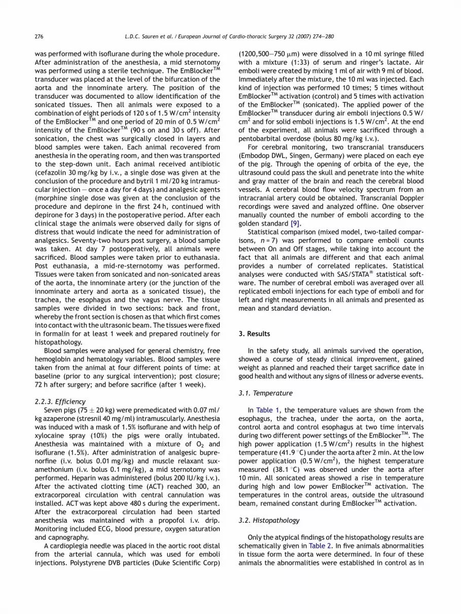
Fig. 1. The EmBlockerTM with the cooling system attached and connected tothe ESTECH stabilizer.
impedance difference between solid emboli and its sur-rounding. [8]. For this matter a lower ultrasonic energy levelof the EmBlockerTM can be sufficient to achieve the sameacoustic radiation force on gaseous emboli as a higherultrasonic level of the EmBlockerTM on solid emboli. Due tothis knowledge and in-vitro testings, an intensity level of theEmBlockerTM of 0.5 W/cm2 is chosen to divert gaseous emboliand an intensity level of the EmBlockerTM of 1.5 W/cm2 ischosen to divert solid emboli.
2.2. Animal preparation
2.2.1. TemperatureOne pig (70 kg) was subcutaneously administrated with
xylazine, diazepam and ketamine HCl 30 min prior tointubation. An endotracheal intubation was conducted andwas followed by mechanical ventilation. Maintenance ofgeneral anesthesia was performed with isoflurane during thewhole procedure. After administration of the anesthesia, amid sternotomy was performed. The EmBlockerTM transducerwas placed at the level of the bifurcation of the aorta and theinnominate artery (Fig. 2). Temperature sensors were placedin the beam of the transducer in the esophagus, trachea,under aorta and on aorta. Also, temperature sensors werepositioned in control areas on the aorta and in the esophagusoutside the ultrasonic beam of the transducer. All animalswere exposed twice to the pretended clinical duration ofsonication: 120 s of 1.5 W/cm2 intensity of the EmBlockerTM
and 600 s of 0.5 W/cm2 intensity of the EmBlockerTM. Thisprotocol was repeated four times in the same animal.
2.2.2. Histopathology and hematologySix female domestic pigs approximately 70 kg (about 4
months old) were subcutaneously administrated withxylazine, diazepam and ketamine HCl 30 min prior tointubation. After the baseline blood sample was taken,endotracheal intubation was conducted andwas followed bymechanical ventilation. Maintenance of general anesthesia
L.D.C. Sauren et al. / European Journal of Cardio-thoracic Surgery 32 (2007) 274—280276
was performed with isoflurane during the whole procedure.After administration of the anesthesia, a mid sternotomywas performed using a sterile technique. The EmBlockerTM
transducer was placed at the level of the bifurcation of theaorta and the innominate artery. The position of thetransducer was documented to allow identification of thesonicated tissues. Then all animals were exposed to acombination of eight periods of 120 s of 1.5 W/cm2 intensityof the EmBlockerTM and one period of 20 min of 0.5 W/cm2
intensity of the EmBlockerTM (90 s on and 30 s off). Aftersonication, the chest was surgically closed in layers andblood samples were taken. Each animal recovered fromanesthesia in the operating room, and then was transportedto the step-down unit. Each animal received antibiotic(cefazolin 30 mg/kg by i.v., a single dose was given at theconclusion of the procedure and bytril 1 ml/20 kg intramus-cular injection — once a day for 4 days) and analgesic agents(morphine single dose was given at the conclusion of theprocedure and depirone in the first 24 h, continued withdepirone for 3 days) in the postoperative period. After eachclinical stage the animals were observed daily for signs ofdistress that would indicate the need for administration ofanalgesics. Seventy-two hours post surgery, a blood samplewas taken. At day 7 postoperatively, all animals weresacrificed. Blood samples were taken prior to euthanasia.Post euthanasia, a mid-re-sternotomy was performed.Tissues were taken from sonicated and non-sonicated areasof the aorta, the innominate artery (or the junction of theinnominate artery and aorta as a sonicated tissue), thetrachea, the esophagus and the vagus nerve. The tissuesamples were divided in two sections: back and front,whereby the front section is chosen as that which first comesinto contactwith the ultrasonic beam. The tissueswere fixedin formalin for at least 1 week and prepared routinely forhistopathology.
Blood samples were analysed for general chemistry, freehemoglobin and hematology variables. Blood samples weretaken from the animal at four different points of time: atbaseline (prior to any surgical intervention); post closure;72 h after surgery; and before sacrifice (after 1 week).
2.2.3. EfficiencySeven pigs (75 � 20 kg) were premedicated with 0.07 ml/
kg azaperone (stresnil 40 mg/ml) intramuscularly. Anesthesiawas induced with a mask of 1.5% isoflurane and with help ofxylocaine spray (10%) the pigs were orally intubated.Anesthesia was maintained with a mixture of O2 andisoflurane (1.5%). After administration of analgesic bupre-norfine (i.v. bolus 0.01 mg/kg) and muscle relaxant sux-amethonium (i.v. bolus 0.1 mg/kg), a mid sternotomy wasperformed. Heparin was administered (bolus 200 IU/kg i.v.).After the activated clotting time (ACT) reached 300, anextracorporeal circulation with central cannulation wasinstalled. ACTwas kept above 480 s during the experiment.After the extracorporeal circulation had been startedanesthesia was maintained with a propofol i.v. drip.Monitoring included ECG, blood pressure, oxygen saturationand capnography.
A cardioplegia needle was placed in the aortic root distalfrom the arterial cannula, which was used for emboliinjections. Polystyrene DVB particles (Duke Scientific Corp)
(1200,500—750 mm) were dissolved in a 10 ml syringe filledwith a mixture (1:33) of serum and ringer’s lactate. Airemboli were created by mixing 1 ml of air with 9 ml of blood.Immediately after the mixture, the 10 ml was injected. Eachkind of injection was performed 10 times; 5 times withoutEmBlockerTM activation (control) and 5 times with activationof the EmBlockerTM (sonicated). The applied power of theEmBlockerTM transducer during air emboli injections 0.5 W/cm2 and for solid emboli injections is 1.5 W/cm2. At the endof the experiment, all animals were sacrificed through apentobarbital overdose (bolus 80 mg/kg i.v.).
For cerebral monitoring, two transcranial transducers(Embodop DWL, Singen, Germany) were placed on each eyeof the pig. Through the opening of orbita of the eye, theultrasound could pass the skull and penetrate into the whiteand gray matter of the brain and reach the cerebral bloodvessels. A cerebral blood flow velocity spectrum from anintracranial artery could be obtained. Transcranial Dopplerrecordings were saved and analyzed offline. One observermanually counted the number of emboli according to thegolden standard [9].
Statistical comparison (mixed model, two-tailed compar-isons, n = 7) was performed to compare emboli countsbetween On and Off stages, while taking into account thefact that all animals are different and that each animalprovides a number of correlated replicates. Statisticalanalyses were conducted with SAS/STATAW statistical soft-ware. The number of cerebral emboli was averaged over allreplicated emboli injections for each type of emboli and forleft and right measurements in all animals and presented asmean and standard deviation.
3. Results
In the safety study, all animals survived the operation,showed a course of steady clinical improvement, gainedweight as planned and reached their target sacrifice date ingood health and without any signs of illness or adverse events.
3.1. Temperature
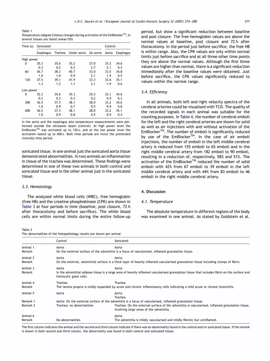
In Table 1, the temperature values are shown from theesophagus, the trachea, under the aorta, on the aorta,control aorta and control esophagus at two time intervalsduring two different power settings of the EmBlockerTM. Thehigh power application (1.5 W/cm2) results in the highesttemperature (41.9 8C) under the aorta after 2 min. At the lowpower application (0.5 W/cm2), the highest temperaturemeasured (38.1 8C) was observed under the aorta after10 min. All sonicated areas showed a rise in temperatureduring high and low power EmBlockerTM activation. Thetemperatures in the control areas, outside the ultrasoundbeam, remained constant during EmBlockerTM activation.
3.2. Histopathology
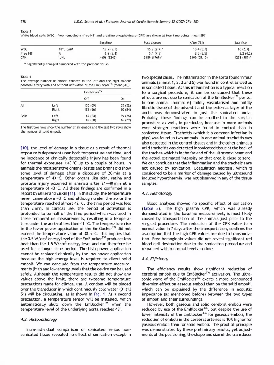
Only the atypical findings of the histopathology results areschematically given in Table 2. In five animals abnormalitiesin tissue form the aorta were determined. In four of theseanimals the abnormalities were established in control as in
L.D.C. Sauren et al. / European Journal of Cardio-thoracic Surgery 32 (2007) 274—280 277
Table 1Temperature (degree Celsius) changes during activation of the EmBlockerTM, inseveral tissues are listed (mean/SD)
Time (s) Sonicated Control
Esophagus Trachea Under aorta On aorta Aorta Esophagus
High power0 35.3 35.6 35.2 27.0 33.2 34.6
0.3 0.2 0.3 2.7 2.1 0.360 36.7 38.3 40.7 33.0 33.5 34.8
1.0 1.0 0.9 2.1 1.9 0.5120 37.5 39.1 41.9 33.3 33.6 35.1
1.3 1.2 1.1 2.1 1.9 0.6
Low power0 35.3 35.6 35.3 25.5 33.1 34.6
0.2 0.3 0.3 0.2 0.9 0.3300 36.5 37.3 38.1 28.0 33.2 34.6
1.0 0.9 0.7 0.5 0.9 0.8600 36.5 37.3 38.1 28.0 33.2 35.1
1.0 0.9 0.8 0.5 0.9 0.4
In the aorta and the esophagus also temperature measurements were per-formed outside the ultrasonic beam (control). At high power level theEmBlockerTM was activated up to 120 s, and at the low power level theactivation lasted up to 600 s. Both time periods are twice the pretendedclinically time period.
sonicated tissue. In one animal just the sonicated aorta tissuedemonstrated abnormalities. In two animals an inflammationin tissue of the trachea was determined. These findings weredetermined in one of these two animals in both control andsonicated tissue and in the other animal just in the sonicatedtissue.
3.3. Hematology
The analyzed white blood cells (WBC), free hemoglobin(free HB) and the creatine phosphokinase (CPK) are shown inTable 3 at four periods in time (baseline, post closure, 72 hafter thoracotomy and before sacrifice). The white bloodcells are within normal limits during the entire follow-up
Table 2The abnormalities of the histopathology results are shown per animal
Control Sonicated
Animal 1 Aorta AortaRemark On the external surface of the adventitia is a focus of vasc
Animal 2 Aorta AortaRemark On the external, adventitial surface is a thick layer of heav
Animal 3 Aorta AortaRemark In the adventitial adipose tissue is a large area of heavily in
histiocytic giant cells.
Animal 4 Trachea TracheaRemark The lamina propria is mildly expanded by acute and chroni
Animal 5 Aorta AortaTrachea
Remark 1 Aorta: On the external surface of the adventitia is a focusRemark 2 Trachea: no abnormalities Trachea: On the e
involving large are
Animal 6 AortaRemark No abnormalities The adventitia is m
The first column indicates the animal and the second and third column indicate if thereis shown in both second and third column, the abnormality was found in both contr
period, but show a significant reduction between baselineand post closure. The free hemoglobin values are above thenormal values at baseline, post closure and 72 h afterthoracotomy. In the period just before sacrifice, the free HBis within range. Also, the CPK values are only within normallimits just before sacrifice and at all three other time pointsthey are above the normal values. Although the first threevalues are higher than normal, there is a significant reductionimmediately after the baseline values were obtained. Justbefore sacrifice, the CPK values significantly reduced tovalues within the normal range.
3.4. Efficiency
In all animals, both left and right velocity spectra of thecerebral arteries could be visualised with TCD. The quality ofthe recorded signals in each animal was suitable for thecounting purposes. In Table 4, the number of cerebral embolifor the left and the right cerebral arteries are shown for solidas well as air injections with and without activation of theEmBlockerTM. The number of emboli is significantly reducedby use of the EmBlockerTM. In the case of air emboliinjections, the number of emboli in the left middle cerebralartery is reduced from 155 emboli to 65 emboli and in theright middle cerebral artery from 182 emboli to 90 emboli,resulting in a reduction of, respectively, 58% and 51%. Theactivation of the EmBlockerTM reduced the number of solidemboli with 42% from 67 emboli to 39 emboli in the leftmiddle cerebral artery and with 44% from 83 emboli to 46emboli in the right middle cerebral artery.
4. Discussion
4.1. Temperature
The absolute temperature in different regions of the bodywas examined in one animal. As stated by Goldstein et al.
ularised, inflamed granulation tissue.
ily inflamed vascularised granulation tissue including clumps of fibrin.
flamed vascularised granulation tissue that includes fibrin on the surface and
c inflammatory cells indicating a mild acute or chronic bronchitis.
of vascularised, inflamed granulation tissue.xternal surface of the adventitia is vascularised, inflamed granulation tissue,as of the adventitia.
ildly vascularised and mildly fibrotic but uninflamed.
was an abnormality found in the control and/or sonicated tissue. If the remarkol and sonicated tissue.
L.D.C. Sauren et al. / European Journal of Cardio-thoracic Surgery 32 (2007) 274—280278
Table 3White blood cells (WBC), free hemoglobin (free HB) and creatine phosphokinase (CPK) are shown at four time points (mean(SD))
Baseline Post closure After 72 h Sacrifice
WBC 10^3 CMM 19.7 (5.1) 15.7 (2.9)a 18.4 (3.7) 16 (2.3)Free HB % 6.9 (5.4) 5.1 (7.5) 8.5 (8.5) 3.2 (4.2)CPK IU/L 4606 (2242) 3189 (1769)a 5109 (25.10) 1228 (589) a
a Significantly changed compared with the previous value.
Table 4The average number of emboli counted in the left and the right middlecerebral artery with and without activation of the EmBlockerTM (mean(SD))
EmBlockerTM
Off On
Air Left 155 (69) 65 (52)Right 182 (96) 90 (84)
Solid Left 67 (34) 39 (26)Right 82 (38) 46 (29)
The first two rows show the number of air emboli and the last two rows showthe number of solid emboli.
[10], the level of damage in a tissue as a result of thermalexposure is dependent upon both temperature and time. Andno incidence of clinically detectable injury has been foundfor thermal exposures �43 8C up to a couple of hours. Inanimals the most sensitive organs (testes and brain) did showsome level of damage after a disposure of 20 min at atemperature of 43 8C. Other organs like skin, retina andprostate injury occurred in animals after 21—40 min at atemperature of 43 8C. All these findings are confirmed in areport by Miller and Ziskin [11]. In this study, the temperaturenever came above 43 8C and although under the aorta thetemperature reached almost 42 8C, the time period was lessthan 2 min. In clinical use, the period of activation ispretended to be half of the time period which was used inthese temperature measurements, resulting in a tempera-ture under the aorta of less then 41 8C. The temperature risein the lower power application of the EmBlockerTM did notexceed the temperature value of 38.5 8C. This implies thatthe 0.5 W/cm2 energy level of the EmBlockerTM produces lessheat than the 1.5 W/cm2 energy level and can therefore beused for a longer time period. The high power applicationcannot be replaced clinically by the low power applicationbecause the high energy level is required to divert solidemboli. We can conclude from the temperature measure-ments (high and low energy level) that the device can be usedsafely. Although the temperature results did not show anyvalues above the limit, there are twosome temperatureprecautions made for clinical use. A condom will be placedover the transducer in which continuously cold water (08 till58) will be circulating, as is shown in Fig. 1. As a secondprecaution, a temperature sensor will be installed, whichautomatically shuts down the EmBlockerTM when thetemperature level of the underlying aorta reaches 438.
4.2. Histopathology
Intra-individual comparison of sonicated versus non-sonicated tissue revealed no effect of sonication except in
two special cases. The inflammation in the aorta found in fouranimals (animal 1, 2, 3 and 5) was found in control as well asin sonicated tissue. As this inflammation is a typical reactionto a surgical procedure, it can be concluded that theseeffects are not due to sonication of the EmBlockerTM per se.In one animal (animal 6) mildly vascularised and mildlyfibrotic tissue of the adventitia of the external layer of theaorta was demonstrated in just the sonicated aorta.Probably, these findings can be ascribed to the surgicalprocedure as well, in particular, because in more animalseven stronger reactions were found in control than insonicated tissue. Tracheitis (which is a common infection inpigs) was found in two animals. In one animal tracheitis wasalso detected in the control tissues and in the other animal amild tracheitis was detected in sonicated tissue at the back ofthe trachea which is in the far end of the ultrasonic beam andthe actual estimated intensity on that area is close to zero.We can conclude that the inflammation and the tracheitis arenot caused by sonication. Coagulation necrosis, which isconsidered to be a marker of damage caused by ultrasoundinduced hyperthermia, was not observed in any of the tissuesamples.
4.3. Hematology
Blood analyses showed no specific effect of sonication(Table 3). The high plasma CPK, which was alreadydemonstrated in the baseline measurement, is most likelycaused by transportation of the animals just prior to thesurgical procedure. The reduction of the CPK value to anormal value in 7 days after the transportation, confirms theassumption that the high CPK values are due to transporta-tion. Free hemoglobin values did not reveal significant redblood cell destruction due to the sonication procedure andremained within normal levels in time.
4.4. Efficiency
The efficiency results show significant reduction ofcerebral emboli due to EmBlockerTM activation. The ultra-sonic wave of the EmBlockerTM exerts a more pronounceddiversion effect on gaseous emboli than on the solid emboli,which can be explained by the difference in acousticimpedance (as mentioned before) between the two typesof emboli and their surroundings.
However, both gaseous and solid cerebral emboli werereduced by use of the EmBlockerTM, but despite the use oflower intensity of the EmBlockerTM for gaseous emboli, thereduction of emboli in the cerebral arteries is 10% higher forgaseous emboli than for solid emboli. The proof of principlewas demonstrated by these preliminary results; yet adjust-ments of the positioning, the shape and size of the transducer

L.D.C. Sauren et al. / European Journal of Cardio-thoracic Surgery 32 (2007) 274—280 279
could improve the efficiency. The EmBlockerTM seems to bean efficient device to reduce cerebral emboli duringextracorporeal circulation and thereby could reduce post-operative neurological complications [4,5,12].
5. Clinical application
Neurological complications after cardiac surgery arewell recognised and can vary from cognitive decline, withan occurrence of 5—43%, to stroke with an occurrence of2—8% [1—5]. A correlation between cerebral emboli andneurological complications has been described in theliterature presuming that a reduction of cerebral embolicould reduce these neurological complications. Specificinterventions during coronary bypass grafting (CABG) andopen heart surgery were identified to generate most of thecerebral emboli; cannulation, cardioplegia needle incision,start and stop CPB, cross-clamping, cross-clamp release,side-clamping, side-clamp release, de-airing and de-cannulation [2]. According to the findings of this presentstudy, the EmBlockerTM is able to divert solid as well asgaseous emboli to the descending aorta and reduce thenumber of emboli in the cerebral vessels. The EmBlockerTM
should be placed after thoracotomy on the aorta ascendensabove the bifurcation of the aorta and the innominateartery (Fig. 2). One minute of activation of the EmBlock-erTM during the interventions mentioned before should besufficient to divert the generated emboli. Because allinterventions (except de-airing) are associated with solidemboli the higher intensity (1.5 W/cm2) of the EmBlock-erTM is recommended. In open-heart surgery, there is anextra procedure in which cerebral gaseous emboli areexpected. This time period is from the moment the heartstarts ejection and can continue for approximately 5—10 min. Due to the fact that the emboli in this period aremainly gaseous emboli the lower intensity level for 5 min isrecommended. In this present study, the clinical activationtime was examined twice as a result of which it can beconcluded that the recommended clinical activation timescould be used safely.
Although this animal study already demonstrates adiversion effect, a possible change in intensities or designof the EmBlockerTM could improve the efficiency.
6. Limitations
The high standard deviation of the number of emboli in thecontrol injections, as shown in Table 4, could be due to thehigh standard deviation of the weight of the animals. Theanatomy difference, particularly in innominate and in rightcommon carotid artery, could cause a difference in absoluteflow to the cerebral vessels. No flow measurements weredone to confirm this hypothesis.
In this preliminary study a surrogate marker (number ofemboli) was used to examine the efficiency of the EmBlock-erTM, no brain MRI or brain pathology has been performed toexamine the cerebral damage of the emboli (gas and solid)injections.
7. Summary
Temperature, hematological and histopathological resultsshow no effect although the clinically pretended period ofactivation of the EmBlockerTM was used twice. Efficiencymeasurements show a significant decrease in cerebral emboliduring emboli injections during extracorporeal circulation.Use of the EmBlockerTM in cardiac surgery bears the potentialto lower the risk of postoperative neurological complica-tions.
Acknowledgments
The authors want to express their gratitude to Mr T. vander Nagel and Ms M. de Jong for their technical support andeffort.
References
[1] Bucerius J, Gummert JF, Borger MA, Walther T, Doll N, Onnasch JF, Metz S,Falk V, Mohr FW. Stroke after cardiac surgery: a risk factor analysis of16,184 consecutive adult patients. Ann Thorac Surg 2003;75(2):472—8.
[2] Fearn SJ, Pole R, Wesnes K, Faragher EB, Hooper TL, McCollum CN.Cerebral injury during cardiopulmonary bypass: emboli impair memory.J Thorac Cardiovasc Surg 2001;121(6):1150—60.
[3] Naylor AR, Mehta Z, Rothwell PM, Bell PR. Carotid artery disease andstroke during coronary artery bypass: a critical review of the literature.Eur J Vasc Endovasc Surg 2002;23(4):283—94.
[4] Nollert G, Mohnle P, Tassani-Prell P, Uttner I, Borasio GD, Schmoeckel M,Reichart B. Postoperative neuropsychological dysfunction and cerebraloxygenation during cardiac surgery. Thorac Cardiovasc Surg 1995;43(5):260—4.
[5] Pugsley W, Klinger L, Paschalis C, Treasure T, Harrison M, Newman S. Theimpact of microemboli during cardiopulmonary bypass on neuropsycho-logical functioning. Stroke 1994;25(7):1393—9.
[6] Palanchon P, Tortoli P, Bouakaz A, Versluis M, de Jong N. Optical observa-tions of acoustical radiation force effects on individual air bubbles. IEEETrans Ultrason Ferroelectr Freq Control 2005;52(1):104—10.
[7] Michishita K, Hasegawa H, Kanai H. Ultrasonic measurement of minutedisplacement of object cyclically actuated by acoustic radiation force.Jpn Soc Appl Phy 2003;42:4608—12.
[8] Markus H. Transcranial Doppler detection of circulating cerebral emboli.A review. Stroke 1993;24(8):1246—50.
[9] Van Zuilen EV, Mess WH, Jansen C, Van der Tweel I, Van Gijn J, AckerstaffGA. Automatic embolus detection compared with human experts. ADoppler ultrasound study. Stroke 1996;27(10):1840—3.
[10] Goldstein LS, Dewhirst MW, Repacholi M, Kheifets L. Summary, conclu-sions and recommendations: adverse temperature levels in the humanbody. Int J Hyperthermia 2003;19(3):373—84.
[11] Miller MW, Ziskin MC. Biological consequences of hyperthermia. Ultra-sound Med Biol 1989;15(8):707—22.
[12] Stump DA, Kon NA, Rogers AT, Hammon JW. Emboli and neuropsycholo-gical outcome following cardiopulmonary bypass. Echocardiography1996;13(5):555—8.
Appendix A. Conference discussion
Dr U. Lockowandt (Stockholm, Sweden): It’s quite fascinating to moveobjects with an ultrasonic beam.
Do you think it will influence the thrombocytes, lymphocytes orerythrocytes?
Dr Sauren: No. It was also tested. Because this mechanism only works whenthere is a difference in acoustic impedance and the difference between bloodand erythrocytes or any other particles, it’s not that much, it won’t be able tomove that particles. That’s also why air is much better to divert because theacoustic impedance is quite large.
L.D.C. Sauren et al. / European Journal of Cardio-thoracic Surgery 32 (2007) 274—280280
Dr P. Grundeman (Utrecht, The Netherlands): I think it’s a great idea to getthe air down in the descending aorta. Suppose there is debris in the ascendingaorta that’s attached to the inner wall, for instance, calcification, can that(material) be dislodged by this technology?
Dr Sauren: That is, of course, an interesting question, but it’s somethingwe still need to take a look at. Of course, it’s different material then particlesin the blood circulation, I mean, it’s attached to the aorta, if you’re talkingabout calcification. but it’s something we still need to take a look at for sure.
Dr Van Der Linden (Stockholm, Sweden): I have a question regarding thesize of the particles you can redirect. Howmuch power do you need to redirectbig particles as opposed to microemboli, very small particles?
Dr Sauren: Well, this is something we are looking at in the in vitro setupactually. We want to use different kind of sizes. And this animal study, quite bigparticles, were used between 500 and 750 m. And also we want to have a lookat which power should be used for each different size. But I cannot say anythingabout it now.
Dr Van Der Linden: Did you use transcranial Doppler in these pigsinsonating the middle cerebral artery or did you insonate the carotid artery?
Dr Sauren: No, I placed the probes on the eyes, because it was possible toget the cerebral arteries through the eyes.
Dr Van Der Linden: So you were looking at retinal emboli?
Dr Sauren: No, no. Because of the eye there is no bone at the spot wherethe nerves are going into the brain. There is a spot where there no bone is so Ican get through with the ultrasound.
Dr Van Der Linden: Because the anatomy of the cerebral circulation ofthe pig is a bit difficult and it’s difficult to insonate the middle cerebralartery.
Dr Sauren: I’m aware that we are not completely sure that we havethe middle cerebral artery and that is also why these results could bethe results are not that perfect. Maybe the results could be betterbecause of this, but we didn’t see them. So we still need to take a look at itfurther.
Dr Van Der Linden: And which Doppler technique did you use? Did you usethe EmboDop?
Dr Sauren: Yes, it was the EmboDop.


