Embed Size (px)
Citation preview
Biochemical Revision of Penicilliumhordei
El-meleigy Magda A.1, M.M. Mokhtar2 and Helmy Eman A.2
1-Botany and Microbiology Department, Faculty of Sciences (Girls), Al-AzharUniversity, Cairo, Egypt.
2-The Regional Center for Mycology and Biotechnology (RCMB), Al-AzharUniversity, Cairo, Egypt.
ABSTRACTEight type strains belonging to Penicillium hordei were
classified from the analysis of their crude extracts by a
polyphasic approach with data processing, using the profiles
of their fatty acid; secondary and volatile metabolites, as
well as random amplified polymorphic DNA-polymerase chain
reaction pattern (RAPD-PCR) as taxonomic markers for these
strains. The study showed that with the harmony of all the
four investigated markers, about all of the investigated type
strains could be classified correctly at the intraspecific
level using only the analysis of metabolites produced on one
growth medium (YES), except in case of the volatile profile
which succeeded as a cladogenetic profile but not as a strain
marker. The study revealed also the ability of RAPD-PCR
technique to evaluate the genetic diversity among the
investigated isolates at the sub-species level, as well as a
rapid and easy method than traditional characterization
techniques. Other relations between type strains could be read
from the dendrograms and the efficient classification showed
the potential of this polyphasic approach identification
system.
1
Keywords: Volatile metabolites; Fatty acids; Secondary metabolites; RAPD-
PCR; Penicillium hordei, Chemotaxonomy.
Introduction
Fungal taxonomy is a dynamic, progressive discipline that
consequently requires changes in nomenclature. These changes
are often difficult for microbiologists to understand.
However, some groups of fungi, because of their economical or
pathological importance, have been studied more extensively.
Other features beside morphology, such as the use of molecular
techniques, physiological and biochemical tests, have been used
in classification and also in identification. The increased use
and availability of modern techniques have opened up many new
areas within systematics and have enabled more traditional ones
to be developed further (Josepa and Alberto, 1999).
Identification of Penicillium species is still never to be
easy, so that, (Pitt, 1991) had more taxonomic handless to
help Penicillium classification. Penicillium hordei was found to be one
of the thirteen different allergens causative agents
discovered in the study of ( Graham et al 1995) in Brazil
evaluating 1,410 patients with asthma and/or rhinitis. This
species produces several odor and volatile metabolites such as
isobutanol, isopentanol (Larsen and Frisvad, 1995) as well as
some extrolites and mycotoxins such as terresteric acid,
carolic acid, carlosic acid and roquefortineC (Samson and
Frisvad, 2004).
Promising results regarding the use of fatty acids (FA)
for identification of filamentous fungi had been reported by
2
many studies including (Losel, 1989; Augustyn, 1992; Blomquist
et al 1992) and which reported that with the aid of FA profile, it
was possible to differentiate between various Aspergillus, Mucor
and Penicillium species. Unfortunately, only a limited number of
strains and species have been included in most of these
studies, which makes the evaluation of this method as an
indication parameter difficult.
Chemotaxonomic studies of large number of isolates in
Penicillium have shown, however, that secondary metabolites (SMs)
have a potential for the characterization of its species and
for phylogenetic relationships. Thus, it complements
morphological data to give a fuller description of an important
part of the phenotype that may be perceived by other organisms.
Chemical analysis of secondary metabolites will provide more
objective and comparable results than traditional description
of color and odor (Smedsgaard and Nielsen, 2005).
Fungi are known to biosynthesize a variety of metabolic
products, including volatile metabolites (VMs) which can be
products of both primary and secondary metabolism. Despite the
advance in technology by the development of the modern
analytical techniques such as (GC, GLC and GC/MS), only a few
reports on the fungal chemotaxonomic studies based on the
production of (VMs) were found (Frisvad et al 2008).
Some studies have evaluated a large number of primers to
identify only a selected few isolates that can successfully
discriminate genetic strain types. (Kac et al 1999) evaluated
fifteen RAPD primers identifying merely one that was highly
discriminatory for strains of Trichophyton mentagrophytes. So, RAPD
technique was evaluated as a reliable tool with good
3
reproducibility of the patterns for each investigated strain as
affirmed by (Stemmler et al 2001). But problems of interpretation
due to inconsistent intensity of bands in different PCR runs
may arise for less experienced personnel. RAPD analysis can be
performed within one working day and needs less DNA compared
with RFLP, so, costs will be reduced.
Most of the known established techniques and designing
options of fungal taxonomy have been validated for only a few
dozen of fungal strains and the lack of efficient genetic
engineering strategic forms still an obstacle for a multitude
of identifying fungi producing commercially interesting
metabolites. To fully explore their biotechnological
capacities, these constraints have to be solved (Vera, 2008).
Materials and methodsFungal strains: all of the tested Penicillium hordei type strains
were purchased from the International Mycological Institute
(IMI) culture collections and coded here as; (30) P.hordei (IMI
246204), (31) P.hordei (IMI 286971), (32) P.hordei (IMI 297900), (33)
P.hordei (IMI 284723), (34) P.hordei (IMI 264173), (35) P.hordei (IMI
223651), (36) P.hordei (IMI 151748) and (37) P. hordei (IMI 040213).
Media: two types of media were used; Malt Extract Agar (MEA)
medium was used for maintenance of the strains according to
(Smith and Onions, 1983). The other medium type was Yeast
Extract Sucrose (YES). This semi-synthetic medium was used in
liquid form for the production of intracellular fatty acids
from the cultivated strains (Peter and Michael, 1996),
intracellular secondary metabolites (Frisvad and Samson, 2004)
and intracellular volatile metabolites (Larsen and Frisvad,
1995b; Kristian and Thomas, 2005) as well as for the DNA study
4
(Zhou and Linz, 1999). Media were sterilized by autoclaving
at 121οC for 20 min. Mycelia growth from 7 days old cultures on
MEA slopes were scraped by using 2 ml of sterile distilled
water. Then, 2.0 ml of 4x10² cells/ml spore suspension of each
type strain were used to inoculate a 100 ml YES medium in a
universal 250 ml flask, and then incubated at 25°C for 7 days,
except in case of the DNA analysis that all flasks were
incubated with a gentle shaking at 180 rpm. at 25οC for 2 days.
Fungal mycelia and pellets were harvested by filtration under
aseptic conditions using microcloth and washed thoroughly with
sterile distilled water then weighed, decanted in sterile
containers and stored at -4°C for further analysis. While for
complete DNA analysis, fungal pellets were lyophilized using a
freeze dryer system (Heto lyophilizer model Maxi Dry plus). The
lyophilized pellets were grounded in a sterile cold mortar
using sterile pestle and decanted in a sterile 1.5 ml microfuge
tube.
It is worthy to mention that all of the experimental work
throughout this research was carried out at the Regional Center
for Mycology and Biotechnology (RCMB), at Al-Azhar University
except for the fatty acids analysis which was achieved at the
Central Lab of the Ain Shams University.
Volatile Metabolite Analysis: Intracellular volatile
metabolites were extracted from fungal mycelia according to
(Evans, 2002) then analyzed using Shimadzu QP 5050A GC /MS
supported with a Class 5000 software and Whiley mass spectral
data base searchable library.
Fatty Acid Analysis: Intracellular fatty acids were extracted
according to (Peter and Michael, 1996). Gas chromatographic
5
analysis was achieved using Dani GLC-FID 1000. For the complete
identification of the resulted compounds, a fatty acids
standard was used. This standard was manufactured by Supelcotm,
containing mixture of 37 fatty acids methyl ester (C4- C24)
dissolved in methylene chloride.
Secondary Metabolite Analysis: Extraction, analysis and
identification of intracellular secondary metabolites were
carried out using the TLC plate technique of the automatic
scanner system (HPTLC Scanner 3 -CAMAG, Switzerland) using
griseofulvin as reference standard. The identity of the
metabolites was performed by comparing shape, color and Rf values
of the recorded spots with those given at (Paterson and Bridge,
1994).
Fungal DNA Extraction: DNA extraction was conducted using
DNeasy kit (Qiagen, Germany).
(RAPD-PCR): Amplification reaction mixture solution was
prepared in a final volume of 50 µl containing: 3 µl (200 ng)
of genomic DNA; 1 μl of 50 pmole of each desired primer; 25 µl
of the Go Taq Green Master Mixture (Promega Co.) and deionized
RNase-DNase free water in sufficient amount to give the total
reaction mixture volume of 50 µl. The amplification was
performed using Research Programmable Thermal Cycler (gradient
Robocycler 96 Stratagene, USA) where the applied program was as
follows: universal denaturation cycle (5 min. at 94°C), 45
cycles of annealing/extension reactions (30 sec. at 94°C, 1
min. at an optimum annealing temperature 36°C for each used
universal primer and 2 min. at 72°C) and cycle of final
extension step (5 min. at 72°C) was followed by soaking at
4°C.The sequence of six oligonucleotide universal primers used
6
in the current search were: primer 1: (5'-GGTGCGGGAA-3'),
primer 2: (5'-GTTTCGCTCC-3'), primer 3: (5'-GTAGACCCGT-3'),
primer 4: (5'-AAGAGCCCGT-3'), primer 5: (5'-AACGCGCAAC-3') and
primer 6: (5'-CCCGTCAGCA-3'). These applied primers were of
HPSF grade and obtained from the MGW Biotech. Ag. Co.
Horizontal Gel Electrophoresis: The amplified products were
separated by agarose gel electrophoresis using a horizontal
submarine gel system (Agagel Maxi, Biometra) as well as agarose
(Gibco BRL Life Technologies) at a concentration of 2% (w/v).
Electrophoresis was conducted in 0.5× TBE buffer (5.4 g of Tris
base, 2.75 g of Boric acid, and 2 ml of 0.5 M EDTA [pH 8.0] in
1 liter of distilled water) at 10 v/cm for various times,
depending on the size of the gel unit (Weising et al 1995).
DNA bands were stained with ethidium bromide (10 mg/ml) then
visualized and photographed under a UV Transilluminator system
using a Gel Doc. 2000 (Bio-RAD).
Statistical Cluster Analysis of the Phylogenetic Relationships:
The role of the RAPD-PCR patterns as well as the fatty acid,
volatile metabolite and secondary metabolite profiles as useful
criteria for studying phylogenetic relationships among the
investigated penicillia strains was evaluated by using
statistical cluster analysis with joining (tree clustering)
being the clustering method. Genetic relationships and
divergence between RAPD –PCR patterns of the investigated
strains were calculated from the decimal coefficient using the
Quantity One (4.0.3) software and were illustrated in
dendrograms constructed using the unweighed pair-group method
with arithmetic averages (UPGMA). While, each of fatty acid,
volatile metabolite and secondary metabolite profiles were
7
amalgamated by a complete linkage using the Elucidation
distance as the distance metric as well as the dice coefficient
as the calculation method using the Statistica software for
Windows release (4.5 F, State Soft. Inc. 1993).
ResultsVolatile Metabolite Profile: The available results from the
volatile metabolite (VM) profile of the investigated P.hordei type
strains (eight type strains), revealed twelve distinct types of
volatile compounds, which studied effectively to clear up a
specific dilemma tied to the morphological grouping of the
examined strains, (Table 1). From the pointed out results, no
single volatile compound could be used as a chemotaxonomic
marker due to their widely distribution among the tested
strains. However, the VM profile as a whole succeeded in
differentiation between the tested strains into two groups. On
the other hand, the aforementioned illustration of the VM
profile gives the impression to be perplexing. For that reason,
the cluster analysis tree dendrogram (Figure1), revealed the
grouping of the investigated strains into two clades based on
their volatile metabolite content. The first clade grouped
P.hordei numbers 33, 34, 32 and 30 together at a linkage distance
ranged between zero and 1.0, through which, each of P.hordei
numbers 33, 34 and 32 were linked together at a linkage
distance of zero, which looked as if they are the same
subspecies. While, P.hordei number 30 appeared to be more likely
the same subspecies also, since it connected to the rest
strains of this group at a linkage distance of 1.0, which is so
close to the similarity matrix value of the rest strains across
this clade (zero). Similarly, the second clade included P.hordei
8
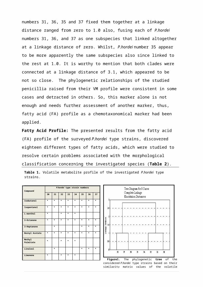
numbers 31, 36, 35 and 37 fixed them together at a linkage
distance ranged from zero to 1.0 also, fusing each of P.hordei
numbers 31, 36, and 37 as one subspecies that linked altogether
at a linkage distance of zero. Whilst, P.hordei number 35 appear
to be more apparently the same subspecies also since linked to
the rest at 1.0. It is worthy to mention that both clades were
connected at a linkage distance of 3.1, which appeared to be
not so close. The phylogenetic relationships of the studied
penicillia raised from their VM profile were consistent in some
cases and detracted in others. So, this marker alone is not
enough and needs further assessment of another marker, thus,
fatty acid (FA) profile as a chemotaxonomical marker had been
applied.
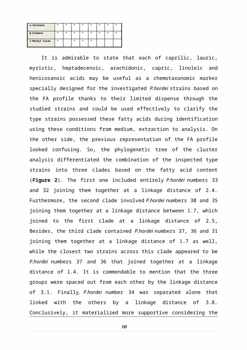
Fatty Acid Profile: The presented results from the fatty acid
(FA) profile of the surveyed P.hordei type strains, discovered
eighteen different types of fatty acids, which were studied to
resolve certain problems associated with the morphological
classification concerning the investigated species (Table 2). Table 1. Volatile metabolite profile of the investigated P.hordei type strains.
CompoundP.hordei type strain numbers
30 31 32 33 34 35 36 37
Isobutanol * * * * * * * *
Isopentanol * * * * * * * *
L-menthol * * * *
3-Octanone * * * * * * * *
3-Heptanone * * * *
Bornyl Acetate * * * * * * * *
Methyl Palmitate * * * *
Linalool * * * *
Limonene * * * *
9 Figure1. The phylogenetic tree of theconsidered P.hordei type strains based on theirsimilarity matrix values of the volatile
α-Selinene * * * * * * * *
β-Elemene * * * * * * * *
3-Methyl Furan * * * *
It is admirable to state that each of caprilic, lauric,
myristic, heptadecenoic, arachidonic, capric, linoleic and
henicosanoic acids may be useful as a chemotaxonomic marker
specially designed for the investigated P.hordei strains based on
the FA profile thanks to their limited dispense through the
studied strains and could be used effectively to clarify the
type strains possessed these fatty acids during identification
using these conditions from medium, extraction to analysis. On
the other side, the previous representation of the FA profile
looked confusing. So, the phylogenetic tree of the cluster
analysis differentiated the combination of the inspected type
strains into three clades based on the fatty acid content
(Figure 2). The first one included entirely P.hordei numbers 33
and 32 joining them together at a linkage distance of 2.4.
Furthermore, the second clade involved P.hordei numbers 30 and 35
joining them together at a linkage distance between 1.7, which
joined to the first clade at a linkage distance of 2.5,
Besides, the third clade contained P.hordei numbers 37, 36 and 31
joining them together at a linkage distance of 1.7 as well,
while the closest two strains across this clade appeared to be
P.hordei numbers 37 and 36 that joined together at a linkage
distance of 1.4. It is commendable to mention that the three
groups were spaced out from each other by the linkage distance
of 3.1. Finally, P.hordei number 34 was separated alone that
linked with the others by a linkage distance of 3.8.
Conclusively, it materialized more supportive considering the
10
use of fatty acid outline in penicillia chemotaxonomy, since
this marker confirmed the former results obtained from the VM
marker, however, P.hordei number 35 appeared to be located at
different clades throughout both
profiles as illustrated before.
Table 2. Fatty acid profile of the investigated P.hordei type strains.
So, the FA profile provided us
a valuable chemo-categorization
indicator for the studied strains.
But it wouldn't be possible to
substantiate such relationships
without a supplementary assessment
using other marker, such as the application of the secondary
metabolite (SM) profile.
Secondary Metabolite Profile: The readily available results
from the secondary metabolite (SM) profile of the considered
P.hordei type strains, showed out twenty one discrete types of
11
Fatty Acid
P.hordei type strain numbers
30
31
32
33
34
35
36
37
Butyric * * * * * * * *
Caproic * * * * * * *
Caprilic * * *
Capric * *
Lauric * * *
Myristic * * *
Pentadecanoic * * * * *
Palmitic * * * * *
Palmitoleic * * * * * * * *
Margaric * * * * * *
Heptadecenoic * * *
Stearic * * * * * * * *
oleic * * * * * * * *
Linoleic *
Linolelaidic * * * *
γ-Linolenic * * * * * *
Arachidonic * * *
Henicosanoic *
Figure.2. The phylogenetic tree of the considered P.hordei type strains based on their similarity matrix values of the fatty acid profile.
secondary metabolites, which inspected to resolve certain
crisis associated with the morphological establishments of the
surveyed isolates of this species, (Table 3 and Figure 3). It
is remarkable to declare that due to their restricted
distribution throughout the studied P.hordei strains, all of
(-)flavoskyrin, cinnamic acid, dehydrocarolic acid,
cyclopenin, viridicatin and terresteric acid could be
advantageously accepted as chemotaxonomical markers as well
for those species.
Fortunately, four unidentified compounds with different Rf
values of 11, 38, 46 and 54 that regained from the tested type
strains were might be considered to be good taxonomical
markers also, owing to their minor and restricted spreading
throughout the tested strains that ranged from one to two
strains at maximum for all and need further categorization
later on. Alternatively, the prior illustration of the SM
profile seemed to be puzzling. Hence, the cluster analysis
tree (Figure 4), revealed the federation of the considered
strains into three clades based on their SM content.
The first one included P.hordei numbers 33 and 32 joining
them together at a linkage distance of 2.4. While, the second
clade fusing P.hordei numbers 30 and 35 together at a linkage
distance of 1.7, then joined them closely to the first clade
at linkage distance of 2.5. Besides, the third clade contained
P.hordei numbers 37, 36 and 31, joining them together at a
linkage distance between 1.7 and 1.46, in which the closest
two strains were P.hordei numbers 36 and 37 that joined together
at a linkage distance of 1.4. It is commendable to mention
that the three groups were joined together at a linkage
12
distance of 3.1 which considered apart from each other. Only
P.hordei number 34 was discarded alone from the rest groups,
indicating the uniqueness of it as a separate subspecies that
a parted from the rest clades by a linkage distance of 3.8.
Conclusively, it looked more supportive considering the use
of secondary metabolite profiling in P.hordei chemotaxonomy,
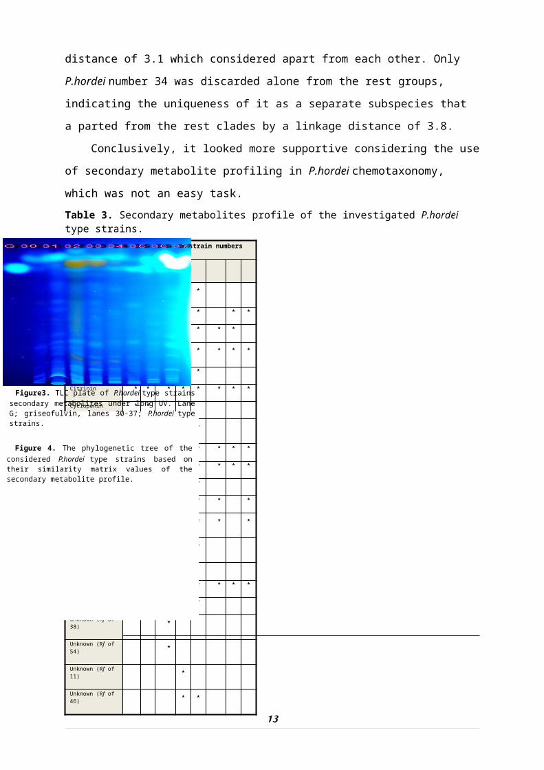
which was not an easy task. Table 3. Secondary metabolites profile of the investigated P.hordei type strains.
13
Compound Name
P.hordei type strain numbers
32 35
Acetyl Carbinol * * * *
Carlosic Acid * * * * * *
Carolic Acid * * * * * * *
ChaetoglobosinC * * * * * * * *
Cinnamic Acid * * *
Citrinin * * * * * * * *
Cyclopenin * *
DehydrocarolicAcid * * *
Fulvic Acid * * * * * * * *
Hevalonic Acid * * * * * *
Patulin * * * * *
Physodic Acid * * * * * * *
Roquefortine B * * * * *
Terresteric Acid * *
Viridicatin * * *
Xanthocillin * * * * * * *
(-)Flavoskyrin * * *
Unknown (Rf of 38) *
Unknown (Rf of 54) *
Unknown (Rf of 11) *
Unknown (Rf of 46) * *
Figure 4. The phylogenetic tree of the considered P.hordei type strains based on their similarity matrix values of the secondary metabolite profile.
Figure3. TLC plate of P.hordei type strains secondary metabolites under long UV. Lane G; griseofulvin, lanes 30-37; P.hordei type strains.
Since, the results of secondary metabolite marker confirmed
the results gained by both of volatile and fatty acid markers
concerning the grouping of each of P.hordei numbers 37, 36 and 31
together as well as P.hordei numbers 32 and 33 in their own
groups, but not supporting them in the rest of clades
speciation.
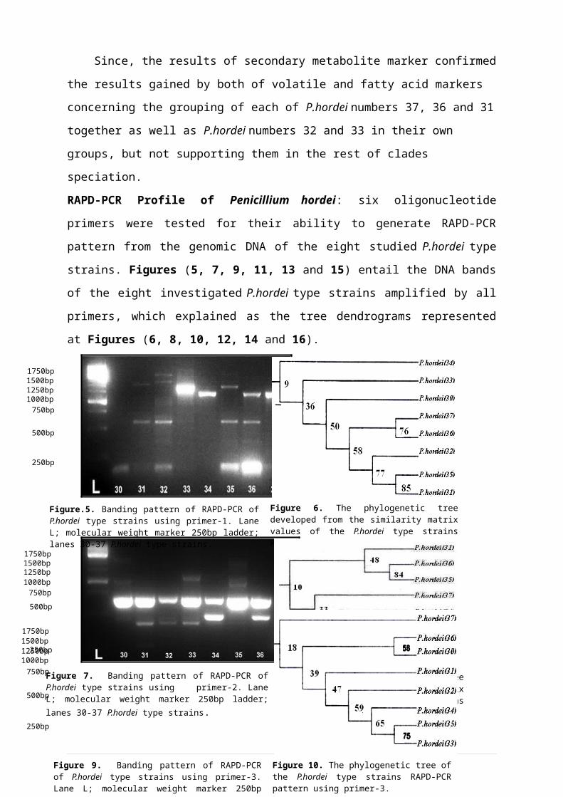
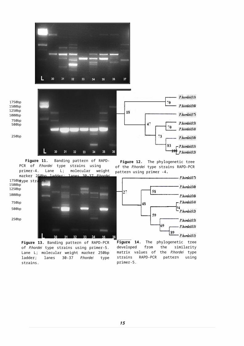
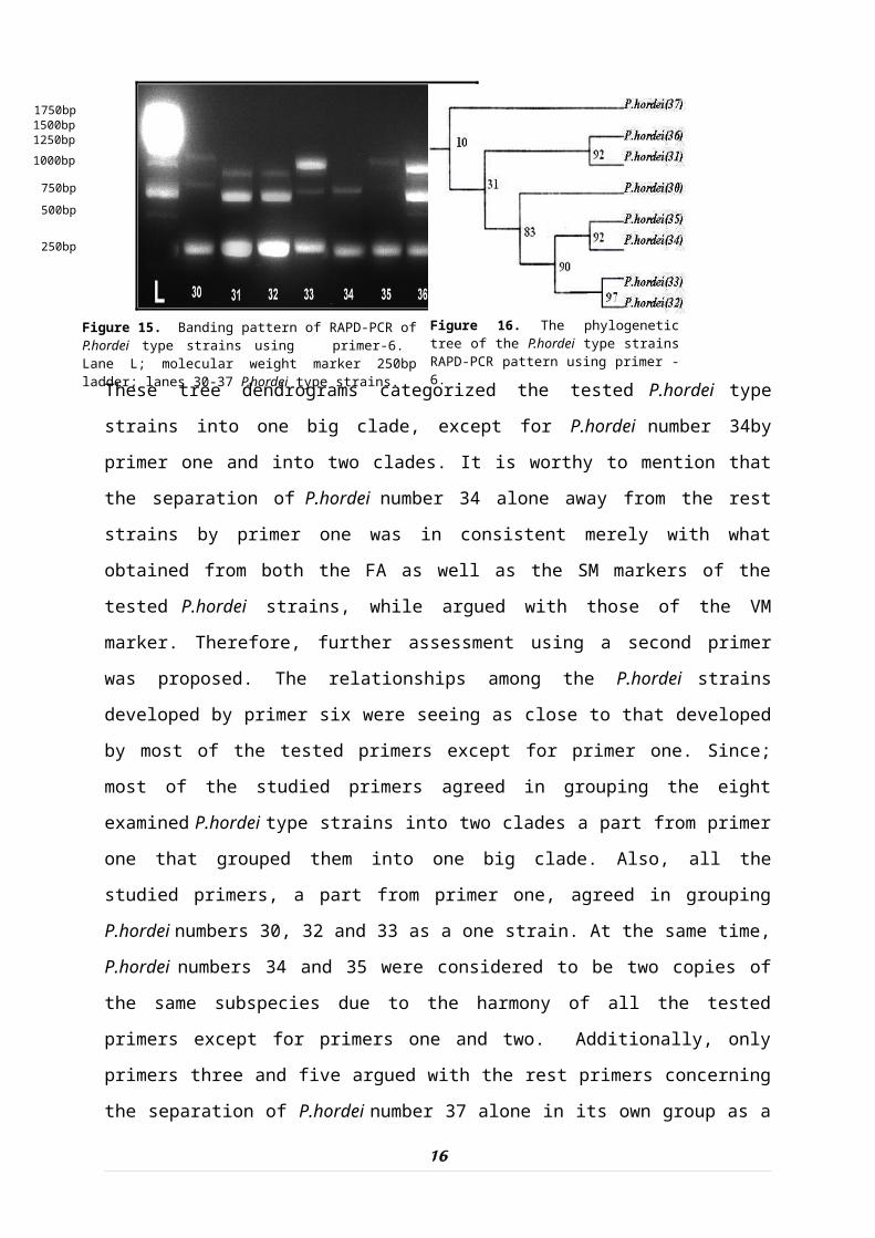
RAPD-PCR Profile of Penicillium hordei: six oligonucleotide
primers were tested for their ability to generate RAPD-PCR
pattern from the genomic DNA of the eight studied P.hordei type
strains. Figures (5, 7, 9, 11, 13 and 15) entail the DNA bands
of the eight investigated P.hordei type strains amplified by all
primers, which explained as the tree dendrograms represented
at Figures (6, 8, 10, 12, 14 and 16).
14
250bp
500bp
750bp1000bp1250bp1500bp1750bp
Figure.5. Banding pattern of RAPD-PCR of P.hordei type strains using primer-1. Lane L; molecular weight marker 250bp ladder; lanes 30-37 P.hordei type strains.
Figure 6. The phylogenetic tree developed from the similarity matrix values of the P.hordei type strains RAPD-PCR pattern using primer-1.
250bp
500bp750bp
1000bp1250bp1500bp1750bp
Figure 7. Banding pattern of RAPD-PCR of P.hordei type strains using primer-2. Lane L; molecular weight marker 250bp ladder; lanes 30-37 P.hordei type strains.
Figure 8. The phylogenetic tree developed from the similarity matrix values of the P.hordei type strains RAPD-PCR pattern using primer-2.
250bp
500bp
750bp1000bp1250bp1500bp1750bp
Figure 9. Banding pattern of RAPD-PCR of P.hordei type strains using primer-3. Lane L; molecular weight marker 250bp ladder;lanes 30-37 P.hordei type strains.
Figure 10. The phylogenetic tree of the P.hordei type strains RAPD-PCR pattern using primer-3.
15
250bp
500bp750bp
1000bp1250bp1500bp1750bp
Figure 11. Banding pattern of RAPD-PCR of P.hordei type strains using primer-4. Lane L; molecular weight marker 250bp ladder; lanes 30-37 P.hordei type strains.
Figure 12. The phylogenetic tree of the P.hordei type strains RAPD-PCR pattern using primer -4.
Figure 13. Banding pattern of RAPD-PCR of P.hordei type strains using primer-5. Lane L; molecular weight marker 250bp ladder; lanes 30-37 P.hordei type strains.
Figure 14. The phylogenetic tree developed from the similarity matrix values of the P.hordei type strains RAPD-PCR pattern using primer-5.
250bp
500bp750bp
1000bp1250bp1500bp1750bp
These tree dendrograms categorized the tested P.hordei type
strains into one big clade, except for P.hordei number 34by
primer one and into two clades. It is worthy to mention that
the separation of P.hordei number 34 alone away from the rest
strains by primer one was in consistent merely with what
obtained from both the FA as well as the SM markers of the
tested P.hordei strains, while argued with those of the VM
marker. Therefore, further assessment using a second primer
was proposed. The relationships among the P.hordei strains
developed by primer six were seeing as close to that developed
by most of the tested primers except for primer one. Since;
most of the studied primers agreed in grouping the eight
examined P.hordei type strains into two clades a part from primer
one that grouped them into one big clade. Also, all the
studied primers, a part from primer one, agreed in grouping
P.hordei numbers 30, 32 and 33 as a one strain. At the same time,
P.hordei numbers 34 and 35 were considered to be two copies of
the same subspecies due to the harmony of all the tested
primers except for primers one and two. Additionally, only
primers three and five argued with the rest primers concerning
the separation of P.hordei number 37 alone in its own group as a
16
250bp
500bp750bp
1000bp1250bp1500bp1750bp
Figure 15. Banding pattern of RAPD-PCR of P.hordei type strains using primer-6. Lane L; molecular weight marker 250bp ladder; lanes 30-37 P.hordei type strains.
Figure 16. The phylogenetic tree of the P.hordei type strains RAPD-PCR pattern using primer -6.
matchless strain. Finally, both of P.hordei numbers 31 and 36
were the reminder strains of great argument throughout the
studied primers, being in consistent with some and detracted
in others.
Conclusively, four markers in total; namely; VM, FA, SM
profiles, as well as six oligonucleotide primers of the RAPD-
PCR pattern, agreed in classifying the eight studied P.hordei
type strains into three major groups. The first group
separated P.hordei numbers 30, 32 and 33 being that these strains
were the same subspecies of this Penicillium species, according
to the harmony of all the tested markers except for primer
three as well as both of the FA and SM profiles. It is worthy
to state that all of the examined markers agreed in gathering
both P.hordei numbers 32 and 33 together as an identical versions
of one subspecies.
Concurrently, at the second group, either of P.hordei numbers
34 and 35 were said to be alike subspecies as a results of the
harmony of all markers apart from primers two and one as well
as the VM profile. The third group gathering P.hordei numbers 31,
36 and 37 as one strain according to the synchronization of
all studied markers despite primers three, five and six. It
is precious to point out that all the studied markers, except
for primers three and five, harmonized in separating P.hordei
numbers 31 and 36 as a duplicate of one strain. Thus it is
promising to handle the examined four markers exclusively or
with each other to categorize the investigated P.hordei ensuring
the value of these markers as exceptional techniques in the
intraspecific grouping of these penicillia.
Discussion
17
In recent years, there has been substantial progress in
the development of innovative methods to analyze fungi (and
other organisms) at the molecular level as well as at
biochemical level (Jianping, 2008).
Referring to the pointed out results of the VM profile as
a chemotaxonomic marker for the eight studied P.hordei type
strains of the present work, since, no specific volatile
compound was unique to a specific isolate of the studied
P.hordei strains. These fallouts could be consistent with
(Borjesson, 1993), who concluded that VM couldn’t be used in
the classification of penicillia if few isolates of the same
species investigated since significant differences could be
observed between isolates of the same species. As well, a
major reason why some literatures reports question the use of
fungal metabolites in taxonomy is probably the use of only one
or very few isolates of the species studied. Furthermore, some
of these few isolates might be incorrectly identified (Frisvad
and Samson, 2004). These results might be concurrent with the
theory that “if some of the few isolates used, it was found to
be misidentified, then false taxonomic conclusions might be
made” stated by (Lund and Frisvad, 1994 and 1995).
However, the volatile profile might be useful to
differentiate among these strains merely at the clade (group)
level. The succession of the VM profile of summation of some
isolates of the investigated strains as one species was noted.
It materialized more supportive considering the use of
fatty acid (FA) outline in the chemotaxonomy of the studied
eight type strains of P.hordei. These results were harmonized
with the promising results regarding the use of FAs for the
18
identification of filamentous fungi that have been reported
for several years by many authors including (Losel, 1989;
Augustyn, 1992; Blomquist et al 1992) they declared that “with
aid of FA profile, it was possible to differentiate between
various Aspergillus, Mucor and Penicillium species”. Also, current
results were in agreement with those of the FA composition of
eighteen species of Penicillium studied by (Smedsgaard and
Nielsen, 2005) to investigate its taxonomic usefulness.
Pertaining to the perceived FAs from the eight examined P.hordei
type strains, all which are harmonized with the results of
(Kock and Botha, 1998) as well as (Abu-Seidah, 2002) who
reported that the most abundant FAs produced by fungi are
palmitic, palmitoleic, stearic, oleic, linoleic and linolenic
acids. Relating to what previously stated at the present work
concerning each of caprilic, lauric, myristic, cis-
heptadecenoic, arachidonic, capric, linoleic and henicosanoic
acids as they were useful chemotaxonomical markers specially
designed for the investigated P.hordei strains based on the FA
profile, their limited dispense throughout the studied
strains could be used effectively to clarify the type strains
possessed these fatty acids during identification applying
these conditions from medium, extraction to analysis. These
findings were in conformity with what affirmed by (Larsen and
Frisvad, 1995a) that “those compounds for being of restricted
distribution might be considered as taxonomic markers for the
rapid identification of the species, however to ensure their
species specificity, a number of isolates belonging to the
same species needed to be analyzed”.
19
It looked more supportive considering the use of
secondary metabolite (SM) profiling in P.hordei chemotaxonomy.
These outcomes were supported by the statement of (Frisvad et
al 2008) that it was promising to use the SM profile in fungal
phylogeny, because the individual metabolites have a limited
distribution throughout the fungal kingdom. Hence, this is the
very quality that makes SMs so useful in classification and
identification. The results of the SM profile especially,
(-)flavoskyrin, cinnamic acid, cyclopenin, terresteric acid,
dehydrocarolic acid and viridicatin as well as the four
unidentified compounds with different Rf values, that were
considered to be useful as chemotaxonomical markers thanks to
their limited distribution range among the explored P.hordei
type strains, were in consistency with what stated by (Larsen
and Frisvad, 1995a) that “those compounds for being of
restricted distribution might be considered as taxonomic
markers for the rapid identification of the species, however
to ensure their species specificity, a number of isolates
belonging to the same species needed to be analyzed”.
Similarly, these findings were of the harmony with the results
of (Mokhtar, 2001) who had been reported that P.hordei produced
a set of SMs that helped in classifying this species such as
citrinin, hevalonic acid, (-)flavoskyrin, xanthocillin,
cyclopenin, patulin, carlosic acid, physodic acid and
palitantin.
Referring to all of the SMs detected from all of the
investigated P.hordei type strains, they couldn't be considered
here to be useful chemotaxonomical markers due to wide
spreading among the testes strains, which was coherently
20
accepted with what concluded by (Ciegler et al 1973) that
"production of similar metabolic products does not provide an
adequate basis for recognition of a new taxon“, based on the
advice of (Frisvad and Filtenborg, 1990) whom was the first to
suggest that "only unique extrolites could be used directly in
Penicillium taxonomy" and this was followed up by a study on many
of the species in subgenus Penicillium by (Frisvad et al 1998).),
where it was shown that only restricted extrolites were of
particularly high value in the taxonomic sense. Conclusively,
the succession of each of the FA and SM profiles as
chemotaxonomical markers was noticed. While, the VM profile
was failed at this level but considered as a good strain
profile. So, the use of chemotaxonomy was not a replacement of
morphology but for support, aid and enhancement.
RAPD pattern produced a profile of bands that allowed the
identification of intraspecific polymorphisms among the
investigated penicillia. Each primer yielded a strong
distinctive pattern for the studied strains, while the number
and the size of the generated fragments were entirely
different from each other.
The relationships among the P.hordei strains developed by all
of the six studied primers seemed as close to each other
except for primer one. The presented results regarding the
generated RAPD-PCR pattern, using six oligonucleotide primers
of the genomic DNA of eight studied P.hordei type strains, that
explained as the phylogenetic relationships and categorized
the tested type strains into one big clade, except for P.hordei
number 34, using primer one and into two clades thanks to the
rest five primers are in harmony with those stated by (Pina et al
21
2005), they used the combination of PCR-fingerprinting and
RAPD assays to discriminate fifty eight yeast isolates from
carbonated orange juice factory that showed to be very useful
in tracking the route of contamination in a carbonated juice
production chain. Current study and those of others (Gil et al
2003, Vasdinyei and Deak, 2003; Fadda et al 2004) have shown that
RAPD-PCR methods performed with different oligonucleotide
primers basically generated consistent patterns, with several
shared fragments unique to each species. According to the
current results obtained from the RAPD-PCR fingerprinting
pattern and those of (Abulhamd et al 2007), it was useful when
discriminating similar organisms to consider RAPD assay as an
important tool to identify as well as study the intra-specific
genetic variability among several yeast isolates. Their RAPD-
PCR results detected genetic diversity between related
representative isolates of the same species. In addition,
presented RAPD marker results evolved more rapidly than other
studied markers such as fatty acid or volatile and non
volatile secondary metabolites. Considering the total four
markers, they all agreed in classifying the eight studied
P.hordei type strains into three major groups. Thus, it is
promising to handle the examined four markers exclusively or
with each other to categorize the investigated P.hordei type
strains ensuring the value of these markers as exceptional
techniques in the intraspecific grouping of these penicillia
on one growth medium. These fallouts are in consensus with the
obtained results by (Zain, 2004) who confirmed the rationale
for using more than one tool in fungal taxonomy, such as the
profiles of either of secondary metabolites and fatty acids
22
which referred to be appropriate tools for this purpose. Also,
it harmonized with what declared by (Johnson et al 2006) that the
fatty acid profile can confirm the molecular classification of
microbes.
Considering, the entirety of the four investigated markers
that fall in classifying the eight studied P.hordei type strains
into four chief groups, it concurred with the results of
(Svendsen and Frisvad, 1994) that a correlation in the
biosynthesis of volatile and non volatile secondary
metabolites probably exists for many mycotoxins-producing
fungi such as penicillia. This hypothesis is strongly
supported by the successful use of both non-volatile (Lund and
Frisvad, 1994; Lund and Frisvad, 1995; Lund, 1995) and
volatile secondary metabolites in the chemosystematics of
penicillia (Larsen and Frisvad, 1995a and 1995b).
Hence, it was possible by using the investigated four
markers solely or with each other to discriminate between the
eight investigated P.hordei type strains ensuring the usefulness
of these markers as extraordinary techniques in the
intraspecific taxonomy of these penicillia.
This work revealed that with the harmony of all the four
investigated markers; the profiles of the fatty acid,
secondary and volatile metabolites, as well as RAPD-PCR
pattern, about all of the investigated type strains could be
classified correctly at the intraspecific level using only the
analysis of metabolites produced on one growth medium (YES),
except in case of the volatile profile which succeeded as a
cladogenetic profile but not as a strain marker.
References
23
Abulhamd, A.; M.M. Mokhtar and R.M. Farrag (2007). Biochemical and Molecular Characterization of Some Yeast Isolates. J. Agric. Sci., 15 (2): 315-324. Abu-Seidah, A.A. (2002). Volatalizable metabolites as differentiating markers of certain Penicillia. Proceeding of the Third International Conference on Fungi: Hopes and Challenges. Cairo, 30th Oct. vol. (I):155-166.Augustyn, O. (1992). Capillary GC-MS Fatty Acids and YeastIdentification. pp. 16-22. Ph.D. Thesis. Department of Microbiology and Biochemistry, University of the Orange Free State. Blomquist, G.; B. Anderson; K. Anderson and I. Brondz (1992). Analysis of fatty acids. A new method for characterization of moulds. J. Microbiol. Meth., 60:16:59.Borjesson, T. (1993). Volatile Fungal Metabolites as Indicator of Mould Growth in Stored Cereals. Ph.D. Thesis.Swedish University of Agricultural Sciences. Uppsala. ISBN. 91-576-4706-2.Ciegler, A.; D.I. Fennell; G.A. Sansing; R.W. Detroy and G.A. Bennett (1973) Mycotoxin-producing strains of Penicillium viridicatum: Classification into subgroups. Applied Microbiology, 26: 271-278.Evans, W.C. (2002). Pharmacopoeial and Related Drugs of Biological Origin. In: Treas, T. and S. Evans, Pharmacognosy. 15th Ed. pp. 253-288. (Saunders W.B. Ed.) Harcourt Publisher Ltd., London. Fadda, M.E.; V. Mossa; M.B. Pisano; M. Deplano and S. Cosentino (2004). Occurrence and characterization of yeasts isolated from artisanal Fiore Sardo cheese. Int. J.Food Microbiol., 95(1):51-59.Frisvad, J.C. and O. Filtenborg (1990). Simple screening method for moulds producing intracellular mycotoxins in pure cultures. Appl. Environ. Microbiol., 45: 581-585.Frisvad, J.C. and R.A. Samson (2004). Polyphasic taxonomy of Penicillium subgenus Penicillium. A guide to identification of food and air-borne terverticillate Penicillia and theirmycotoxins. In: Samson, R.A. and J.C. Frisvad, Penicillium subgenus Penicillium: new taxonomic schemes, mycotoxins and other extrolites. (Samson, R.A.; Ed.) Studies in
24
Mycology, No. 49. pp.1-174. Centraalbureau voor Schimmelcultures. Netherlands. Frisvad, J.C.; O. Filtenborg and U. Thrane (1998). The role of the use of secondary metabolites in fungal taxonomy. In: (Frisvad J.C.; P.D. Bridge and K.D. Arora, Eds.). Chemical Fungal Taxonomy. pp. 289-320. Marcel Dekker Inc. New York, USA. Frisvad, J.C.; B. Andersen and U. Thrane (2008). The use of secondary metabolite profiling in chemotaxonomy of filamentous fungi. Mycol. Research, 112: 231–240.Gil, C.; E. Roilides; J. Hacker and F.M.C. Muller (2003). Molecular typing for fungi. A critical review of the possibilities and limitations of currently and future methods. Clin. Microbiol. Infect., 9:172–185.Graham, L.; H. James; C. Nancy; T. Hodge and B. Joseph (1995). Fatty acid methyl ester profiles for characterization of Glomalean fungi and their Endomycorrhizae. Applied and Environmental Microbiology, 61 (1):58–64.Jianping, X.U. (2008). Fundamentals of fungal molecular population genetic analyses. Curr. Issues Mol. Biol., 8: 75–90. Johnson, S.A.; S. Jackson; V.R. Abratt; G.M. Wolfaardt; R.Cordero-Otero and S.W. Nicolson (2006). Xylose utilizationand short-chain fatty acids production by selected components of the intestinal microflora of a rodent pollinator (Aethomys namaquensis). J. Comp. Physiol., [B].176(7):631-641. Josepa, G. and M.S. Alberto (1999). Developments in FungalTaxonomy. Clinical Microbiology Reviews, 12 (3):454-500.Kac, G.; M.E. Bougnoux; D. Feuilhade; M. Chauvin; S. Sene and F. Derouin (1999). Genetic diversity among T.mentagrophytes isolates using random amplified polymorphicDNA method. Br. J. Dermatol., 140:839–44.Kock, J.L.F and A. Botha (1998). Fatty acids in fungal taxonomy. In: (Frisvad J.C., P.D. Bridge and K.D. Arora, Eds.). Chemical Fungal Taxonomy. pp. 219-246. Marcel Dekker Inc. NewYork, USA. Kristian, K. and O.L. Thomas (2005). Differentiation of Species from the Penicillium roqueforti Group by Volatile
25
Metabolite Profiling. J. Agric. Food Chem., 53 (3): 708 -715.Larsen, T.O. and J.C. Frisvad (1995). Aurantiamine, a diketopiperazine from two varieties of Penicillium aurantiogriseum. Phytochemistry, 31: 1613-1615.Larsen, T.O. and J.C. Frisvad (1995a). Characterization ofvolatile metabolites from 47 Penicillium taxa. Mycological Research, 99: 1153-1166.Larsen, T.O. and J.C. Frisvad (1995b). Chemosystematics ofPenicillium based on profiles of volatile metabolites. Mycological Research, 99: 1167-1174.Losel, D.M. (1989). Fungal Lipids. In: Microbial Lipids, Vol. 1, pp. 699-806. (Ratldge C. and S. Wiilkinson Ed.). Academic Press. London.Lund, F. (1995). Differentiating Penicillium species by detection of indole metabolites using a filter paper method. Lett. Appl. Microbiology, 20: 228-231.Lund, F. and J.C. Frisvad (1994). Chemotaxonomy of Penicillium auratiogriseum and related species. Mycological Research, 98: 481-492.Lund, F. and J.C. Frisvad (1995). Penicillium verrucosum in wheat and barley indicates presence of ochratoxin A. J. Appl. Microbiology, 95: 1117-1123.Mokhtar, M.M. (2001). Chemotaxonomical Revision of Fungal Speciation. pp. 15-21. M.Sc., Department of Microbiology, Faculty of Sciences, Al-Azhar University. Cairo. Paterson, J.M. and C. Bridge (1994). Biochemical Techniques for Filamentous Fungi. pp. 96-121. CAB International Press, London. Peter, D.S. and J.K. Michael (1996). Characterization and differentiation of filamentous fungi based on fatty acid composition. Appl. Envron. Microbiol, 62:4182-4191. Pina, C.; P. Teixeiro; P. Leite; M. Villa; C. Belloch andL. Brito (2005). PCR-fingerprinting and RAPD approaches for tracing the source of yeast contamination in a carbonated orange juice production chain. J. Appl. Microbiol., 98(5):1107-14.Pitt, J.I. (1991). The genus Penicillium. Mycologia, 65: 353-360.
26
Samson, A. R. and J.C. Frisvad (2004). Phylogenetic analysis of Penicillium subgenus Penicillium using partial β-tubulin sequences. Studies in Mycology, 49: 175-200.Smedsgaard, J. and J. Nielsen (2005). Metabolite profiling of fungi and yeast: from phenotype to metabolomeby MS and informatics. J. Exp. Bot., 56(4):273-286.Smith, D. and A.H.S. Onions (1983). The Preservation and Maintenance of Living Fungi. pp. 214-219. Kew., Commonwealth, Mycological Institute, Australia. Stemmler, M.; H. Neubauer and H. Meyer (2001). Comparison of closely related orthopoxvirus isolates by random amplified polymorphic DNA and restriction fragment length polymorphism analysis. J. Vet. Med. B. Infect. Dis. Vet. Public Health, 48(9):647-654.Svendsen, A. and J.C. Frisvad (1994). A chemotaxonomic study of the terverticillate penicillia based on high performance liquid chromatography of secondary metabolites. Mycological Research, 98: 1317-1328.Vasdinyei, R. and T. Deak (2003). Characterization of yeast isolates originating from Hungarian dairy products using traditional and molecular identification techniques.Int. J. Food Microbiol., 86(1-2):123-130.Vera M. (2008). Genetic engineering of filamentous fungi:Progress, obstacles and future trends. Biotechnology Advances, 26: 177–185. Weising, K.; H. Nybom; K. Wolff and W. Meyer (1995). DNA Fingerprinting in Plants and Fungi. pp. 251-269. CRC Press, Inc. NewYork. Zain, M.E. (2004). Secondary metabolites as taxonomic markers in fungal taxonomy. The African Journal of Mycology and Biotechnology. 12 (1): 45-54.Zhou, R. and J.F. Linz (1999). Enzymatic function of the Nor-1 protein in aflatoxin biosynthysis in Aspergillus parasiticus. Appl. Environ. Microbiol., 65(12): 5639-5641.
27

28
عة� ة� مراج�� ي� ئ�� ا مي� وك�ي� ي� طرة� ئ�! hordei ل�ف%
Penicillium دة� د م�اج�� ف% ع�ي� ى ال�لطي� ج! ار م�حمد 1ال�ملي� ت� ن= م�خ% ي?� مـان= 2م�حمد ي?� Dح�ـمـد ا 2ج�ـلـمـى ا�
سم1 اتL� - ق�� ي� ةL� ال�ن% ى-ك�لي� ول�وج� ي� !Tئ كرو ام�عة� ال�علوم وال�مي� ( - ج�� ات� ي% ه�ر )ئ�� ر% ة�الأ� ي% �Tئ . - م�د اه�رة� صر- ال�ق� ن�%مى - ال�مرك�ز2% لي� ق�� اتL� الأ� �Tي طر ها و ل�لف% ات�� ق� ي� طب! ام�عة� ن�� ه�ر - ج�� ر% ة�الأ� ي% �Tئ صر - م�د .L ن�% اه�رة� - ال�ق�
م ا اه�ت� د% �LLLLLLLLه sخث دة� دراسLLLLLLLLة�ب�� ال�ي� �LLLLLLLLة� دلألأت� ع �LLLLLLLLي ئ�� ا مي� وك�ي� ي� ة� و ئ�! �LLLLLLLLي ن� ب�� ر% ة� ج�� �LLLLLLLLي ئ�% ما� sلألأت� ل�ي �LLLLLLLLة� س �LLLLLLLLي ج�� مود% س م�ن= ي�% ن% ج��
Penicillium ى و ت� �LLي�مى ال ن� ك� و P.hordei وعب�LL% الى ب�� �LLل ة� ع�لى د% �LLي ن� ة� ب�! �LLي ائ�� د% دة� ع�% �LLة� وهى م�وج �LLي ن� لص ب�! خ% رة� م�سLLت� مLLي� و ال�ح%
ك� و(YES L) ال�سLLكرور% �LLل ة� د% %LLدى ل�معرف �LLادة� م ق% ي� �LLة م�ن= الأس د% �LLص ه صLLان�� دلألأت� ال�خ% �LLة� ك ي� ف% ي� صLLن% ف% ن�� ��LLعري ا ل�ي� د% �Lس ه ن% ع�لى ال�خ�
ة ة و ال�عمومL وج� ة ع�لى ال�سلألأت� ه�د% صوص. وج� ال�خ%
ان�%ت� و �LLعلأ�ا� ك ب§� ة�ال دلألأت� ر ي� ف% ي� صLLن% وى ه�م ن� �LLات�ال : م�خي �LLمرك�ي %LLص ن�� ة�الأ� ارة� ي� �LLوى ال�طي �LLاص% وم�خيLLح�م ة� الأ� �LLي و ال�ده�ن%وى �LLLLLى� ال�مخي %LLLLLض ن�� وى� الأ� ن?% ا� sLLLLLة� ال�ي اف% %�LLLLLالأض ة� دراسLLLLLة� الى ي�� �LLLLLي ب% ق� م ت�� خ% %LLLLLض �ى ال�ت� وائ� sLLLLLد ال�عش ة ع�دي�� وج� اع�ل ع�لى ال�معي�مد الأ� ق% لمLLLLLرة� ت�� ال�ي�
سلسل ك�RAPD-PCR ال�مت� �LLLLل دام ود% خ% ت� �LLLLاس ة� ي�� ي� �LLLLواع س %?LLLLن ة� ا� %LLLLي ن� ب�! ا ات� م�ن= م�ي� ��LLLLادي ة� ال�ي� ي� وائ�� sLLLLع ع�ش ب?� ا �LLLLي دام�هم ال�ن� خ% ت� �LLLLلأسج= صمة� ك�ي%مود% ع ن�� ص�ي� Dة� الأ ف%رف� ي�ن= ل�لي� . م�خل ال�سلألأت� ب�� ال�دراسة�
29

د و ي�ن= ق�� ي� ��LLاج ئ �LLخ ع ن�% ب§� ر دمة� دلألأت� الأ� خ% دلألأت� ال�مسLLت� �LLة� ك ي� ف% ي� صLLن% ة� ن�� ��LLوي ة� ق� �LLف%رف ي�ن= ل�لي� لأت� ب�� م�ن= ال�مدروسLLة� ال�عLLر%
ا س ه�د% ن% ث� ع�لى ال�خ� ب� �ى م�ن% ائ� د% ة م�وج�د ع�% , و ل�هد% دلألأت� �Lك� ال�Lل ما د% ي� دا ق�% �LLوى ع �Lواد م�خيLLاي��رة� ال�مLط ك� و ال�مت� �LLل رًا د% ÍLط لLLة� ن�% دد ل�ق� �LLع
لأت� اجة� ال�عر% ا م�ن= ال�مي� وع. ه�د% ال�ي%
م: خكت� ن��.د/ م�حمد ح�مد ع�لى ا� ا�ة� ك�ي� .د/ ر% ح�مد ا� و ا� ن�� ر ا� ي� ال�خ%
30


![Nietzsche - Revision [Full]](https://img.dokumen.tips/doc/110x75/63618ee01b0df99ce1022aa0/nietzsche-revision-full.jpg)
