Embed Size (px)
Citation preview
materials
Review
Applications, Surface Modification andFunctionalization of Nickel Nanorods
Stefan Schrittwieser *, Daniela Reichinger and Joerg Schotter
Molecular Diagnostics, AIT Austrian Institute of Technology, 1220 Vienna, Austria;[email protected] (D.R.); [email protected] (J.S.)* Correspondence: [email protected]; Tel.: +43-50-550-4309
Received: 27 November 2017; Accepted: 22 December 2017; Published: 28 December 2017
Abstract: The growing number of nanoparticle applications in science and industry is leading toincreasingly complex nanostructures that fulfill certain tasks in a specific environment. Nickelnanorods already possess promising properties due to their magnetic behavior and their elongatedshape. The relevance of this kind of nanorod in a complex measurement setting can be furtherimproved by suitable surface modification and functionalization procedures, so that customizednanostructures for a specific application become available. In this review, we focus on nickel nanorodsthat are synthesized by electrodeposition into porous templates, as this is the most common typeof nickel nanorod fabrication method. Moreover, it is a facile synthesis approach that can be easilyestablished in a laboratory environment. Firstly, we will discuss possible applications of nickelnanorods ranging from data storage to catalysis, biosensing and cancer treatment. Secondly, we willfocus on nickel nanorod surface modification strategies, which represent a crucial step for thesuccessful application of nanorods in all medical and biological settings. Here, the immobilization ofantibodies or peptides onto the nanorod surface adds another functionality in order to yield highlypromising nanostructures.
Keywords: nickel; nanorod; electrodeposition; porous membrane; template synthesis; surface chemistry;functionalization; biosensing; nickel nanoparticle application
1. Introduction
Nickel (Ni) is a material that in the form of nanostructures is already widely employed inmodern industry. Furthermore, it is also in the focus of current scientific interests. An example isin photovoltaics, where nickel oxide is used due to its semiconducting characteristics [1]. Similarly,Ni and its oxides are employed for smart windows, which control the transmission of light and solarradiation [2]. Moreover, Ni and nickel oxide also possess catalytic behavior, which, for example, isemployed for the oxidation of carbon monoxide [3]. Another possible application of Ni is the fieldof platinum-group-metal-free hydroxide exchange membrane fuel cells. Here Ni can be employed tocatalyze the hydrogen oxidation at the anode [4]. Furthermore, the ferromagnetic behavior of Ni canbe employed to realize memory applications for long-term data storage [5]. Generally, the magneticproperties of nanoparticles represent an important feature that can be employed for many differentapplications [6–10]. Within this review, we focus on elongated Ni nanostructures, i.e., nanorods ofcylindrical shape. Moreover, we focus on Ni nanorods that are synthesized by electrodeposition intonanoporous templates.
The underlying step for all applications involving nanorods is the nanorod fabrication itself.The different methods employed for the synthesis of metallic nanorods in general can be classifiedinto either template-based methods (i.e., the ones focused on by this review) or template-free methods.The latter can rely, for example, on a seed-mediated growth, where metal salts are first reduced to
Materials 2018, 11, 45; doi:10.3390/ma11010045 www.mdpi.com/journal/materials
Materials 2018, 11, 45 2 of 28
form small nanoparticles, which act as seeds for following nanorod synthesis. To achieve elongatednanoparticle growth, structure-directing additives are employed in a second metal salt reducing step,as, for example, described by Murphy et al. [11]. Similarly, it was shown that the high-temperaturedecomposition of organometallic or coordination metal precursors by chemical reduction underhydrogen is a feasible method to fabricate metallic nanorods [12–14]. By this method, cobalt (Co)and Ni nanorods with precise geometry were prepared [15–18]. Furthermore, this method canalso be employed to synthesize metallic core-shell nanorods, where a shell composed of noblemetals is used to protect the inner ferromagnetic core from oxidation and, thus, degradation ofthe magnetic nanorod properties [19]. Another possibility for a template-free nanorod synthesismethod in solution is the polyol process [20–22], which was employed to synthesize magnetic Conanorod structures [23–25]. Recently, iron oxide nanorods have been synthesized in aqueous solutionvia a hydrothermal method [26] and the use of nicotinic acid as structure-directing additive, which inaddition facilitates the water solubility of nanorods [27].
When specifically regarding the synthesis of Ni nanoparticles of various shapes, several differentfabrication methods are reported in the literature. These include the already mentioned decompositionof organometallic precursors [12,13,17], electrochemical deposition onto flat graphite surfaces [28],chemical reduction of nickel salts in solution [29–32] and the thermal decomposition of metal-surfactantcomplexes under argon atmosphere [33].
The combination of the latter two paragraphs, which deal with nanorod synthesis in general andNi nanoparticle fabrication of various shapes, is the specific fabrication of Ni nanorods. The mostcommon synthesis method for Ni nanorods is electrodeposition into nanoporous templates. Here,the pores of a suitable template are filled with Ni to yield cylindrical nanorods within thesepores. In more detail, the template is employed as electrode and immersed in a solution of Nications. In a next step, a voltage is applied between the template and a counter electrode sothat the Ni cations are deposited inside the pores and reduced to bulk Ni. The most commonemployed template types are porous aluminum oxide membranes, ion track-etched polycarbonatemembranes and porous silicon [34–36]. This synthesis method was established in the mid-1990s byMartin and Al-Mawlawi et al. [37–39] and is in the meantime well documented in the literature byvarious reviews [36,40–43]. Generally, a huge variety of different materials has been explored withregard to the synthesis of nanorods by electrodeposition into porous templates. In a recent review,Davydov and Volgin report on template-assisted metal electrodeposition and list metals that have beencommunicated in this context [42]. This includes next to the ferromagnetic materials iron, nickel andcobalt, also gold, platinum, silver, tin, tellurium, lead, silicon and many more, including various alloysand chemical compounds. Other examples of materials employed for deposition into porous templatesare perovskite [44], palladium [45], rhodium [46] and carbon atoms arranged as fullerenes [47]. Next tothe synthesis of nanorods consisting of a single metal, segmented nanorods composed of multiplelayers of different metals have also been reported [48–51]. Thus, the electrodeposition techniquerepresents a very versatile method to fabricate a wide range of different nanostructures.
Once the nanostructures are fabricated, they have to be released from the template first before theycan be stabilized in solution. A suitable surface modification allows provision of them with a specificfunctionality. This is especially true for medical and biological applications, where the nanoparticlesneed to be stabilized in a complex biological medium and, moreover, need to fulfill a certain medicalor sensing task. Different methods to modify and functionalize the surface of nanoparticles of variouscomposition and size are documented in the literature [52–57]. Within the current review, we willdiscuss surface modification approaches that are reported for electrodeposited Ni nanorods.
In the following sections, we will, first, discuss the applications of nickel nanorods and then moveon to strategies for surface modification and functionalization. Finally, we conclude with an outlook inwhich we also propose alternative possibilities for nanorod surface modifications.
Materials 2018, 11, 45 3 of 28
2. Nickel Nanorod Applications
This section discusses applications of electrochemically synthesized Ni nanorods that havebeen reported in the literature. An already widely discussed application is so called micro- andnanomotors, which are self-propelled by locally provided chemical fuels to drive these devicesin solution. They often rely on particles with a Ni content, which is used to guide the particlesthrough a sample solution by applying external magnetic fields. Numerous examples for differentlysurface-functionalized nanomotors that fulfill certain application-dependent tasks can be found in theliterature [58–62].
For all possible applications, it is of fundamental importance to have a suitable Ni nanorodcharacterization. This allows understanding of the physical behavior of nanorods in various settingsas well as the results gained by their application. Consequently, many studies in the correspondingliterature deal with the different properties of Ni nanorods that are synthesized by electrodepositioninto porous templates. Among others, the studied topics account for optical, magnetic, crystallographicand compositional properties [63–88]. Within the current review, we focus on Ni nanorod applicationsthat enable to examine phenomena that are additional to the determination of the physical propertiesof bare Ni nanorods only. The various fields of Ni nanorod applications are discussed in the chaptersbelow. A short summary of these is given in Table 1.
Table 1. Summary of Ni nanorod applications.
Field of Application Comments References
Rheological fluid propertiesHydrogels [89–93]
Micellar solutions [94,95]Interfacial shear rheology [96,97]
Data storage Multilayered Ni-Cu nanorods [98]
Electronics Microwave electronics [99–104]
Catalysis Catalysis of methanol [51,105]Oxygen reduction [106]
Optical phenomenaLocalized surface plasmon resonance [107–109]Surface-enhanced Raman scattering [110–112]
Liquid crystal technology [113,114]
Sensing and biosensing
Carbohydrates [115–120]Proteins [121]
Magnetic resonance imaging [122]Heavy metal ions [123]
Cell biology
Internalized nanorods with external agitation [124–126]Release/presentation of target molecules [127–132]
Cell guidance/cell growth guidance [133–136]Cell separation [137–140]
Inducing cell death [141–144]
2.1. Rheological Fluid Properties
The first to employ magnetic particles to determine rheological fluid properties were Crick andHughes in 1950, who examined the elastic properties of cytoplasm in chick fibroblasts [145,146]. To thatend, they employed cell cultures, magnetic particles and external magnetic fields [145,146]. Protocolsthat have been established in this work are still of interest today, as recently demonstrated by the groupof Andrejs Cebers, who applied iron oxide nanorods for microrheological measurements of gels of thebacteriophage Pf1 [147].
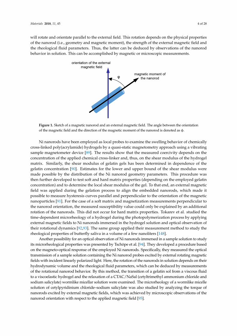
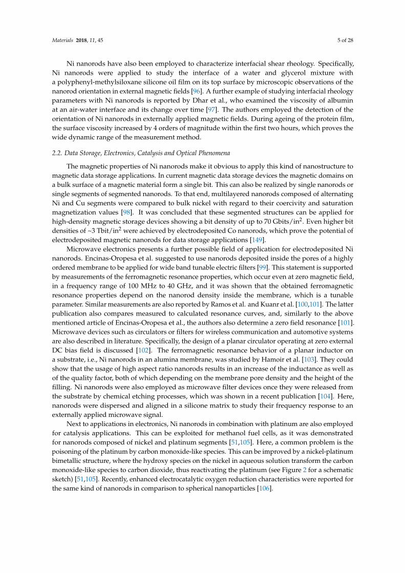
Generally, in order to examine rheological fluid parameters, magnetic nanorods are immersedin a sample solution and their movement under application of an external magnetic field is studied.A schematic illustration of a magnetic nanorod in an external magnetic field is shown in Figure 1.The angle φ describes the orientation of the magnetic moment of the nanorod to the direction of theexternal magnetic field. When being dispersed in solution or embedded in a soft matter, the nanorod
Materials 2018, 11, 45 4 of 28
will rotate and orientate parallel to the external field. This rotation depends on the physical propertiesof the nanorod (i.e., geometry and magnetic moment), the strength of the external magnetic field andthe rheological fluid parameters. Thus, the latter can be deduced by observations of the nanorodbehavior in solution. This can be accomplished by magnetic or microscopic measurements.
Materials 2018, 11, 45 4 of 28
external magnetic field. When being dispersed in solution or embedded in a soft matter, the nanorod will rotate and orientate parallel to the external field. This rotation depends on the physical properties of the nanorod (i.e., geometry and magnetic moment), the strength of the external magnetic field and the rheological fluid parameters. Thus, the latter can be deduced by observations of the nanorod behavior in solution. This can be accomplished by magnetic or microscopic measurements.
Figure 1. Sketch of a magnetic nanorod and an external magnetic field. The angle between the orientation of the magnetic field and the direction of the magnetic moment of the nanorod is denoted as ϕ.
Ni nanorods have been employed as local probes to examine the swelling behavior of chemically cross-linked poly(acrylamide) hydrogels by a quasi-static magnetometry approach using a vibrating sample magnetometer device [89]. The results show that the measured coercivity depends on the concentration of the applied chemical cross-linker and, thus, on the shear modulus of the hydrogel matrix. Similarly, the shear modulus of gelatin gels has been determined in dependence of the gelatin concentration [90]. Estimates for the lower and upper bound of the shear modulus were made possible by the distribution of the Ni nanorod geometry parameters. This procedure was then further developed to test soft and hard matrix properties (depending on the employed gelatin concentration) and to determine the local shear modulus of the gel. To that end, an external magnetic field was applied during the gelation process to align the embedded nanorods, which made it possible to measure hysteresis curves parallel and perpendicular to the orientation of the magnetic nanoparticles [91]. For the case of a soft matrix and magnetization measurements perpendicular to the nanorod orientation, the measured susceptibility value could only be explained by an additional rotation of the nanorods. This did not occur for hard matrix properties. Tokarev et al. studied the time-dependent microrheology of a hydrogel during the photopolymerization process by applying external magnetic fields to Ni nanorods immersed in the hydrogel solution and optical observation of their rotational dynamics [92,93]. The same group applied their measurement method to study the rheological properties of butterfly saliva in a volume of a few nanoliters [148].
Another possibility for an optical observation of Ni nanorods immersed in a sample solution to study its microrheological properties was presented by Tschöpe et al. [94]. They developed a procedure based on the magneto-optical response of the employed Ni nanorods. Specifically, they measured the optical transmission of a sample solution containing the Ni nanorod probes excited by external rotating magnetic fields with incident linearly polarized light. Here, the rotation of the nanorods in solution depends on their hydrodynamic volume and the rheological fluid parameters, which can be deduced by measurements of the rotational nanorod behavior. By this method, the transition of a gelatin sol from a viscous fluid to a viscoelastic hydrogel and the relaxation of a CTAC/NaSal (cetyltrimethyl ammonium chloride and sodium salicylate) wormlike micellar solution were examined. The microrheology of a wormlike micelle solution of cetylpyridinium chloride–sodium salicylate was also studied by analyzing the torque of nanorods excited by external magnetic fields, which was achieved by microscopic observations of the nanorod orientation with respect to the applied magnetic field [95].
Figure 1. Sketch of a magnetic nanorod and an external magnetic field. The angle between the orientationof the magnetic field and the direction of the magnetic moment of the nanorod is denoted as φ.
Ni nanorods have been employed as local probes to examine the swelling behavior of chemicallycross-linked poly(acrylamide) hydrogels by a quasi-static magnetometry approach using a vibratingsample magnetometer device [89]. The results show that the measured coercivity depends on theconcentration of the applied chemical cross-linker and, thus, on the shear modulus of the hydrogelmatrix. Similarly, the shear modulus of gelatin gels has been determined in dependence of thegelatin concentration [90]. Estimates for the lower and upper bound of the shear modulus weremade possible by the distribution of the Ni nanorod geometry parameters. This procedure wasthen further developed to test soft and hard matrix properties (depending on the employed gelatinconcentration) and to determine the local shear modulus of the gel. To that end, an external magneticfield was applied during the gelation process to align the embedded nanorods, which made itpossible to measure hysteresis curves parallel and perpendicular to the orientation of the magneticnanoparticles [91]. For the case of a soft matrix and magnetization measurements perpendicular tothe nanorod orientation, the measured susceptibility value could only be explained by an additionalrotation of the nanorods. This did not occur for hard matrix properties. Tokarev et al. studied thetime-dependent microrheology of a hydrogel during the photopolymerization process by applyingexternal magnetic fields to Ni nanorods immersed in the hydrogel solution and optical observation oftheir rotational dynamics [92,93]. The same group applied their measurement method to study therheological properties of butterfly saliva in a volume of a few nanoliters [148].
Another possibility for an optical observation of Ni nanorods immersed in a sample solution to studyits microrheological properties was presented by Tschöpe et al. [94]. They developed a procedure basedon the magneto-optical response of the employed Ni nanorods. Specifically, they measured the opticaltransmission of a sample solution containing the Ni nanorod probes excited by external rotating magneticfields with incident linearly polarized light. Here, the rotation of the nanorods in solution depends on theirhydrodynamic volume and the rheological fluid parameters, which can be deduced by measurementsof the rotational nanorod behavior. By this method, the transition of a gelatin sol from a viscous fluidto a viscoelastic hydrogel and the relaxation of a CTAC/NaSal (cetyltrimethyl ammonium chloride andsodium salicylate) wormlike micellar solution were examined. The microrheology of a wormlike micellesolution of cetylpyridinium chloride–sodium salicylate was also studied by analyzing the torque ofnanorods excited by external magnetic fields, which was achieved by microscopic observations of thenanorod orientation with respect to the applied magnetic field [95].
Materials 2018, 11, 45 5 of 28
Ni nanorods have also been employed to characterize interfacial shear rheology. Specifically,Ni nanorods were applied to study the interface of a water and glycerol mixture witha polyphenyl-methylsiloxane silicone oil film on its top surface by microscopic observations of thenanorod orientation in external magnetic fields [96]. A further example of studying interfacial rheologyparameters with Ni nanorods is reported by Dhar et al., who examined the viscosity of albuminat an air-water interface and its change over time [97]. The authors employed the detection of theorientation of Ni nanorods in externally applied magnetic fields. During ageing of the protein film,the surface viscosity increased by 4 orders of magnitude within the first two hours, which proves thewide dynamic range of the measurement method.
2.2. Data Storage, Electronics, Catalysis and Optical Phenomena
The magnetic properties of Ni nanorods make it obvious to apply this kind of nanostructure tomagnetic data storage applications. In current magnetic data storage devices the magnetic domains ona bulk surface of a magnetic material form a single bit. This can also be realized by single nanorods orsingle segments of segmented nanorods. To that end, multilayered nanorods composed of alternatingNi and Cu segments were compared to bulk nickel with regard to their coercivity and saturationmagnetization values [98]. It was concluded that these segmented structures can be applied forhigh-density magnetic storage devices showing a bit density of up to 70 Gbits/in2. Even higher bitdensities of ~3 Tbit/in2 were achieved by electrodeposited Co nanorods, which prove the potential ofelectrodeposited magnetic nanorods for data storage applications [149].
Microwave electronics presents a further possible field of application for electrodeposited Ninanorods. Encinas-Oropesa et al. suggested to use nanorods deposited inside the pores of a highlyordered membrane to be applied for wide band tunable electric filters [99]. This statement is supportedby measurements of the ferromagnetic resonance properties, which occur even at zero magnetic field,in a frequency range of 100 MHz to 40 GHz, and it was shown that the obtained ferromagneticresonance properties depend on the nanorod density inside the membrane, which is a tunableparameter. Similar measurements are also reported by Ramos et al. and Kuanr et al. [100,101]. The latterpublication also compares measured to calculated resonance curves, and, similarly to the abovementioned article of Encinas-Oropesa et al., the authors also determine a zero field resonance [101].Microwave devices such as circulators or filters for wireless communication and automotive systemsare also described in literature. Specifically, the design of a planar circulator operating at zero externalDC bias field is discussed [102]. The ferromagnetic resonance behavior of a planar inductor ona substrate, i.e., Ni nanorods in an alumina membrane, was studied by Hamoir et al. [103]. They couldshow that the usage of high aspect ratio nanorods results in an increase of the inductance as well asof the quality factor, both of which depending on the membrane pore density and the height of thefilling. Ni nanorods were also employed as microwave filter devices once they were released fromthe substrate by chemical etching processes, which was shown in a recent publication [104]. Here,nanorods were dispersed and aligned in a silicone matrix to study their frequency response to anexternally applied microwave signal.
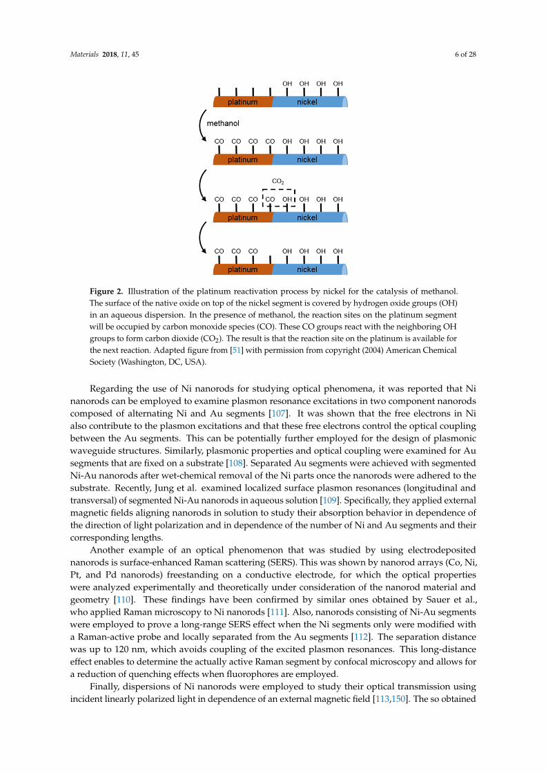
Next to applications in electronics, Ni nanorods in combination with platinum are also employedfor catalysis applications. This can be exploited for methanol fuel cells, as it was demonstratedfor nanorods composed of nickel and platinum segments [51,105]. Here, a common problem is thepoisoning of the platinum by carbon monoxide-like species. This can be improved by a nickel-platinumbimetallic structure, where the hydroxy species on the nickel in aqueous solution transform the carbonmonoxide-like species to carbon dioxide, thus reactivating the platinum (see Figure 2 for a schematicsketch) [51,105]. Recently, enhanced electrocatalytic oxygen reduction characteristics were reported forthe same kind of nanorods in comparison to spherical nanoparticles [106].
Materials 2018, 11, 45 6 of 28Materials 2018, 11, 45 6 of 28
Figure 2. Illustration of the platinum reactivation process by nickel for the catalysis of methanol. The surface of the native oxide on top of the nickel segment is covered by hydrogen oxide groups (OH) in an aqueous dispersion. In the presence of methanol, the reaction sites on the platinum segment will be occupied by carbon monoxide species (CO). These CO groups react with the neighboring OH groups to form carbon dioxide (CO2). The result is that the reaction site on the platinum is available for the next reaction. Adapted figure from [51] with permission from copyright (2004) American Chemical Society (Washington, DC, USA).
Regarding the use of Ni nanorods for studying optical phenomena, it was reported that Ni nanorods can be employed to examine plasmon resonance excitations in two component nanorods composed of alternating Ni and Au segments [107]. It was shown that the free electrons in Ni also contribute to the plasmon excitations and that these free electrons control the optical coupling between the Au segments. This can be potentially further employed for the design of plasmonic waveguide structures. Similarly, plasmonic properties and optical coupling were examined for Au segments that are fixed on a substrate [108]. Separated Au segments were achieved with segmented Ni-Au nanorods after wet-chemical removal of the Ni parts once the nanorods were adhered to the substrate. Recently, Jung et al. examined localized surface plasmon resonances (longitudinal and transversal) of segmented Ni-Au nanorods in aqueous solution [109]. Specifically, they applied external magnetic fields aligning nanorods in solution to study their absorption behavior in dependence of the direction of light polarization and in dependence of the number of Ni and Au segments and their corresponding lengths.
Another example of an optical phenomenon that was studied by using electrodeposited nanorods is surface-enhanced Raman scattering (SERS). This was shown by nanorod arrays (Co, Ni, Pt, and Pd nanorods) freestanding on a conductive electrode, for which the optical properties were analyzed experimentally and theoretically under consideration of the nanorod material and geometry [110]. These findings have been confirmed by similar ones obtained by Sauer et al., who applied Raman microscopy to Ni nanorods [111]. Also, nanorods consisting of Ni-Au segments were employed to prove a long-range SERS effect when the Ni segments only were modified with a Raman-active probe and locally separated from the Au segments [112]. The separation distance was up to 120 nm, which avoids coupling of the excited plasmon resonances. This long-distance effect enables to determine the actually active Raman segment by confocal microscopy and allows for a reduction of quenching effects when fluorophores are employed.
Finally, dispersions of Ni nanorods were employed to study their optical transmission using incident linearly polarized light in dependence of an external magnetic field [113,150]. The so obtained results suggest the use of Ni nanorods for optical switches that are controlled by magnetic fields, which can be applied for liquid crystal technology [113]. Similarly, cold cathode fluorescent
Figure 2. Illustration of the platinum reactivation process by nickel for the catalysis of methanol.The surface of the native oxide on top of the nickel segment is covered by hydrogen oxide groups (OH)in an aqueous dispersion. In the presence of methanol, the reaction sites on the platinum segmentwill be occupied by carbon monoxide species (CO). These CO groups react with the neighboring OHgroups to form carbon dioxide (CO2). The result is that the reaction site on the platinum is available forthe next reaction. Adapted figure from [51] with permission from copyright (2004) American ChemicalSociety (Washington, DC, USA).
Regarding the use of Ni nanorods for studying optical phenomena, it was reported that Ninanorods can be employed to examine plasmon resonance excitations in two component nanorodscomposed of alternating Ni and Au segments [107]. It was shown that the free electrons in Nialso contribute to the plasmon excitations and that these free electrons control the optical couplingbetween the Au segments. This can be potentially further employed for the design of plasmonicwaveguide structures. Similarly, plasmonic properties and optical coupling were examined for Ausegments that are fixed on a substrate [108]. Separated Au segments were achieved with segmentedNi-Au nanorods after wet-chemical removal of the Ni parts once the nanorods were adhered to thesubstrate. Recently, Jung et al. examined localized surface plasmon resonances (longitudinal andtransversal) of segmented Ni-Au nanorods in aqueous solution [109]. Specifically, they applied externalmagnetic fields aligning nanorods in solution to study their absorption behavior in dependence ofthe direction of light polarization and in dependence of the number of Ni and Au segments and theircorresponding lengths.
Another example of an optical phenomenon that was studied by using electrodepositednanorods is surface-enhanced Raman scattering (SERS). This was shown by nanorod arrays (Co, Ni,Pt, and Pd nanorods) freestanding on a conductive electrode, for which the optical propertieswere analyzed experimentally and theoretically under consideration of the nanorod material andgeometry [110]. These findings have been confirmed by similar ones obtained by Sauer et al.,who applied Raman microscopy to Ni nanorods [111]. Also, nanorods consisting of Ni-Au segmentswere employed to prove a long-range SERS effect when the Ni segments only were modified witha Raman-active probe and locally separated from the Au segments [112]. The separation distancewas up to 120 nm, which avoids coupling of the excited plasmon resonances. This long-distanceeffect enables to determine the actually active Raman segment by confocal microscopy and allows fora reduction of quenching effects when fluorophores are employed.
Finally, dispersions of Ni nanorods were employed to study their optical transmission usingincident linearly polarized light in dependence of an external magnetic field [113,150]. The so obtained
Materials 2018, 11, 45 7 of 28
results suggest the use of Ni nanorods for optical switches that are controlled by magnetic fields,which can be applied for liquid crystal technology [113]. Similarly, cold cathode fluorescent lampsused for optical applications can be realized by using Ni nanorods as reported by Feizi et al. [114].These are of interest for liquid crystal displays. To that end, Feizi et al. demonstrated an enlargedelectron emission by a factor of four at a bias potential of 1 V in comparison to bulk nickel substrates.
2.3. Sensing and Biosensing Applications
Nickel nanoparticles synthesized in porous membranes can be applied for the sensing ofcarbohydrates. To that end, Lu et al. synthesized nickel nanorods in polycarbonate membranesthat were placed on a glassy carbon electrode to end up with a freestanding nanorod array on theelectrode after chemically dissolving the polycarbonate template [115]. This electrode served asworking electrode for non-enzymatic electrochemical detection of glucose by cyclic voltammetricand amperometric measurements. A detection limit of 100 nM was achieved in an alkaline solution.Carbohydrates were also detected in a non-enzymatic electrochemical detection by first droppinga solution of dispersed nickel nanorods onto the surface of a screen printed electrode [116]. Then,the Ni nanorods were activated by an amperometry step in alkaline solution to generate oxygengroups on the surface, which were employed for catalytic oxidation of carbohydrates. A detection limitof 20 µM for glucose was achieved in serum samples, which typically show glucose concentrationsin the lower millimolar regime. The same measurement method was also applied for the sensingof inulin in buffer solutions [117]. Recently, this measurement method was further developed intoa miniaturized flow injection analysis system to detect galactose in urine of newborns for the diagnosisof galactosemia, a genetic disorder impairing galactose metabolization [118]. Nickel nanorods coatedby electroless deposition with a gold layer freestanding on a substrate were used as electrode for thedetection of glucose [119]. The actual sensing was carried out by cyclic voltammetry measurements.Here, nanorods were employed to increase the electrode surface, while the detection of glucose wasachieved via the enzyme glucose oxidase. A non-enzymatic approach for the detection of glucose withthe help of a nanorod array was recently presented by Qin et al. [120]. To that end, they synthesizedmultilayered nickel gold nanorods on the surface of an electrode by using an alumina membrane thatwas chemically dissolved after nanorod synthesis. Amperometric measurements were conductedto detect glucose in sodium hydroxide solutions with a detection limit of 0.1 µM and, furthermore,glucose was also detected in spiked urine samples.
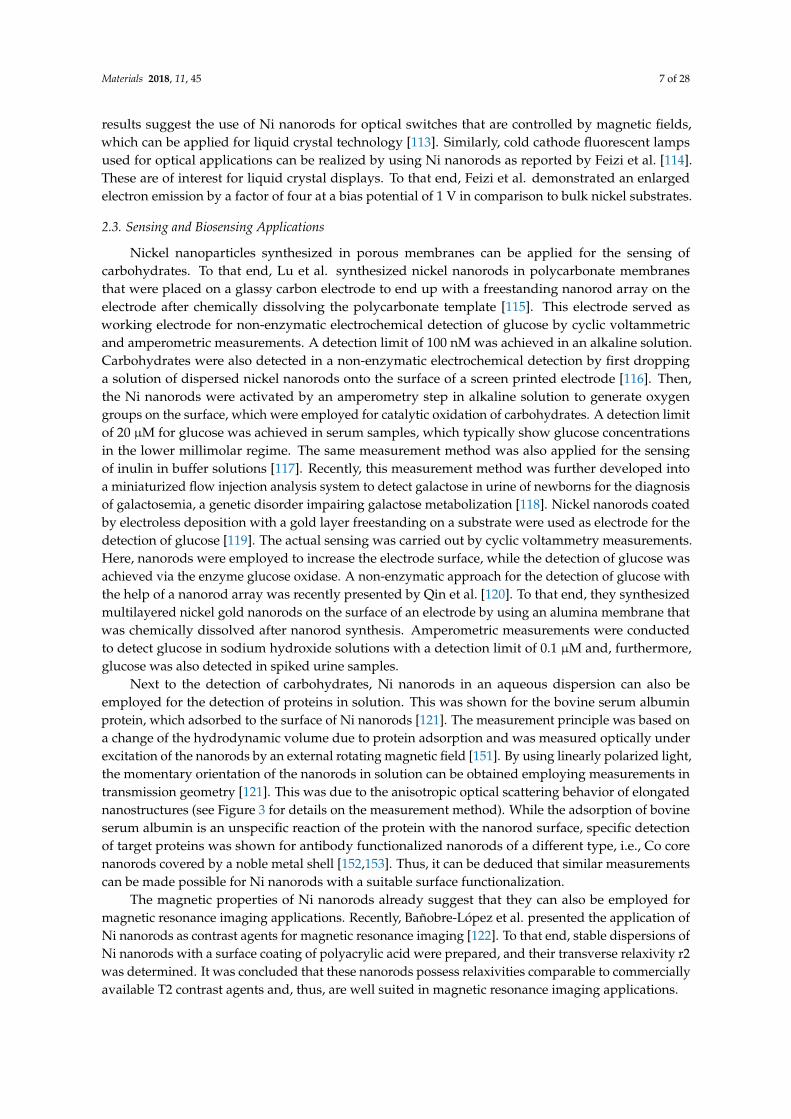
Next to the detection of carbohydrates, Ni nanorods in an aqueous dispersion can also beemployed for the detection of proteins in solution. This was shown for the bovine serum albuminprotein, which adsorbed to the surface of Ni nanorods [121]. The measurement principle was based ona change of the hydrodynamic volume due to protein adsorption and was measured optically underexcitation of the nanorods by an external rotating magnetic field [151]. By using linearly polarized light,the momentary orientation of the nanorods in solution can be obtained employing measurements intransmission geometry [121]. This was due to the anisotropic optical scattering behavior of elongatednanostructures (see Figure 3 for details on the measurement method). While the adsorption of bovineserum albumin is an unspecific reaction of the protein with the nanorod surface, specific detectionof target proteins was shown for antibody functionalized nanorods of a different type, i.e., Co corenanorods covered by a noble metal shell [152,153]. Thus, it can be deduced that similar measurementscan be made possible for Ni nanorods with a suitable surface functionalization.
The magnetic properties of Ni nanorods already suggest that they can also be employed formagnetic resonance imaging applications. Recently, Bañobre-López et al. presented the application ofNi nanorods as contrast agents for magnetic resonance imaging [122]. To that end, stable dispersions ofNi nanorods with a surface coating of polyacrylic acid were prepared, and their transverse relaxivity r2was determined. It was concluded that these nanorods possess relaxivities comparable to commerciallyavailable T2 contrast agents and, thus, are well suited in magnetic resonance imaging applications.
Materials 2018, 11, 45 8 of 28Materials 2018, 11, 45 8 of 28
Figure 3. Measurement principle for detecting proteins in solution according to reference [121]. Nanorods immersed in solution were excited by an external rotating magnetic field, which caused the nanorods to rotate with field, but delayed by a phase lag ϕ. This is caused by the drag torque that the nanorods experienced in the solution. The phase lag ϕ was a function of the hydrodynamic nanorod volume. The orientation of nanorods immersed in solution was measured optically by linearly polarized light. By measurements in transmission geometry, the detected intensity depended on the angle α between the direction of light polarization and the long axis of the nanorod. An increase of the hydrodynamic nanorod volume was observed upon addition of a protein to the sample solution, which leads to protein adhesion to the nanorod surface. This resulted in a change of the angle ϕ and, thus, of the angle α, thereby leading to a measurable change in the optical signal. Adapted figure from [121] reprinted with permission from copyright 2014, John Wiley and Sons (Hoboken, NJ, USA).
Alternatively, the magnetic properties of Ni nanorods can be applied for magnetic separation techniques. To that end, a sensing method was presented by Pinheiro et al. for the specific detection and targeting of mercury ions [123]. Here, the heavy metal ions bound to the nanorod surface so that they could be removed from a solution by magnetic separation. In order to target heavy metal ions, the surface of the Ni nanorods was modified to finally achieve silica shell coated nanorods with incorporated dithiocarbamate groups that show a strong affinity for binding heavy metal ions. The feasibility of this method was shown by applying the nanorods to remove mercury from an aqueous solution by magnetic separation with a removal rate of 99.8% of mercury ions. The resulting final mercury concentration value was below the recommended guideline threshold for drinking water quality in Europe.
2.4. Cell Biology Applications
The first reported applications of magnetic particles for cell biology dealt with the characterization of the rheological parameters of the cytoplasm in chick fibroblasts as detailed above in Section 2.1. In recent years a growing number of reports can be found that focus on the cellular response once nanorods were internalized by cells and then agitated externally by magnetic fields. For example, Castillo et al. studied the torques of segmented Ni-Pt nanorods when incubated with fibroblast cells under excitation by an external magnetic field [124]. The nanorod rotation was observed microscopically and as a result, they were able to define a minimum torque for nanorod rotation for which they deduced a non-Newtonian behavior of cytoplasm. A further example for a cellular response to external force was investigated by pulmonary artery smooth muscle cells with internalized Ni nanorods and an array of flexible micropost force sensors as a substrate for cell adhesion [125]. External magnetic fields of low frequency up to 10 Hz were applied, and the cellular contractile force was studied. Furthermore, rotational motion of nanorods internalized by different cells was used to study the effect of gene mutations as demonstrated by Celedon et al. using Lamin A/C knockout cells that showed defective nuclear mechanics [126]. To that end, the nanorods were incubated with the cells so that they got internalized, and their rotational motion excited by an external magnetic field was studied optically to deduce viscosity and elasticity parameters of the cell nucleus.
Ni nanorods can also be employed to release a specific molecule inside cells or to present a chosen molecule to cells. The aim of both approaches is to trigger a cellular response. For this purpose,
Figure 3. Measurement principle for detecting proteins in solution according to reference [121].Nanorods immersed in solution were excited by an external rotating magnetic field, which causedthe nanorods to rotate with field, but delayed by a phase lag φ. This is caused by the drag torquethat the nanorods experienced in the solution. The phase lag φ was a function of the hydrodynamicnanorod volume. The orientation of nanorods immersed in solution was measured optically by linearlypolarized light. By measurements in transmission geometry, the detected intensity depended on theangle α between the direction of light polarization and the long axis of the nanorod. An increase ofthe hydrodynamic nanorod volume was observed upon addition of a protein to the sample solution,which leads to protein adhesion to the nanorod surface. This resulted in a change of the angle φ and,thus, of the angle α, thereby leading to a measurable change in the optical signal. Adapted figurefrom [121] reprinted with permission from copyright 2014, John Wiley and Sons (Hoboken, NJ, USA).
Alternatively, the magnetic properties of Ni nanorods can be applied for magnetic separationtechniques. To that end, a sensing method was presented by Pinheiro et al. for the specific detectionand targeting of mercury ions [123]. Here, the heavy metal ions bound to the nanorod surface sothat they could be removed from a solution by magnetic separation. In order to target heavy metalions, the surface of the Ni nanorods was modified to finally achieve silica shell coated nanorodswith incorporated dithiocarbamate groups that show a strong affinity for binding heavy metal ions.The feasibility of this method was shown by applying the nanorods to remove mercury from anaqueous solution by magnetic separation with a removal rate of 99.8% of mercury ions. The resultingfinal mercury concentration value was below the recommended guideline threshold for drinking waterquality in Europe.
2.4. Cell Biology Applications
The first reported applications of magnetic particles for cell biology dealt with the characterizationof the rheological parameters of the cytoplasm in chick fibroblasts as detailed above in Section 2.1.In recent years a growing number of reports can be found that focus on the cellular response oncenanorods were internalized by cells and then agitated externally by magnetic fields. For example,Castillo et al. studied the torques of segmented Ni-Pt nanorods when incubated with fibroblastcells under excitation by an external magnetic field [124]. The nanorod rotation was observedmicroscopically and as a result, they were able to define a minimum torque for nanorod rotationfor which they deduced a non-Newtonian behavior of cytoplasm. A further example for a cellularresponse to external force was investigated by pulmonary artery smooth muscle cells with internalizedNi nanorods and an array of flexible micropost force sensors as a substrate for cell adhesion [125].External magnetic fields of low frequency up to 10 Hz were applied, and the cellular contractile forcewas studied. Furthermore, rotational motion of nanorods internalized by different cells was used tostudy the effect of gene mutations as demonstrated by Celedon et al. using Lamin A/C knockout cellsthat showed defective nuclear mechanics [126]. To that end, the nanorods were incubated with thecells so that they got internalized, and their rotational motion excited by an external magnetic fieldwas studied optically to deduce viscosity and elasticity parameters of the cell nucleus.
Ni nanorods can also be employed to release a specific molecule inside cells or to present a chosenmolecule to cells. The aim of both approaches is to trigger a cellular response. For this purpose, specially
Materials 2018, 11, 45 9 of 28
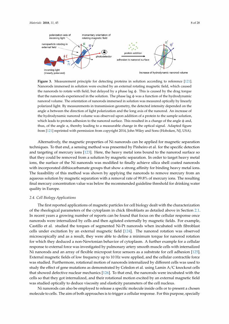
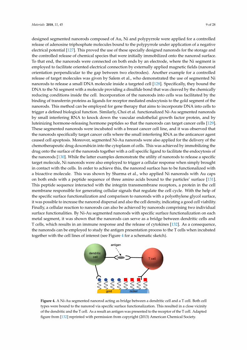
designed segmented nanorods composed of Au, Ni and polypyrrole were applied for a controlledrelease of adenosine triphosphate molecules bound to the polypyrrole under application of a negativeelectrical potential [127]. This proved the use of these specially designed nanorods for the storage andthe controlled release of chemical species that were initially immobilized onto the nanorod surface.To that end, the nanorods were connected on both ends by an electrode, where the Ni segment isemployed to facilitate oriented electrical connection by externally applied magnetic fields (nanorodorientation perpendicular to the gap between two electrodes). Another example for a controlledrelease of target molecules was given by Salem et al., who demonstrated the use of segmented Ninanorods to release a small DNA molecule inside a targeted cell [128]. Specifically, they bound theDNA to the Ni segment with a molecule providing a disulfide bond that was cleaved by the chemicallyreducing conditions inside the cell. Incorporation of the nanorods into cells was facilitated by thebinding of transferrin proteins as ligands for receptor mediated endocytosis to the gold segment of thenanorods. This method can be employed for gene therapy that aims to incorporate DNA into cells totrigger a defined biological function. Similarly, Choi et al. functionalized Ni-Au segmented nanorodsby small interfering RNA to knock down the vascular endothelial growth factor protein, and byluteinizing hormone-releasing hormone peptides so that the nanorods can target cancer cells [129].These segmented nanorods were incubated with a breast cancer cell line, and it was observed thatthe nanorods specifically target cancer cells where the small interfering RNA as the anticancer agentcaused cell apoptosis. Moreover, segmented Ni-Au nanorods were also applied for the delivery of thechemotherapeutic drug doxorubicin into the cytoplasm of cells. This was achieved by immobilizing thedrug onto the surface of the nanorods together with a cell specific ligand to facilitate the endocytosis ofthe nanorods [130]. While the latter examples demonstrate the utility of nanorods to release a specifictarget molecule, Ni nanorods were also employed to trigger a cellular response when simply broughtin contact with the cells. In order to achieve this, the nanorod surface has to be functionalized witha bioactive molecule. This was shown by Sharma et al., who applied Ni nanorods with Au capson both ends with a peptide sequence of three amino acids bound to the particles’ surface [131].This peptide sequence interacted with the integrin transmembrane receptors, a protein in the cellmembrane responsible for generating cellular signals that regulate the cell cycle. With the help ofthe specific surface functionalization and comparison to nanorods with a polyethylene glycol surface,it was possible to increase the nanorod dispersal and also the cell density, indicating a good cell viability.Finally, a cellular reaction to nanorods can also be achieved by nanorods comprising two individualsurface functionalities. By Ni-Au segmented nanorods with specific surface functionalization on eachmetal segment, it was shown that the nanorods can serve as a bridge between dendritic cells andT cells, which results in an immune response and the release of cytokines [132]. As a consequence,the nanorods can be employed to study the antigen presentation process to the T cells when incubatedtogether with the cell lines of interest (see Figure 4 for a schematic sketch).
Materials 2018, 11, 45 9 of 28
specially designed segmented nanorods composed of Au, Ni and polypyrrole were applied for a controlled release of adenosine triphosphate molecules bound to the polypyrrole under application of a negative electrical potential [127]. This proved the use of these specially designed nanorods for the storage and the controlled release of chemical species that were initially immobilized onto the nanorod surface. To that end, the nanorods were connected on both ends by an electrode, where the Ni segment is employed to facilitate oriented electrical connection by externally applied magnetic fields (nanorod orientation perpendicular to the gap between two electrodes). Another example for a controlled release of target molecules was given by Salem et al., who demonstrated the use of segmented Ni nanorods to release a small DNA molecule inside a targeted cell [128]. Specifically, they bound the DNA to the Ni segment with a molecule providing a disulfide bond that was cleaved by the chemically reducing conditions inside the cell. Incorporation of the nanorods into cells was facilitated by the binding of transferrin proteins as ligands for receptor mediated endocytosis to the gold segment of the nanorods. This method can be employed for gene therapy that aims to incorporate DNA into cells to trigger a defined biological function. Similarly, Choi et al. functionalized Ni-Au segmented nanorods by small interfering RNA to knock down the vascular endothelial growth factor protein, and by luteinizing hormone-releasing hormone peptides so that the nanorods can target cancer cells [129]. These segmented nanorods were incubated with a breast cancer cell line, and it was observed that the nanorods specifically target cancer cells where the small interfering RNA as the anticancer agent caused cell apoptosis. Moreover, segmented Ni-Au nanorods were also applied for the delivery of the chemotherapeutic drug doxorubicin into the cytoplasm of cells. This was achieved by immobilizing the drug onto the surface of the nanorods together with a cell specific ligand to facilitate the endocytosis of the nanorods [130]. While the latter examples demonstrate the utility of nanorods to release a specific target molecule, Ni nanorods were also employed to trigger a cellular response when simply brought in contact with the cells. In order to achieve this, the nanorod surface has to be functionalized with a bioactive molecule. This was shown by Sharma et al., who applied Ni nanorods with Au caps on both ends with a peptide sequence of three amino acids bound to the particles’ surface [131]. This peptide sequence interacted with the integrin transmembrane receptors, a protein in the cell membrane responsible for generating cellular signals that regulate the cell cycle. With the help of the specific surface functionalization and comparison to nanorods with a polyethylene glycol surface, it was possible to increase the nanorod dispersal and also the cell density, indicating a good cell viability. Finally, a cellular reaction to nanorods can also be achieved by nanorods comprising two individual surface functionalities. By Ni-Au segmented nanorods with specific surface functionalization on each metal segment, it was shown that the nanorods can serve as a bridge between dendritic cells and T cells, which results in an immune response and the release of cytokines [132]. As a consequence, the nanorods can be employed to study the antigen presentation process to the T cells when incubated together with the cell lines of interest (see Figure 4 for a schematic sketch).
Figure 4. A Ni-Au segmented nanorod acting as bridge between a dendritic cell and a T cell. Both cell types were bound to the nanorod via specific surface functionalization. This resulted in a close vicinity of the dendritic and the T cell. As a result an antigen was presented to the receptor of the T cell. Adapted figure from [132] reprinted with permission from copyright (2013) American Chemical Society.
Figure 4. A Ni-Au segmented nanorod acting as bridge between a dendritic cell and a T cell. Both celltypes were bound to the nanorod via specific surface functionalization. This resulted in a close vicinityof the dendritic and the T cell. As a result an antigen was presented to the receptor of the T cell. Adaptedfigure from [132] reprinted with permission from copyright (2013) American Chemical Society.
Materials 2018, 11, 45 10 of 28
A further possibility to apply Ni nanorods together with cells is to guide the entire cell to a specificlocation or to guide the cell growth along a specific direction. This can be achieved by making useof the magnetic properties of nanorods and external magnetic fields to manipulate the orientationof nanorods. For example, cells that internalized Ni nanorods were guided to the gap betweena ferromagnetic and a gold electrode by magnetic interaction of the electrode with the Ni nanorods toallow for non-destructive examination of neurons, i.e., a cell type responding to electrical signals [133].A further example was demonstrated by Tanase et al. who controlled the organization of mammaliancells using Ni nanorods in conjunction with a patterned micromagnetic substrate [134]. Cells couldbe adsorbed to the nanorod surface, which did not hinder the external orientation of the nanorodsby the micromagnets and by an additional external magnetic field so that controlled organization oftwo dimensional cell structures was achieved. An alternative method for cell guidance was presentedby Johansson et al. who employed Ni nanorods that were dispersed on a substrate and orientedby an external magnetic field to form structures for subsequent cell adhesion [135]. Here, not onlythe employed fibroblast but also the axons showed oriented adhesion along the main nanorod axis.Another example of lateral cell displacement by the effect of nanorod rotational motion caused byexternal magnetic fields was demonstrated by using Ni nanorods and skeletal myoblasts, which canbe employed to create three dimensional cell clusters [136].
Comparable to the guidance of the cells as described in the paragraph above, Ni nanorods canalso be employed to execute cell separation. Hultgren et al. demonstrated the application of Ninanorods for cell separation by an external magnetic field after incubation of the employed cells withthe magnetic nanorods [137]. The same research group further studied the influence of the nanorodlength on the cell separation performance and concluded that the cell separation performance is bestwhen the nanorod length is comparable to the cell diameter [138]. Similarly, Nanorod dispersions withtwo different nanorod lengths were employed to capture cells of a specific diameter in accordance tothe nanorod length and cell diameter correlation [139]. More recently, a cell separation technique waspresented by Gao et al. who applied nanorods with an antibody surface functionalization for a proofof principle of cell separation in an external static magnetic field [140]. Briefly, the authors incubatedcells and Ni nanorods and placed the mixture in a static magnetic gradient field to separate cells withinternalized nanorods from nanorod-free cells by removing the supernatant and, finally, counted thenumber of separated cells by cytometry.
Inducing a controlled cell death is an important application of magnetic nanoparticles that isemployed for cancer therapy [154]. In that regard, Fung et al. demonstrated the application of Ninanorods internalized by fibroblasts to induce an inflammatory response by the cell and subsequentcell death [141]. This was achieved by external magnetic fields at a low frequency of 1 Hz to inducenanorod motion, which caused upregulation of the IL-6 cytokine and cell death. An experimentalproof was given by colorimetric cell viability assays. Similar results with a similar nanorod excitationwere obtained for human embryonic kidney cells [142]. Additionally, induced cell death of cancercells with internalized Ni nanorods by manipulation of nanorod motion with external AC magneticfields was demonstrated by Contreras et al. [143]. Here, a weak magnetic field with an amplitudeof 0.5 mT only was applied and it was shown that this field strength is enough to induce mechanicalcell disturbances that result in reduced cell viability. This was explained by cell membrane rupturescaused by the nanorods and their motion due to the external excitation. Recently, Hopkins et al.presented a method for tumor therapy based on a radio frequency induction of heat in Ni nanorodscovered by a Au shell [144]. Here, Ni nanorods were fabricated by electrodeposition into aluminamembranes followed by a Au shell synthesis achieved via electroless plating of the Ni cores. Theseparticles were injected into xenograft mice models. Specifically, the tumor cells of a pancreatic tumorwere transplanted into the animals to grow the tumor into which the nanorods were injected. The radiofrequency signal was generated by an external microstrip spiral antenna and the effect of heat inductionwas based on eddy currents on the nanorod surface and a loss of hysteresis due to the applied AC
Materials 2018, 11, 45 11 of 28
field. These resulted in a nanorod heating of up to 45 ◦C that causes severe damage to the neighboringtumor cells.
An important topic for all medical applications involving nanostructures of any geometry is thecytotoxicity of the applied nanostructures. The cytotoxicity of Ni nanorods depends on many factors,as for example the geometry, the surface coating, but also the measurement procedure for evaluationof cytotoxic effects. Consequently, while some studies can be found in literature on the cytotoxic effectsof Ni nanorods, they can hardly be compared [155–157]. A reasonable examination of the cytotoxiceffects of Ni nanorods is in our opinion best conducted for the specific type of nanorods and underconsideration of the envisaged application. Thus, a deeper discussion of this topic is omitted withinthis review.
3. Nickel Nanorod Surface Chemistry Modification
Chemical modification of the surface of Ni nanorods is essential for a lot of complex applicationsas they have been detailed in the previous section. A prerequisite for applying nanorods is to separatethem from the nanoporous template in which they were synthesized. The two most common templatematerials for electrodeposition are aluminum oxide and polycarbonate, which have to be removed withoutdamaging the Ni nanorods in the pores. This can be achieved by chemically selective etching processes.While the polycarbonate membrane can be rapidly dissolved in dichloromethane, the aluminum oxidetemplate can be dissolved in sodium hydroxide solutions or in mixtures of chromic acid and phosphoricacid [42]. Though functional groups can be immobilized onto the nanorod surface by an unspecificbinding process (e.g., as demonstrated for binding of streptavidin protein by an incubation step in a cellculture medium [133]), more specific binding processes are addressed by most research groups. Anotherexception for an unspecific binding process to the nanorod surface was presented by Sharma et al.,who modified the negatively charged surface of Ni nanorods by positively charged amine groups viaionic binding processes [131]. This was controlled by the adjustment of the pH value of the dispersionsolution under consideration of the isoelectric point of the employed molecules. Similarly, Magnin et al.employed the layer-by-layer technique [158], which is based on the coating of the surface by multiplepolymer layers of opposite charge [159]. Specifically, positively charged chitosan and negatively chargedcarboxymethylpullulan layers were immobilized onto the nanorod surface. In the following subsections,different types of chemical surface modifications that are structured with respect to the employed moleculeand the anchor group for linkage to the nanorod surface will be discussed. An overview is given inTable 2. For this report, we only considered references with a detailed description of the experimentalprocedure so that it can be reproduced by others.
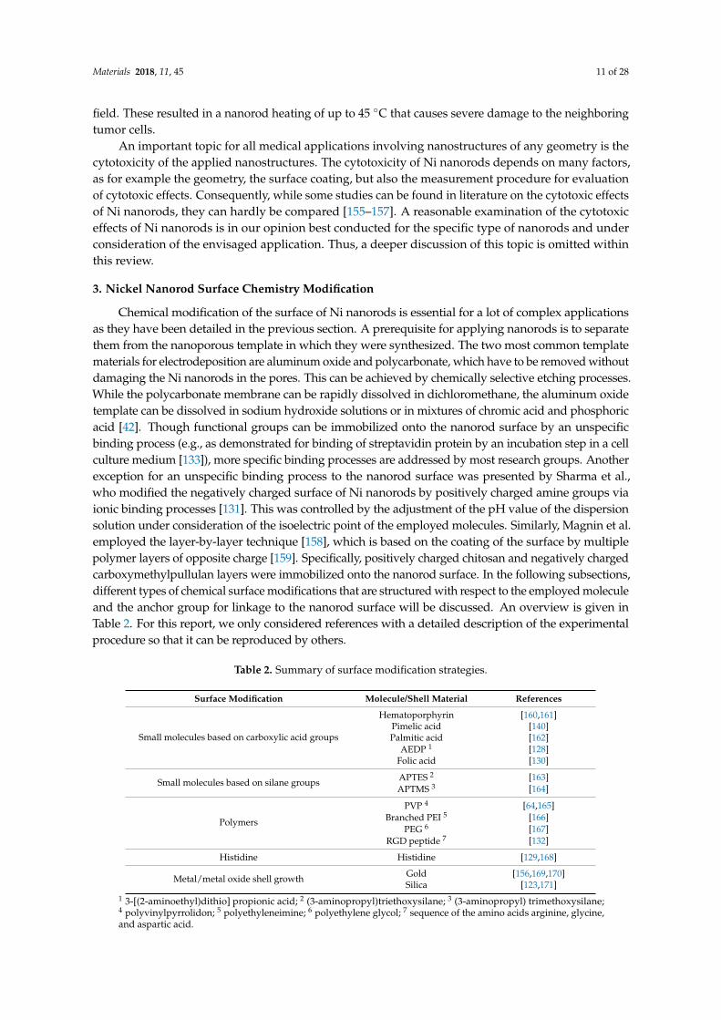
Table 2. Summary of surface modification strategies.
Surface Modification Molecule/Shell Material References
Small molecules based on carboxylic acid groups
Hematoporphyrin [160,161]Pimelic acid [140]Palmitic acid [162]
AEDP 1 [128]Folic acid [130]
Small molecules based on silane groups APTES 2 [163]APTMS 3 [164]
Polymers
PVP 4 [64,165]Branched PEI 5 [166]
PEG 6 [167]RGD peptide 7 [132]
Histidine Histidine [129,168]
Metal/metal oxide shell growth Gold [156,169,170]Silica [123,171]
1 3-[(2-aminoethyl)dithio] propionic acid; 2 (3-aminopropyl)triethoxysilane; 3 (3-aminopropyl) trimethoxysilane;4 polyvinylpyrrolidon; 5 polyethyleneimine; 6 polyethylene glycol; 7 sequence of the amino acids arginine, glycine,and aspartic acid.
Materials 2018, 11, 45 12 of 28
Commonly and as mentioned above, the surface modification of the synthesized nanorods isemployed after removal of the nanoporous template, but it is also possible to modify the walls ofthe pore channels before electrodeposition to yield nanorods with a specific surface functional group.An example for a surface modification inside the pore channels before electrodeposition was presentedby Skinner et al., who synthesized segmented Au-CdSe nanorods with exclusive modification ofthe Au sections [172]. Specifically, they employed vapor deposition to cover the walls of the porechannels by (3-mercaptopropyl)trimethoxysilane (MPTMS), a molecule comprising a silane groupand a thiol group separated by a short carbon chain. Modification of the nanoporous template wasexecuted before each Au deposition step through the silane group that reacted with the aluminumoxide, and once the gold was deposited, the thiol groups attached to the gold surface. A cleaning stepto remove the MPTMS molecules from the surface was executed by an oxygen plasma etching processbefore electrodepositing CdSe. Channel wall surface modification prior to the electrodeposition wasalso presented by Sanz et al. for Ni nanorods, which were coated by a polystyrene layer inside thepores [173]. This was achieved via formation of polystyrene nanotubes in the pores of an aluminatemplate by placing a polystyrene film on top of the template, followed by an incubation at 200 ◦Cin a nitrogen atmosphere, which resulted in the adhesion of a polymer film on the pore walls. Next,the polystyrene nanotubes were filled with Ni in an electrodeposition step, and the alumina templatewas removed in a sodium hydroxide solution so that, finally, Ni nanorods in a polystyrene shell wereobtained in solution.
3.1. Small Molecules Based on Carboxylic Acid Groups
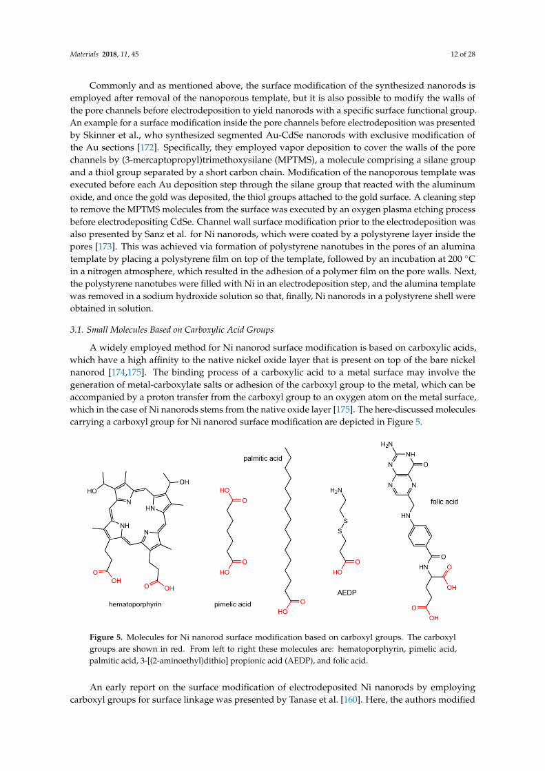
A widely employed method for Ni nanorod surface modification is based on carboxylic acids,which have a high affinity to the native nickel oxide layer that is present on top of the bare nickelnanorod [174,175]. The binding process of a carboxylic acid to a metal surface may involve thegeneration of metal-carboxylate salts or adhesion of the carboxyl group to the metal, which can beaccompanied by a proton transfer from the carboxyl group to an oxygen atom on the metal surface,which in the case of Ni nanorods stems from the native oxide layer [175]. The here-discussed moleculescarrying a carboxyl group for Ni nanorod surface modification are depicted in Figure 5.
Materials 2018, 11, 45 12 of 28
Commonly and as mentioned above, the surface modification of the synthesized nanorods is employed after removal of the nanoporous template, but it is also possible to modify the walls of the pore channels before electrodeposition to yield nanorods with a specific surface functional group. An example for a surface modification inside the pore channels before electrodeposition was presented by Skinner et al., who synthesized segmented Au-CdSe nanorods with exclusive modification of the Au sections [172]. Specifically, they employed vapor deposition to cover the walls of the pore channels by (3-mercaptopropyl)trimethoxysilane (MPTMS), a molecule comprising a silane group and a thiol group separated by a short carbon chain. Modification of the nanoporous template was executed before each Au deposition step through the silane group that reacted with the aluminum oxide, and once the gold was deposited, the thiol groups attached to the gold surface. A cleaning step to remove the MPTMS molecules from the surface was executed by an oxygen plasma etching process before electrodepositing CdSe. Channel wall surface modification prior to the electrodeposition was also presented by Sanz et al. for Ni nanorods, which were coated by a polystyrene layer inside the pores [173]. This was achieved via formation of polystyrene nanotubes in the pores of an alumina template by placing a polystyrene film on top of the template, followed by an incubation at 200 °C in a nitrogen atmosphere, which resulted in the adhesion of a polymer film on the pore walls. Next, the polystyrene nanotubes were filled with Ni in an electrodeposition step, and the alumina template was removed in a sodium hydroxide solution so that, finally, Ni nanorods in a polystyrene shell were obtained in solution.
3.1. Small Molecules Based On Carboxylic Acid Groups
A widely employed method for Ni nanorod surface modification is based on carboxylic acids, which have a high affinity to the native nickel oxide layer that is present on top of the bare nickel nanorod [174,175]. The binding process of a carboxylic acid to a metal surface may involve the generation of metal-carboxylate salts or adhesion of the carboxyl group to the metal, which can be accompanied by a proton transfer from the carboxyl group to an oxygen atom on the metal surface, which in the case of Ni nanorods stems from the native oxide layer [175]. The here-discussed molecules carrying a carboxyl group for Ni nanorod surface modification are depicted in Figure 5.
Figure 5. Molecules for Ni nanorod surface modification based on carboxyl groups. The carboxyl groups are shown in red. From left to right these molecules are: hematoporphyrin, pimelic acid, palmitic acid, 3-[(2-aminoethyl)dithio] propionic acid (AEDP), and folic acid.
An early report on the surface modification of electrodeposited Ni nanorods by employing carboxyl groups for surface linkage was presented by Tanase et al. [160]. Here, the authors modified the Ni nanorod surface by Hematoporphyrin IX, a fluorescent porphyrin that possesses two carboxyl groups for the binding process. The modification of the nanorod surface was achieved after
Figure 5. Molecules for Ni nanorod surface modification based on carboxyl groups. The carboxylgroups are shown in red. From left to right these molecules are: hematoporphyrin, pimelic acid,palmitic acid, 3-[(2-aminoethyl)dithio] propionic acid (AEDP), and folic acid.
An early report on the surface modification of electrodeposited Ni nanorods by employingcarboxyl groups for surface linkage was presented by Tanase et al. [160]. Here, the authors modified
Materials 2018, 11, 45 13 of 28
the Ni nanorod surface by Hematoporphyrin IX, a fluorescent porphyrin that possesses two carboxylgroups for the binding process. The modification of the nanorod surface was achieved after dissolvingthe alumina template in a solution of potassium hydroxide to remove the nanorods from thetemplate. This was followed by a washing and transfer step of the nanorods in solution to ethanol bycentrifugation and magnetic separation using permanent magnets. The binding of HematoporphyrinIX was executed by mixing the porphyrin with the nanorods and immersion for 24 h at roomtemperature followed by another solvent washing step with ethanol to remove unbound porphyrinspecies. This procedure was expanded to also transfer the nanorods in solvents of water and ethyleneglycol [161].
Gao et al. presented a method to functionalize the nanorod surface by biotinylated antibodies viamodification of the nickel surface with carboxyl groups, followed by binding of a streptavidin protein layer,which has a very high affinity to the biotin groups of the antibody [140]. The reported protocol involvedthe use of pimelic acid, a dicarboxylic acid molecule that serves for both the binding to the nickel nanorodsurface and as anchor for amine groups of the streptavidin. The latter is subsequently bound to theseanchors via EDC/S-NHS linker chemistry (EDC: N-(3-(Dimethylamino)propyl)-N′-ethylcarbodiimidehydrochloride; S-NHS: N-Hydroxysulfosuccinimide sodium salt). In a first step, the alumina templatewas dissolved in a sodium hydroxide solution, and the nanorods were transferred into ethanol bycentrifugation. Then, the nanorods were immersed in ethanol containing pimelic acid and incubatedfor 24 h. The EDC/S-NHS coupling chemistry was conducted by adding phosphate buffered saline(PBS) solutions of EDC and S-NHS together with the streptavidin protein, followed by incubation for 3 h.Finally, the biotinylated antibody was immobilized by incubation with the streptavidin-coated nanorodsfor 30 min in PBS solution. Washing and solvent transfer steps were conducted after each individualsurface modification step.
Several examples for surface modification of segmented Ni-Au nanorods can be found inliterature, all of which employ thiol groups for modifying the gold segments and carboxyl groupsfor nickel surface modification [128,130,132,162]. Birenbaum et al. employed palmitic acid formodifying the surface of the Ni segments to yield hydrophobic Ni segments (due to the alkylgroup of palmitic acid) [162]. The palmitic acid was immobilized on the nanorods by immersionin a solution of ethanol overnight. Salem et al. presented functionalization of nickel segmentsby 3-[(2-aminoethyl)dithio] propionic acid (AEDP), which is a molecule providing a carboxyl andan amine group spaced by a short alkyl segment and a disulfide bridge [128]. Here, the binding wasachieved in a solution of AEDP in water after 24 h. At a pH value of 5.7, small plasmid molecules(circular double-stranded DNA) were bound by electrostatic interaction to the positively chargedamine groups of the AEDP in a further incubation step that lasted for 24 h. It was also reported to bindfolic acid to the Ni segments of a bimetallic nanorod via the carboxyl groups of folic acid by immersionin methanol at 4 ◦C for 12 h [130].
3.2. Small Molecules Based on Silane Groups
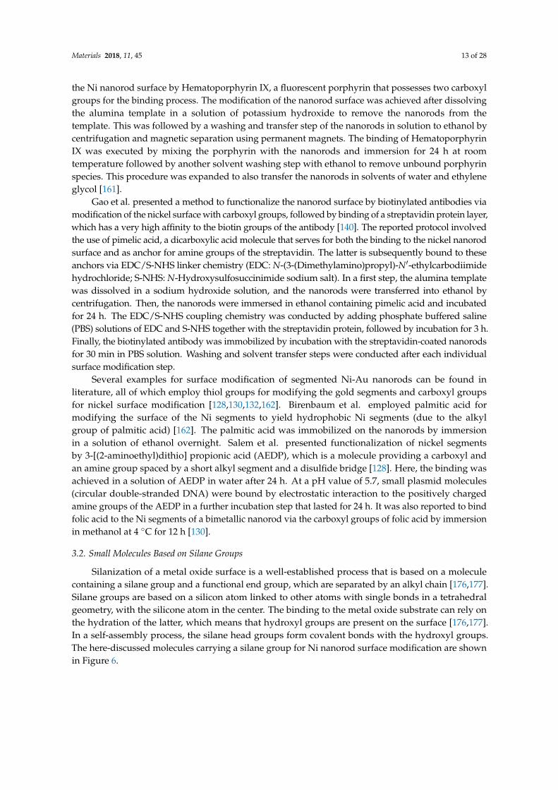
Silanization of a metal oxide surface is a well-established process that is based on a moleculecontaining a silane group and a functional end group, which are separated by an alkyl chain [176,177].Silane groups are based on a silicon atom linked to other atoms with single bonds in a tetrahedralgeometry, with the silicone atom in the center. The binding to the metal oxide substrate can rely onthe hydration of the latter, which means that hydroxyl groups are present on the surface [176,177].In a self-assembly process, the silane head groups form covalent bonds with the hydroxyl groups.The here-discussed molecules carrying a silane group for Ni nanorod surface modification are shownin Figure 6.
Materials 2018, 11, 45 14 of 28Materials 2018, 11, 45 14 of 28
Figure 6. Molecules for Ni nanorod surface modification based on silane groups. The silane groups are shown in red. From left to right, these molecules are: (3-aminopropyl)triethoxysilane (APTES) and (3-aminopropyl) trimethoxysilane (APTMS).
Wildt et al. presented surface modification of Ni in segmented Ni-Au nanorods by using a silane group for the coupling reaction to the native oxide on the nickel segment [163]. Here, the authors first dissolved the alumina template in a solution of potassium hydroxide, followed by washing and solvent transfer steps to yield nanorod solutions in ethanol. Coupling of a silane group to the nanorod surface was executed by immersion of the nanorods in ethanol with (3-aminopropyl)triethoxysilane overnight. This resulted in a nanorod surface that is now terminated by amine groups that can be further employed to do a subsequent surface modification step, e.g., by N-hydroxysuccinimide esters. Wildt et al. employed a methoxypoly(ethylene glycol) succinate N-hydroxysuccinimide ester to bind polyethylene glycol to the nanorod surface, which was done by immersion for 1 hour in a solution of sodium bicarbonate.
Recently, Kozlovskiy et al. demonstrated the surface modification of Ni nanotubes by (3-aminopropyl) trimethoxysilane, which was achieved by mixing the nanotubes with the silane reagent in ethanol [164]. The so obtained solution was incubated for 12 h after an initial sonication step. The resultant amine terminated surface was further modified to bind bovine serum albumin (BSA) protein via its carboxyl groups. The protein linking was done in an acetate buffer solution of acidic pH and under addition of N-(3-(Dimethylamino)propyl)-N′-ethylcarbodiimide and pentafluorophenol to activate carboxyl groups of BSA, thus making them bind to the amine groups on the nanorods.
3.3. Surface Modification by Polymers
The use of polymers for nanoparticle surface coating offers some distinct advantages. Very important is the enhanced steric interaction of nanoparticles in solution that results in better stabilization of the nanoparticle dispersion, which is due to the large molecular weight of polymers. Polymers can be synthesized comprising different structural subgroups to allow both further functionalization by additional molecules and binding to the nanoparticle surface. Reviews can be found in the literature for a detailed overview on the application of polymers for nanoparticle surface coating [178–181]. The here-discussed polymers are sketched in Figure 7.
Polyvinylpyrrolidone (PVP) binds to the surface of metal and metal oxide nanoparticles through the carbonyl group of the lactam ring [182]. This can be exploited to coat the surface of Ni nanorods with PVP as dispersion stabilizing surfactant, which has been reported by adding PVP to either a 20 mM sodium hydroxide solution or a sodium hydroxide solution at pH 11.5 [64,165]. The so obtained solution was employed for slowly dissolving the aluminum oxide of the template with the goal that the PVP binds to the nanorods before the template is entirely dissolved. A nanorod dispersion in water was obtained by succeeding washing steps that were assisted by centrifugation and permanent magnets [64,165].
Figure 6. Molecules for Ni nanorod surface modification based on silane groups. The silane groups areshown in red. From left to right, these molecules are: (3-aminopropyl)triethoxysilane (APTES) and(3-aminopropyl) trimethoxysilane (APTMS).
Wildt et al. presented surface modification of Ni in segmented Ni-Au nanorods by using a silanegroup for the coupling reaction to the native oxide on the nickel segment [163]. Here, the authorsfirst dissolved the alumina template in a solution of potassium hydroxide, followed by washing andsolvent transfer steps to yield nanorod solutions in ethanol. Coupling of a silane group to the nanorodsurface was executed by immersion of the nanorods in ethanol with (3-aminopropyl)triethoxysilaneovernight. This resulted in a nanorod surface that is now terminated by amine groups that can befurther employed to do a subsequent surface modification step, e.g., by N-hydroxysuccinimide esters.Wildt et al. employed a methoxypoly(ethylene glycol) succinate N-hydroxysuccinimide ester to bindpolyethylene glycol to the nanorod surface, which was done by immersion for 1 hour in a solution ofsodium bicarbonate.
Recently, Kozlovskiy et al. demonstrated the surface modification of Ni nanotubes by(3-aminopropyl) trimethoxysilane, which was achieved by mixing the nanotubes with the silanereagent in ethanol [164]. The so obtained solution was incubated for 12 h after an initial sonication step.The resultant amine terminated surface was further modified to bind bovine serum albumin (BSA)protein via its carboxyl groups. The protein linking was done in an acetate buffer solution of acidic pHand under addition of N-(3-(Dimethylamino)propyl)-N′-ethylcarbodiimide and pentafluorophenol toactivate carboxyl groups of BSA, thus making them bind to the amine groups on the nanorods.
3.3. Surface Modification by Polymers
The use of polymers for nanoparticle surface coating offers some distinct advantages.Very important is the enhanced steric interaction of nanoparticles in solution that results in betterstabilization of the nanoparticle dispersion, which is due to the large molecular weight of polymers.Polymers can be synthesized comprising different structural subgroups to allow both furtherfunctionalization by additional molecules and binding to the nanoparticle surface. Reviews canbe found in the literature for a detailed overview on the application of polymers for nanoparticlesurface coating [178–181]. The here-discussed polymers are sketched in Figure 7.
Materials 2018, 11, 45 15 of 28
Materials 2018, 11, 45 15 of 28
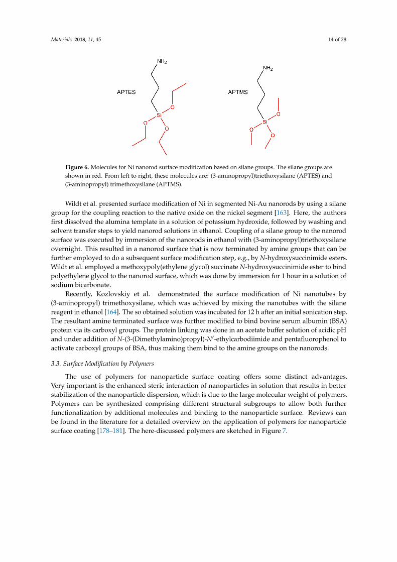
Figure 7. Polymers for Ni nanorod surface modification. From left to right, these polymers are: polyvinylpyrrolidone (PVP), branched polyethyleneimine (PEI), polyethylene glycol (PEG), and a RGD peptide bound to a polyethylene glycol modified with a carboxyl and an amine group. Carboxyl groups are shown in red. The amine group of the PEG is bound to the RGD peptide via EDC/NHS linker chemistry.
It is reported in the literature that branched polyethyleneimine (PEI) can be used for surface modification of Ni nanorods [166]. To that end, an aqueous solution of branched PEI at a pH of 5.5 was added to an aqueous solution of Ni nanorods and stirred for 1 h. Though this protocol is mainly based on electrostatic binding of branched PEI to the nanorod surface, hydrogen bonding between the amino groups of the polymer and the hydrogen oxide of the native oxide may also be involved in the surface coating process. The advantage of using branched PEI is that it can be modified prior to the nanorod surface coating step by making use of the amino groups of the polymer. This was shown by labeling the branched PEI with a fluorescent organic dye to yield fluorescent nanorods.
An alternative approach to yield polymer coated Ni nanorods was presented by Tripathy et al., who added polyethylene glycol (PEG) to the electrodeposition solution containing the Ni cations (Ni2+) [167]. This lead to the formation of coordination bonds between the metal cations and the polymer. By employing this solution for the electrodeposition process into alumina templates, the polymer was deposited at the same time as the nickel. The encapsulation effect of the nickel nanorod by the polymer was due to the positively charged pore walls that lead to the reduction of the Ni cations in the pore center, while the polymer adsorbed to the pore walls. After immersion of the template in a 0.1 M sodium hydroxide solution overnight, nanorod dispersions were obtained.
Finally, Son et al. applied immersion in methanol at 4 °C to bind RGD peptides (a sequence of the three amino acids arginine, glycine, and aspartic acid) to the Ni nanorod surface [132]. Here, for linking to the nanorod, a polyethylene glycol (PEG) spacer comprising an amine and a carboxyl group was applied. First, the PEG was bound by EDC/NHS linker chemistry with its amine end to the peptide (specifically to the carboxyl groups of the aspartic acid), followed by another binding step of the carboxyl end group to the nanorod.
3.4. Surface Modification by Histidine
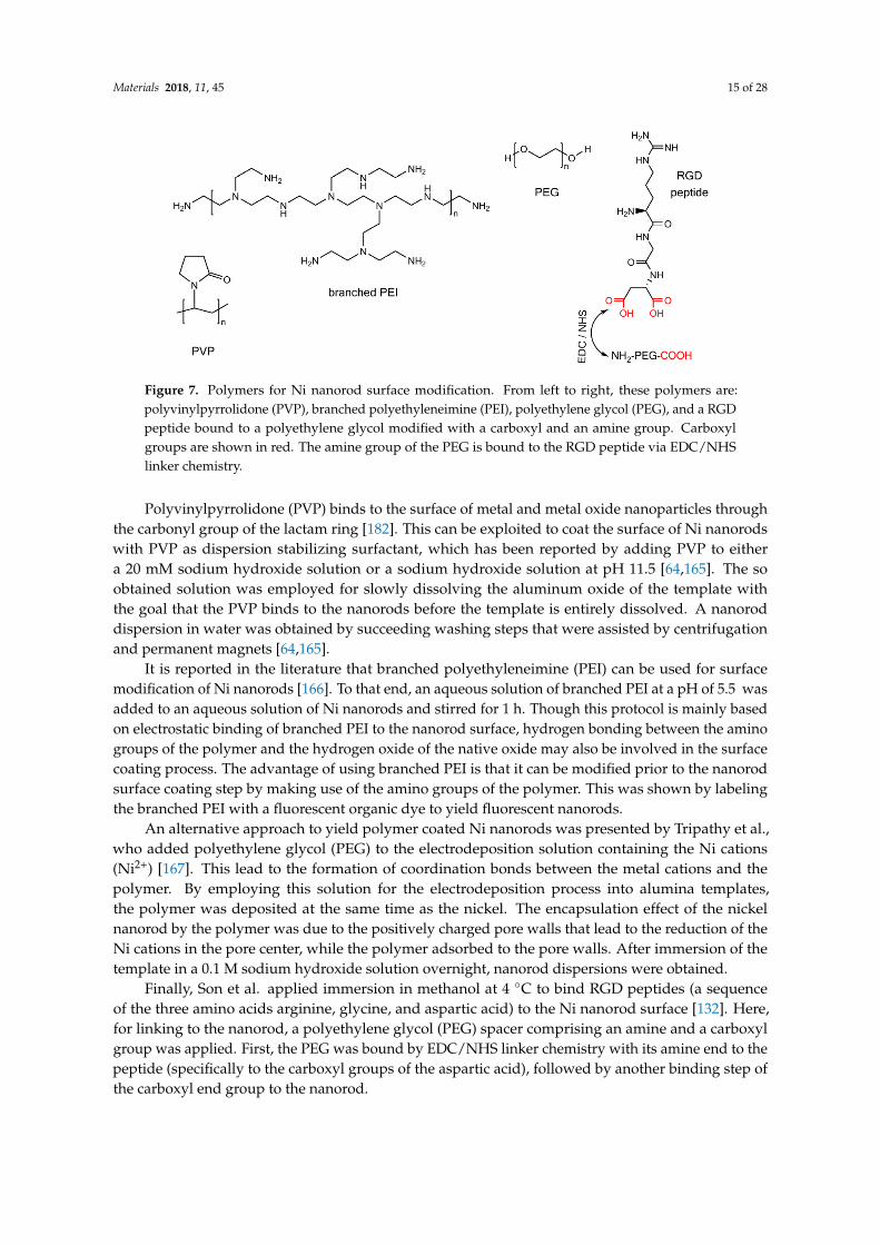
Proteins with histidine amino acid side chains are reported to have a high affinity for nickel, which is also employed for protein purification processes [183,184]. Thus, molecules with a histidine tag can be employed for surface modification of Ni nanorods. The structure of histidine is depicted in Figure 8, with the imidazole group that is a part of the amino acid side chain shown in red.
Figure 7. Polymers for Ni nanorod surface modification. From left to right, these polymers are:polyvinylpyrrolidone (PVP), branched polyethyleneimine (PEI), polyethylene glycol (PEG), and a RGDpeptide bound to a polyethylene glycol modified with a carboxyl and an amine group. Carboxylgroups are shown in red. The amine group of the PEG is bound to the RGD peptide via EDC/NHSlinker chemistry.
Polyvinylpyrrolidone (PVP) binds to the surface of metal and metal oxide nanoparticles throughthe carbonyl group of the lactam ring [182]. This can be exploited to coat the surface of Ni nanorodswith PVP as dispersion stabilizing surfactant, which has been reported by adding PVP to eithera 20 mM sodium hydroxide solution or a sodium hydroxide solution at pH 11.5 [64,165]. The soobtained solution was employed for slowly dissolving the aluminum oxide of the template withthe goal that the PVP binds to the nanorods before the template is entirely dissolved. A nanoroddispersion in water was obtained by succeeding washing steps that were assisted by centrifugationand permanent magnets [64,165].
It is reported in the literature that branched polyethyleneimine (PEI) can be used for surfacemodification of Ni nanorods [166]. To that end, an aqueous solution of branched PEI at a pH of 5.5 wasadded to an aqueous solution of Ni nanorods and stirred for 1 h. Though this protocol is mainly basedon electrostatic binding of branched PEI to the nanorod surface, hydrogen bonding between the aminogroups of the polymer and the hydrogen oxide of the native oxide may also be involved in the surfacecoating process. The advantage of using branched PEI is that it can be modified prior to the nanorodsurface coating step by making use of the amino groups of the polymer. This was shown by labelingthe branched PEI with a fluorescent organic dye to yield fluorescent nanorods.
An alternative approach to yield polymer coated Ni nanorods was presented by Tripathy et al.,who added polyethylene glycol (PEG) to the electrodeposition solution containing the Ni cations(Ni2+) [167]. This lead to the formation of coordination bonds between the metal cations and thepolymer. By employing this solution for the electrodeposition process into alumina templates,the polymer was deposited at the same time as the nickel. The encapsulation effect of the nickelnanorod by the polymer was due to the positively charged pore walls that lead to the reduction of theNi cations in the pore center, while the polymer adsorbed to the pore walls. After immersion of thetemplate in a 0.1 M sodium hydroxide solution overnight, nanorod dispersions were obtained.
Finally, Son et al. applied immersion in methanol at 4 ◦C to bind RGD peptides (a sequenceof the three amino acids arginine, glycine, and aspartic acid) to the Ni nanorod surface [132]. Here,for linking to the nanorod, a polyethylene glycol (PEG) spacer comprising an amine and a carboxylgroup was applied. First, the PEG was bound by EDC/NHS linker chemistry with its amine end to thepeptide (specifically to the carboxyl groups of the aspartic acid), followed by another binding step ofthe carboxyl end group to the nanorod.
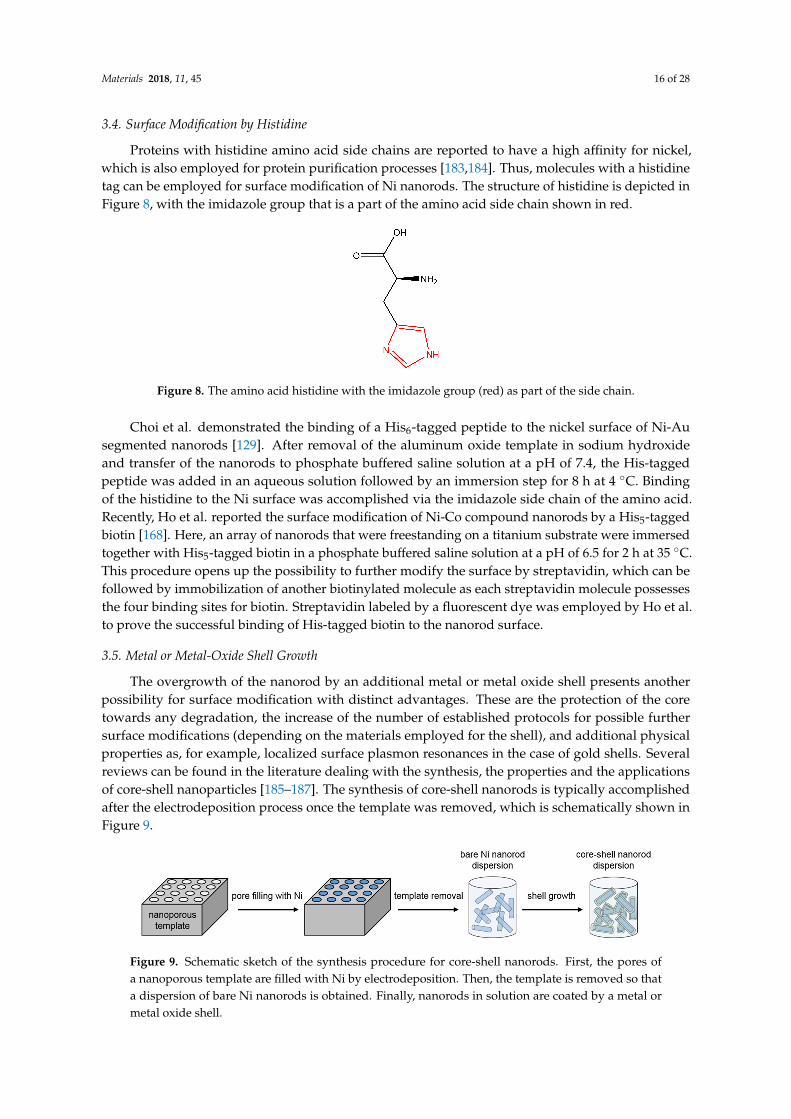
Materials 2018, 11, 45 16 of 28
3.4. Surface Modification by Histidine
Proteins with histidine amino acid side chains are reported to have a high affinity for nickel,which is also employed for protein purification processes [183,184]. Thus, molecules with a histidinetag can be employed for surface modification of Ni nanorods. The structure of histidine is depicted inFigure 8, with the imidazole group that is a part of the amino acid side chain shown in red.Materials 2018, 11, 45 16 of 28
Figure 8. The amino acid histidine with the imidazole group (red) as part of the side chain.
Choi et al. demonstrated the binding of a His6-tagged peptide to the nickel surface of Ni-Au segmented nanorods [129]. After removal of the aluminum oxide template in sodium hydroxide and transfer of the nanorods to phosphate buffered saline solution at a pH of 7.4, the His-tagged peptide was added in an aqueous solution followed by an immersion step for 8 h at 4 °C. Binding of the histidine to the Ni surface was accomplished via the imidazole side chain of the amino acid. Recently, Ho et al. reported the surface modification of Ni-Co compound nanorods by a His5-tagged biotin [168]. Here, an array of nanorods that were freestanding on a titanium substrate were immersed together with His5-tagged biotin in a phosphate buffered saline solution at a pH of 6.5 for 2 h at 35 °C. This procedure opens up the possibility to further modify the surface by streptavidin, which can be followed by immobilization of another biotinylated molecule as each streptavidin molecule possesses the four binding sites for biotin. Streptavidin labeled by a fluorescent dye was employed by Ho et al. to prove the successful binding of His-tagged biotin to the nanorod surface.
3.5. Metal or Metal-Oxide Shell Growth
The overgrowth of the nanorod by an additional metal or metal oxide shell presents another possibility for surface modification with distinct advantages. These are the protection of the core towards any degradation, the increase of the number of established protocols for possible further surface modifications (depending on the materials employed for the shell), and additional physical properties as, for example, localized surface plasmon resonances in the case of gold shells. Several reviews can be found in the literature dealing with the synthesis, the properties and the applications of core-shell nanoparticles [185–187]. The synthesis of core-shell nanorods is typically accomplished after the electrodeposition process once the template was removed, which is schematically shown in Figure 9.
Figure 9. Schematic sketch of the synthesis procedure for core-shell nanorods. First, the pores of a nanoporous template are filled with Ni by electrodeposition. Then, the template is removed so that a dispersion of bare Ni nanorods is obtained. Finally, nanorods in solution are coated by a metal or metal oxide shell.
The overgrowth of electrodeposited Ni nanorods by a gold shell was accomplished by Jeon et al. employing an electroless gold plating process [169]. To that end, Ni nanorods were first released from the alumina template by dissolving the aluminum oxide in a solution of sodium hydroxide. The dispersed nanorods in solution were then washed and transferred to water. Gold shell synthesis was achieved by immersing the nanorods in an aqueous potassium gold cyanide solution at a temperature
Figure 8. The amino acid histidine with the imidazole group (red) as part of the side chain.
Choi et al. demonstrated the binding of a His6-tagged peptide to the nickel surface of Ni-Ausegmented nanorods [129]. After removal of the aluminum oxide template in sodium hydroxideand transfer of the nanorods to phosphate buffered saline solution at a pH of 7.4, the His-taggedpeptide was added in an aqueous solution followed by an immersion step for 8 h at 4 ◦C. Bindingof the histidine to the Ni surface was accomplished via the imidazole side chain of the amino acid.Recently, Ho et al. reported the surface modification of Ni-Co compound nanorods by a His5-taggedbiotin [168]. Here, an array of nanorods that were freestanding on a titanium substrate were immersedtogether with His5-tagged biotin in a phosphate buffered saline solution at a pH of 6.5 for 2 h at 35 ◦C.This procedure opens up the possibility to further modify the surface by streptavidin, which can befollowed by immobilization of another biotinylated molecule as each streptavidin molecule possessesthe four binding sites for biotin. Streptavidin labeled by a fluorescent dye was employed by Ho et al.to prove the successful binding of His-tagged biotin to the nanorod surface.
3.5. Metal or Metal-Oxide Shell Growth
The overgrowth of the nanorod by an additional metal or metal oxide shell presents anotherpossibility for surface modification with distinct advantages. These are the protection of the coretowards any degradation, the increase of the number of established protocols for possible furthersurface modifications (depending on the materials employed for the shell), and additional physicalproperties as, for example, localized surface plasmon resonances in the case of gold shells. Severalreviews can be found in the literature dealing with the synthesis, the properties and the applicationsof core-shell nanoparticles [185–187]. The synthesis of core-shell nanorods is typically accomplishedafter the electrodeposition process once the template was removed, which is schematically shown inFigure 9.
Materials 2018, 11, 45 16 of 28
Figure 8. The amino acid histidine with the imidazole group (red) as part of the side chain.
Choi et al. demonstrated the binding of a His6-tagged peptide to the nickel surface of Ni-Au segmented nanorods [129]. After removal of the aluminum oxide template in sodium hydroxide and transfer of the nanorods to phosphate buffered saline solution at a pH of 7.4, the His-tagged peptide was added in an aqueous solution followed by an immersion step for 8 h at 4 °C. Binding of the histidine to the Ni surface was accomplished via the imidazole side chain of the amino acid. Recently, Ho et al. reported the surface modification of Ni-Co compound nanorods by a His5-tagged biotin [168]. Here, an array of nanorods that were freestanding on a titanium substrate were immersed together with His5-tagged biotin in a phosphate buffered saline solution at a pH of 6.5 for 2 h at 35 °C. This procedure opens up the possibility to further modify the surface by streptavidin, which can be followed by immobilization of another biotinylated molecule as each streptavidin molecule possesses the four binding sites for biotin. Streptavidin labeled by a fluorescent dye was employed by Ho et al. to prove the successful binding of His-tagged biotin to the nanorod surface.
3.5. Metal or Metal-Oxide Shell Growth
The overgrowth of the nanorod by an additional metal or metal oxide shell presents another possibility for surface modification with distinct advantages. These are the protection of the core towards any degradation, the increase of the number of established protocols for possible further surface modifications (depending on the materials employed for the shell), and additional physical properties as, for example, localized surface plasmon resonances in the case of gold shells. Several reviews can be found in the literature dealing with the synthesis, the properties and the applications of core-shell nanoparticles [185–187]. The synthesis of core-shell nanorods is typically accomplished after the electrodeposition process once the template was removed, which is schematically shown in Figure 9.
Figure 9. Schematic sketch of the synthesis procedure for core-shell nanorods. First, the pores of a nanoporous template are filled with Ni by electrodeposition. Then, the template is removed so that a dispersion of bare Ni nanorods is obtained. Finally, nanorods in solution are coated by a metal or metal oxide shell.
The overgrowth of electrodeposited Ni nanorods by a gold shell was accomplished by Jeon et al. employing an electroless gold plating process [169]. To that end, Ni nanorods were first released from the alumina template by dissolving the aluminum oxide in a solution of sodium hydroxide. The dispersed nanorods in solution were then washed and transferred to water. Gold shell synthesis was achieved by immersing the nanorods in an aqueous potassium gold cyanide solution at a temperature
Figure 9. Schematic sketch of the synthesis procedure for core-shell nanorods. First, the pores ofa nanoporous template are filled with Ni by electrodeposition. Then, the template is removed so thata dispersion of bare Ni nanorods is obtained. Finally, nanorods in solution are coated by a metal ormetal oxide shell.
Materials 2018, 11, 45 17 of 28
The overgrowth of electrodeposited Ni nanorods by a gold shell was accomplished by Jeon et al.employing an electroless gold plating process [169]. To that end, Ni nanorods were first releasedfrom the alumina template by dissolving the aluminum oxide in a solution of sodium hydroxide.The dispersed nanorods in solution were then washed and transferred to water. Gold shell synthesiswas achieved by immersing the nanorods in an aqueous potassium gold cyanide solution ata temperature of 90 ◦C. The so-fabricated core-shell nanorods were functionalized by using thiollinkers for binding to the gold shell.
Pondman et al. reported the deposition of a gold shell onto the surface of Ni nanorods thatwere electrodeposited into polycarbonate membranes [156]. After washing steps and transfer ofthe nanorods into aqueous solution assisted by centrifugation, the nanorods were coated by a goldlayer based on a method presented by Kohli et al. for electroless synthesis of gold nanorods [188].Here, we briefly describe the procedure, while the details can be found in the original report. First,the nanorods were coated with aminoundecanoic acid, which resulted in the formation of aminegroups on top of the nanorod surface [156]. In a subsequent step, tin cations (Sn2+) were bound to theamine groups by immersion in a tin chloride solution, followed by oxidation to Sn4+ by silver nitrate,which resulted in the formation of a layer of silver atoms on top of the nanorod surface. This silverlayer was then replaced by gold atoms via immersion into a commercially available gold solution,which was followed by a continuous growth of a metallic gold layer. Biofunctionalization of the goldlayer was then possible by the well-known interaction of gold with thiol groups.
Serrà et al. recently presented the deposition of gold onto electrodeposited Ni nanorods byimmersing the nanorods in an aqueous solution of chloroauric acid, which lead to the formation ofgold by galvanic displacement [170]. A subsequent functionalization step was carried out by bindinga polyethylene glycol polymer comprising a thiol group to the nanorod surface.
Nanoparticles can also be coated by a shell of silica (silicon dioxide), which is a biocompatiblematerial that protects the core and allows for further functionalization of the surface, e.g., by silanegroups. A widely employed method for the growth of a silica layer is the so called Stöber process,where the silica shell is grown by condensation of the precursor tetraethoxysilane (TEOS) in a solutionof ethanol and ammonia [189]. Pinheiro et al. employed the silica shell growth for electrodeposited Ninanorods once they were released from the alumina template and transferred to ethanol [123]. Specifically,they employed co-condensation of TEOS and siloxydithiocarbamate to generate dithiocarbamategroups on the silica shell. Dithiocarbamate groups have a high binding affinity for heavy metal ions.Here, the siloxydithiocarbamate precursor was prepared prior to the silica shell growth by linkinga dithiocarbamate group to (3-aminopropyl)triethoxysilane.
A further possibility to overcoat a nanorod surface by a silica shell was presented byGraf et al. [190]. Here, they coated a range of different nanoparticles by employing PVP as a sub-layerfor subsequent silica coating in a Stöber growth process. This has been employed for electrodepositedNi nanorods that were released form the template in a sodium hydroxide solution containing PVP ata pH of 11.5 [171]. After some washing steps and a transfer to water, the nanorods were dispersed inan aqueous solution of 2-propanol and ammonia (25%), and then TEOS was added to induce the silicashell growth. After additional washing steps assisted by centrifugation and magnetic separation, silicacoated Ni nanorod dispersions in water were obtained.
4. Conclusions and Outlook
In this review, we focused on Ni nanorods that have been fabricated by electrodeposition intoporous membranes. Such ferromagnetic nanorods offer the advantage that they already intrinsicallypossess different functionalities. Specifically, their elongated form leads to magnetic shape anisotropythat fixes their magnetic moment in the direction of the long nanorod axis. At the same time,the rod-shaped form also results in an optical anisotropy. Consequently, it is possible to examine theactual nanorod orientation in a solution either by magnetic or optical sensing methods. Moreover,
Materials 2018, 11, 45 18 of 28
the orientation of nanorods can be manipulated by external magnetic fields, so that a wide field ofapplications can be realized.
We presented the extensive scope of applications of these nanorods, which span from electronicsand data storage to biosensing and to cell biology, including both fundamental cell biology andcancer treatment. The second main part of the review focused on nanorod surface modificationstrategies to allow for fabrication of customized nanorods that can be applied for a specific application.A suitable surface modification of nanorods is crucial for their application in complex measurementsettings. Thus, it will play an important role for a future broadening of the range of Ni nanorodapplications. Regarding this broadening of the applicability, several different possibilities can beeasily envisaged. Firstly, segmented nanorods with different materials to increase the functionalityof the bare nanorods itself present a promising future direction that can open up new fields ofapplication. Secondly, Ni nanorods could be employed for all imaging and detection techniquesthat rely on either a magnetic or an optical detection. Examples are magnetic particle imaging,photoacoustic imaging or optical coherence tomography, next to the already presented magneticresonance imaging and surface-enhanced Raman spectroscopy. This is of special interest whenbiofunctionalized nanorods can be employed. Alternatively, one might expect that Ni nanorods can beemployed for any kind of tracking measurements, as nanorods can be sensitively detected by theirmagnetic and optical characteristics. An example would be to track the path of a sample fluid withina complex setting. Moreover, Ni nanorods can be employed for measurements at a single cell level toexamine processes inside living cells. Thirdly, in order to broaden the applicability of Ni nanorods,the surface modification can be improved. This accounts for advanced protocols for the binding ofantibodies and peptides, which will be an improvement for all sensing and biosensing applications.Furthermore, a surface modification that results in nanorods that withstand harsh environmentalconditions can trigger the development of new applications. Finally, the synthesis of more complexpolymers that already possess an intrinsic multifunctionality can be an important future step. In thefollowing final paragraphs, we will discuss alternative possible surface modification approaches forwhich, to our knowledge, no protocol for the here discussed Ni nanorods can be found in the literature.This, for sure, is an incomplete list, but it might be a motivation that triggers new developments.
As discussed above, carboxylic acid groups can be employed for Ni nanorod surface coatingas a result of their affinity to nickel oxide. Similarly, all kinds of fatty acids could be employed forsurface modification. Moreover, polyacrylic acid (PAA) with a carboxyl group in every monomerunit is a promising candidate for Ni nanorod surface modification. It offers the additional advantagethat some of the carboxyl groups of the polymer can be used to modify the polymer by furtherfunctional molecules.
Alternatively, alkylphosphonic acids were reported to bind to native nickel oxide surfaces [191].Here, the presented immersion method can be a promising approach to attach alkylphosphonic acidsto electrodeposited Ni nanorods. These molecules, which comprise a phosphonic group, can alsobe modified with another functional group at the opposite end of the alkyl chain. For example,16-phosphonohexadecanoic acid that possesses a carboxyl group. This additional functionality couldthen be employed to add another molecule for further nanorod surface modification.
Another simple approach for a surface modification was based on the use of commercially availablespherical Ni nanoparticles. These were coated by the also commercially available Brij 76 ligand thathas poly(ethylene glycol) octadecyl ether as main ingredient, which allows for a transfer from aqueousto organic solutions [192]. This could be exploited for all applications that do not necessarily rely onwater-based nanorod dispersions.
Regarding the synthesis of metal or metal-oxide shells on top of the nanorod surface, variousalternative materials could be employed. For example, it has been reported that Ni nanorods that weresynthesized by a template-free method were coated by tellurium and also by zinc oxide shells [193].This resulted in hybrid nanorods with magnetic and semiconducting properties, which can also be
Materials 2018, 11, 45 19 of 28
a very interesting approach for template-synthesized nanorods. Therefore, an additional functionalitycould already be realized for the bare nanorod prior to any surface modification.
Finally, Solanki et al. immobilized protein A on the surface of nickel oxide nanowires [194].Though this method was based on an electrostatic interaction between the nickel oxide and protein A,the authors were able to demonstrate its application for biosensing. By making use of the well-knownaffinity of the Fc fragment of antibodies to protein A, they were able to further modify the nanowiresurface by antibodies. In principle, it could be possible to expand this approach to electrodeposited Ninanorods, but also to alternative established protein-antibody interactions. These approaches could bebased, for example, on protein G or on protein A/G.
In summary, it can be concluded that the already existing wide range of applications of Ninanorods will further grow in the future. This will be a result of the multifunctionality of these kindsof nanoparticles. The development of new advanced surface modification procedures will furtherboost the application of Ni nanorods and will open new fields of applications. Thus, one can be verycurious to see the advances of this scientific field in the coming years.
Acknowledgments: Parts of this work were supported by the Austrian Research Promotion Agency FFG (projectnumber 861414 LAMPION). Daniela Reichinger acknowledges funding by the Austrian Research PromotionAgency FFG within the frame of the FEMtech initiative.
Conflicts of Interest: The authors declare no conflict of interest.
References
1. Odobel, F.; Le Pleux, L.; Pellegrin, Y.; Blart, E. New photovoltaic devices based on the sensitization of p-typesemiconductors: Challenges and opportunities. Acc. Chem. Res. 2010, 43, 1063–1071. [CrossRef] [PubMed]
2. Granqvist, C.G. Electrochromics for smart windows. Oxide-based thin films and devices. Thin Solid Films2014, 564, 1–38. [CrossRef]
3. Morozov, Y.G.; Belousova, O.V.; Kuznetsov, M.V. Preparation of nickel nanoparticles for catalytic applications.Inorg. Mater. 2011, 47, 36–40. [CrossRef]
4. Zhuang, Z.; Giles, S.A.; Zheng, J.; Jenness, G.R.; Caratzoulas, S.; Vlachos, D.G.; Yan, Y. Nickel supportedon nitrogen-doped carbon nanotubes as hydrogen oxidation reaction catalyst in alkaline electrolyte.Nat. Commun. 2016, 7, 10141. [CrossRef] [PubMed]
5. Verrelli, E.; Tsoukalas, D.; Giannakopoulos, K.; Kouvatsos, D.; Normand, P.; Ioannou, D.E. Nickel nanoparticledeposition at room temperature for memory applications. Microelectron. Eng. 2007, 84, 1994–1997. [CrossRef]
6. Kudr, J.; Haddad, Y.; Richtera, L.; Heger, Z.; Cernak, M.; Adam, V.; Zitka, O. Magnetic Nanoparticles: FromDesign and Synthesis to Real World Applications. Nanomaterials 2017, 7, 243. [CrossRef] [PubMed]
7. Schrittwieser, S.; Pelaz, B.; Parak, W.; Lentijo-Mozo, S.; Soulantica, K.; Dieckhoff, J.; Ludwig, F.; Guenther, A.;Tschöpe, A.; Schotter, J. Homogeneous Biosensing Based on Magnetic Particle Labels. Sensors 2016, 16, 828.[CrossRef] [PubMed]
8. Xu, S.; Habib, A.H.; Pickel, A.D.; McHenry, M.E. Magnetic nanoparticle-based solder composites forelectronic packaging applications. Prog. Mater. Sci. 2015, 67, 95–160. [CrossRef]
9. Gómez-Pastora, J.; Dominguez, S.; Bringas, E.; Rivero, M.J.; Ortiz, I.; Dionysiou, D.D. Review and perspectiveson the use of magnetic nanophotocatalysts (MNPCs) in water treatment. Chem. Eng. J. 2017, 310, 407–427.[CrossRef]
10. Reddy, L.H.; Arias, J.L.; Nicolas, J.; Couvreur, P. Magnetic nanoparticles: Design and characterization,toxicity and biocompatibility, pharmaceutical and biomedical applications. Chem. Rev. 2012, 112, 5818–5878.[CrossRef] [PubMed]
11. Murphy, C.J.; Sau, T.K.; Gole, A.M.; Orendorff, C.J.; Gao, J.; Gou, L.; Hunyadi, S.E.; Li, T. Anisotropicmetal nanoparticles: Synthesis, assembly, and optical applications. J. Phys. Chem. B 2005, 109, 13857–13870.[CrossRef] [PubMed]
12. Koltypin, Y.; Fernandez, A.; Rojas, T.C.; Campora, J.; Palma, P.; Prozorov, R.; Gedanken, A. Encapsulation ofNickel Nanoparticles in Carbon Obtained by the Sonochemical Decomposition of Ni(C8H12)2. Chem. Mater.1999, 11, 1331–1335. [CrossRef]
Materials 2018, 11, 45 20 of 28
13. Ely, T.O.; Amiens, C.; Chaudret, B.; Snoeck, E.; Verelst, M.; Respaud, M.; Broto, J.-M. Synthesis of NickelNanoparticles. Influence of Aggregation Induced by Modification of Poly(vinylpyrrolidone) Chain Lengthon Their Magnetic Properties. Chem. Mater. 1999, 11, 526–529. [CrossRef]
14. Amiens, C.; Chaudret, B.; Ciuculescu-Pradines, D.; Collière, V.; Fajerwerg, K.; Fau, P.; Kahn, M.;Maisonnat, A.; Soulantica, K.; Philippot, K. Organometallic approach for the synthesis of nanostructures.New J. Chem. 2013, 37, 3374–3401. [CrossRef]
15. Dumestre, F.; Chaudret, B.; Amiens, C.; Respaud, M.; Fejes, P.; Renaud, P.; Zurcher, P. Unprecedentedcrystalline super-lattices of monodisperse cobalt nanorods. Angew. Chem. Int. Ed. 2003, 42, 5213–5216.[CrossRef] [PubMed]
16. Wetz, F.; Soulantica, K.; Respaud, M.; Falqui, A.; Chaudret, B. Synthesis and magnetic properties of Conanorod superlattices. Mater. Sci. Eng. C 2007, 27, 1162–1166. [CrossRef]
17. Cordente, N.; Respaud, M.; Senocq, F.; Casanove, M.-J.; Amiens, C.; Chaudret, B. Synthesis and MagneticProperties of Nickel Nanorods. Nano Lett. 2001, 1, 565–568. [CrossRef]
18. Soulantica, K.; Wetz, F.; Maynadié, J.; Falqui, A.; Tan, R.P.; Blon, T.; Chaudret, B.; Respaud, M. Magnetism ofsingle-crystalline Co nanorods. Appl. Phys. Lett. 2009, 95, 152504. [CrossRef]
19. Lentijo-Mozo, S.; Tan, R.P.; Garcia-Marcelot, C.; Altantzis, T.; Fazzini, P.-F.; Hungria, T.; Cormary, B.;Gallagher, J.R.; Miller, J.T.; Martinez, H.; et al. Air- and water-resistant noble metal coated ferromagneticcobalt nanorods. ACS Nano 2015, 9, 2792–2804. [CrossRef] [PubMed]
20. Toneguzzo, P.; Viau, G.; Acher, O.; Fiévet-Vincent, F.; Fiévet, F. Monodisperse Ferromagnetic Particles forMicrowave Applications. Adv. Mater. 1998, 10, 1032–1035. [CrossRef]
21. Viau, G.; Ravel, F.; Acher, O.; Fiévet-Vincent, F.; Fiévet, F. Preparation and microwave characterization ofspherical and monodisperse Co-Ni particles. J. Magn. Magn. Mater. 1995, 140–144, 377–378. [CrossRef]
22. Viau, G.; Ravel, F.; Acher, O.; Fiévet-Vincent, F.; Fiévet, F. Preparation and microwave characterization ofspherical and monodisperse Co20Ni80 particles. J. Appl. Phys. 1994, 76, 6570–6572. [CrossRef]
23. Atmane, K.A.; Michel, C.; Piquemal, J.-Y.; Sautet, P.; Beaunier, P.; Giraud, M.; Sicard, M.; Nowak, S.; Losno, R.;Viau, G. Control of the anisotropic shape of cobalt nanorods in the liquid phase: From experiment to theory. . . and back. Nanoscale 2014, 6, 2682–2692. [CrossRef] [PubMed]
24. Pousthomis, M.; Anagnostopoulou, E.; Panagiotopoulos, I.; Boubekri, R.; Fang, W.; Ott, F.; Atmane, K.A.;Piquemal, J.-Y.; Lacroix, L.-M.; Viau, G. Localized magnetization reversal processes in cobalt nanorods withdifferent aspect ratios. Nano Res. 2015, 8, 2231–2241. [CrossRef]
25. Soumare, Y.; Garcia, C.; Maurer, T.; Chaboussant, G.; Ott, F.; Fiévet, F.; Piquemal, J.-Y.; Viau, G. KineticallyControlled Synthesis of Hexagonally Close-Packed Cobalt Nanorods with High Magnetic Coercivity.Adv. Funct. Mater. 2009, 19, 1971–1977. [CrossRef]
26. Lian, S.; Wang, E.; Kang, Z.; Bai, Y.; Gao, L.; Jiang, M.; Hu, C.; Xu, L. Synthesis of magnetite nanorods andporous hematite nanorods. Solid State Commun. 2004, 129, 485–490. [CrossRef]
27. Attallah, O.A.; Girgis, E.; Abdel-Mottaleb, M.M. Synthesis of non-aggregated nicotinic acid coated magnetitenanorods via hydrothermal technique. J. Magn. Magn. Mater. 2016, 399, 58–63. [CrossRef]
28. Zach, M.P.; Penner, R.M. Nanocrystalline Nickel Nanoparticles. Adv. Mater. 2000, 12, 878–883. [CrossRef]29. Sun, Y.-P.; Rollins, H.W.; Guduru, R. Preparations of Nickel, Cobalt, and Iron Nanoparticles through the
Rapid Expansion of Supercritical Fluid Solutions (RESS) and Chemical Reduction. Chem. Mater. 1999, 11,7–9. [CrossRef]
30. Duteil, A.; Schmid, G.; Meyer-Zaika, W. Ligand stabilized nickel colloids. J. Chem. Soc. Chem. Commun. 1995, 31.[CrossRef]
31. Boudjahem, A.-G.; Monteverdi, S.; Mercy, M.; Ghanbaja, D.; Bettahar, M.M. Nickel Nanoparticles Supportedon Silica of Low Surface Area. Hydrogen Chemisorption and TPD and Catalytic Properties. Catal. Lett. 2002,84, 115–122. [CrossRef]
32. Murray, C.B.; Sun, S.; Doyle, H.; Betley, T. Monodisperse 3d Transition-Metal (Co,Ni,Fe) Nanoparticles andTheir Assembly into Nanoparticle Superlattices. MRS Bull. 2001, 26, 985–991. [CrossRef]
33. Park, J.; Kang, E.; Son, S.U.; Park, H.M.; Lee, M.K.; Kim, J.; Kim, K.W.; Noh, H.-J.; Park, J.-H.; Bae, C.J.; et al.Monodisperse Nanoparticles of Ni and NiO. Synthesis, Characterization, Self-Assembled Superlattices,and Catalytic Applications in the Suzuki Coupling Reaction. Adv. Mater. 2005, 17, 429–434. [CrossRef]
34. Cao, G.; Liu, D. Template-based synthesis of nanorod, nanowire, and nanotube arrays. Adv. Colloid Interface Sci.2008, 136, 45–64. [CrossRef] [PubMed]
Materials 2018, 11, 45 21 of 28
35. Schönenberger, C.; van der Zande, B.M.I.; Fokkink, L.G.J.; Henny, M.; Schmid, C.; Krüger, M.; Bachtold, A.;Huber, R.; Birk, H.; Staufer, U. Template Synthesis of Nanowires in Porous Polycarbonate Membranes:Electrochemistry and Morphology. J. Phys. Chem. B 1997, 101, 5497–5505. [CrossRef]
36. Lee, W.; Park, S.-J. Porous anodic aluminum oxide: Anodization and templated synthesis of functionalnanostructures. Chem. Rev. 2014, 114, 7487–7556. [CrossRef] [PubMed]
37. Al-Mawlawi, D.; Liu, C.Z.; Moskovits, M. Nanowires formed in anodic oxide nanotemplates. J. Mater. Res.1994, 9, 1014–1018. [CrossRef]
38. Martin, C.R. Nanomaterials: A membrane-based synthetic approach. Science 1994, 266, 1961–1966. [CrossRef][PubMed]
39. Martin, C.R. Membrane-Based Synthesis of Nanomaterials. Chem. Mater. 1996, 8, 1739–1746. [CrossRef]40. García, M.; Batalla, P.; Escarpa, A. Metallic and polymeric nanowires for electrochemical sensing and
biosensing. Trends Anal. Chem. 2014, 57, 6–22. [CrossRef]41. Bicelli, L.P.; Bozzini, B.; Mele, C.; D’Urzo, L. A Review of Nanostructural Aspects of Metal Electrodeposition.
Int. J. Electrochem. Sci. 2008, 3, 356–408.42. Davydov, A.D.; Volgin, V.M. Template electrodeposition of metals. Review. Russ. J. Electrochem. 2016, 52,
806–831. [CrossRef]43. Huczko, A. Template-based synthesis of nanomaterials. Appl. Phys. A Mater. Sci. Process. 2000, 70, 365–376.
[CrossRef]44. Tavakoli, M.M.; Waleed, A.; Gu, L.; Zhang, D.; Tavakoli, R.; Lei, B.; Su, W.; Fang, F.; Fan, Z. A non-catalytic
vapor growth regime for organohalide perovskite nanowires using anodic aluminum oxide templates.Nanoscale 2017, 9, 5828–5834. [CrossRef] [PubMed]
45. Kim, K.; Kim, M.; Cho, S.M. Pulsed electrodeposition of palladium nanowire arrays using AAO template.Mater. Chem. Phys. 2006, 96, 278–282. [CrossRef]
46. Zhang, L.; Liu, L.; Wang, H.; Shen, H.; Cheng, Q.; Yan, C.; Park, S. Electrodeposition of Rhodium NanowiresArrays and Their Morphology-Dependent Hydrogen Evolution Activity. Nanomaterials 2017, 7, 103. [CrossRef][PubMed]
47. Li, C.; Gao, X.; Li, X. Controllable Growth of Fullerene Nanostructures. J. Macromol. Sci. Pure Appl. Chem.2006, 43, 1679–1685. [CrossRef]
48. Martin, B.R.; Dermody, D.J.; Reiss, B.D.; Fang, M.; Lyon, L.A.; Natan, M.J.; Mallouk, T.E. OrthogonalSelf-Assembly on Colloidal Gold-Platinum Nanorods. Adv. Mater. 1999, 11, 1021–1025. [CrossRef]
49. Nicewarner-Pena, S.R.; Freeman, R.G.; Reiss, B.D.; He, L.; Pena, D.J.; Walton, I.D.; Cromer, R.; Keating, C.D.;Natan, M.J. Submicrometer metallic barcodes. Science 2001, 294, 137–141. [CrossRef] [PubMed]
50. Liu, R.S.; Chang, S.C.; Hu, S.F.; Huang, C.Y. Highly ordered magnetic multilayer Ni/Cu nanowires.Phys. Status Solidi C 2006, 3, 1339–1342. [CrossRef]
51. Liu, F.; Lee, J.Y.; Zhou, W. Template Preparation of Multisegment PtNi Nanorods as MethanolElectro-Oxidation Catalysts with Adjustable Bimetallic Pair Sites. J. Phys. Chem. B 2004, 108, 17959–17963.[CrossRef]
52. Sapsford, K.E.; Algar, W.R.; Berti, L.; Gemmill, K.B.; Casey, B.J.; Oh, E.; Stewart, M.H.; Medintz, I.L.Functionalizing nanoparticles with biological molecules: Developing chemistries that facilitatenanotechnology. Chem. Rev. 2013, 113, 1904–2074. [CrossRef] [PubMed]
53. Conde, J.; Dias, J.T.; Grazú, V.; Moros, M.; Baptista, P.V.; de la Fuente, J.M. Revisiting 30 years ofbiofunctionalization and surface chemistry of inorganic nanoparticles for nanomedicine. Front. Chem.2014, 2, 48. [CrossRef] [PubMed]
54. Hühn, J.; Carrillo-Carrion, C.; Soliman, M.G.; Pfeiffer, C.; Valdeperez, D.; Masood, A.; Chakraborty, I.; Zhu, L.;Gallego, M.; Yue, Z.; et al. Selected Standard Protocols for the Synthesis, Phase Transfer, and Characterizationof Inorganic Colloidal Nanoparticles. Chem. Mater. 2017, 29, 399–461. [CrossRef]
55. Sperling, R.A.; Parak, W.J. Surface modification, functionalization and bioconjugation of colloidal inorganicnanoparticles. Philos. Trans. A Math. Phys. Eng. Sci. 2010, 368, 1333–1383. [CrossRef] [PubMed]
56. Thanh, N.T.; Green, L.A. Functionalisation of nanoparticles for biomedical applications. Nano Today 2010, 5,213–230. [CrossRef]
57. Yan, K.; Li, P.; Zhu, H.; Zhou, Y.; Ding, J.; Shen, J.; Li, Z.; Xu, Z.; Chu, P.K. Recent advances in multifunctionalmagnetic nanoparticles and applications to biomedical diagnosis and treatment. RSC Adv. 2013, 3, 10598.[CrossRef]
Materials 2018, 11, 45 22 of 28
58. Chałupniak, A.; Morales-Narváez, E.; Merkoçi, A. Micro and nanomotors in diagnostics. Adv. Drug Deliv. Rev.2015, 95, 104–116. [CrossRef] [PubMed]
59. Guix, M.; Mayorga-Martinez, C.C.; Merkoçi, A. Nano/micromotors in (bio)chemical science applications.Chem. Rev. 2014, 114, 6285–6322. [CrossRef] [PubMed]
60. Li, J.; Rozen, I.; Wang, J. Rocket Science at the Nanoscale. ACS Nano 2016, 10, 5619–5634. [CrossRef] [PubMed]61. Wang, H.; Pumera, M. Fabrication of Micro/Nanoscale Motors. Chem. Rev. 2015, 115, 8704–8735. [CrossRef]
[PubMed]62. Gao, W.; Wang, J. The environmental impact of micro/nanomachines: A review. ACS Nano 2014, 8, 3170–3180.
[CrossRef] [PubMed]63. Bender, P.; Krämer, F.; Tschöpe, A.; Birringer, R. Influence of dipolar interactions on the angular-dependent
coercivity of nickel nanocylinders. J. Phys. D Appl. Phys. 2015, 48, 145003. [CrossRef]64. Günther, A.; Bender, P.; Tschöpe, A.; Birringer, R. Rotational diffusion of magnetic nickel nanorods in
colloidal dispersions. J. Phys. Condens. Matter 2011, 23, 325103. [CrossRef] [PubMed]65. Günther, A.; Monz, S.; Tschöpe, A.; Birringer, R.; Michels, A. Angular dependence of coercivity and
remanence of Ni nanowire arrays and its relevance to magnetoviscosity. J. Magn. Magn. Mater. 2008,320, 1340–1344. [CrossRef]
66. Tschöpe, A.; Krämer, F.; Birster, K.; Gratz, M.; Birringer, R. Quantification of magneto-optically activenanorods and inactive aggregates in nickel nanorod colloids. J. Colloid Interface Sci. 2016, 10–11, 11–14.[CrossRef]
67. Roeder, L.; Bender, P.; Kundt, M.; Tschöpe, A.; Schmidt, A.M. Magnetic and geometric anisotropy inparticle-crosslinked ferrohydrogels. Phys. Chem. Chem. Phys. 2015, 17, 1290–1298. [CrossRef] [PubMed]
68. Krämer, F.; Gratz, M.; Tschöpe, A. Analysis of the static magnetic field-dependent optical transmission of Ninanorod colloidal suspensions. J. Appl. Phys. 2016, 120, 44301. [CrossRef]
69. Gratz, M.; Tschöpe, A. Optical transmission versus ac magnetization measurements for monitoring colloidalNi nanorod rotational dynamics. J. Phys. D Appl. Phys. 2017, 50, 15001. [CrossRef]
70. Bender, P.; Günther, A.; Honecker, D.; Wiedenmann, A.; Disch, S.; Tschöpe, A.; Michels, A.; Birringer, R.Excitation of Ni nanorod colloids in oscillating magnetic fields: A new approach for nanosensing investigatedby TISANE. Nanoscale 2015, 7, 17122–17130. [CrossRef] [PubMed]
71. Aravamudhan, S.; Singleton, J.; Goddard, P.A.; Bhansali, S. Magnetic properties of Ni–Fe nanowire arrays.Effect of template material and deposition conditions. J. Phys. D Appl. Phys. 2009, 42, 115008. [CrossRef]
72. Cao, H.; Tie, C.; Xu, Z.; Hong, J.; Sang, H. Array of nickel nanowires enveloped in polyaniline nanotubulesand its magnetic behavior. Appl. Phys. Lett. 2001, 78, 1592–1594. [CrossRef]
73. Cao, H.; Wang, L.; Qiu, Y.; Wu, Q.; Wang, G.; Zhang, L.; Liu, X. Generation and growth mechanism of metal(Fe, Co, Ni) nanotube arrays. ChemPhysChem 2006, 7, 1500–1504. [CrossRef] [PubMed]
74. Chérif, S.M.; Roussigné, Y.; Stashkevich, A.A.; Darques, M.; Bouziane, K.; Piraux, L. Ferromagneticnanocylinders electrodeposited into nanoporous alumina template. A magnetometry and Brillouin lightscattering study. J. Appl. Phys. 2011, 109, 103912. [CrossRef]
75. Escrig, J.; Lavín, R.; Palma, J.L.; Denardin, J.C.; Altbir, D.; Cortés, A.; Gómez, H. Geometry dependence ofcoercivity in Ni nanowire arrays. Nanotechnology 2008, 19, 75713. [CrossRef] [PubMed]
76. Hamidi, S.M.; Sobhani, A.; Aftabi, A.; Najafi, M. Optical and magneto-optical properties of aligned Ninanowires embedded in polydimethylsiloxane. J. Magn. Magn. Mater. 2015, 374, 139–143. [CrossRef]
77. Joo, J.; Lee, S.J.; Park, D.H.; Kim, Y.S.; Lee, Y.; Lee, C.J.; Lee, S.-R. Field emission characteristics ofelectrochemically synthesized nickel nanowires with oxygen plasma post-treatment. Nanotechnology 2006,17, 3506–3511. [CrossRef] [PubMed]
78. Joo, S.W.; Banerjee, A.N. FESEM studies of densely packed aligned nickel nanopillars on silicon substrateby electrochemical deposition through porous alumina membrane. Mater. Sci. Eng. B 2010, 175, 36–40.[CrossRef]
79. Kumar, A.; Fähler, S.; Schlörb, H.; Leistner, K.; Schultz, L. Competition between shape anisotropy andmagnetoelastic anisotropy in Ni nanowires electrodeposited within alumina templates. Phys. Rev. B 2006, 73,064421. [CrossRef]
80. Lavín, R.; Denardin, J.C.; Escrig, J.; Altbir, D.; Cortés, A.; Gómez, H. Angular dependence of magneticproperties in Ni nanowire arrays. J. Appl. Phys. 2009, 106, 103903. [CrossRef]
Materials 2018, 11, 45 23 of 28
81. Melle, S.; Menéndez, J.L.; Armelles, G.; Navas, D.; Vázquez, M.; Nielsch, K.; Wehrspohn, R.B.; Gösele, U.Magneto-optical properties of nickel nanowire arrays. Appl. Phys. Lett. 2003, 83, 4547–4549. [CrossRef]
82. Oh, S.-L.; Kim, Y.-R.; Malkinski, L.; Vovk, A.; Whittenburg, S.L.; Kim, E.-M.; Jung, J.-S. Magnetic propertiesof nickel nanostructures grown in AAO membrane. J. Magn. Magn. Mater. 2007, 310, e827–e829. [CrossRef]
83. Xia, Z.; Wen, W. Synthesis of Nickel Nanowires with Tunable Characteristics. Nanomaterials 2016, 6, 19.[CrossRef] [PubMed]
84. Xie, L.; Yao, H.; Duan, J.; Chen, Y.; Lyu, S.; Maaz, K.; Mo, D.; Liu, J.; Sun, Y.; Hou, M. Investigation of opticalproperties of Cu/Ni multilayer nanowires embedded in etched ion-track template. Appl. Surf. Sci. 2016, 388,155–159. [CrossRef]
85. Kröll, M.; Blau, W.; Grandjean, D.; Benfield, R.; Luis, F.; Paulus, P.; de Jongh, L. Magnetic properties offerromagnetic nanowires embedded in nanoporous alumina membranes. J. Magn. Magn. Mater. 2002, 249,241–245. [CrossRef]
86. Sorop, T.G.; Untiedt, C.; Luis, F.; Kröll, M.; Rasa, M.; de Jongh, L.J. Magnetization reversal of ferromagneticnanowires studied by magnetic force microscopy. Phys. Rev. B 2003, 67, 014402. [CrossRef]
87. Maurer, T.; Gautrot, S.; Ott, F.; Chaboussant, G.; Zighem, F.; Cagnon, L.; Fruchart, O. Ordered arrays ofmagnetic nanowires investigated by polarized small-angle neutron scattering. Phys. Rev. B 2014, 89, 184423.[CrossRef]
88. Napolskii, K.S.; Roslyakov, I.V.; Eliseev, A.A.; Petukhov, D.I.; Lukashin, A.V.; Chen, S.-F.; Liu, C.-P.;Tsirlina, G.A. Tuning the microstructure and functional properties of metal nanowire arrays via depositionpotential. Electrochim. Acta 2011, 56, 2378–2384. [CrossRef]
89. Roeder, L.; Bender, P.; Tschöpe, A.; Birringer, R.; Schmidt, A.M. Shear modulus determination in modelhydrogels by means of elongated magnetic nanoprobes. J. Polym. Sci. B Polym. Phys. 2012, 50, 1772–1781.[CrossRef]
90. Bender, P.; Tschöpe, A.; Birringer, R. Determination of the shear modulus of gelatine hydrogels by magnetizationmeasurements using dispersed nickel nanorods as mechanical probes. J. Magn. Magn. Mater. 2013, 346, 152–160.[CrossRef]
91. Bender, P.; Tschöpe, A.; Birringer, R. Magnetization measurements reveal the local shear stiffness of hydrogelsprobed by ferromagnetic nanorods. J. Magn. Magn. Mater. 2014, 372, 187–194. [CrossRef]
92. Tokarev, A.; Luzinov, I.; Owens, J.R.; Kornev, K.G. Magnetic rotational spectroscopy with nanorods to probetime-dependent rheology of microdroplets. Langmuir 2012, 28, 10064–10071. [CrossRef] [PubMed]
93. Tokarev, A.; Aprelev, A.; Zakharov, M.N.; Korneva, G.; Gogotsi, Y.; Kornev, K.G. Multifunctional magneticrotator for micro and nanorheological studies. Rev. Sci. Instrum. 2012, 83, 065110. [CrossRef] [PubMed]
94. Tschöpe, A.; Birster, K.; Trapp, B.; Bender, P.; Birringer, R. Nanoscale rheometry of viscoelastic soft matter byoscillating field magneto-optical transmission using ferromagnetic nanorod colloidal probes. J. Appl. Phys.2014, 116, 184305. [CrossRef]
95. Cappallo, N.; Lapointe, C.; Reich, D.H.; Leheny, R.L. Nonlinear microrheology of wormlike micelle solutionsusing ferromagnetic nanowire probes. Phys. Rev. E Stat. Nonlinear Soft Matter Phys. 2007, 76, 031505.[CrossRef] [PubMed]
96. Anguelouch, A.; Leheny, R.L.; Reich, D.H. Application of ferromagnetic nanowires to interfacialmicrorheology. Appl. Phys. Lett. 2006, 89, 111914. [CrossRef]
97. Dhar, P.; Cao, Y.; Fischer, T.M.; Zasadzinski, J.A. Active interfacial shear microrheology of aging protein films.Phys. Rev. Lett. 2010, 104, 016001. [CrossRef] [PubMed]
98. Guo, Y.; Wan, L.-J.; Zhu, C.-F.; Yang, D.-L.; Chen, D.-M.; Bai, C.-L. Ordered Ni−Cu Nanowire Array withEnhanced Coercivity. Chem. Mater. 2003, 15, 664–667. [CrossRef]
99. Encinas-Oropesa, A.; Demand, M.; Piraux, L.; Ebels, U.; Huynen, I. Effect of dipolar interactions on theferromagnetic resonance properties in arrays of magnetic nanowires. J. Appl. Phys. 2001, 89, 6704–6706.[CrossRef]
100. Ramos, C.A.; Vazquez, M.; Nielsch, K.; Pirota, K.; Rivas, J.; Wehrspohn, R.B.; Tovar, M.; Sanchez, R.D.;Gösele, U. FMR characterization of hexagonal arrays of Ni nanowires. J. Magn. Magn. Mater. 2004, 272–276,1652–1653. [CrossRef]
101. Kuanr, B.K.; Veerakumar, V.; Marson, R.; Mishra, S.R.; Kuanr, A.V.; Camley, R.E.; Celinski, Z. NickelNano-Wires Filled Alumina Templates for Microwave Electronics. IEEE Trans. Magn. 2009, 45, 4052–4055.[CrossRef]
Materials 2018, 11, 45 24 of 28
102. Darques, M.; Spiegel, J.; De la Torre Medina, J.; Huynen, I.; Piraux, L. Ferromagnetic nanowire-loadedmembranes for microwave electronics. J. Magn. Magn. Mater. 2009, 321, 2055–2065. [CrossRef]
103. Hamoir, G.; Piraux, L.; Huynen, I. Q-factor improvement of integrated inductors using high aspect ratioferromagnetic nanowires. Microw. Opt. Technol. Lett. 2012, 54, 1633–1637. [CrossRef]
104. Hamidi, S.M.; Sobhani, A.; Aftabi, A. Fabrication and Characterization of a Microwave Filter Based ona Nanowire-Supported Magnetic Photonic Band Gap Material. J. Supercond. Nov. Magn. 2015, 28, 3565–3569.[CrossRef]
105. Liu, F.; Lee, J.Y.; Zhou, W.J. Segmented Pt/Ru, Pt/Ni, and Pt/RuNi nanorods as model bifunctional catalystsfor methanol oxidation. Small 2006, 2, 121–128. [CrossRef] [PubMed]
106. Chang, F.; Yu, G.; Shan, S.; Skeete, Z.; Wu, J.; Luo, J.; Ren, Y.; Petkov, V.; Zhong, C.-J. Platinum–nickel nanowirecatalysts with composition-tunable alloying and faceting for the oxygen reduction reaction. J. Mater. Chem. A2017, 5, 12557–12568. [CrossRef]
107. Kim, S.; Shuford, K.L.; Bok, H.-M.; Kim, S.K.; Park, S. Intraparticle surface plasmon coupling inquasi-one-dimensional nanostructures. Nano Lett. 2008, 8, 800–804. [CrossRef] [PubMed]
108. Gupta, M.K.; König, T.; Near, R.; Nepal, D.; Drummy, L.F.; Biswas, S.; Naik, S.; Vaia, R.A.; El-Sayed, M.A.;Tsukruk, V.V. Surface assembly and plasmonic properties in strongly coupled segmented gold nanorods.Small 2013, 9, 2979–2990. [CrossRef] [PubMed]
109. Jung, I.; Jang, H.-J.; Han, S.; Acapulco, J.A.I.; Park, S. Magnetic Modulation of Surface Plasmon Resonanceby Tailoring Magnetically Responsive Metallic Block in Multisegment Nanorods. Chem. Mater. 2015, 27,8433–8441. [CrossRef]
110. Yao, J.-L.; Tang, J.; Wu, D.-Y.; Sun, D.-M.; Xue, K.-H.; Ren, B.; Mao, B.-W.; Tian, Z.-Q. Surface enhancedRaman scattering from transition metal nano-wire array and the theoretical consideration. Surf. Sci. 2002,514, 108–116. [CrossRef]
111. Sauer, G.; Brehm, G.; Schneider, S.; Graener, H.; Seifert, G.; Nielsch, K.; Choi, J.; Göring, P.; Gösele, U.;Miclea, P.; et al. Surface-enhanced Raman spectroscopy employing monodisperse nickel nanowire arrays.Appl. Phys. Lett. 2006, 88, 23106. [CrossRef]
112. Wei, W.; Li, S.; Millstone, J.E.; Banholzer, M.J.; Chen, X.; Xu, X.; Schatz, G.C.; Mirkin, C.A. Surprisinglylong-range surface-enhanced Raman scattering (SERS) on Au-Ni multisegmented nanowires. Angew. Chem.Int. Ed. 2009, 48, 4210–4212. [CrossRef] [PubMed]
113. Bentley, A.K.; Ellis, A.B.; Lisensky, G.C.; Crone, W.C. Suspensions of nickel nanowires as magneto-opticalswitches. Nanotechnology 2005, 16, 2193–2196. [CrossRef] [PubMed]
114. Feizi, E.; Scott, K.; Baxendale, M.; Pal, C.; Ray, A.K.; Wang, W.; Pang, Y.; Hodgson, S. Synthesis andcharacterisation of nickel nanorods for cold cathode fluorescent lamps. Mater. Chem. Phys. 2012, 135, 832–836.[CrossRef]
115. Lu, L.-M.; Zhang, L.; Qu, F.-L.; Lu, H.-X.; Zhang, X.-B.; Wu, Z.-S.; Huan, S.-Y.; Wang, Q.-A.; Shen, G.-L.;Yu, R.-Q. A nano-Ni based ultrasensitive nonenzymatic electrochemical sensor for glucose: Enhancingsensitivity through a nanowire array strategy. Biosens. Bioelectron. 2009, 25, 218–223. [CrossRef] [PubMed]
116. García, M.; Escarpa, A. Disposable electrochemical detectors based on nickel nanowires for carbohydratesensing. Biosens. Bioelectron. 2011, 26, 2527–2533. [CrossRef] [PubMed]
117. García, M.; García-Carmona, L.; Escarpa, A. Microfluidic system for enzymeless electrochemicaldetermination of inulin using catalytically active metal nanowires. Microchim. Acta 2015, 182, 745–752.[CrossRef]
118. García-Carmona, L.; González, M.C.; Escarpa, A. Vertically-Oriented and Shape-Tailored ElectrocatalyticMetal Nanowire Arrays for Enzyme-Free Galactosemia Rapid Diagnosis. Chem. Eur. J. 2017, 23, 9048–9053.[CrossRef] [PubMed]
119. Hsu, C.-W.; Wang, G.-J. Highly sensitive glucose biosensor based on Au-Ni coaxial nanorod array havinghigh aspect ratio. Biosens. Bioelectron. 2014, 56, 204–209. [CrossRef] [PubMed]
120. Qin, L.; He, L.; Zhao, J.; Zhao, B.; Yin, Y.; Yang, Y. Synthesis of Ni/Au multilayer nanowire arrays forultrasensitive non-enzymatic sensing of glucose. Sens. Actuator B Chem. 2017, 240, 779–784. [CrossRef]
121. Schrittwieser, S.; Ludwig, F.; Dieckhoff, J.; Tschoepe, A.; Guenther, A.; Richter, M.; Huetten, A.; Brueckl, H.;Schotter, J. Direct protein detection in the sample solution by monitoring rotational dynamics of nickelnanorods. Small 2014, 10, 407–411. [CrossRef] [PubMed]
Materials 2018, 11, 45 25 of 28
122. Bañobre-López, M.; Bran, C.; Rodríguez-Abreu, C.; Gallo, J.; Vázquez, M.; Rivas, J. A colloidally stablewater dispersion of Ni nanowires as an efficient T2-MRI contrast agent. J. Mater. Chem. B 2017, 5, 3338–3347.[CrossRef]
123. Pinheiro, P.C.; Tavares, D.S.; Daniel-da-Silva, A.L.; Lopes, C.B.; Pereira, E.; Araújo, J.P.; Sousa, C.T.;Trindade, T. Ferromagnetic sorbents based on nickel nanowires for efficient uptake of mercury from water.ACS Appl. Mater. Interfaces 2014, 6, 8274–8280. [CrossRef] [PubMed]
124. Castillo, M.; Ebensperger, R.; Wirtz, D.; Walczak, M.; Hurtado, D.E.; Celedon, A. Local mechanical responseof cells to the controlled rotation of magnetic nanorods. J. Biomed. Mater. Res. Part B Appl. Biomater. 2014, 102,1779–1785. [CrossRef] [PubMed]
125. Lin, Y.-C.; Kramer, C.M.; Chen, C.S.; Reich, D.H. Probing cellular traction forces with magnetic nanowiresand microfabricated force sensor arrays. Nanotechnology 2012, 23, 075101. [CrossRef] [PubMed]
126. Celedon, A.; Hale, C.M.; Wirtz, D. Magnetic manipulation of nanorods in the nucleus of living cells. Biophys. J.2011, 101, 1880–1886. [CrossRef] [PubMed]
127. Lee, D.J.; Kim, E.; Kim, D.; Park, J.; Hong, S. Nano-storage wires. ACS Nano 2013, 7, 6906–6913. [CrossRef][PubMed]
128. Salem, A.K.; Searson, P.C.; Leong, K.W. Multifunctional nanorods for gene delivery. Nat. Mater. 2003, 2,668–671. [CrossRef] [PubMed]
129. Choi, J.-H.; Oh, B.-K. Development of two-component nanorod complex for dual-fluorescence imaging andsiRNA delivery. J. Microbiol. Biotechnol. 2014, 24, 1291–1299. [CrossRef] [PubMed]
130. Park, S.; Son, Y.J.; Leong, K.W.; Yoo, H.S. Therapeutic nanorods with metallic multi-segments: Thermallyinducible encapsulation of doxorubicin for anti-cancer therapy. Nano Today 2012, 7, 76–84. [CrossRef]
131. Sharma, A.; Orlowski, G.M.; Zhu, Y.; Shore, D.; Kim, S.Y.; DiVito, M.D.; Hubel, A.; Stadler, B.J.H. Inducingcells to disperse nickel nanowires via integrin-mediated responses. Nanotechnology 2015, 26, 135102.[CrossRef] [PubMed]
132. Son, Y.J.; Kim, H.; Leong, K.W.; Yoo, H.S. Multifunctional nanorods serving as nanobridges to modulate Tcell-mediated immunity. ACS Nano 2013, 7, 9771–9779. [CrossRef] [PubMed]
133. Choi, D.; Fung, A.; Moon, H.; HO, D.; Chen, Y.; Kan, E.; Rheem, Y.; Yoo, B.; Myung, N. Transport of livingcells with magnetically assembled nanowires. Biomed. Microdevices 2007, 9, 143–148. [CrossRef] [PubMed]
134. Tanase, M.; Felton, E.J.; Gray, D.S.; Hultgren, A.; Chen, C.S.; Reich, D.H. Assembly of multicellular constructsand microarrays of cells using magnetic nanowires. Lab Chip 2005, 5, 598–605. [CrossRef] [PubMed]
135. Johansson, F.; Jonsson, M.; Alm, K.; Kanje, M. Cell guidance by magnetic nanowires. Exp. Cell Res. 2010, 316,688–694. [CrossRef] [PubMed]
136. Zhao, Y.; Zeng, H. Rotational maneuver of ferromagnetic nanowires for cell manipulation. IEEE Trans. Nanobiosci.2009, 8, 226–236. [CrossRef] [PubMed]
137. Hultgren, A.; Tanase, M.; Chen, C.S.; Meyer, G.J.; Reich, D.H. Cell manipulation using magnetic nanowires.J. Appl. Phys. 2003, 93, 7554–7556. [CrossRef]
138. Hultgren, A.; Tanase, M.; Chen, C.S.; Reich, D.H. High-Yield Cell Separations Using Magnetic Nanowires.IEEE Trans. Magn. 2004, 40, 2988–2990. [CrossRef]
139. Hultgren, A.; Tanase, M.; Felton, E.J.; Bhadriraju, K.; Salem, A.K.; Chen, C.S.; Reich, D.H. Optimization ofyield in magnetic cell separations using nickel nanowires of different lengths. Biotechnol. Prog. 2005, 21,509–515. [CrossRef] [PubMed]
140. Gao, N.; Wang, H.; Yang, E.-H. An experimental study on ferromagnetic nickel nanowires functionalizedwith antibodies for cell separation. Nanotechnology 2010, 21, 105107. [CrossRef] [PubMed]
141. Fung, A.O.; Kapadia, V.; Pierstorff, E.; Ho, D.; Chen, Y. Induction of Cell Death by Magnetic Actuation ofNickel Nanowires Internalized by Fibroblasts. J. Phys. Chem. C 2008, 112, 15085–15088. [CrossRef]
142. Choi, D.S.; Hopkins, X.; Kringel, R.; Park, J.; Jeon, I.T.; Keun Kim, Y. Magnetically driven spinning nanowiresas effective materials for eradicating living cells. J. Appl. Phys. 2012, 111, 07B329. [CrossRef]
143. Contreras, M.F.; Sougrat, R.; Zaher, A.; Ravasi, T.; Kosel, J. Non-chemotoxic induction of cancer cell deathusing magnetic nanowires. Int. J. Nanomed. 2015, 10, 2141–2153. [CrossRef] [PubMed]
144. Hopkins, X.; Gill, W.A.; Kringel, R.; Wang, G.; Hass, J.; Acharya, S.; Park, J.; Jeon, I.T.; An, B.H.; Lee, J.S.; et al.Radio frequency-mediated local thermotherapy for destruction of pancreatic tumors using Ni-Au core-shellnanowires. Nanotechnology 2017, 28, 03LT01. [CrossRef] [PubMed]
Materials 2018, 11, 45 26 of 28
145. Crick, F. The physical properties of cytoplasm. A study by means of the magnetic particle method. Part II.Theoretical treatment. Exp. Cell Res. 1950, 1, 505–533. [CrossRef]
146. Crick, F.; Hughes, A. The physical properties of cytoplasm. Exp. Cell Res. 1950, 1, 37–80. [CrossRef]147. Brasovs, A.; Cımurs, J.; Erglis, K.; Zeltins, A.; Berret, J.-F.; Cebers, A. Magnetic microrods as a tool for
microrheology. Soft Matter 2015, 11, 2563–2569. [CrossRef] [PubMed]148. Tokarev, A.; Kaufman, B.; Gu, Y.; Andrukh, T.; Adler, P.H.; Kornev, K.G. Probing viscosity of nanoliter
droplets of butterfly saliva by magnetic rotational spectroscopy. Appl. Phys. Lett. 2013, 102, 33701. [CrossRef]149. Shimizu, T.; Aoki, K.; Tanaka, Y.; Terui, T.; Shingubara, S. Preparation of Ultrahigh-Density Magnetic
Nanowire Arrays beyond 1 Terabit/Inch2 on Si Substrate Using Anodic Aluminum Oxide Template. Jpn. J.Appl. Phys. 2011, 50, 06GE01. [CrossRef]
150. Klein, T.; Laptev, A.; Günther, A.; Bender, P.; Tschöpe, A.; Birringer, R. Magnetic-field-dependent opticaltransmission of nickel nanorod colloidal dispersions. J. Appl. Phys. 2009, 106, 114301. [CrossRef]
151. Schrittwieser, S.; Ludwig, F.; Dieckhoff, J.; Soulantica, K.; Viau, G.; Lacroix, L.-M.; Lentijo, S.M.; Boubekri, R.;Maynadié, J.; Huetten, A.; et al. Modeling and development of a biosensor based on optical relaxationmeasurements of hybrid nanoparticles. ACS Nano 2012, 6, 791–801. [CrossRef] [PubMed]
152. Schrittwieser, S.; Pelaz, B.; Parak, W.J.; Lentijo-Mozo, S.; Soulantica, K.; Dieckhoff, J.; Ludwig, F.; Altantzis, T.;Bals, S.; Schotter, J. Homogeneous Protein Analysis by Magnetic Core–Shell Nanorod Probes. ACS Appl.Mater. Interfaces 2016, 8, 8893–8899. [CrossRef] [PubMed]
153. Schrittwieser, S.; Pelaz, B.; Parak, W.J.; Lentijo-Mozo, S.; Soulantica, K.; Dieckhoff, J.; Ludwig, F.; Schotter, J.Direct protein quantification in complex sample solutions by surface-engineered nanorod probes. Sci. Rep.2017, 7, 4752. [CrossRef] [PubMed]
154. Pankhurst, Q.A.; Thanh, N.T.K.; Jones, S.K.; Dobson, J. Progress in applications of magnetic nanoparticles inbiomedicine. J. Phys. D Appl. Phys. 2009, 42, 224001. [CrossRef]
155. Byrne, F.; Prina-Mello, A.; Whelan, A.; Mohamed, B.M.; Davies, A.; Gun’ko, Y.K.; Coey, J.; Volkov, Y. Highcontent analysis of the biocompatibility of nickel nanowires. J. Magn. Magn. Mater. 2009, 321, 1341–1345.[CrossRef]
156. Pondman, K.M.; Maijenburg, A.W.; Celikkol, F.B.; Pathan, A.A.; Kishore, U.; Haken, B.T.; Johan, E.T.Au coated Ni nanowires with tuneable dimensions for biomedical applications. J. Mater. Chem. B 2013, 1,6129. [CrossRef]
157. Felix, L.P.; Perez, J.E.; Contreras, M.F.; Ravasi, T.; Kosel, J. Cytotoxic effects of nickel nanowires in humanfibroblasts. Toxicol. Rep. 2016, 3, 373–380. [CrossRef] [PubMed]
158. Decher, G. Fuzzy Nanoassemblies. Toward Layered Polymeric Multicomposites. Science 1997, 277, 1232–1237.[CrossRef]
159. Magnin, D.; Callegari, V.; Mátéfi-Tempfli, S.; Mátéfi-Tempfli, M.; Glinel, K.; Jonas, A.M.;Demoustier-Champagne, S. Functionalization of magnetic nanowires by charged biopolymers.Biomacromolecules 2008, 9, 2517–2522. [CrossRef] [PubMed]
160. Tanase, M.; Bauer, L.A.; Hultgren, A.; Silevitch, D.M.; Sun, L.; Reich, D.H.; Searson, P.C.; Meyer, G.J. MagneticAlignment of Fluorescent Nanowires. Nano Lett. 2001, 1, 155–158. [CrossRef]
161. Chien, C.L.; Sun, L.; Tanase, M.; Bauer, L.A.; Hultgren, A.; Silevitch, D.M.; Meyer, G.J.; Searson, P.C.;Reich, D.H. Electrodeposited magnetic nanowires: Arrays, field-induced assembly, and surfacefunctionalization. J. Magn. Magn. Mater. 2002, 249, 146–155. [CrossRef]
162. Birenbaum, N.S.; Lai, B.T.; Chen, C.S.; Reich, D.H.; Meyer, G.J. Selective Noncovalent Adsorption of Proteinto Bifunctional Metallic Nanowire Surfaces. Langmuir 2003, 19, 9580–9582. [CrossRef]
163. Wildt, B.; Mali, P.; Searson, P.C. Electrochemical template synthesis of multisegment nanowires: Fabricationand protein functionalization. Langmuir 2006, 22, 10528–10534. [CrossRef] [PubMed]
164. Kozlovskiy, A.L.; Korolkov, I.V.; Kalkabay, G.; Ibragimova, M.A.; Ibrayeva, A.D.; Zdorovets, M.V.;Mikulich, V.S.; Yakimchuk, D.V.; Shumskaya, A.E.; Kaniukov, E.Y. Comprehensive Study of Ni Nanotubesfor Bioapplications. From Synthesis to Payloads Attaching. J. Nanomater. 2017, 2017, 3060972. [CrossRef]
165. Bender, P.; Günther, A.; Tschöpe, A.; Birringer, R. Synthesis and characterization of uniaxial ferrogels with Ninanorods as magnetic phase. J. Magn. Magn. Mater. 2011, 323, 2055–2063. [CrossRef]
166. Pinheiro, P.C.; Sousa, C.T.; Araújo, J.P.; Guiomar, A.J.; Trindade, T. Functionalization of nickel nanowireswith a fluorophore aiming at new probes for multimodal bioanalysis. J. Colloid Interface Sci. 2013, 410, 21–26.[CrossRef] [PubMed]
Materials 2018, 11, 45 27 of 28
167. Tripathy, J.; Khanal, S.; Vargas, J.M.; Spinu, L.; Wiley, J.B. Electrochemically synthesized polyethylene glycolcoated ferromagnetic nanowire arrays. Mater. Res. Bull. 2015, 68, 60–65. [CrossRef]
168. Ho, C.-Y.; Fan, Y.-H.; Chang, Y.-J. High Efficiency of Electroplating Ni–Co Nanowires Upon the Capture ofImmobilization of Histidine-Tagged Protein. J. Nanosci. Nanotechnol. 2017, 17, 4778–4783. [CrossRef]
169. Jeon, I.T.; Cho, M.K.; Cho, J.W.; An, B.H.; Wu, J.H.; Kringel, R.; Choi, D.S.; Kim, Y.K. Ni–Au core–shellnanowires. Synthesis, microstructures, biofunctionalization, and the toxicological effects on pancreaticcancer cells. J. Mater. Chem. 2011, 21, 12089. [CrossRef]
170. Serrà, A.; Gimeno, N.; Gómez, E.; Mora, M.; Sagristá, M.L.; Vallés, E. Magnetic Mesoporous Nanocarriers forDrug Delivery with Improved Therapeutic Efficacy. Adv. Funct. Mater. 2016, 26, 6601–6611. [CrossRef]
171. Krämer, F. Quantitative Modeling of the Magnetic Field-Dependent Optical Transmission of Silica CoatedNickel Nanorod Colloids. Ph.D. Thesis, Universität des Saarlandes, Saarbrücken, Germany, 31 May 2017.
172. Skinner, K.; Dwyer, C.; Washburn, S. Selective functionalization of arbitrary nanowires. Nano Lett. 2006, 6,2758–2762. [CrossRef] [PubMed]
173. Sanz, B.; Palmero, E.M.; del Real, R.P.; Vázquez, M.; Mijangos, C. Arrays of Magnetic Ni Nanowires GrownInside Polystyrene Nanotubes. Ind. Eng. Chem. Res. 2015, 54, 13005–13008. [CrossRef]
174. Allara, D.L.; Nuzzo, R.G. Spontaneously organized molecular assemblies. 2. Quantitative infraredspectroscopic determination of equilibrium structures of solution-adsorbed n-alkanoic acids on an oxidizedaluminum surface. Langmuir 1985, 1, 52–66. [CrossRef]
175. Allara, D.L.; Nuzzo, R.G. Spontaneously organized molecular assemblies. 1. Formation, dynamics,and physical properties of n-alkanoic acids adsorbed from solution on an oxidized aluminum surface.Langmuir 1985, 1, 45–52. [CrossRef]
176. Haensch, C.; Hoeppener, S.; Schubert, U.S. Chemical modification of self-assembled silane based monolayersby surface reactions. Chem. Soc. Rev. 2010, 39, 2323–2334. [CrossRef] [PubMed]
177. Aswal, D.K.; Lenfant, S.; Guerin, D.; Yakhmi, J.V.; Vuillaume, D. Self assembled monolayers on silicon formolecular electronics. Anal. Chim. Acta 2006, 568, 84–108. [CrossRef] [PubMed]
178. Shan, J.; Tenhu, H. Recent advances in polymer protected gold nanoparticles: Synthesis, properties andapplications. Chem. Commun. 2007, 4580–4598. [CrossRef] [PubMed]
179. Quarta, A.; Curcio, A.; Kakwere, H.; Pellegrino, T. Polymer coated inorganic nanoparticles. Tailoring thenanocrystal surface for designing nanoprobes with biological implications. Nanoscale 2012, 4, 3319–3334.[CrossRef] [PubMed]
180. Ladj, R.; Bitar, A.; Eissa, M.M.; Fessi, H.; Mugnier, Y.; Le Dantec, R.; Elaissari, A. Polymer encapsulation ofinorganic nanoparticles for biomedical applications. Int. J. Pharm. 2013, 458, 230–241. [CrossRef] [PubMed]
181. Zhang, F.; Lees, E.; Amin, F.; Rivera Gil, P.; Yang, F.; Mulvaney, P.; Parak, W.J. Polymer-coated nanoparticles:A universal tool for biolabelling experiments. Small 2011, 7, 3113–3127. [CrossRef] [PubMed]
182. Johnson, N.J.J.; Sangeetha, N.M.; Boyer, J.-C.; van Veggel, F.C.J.M. Facile ligand-exchange withpolyvinylpyrrolidone and subsequent silica coating of hydrophobic upconverting β-NaYF4:Yb3+/Er3+
nanoparticles. Nanoscale 2010, 2, 771–777. [CrossRef] [PubMed]183. Agarwal, G.; Naik, R.R.; Stone, M.O. Immobilization of histidine-tagged proteins on nickel by electrochemical
dip pen nanolithography. J. Am. Chem. Soc. 2003, 125, 7408–7412. [CrossRef] [PubMed]184. Hainfeld, J.F.; Liu, W.; Halsey, C.M.; Freimuth, P.; Powell, R.D. Ni-NTA-gold clusters target His-tagged
proteins. J. Struct. Biol. 1999, 127, 185–198. [CrossRef] [PubMed]185. Ghosh Chaudhuri, R.; Paria, S. Core/shell nanoparticles: Classes, properties, synthesis mechanisms,
characterization, and applications. Chem. Rev. 2012, 112, 2373–2433. [CrossRef] [PubMed]186. Kwizera, E.A.; Chaffin, E.; Wang, Y.; Huang, X. Synthesis and Properties of Magnetic-Optical Core-Shell
Nanoparticles. RSC Adv. 2017, 7, 17137–17153. [CrossRef] [PubMed]187. Lu, A.-H.; Salabas, E.L.; Schüth, F. Magnetic nanoparticles: Synthesis, protection, functionalization,
and application. Angew. Chem. Int. Ed. 2007, 46, 1222–1244. [CrossRef] [PubMed]188. Kohli, P.; Wharton, J.E.; Braide, O.; Martin, C.R. Template Synthesis of Gold Nanotubes in an Anodic Alumina
Membrane. J. Nanosci. Nanotechnol. 2004, 4, 605–610. [CrossRef] [PubMed]189. Stöber, W.; Fink, A.; Bohn, E. Controlled growth of monodisperse silica spheres in the micron size range.
J. Colloid Interface Sci. 1968, 26, 62–69. [CrossRef]190. Graf, C.; Vossen, D.L.J.; Imhof, A.; van Blaaderen, A. A General Method To Coat Colloidal Particles with
Silica. Langmuir 2003, 19, 6693–6700. [CrossRef]
Materials 2018, 11, 45 28 of 28
191. Quiñones, R.; Raman, A.; Gawalt, E.S. Functionalization of nickel oxide using alkylphosphonic acidself-assembled monolayers. Thin Solid Films 2008, 516, 8774–8781. [CrossRef]
192. Romio, A.P.; Rodrigues, H.H.; Peres, A.; Da Cas Viegas, A.; Kobitskaya, E.; Ziener, U.; Landfester, K.; Sayer, C.;Araújo, P.H.H. Encapsulation of magnetic nickel nanoparticles via inverse miniemulsion polymerization.J. Appl. Polym. Sci. 2013, 129, 1426–1433. [CrossRef]
193. Krishnadas, K.R.; Sajanlal, P.R.; Pradeep, T. Pristine and Hybrid Nickel Nanowires: Template-, MagneticField-, and Surfactant-Free Wet Chemical Synthesis and Raman Studies. J. Phys. Chem. C 2011, 115, 4483–4490.[CrossRef]
194. Solanki, P.R.; Ali, M.A.; Agrawal, V.V.; Srivastava, A.K.; Kotnala, R.K.; Malhotra, B.D. Highly sensitivebiofunctionalized nickel oxide nanowires for nanobiosensing applications. RSC Adv. 2013, 3, 16060.[CrossRef]
© 2017 by the authors. Licensee MDPI, Basel, Switzerland. This article is an open accessarticle distributed under the terms and conditions of the Creative Commons Attribution(CC BY) license (http://creativecommons.org/licenses/by/4.0/).


