Embed Size (px)
Citation preview

Macromol. Symp. 2008, 264, 107–112 DOI: 10.1002/masy.200850417 107
1 D
M
E-2 D
M3 D
Sc
Na
Cop
Molecular Functionalization of
Cold-Plasma-Treated Bombyx mori Silk
Piyarat Nimmanpipug,*1 Vannajan Sanghiran Lee,1 Sorapong Janhom,1
Pradoong Suanput,2 Dherawan Boonyawan,2 Kohji Tashiro3
Summary: Bombyx mori silk treated by cold SF6 plasma was found to show higher
hydrophobic property than the original silk. In order to clarify the chemical changes
occurring in this treatment, the changes of functional groups of silk surface were
investigated by attenuated total reflection (ATR) infrared spectroscopy, surface
charge determination, and quantum mechanical calculation. Infrared spectra of
original and plasma-treated silks do not show any change in the frequency regions
of amide I, II, and III bands which locate at around 1627, 1513, and 1228 cm�1,
respectively. Slight changes were detected for the peak intensities of the bands
locating in the frequency region of 1000–1050 cm�1 after plasma treatment. This
suggests the formation of CF groups in the Bombyx mori silk chain. The zeta potential
experiment suggested that the electrostatic charges of the silk surface were not
affected by the plasma treatment. In order to investigate the surface state of the
plasma treated silk, the density functional calculation was performed for the model
compounds with similar chemical structure as that of Bombyx mori silk. In this
calculation, a fluorine radical was located at the various positions of the model
compound, and the energetically most plausible structures were extracted to show
the chemical reaction of CHþ F�! CFþH�.
Keywords: Bombyx mori silk; cold SF6 plasma; computer simulation
Introduction
At present stage, the use of plasma to
modify surface properties of materials is
experiencing rapid growth. The advantage
of this technique is that plasma treatment
changes only the uppermost atomic layers
of a material surface without interfering
the bulk properties. For example, Bombyx
mori silk has been treated in a low-
temperature SF6 radio frequency of around
50 W with a pressure of 3–5 mTorr and
found to increase the hydrophobic property
epartment of Chemistry, Faculty of Science, Chiang
ai University, Chiang Mai 50200, Thailand
mail: [email protected]
epartment of Physics, Faculty of Science, Chiang
ai University, Chiang Mai 50200, Thailand
epartment of Future Industry-oriented Basic
ience and Materials, Toyota Technology Institute,
goya 468-8511, Japan
yright � 2008 WILEY-VCH Verlag GmbH & Co. KGaA
of silk surface. After plasma treatment, the
presence of CF–CF and –CH2–CHF–
groups were found. High-resolution XPS
spectra indicated the present of chemical
bonding of fluorine on the treated sur-
face.[1–4]
In this study, the change in functional
groups of fibers surface were analyzed by
attenuated total reflectance/infrared spec-
troscopy (ATR/IR) and Raman spectro-
scopy whereas electrical properties of the
surfaces were characterized on the basis of
zeta potential measurement. The wide
angle X-ray diffraction (WAXD) and small
angle X-ray scattering (SAXS) were used to
determine the aggregation structure of
these polymers. In molecular level investi-
gation, the molecular quantum simulation
works have been performed. As a method
of calculation, we have chosen the BLYP
functional of the generalized gradient
, Weinheim

Macromol. Symp. 2008, 264, 107–112108
approximation (GGA). Physical, chemical,
and hydrophobic properties were investi-
gated in order to clarify the functionaliza-
tion of Bombyx mori silk surface.
Experimental Part
Materials
Bombyxmori silk fabric (GRAZIE, 52.9 g/m2,
Thanapaisal R.O.P, Thailand) was treated
utilizing an inductively coupled 13.56 MHz
rf plasma system described elsewhere.[3]
The 60� 60 cm cylindrical plasma chamber
is powered about 50–100 W. The sample
(7� 7 cm) was suspended in the middle of
plasma chamber, 6–21 cm apart from the
antenna. Working pressure of SF6 gas was
retained at 2.5 Pa. The treatment time was
normally around 10 minutes.
X-ray Scattering Measurement
A Rigaku Nanoviewer (Micro-source gen-
erator, MicroMax 007, Japan) equipped
with a rotating Cu anode generator
(l¼ 1.5418 A) and coupled with a Confocal
Maxflux Mirror was used at 40 kV and 20
mA. The scattering pattern was measured
using an imaging plate and the 2D image
was read by TRY XIA-23� 25 IP reader.
The exposure time was 4 hr for WAXD and
12 hr for SAXS.
Raman and IR Spectroscopic
Measurements
The infrared spectra were measured with a
Varian FTS 7000 series FT-IR spectro-
meter with 64 scans at a resolution power
4 cm�1. The reflective spectra were col-
lected using diamond crystal plates of
MIRacle ATR accessory. Raman spectra
were collected using a Jasco NRS-2100
green-laser Raman spectrophotometer at a
resolution power 4 cm�1.
Surface Charge Determination
The untreated and treated fibers were cut to
the size of about 1 mm. The 0.10% w/v
solids were prepared in 0.0010 M KCl at pH
range 2–11 and stirred using magnetic
stirrer for 10 minutes. The zeta potentials
Copyright � 2008 WILEY-VCH Verlag GmbH & Co. KGaA
were then measured using zeta meter (Zeta
meter 3.0).
Computer Simulation
The reaction mechanism of silk treated
by plasma ion was tried to clarify using
DMol3 module of Material Studio pro-
gram. The nonlocal exchange and correla-
tion energies of the reactant, transition
states, and products of silk treated by
plasma were calculated with the BLYP
functional of the generalized gradient
approximation (GGA). A Fermi smearing
of 0.005 hartree was used to improve
computational performance. The geome-
tries of all stationary points were fully
optimized. Frequency analysis at the same
level determines the nature of the station-
ary points and each transition state with one
imaginary frequency. The linear synchro-
nous transit (LST) and quadratic synchro-
nous transit (QST) methods were used to
study the transition state.
Results and Discussion
Characterization of Plasma-Treated
Bombyx mori Silk
A comparison between the ATR/IR spec-
trum of original silk (A) and silk treated
by SF6 plasma (B) is made as shown in
Figure 1a. We observe common absorption
bands of amide I (C¼O stretching) at
1627 cm�1, amide II (N–H deformation) at
1513 cm�1, and amide III (C–N stretching)
at 1228 cm�1. There are changes of peak
intensity located at 1000–1050 cm�1 within
spectra A, which could be explained by the
appearance of the bands characteristic of
the stretching vibration of C-F bond.[5] The
peak intensity changes are relatively low for
both spectra due to the low concentration
of fluorine on the treated SF surface. The
slight change of the peak position can also
be observed from Raman measurement in
Figure 1b. A comparison between the
ATR/IR spectra of silk treated by SF6
plasma at difference sample-to-antenna
distance of 6, 11, 16, and 21 cm was made
as shown in Figure 2. There is an increase in
the peak intensity corresponding to the C-F
, Weinheim www.ms-journal.de

Figure 1.
(a) ATR/IR and (b) Raman spectra of Bombyx mori silk treated by SF6 plasma.
Macromol. Symp. 2008, 264, 107–112 109
bond stretching when the sample-
to-antenna distance �11 cm.
An electrokinetic property of Bombyx
mori silk was studied via zeta potential
measurement. The zeta potentials were
investigated over the pH ranges of 2–11.
For untreated silk, it exhibits a negative
charge (zplatau¼�40 mV) on the surface in
the range of pH 4–11. The similar trend was
also shown for the treated sample. In
addition, points of zero charges for
untreated and treated samples are not
significantly changed. It indicates that the
pre-plasma-treated silk fiber slightly affects
on the presence of negative charge on the
surface. This appearance may occur as a
result of the effect of plasma concentration
used for pretreatment process.
The WAXS patterns of all silk samples
indicate the present of crystalline structure
Figure 2.
ATR/IR spectra of Bombyx mori silk treated by SF6 plasm
spectrum are for the original and the silk treated by SF
Copyright � 2008 WILEY-VCH Verlag GmbH & Co. KGaA
in high portion. The SAXS patterns show a
strong central diffuse scattering. This pattern
indicates that voids dominate the central
scattering so called the void streak found in
silk fibers from Bombyx mori. No change in
diffraction pattern among various sample-
to-antenna distance plasma treated silk was
found. So the plasma treatment at low
temperature does not affect the bulk region.
Computer Simulation
The crystal structure of Bombyx mori silk can
be simplified as a repetition of alanine (Ala)
and glycine (Gly) linked with b-pleated
conformation.[6–8]
Model compound of Bombyx mori silk
a at difference sample-to-antenna distance: A, and B
6 plasma, respectively.
, Weinheim www.ms-journal.de

Figure 3.
(a) HOMO-LUMO and (b) electrostatic potential plots of Bombyx mori silk model compound.
Macromol. Symp. 2008, 264, 107–112110
Energy minimization was performed for
the model compound of silk to obtain the
optimized molecular conformation. Mini-
mized molecular structure of the model
compound is essentially the same as
b-pleated conformation of protein, corre-
sponding well to the crystallographic data.
By considering the molecular orbitals of
LUMO (Figure 3a) and the electrostatic
potential energy profile (Figure 3b) of this
model, an F anion in SF6 plasma should
react with methyl group of Ala part of silk
model.
As shown in Table 1, we calculated three
possible reaction pathways of F anion reacting
with CH4, C2H6, and NH2–CH(CH3)–CO–H
to simulate side chain of Ala in order to
search the reaction path possible for the silk
macromolecule.
Copyright � 2008 WILEY-VCH Verlag GmbH & Co. KGaA
The energy barrier of each reaction shown
in Table 1 is less than 3 eV which is in the
range of experimental condition.[3] The
reaction pathway of NH2–CH(CH3)–CO–H
reacts with F anion was shown in Figure 4
for an example of the reactant, transition
states, and products energies calculation.
Conclusions
In this work we have demonstrated a
chemical characterization utilizing a com-
bination of X-ray diffraction, ATR-IR
spectroscopy, Raman spectroscopy, surface
charge determination, and density func-
tional calculation. The results indicate that
the change in functional groups of Bombyx
mori silk fibers modified by SF6 plasma
treatment may be detected as the creation
, Weinheim www.ms-journal.de
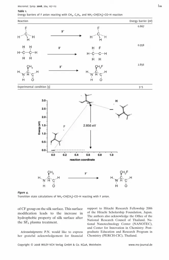
Figure 4.
Transition state calculations of NH2–CH(CH3)–CO–H reacting with F anion.
Table 1.Energy barriers of F anion reacting with CH4, C2H6, and NH2–CH(CH3)–CO–H reaction
Reaction Energy barrier (eV)
0.867
0.958
2.856
Experimental condition [3] 3–5
Macromol. Symp. 2008, 264, 107–112 111
of CF group on the silk surface. This surface
modification leads to the increase in
hydrophobic property of silk surface after
the SF6 plasma treatment.
Acknowledgments: P.N. would like to expressher grateful acknowledgement for financial
Copyright � 2008 WILEY-VCH Verlag GmbH & Co. KGaA
support to Hitachi Research Fellowship 2006of the Hitachi Scholarship Foundation, Japan.The authors also acknowledge the Office of theNational Research Council of Thailand, Na-tional Nanotechnology Center (NANOTEC),and Center for Innovation in Chemistry: Post-graduate Education and Research Program inChemistry (PERCH-CIC), Thailand.
, Weinheim www.ms-journal.de

Macromol. Symp. 2008, 264, 107–112112
[1] P. Komhoi, S. Janhom, V. S. Lee, P. Nimmanpipug,
Proceedings of Asian Workshop on Polymer Proces-
sing, 2006, 112–114.
[2] P. Zhou, G. Li, Z. Shao, X. Pan, T. Yu, J. Phys. Chem. B
2001, 105, 12469–12476.
[3] P. Chaivan, N. Pasaja, D. Boonyawan, P. Suanpoot,
T. Vilaithong, Surf. Coat Technol 2005, 193, 356–360.
[4] E. Selli, C. Riccardi, Rosaria. M. Massafra, B.
Marcandalli, Macromol. Chem. Phys. 2001, 202,
1672–1678.
Copyright � 2008 WILEY-VCH Verlag GmbH & Co. KGaA
[5] K. Nakanishi, P. H. Solomon, Infrared. Absorption.
Spectroscopy, 2nd ed., Holden- Day. San Francisco
1977, p. 25.
[6] T. Asakura, D. L. Kaplan, Encyclopedia ofAgricultural
Science, Vol. 4, C. J. Arutzen, Ed., Academic Press, New
York 1994, p. 1–11.
[7] R. E. Marsh, R. B. Corey, L. Pauling, Biochim. Bio-
phys. Acta 1955, 16, 1–34.
[8] Y. Takahashi, M. Gehoh, K. Yuzuriha, J. Polym. Sci.,
Part B: Polym. Phys. 1991, 29, 889.
, Weinheim www.ms-journal.de


