Embed Size (px)
Citation preview
Georgia Southern University
Digital Commons@Georgia Southern
Electronic Theses and Dissertations Graduate Studies, Jack N. Averitt College of
Spring 2013
Acute Alterations of Scapular Upward Rotation following a Functional Fatiguing Protocol in Male Tennis Players R. Lyndsey Ingram
Follow this and additional works at: https://digitalcommons.georgiasouthern.edu/etd
Part of the Kinesiology Commons
Recommended Citation Ingram, R. Lyndsey, "Acute Alterations of Scapular Upward Rotation following a Functional Fatiguing Protocol in Male Tennis Players" (2013). Electronic Theses and Dissertations. 53. https://digitalcommons.georgiasouthern.edu/etd/53
This thesis (open access) is brought to you for free and open access by the Graduate Studies, Jack N. Averitt College of at Digital Commons@Georgia Southern. It has been accepted for inclusion in Electronic Theses and Dissertations by an authorized administrator of Digital Commons@Georgia Southern. For more information, please contact [email protected].
1
ACUTE ALTERATIONS OF SCAPULAR UPWARD ROTATION FOLLOWING A
FUNCTIONAL FATIGUING PROTOCOL IN MALE TENNIS PLAYERS
by
R. LYNDSEY INGRAM, ATC, LAT
(Under the Direction of Thomas Buckley)
Context: Alterations in scapular kinematics, specifically upward rotation, are associated with a variety of chronic shoulder conditions. Fatigue may exacerbate the mechanisms potentially resulting in microtrauma and impingement syndrome. Objective: To identify acute alterations of scapular upward rotation following a functional fatigue protocol. Design: Prospective longitudinal. Setting: Biomechanics research laboratory. Patients or Other Participants: Twenty healthy, male competitive tennis players with no history of shoulder injury participated in this study: 10 experimental subjects (19.4 ± 1.07 yrs., 180.09 + 8.92 cm 72 + 11.56 kg) and 10 control subjects (19.6 ± 1.17 yrs., 181.1 + 6.56 cm 81.56 + 13.51 kg). Interventions: Scapular upward rotation was measured three times per session on the dominant arm while at rest, 60˚, 90˚ and 120˚ of humeral elevation in the scapular plane. Participants in the experimental group performed a tennis serving protocol and maintained at least 90% maximal exertion of the tennis serve. Fatigue was defined as reaching a rating of 15 using Borg’s rate of perceived exertion scale as well as 70% HR max. Upward rotation measurements were taken before and immediately following the fatigue protocol, and 24, 48, and 72 hours post exercise. Control participants were tested at the same time intervals without the fatiguing protocol. Main Outcome Measures: Four 2 x 5 (group x session) repeated measures ANOVA’s were performed followed by simple contrasts as appropriate. Results: Significant group by time interaction for scapular upward rotation was found at all testing positions (rest, 60˚, 90˚ and 120˚). Contrasts revealed differences between the experimental group’s pre fatigue and post fatigue values at all testing positions (pre fatigue rest: 1.48 + 2.66 post fatigue rest: -.68+ 2.66 p<.001; pre fatigue 60˚: 7.87+ 4.46 post fatigue 60˚: 5.67+ 4.72 p=.010; pre fatigue 90˚: 22.51+ 5.40 post fatigue 90˚: 19.29+ 5.16 p<.001; pre fatigue 120˚: 37.34+ 6.91 post fatigue 120˚: 33.35+ 6.49 p<.001; as well as at 60˚ pre fatigue and day four measurements (pre fatigue 60˚: 7.87+ 4.46 day 4 60˚: 7.67+ 4.55 p=.031) Conclusions: Fatigue appears to affect, specifically impairs, scapular upward rotation in male tennis players but returns to baseline values within twenty-four hours. Further research should identify when it returns to baseline to provide guidance for rest intervals for healthy male tennis players as well as if these changes are similar in a pathologic group of players. Word Count: 387
INDEX WORDS: Tennis, Scapula Kinematics, Upward rotation, Fatigue,
2
ACUTE ALTERATIONS IN SCAPULAR UPWARD ROTATION FOLLOWING A
FUNCTIONAL FATIGUING PROTOCOL IN MALE TENNIS PLAYERS
by
R. LYNDSEY INGRAM, ATC, LAT
B.S., University of Central Arkansas, 2011
M.S., Georgia Southern University, 2013
A Thesis Submitted to the Graduate Faculty of Georgia Southern University in Partial Fulfillment
of the Requirements for the Degree
MASTER OF SCIENCE
STATESBORO, GEORGIA
2012

3
© 2013
R. LYNDSEY INGRAM
All Rights Reserved
4
ACUTE ALTERATION IN SCAPULAR UPWARD ROTATION FOLLOWING A
FUNCTIONAL FATIGUING PROTOCOL IN MALE TENNIS PLAYERS
by
R. LYNDSEY INGRAM
Major Professor: Thomas Buckley
Committee: Barry Munkasy
Barry Joyner
W. Steven Tucker
Electronic Version Approved:
May 2013
5
TABLE OF CONTENTS
Page
TABLE OF CONTENTS………………………………………………………………….6
LIST OF FIGURES………………………………………………………………………..7
CHAPTER
1 INTRODUCTION…………………………………………………………..8
2 METHODS.......................................................... ........................................12
3 RESULTS………………………………………………………………….18
4 DISCUSSIONS…………………………………………………………….19
REFERENCES ................................................................................................31
APPENDICES
A – Research Hypothesis, Limitations, Assumptions, and Definitions ...........34
B – Extended Review of Literature…………………………………………..29
6
LIST OF FIGURES
Figure 1: Modified Digital Protractor……………………….……………………………60
Figure 2: Borg 6-20 Scale……………………….………………………………………..61
Figure 3: Health History Questionnaire……………………….………………………….62
Figure 4: Scapular Plane with Plum line and pole………………………………………..63
Figure 5: Measurement of 120 degrees of humeral elevation………………………….....64
Figure 5:Bony Landmark.…………………………….……………………………….….65
Figure 6: FFP set up .…………………………………………………………….……….66
Table 1: Demographic Data...…………………………………………………………….
Figure 7: Resting position over 5 testing times between groups.…….…………..............71
Figure 8: 60° humeral abduction over 5 testing times between groups….…..........……..72
Figure 9: 90° humeral abduction over 5 testing times between groups........….…………73
Figure 10: 120° humeral abduction over 5 testing times between groups.…........….…...74
Table 2: Minimal detectable change scores vs. FFP values………………..……….…....75
Table 3: Serve velocity, HR, time to fatigue, % serve error, Borg scale..…………Figure 11:
Participant RR Serve Velocity Chart………..……..….……………………...75
Figure 12: Participant KH Serve Velocity Chart………..……..….……………………...76
Figure 13: Participant PD Serve Velocity Chart………..……..………………………....77
Figure 14: Participant MP Serve Velocity Chart………..……..………………………...78
Figure 15: Participant RT Serve Velocity Chart………..……..………………………....79
Figure 16: Participant RA Serve Velocity Chart………..……..………………………...80
Figure 17: Participant MO Serve Velocity Chart………..……..………………………...81
Figure 18: Participant MV Serve Velocity Chart………..……..………………………...82
7
Figure 19: Participant SU Serve Velocity Chart………..……..………………………...83
Figure 20: Participant DA Serve Velocity Chart………..……..………………………...84
8
CHAPTER 1
INTRODUCTION
Synchronous movements of the scapula, clavicle, and humerus are required for
the shoulder complex to function properly – predominantly when elevating the arm above
ninety degrees.1 Coordinated movement of the scapula and glenohumeral joint during
elevation of the arm is known as scapulohumeral rhythm.2 As the arm is elevated, the
scapula upwardly rotates, posteriorly tilts and externally rotates; the clavicle elevates and
retracts, and the humerus elevates and externally rotates.3 Abnormalities in either static
scapular position and/or dynamic scapular motion is referred to as scapula dyskinesia.4
Scapula dyskinesia, as it relates to scapular upward rotation, has been observed in
patients with a diverse range of shoulder pathologies including rotator cuff tendon failure,
impingement syndrome, and glenohumeral instability.5-10 When the shoulder muscles are
weak or fatigued, scapulohumeral rhythm may be compromised and shoulder dysfunction
can result.11 This impairment may result in microtrauma in the shoulder muscles,
capsule, and ligamentous tissue and potentially lead to impingement.11
Fatigue, classified as either central or peripheral, is the natural physiological
response to exercise describing the decline in performance or work output associated with
repetitive or sustained activities.12 The loss of work output observed in athletic
performance, excluding environmental or temporal extremes, is generally considered to
be peripheral in nature.13 Specifically, within the motor unit, two different sites may
become impaired due to repeated contractions – the transmission site and contractile
site.13 The transmission site includes the neuromuscular junction, muscle membrane and
endoplasmic reticulum whereas the contractile site is the muscle filament itself.13
9
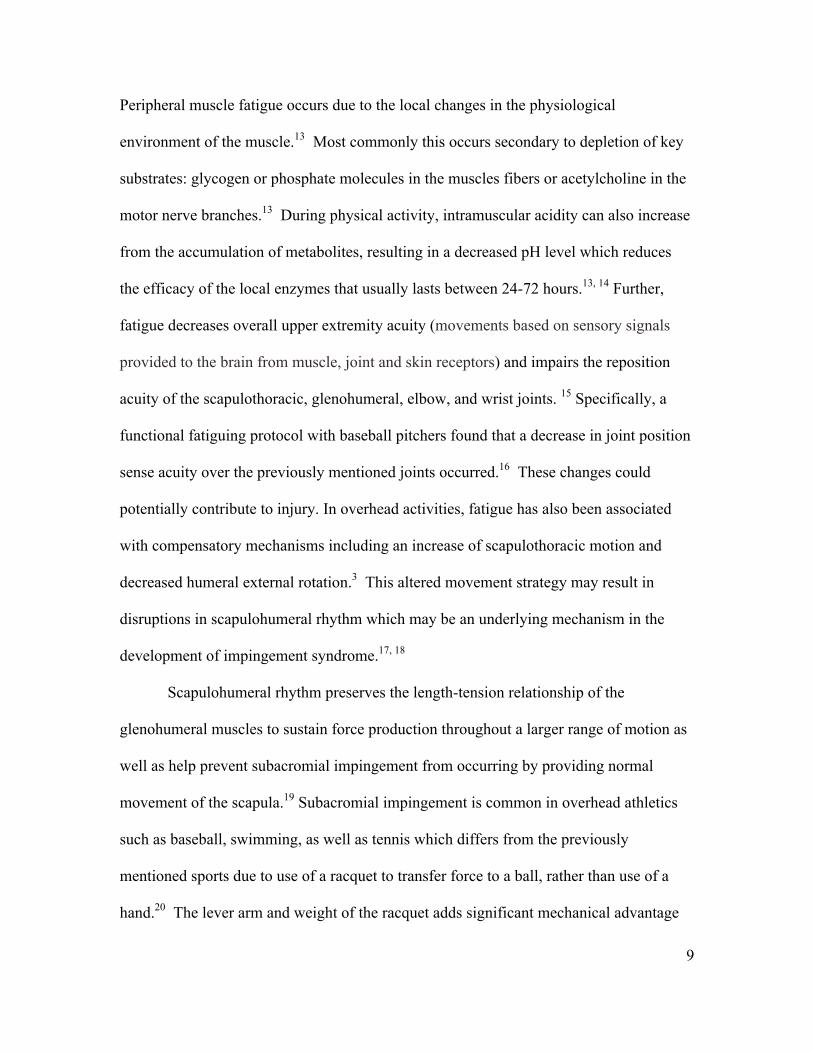
Peripheral muscle fatigue occurs due to the local changes in the physiological
environment of the muscle.13 Most commonly this occurs secondary to depletion of key
substrates: glycogen or phosphate molecules in the muscles fibers or acetylcholine in the
motor nerve branches.13 During physical activity, intramuscular acidity can also increase
from the accumulation of metabolites, resulting in a decreased pH level which reduces
the efficacy of the local enzymes that usually lasts between 24-72 hours.13, 14 Further,
fatigue decreases overall upper extremity acuity (movements based on sensory signals
provided to the brain from muscle, joint and skin receptors) and impairs the reposition
acuity of the scapulothoracic, glenohumeral, elbow, and wrist joints. 15 Specifically, a
functional fatiguing protocol with baseball pitchers found that a decrease in joint position
sense acuity over the previously mentioned joints occurred.16 These changes could
potentially contribute to injury. In overhead activities, fatigue has also been associated
with compensatory mechanisms including an increase of scapulothoracic motion and
decreased humeral external rotation.3 This altered movement strategy may result in
disruptions in scapulohumeral rhythm which may be an underlying mechanism in the
development of impingement syndrome.17, 18
Scapulohumeral rhythm preserves the length-tension relationship of the
glenohumeral muscles to sustain force production throughout a larger range of motion as
well as help prevent subacromial impingement from occurring by providing normal
movement of the scapula.19 Subacromial impingement is common in overhead athletics
such as baseball, swimming, as well as tennis which differs from the previously
mentioned sports due to use of a racquet to transfer force to a ball, rather than use of a
hand.20 The lever arm and weight of the racquet adds significant mechanical advantage
10
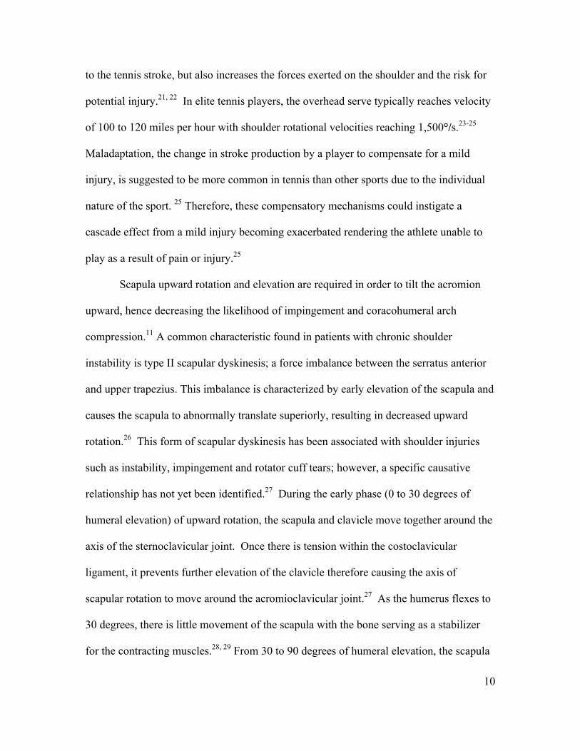
to the tennis stroke, but also increases the forces exerted on the shoulder and the risk for
potential injury.21, 22 In elite tennis players, the overhead serve typically reaches velocity
of 100 to 120 miles per hour with shoulder rotational velocities reaching 1,500°/s.23-25
Maladaptation, the change in stroke production by a player to compensate for a mild
injury, is suggested to be more common in tennis than other sports due to the individual
nature of the sport. 25 Therefore, these compensatory mechanisms could instigate a
cascade effect from a mild injury becoming exacerbated rendering the athlete unable to
play as a result of pain or injury.25
Scapula upward rotation and elevation are required in order to tilt the acromion
upward, hence decreasing the likelihood of impingement and coracohumeral arch
compression.11 A common characteristic found in patients with chronic shoulder
instability is type II scapular dyskinesis; a force imbalance between the serratus anterior
and upper trapezius. This imbalance is characterized by early elevation of the scapula and
causes the scapula to abnormally translate superiorly, resulting in decreased upward
rotation.26 This form of scapular dyskinesis has been associated with shoulder injuries
such as instability, impingement and rotator cuff tears; however, a specific causative
relationship has not yet been identified.27 During the early phase (0 to 30 degrees of
humeral elevation) of upward rotation, the scapula and clavicle move together around the
axis of the sternoclavicular joint. Once there is tension within the costoclavicular
ligament, it prevents further elevation of the clavicle therefore causing the axis of
scapular rotation to move around the acromioclavicular joint.27 As the humerus flexes to
30 degrees, there is little movement of the scapula with the bone serving as a stabilizer
for the contracting muscles.28, 29 From 30 to 90 degrees of humeral elevation, the scapula
11
abducts and upwardly rotates one degree for every two degrees of humeral elevation with
this ratio changing to a 1:1 ratio from 90 degrees to full abduction of the humerus.28
Functional fatiguing protocols have demonstrated changes within all upper
extremity joints in the sport of baseball including an increase in scapular upward rotation;
however, tennis, with its unique biomechanics and additional external load of the tennis
racket, has not been explored. Therefore, the purpose of this study was to determine if
scapular upward rotation was affected by a functional fatigue protocol, in male tennis
players. Specifically, we aim to identify length of time for upward rotation to return to its
pre-fatigue values. We hypothesized that a functional fatiguing protocol would impair
scapular upward rotation and we attempted to identify the duration of this impairment .
By understanding fatigue’s role on acute changes of the scapula, implication on practice
routines, rest time, and days off could be made to help reduce the risk of further injury.
12
CHAPTER 2
METHODS
PARTICIPANTS
We recruited 20 men’s tennis players to volunteer to enroll in the research study.
The inclusion criteria for participation included being an active member of the
intercollegiate athletic tennis program who had been a tennis team member for the
duration of the semester they were being tested or an active member of a men’s tennis
recreational program who had been competitive within the sport for at least one year. The
exclusion criteria for the study included upper extremity injury such as a history of
shoulder dislocation, subluxation, congenital scapular deficit, fracture or surgery of the
glenohumeral joint, cervical injuries, thoracic outlet syndrome, SLAP tear, or decreased
sensation in the upper extremities; also participants could not have history of any type of
impingement (primary, secondary, or internal) within six months prior to participation.5-8,
10, 19, 23, 27, 30-33 All participants had not been active in any strenuous shoulder activity for
at least three days prior to the day of initial testing. The participants were not allowed to
take place in any type of exertional shoulder activity (e.g., weight lifting) for three days
following the functional fatigue protocol. All participants were provided written informed
consent prior to participating in the study as approved by the university’s institutional
review board.
13
INSTRUMENTATION
The primary investigator showed proficiency in goniometer and palpation skills
prior to data collection by being compared to experts in the field of athletic training. The
primary investigator also demonstrated reliability use of her measurement by comparing
known angles multiple times that were randomly assigned to her. Each participant’s
anthropometric data, height and weight, was recorded utilizing a manufactured, calibrated
eye-level physician scale (Detecto Inc, Webb City, MO, USA). A manual, handheld 12”
goniometer (Bionetics, Model J00240 12.5 plastic goniometer) was used to ensure proper
arm position of all testing positions.
All participants’ scapular upward rotation was measured with a reliable and
validated (ICC [3,1] .89-.96 validity r= .74-.92) and calibrated Pro 360 digital protractor
(Macklanburg Duncan, Oklahoma City, OK, USA) (Tester reliability ICC [3,1] .95) 34
The digital protractor was modified to measure scapular upward rotation by having two
adjustable 10 cm locator rods on the inferior side of the instrument used to make contact
with the landmarks on the scapular spine – the root of the scapular spine and the
posterolateral acromion.35 Moleskin was placed on the end of each rod for subject
comfort. The digital protractor was modified to measure scapular upward rotation with a
bubble level to prevent anterior or posterior positioning of the instrument (Appendix C;
Figure 1).34 Prior to each participant’s testing session, the digital protractor was
calibrated by placing the instrument on a 3 way level and pushing the “alt zero” button on
the instrument once it was ensured that the instrument was level.
A standard tennis ball, (56-59.4 g and 6.7 cm in diameter, Wilson Sports,
14
Chicago, IL, USA) was used during the functional fatiguing protocol (FFP). Each
participant’s maximum serve velocity was detected by a Stalker radar gun (Stalker
Digital Sports Radar, Plano, TX, USA). Both the Borg Rating of Perceived Exertion
(RPE) 6-20 scale (Reliability .93 Validity .85) as well as heart-rate detected by a Polar
wrist heart rate monitor (Polar Electro Inc. Lake Success, NY) (Appendix C; Figure 2)
were used to assess fatigue level.36, 37 Maximum heart rate was calculated using the
Londeree and Moeschberger method that has been shown to be reliable and valid for an
active population: (206.3 – (.711*age) then a 70% HR maximum was calculated by
multiplying by .70. 38 The local upper extremity RPE rating of 15 on a scale of 6 to 20
has been reported to be highly correlated with the metabolic responses of fatigue:
including respiratory exchange, heart rate, absolute oxygen consumption, and blood
lactate.39, 40
PROCEDURES
Participants were divided into control and experimental groups quasi-randomly by
a random number generator application compatible with Apple products. Once the
experimental group had acquired 10 subjects, the rest of the participants filled the control
group. The control group was then quasi-randomly paired with experimental subjects by
the random number generator. The participants first completed the health history
questionnaire (Appendix C; Figure 3) to ensure that inclusion and exclusion criteria are
met. All enrolled participants reported to the biomechanics laboratory for anthropometric
measurements prior to the exercise protocol.
To quantify baseline measurements of scapular upward rotation, tape was placed
on the floor in line with the frontal plane and at a 40˚ from the frontal plane. Participants
15
stood in a relaxed resting position next to a pole placed along the 40˚ angle tapeline at an
arm’s distance from the participant (Appendix C; Figure 4). The pole ensured proper
position of the participant’s arm in the 40˚ of forward flexion, also referred to as the
scapular plane, during testing trials.35 The four testing positions were based on previous
reports of valid and reliable measures of scapular positioning utilizing a digital
protractor.34, 41 To accurately position the participant’s body along the frontal plane, a
visual plum line was used from the participant’s dominant side, the self-reported
preferred serving extremity, acromion process to the tape on the floor representing the
frontal plane. Using a manual hand-held goniometer, the investigator moved the subject’s
dominant arm to 60˚, 90˚, and 120˚ of abduction in the scapular plane. Participants were
instructed to maintain an open hand with their thumb pointing up.34 For each of the three
positions, tape was placed on the pole representing the three measured positions. The tape
marks aided to ensure consistency of arm angles between testing trials (Appendix C;
Figure 5).
All participants performed the testing trials while scapular upward rotation was
measured with the digital protractor.34 Each participant’s scapular rotation was assessed
three times at the four testing positions and the order of positions was randomized by
using a random number generator application compatible with all Apple products. The
participants performed active abduction in the scapular plane to the predetermined
positions of a resting position, 60˚, 90˚ and 120˚. The investigator palpated and marked,
with a skin marker, the root of the scapular spine and the posterolateral acromion at each
predetermined position (Appendix C; Figure 6).34 During the testing trials, the two
adjustable locator rods of the digital protractor were placed in line with the two marks.
16
Using the bubble levels as a guide, the digital protractor was held level in place for five
seconds against the skin by the investigator. During this time, the investigator monitored
and recorded the value displayed on the digital screen at a point when equilibrium was
reached. The results of each trial at each position were recorded and the mean of the
three trials were analyzed.
After baseline values were established, the experimental participants then
performed a three-minute serving warm up. This was followed by the assessment of each
participant’s maximum serve velocity from whichever service side they were most
comfortable. To determine the velocity used for statistical assessment, the mean velocity
of the first five serves was used. During the FFP, the participant was asked to serve each
ball exceeding their 90% maximum velocity every ten seconds until they reached fatigue.
The participant was verbally prompted by “try harder” when maximum velocity fell
below 90% to ensure maximal effort. This FFP was adapted from a baseball fatigue
protocol used by Tripp et al in 2007.The participant was also required to serve the ball
within the service court during the protocol to reduce the possibility of altered mechanics.
Participants continued to serve every 10 seconds (6 serves per minute) until fatigue was
reached.15 The participant’s fatigue level on the Borg RPE scale as well as heart rate was
recorded each minute.15 Fatigue was considered to have been reached when he reported
an exertion level of 15 or more and had a 70% HR max.
Once the participant reached fatigue, as previously described, the FFP was
concluded and the participant returned to the biomechanics lab in which posttest
measurements of the scapula’s upward rotation, as previously described, were performed.
17
The participant was then tested on these measurements on each of the 3 days following
the FFP at the same 4 hour interval due to circadian rhythms.
The control group performed the same baseline measurements as the experiential
group. Subsequent, measurements were taken that pseudo matched to an experimental
subject by replicating the time allotted for the FFP. The control group was also tested for
3 days to access scapular position.
DATA ANALYSIS
This study was a prospective, longitudinal, quasi-randomized study. The
dependent variables were the mean upward rotation at the four positions of humeral
elevation (rest, 60˚, 90˚ and 120˚) at the five testing times; Pre Exercise, Post Exercise,
Day 1, Day 2, and Day 3 with the independent variable being group; control and
experimental.
Demographic information was reported with means and standard deviations. To
compare between and within groups at each of the testing positions; four 2 x 5 (group x
session) repeated measures ANOVA’s, with repeated measures on the second factor,
were performed with an alpha level set at .05. As appropriate, simple repeated contrasts
was also performed. The alpha level was set at p<0.05. Data were analyzed using IBM
SPSS 19.0 for Windows package (SPSS Inc, Chicago, IL).
18
CHAPTER 3
RESULTS
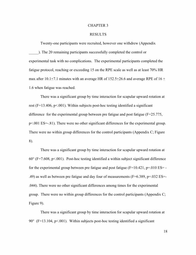
Twenty-one participants were recruited, however one withdrew (Appendix
_____). The 20 remaining participants successfully completed the control or
experimental task with no complications. The experimental participants completed the
fatigue protocol, reaching or exceeding 15 on the RPE scale as well as at least 70% HR
max after 10.1+7.1 minutes with an average HR of 152.5+26.6 and average RPE of 16 +
1.6 when fatigue was reached.
There was a significant group by time interaction for scapular upward rotation at
rest (F=13.406, p<.001). Within subjects post-hoc testing identified a significant
difference for the experimental group between pre fatigue and post fatigue (F=25.775,
p<.001 ES=-.81). There were no other significant differences for the experimental group.
There were no within group differences for the control participants (Appendix C; Figure
8).
There was a significant group by time interaction for scapular upward rotation at
60° (F=7.608, p<.001). Post-hoc testing identified a within subject significant difference
for the experimental group between pre fatigue and post fatigue (F=10.421, p=.010 ES= -
.49) as well as between pre fatigue and day four of measurements (F=6.389, p=.032 ES=-
.044). There were no other significant differences among times for the experimental
group. There were no within group differences for the control participants (Appendix C;
Figure 9).
There was a significant group by time interaction for scapular upward rotation at
90° (F=13.104, p<.001). Within subjects post-hoc testing identified a significant

19
difference for the experimental group between pre fatigue and post fatigue (F=23.753,
p<.001 ES=-.60). There were no other significant differences among times for the
experimental group. There were no within group differences for the control participants
(Appendix C; Figure 10).
There was a significant group by time interaction for scapular upward rotation at
120° (F=19.875, p<.001). Within subjects post-hoc testing identified a significant
difference for the experimental group between pre fatigue and post fatigue (F=95.214,
p<.001 ES=-.58). There were no other significant differences among times for the
experimental group. There were no within group differences for the control participants
(Appendix C; Figure 11).
20
CHAPTER 4
DISCUSSION
Compensatory mechanisms in scapular upward rotation, secondary to fatigue,
may be associated with chronic overuse injury; however, the specific effects of fatigue on
scapular upward rotation have not previously been elucidated.5-10 Therefore, the purpose
of this study was to determine if scapular upward rotation was affected by a FFP. The
main finding of this study identified that scapular upward rotation was acutely impaired
following a sport- specific FFP in male tennis players. Specifically, the FFP resulted in a
significant decrease in scapular upward rotation at all 4 testing positions (rest, 60°, 90°,
and 120º); however, this change was only noted immediately post-activity and was
recovered by 24 hours post-exercise. These findings may have significant implications of
fatigue’s role on male tennis players’ upward rotation and give insight on rest intervals
needed for scapulohumeral rhythm to return to baseline, thus potentially reducing the risk
of injury.
The results of this study suggest that a FFP acutely impairs, specifically reduces,
scapular upward rotation at all four positions examined in this study. Interestingly, each
participant’s scapular upward rotation returning to pre-exercise levels within 24 hours
post FFP. These changes were consistent at all four test positions, which imply that the
FFP affected the scapular stabilizers, in a static position, consistently regardless of the
humeral head positioning. Although this study was kinematic in nature and did not
include electromyography, Joshi et al. suggested that FFP increased infraspinatus firing
ratios to that of lower and middle trapezius following fatigue.42 This could have
implication on the consistency of arm position due to alterations of muscle firing patterns
21
for compensation of the fatigued muscles. With the values returning within 24 hours, it
seems lingering fatigue does not effect a healthy tennis player’s scapular upward rotation
statically. However, results of dynamic measurements are still unknown.
The impairments seen in scapular upward rotation following a FFP may provide a
greater understanding of an underlying mechanism of injury to the shoulder - more
specifically impingement syndrome. Given the complex neuromuscular controlling
mechanisms and tremendous demands imposed on the shoulder by upper extremity
sports, it is not surprising that a small deficiency could have a cumulative effect on the
shoulder.43 Sports that require overhead and throwing maneuvers, including tennis, stress
the tissues to near their physiological limits.43 Specifically, reductions in scapular upward
rotation are associated with increased risk of shoulder pathology based on two primary
mechanisms. First, the shoulder exercising near these physiological limits without proper
position of anatomical structures, including the scapula, may place the glenoid in a sub-
optimal position for the activities being performed, potentially predisposing the shoulder
to injury.43 Secondly, compensatory motions of scapular stabilizers and rotator cuff
muscles may disrupt synchronous firing of these muscles, which in turn can predispose to
further injury.43,28 This dyssynchronous firing pattern can also occur secondary to weak,
injured, or fatigued musculature.42 These altered or impaired shoulder movement
patterns may cause microtrauma in the shoulder muscles, capsule, and ligamentous tissue
potentially leading to secondary impingement.11,17, 18 Other abnormal movement and /or
injuries such as scapular dyskinesis, rotator cuff pathology, and instability can also occur
from lack of upward rotation of the scapula from repeated microtrauma of certain
muscles in the area due to this subacromial impingement.11
22
Following the FFP, scapular upward rotation was significantly reduced (p<0.05);
however, the mean reduction across the four testing positions ranged from 2.16° to 3.99°
Although this degree of change may appear small, there were large effect sizes (-.44 to -
.81) as well as the numbers being clinically significant when compared to minimal
detectable change, a value that allows clinicians to identify the difference between
normal daily variation and actual differences, values in scapular upward rotation.8 All
post-exercise scapular upward rotation measures fall above the MDC values (Appendix
C; Figure 12); therefore, not only are the results statically significant, but likely clinically
significant as well. It should be noted such a small change could potentially be an
underlying factor of impingement syndrome due to the height of the subacromial space
only being 7 to 12 mm.30 These changes could potentially contribute to overuse injury
and subtle instability. In overhead activities, fatigue has also been associated with
compensatory mechanisms including an increase of scapulothoracic motion and
decreased humeral external rotation.3 This altered movement strategy may result in
disruptions in scapulohumeral rhythm, which may be an underlying mechanism in the
development of impingement syndrome.17, 18 It is unknown at this time for how long an
individual must exhibit these small changes to develop shoulder pathology.
Interestingly, there was also a significant difference at 60° of scapular upward
between pre fatigue values and day four values (F=6.389, p=.032 ES=-.044). With such a
small effect size (.044) and the value not reaching above the MDC value for 60° (1.23) it
is questionable that this is clinically significant to the population of male tennis players. It
is unclear why differences were seen exclusively at this angle between pre fatigue and
day four data was observed as it seems highly unlikely for changes to occur with the
23
conservative delimitations presented in this study- not allowing any upper extremity
activity, measurements taken in the same circadian time interval, as well as the
movement of the scapula within the scapulothoracic interval ratio being 2:1 at this angle.
All participants verbally reported no activity and no other differences between the
experimental group at pre fatigue and day four data were observed by the primary
investigator that may have influenced this finding.
The participants in this study served until they reached fatigue; herein defined as a
15 on the Borg 6-20 scale and 70% of their HR max.38, 44 The participants in this study
averaged 58.2 + 41.7 serves before meeting the criteria for fatigue (Appendix C; Figure
13). In a regular tennis match for a collegiate player, the absolute least amount of serves
that could be preformed is 48; however this rarely occurs due to similarly matched level
of competition with the likelihood of a person winning every single point in every single
game is rare. For example, the winner of the 2003 US Open averaged 7.8 (3.2) serves per
game for 31 service games or approximately 242 serves. During the tournament it is
estimated that he hit over 1000 serves.45 By contrast, a professional baseball pitcher
typically pitches every four to five days with an average of approximately 100 pitches per
game. Given the combination of high demand, limited rest, and the mechanical impact of
the tennis racket acting as a lever arm for tennis players, it is not surprising that shoulder
injuries account for 20-45.7% of all injuries in tennis players.46 By providing information
directly related to tennis players, with inclusion of possible mechanism of injury, this
study may begin the process of understanding the neuromuscular adaptations potentially
associated with injury.
24
An alteration in scapular upward rotation following a FFP has received limited
attention in the literature. The results of this study, a decrease in scapular upward rotation
following a FFP in male tennis players, agrees with two earlier investigations that
reported a decrease in scapular upward rotation following fatigue; however, both studies
investigated the general public, not an athletic population.42, 47 These results however
differ from a previous investigation on baseball pitchers. Specifically, Tripp et al,
reported an increase in scapular upward rotation following a FFP in which baseball
pitchers threw from a kneeling position every five seconds until a 15 was reached on the
Borg 6-20 scale.15 They speculated that the sensorimotor system was impaired causing
impaired joint angle acuity to occur. Similarly, Joshi et al also reported an increase in
scapular upward of less than 3° rotation following a fatigue protocol of males and
females from the general population. For their study, participants rhythmically externally
rotated in their arm with weight while lying prone from 0 to 75 per second until they
could not keep pace or continue. Participants were then given a thirty second rest period
and then continued. This continued for a minimum of five sets and until the number of
repetitions was less than 50% performed during the first set. They speculated that either
clavicular elevation or compensatory mechanisms by which the fatigued shoulder
maintained a normal subacromial space by activating different shoulder muscles could be
an explanation for this increase. These inconsistent reports could be a result of the
difference in the upper extremities tested (dominant vs. non-dominant), fatigue protocol
(static vs. dynamic), as well as activity prior to the study. The difference between studies,
although similar in conceptual design, could be a result of a number of factors. First,
positioning of the participant was different. Within Tripp’s study, the participants were
25
positioned in a kneeling position to reduce lower extremity force production. However, in
this study, with the inclusion of the increased lever arm due to the tennis racquet, this
position was not feasible. Second, the time between throws/serves were different by five
seconds which could account for a longer time to fatigue for tennis, however, time to
position one’s self for a tennis serve is longer than that of a throw from a kneeling
position. Third, in Joshi’s study, the participants were not completing a true sport specific
FFP. They were also asked to perform a task until failure five times with thirty-second
rest intervals. Research on scapular kinematics in volleyball players showed no variance
in scapular upward rotation following an entire season; however none of these athletes
suffered a shoulder injury during play.48 Although the limited differences between these
studies seem trivial, they are in fact crucial in identifying issues of scapular upward
rotation to the specific population of male tennis players.
When comparing differences between intercollegiate and club tennis players’
FFP, there are significant differences to report. Intercollegiate players demonstrated
better serving performance including significantly higher service velocity (97.7+10.3 vs.
75.0 + 10.3 mph; P=0.009) and serve accuracy (56.3%+15.2% vs. 31.5% + 13.1%
P=0.024). Conversely, there were no differences between groups for time to fatigue
(9.4+7.0 vs. 10.8+ 7.9; P=0.780), HR (146.6 + 9.7 P=0.530), and Borg scale (15.6 + .89
vs. 16.4 + 2.2, P=0.471) (Appendix C; Figure 13).
The differences found in the current study, along with previous studies,
demonstrates further research is needed on upward scapular rotation. Specifically,
research should address tracking scapular upward rotation over the course of multiple
seasons and multiple teams and tracking injury rates of impingement, instability, rotator
26
cuff tears, labrum and identify the role of the scapula with these injuries. Measuring
scapular upward rotation during pre season and through multiple time points throughout a
season or within a multi-match day may help identify an indicator of shoulder pathology
and help identify recovery time for scapular upward rotation following fatigue. This
accounts for tennis players participating in multiple matches in one day in which the
scapular stabilizers may not recover between matches, thus leading to a cumulative effect
of compensatory motions by tennis players. Longitudinal tracking of scapular upward
rotation, especially following a fatigued state, would help to identify the role of the
scapula in shoulder pathology. Finally, tracking injury incidence and scapular upward
rotation may help with prevention of shoulder injuries by correcting scapular posture and
range of motion with rehabilitation.
Although the entire available population was recruited to participate in this study,
more participants may have made for stronger results. The digital protractor used has
been shown to be reliable and valid; however breathing pattern and investigator error
may have influenced the manual application.28 Additionally, the upward rotation
measurements that were taken during this study were static. Although decreased upward
rotation is associated with shoulder injuries such as instability, impingement and rotator
cuff tears, caution should be taken when making inferences with regard to dynamic
movements, such as a tennis serve, based on static upward rotation measurements. Only
male tennis players were allowed to participate in this study due the laxity of the
costoclavicular ligament during a female’s menstrual cycle as well as for ease of
measurement techniques as marker accessibility is substantially more accurate when the
subject is shirtless. Also, previous research has found quantification of static scapular
27
upward rotation alone to not be an effective diagnostic tool for determining shoulder
dysfunction.49 For this reason, only healthy individuals with no previous history of
shoulder dysfunction were used. During the FFP one participant did not meet the
inclusion requirements of the study’s definition of fatigue with a HR above his 70% HR
max. He discontinued the study with a HR of 127 while it should have been when 134
was the 70% HR max threshold. For another participate, the radar gun malfunctioned two
minutes into the study with the back up battery not working as well. Therefore, only
serve accuracy was recorded for these participants. Lastly, the FFP was discontinued for
safety reasons for one participant. Twenty-two minutes into the FFP, the participant’s HR
did not exceed 119 beats per minute despite reporting a maximum rating of 20 on the
Borg scale.
In conclusion, this study provides information to clinicians about the role of acute
fatigue on scapular upward rotation in male tennis players. The findings suggest that
following a FFP decreases scapular upward rotation at rest, 60°, 90°, and 120º of humeral
abduction in the scapular plane but returns to baseline values within 24 hours. This may
help clinicians understand the influence of lingering fatigue on scapular kinematics as
well as potentially identify an underlying mechanism of injury. Future studies should
identify alterations of scapular upward rotation within a 24-hour period following fatigue
for male tennis players to assist in suggested rest intervals to potentially decrease the risk
of injury.
28
1. Culham E, Peat M. Functional anatomy of the shoulder complex. The Journal of orthopaedic and sports physical therapy 1993;18(1):342-50.
2. Inman VT, Saunders JB, Abbott LC. Observations of the function of the shoulder joint. 1944. Clin Orthop Relat Res 1996(330):3-12.
3. Ebaugh DD, McClure PW, Karduna AR. Effects of shoulder muscle fatigue caused by repetitive overhead activities on scapulothoracic and glenohumeral kinematics. Journal of electromyography and kinesiology : official journal of the International Society of Electrophysiological Kinesiology 2006;16(3):224-35.
4. Kibler WB, McMullen J. Scapular dyskinesis and its relation to shoulder pain. The Journal of the American Academy of Orthopaedic Surgeons 2003;11(2):142-51.
5. Burkhart SS, Morgan CD, Kibler WB. Shoulder injuries in overhead athletes. The "dead arm" revisited. Clinics in sports medicine 2000;19(1):125-58.
6. Kibler WB. The role of the scapula in athletic shoulder function. The American journal of sports medicine 1998;26(2):325-37.
7. Ozaki J. Glenohumeral movements of the involuntary inferior and multidirectional instability. Clinical orthopaedics and related research 1989(238):107-11.
8. Paletta GA, Jr., Warner JJ, Warren RF, Deutsch A, Altchek DW. Shoulder kinematics with two-plane x-ray evaluation in patients with anterior instability or rotator cuff tearing. Journal of shoulder and elbow surgery / American Shoulder and Elbow Surgeons ... [et al.] 1997;6(6):516-27.
9. Solem-Bertoft E, Thuomas KA, Westerberg CE. The influence of scapular retraction and protraction on the width of the subacromial space. An MRI study. Clinical orthopaedics and related research 1993(296):99-103.
10. Warner JJ, Micheli LJ, Arslanian LE, Kennedy J, Kennedy R. Scapulothoracic motion in normal shoulders and shoulders with glenohumeral instability and impingement syndrome. A study using Moire topographic analysis. Clinical orthopaedics and related research 1992(285):191-9.
11. Voight ML, Thomson BC. The role of the scapula in the rehabilitation of shoulder injuries. Journal of athletic training 2000;35(3):364-72.
29
12. Mannion AF, Dolan P. Relationship between myoelectric and mechanical manifestations of fatigue in the quadriceps femoris muscle group. European journal of applied physiology and occupational physiology 1996;74(5):411-9.
13. Asmussen E. Muscle fatigue. June 1979. Medicine and science in sports and exercise 1993;25(4):411-20.
14. Kraemer WJ, Fleck SJ, Deschenes MR. Exercise physiology : integrated from theory to practical applications. 1st ed. Philadelphia: Wolters Kluwer/Lippincott Williams & Wilkins Health; 2012.
15. Tripp BL, Yochem EM, Uhl TL. Functional fatigue and upper extremity sensorimotor system acuity in baseball athletes. Journal of athletic training 2007;42(1):90-8.
16. Kent-Braun JA, Ng AV, Doyle JW, Towse TF. Human skeletal muscle responses vary with age and gender during fatigue due to incremental isometric exercise. Journal of applied physiology 2002;93(5):1813-23.
17. Jobe FW, Pink M. Classification and treatment of shoulder dysfunction in the overhead athlete. The Journal of orthopaedic and sports physical therapy 1993;18(2):427-32.
18. Bak K, Fauno P. Clinical findings in competitive swimmers with shoulder pain. The American journal of sports medicine 1997;25(2):254-60.
19. Sevinsky S. Scapular Dyskinesis. Bethlehem, PA. 20. Page P. Shoulder muscle imbalance and subacromial impingement syndrome in
overhead athletes. International journal of sports physical therapy 2011;6(1):51-8. 21. Marx RG, Sperling JW, Cordasco FA. Overuse injuries of the upper extremity in
tennis players. Clinics in sports medicine 2001;20(3):439-51. 22. Pluim BM, Staal JB, Windler GE, Jayanthi N. Tennis injuries: occurrence,
aetiology, and prevention. British journal of sports medicine 2006;40(5):415-23. 23. Di Sumant G, Krishnan, Richard J., Hawkins, Russell, F. Warren. The shoulder
and the overhead athlete. Philadelphia: Lippincott, Williams, and Wilkins; 2004. 24. Kibler WB. Biomechanical analysis of the shoulder during tennis activities.
Clinics in sports medicine 1995;14(1):79-85. 25. Krishnan SG, Hawkins, Richard J., Warren Russell F. The Shoulder and the
Overhead Athlete . 1 ed. Philadelphia: Lippincott Williams & Wilkins; 2004. 26. Ludewig PM, Cook TM. Alterations in shoulder kinematics and associated
muscle activity in people with symptoms of shoulder impingement. Physical therapy 2000;80(3):276-91.
27. Inman VT, Saunders JB, Abbott LC. Observations of the function of the shoulder joint. 1944. Clinical orthopaedics and related research 1996(330):3-12.
28. Prentice WE, Arnheim DD. Arnheim's principles of athletic training : a competency-based approach. 13th ed. Boston: McGraw-Hill Higher Education; 2009.
29. Starkey C, Ryan JL. Evaluation of orthopedic and athletic injuries. 2nd ed. Philadelphia, PA: F.A. Davis Co.; 2002.
30. Andrews JRW, Kevin E. The Athlete's Shoulder. New York: Churchill Livingstone Inc.; 1994.
31. Ciullo J. Shoulder Injuries in Sport. Troy; 1996. 32. Micheo W. Rehabiliation of shoulder injury in the throwing athlete. Critical
Reviews in Physical and Rehabilitation Medicine 2008;20(1):87. 33. Pinsent C, Rae, Lisa, Palmer, Jillian. Return to activity guidelines for the
overhead athlete after shoulder injury. Edmonton: University of Alberta; 2010. p. 38.
30
34. Johnson MP, McClure PW, Karduna AR. New method to assess scapular upward rotation in subjects with shoulder pathology. The Journal of orthopaedic and sports physical therapy 2001;31(2):81-9.
35. Tucker WS, Ingram RL. Reliability and validity of measuring scapular upward rotation using an electrical inclinometer. Journal of electromyography and kinesiology : official journal of the International Society of Electrophysiological Kinesiology 2012;22(3):419-23.
36. Edwards RH, Melcher A, Hesser CM, Wigertz O, Ekelund LG. Physiological correlates of perceived exertion in continuous and intermittent exercise with the same average power output. European journal of clinical investigation 1972;2(2):108-14.
37. Borg G. Borg's Perceived exertion and pain scales. Champaign, IL: Human Kinetics; 1998.
38. Londeree BR MM. Influence of age and other factors on maximal heart rate. J Cardiac Rehabil 1984;4:44–49.
39. Kang J, Chaloupka EC, Mastrangelo MA, Donnelly MS, Martz WP, Robertson RJ. Regulating exercise intensity using ratings of perceived exertion during arm and leg ergometry. European journal of applied physiology and occupational physiology 1998;78(3):241-6.
40. Marais G, Weissland T, Robin H, Vanvelcenaher JM, Lavoie JM, Pelayo P. Physiological effects of variations in spontaneously chosen crank rate during sub-maximal and supra-maximal upper body exercises. International journal of sports medicine 1999;20(4):239-45.
41. Downar JM, Sauers EL. Clinical Measures of Shoulder Mobility in the Professional Baseball Player. Journal of athletic training 2005;40(1):23-29.
42. Joshi M, Thigpen CA, Bunn K, Karas SG, Padua DA. Shoulder external rotation fatigue and scapular muscle activation and kinematics in overhead athletes. Journal of athletic training 2011;46(4):349-57.
43. Rockwood CA. The Shoulder. Philadelphia, PA: W. B. Saunders Company; 1990. 44. Tripp BL, Boswell L, Gansneder BM, Shultz SJ. Functional Fatigue Decreases 3-
Dimensional Multijoint Position Reproduction Acuity in the Overhead-Throwing Athlete. Journal of athletic training 2004;39(4):316-20.
45. Johnson CD, McHugh MP, Wood T, Kibler B. Performance demands of professional male tennis players. British journal of sports medicine 2006;40(8):696-9; discussion 99.
46. Caine DJ, Maffulli N. Epidemiology of pediatric sports injuries. Basel ; New York: Karger; 2005.
47. McQuade KJ, Dawson J, Smidt GL. Scapulothoracic muscle fatigue associated with alterations in scapulohumeral rhythm kinematics during maximum resistive shoulder elevation. The Journal of orthopaedic and sports physical therapy 1998;28(2):74-80.
48. Brewer BE TW. Shoulder Kinematics in Elite Volleyball Players Following a Full Season of Play Conway, AR: University of Central Arkansas; 2012.
49. McClure PW, Michener LA, Karduna AR. Shoulder function and 3-dimensional scapular kinematics in people with and without shoulder impingement syndrome. Physical therapy 2006;86(8):1075-90.
31
APPENDIX A
RESEARCH HYPOTHESIS, DELIMITATIONS, LIMITATIONS, ASSUMPTIONS,
AND DEFINITIONS
Research hypotheses
Limitations
Breathing pattern and investigator error may influence manual application of the
digital protractor; however, this method has been identified as valid and reliable.28
Additionally, the upward rotation measurements that will be taken during this study will
be static. Although decreased upward rotation is associated with shoulder injuries such as
instability, impingement and rotator cuff tears, caution should be taken when making
inferences with regard to dynamic movements, such as throwing, based on static upward
rotation measurements.
Delimitations
This study’s delimitations include only allowing male tennis players to participate
in this study with no previous history of shoulder injury. Male athletes will be used for
ease of measurement techniques as marker accessibility is substantially more accurate
when the subject is shirtless. Also, previous research has found quantification of static
scapular upward rotation alone to not be an effective diagnostic tool for determining
shoulder dysfunction. For this reason, only healthy individuals with no previous history
of shoulder dysfunction will be used.
Exclusion Criteria
32
No history of shoulder dislocation, subluxation, fracture or surgery, cervical injuries,
thoracic outlet syndrome, impingement syndrome within the last six months, or self
reported decreased sensation in the upper extremities.
Assumptions
1. Participants will give full effort.
2. Participants will follow directions.
3. Participants will not try to alter their performance during the protocol or
measurements.
Definitions
1. Scapular upward rotation — superior rotation; moves scapula’s away from one another
2. Functional Fatiguing Protocol – for the purpose of this study, the FFP will be defined as a tennis serving protocol every 10 seconds within the service court until fatigue is reached
3. Fatigue – for the purpose of this study, fatigue is defined as a 15 on the Borg
6-20 RPE scale and 70% heart rate maximum
33
EXTENDED REVIEW OF LITERATURE
The purpose of this biomechanical study is to examine acute alterations in
scapular upward rotation following a functional fatiguing protocol in male tennis players.
To carry out this study it is necessary to complete a critical review of the literature, which
will continue through the data collection, data analysis, and synthesis stages of the
research.
To conduct the literature review, the researcher is using multiple information
sources including books, Internet resources, and professional journals. These sources
were primarily accessed through PubMed, MEDLINE, CINAHL, and SPORT Discus.
There is no delimiting timeframe, however, the majority of research has been performed
34
within the last 20 years. The researcher will be attempting to point out existing gaps in
the literature throughout this review.
Anatomy
The shoulder complex consists of three joints and one region: the glenohumeral
joint, acromioclavicular joint, sternoclavicular joint, and scapulothoracic region.21
Because of these joints and region, the shoulder allows for all three degrees of freedom.
The immense amount of range of motion allowed within the shoulder complex is due in
part to the large surface of the humeral head articulating with the relatively small glenoid
fossa of the scapula.28
The glenohumeral joint is a multi-axial ball-and-socket synovial joint comprised
of the hemispherical head of the humerus and where it articulates with the glenoid cavity
of the scapula.39 The muscles of the glenohumeral joint can be subdivided several
different ways – by their anatomical, functional, or mechanical properties. Anatomically,
muscles of the shoulder complex can be divided into three groups depending on their
attachment site: scapula to humerus, radius, or ulna; scapula to trunk; or humerus to
trunk.28 Functionally, muscles of the shoulder complex can also be divided into those that
act as prime movers and those that stabilize for the shoulder complex. Prime movers of
the shoulder girdle include large muscles such as the pectoralis major, latissimus dorsi,
and deltoid.28 The smaller muscles are classified as stabilizers, which include the rotator
cuff muscles, rhomboid major, rhomboid minor, and serratus anterior.28 Mechanically,
muscles of the shoulder complex can be divided into those that rotate the body segment
around an axis and those that translate the segment toward or away from the axis.28 The
orientation of the musculature of the glenohumeral joint has large reaction forces that acts
35
perpendicular to the glenoid fossa to compress the concave humeral head into the glenoid
fossa.21 This concavity-compression maintains anterior joint stability over a large range
of shoulder motion and has been found to resist inferior translation of the humeral head.
40,41 The musculature of the glenohumeral joint is comprised of muscles of the rotator
cuff and deltoid.23 The rotator cuff is composed of four muscles: the supraspinatus,
subscapularis, infraspinatus, and teres minor.42 The supraspinatus originates on the
posterosuperior scapula, superior to the scapular spine.23 It passes under the acromion,
through the supraspinatus outlet, and inserts on the greater tuberosity.23 The
supraspinatus is active during the entire motion of scapular plane abduction.23 The
infraspinatus and teres minor muscles originate on the posterior scapula, inferior to the
scapular spine and both muscles insert on the posterior aspect of the greater tuberosity.23
These two muscles act together to externally rotate and extend the humerus.23
Collectively these muscles contribute approximately 80% of the external rotation strength
with the arm in an adducted position.23 The subscapularis muscle arises from the anterior
scapula and is the only muscle to insert on the lesser tuberosity.23 The subscapularis
internally rotates and flexes the humerus.23 The tendon of the subscapularis inserts on the
lateral aspect of the anterior capsule; therefore, this muscle is the most responsible for
providing anterior glenohumeral stability because it is the most active stabilizer muscle.43,
44 The deltoid is the largest muscle of the shoulder girdle. It has three different
components – anterior, middle, and posterior that has three different origins: the clavicle,
the acromion, and the scapular spine. The deltoid inserts at the deltoid tubercle on the
lateral aspect of the humeral shaft.23 The anterior portion of this muscle forward flexes
and abducts the humerus, while the middle and posterior heads provide more

36
glenohumeral compression and a less shear force than does the anterior component.45
The humeral head is approximately one third of a sphere with an average diameter
being 45 mm.23 The proximal humerus is comprised of four parts: the articular surface,
the greater tuberosity, the lesser tuberosity, and the diaphyseal shaft.42 The humeral head
is angulated medially 45 degrees to the long axis of the humeral shaft, and on average it
is retroverted 20-25 degrees relative to the transcondylar axis of the distal humerus.25
Between the greater and lesser tuberosity is the intertubercular groove in which lies the
tendon of the long head of the biceps brachii.23 The tendon is held in place by the
coracohumeral ligament, superior glenohumeral ligament, and the transverse humeral
ligament.23 In the glenohumeral joint, three different types of motion may occur:
spinning, sliding, and rolling.26 Spinning occurs when the contact point on the glenoid
remains the same while the humeral head contact point is changing.23 Sliding is pure
translation of the humeral head on the articular surface of the glenoid.23 This action
occurs most at end ranges of motion or in unstable joints.23 In the glenohumeral joint, the
contact point on the glenoid is moving, while that of the humerus remains neutral.23
Rolling is a combination of humeral head sliding and spinning relative to the glenoid
such that the contact point changes on both the glenoid and the humeral head.23
Normal movement of the scapula on the wall of the thoracic cage is an essential
component of normal function at the glenohumeral joint.23, 25 This region is referred to as
the scapulothoracic region and is the least congruent area in the body with the only
osseous link between the scapula and the axial skeleton occurring at the clavicle.17
Therefore, this region is highly dependent on the surrounding musculature for stability
and normal motion.1, 15, 46-48 Because 18 muscles (rhomboid major, rhomboid minor,
37
levator scapulae, pectoralis minor, trapezius, serratus anterior, subscapularis,
supraspinatus, infraspinatus, teres minor, teres major, deltoid, pectoralis major, latissimus
dorsi, coracobrachialis, long head of the triceps, long head of the biceps, and omohyoid)17
originate or insert on the scapula, it also plays into the role of stability within the
glenohumeral joint.17
The scapula is a flat, triangular shaped bone that is classified as metaphyseal,
which refers to a thin cortex with frequent vascular perforation.26, 49 It moves through a
gliding mechanism in which the concave anterior surface of the scapula moves on the
convex posterolateral surface of the thoracic cage.39 The scapula serves mainly as an
articulating surface for the head of the humerus within the glenohumeral joint as well as a
bony structure for muscles’ origin and insertion sites.26 The scapula is also linked in the
proximal-to-distal transfer of energy that allows for the most appropriate shoulder
positioning for optimal function.22, 50 The scapula helps transfer the large forces and high
energy from the major sources of force and energy, the legs and trunk, to the actual
delivery mechanism of the energy and form, the arm and hands.10
The scapula is located on the dorsal aspect of the thorax and has four predominant
processes – the spine, acromion, coracoid, and glenoid.51 The spine of the scapula divides
the posterior scapula unequally in the frontal plane.42 The superior aspect of the spine
creates a deep depression called the supraspinatus fossa and the area below the spine is
called the infraspinatus fossa, which is shallower.26 The scapula spine serves as an
insertion site for the middle and lower trapezius and the origin for the posterior third of
the deltoid muscle.51 Because of the location and size of this process, the scapular spine
also adds to the lever arm of these muscles.51 The acromion is a process at the lateral tip
38
of the spine and is an attachment site for both the middle trapezius and deltoid and serves
as a lever arm for these muscles. Average thickness of a male’s acromion is 7.7mm and
average thickness of a female’s acromion is 6.7 mm.23 Three distinct types of acromion
can be viewed by diagnostic measures.23 Type I acromion is a flat shaped acromion and
considered “normal.” Type II acromion is curved and downward dipped. Type III
acromion is referred to as a hooked acromion, which refers to the anterior bone spur. This
type of acromion also downward dips and obstructs the outlet for the supraspinatus
tendon. Cadaver studies have shown an increased incidence of rotator cuff tears in
individuals with type II or type III acromion, likely because of the abnormal skeletal
features that allow for more microtrauma to the musculature. The coracoid process is a
hook-like projection on the anterior aspect of the scapula and serves as the insertion site
for the pectoralis minor and origin site for the coracobrachialis and short head of the
biceps brachii. The coracoid process curves upward, forward and outward in front of the
glenoid fossa.26 The base of the coracoid is the attachment site for the coracoclavicular
ligaments, and serves as the origin of the short head of the biceps and the
coracobrachialis, as well as the insertion of the pectoralis minor. The arm of the coracoid,
behind the tip, forms the anterior limit of the coracoacromial arch.51 The coracoid also is
the origin of the coracohumeral ligament and the roof of the axillary space.
The glenoid is the articular process on the lateral portion of the scapula and lies
almost perpendicular to the body of the scapula.51 The surface of the glenoid is covered in
hyaline cartilage and around the rim is the labrum. This fibrocartilaginous labrum helps
increase the depth of the articulation, but even so the glenoid and labrum’s combined
surface area totals 28% of the humeral articular surface area.23, 26 The glenoid has a 35
39
mm vertical diameter and a 25 mm horizontal diameter.25, 52, 53 Relative to the scapular
plane, the glenoid is angled superiorly and posteriorly approximately five degrees.54
Because of the orientation, the glenoid offers little restraint to inferior instability.21 The
anterosuperior aspect of the glenoid is the area of maximum contact stresses in the
shoulder.55
The scapula’s various movements include depression, elevation, protraction,
retraction, downward rotation, and upward rotation. The scapulothoracic muscles –
trapezius, rhomobids, and serratus anterior, helps maintain the glenoid in an optimal
position by directly controlling scapular position.17 The muscles in this group include the
upper, middle, and lower trapezius, the levator scapulae, the serratus anterior, the
pectoralis minor, and the rhomboids.42 The levator scapulae and the upper trapezius
provide postural support as well as elevate the scapula.42 The levator scapula originates
on the transverse processes and posterior tubercles of vertebrae C1 through C4 and
inserts on the superior angle of the scapula and assists in upward rotation of the scapula.42
The upper trapezius originates from the occipital bone and the nuchal ligament on the
cervical spinous processes and inserts over the distal one-third of the clavicle and
acromion process.42 Besides elevation, the upper trapezius also helps upwardly rotate and
retract the scapula.42 The middle trapezius and rhomboids, both major and minor, retract
the scapula.42 The middle trapezius originates on the spinous processes of C7 through T3
and inserts over the acromion and the scapular spine and assists in stabilizing the
scapula.42 The rhomboids have two components – major and minor.42 The rhomboid
minor originates from the ligamentum nuchae and vertebrae C7 and T1 and inserts on the
posterior portion of the superior medial angle of the scapula.42 The rhomboid major
40
originates at T2 through T5 and inserts into the entire posteromedial edge of the scapula
below its spine.42 Besides retraction, the rhomboids major and minor assist in elevation of
the scapula and downward rotation.42 The lower trapezius originates on the spinous
processes of the middle and lower thoracic vertebrae and inserts on the base of the
scapular spine and depresses and stabilizees the scapula.42 The upper trapezius and
serratus anterior both upwardly rotate the scapula.42 The serratus anterior has three
sections originating from the anterolateral ribs.42 The first section from ribs 1 and 2
progresses to the superior angle of the scapula.42 The second section of the serratus
anterior originates from ribs 2 through 9 and inserts on the inferior angle of the scapula.42
The primary function of the serratus anterior is to assist in upward rotation as well as hold
the scapula down during this motion to prevent scapular winging.29
Other than muscular attachments, the scapula is supported only by the
acromioclavicular joint and the coracoclavicular ligaments which allows the scapula is
able to move in many directions.21 The coordinated movement of the scapula and the
glenohumeral joint during elevation of the arm is known as scapulohumeral rhythm.25
This rhythm serves two purposes. First, scapulohumeral rhythm preserves the length-
tension relationship of the glenohumeral muscles so that the muscles do not shorten as
much as they would without scapular upward rotation.17 The reason for this is so they can
sustain force production throughout a larger range of motion.17 Secondly, this rhythm
also helps reduce subacromial impingement from occurring by providing normal
movement of the scapula.17 As mentioned previously, simultaneous movement of the
humerus, clavicle and scapula is needed for the shoulder complex to properly function.1
Upward Rotation of the Scapula
41
The generally accepted pattern of motion during scapular upward rotation is as the
arm is raised, the scapula upwardly rotates, the superior aspect posteriorly tilts, and the
scapula externally rotates; the clavicle elevates and retracts, and the humerus elevates and
externally rotates.3 Upward rotation and elevation are required in order to tilt the
acromion upward, hence decreasing the likelihood of impingement and coracohumeral
arch compression.10 During the early phase (0 to 30 degrees of humeral elevation) of
upward rotation, the scapula and clavicle move together around the axis of the
sternoclavicular joint. Once there is tension within the costoclavicular ligament, this
ligament prevents further elevation of the clavicle therefore causing the axis of scapular
rotation to move to around the acromioclavicular joint.25
As the humerus flexes to 30 degrees, there is no movement of the scapula.26 In
these beginning movements, the scapula serves as the stabilizer for the contracting
muscles.26, 27 From 30 to 90 degrees, the scapula abducts and upwardly rotates one degree
for every two degrees of humeral elevation.26 From 90 degrees to full abduction, the
scapula abducts and upwardly rotates one degree for each one degree of humeral
elevation.26 Maintenance of this rhythm is based on the coordination of the prime movers
during humeral elevation– the deltoid and the supraspinatus, and the contractions of the
scapular stabilizers which include all fibers of the trapezius, the serratus anterior, the
levator scapulae, and the rhomboid major and minor.27 A prime mover is a muscle that
acts directly to produce a desired movement amid other muscles acting simultaneously to
produce the same movement indirectly.26 Stabilizer muscles are defined as muscles that
control the joint neutral position. They work at low load and do not produce movement.26
Primary scapular stabilization during upward rotation on the thorax requires the
42
involvement of the upper and lower fibers of the trapezius muscle as well as the serratus
anterior and rhomboid major and minor muscles.27 During this motion, the lower
trapezius is especially important to help maintain the position of the scapula. The lower
trapezius also helps maintain the normal path of the scapula motion in arm elevation, due
to the mechanical advantage of its attachment at the medial aspect of the scapular spine.23
The serratus anterior assists not only in scapula upward rotation, but also with the
posterior tilt and external rotation while stabilizing the medial board and the inferior
angle thus prevents scapular winging.56 Without an appropriate amount of scapular
upward rotation, the shoulder cannot be elevated above 90 degrees due to the humeral
elevation to scapular rotation ratio.26
When the muscles of the shoulder complex are weak, injured, or fatigued,
scapulohumeral rhythm is compromised and shoulder dysfunction results.10 This
dysfunction can cause microtrauma in the shoulder muscles, capsule, and ligamentous
tissue and lead to impingement.10 Research conducted by Jobe and Pink as well as Bak
and Faunl demonstrated that if weakness or fatigue of any of the structures mentioned
previously occurs, scapulohumeral rhythm is disrupted, and secondary impingement may
ensue.15, 16 Other injuries such as scapular dyskinesis, rotator cuff pathology, and
instability can also occur from lack of upward rotation of the scapula from repeated
microtrauma of certain muscles in the area due to this subacromial impingement.10
Overhead Sports
The overhead athlete is defined as an athlete who uses his/her hand in an overhead
position.57 Sports such as baseball, football, swimming, volleyball, javelin, water polo,
and tennis are examples of overhead sports that potentially expose the shoulder to
43
extreme ranges of motion, forces and accelerations/decelerations over many repetitions.57
As a result, overhead sports are associated with a high prevalence of overuse shoulder
pathologies, most of which occur as a result to microtrauma of the anatomical
structures.57 Microtrauma is the culmination of many small stresses to a tissue or
structure that alone is insufficient to cause injury.57 Repeated microtrauma produces an
inflammatory process that can lead to a larger injury if appropriate healing time or
treatment is not received.30 Overhead athletes are prone to shoulder injuries due to the
anatomy of the glenohumeral joint, the biomechanics of overhead sport and the resultant
repetitive trauma to the structures within the joint.30
Baseball
In baseball pitching, the components of throwing are divided into five
components: wind-up, stride, arm cocking, acceleration, deceleration, and follow-
through. Wind-up is the phase in which the thrower plants the back foot on the ground
and places the body perpendicular to the target.28 Usually, with most pitchers, this is also
the phase in which the leg is lifted high. At this balancing point, both hands are together
anterior to the chest.28 The pitcher then proceeds with the pitch by stepping towards the
target and at the same time moves the arms away from each other. Little energy is shown
to be generated during this phase.28 During the arm-cocking phase, the front leg strides
toward the targets as the arms swing apart. These motions cause the body to stretch and
create elastic energy, that is, potential energy within body that is stored to drive the upper
body forward. The stride foot lands almost directly in front of the back foot with the knee
flexed at 45 degrees.28 The throwing arm is then flexed at the elbow and maximum
external rotation takes place at the shoulder.28 Maximum external rotation of the shoulder
44
joint ranges from 150 to 180 degrees for baseball pitchers.58 Internal rotation torque of
the shoulder averages at 67 Nm with an anterior superior shear force of 250 to 600 N,
which is approximately 50% of the pitcher’s body weight.59 The hips and shoulders rotate
forward to directly face the target and the throwing arm is held back as long as possible
to create more elastic energy.28 The acceleration phase is defined as the point in which
the elbow extends and internal rotation begins in the shoulder at 90 to 100 degrees of
abduction.28 During this phase, the shoulder generates torques at the glenohumeral joint
between 800-900 Newtons (180-202 lbs.)distraction force at the glenohumeral joint and
greater than 7,000 degrees/second shoulder internal rotation.60, 61 The shoulder also often
generates up to 7,000 degrees of internal rotation velocity per second in this phase which
is the fastest movement all sports.62, 63 The scapula is protracted laterally and then
anteriorly around the thoracic wall to allow the scapula to maintain a normal positional
relationship with the humerus.10 This motion is controlled through eccentric contraction
of the medial-stabilizing musculature, mainly of the rhomboids and the middle trapezius,
thus facilitating the dissipation of some of the deceleration forces that occur in the
follow-through phase.10 Elbow extension speeds as high as 3000 degrees per second have
been observed.64 The arm position and extreme amount of force placed can lead to
pathology of the anatomical stability components of the shoulder, especially the
capsuloligamentous structures and the rotator cuff.23, 31 The arm deceleration phase refers
to the arm continually extending at the elbow and internally rotating at the shoulder as the
hand pronates. During this phase, external rotation muscles are eccentrically contracting
and also trying to prevent distraction at the glenohumeral joint.28 The shoulder
compressive forces in this phase average at 1090 N.59 The elbow is also decelerated and
45
distraction is prevented. The final, follow-through, stage is critical in preventing injury. A
good follow-through helps dissipate the forces created after ball release.28 A flexed trunk
and extension of the knee can lead to the absorption of energy.28 The generation of these
extreme eccentric forces place baseball pitchers at a significant risk rate for shoulder
injury due to the repetitive nature and maximal external rotation motion followed by a
large deceleration of the arm.65
Tennis
In tennis, 24 percent of tennis players between 12 and 19 years old complain of
shoulder pain.21 This number increases to 50 percent in middle-aged players.21 The
shoulder in tennis is susceptible to injury because of the rapid acceleration and
deceleration forces on the joint, just as with baseball and swimming. Glenohumeral
shoulder rotation in tennis includes an average arc of 146.66 Velocities occur rapidly in
tennis, creating large accelerations at the hip, shoulder, and elbow.22, 67 The tennis serve’s
force is generated in the lower extremity and continues upwards culminating in large
rotational torques at the shoulder. Tennis differs from other overhead sports, however,
due to the use of a racquet. The lever arm and weight of the racquet adds power to the
tennis stroke, which also increases the stress on the shoulder and the risk for potential
injury.19, 20
There are three basic strokes in tennis: the overhead/serve stroke, the forehand
and the backhand.68 The tennis serve is composed of four stages: wind-up, cocking,
acceleration/deceleration, and follow through.19 The wind-up describes the beginning of
the serve in which the ball is thrown from the non-dominant hand. There is very low
muscle activity surrounding the dominant shoulder complex during this phase.69 The
46
cocking phase of the tennis serve begins after the ball is tossed and ends at the point of
maximal external rotation of the glenohumeral joint of the dominant arm.28 Muscle
activity is high, especially in the supraspinatus, infraspinatus, subscapularis, biceps
brachii, and serratus anterior.28 The serratus anterior has the highest maximum voluntary
isometric contraction of all muscles at 70 percent.69 To operationally define, a study
performed by Townsend et al. stated EMG activity of 0% to 20% MVIC was minimal
activity; 20% to 35% moderate activity; and 35% to 50% moderately strong and greater
than 50% of the MVIC represented a significant amount of muscle activation.70 This
significant amount of muscle activity, especially the rate of the serratus anterior, shows
the importance of scapular stabilization during this phase for the proper execution of the
serve. The third phase of the tennis serve is acceleration. This phase begins at maximal
external rotation of the glenohumeral joint and ends when the racquet hits the ball.
Similar to the acceleration phase of a baseball pitch, the muscle activity is found greatest
in the pectoralis major, subscapularis, latissimus dorsi, and serratus anterior during this
concentric internal rotation of the humerus.28, 69 The last phase of the tennis serve is
follow-through. This phase requires high muscle activity in the posterior rotator cuff – the
infraspinatus and teres minor, as well as the serratus anterior, biceps brachii, deltoid, and
latissimus dorsi.69 Immediately following the acceleration phase of the serve, tremendous
amounts of eccentric forces are required to decelerate the striking arm.71 This is because a
tennis player generates substantial force and rotational velocities around the shoulder.
The force on the ball during the overhead serve averages around 100 to 120 miles per
hour with rotational velocities as fast as 1,500 degrees per second during the acceleration
phase in professional players.21, 22 The degree of repetition in tennis varies tremendously,
47
but a player can expect to serve several hundred times per match.72 The serve stroke
accounted for 45% of the total strokes in the French Open and 60% of the total strokes at
Wimbledon.72 This can contribute to injury as the scapular stabilizers become fatigued
earlier, leading to an increased demand on the rotator cuff. 72, 73
The forehand and backhand groundstrokes are characterized by three stages:
racquet preparation, acceleration, and follow-through.69 Muscle activity during the
preparation phase of the forehand and backhand are both low.28 During the acceleration
phase of the forehand the biceps brachii, pectoralis major, and serratus anterior are all
active with the subscapularis being the most activated.69 During the acceleration phase of
the backhand groundstroke, the middle deltoid, supraspinatus, and infraspinatus muscles
are most activated.69 The serratus anterior and the biceps brachii are activated during the
acceleration phase of both strokes. The follow-through phase of the forehand
groundstroke requires high muscle activity of the serratus anterior, subscapularis,
infraspinatus, and biceps brachii.69 The follow-through phase of the backhand
groundstroke involves high muscle activity of the biceps brachii, middle deltoid,
supraspinatus, and infraspinatus.69 The backhand and forehand average rotational
velocities of 895 and 387 °/s respectively.21, 22 During return games, there were more
forehand and backhand return groundstrokes as well as topspin forehands and backhands
than any other stroke.72
Maladaptation is more common in tennis than any other sport and refers to change
in stroke production by a player to compensate for a mild injury.23 Therefore, the
athlete’s mild injury may become exacerbated and he or she is eventually unable to play
through the pain or injury. Metabolic evaluation indicates that metabolic demands in
48
tennis are 70% alactic anaerobic, 20% lactic anaerobic, and 10% aerobic.66 Demands
inherent in tennis are high in magnitude, intensity, and frequency of application.74 The
musculoskeletal base must respond to these demands to protect itself from injury and
allow skillful performance.
Injuries in Overhead Sports
The majority of overhead athlete shoulder injuries occur secondary to
microtrauma of the stabilizing structures surrounding the glenohumeral joint with the
primary cause being changes in the anatomical structures.30 Factors that lead to shoulder
injuries can be divided into extrinsic and intrinsic factors. Extrinsic factors include
volume of activity, rest time, intensity of the sport, time of day, and environmental
conditions such as weather. Intrinsic factors include age, gender, muscle weakness,
laxity, reduced flexibility (i.e. posterior capsule), and poor sport technique.30 These
factors frequently contribute to shoulder injuries in overhead athletes. The most common
injuries reported in overhead athletes in epidemiology studies are those of sprains and
strains (39.6%), dislocations and separations (23.7%), and contusions (11.5%).75 Specific
injuries that are associated with the overhead athlete and this sense of biomechanical
change within the joint are those of scapular dyskinesis, SLAP lesion tears, rotator cuff
tears, instability, and impingement.4-7, 9, 17, 21, 25, 28-31
Scapular Dyskinesis
Kibler described the condition of scapula dyskinesis as an alteration in the normal
position or motion of the scapula during scapulohumeral movements.76 Additionally, it
has been identified by a group of experts at the Scapular Summit as abnormal static
scapular position and/or dynamic scapular motion characterized by three different types
49
that may or may not overlap when identifying.76 Causative factors for scapular dyskinesis
can be grouped into proximal problems to the scapula or distal problems to the scapula.77
Proximal causes of scapular dyskinesis includes postural alterations in the cervical,
thoracic, or lumbar spines, hip and trunk muscle weakness or instability, and neurologic
lesions in the peripheral nerves. These can result in loss of proximal stabilization for
scapular control and diminished muscle activation.23 Distal causes of scapular dyskinesis
include inhibition of muscle activation or muscle strength from overload.23 This can
include labral tears or instability, rotator cuff injury or impingement, or soft tissue
inflexibilities.23 Scapular dyskinesis can also be caused by bony posture.17 A resting
posture that includes thoracic kyphosis or cervical lordosis can result in excessive
scapular protraction and acromial depression in athletic activity, thus increasing the risk
of impingement.23 Fractures of the clavicle as well as third degree AC joint separation
can lead to scapular protraction and acromial depression which may then leads to muscle
weakness and impingement.23 Scapular dyskinesis is most often observed as a result of a
change in muscle activation or coordination.23 Most abnormal mechanics that occur in
scapular dyskinesis can be traced to alterations in the function of the muscles that control
the scapula.23 Nerve injury in either the long thoracic nerve or spinal accessory nerve
accounts for 5% of muscle dysfunction of the scapula.23 These nerve injuries effect the
serratus anterior or the trapezius muscle.23 There are three types of scapular dyskinesis
patterns: type I, II, and III.56 Type I refers to the medial border prominence or inferior
angle prominence.17 This type becomes evident in the cocking position of overhead
sports. It is often associated with tightness at the anterior side of the shoulder – usually
within the pectoralis minor and weakness in the lower trapezius and serratus anterior.76
50
Posterior tipping of the scapula is responsible for functionally narrowing the subacromial
space leading to possible impingement.56, 76 Type II of scapular dyskinesis refers to early
scapular elevation or shrugging on arm elevation.56 A force production imbalance
between the serratus anterior and upper trapezius causes the scapula to abnormally
translate, resulting in decreased upward rotation.24 This form of scapular dyskinesis has
been associated with shoulder injuries such as instability, impingement and rotator cuff
tears; however, there is an unknown cause and effect between these injuries.25 Type III
scapular dyskinesis is a rapid downward rotation during arm lowering.56 It is displayed as
a prominence of the superior medial border of the scapula and is often associated with
impingement and rotator cuff injury.25 Scapular dyskinesis is often seen in
musculoskeletal injuries that result in the inhibition or disorganization of activation
patterns in scapular stabilizing muscles.76 Numerous pathologies, problems and
impairments may result from abnormal scapular control and motion.
SLAP lesions
Studies show labral lesions are higher in those who have scapular dyskinesis.4, 28,
56 Scapular dyskinesis is part of the pathological cascade of labral injury.4 Labral injury
refers to the fraying of the glenoid labrum and/or detachment of the long head of the
biceps from the supraglenoid tubercle.4 During overhead activity, the glenohumeral joint
receives large compressive and shear forces, as well as distraction forces of the humeral
head anteriorly to posteriorly. These forces lead to injury on the superior labrum by
causing entrapment of the labrum between the humeral head and the glenoid rim,
resulting in the labral tearing from degenerative results.5, 78 A common labral lesion is the
SLAP lesion, which refers to a superior labrum tear anterior and posterior in location on
51
the labrum.28 SLAP lesions have four specific types of detachment that have been
classified.28 Type I SLAP tears refers to the superior labrum being frayed. Type II SLAP
tears refers to the superior labrum being frayed, as well as the superior labrum is
detached. Type III SLAP tears refers to a bucket handle tear of the labrum with the biceps
tendon still attached. Lastly, the type IV SLAP tears refers to the tear extending into the
biceps tendon which allows it to sublux into the joint.17, 28 Type II and Type IV cause
shoulder instability that can be repaired by arthroscopic reattachment of the labrum to the
glenoid.17 These SLAP lesions have found to increase anterior translation of the humeral
head up to 6 mm.79 This increase in anterior translation may worsen contact and stresses
seen on the posterior labrum and undersurface of the rotator cuff musculature.80 Type 1
and Type III do not cause instability and are usually treated with arthroscopic
debridement.17
Rotator Cuff tears
Rotator cuff tears have been traditionally attributed to one of three mechanisms:
primary impingement, secondary impingement due to underlying glenohumeral
instability, or tensile overload.30, 81 The subscapularis, supraspinatus, infraspinatus and
teres minor make up this muscle group and function together to compress the humeral
head into the glenoid fossa. This compression stabilizes the glenohumeral joint during
throwing as well as controls the movement of the humeral head and helps “steer” the
shoulder in activity.28 Weakness or fatigue within the muscle group decreases the muscle
efficiency required to decelerate the shoulder properly.28 This decrease in efficiency leads
to muscular fatigue that results in tissue damage. Another common rotator cuff pathology
seen in overhead athletes, specifically throwers, is a small tear of the undersurface of the
52
rotator cuff.28 As the supraspinatus attempts to resist the horizontal adduction, internal
rotation and glenohumeral distraction forces it can become overloaded eccentrically and a
tear occurs because of the repetitive microtrauma at the midsupraspinatus, posterior to the
midinfraspinatus area.82 A rotator cuff tear can also be the continuation of an
impingement pathology, with the most common tear taking place on the supraspinatus
tendon because it is directly under the acromion process and has poor vascularity.28, 30
Due to arm positioning of humeral abduction, horizontal adduction, and internal rotation,
impingement of the greater tuberosity, rotator cuff muscles, or biceps against the inferior
surface of the acromion or coracoacromial ligament could ensue.82 Improper coordination
of the scapular stabilizers - all fibers of the trapezius, the serratus anterior, the levator
scapulae, and the rhomboid major and minor - may lead to altered biomechanics of the
glenohumeral joint and result in excessive stress on the rotator cuff.30 Both instability and
impingement have been linked with rotator cuff tears.6, 7, 9, 23, 48, 83
Instability
Instability describes the unwanted translation of the humeral head on the
glenoid.84 This translation compromises the comfort and function on the shoulder.83 In
overhead athletes, instability is a common problem. The shoulder must be lax/flexible
enough to promote elastic energy - therefore creating a greater force; however, it must be
tight enough to provide stability.28 The static stabilizers, the geometry of the joint, the
ligaments, and the labrum assist in preventing instability. Due to the repetitive movement
and substantial forces (800-900 N) generated, the stabilizers easily fatigue and ligaments
are stretched.84 Instability occurs when not only the dynamic stability is altered, but also
can occur when the rotator cuff muscles are unable to control the humeral head motion
53
within the glenoid during activity. Jobe et al. created a classification system in order to
evaluate for instability: Group I – athletes with pure impingement, Group II- athletes who
have instability secondary to anterior ligament and labral injury with secondary
impingement, Group III- athletes whose instability is due to hyperelasticity and
secondary impingement, and Group IV- athletes who demonstrate pure instability. 23, 28
Four types of directional instability exist – anterior, posterior, inferior, and multi-
directional. Anterior instability is the most common in overhead athletes and occurs in
the acceleration phases of activity such as serving or pitching.4, 28 Posterior instability is
also common for overhead athletes and is seen during the deceleration phase when the
arm horizontally adducts and internally rotate.28 Inferior instability is less common in
overhead athletes. Multidirectional instability is not usually caused by a single traumatic
episode and can be associated with generalized laxity. It could also be related to an
imbalance in the dynamic stabilizers in the shoulder.21
Impingement
Over 13.7 million people visit doctor’s offices in a given year for shoulder pain.85
Of these, approximately 44-65% of these patients are diagnosed with shoulder
impingement syndrome.85 Impingement accounts for 80% of the problems seen in the
overhead athlete’s shoulder.23, 56 There are over 10 specific diagnoses of impingement all
containing a component of either pain or affect the width of the subacromial space.
Alterations in activation amplitude or timing have been identified across various
investigations of subjects with shoulder impingement as compared to healthy controls.86
These include decreased activation of the middle or lower serratus anterior and rotator
cuff, delayed activation of middle and lower trapezius, and increased activation of the
54
upper trapezius and middle deltoid in impingement subjects.86 Primary and secondary
impingement syndrome are related to both instability and rotator cuff tears.23 Primary
impingement refers to subacromial entrapment, which is characterized as a continuum
beginning with an inflammatory process, progressing to fibrous and ending in rotator cuff
rupture.23 The subacromial space is located between the acromion and head of the
humerus and in healthy adult shoulders normally measures 7 to 12 mm.28 As this space
becomes narrowed with the elevation of the arm, structures within can become pinched
against the acromion and the coracoacromial ligament.28 With a space smaller than 7 mm,
there is an increased likelihood of injury, especially with a fuller thickness rotator cuff
tear.23 Neer subcategorized primary impingement into three different stages: I, II, and III.
Stage I subacromial impingement is characterized by edema and hemorrhage of the
subacromial bursa. This stage is usually seen in athletes under the age of 25. Stage II
lesions are seen in athletes who are between the age of 25-40 years old and is
characterized by fibrosis and scarring of the subacromial bursa. Athletes who are 40 years
and older are classified in Stage III in which the subacromial space is narrowed by rotator
cuff or bursal fibrosis, coracoacromial arch calcification, a hooked acromion, bone
spurring, or AC joint degeneration.17, 87 This is considered an anatomical impingement. It
is more common in the younger population and frequently results from GH instability as
well as posterior capsular tightness and/or weakness and fatigue of the scapulohumeral
and scapulothoracic muscles.
Secondary shoulder impingement is the most common cause of shoulder pain in
overhead athletes.88 Secondary impingement occurs in individuals who have anterior
capsular laxity.23 Diminished retroversion of the humeral head, increased anterior
55
capsular laxity, attenuation of the inferior glenohumeral ligament, posterior capsule
tightness, and dysfunction of the rotator cuff musculature explains the structures are all
pathologies involved with secondary impingement.89 The humeral head of the throwing
arm develops an extra 17 degrees of retroversion on average more than the no dominant
side.90 This increase of retroversion provides greater external rotation of the humerus and
less internal rotation as well as possibly providing a protective mechanism against
internal impingement.23, 91 Patients with diminished retroversion are more likely to
impinge the cuff between the posterosuperior glenoid and the greater tuberosity in the
late cocking phase of throwing.23
Internal impingement exists by repeated contact between the undersurface of the
rotator cuff tendon and the posterosuperior glenoid that leads to injury and/or
dysfunction.23 Internal impingement is most often associated with overhead athletes who
perform repetitive abduction and external rotation of the glenohumeral joint.23 As
primary impingement was considered anatomical, internal impingement is considered
functional narrowing that causes impingement.17 The athletes who develop internal
impingement are described as having a chronic, pathologic condition.92 Findings typical
of internal impingement include articular-sided partial-thickness rotator cuff tears and
posterosuperior or posterior labral fraying or tears.92 These stresses often lead to adaptive
changes of the surrounding tissue which leads to multifactorial changes in anatomic
structures of the shoulder.23 These changes have been shown in cadaveric, MRI, and
arthoscpoic studies to be a normal, physiologic occurrence in overhead activities.93-96
Both extrinsic and intrinsic factors play a role in impingement. Extrinsic factors
56
such as mechanical wear of the rotator cuff under the coracoacromial arch have been
described as primary etiology.28 Degenerative tendonopathy and aging of the cuff tendons
as well as lack of blood flow of the supraspinatus tendon have been found to be extrinsic
factors.28 The most common entrapment occurs near the supraspinatus to the greater
tuberosity of the humerus.28
The high demands placed on the shoulder during overhead activity may result in
muscle fatigue, eccentric overload, inflammation, secondary impingement, and eventual
tendon failure.23 The repetitive stressors caused by impingement can lead to secondary
changes in the static stabilizers, which leads to pathologic movement and dysfunction in
the rotator cuff.23 With repetitive overhead motion, the subscapularis can fatigue and
provide less dynamic restraint.23 The anteroinferior glenohumeral ligament may then be
subjected to more force, which over time can lead to plastic deformation and rotational
instability.23 Increased anterior movement of the humeral head may increase the contact
between the posterosuperior glenoid and the rotator cuff.97 A cycle then develops in
which a series of abnormalities support each other and lead to further altered mechanics
in the shoulder.23 Fatigue is believed to play a role in shoulder dysfunction because
normal shoulder mechanics can be altered as an overhead athlete fatigues, thereby
decreasing the amount of force the shoulder muscles produce.14, 98 Not only does this
decrease the muscle force used for activity, but it also can reduces the force within the
scapular stabilizers which leads to possible abnormal scapular position.
Sensorimotor System
Maintaining proper form during overhead athletics is crucial to help dissipate the
substantial eccentric forces placed on the shoulder.98 The upper extremity must control
57
and transfer these forces to be able to continue with performance and avoid injury.98 The
sensorimotor system (SMS) helps the joint by providing awareness, coordination and
feedback to maintain appropriate technique, thereby reducing injury.38 The sensorimotor
system, a subcomponent of the comprehensive motor control system of the body, is
extremely complex. The term sensorimotor system was developed to describe the
sensory, motor, and central integration and processing components involved in
maintaining functional joint stability.99 Functional joint stability is accomplished by both
static and dynamic components because of the flexibility and adaptively needed due to
various activities performed daily.99 Ligaments, joint capsule, cartilage, friction, and the
bony geometry within the articulation comprise the static system.100 Dynamic
contributions arise from feedforward and feedback neuromotor control over the skeletal
muscles crossing the joint.99 Underlying the effectiveness of the dynamic restraints are
the biomechanical and physical characteristics of the joint that include range of motion
and muscle strength and endurance.99 With injury, the SMS is compromised.101 If the
SMS does become compromised, risk of even further injury may occur.101 Just as injury
impairs the SMS, there is evidence that fatigue also causes impairment.101-105 In overhead
athletes, SMS insufficiency may occur because ideal mechanics are lost from
mechanoreceptor feedback failure that then causes increased stress upon the shoulder.38,
99 Lephart describes a model which indicates injury or fatigue may affect the SMS both
directly and indirectly, thus hindering neuromuscular control and leading to functional
instability.106 Tripp found in a functional fatiguing protocol in baseball players, fatigue
decreased overall upper extremity acuity and affected the reposition acuity of the
scapulothoracic, glenohumeral, elbow, and wrist joints. Fatigue also decreased accuracy
58
and increased variability in multiple planes of motion during repositioning tasks.38 This
helps clinicians understand the role of fatigue within the upper extremity region.
Fatigue
Fatigue is the natural physiological response to exercise describing the decline in
performance or work output associated with repetitive or sustained activities.11 Fatigue
can be classified by two basic mechanisms – central and peripheral. The central
mechanism involves motor neurons in the brain.11, 107 According to the Central Governor
Theory, fatigue is merely the physical manifestation of a change in pacing strategy, and
that the cause of this altered pacing must be to insure that internal body homeostasis is
maintained.107 Therefore, as an athlete becomes progressively fatigued he/she shows a
gradual decline in output as a result of an ongoing reduction in the central neural
recruitment. The goal of this central governor in the brain is to reduce the mass of muscle
that can be recruited during prolonged exercise gradually, such as a tennis match, thereby
preventing the development of muscle glycogen depletion, increase in core temperature,
muscle rigor, and hyperthermia.107
Central fatigue is a phenomenon whereby alterations within the CNS decrease the
ability to voluntarily send a signal to the neuromuscular junction, essentially inhibiting
development and/or transfer of the stimulus for muscular contraction.108 The chain of
events during prolonged physical activity increases the plasma level of free tryptophan
and reduces the level of branched-chain amino acids (BCAAs).108 The rise of serotonin
during prolonged exercise suggests importance in its role as a potential mediator of
central fatigue through association with arousal, lethargy, sleepiness and mood.108
Prolonged submaximal and/or exhaustive exercise is recognized to deplete muscle
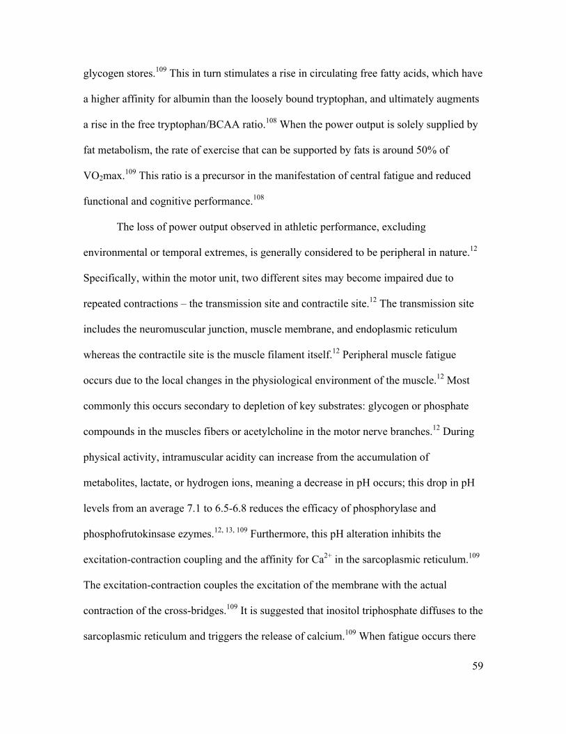
59
glycogen stores.109 This in turn stimulates a rise in circulating free fatty acids, which have
a higher affinity for albumin than the loosely bound tryptophan, and ultimately augments
a rise in the free tryptophan/BCAA ratio.108 When the power output is solely supplied by
fat metabolism, the rate of exercise that can be supported by fats is around 50% of
VO2max.109 This ratio is a precursor in the manifestation of central fatigue and reduced
functional and cognitive performance.108
The loss of power output observed in athletic performance, excluding
environmental or temporal extremes, is generally considered to be peripheral in nature.12
Specifically, within the motor unit, two different sites may become impaired due to
repeated contractions – the transmission site and contractile site.12 The transmission site
includes the neuromuscular junction, muscle membrane, and endoplasmic reticulum
whereas the contractile site is the muscle filament itself.12 Peripheral muscle fatigue
occurs due to the local changes in the physiological environment of the muscle.12 Most
commonly this occurs secondary to depletion of key substrates: glycogen or phosphate
compounds in the muscles fibers or acetylcholine in the motor nerve branches.12 During
physical activity, intramuscular acidity can increase from the accumulation of
metabolites, lactate, or hydrogen ions, meaning a decrease in pH occurs; this drop in pH
levels from an average 7.1 to 6.5-6.8 reduces the efficacy of phosphorylase and
phosphofrutokinsase ezymes.12, 13, 109 Furthermore, this pH alteration inhibits the
excitation-contraction coupling and the affinity for Ca2+ in the sarcoplasmic reticulum.109
The excitation-contraction couples the excitation of the membrane with the actual
contraction of the cross-bridges.109 It is suggested that inositol triphosphate diffuses to the
sarcoplasmic reticulum and triggers the release of calcium.109 When fatigue occurs there
60
is a decrease in inositol triphosphate and results in less calcium released from the
sarcoplasmic reticulum. Less calcium results in loss of muscle contraction.109
Further physiologic changes of fatigue include blood flow impairment and
accumulation of lactic acid.110 While muscles are activated they produce lactic acid, a
product of anaerobic metabolism. Blood flow from the musculature typically removes
this waste product, however, a sustained contraction of ≈20% MVC can restrict the blood
flow.110 This effect is exacerbated in sustained contractions and static poses because the
effects of vascular return are also limited from the lack of movement.110 Higher force
contractions can even stop blood flow, making the muscle ischemic.111 A contraction at
this level would produce a more rapid acute fatigue although muscles would also recover
rapidly.111 Lower level contractions require longer periods of time to elicit a fatigue
response, however the recovery time is also lengthened.111 An accumulation of sub-
maximal contractions of the course of a prolonged period of time has been theorized to
contribute to overuse injuries in overhead athletes.5, 112-114
Fatigue during intermittent exercise is complex, and it is difficult to identify a
single factor in the muscle responsible for the reduction in performance during intense
exercise.109 Based on measurements of adenosine triphosphate and creatine phosphate it
seems that fatigue is not caused by lack of energy and accumulation of lactate and the
disturbance of the acid/base balance of skeletal muscle also does not play a significant
role.109 Fatigue may be associated with an excitation- coupling failure and a reduced
nervous drive due to reflex inhibition at the spinal level as well as the interstitial
potassium in the muscle.109 It is speculated that the accumulation of potassium in the
interstitium also may be correlated to fatigue with intermittent exercise.109 The
61
mechanism behind the possible effect of potassium on the development of fatigue is
unclear. It may be that the accumulating potassium stimulates sensory receptors of group
III and IV nerve fibers leading to the inhibition at the spinal level or potassium could
inhibit the spread of the action potential due to ion disturbances over the sarcolemma and
possible block into the t-tubules.109 The continuous efflux of potassium from the
exercising muscle, together with a limited reuptake and a reduced release to venous blood
leads to a progressive accumulation of potassium in the interstitum, which may have been
implicated in the fatigue process.109
APPENDIX C
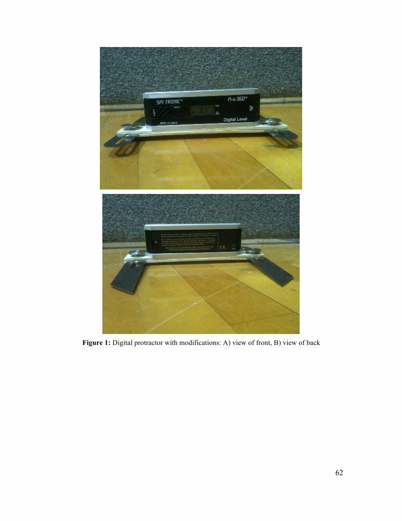
62
Figure 1: Digital protractor with modifications: A) view of front, B) view of back
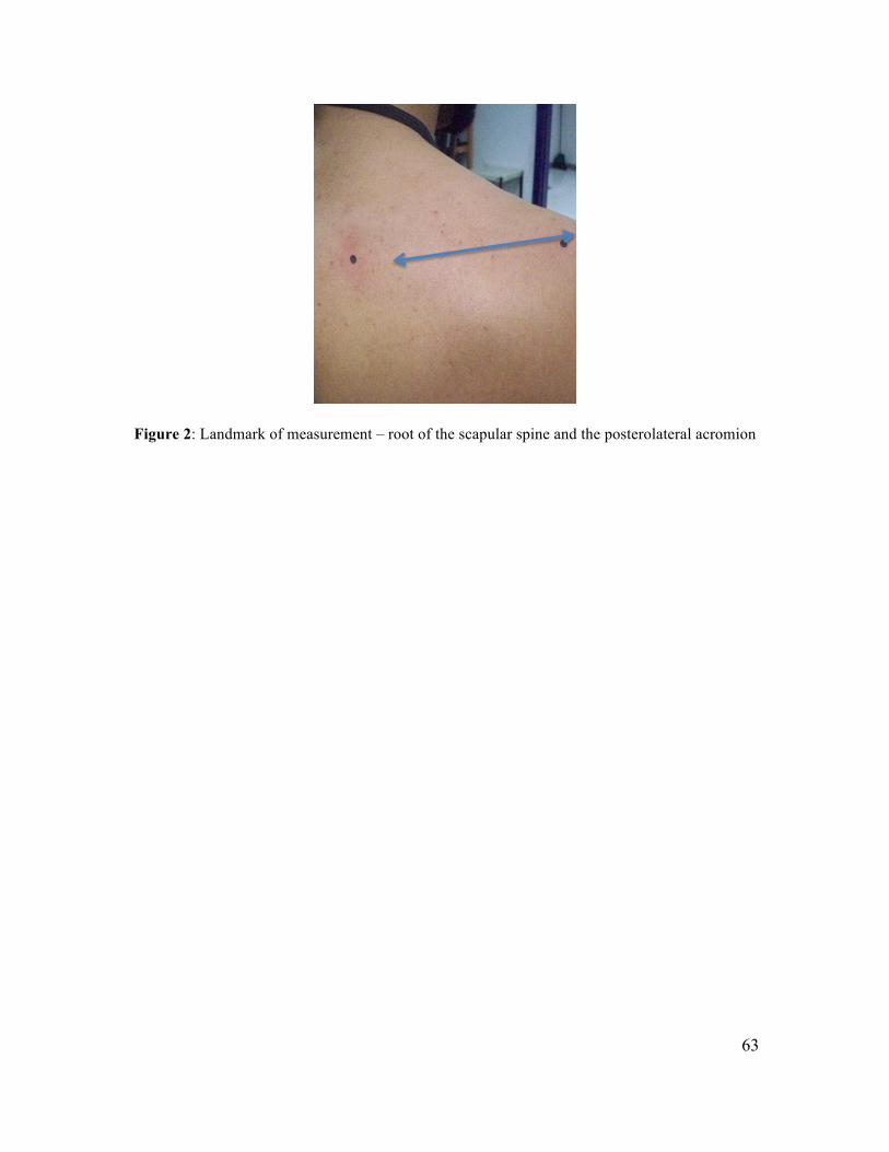
63
Figure 2: Landmark of measurement – root of the scapular spine and the posterolateral acromion
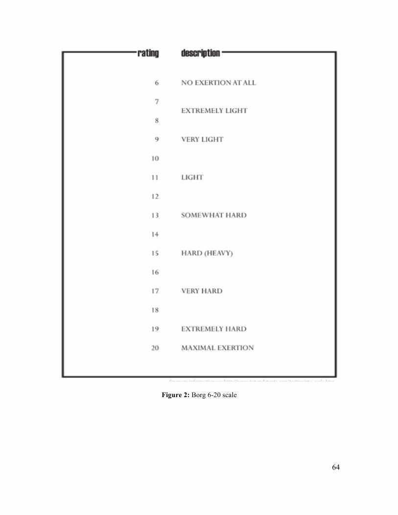
64
Figure 2: Borg 6-20 scale
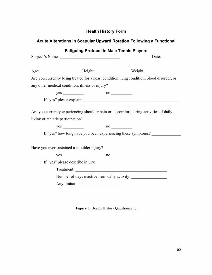
65
Health History Form
Acute Alterations in Scapular Upward Rotation Following a Functional
Fatiguing Protocol in Male Tennis Players Subject’s Name: _____________________________ Date:
______________
Age: ________ Height: ________ Weight: ________
Are you currently being treated for a heart condition, lung condition, blood disorder, or
any other medical condition, illness or injury?
yes __________ no __________
If “yes” please explain: ______________________________________________
Are you currently experiencing shoulder pain or discomfort during activities of daily
living or athletic participation?
yes __________ no __________
If “yes” how long have you been experiencing these symptoms? ______________
Have you ever sustained a shoulder injury?
yes __________ no __________
If “yes” please describe injury: __________________________________
Treatment: ____________________________________________
Number of days inactive from daily activity: _________________
Any limitations: ________________________________________
Figure 3: Health History Questionnaire
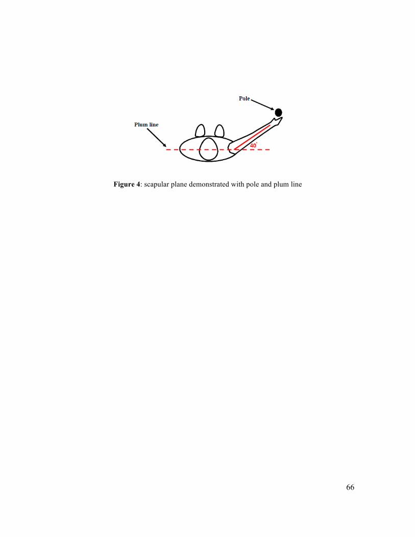
66
Figure 4: scapular plane demonstrated with pole and plum line
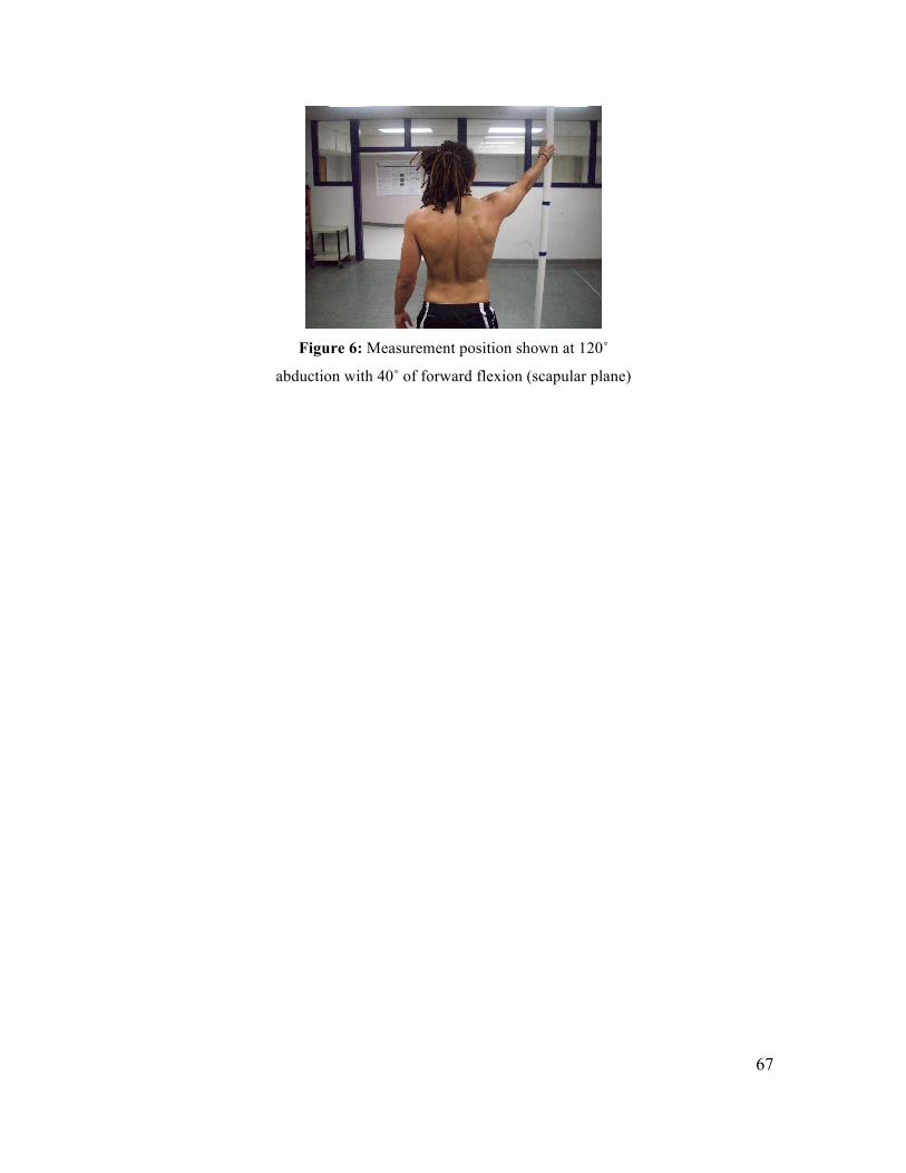
67
Figure 6: Measurement position shown at 120˚
abduction with 40˚ of forward flexion (scapular plane)
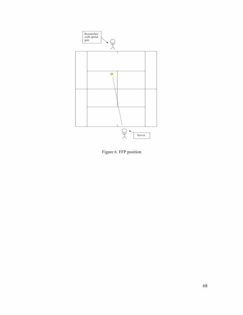
68
Figure 6. FFP position
��
%ECQNA�� %@@IGHF5H=CB�C:�H<9�(5L=A5@�.9FJ9�19@C7=HM�/9GH
��2ANRAN
1AOA=N?DAN
NA?KN@EJC
@=P=
1AOA=N?DAN
FQ@CEJC
>=HH��EJ�KQP
1AOA=N?DAN
SEPD�OLAA@
CQJ
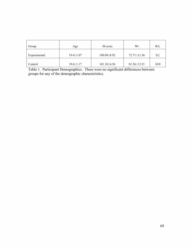
69
Group Age Ht (cm) Wt R/L
Experimental 19.4+1.07 180.09+8.92 72.71+11.56 9/1
Control 19.6+1.17 181.10+6.56 81.56+13.51 10/0 Table 1. Participant Demographics. There were no significant differences between groups for any of the demographic characteristics.
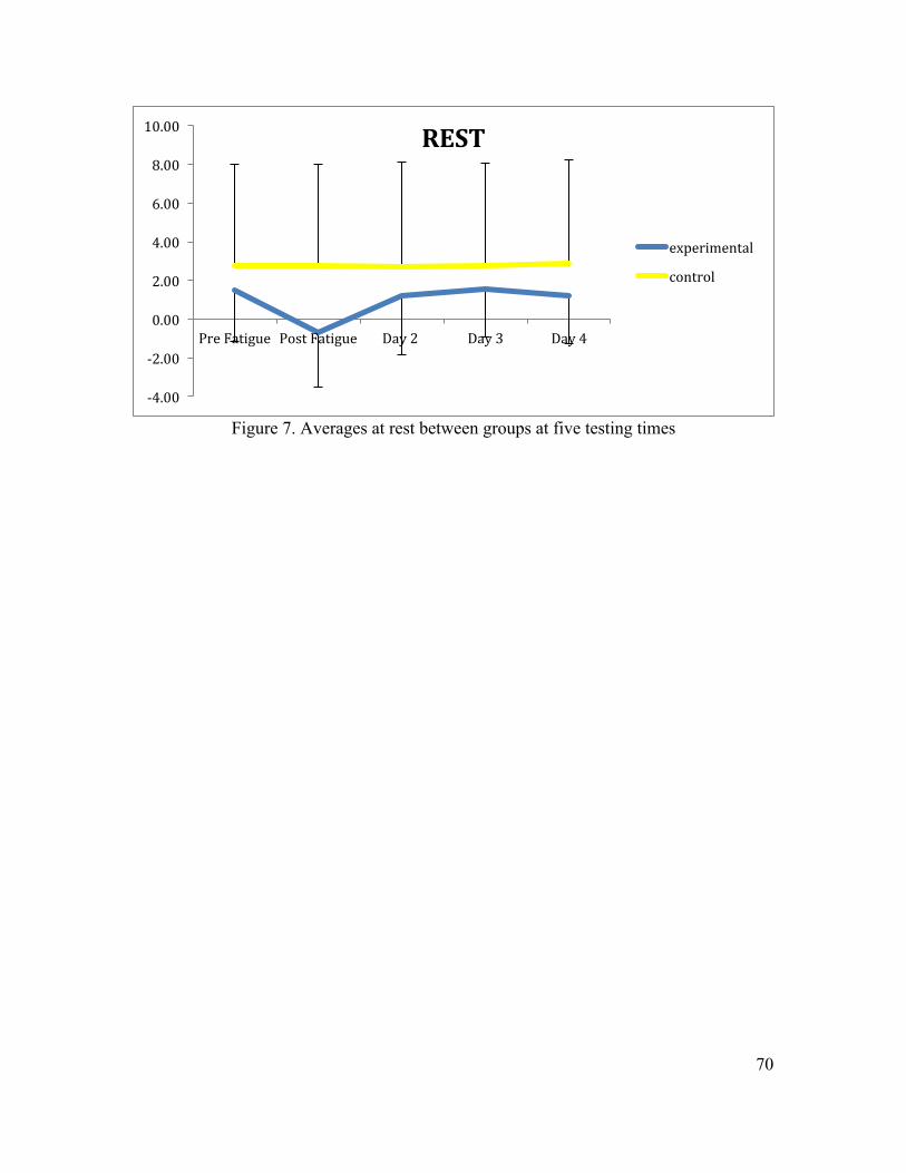
70
Figure 7. Averages at rest between groups at five testing times
-‐4.00
-‐2.00
0.00
2.00
4.00
6.00
8.00
10.00
Pre Fatigue Post Fatigue Day 2 Day 3 Day 4
REST
experimental
control
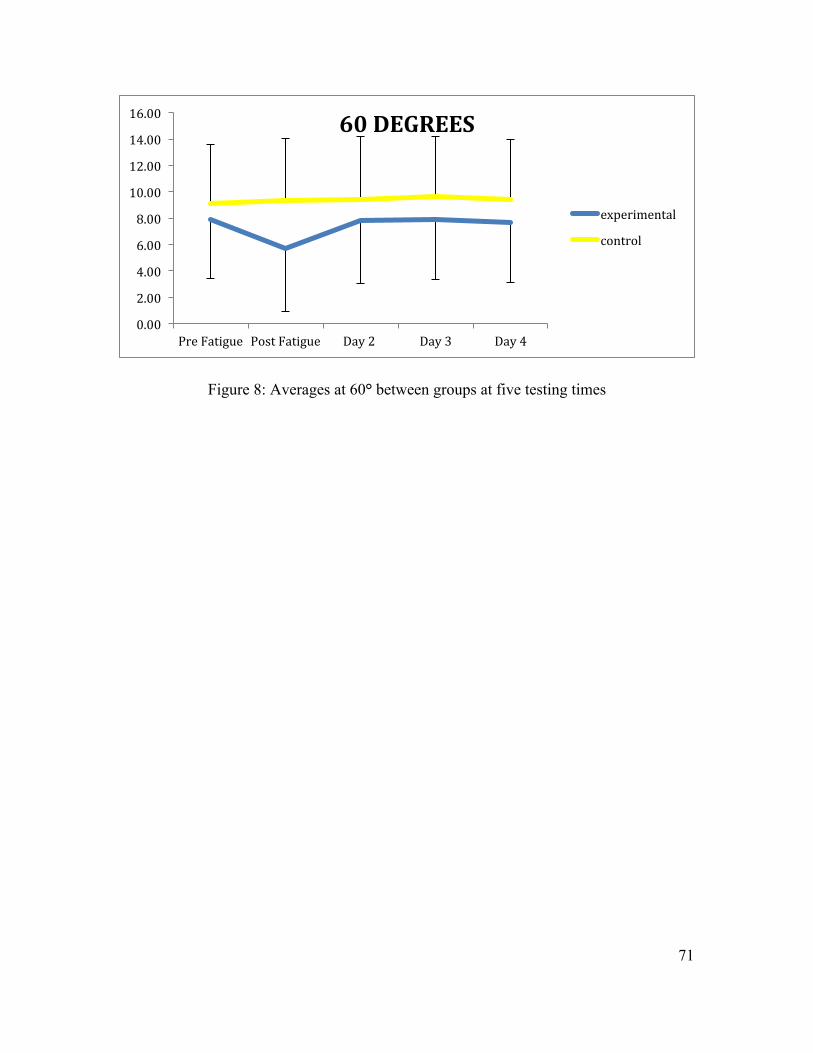
71
Figure 8: Averages at 60° between groups at five testing times
0.00
2.00
4.00
6.00
8.00
10.00
12.00
14.00
16.00
Pre Fatigue Post Fatigue Day 2 Day 3 Day 4
60 DEGREES
experimental
control
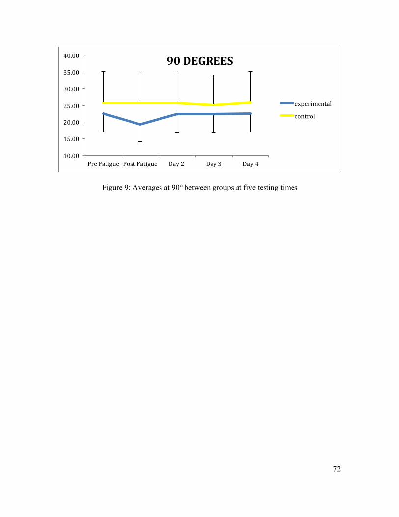
72
Figure 9: Averages at 90° between groups at five testing times
10.00
15.00
20.00
25.00
30.00
35.00
40.00
Pre Fatigue Post Fatigue Day 2 Day 3 Day 4
90 DEGREES
experimental
control
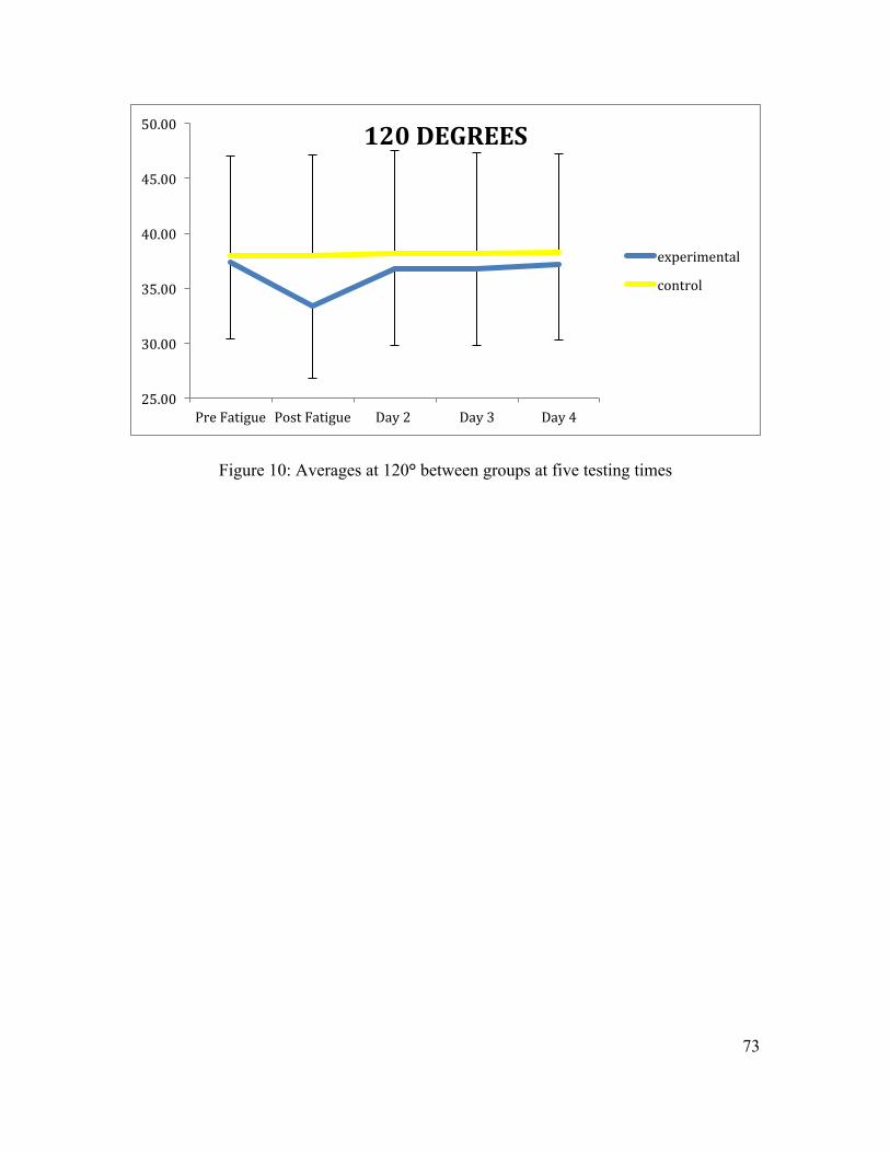
73
Figure 10: Averages at 120° between groups at five testing times
25.00
30.00
35.00
40.00
45.00
50.00
Pre Fatigue Post Fatigue Day 2 Day 3 Day 4
120 DEGREES
experimental
control
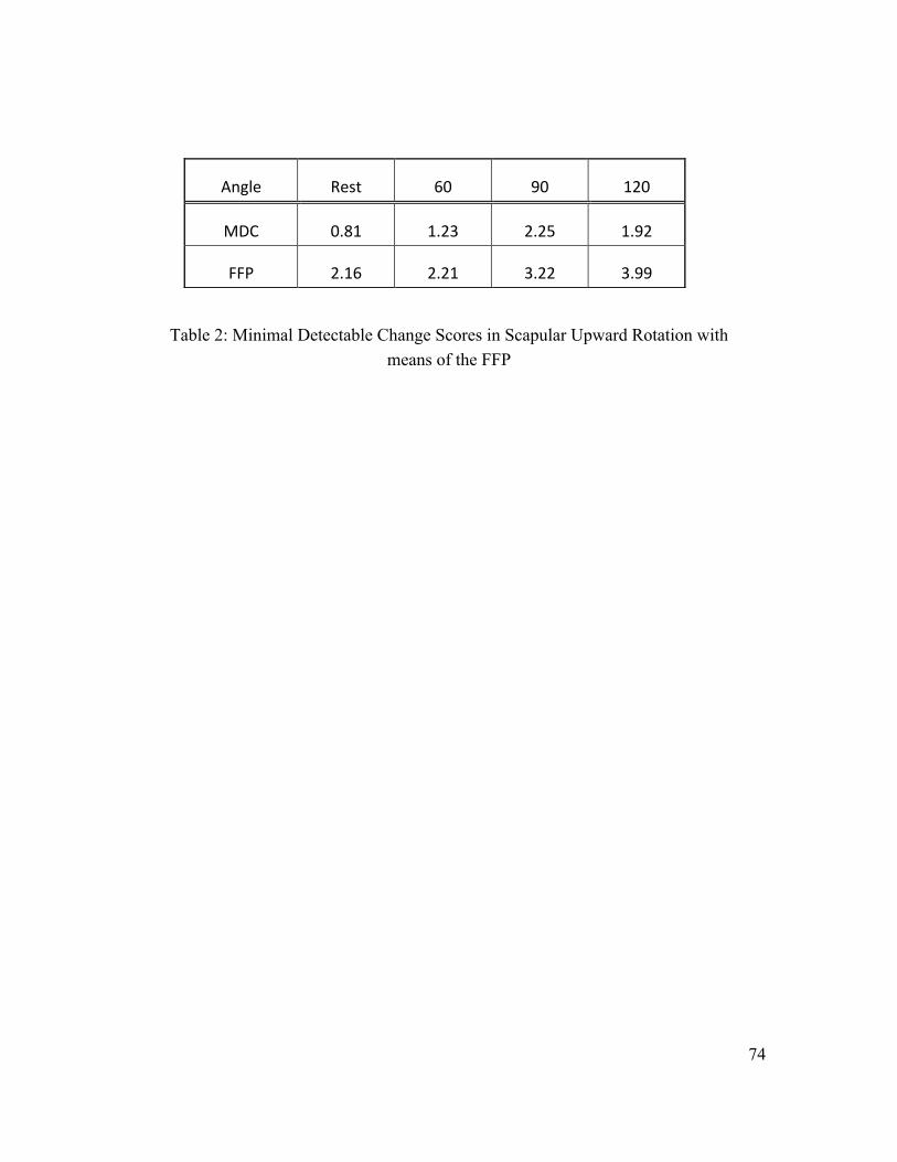
74
Table 2: Minimal Detectable Change Scores in Scapular Upward Rotation with means of the FFP
Angle Rest 60 90 120
MDC 0.81 1.23 2.25 1.92
FFP 2.16 2.21 3.22 3.99
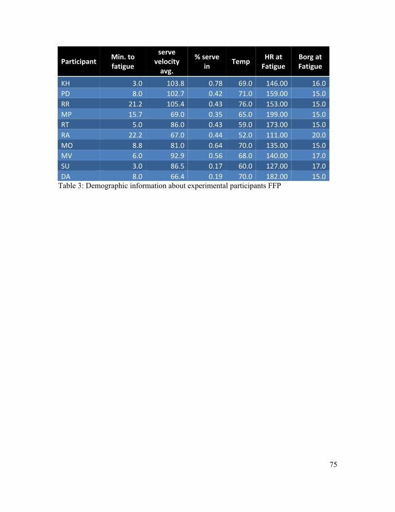
75
Participant Min. to fatigue
serve velocity avg.
% serve in Temp HR at
Fatigue Borg at Fatigue
KH 3.0 103.8 0.78 69.0 146.00 16.0 PD 8.0 102.7 0.42 71.0 159.00 15.0 RR 21.2 105.4 0.43 76.0 153.00 15.0 MP 15.7 69.0 0.35 65.0 199.00 15.0 RT 5.0 86.0 0.43 59.0 173.00 15.0 RA 22.2 67.0 0.44 52.0 111.00 20.0 MO 8.8 81.0 0.64 70.0 135.00 15.0 MV 6.0 92.9 0.56 68.0 140.00 17.0 SU 3.0 86.5 0.17 60.0 127.00 17.0 DA 8.0 66.4 0.19 70.0 182.00 15.0
Table 3: Demographic information about experimental participants FFP
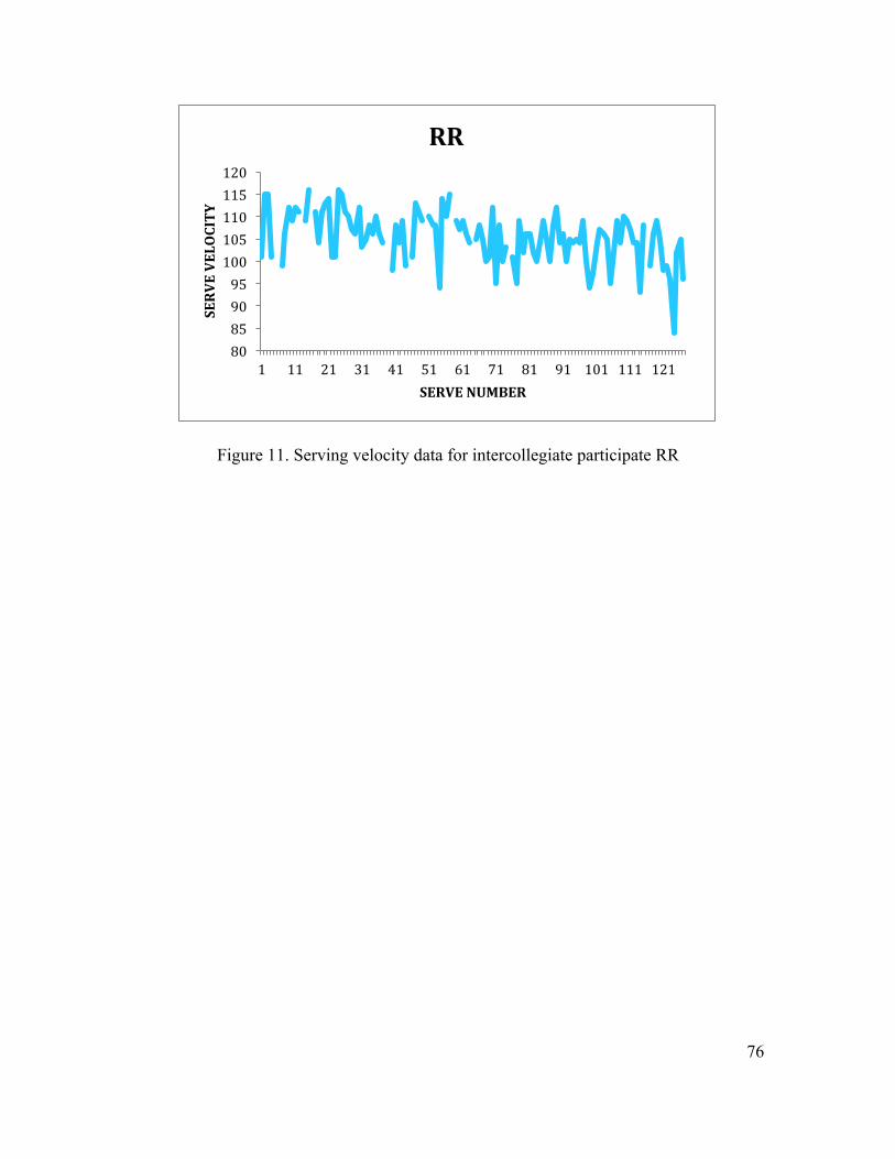
76
Figure 11. Serving velocity data for intercollegiate participate RR
80 85 90 95 100 105 110 115 120
1 11 21 31 41 51 61 71 81 91 101 111 121
SERVE VELOCITY
SERVE NUMBER
RR
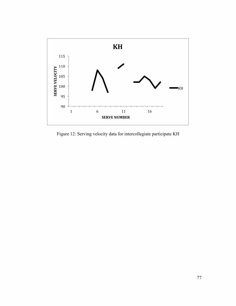
77
Figure 12: Serving velocity data for intercollegiate participate KH
90
95
100
105
110
115
1 6 11 16
SERVE VELOCITY
SERVE NUMBER
KH
KH
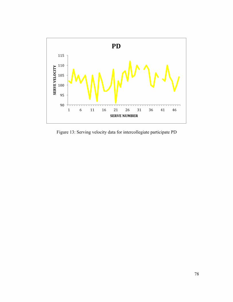
78
Figure 13: Serving velocity data for intercollegiate participate PD
90
95
100
105
110
115
1 6 11 16 21 26 31 36 41 46
SERVE VELOCITY
SERVE NUMBER
PD
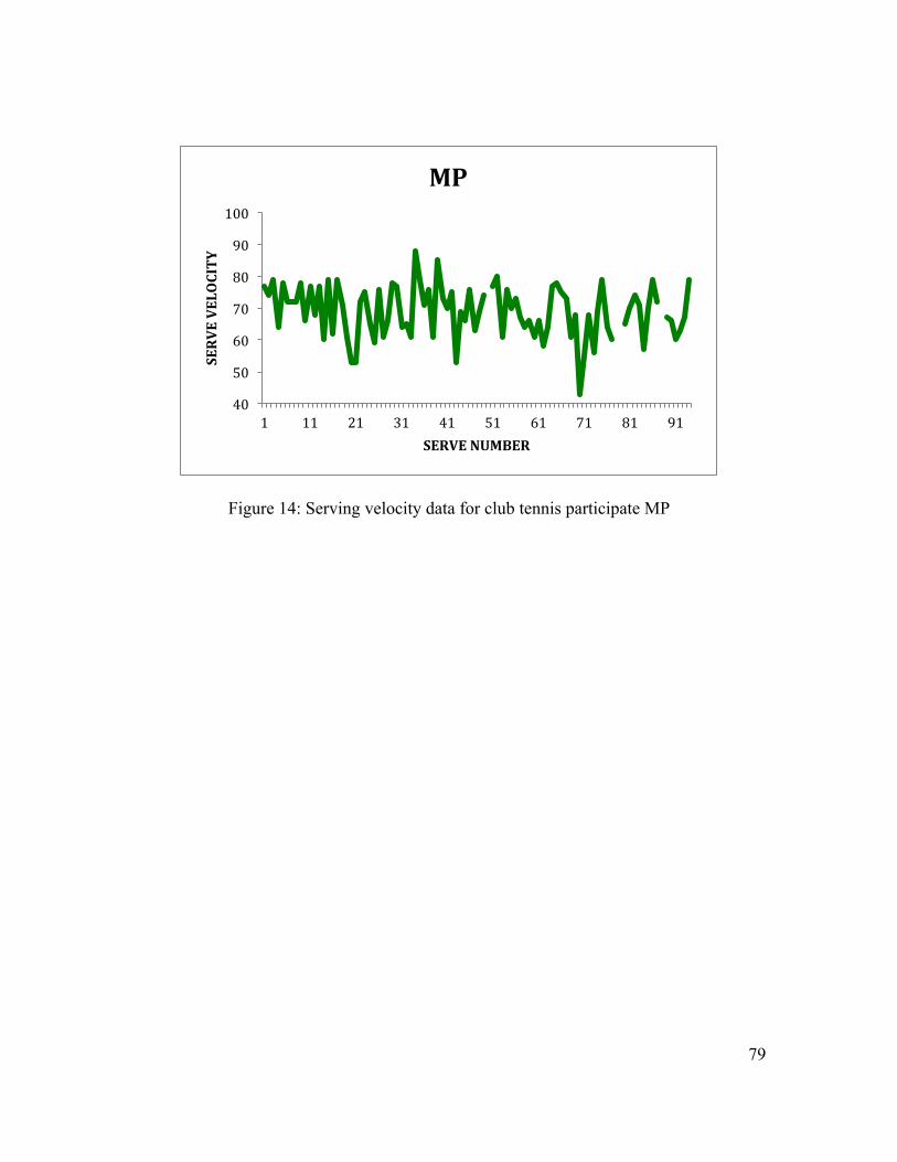
79
Figure 14: Serving velocity data for club tennis participate MP
40
50
60
70
80
90
100
1 11 21 31 41 51 61 71 81 91
SERVE VELOCITY
SERVE NUMBER
MP
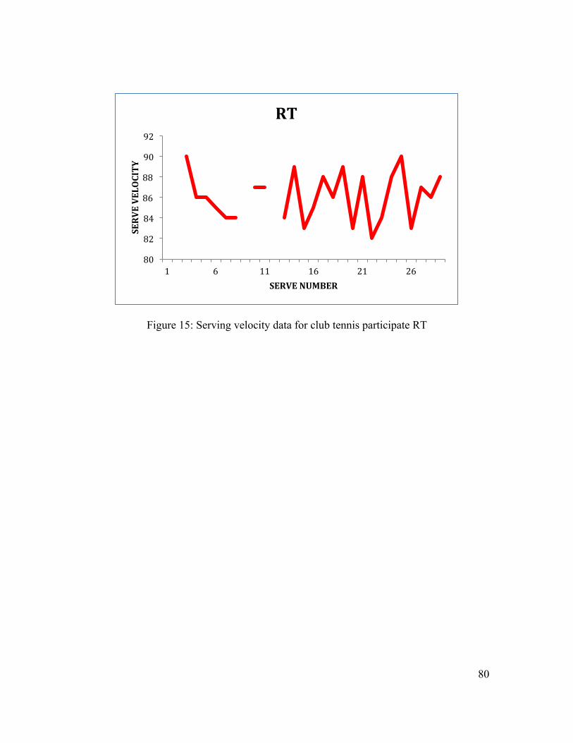
80
Figure 15: Serving velocity data for club tennis participate RT
80
82
84
86
88
90
92
1 6 11 16 21 26
SERVE VELOCITY
SERVE NUMBER
RT
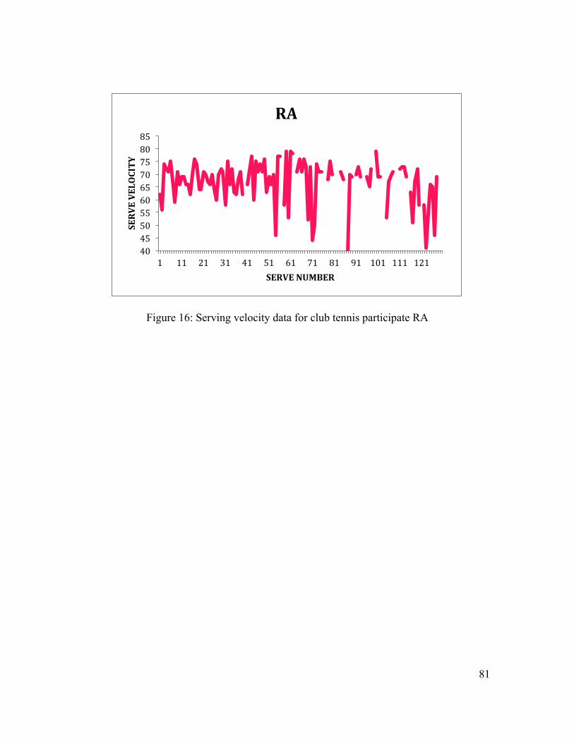
81
Figure 16: Serving velocity data for club tennis participate RA
40 45 50 55 60 65 70 75 80 85
1 11 21 31 41 51 61 71 81 91 101 111 121
SERVE VELOCITY
SERVE NUMBER
RA
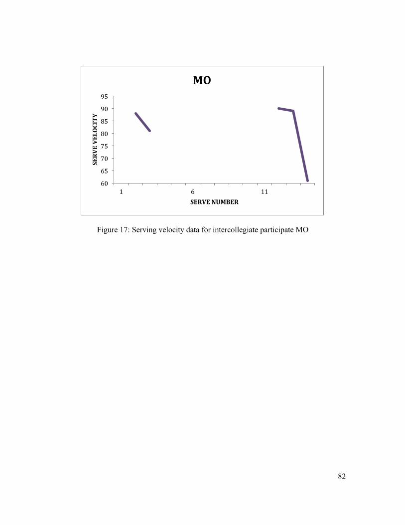
82
Figure 17: Serving velocity data for intercollegiate participate MO
60
65
70
75
80
85
90
95
1 6 11
SERVE VELOCITY
SERVE NUMBER
MO
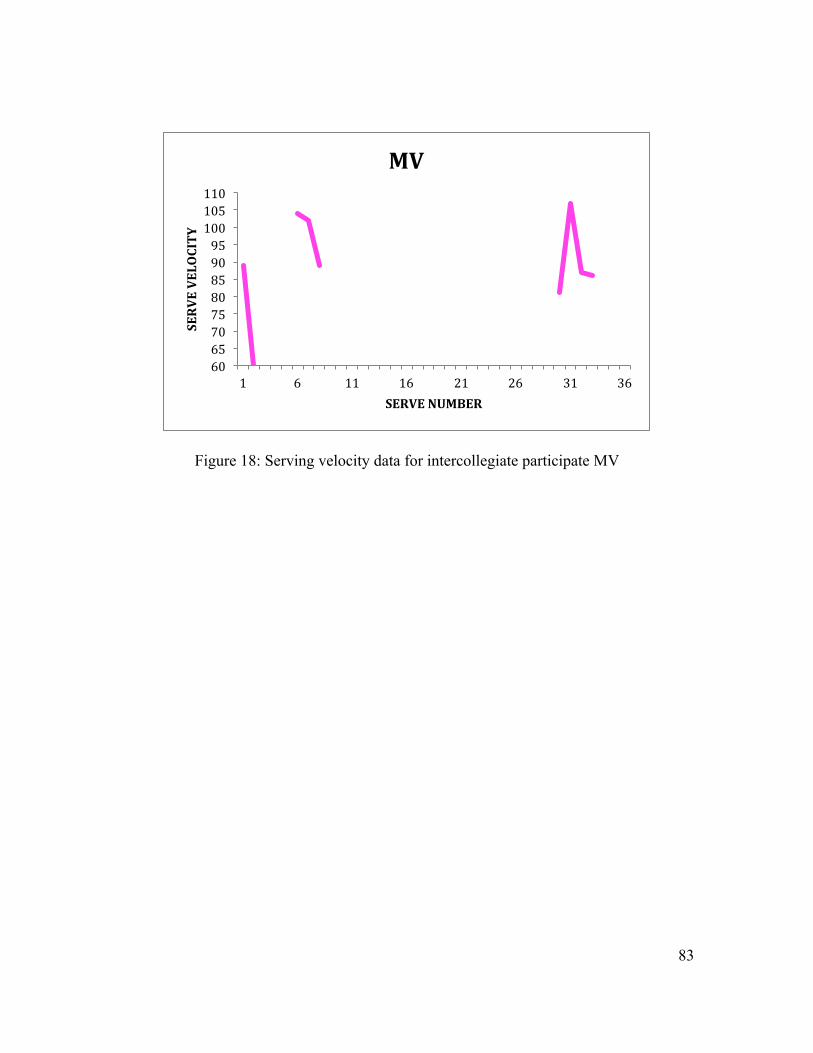
83
Figure 18: Serving velocity data for intercollegiate participate MV
60 65 70 75 80 85 90 95 100 105 110
1 6 11 16 21 26 31 36
SERVE VELOCITY
SERVE NUMBER
MV
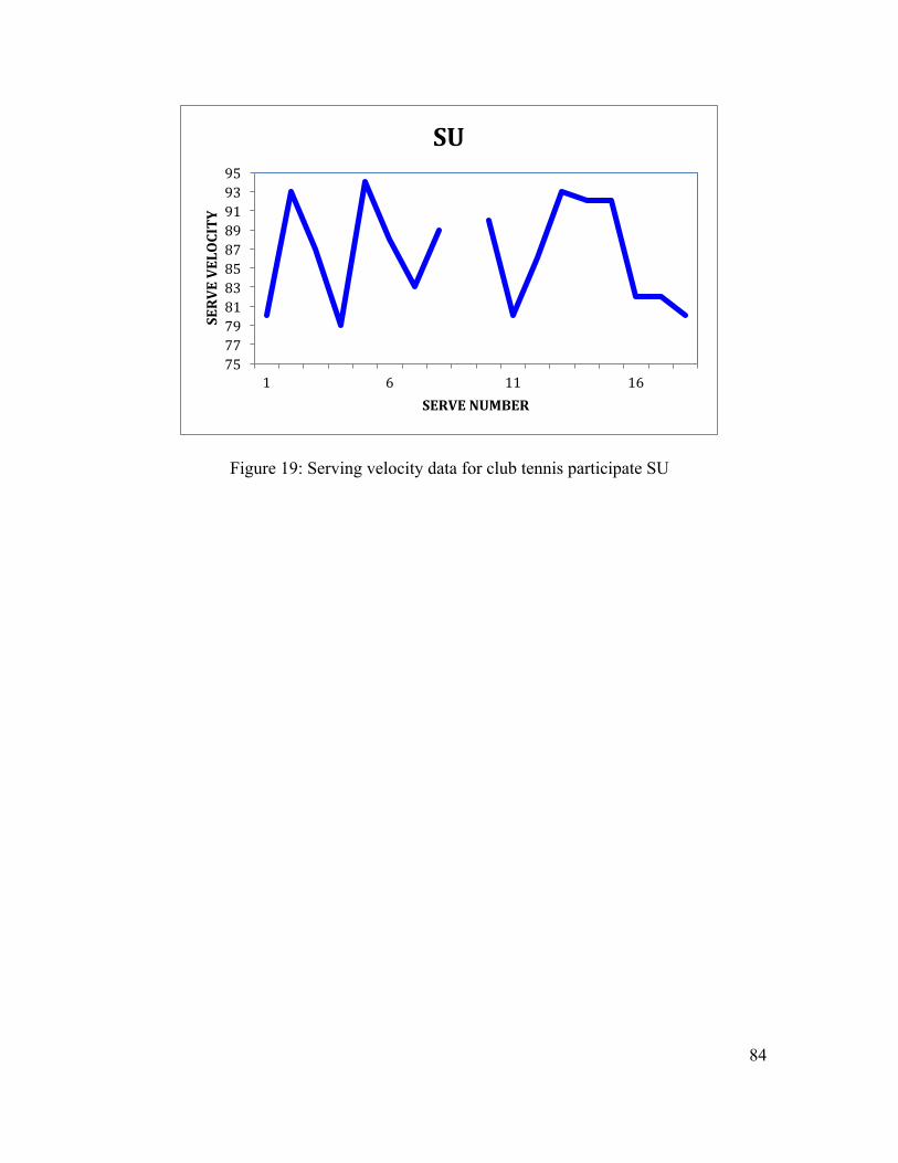
84
Figure 19: Serving velocity data for club tennis participate SU
75 77 79 81 83 85 87 89 91 93 95
1 6 11 16
SERVE VELOCITY
SERVE NUMBER
SU
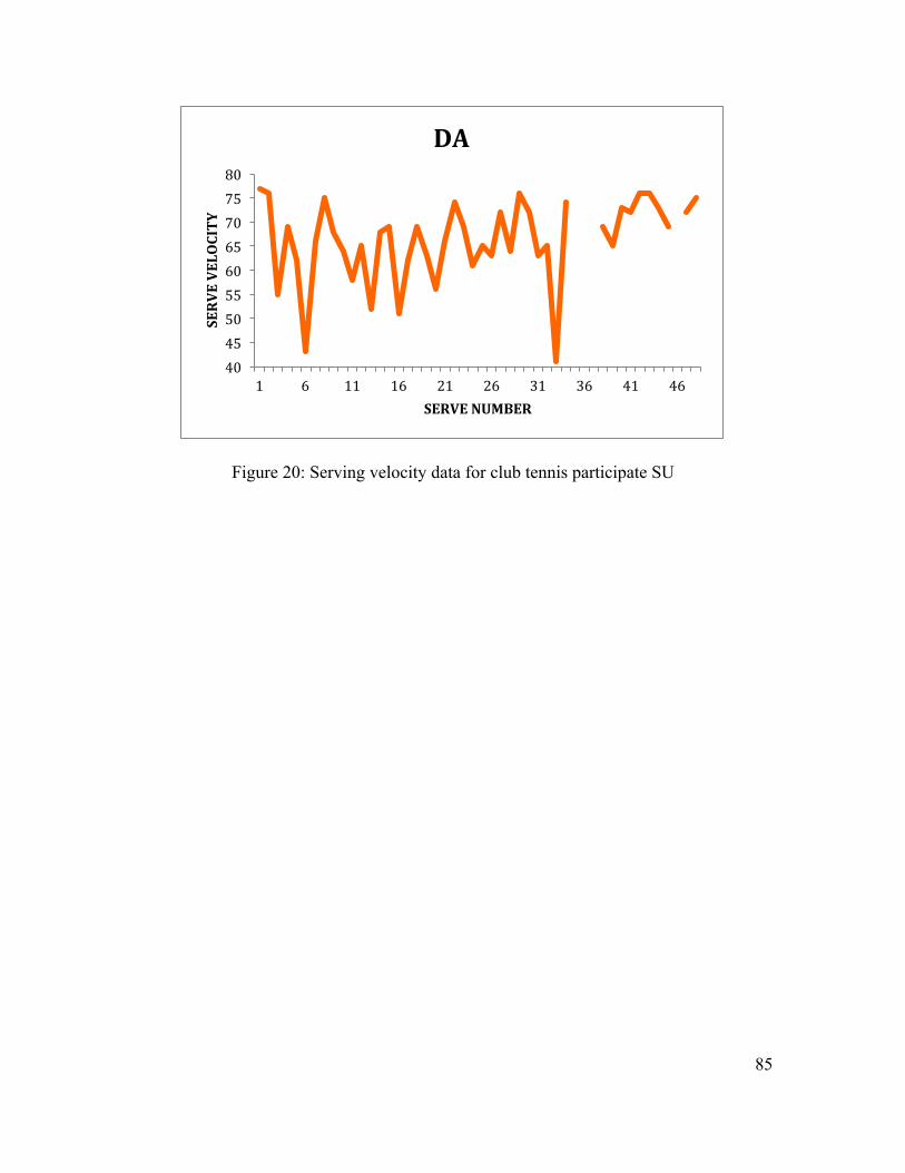
85
Figure 20: Serving velocity data for club tennis participate SU
40 45 50 55 60 65 70 75 80
1 6 11 16 21 26 31 36 41 46
SERVE VELOCITY
SERVE NUMBER
DA

86
APPENDIX D
INSTITUTIONAL REVIEW BOARD FORMS
87
1. Culham E, Peat M. Functional anatomy of the shoulder complex. The Journal of
orthopaedic and sports physical therapy 1993;18(1):342-50. 2. Inman VT, Saunders JB, Abbott LC. Observations of the function of the shoulder
joint. 1944. Clin Orthop Relat Res 1996(330):3-12. 3. Ebaugh DD, McClure PW, Karduna AR. Effects of shoulder muscle fatigue
caused by repetitive overhead activities on scapulothoracic and glenohumeral kinematics. Journal of electromyography and kinesiology : official journal of the International Society of Electrophysiological Kinesiology 2006;16(3):224-35.
4. Burkhart SS, Morgan CD, Kibler WB. Shoulder injuries in overhead athletes. The "dead arm" revisited. Clinics in sports medicine 2000;19(1):125-58.
5. Kibler WB. The role of the scapula in athletic shoulder function. The American journal of sports medicine 1998;26(2):325-37.
6. Ozaki J. Glenohumeral movements of the involuntary inferior and multidirectional instability. Clinical orthopaedics and related research 1989(238):107-11.
7. Paletta GA, Jr., Warner JJ, Warren RF, Deutsch A, Altchek DW. Shoulder kinematics with two-plane x-ray evaluation in patients with anterior instability or rotator cuff tearing. Journal of shoulder and elbow surgery / American Shoulder and Elbow Surgeons ... [et al.] 1997;6(6):516-27.
8. Solem-Bertoft E, Thuomas KA, Westerberg CE. The influence of scapular retraction and protraction on the width of the subacromial space. An MRI study. Clinical orthopaedics and related research 1993(296):99-103.
9. Warner JJ, Micheli LJ, Arslanian LE, Kennedy J, Kennedy R. Scapulothoracic motion in normal shoulders and shoulders with glenohumeral instability and impingement syndrome. A study using Moire topographic analysis. Clinical orthopaedics and related research 1992(285):191-9.
10. Voight ML, Thomson BC. The role of the scapula in the rehabilitation of shoulder injuries. Journal of athletic training 2000;35(3):364-72.
11. Mannion AF, Dolan P. Relationship between myoelectric and mechanical manifestations of fatigue in the quadriceps femoris muscle group. European journal of applied physiology and occupational physiology 1996;74(5):411-9.
12. Asmussen E. Muscle fatigue. June 1979. Medicine and science in sports and exercise 1993;25(4):411-20.
13. Kraemer WJ, Fleck SJ, Deschenes MR. Exercise physiology : integrated from theory to practical applications. 1st ed. Philadelphia: Wolters Kluwer/Lippincott Williams & Wilkins Health; 2012.
14. Kent-Braun JA, Ng AV, Doyle JW, Towse TF. Human skeletal muscle responses vary with age and gender during fatigue due to incremental isometric exercise. Journal of applied physiology 2002;93(5):1813-23.
15. Jobe FW, Pink M. Classification and treatment of shoulder dysfunction in the overhead athlete. The Journal of orthopaedic and sports physical therapy 1993;18(2):427-32.
16. Bak K, Fauno P. Clinical findings in competitive swimmers with shoulder pain. The American journal of sports medicine 1997;25(2):254-60.
17. Sevinsky S. Scapular Dyskinesis. Bethlehem, PA. 18. Page P. Shoulder muscle imbalance and subacromial impingement syndrome in
overhead athletes. International journal of sports physical therapy 2011;6(1):51-8.
88
19. Marx RG, Sperling JW, Cordasco FA. Overuse injuries of the upper extremity in tennis players. Clinics in sports medicine 2001;20(3):439-51.
20. Pluim BM, Staal JB, Windler GE, Jayanthi N. Tennis injuries: occurrence, aetiology, and prevention. British journal of sports medicine 2006;40(5):415-23.
21. Di Sumant G, Krishnan, Richard J., Hawkins, Russell, F. Warren. The shoulder and the overhead athlete. Philadelphia: Lippincott, Williams, and Wilkins; 2004.
22. Kibler WB. Biomechanical analysis of the shoulder during tennis activities. Clinics in sports medicine 1995;14(1):79-85.
23. Krishnan SG, Hawkins, Richard J., Warren Russell F. The Shoulder and the Overhead Athlete
. 1 ed. Philadelphia: Lippincott Williams & Wilkins; 2004. 24. Ludewig PM, Cook TM. Alterations in shoulder kinematics and associated
muscle activity in people with symptoms of shoulder impingement. Physical therapy 2000;80(3):276-91.
25. Inman VT, Saunders JB, Abbott LC. Observations of the function of the shoulder joint. 1944. Clinical orthopaedics and related research 1996(330):3-12.
26. Prentice WE, Arnheim DD. Arnheim's principles of athletic training : a competency-based approach. 13th ed. Boston: McGraw-Hill Higher Education; 2009.
27. Starkey C, Ryan JL. Evaluation of orthopedic and athletic injuries. 2nd ed. Philadelphia, PA: F.A. Davis Co.; 2002.
28. Andrews JRW, Kevin E. The Athlete's Shoulder. New York: Churchill Livingstone Inc.; 1994.
29. Ciullo J. Shoulder Injuries in Sport. Troy; 1996. 30. Micheo W. Rehabiliation of shoulder injury in the throwing athlete. Critical
Reviews in Physical and Rehabilitation Medicine 2008;20(1):87. 31. Pinsent C, Rae, Lisa, Palmer, Jillian. Return to activity guidelines for the
overhead athlete after shoulder injury. Edmonton: University of Alberta; 2010. p. 38.
32. Johnson MP, McClure PW, Karduna AR. New method to assess scapular upward rotation in subjects with shoulder pathology. The Journal of orthopaedic and sports physical therapy 2001;31(2):81-9.
33. Tucker WS, Ingram RL. Reliability and validity of measuring scapular upward rotation using an electrical inclinometer. Journal of electromyography and kinesiology : official journal of the International Society of Electrophysiological Kinesiology 2012;22(3):419-23.
34. Londeree BR MM. Influence of age and other factors on maximal heart rate. J Cardiac Rehabil 1984;4:44–49.
35. Kang J, Chaloupka EC, Mastrangelo MA, Donnelly MS, Martz WP, Robertson RJ. Regulating exercise intensity using ratings of perceived exertion during arm and leg ergometry. European journal of applied physiology and occupational physiology 1998;78(3):241-6.
36. Marais G, Weissland T, Robin H, Vanvelcenaher JM, Lavoie JM, Pelayo P. Physiological effects of variations in spontaneously chosen crank rate during sub-maximal and supra-maximal upper body exercises. International journal of sports medicine 1999;20(4):239-45.
37. Downar JM, Sauers EL. Clinical Measures of Shoulder Mobility in the Professional Baseball Player. Journal of athletic training 2005;40(1):23-29.
38. Tripp BL, Yochem EM, Uhl TL. Functional fatigue and upper extremity sensorimotor system acuity in baseball athletes. Journal of athletic training 2007;42(1):90-8.
89
39. Peat M. Functional anatomy of the shoulder complex. Physical therapy 1986;66(12):1855-65.
40. Lippitt S, Matsen F. Mechanisms of glenohumeral joint stability. Clinical orthopaedics and related research 1993(291):20-8.
41. Warner JJ, Bowen MK, Deng X, Torzilli PA, Warren RF. Effect of joint compression on inferior stability of the glenohumeral joint. Journal of shoulder and elbow surgery / American Shoulder and Elbow Surgeons ... [et al.] 1999;8(1):31-6.
42. Gray H, Clemente CD. Anatomy of the human body. 30th American ed. Philadelphia: Lea & Febiger; 1985.
43. Walch G LJ, Boileau P. Paper presented at: Fifth International Conference on Surgery of the Shoulder, 1992; Paris, France.
44. Werner CM, Favre P, Gerber C. The role of the subscapularis in preventing anterior glenohumeral subluxation in the abducted, externally rotated position of the arm. Clinical biomechanics 2007;22(5):495-501.
45. Lee SB, An KN. Dynamic glenohumeral stability provided by three heads of the deltoid muscle. Clinical orthopaedics and related research 2002(400):40-7.
46. Bigliani LU, Codd TP, Connor PM, Levine WN, Littlefield MA, Hershon SJ. Shoulder motion and laxity in the professional baseball player. The American journal of sports medicine 1997;25(5):609-13.
47. Jobe FW, Pink M. The athlete's shoulder. Journal of hand therapy : official journal of the American Society of Hand Therapists 1994;7(2):107-10.
48. Kamkar A, Irrgang JJ, Whitney SL. Nonoperative management of secondary shoulder impingement syndrome. The Journal of orthopaedic and sports physical therapy 1993;17(5):212-24.
49. Woo SL, Buckwalter JA. AAOS/NIH/ORS workshop. Injury and repair of the musculoskeletal soft tissues. Savannah, Georgia, June 18-20, 1987. Journal of orthopaedic research : official publication of the Orthopaedic Research Society 1988;6(6):907-31.
50. Elliott BC, Marshall, R.N., Noffal, G.J. Contributions of upper limb segment rotations during the power serve in tennis. J Appl Biomech 1995;11(1):433-42.
51. Jobe FW. Operative Techniques in Upper Extremity Sports Injuries. St. Louis: Mosby-Year Book Inc; 1996.
52. Iannotti JP, Gabriel JP, Schneck SL, Evans BG, Misra S. The normal glenohumeral relationships. An anatomical study of one hundred and forty shoulders. The Journal of bone and joint surgery. American volume 1992;74(4):491-500.
53. Jobe CM, Iannotti JP. Limits imposed on glenohumeral motion by joint geometry. Journal of shoulder and elbow surgery / American Shoulder and Elbow Surgeons ... [et al.] 1995;4(4):281-5.
54. Poppen NK, Walker PS. Normal and abnormal motion of the shoulder. The Journal of bone and joint surgery. American volume 1976;58(2):195-201.
55. Schulz CU, Pfahler M, Anetzberger HM, Becker CR, Muller-Gerbl M, Refior HJ. The mineralization patterns at the subchondral bone plate of the glenoid cavity in healthy shoulders. Journal of shoulder and elbow surgery / American Shoulder and Elbow Surgeons ... [et al.] 2002;11(2):174-81.
56. Kibler WB, Sciascia A. Current concepts: scapular dyskinesis. British journal of sports medicine 2010;44(5):300-5.
57. Pinsent C RL, Palmer J. Return to activity guidelines for the overhead athlete after shoulder injury. [Edmonton: University of Alberta; 2010.
58. Werner SL, Guido JA, Jr., Stewart GW, McNeice RP, VanDyke T, Jones DG. Relationships between throwing mechanics and shoulder distraction in collegiate
90
baseball pitchers. Journal of shoulder and elbow surgery / American Shoulder and Elbow Surgeons ... [et al.] 2007;16(1):37-42.
59. Brakeville R. New Concepts in the Management of Upper Extremity Injuries in the Adolescent Throwing Athlete.
60. Davis JT, Limpisvasti O, Fluhme D, Mohr KJ, Yocum LA, Elattrache NS, et al. The effect of pitching biomechanics on the upper extremity in youth and adolescent baseball pitchers. The American journal of sports medicine 2009;37(8):1484-91.
61. Hurov J. Anatomy and mechanics of the shoulder: review of current concepts. Journal of hand therapy : official journal of the American Society of Hand Therapists 2009;22(4):328-42; quiz 43.
62. Dillman CJ, Fleisig GS, Andrews JR. Biomechanics of pitching with emphasis upon shoulder kinematics. The Journal of orthopaedic and sports physical therapy 1993;18(2):402-8.
63. Fleisig GS, Barrentine SW, Escamilla RF, Andrews JR. Biomechanics of overhand throwing with implications for injuries. Sports medicine 1996;21(6):421-37.
64. Werner SL, Murray TA, Hawkins RJ, Gill TJ. Relationship between throwing mechanics and elbow valgus in professional baseball pitchers. Journal of shoulder and elbow surgery / American Shoulder and Elbow Surgeons ... [et al.] 2002;11(2):151-5.
65. Braun S, Kokmeyer D, Millett PJ. Shoulder injuries in the throwing athlete. The Journal of bone and joint surgery. American volume 2009;91(4):966-78.
66. Matsen FA, Fu FH, Hawkins RJ. The Shoulder : a balance of mobility and stability : workshop, Vail, Colorado, September 1992. Rosemont, IL: American Academy of Orthopaedic Surgeons; 1993.
67. Peterson L, Renström P, Grana WA. Sports injuries : their prevention and treatment. Chicago: Year Book Medical Publishers; 1986.
68. Perry J. Anatomy and biomechanics of the shoulder in throwing, swimming, gymnastics, and tennis. Clinics in sports medicine 1983;2(2):247-70.
69. Ryu RK, McCormick J, Jobe FW, Moynes DR, Antonelli DJ. An electromyographic analysis of shoulder function in tennis players. The American journal of sports medicine 1988;16(5):481-5.
70. Townsend H, Jobe FW, Pink M, Perry J. Electromyographic analysis of the glenohumeral muscles during a baseball rehabilitation program. The American journal of sports medicine 1991;19(3):264-72.
71. Elliott B. Biomechanics and tennis. British journal of sports medicine 2006;40(5):392-6.
72. Johnson CD, McHugh MP, Wood T, Kibler B. Performance demands of professional male tennis players. British journal of sports medicine 2006;40(8):696-9; discussion 99.
73. Magee DJ. Orthopedic physical assessment. 5th ed. St. Louis, Mo.: Saunders Elsevier; 2002.
74. Kibler WB, Safran MR. Musculoskeletal injuries in the young tennis player. Clinics in sports medicine 2000;19(4):781-92.
75. Bonza JE, Fields SK, Yard EE, Dawn Comstock R. Shoulder injuries among United States high school athletes during the 2005-2006 and 2006-2007 school years. Journal of athletic training 2009;44(1):76-83.
76. Kibler WB, McMullen J. Scapular dyskinesis and its relation to shoulder pain. The Journal of the American Academy of Orthopaedic Surgeons 2003;11(2):142-51.
91
77. Rubin BD, Kibler WB. Fundamental principles of shoulder rehabilitation: conservative to postoperative management. Arthroscopy : the journal of arthroscopic & related surgery : official publication of the Arthroscopy Association of North America and the International Arthroscopy Association 2002;18(9 Suppl 2):29-39.
78. Andrews JR, Kupferman SP, Dillman CJ. Labral tears in throwing and racquet sports. Clinics in sports medicine 1991;10(4):901-11.
79. Pagnani MJ, Deng XH, Warren RF, Torzilli PA, Altchek DW. Effect of lesions of the superior portion of the glenoid labrum on glenohumeral translation. The Journal of bone and joint surgery. American volume 1995;77(7):1003-10.
80. Morgan CD, Burkhart SS, Palmeri M, Gillespie M. Type II SLAP lesions: three subtypes and their relationships to superior instability and rotator cuff tears. Arthroscopy : the journal of arthroscopic & related surgery : official publication of the Arthroscopy Association of North America and the International Arthroscopy Association 1998;14(6):553-65.
81. Rizio L, Uribe JW. Overuse injuries of the upper extremity in baseball. Clinics in sports medicine 2001;20(3):453-68.
82. Fleisig GS, Andrews JR, Dillman CJ, Escamilla RF. Kinetics of baseball pitching with implications about injury mechanisms. The American journal of sports medicine 1995;23(2):233-9.
83. Matsen FA, Harryman DT, Sidles JA. Mechanics of Glenohumeral Instability. Clinics in sports medicine 1991;10(4):783-88.
84. Wilk KE, Arrigo CA, Andrews JR. Current concepts: the stabilizing structures of the glenohumeral joint. The Journal of orthopaedic and sports physical therapy 1997;25(6):364-79.
85. Statistics NCfH. Health, United States. In: Technology M, editor. Hyattsville: US Government Printing Office; 2009.
86. Phadke V, Camargo P, Ludewig P. Scapular and rotator cuff muscle activity during arm elevation: A review of normal function and alterations with shoulder impingement. Revista brasileira de fisioterapia 2009;13(1):1-9.
87. Meister K, Andrews JR. Classification and treatment of rotator cuff injuries in the overhand athlete. The Journal of orthopaedic and sports physical therapy 1993;18(2):413-21.
88. Altchek DW, Wrren R.F., Wickiewicz T.L., Skyhar M.J., Ortiz, G., Schwartz E. Arthoscopic aromioplasty. Techniques and results. J Bone Joint Surg Am 1990(72):1198-207.
89. Belling Sorensen AK, Jorgensen U. Secondary impingement in the shoulder. An improved terminology in impingement. Scandinavian journal of medicine & science in sports 2000;10(5):266-78.
90. Crockett HC, Gross LB, Wilk KE, Schwartz ML, Reed J, O'Mara J, et al. Osseous adaptation and range of motion at the glenohumeral joint in professional baseball pitchers. The American journal of sports medicine 2002;30(1):20-6.
91. Tyler TF, Nicholas SJ, Roy T, Gleim GW. Quantification of posterior capsule tightness and motion loss in patients with shoulder impingement. The American journal of sports medicine 2000;28(5):668-73.
92. Heyworth B. Internal impingement of the shoulder. american journal of sports medicine 2008;X:1-14.
93. Burkhart SS. Internal impingement of the shoulder. Instr Course Lect. 2006;55:29-54.
94. Edelson GT, C. Internal impingement in the shoulder. Journal of shoulder and elbow surgery / American Shoulder and Elbow Surgeons ... [et al.] 2000(9):308-15.
92
95. Burkhart SSM, CD, Kibler WB. The disabled throwing shoulder: spectrum of pathology Part I, pathoanatomy and biomechanics. Arthroscopy : the journal of arthroscopic & related surgery : official publication of the Arthroscopy Association of North America and the International Arthroscopy Association 2003;19:404-20.
96. McFarland ES, HS, Keyurpan, E. Internal impingement of the shoulder: a clinical and arthroscopic analysis. Journal of shoulder and elbow surgery / American Shoulder and Elbow Surgeons ... [et al.] 1999;8:458-60.
97. Paley KJ, Jobe FW, Pink MM, Kvitne RS, ElAttrache NS. Arthroscopic findings in the overhand throwing athlete: evidence for posterior internal impingement of the rotator cuff. Arthroscopy : the journal of arthroscopic & related surgery : official publication of the Arthroscopy Association of North America and the International Arthroscopy Association 2000;16(1):35-40.
98. Davies GJ, Dickoff-Hoffman S. Neuromuscular testing and rehabilitation of the shoulder complex. The Journal of orthopaedic and sports physical therapy 1993;18(2):449-58.
99. Riemann BL, Lephart SM. The sensorimotor system, part I: the physiologic basis of functional joint stability. Journal of athletic training 2002;37(1):71-9.
100. Lew WLJ, Craig, EV. Stabilization by capsule, ligaments, and labrum: stability at the extremes of motion. In: Matsen FF, FH Hawkins, RJ, editor. The Shoulder: A Balance of Mobility and Stability. Rosemont IL: American Academy of Orthopaedic Surgeons; 1993. p. 69-89.
101. Voight ML, Hardin JA, Blackburn TA, Tippett S, Canner GC. The effects of muscle fatigue on and the relationship of arm dominance to shoulder proprioception. The Journal of orthopaedic and sports physical therapy 1996;23(6):348-52.
102. Myers JB, Guskiewicz KM, Schneider RA, Prentice WE. Proprioception and neuromuscular control of the shoulder after muscle fatigue. Journal of athletic training 1999;34(4):362-7.
103. Pedersen J, Lonn J, Hellstrom F, Djupsjobacka M, Johansson H. Localized muscle fatigue decreases the acuity of the movement sense in the human shoulder. Medicine and science in sports and exercise 1999;31(7):1047-52.
104. Plancher KD, Litchfield R, Hawkins RJ. Rehabilitation of the shoulder in tennis players. Clinics in sports medicine 1995;14(1):111-37.
105. Tripp BL, Boswell L, Gansneder BM, Shultz SJ. Functional Fatigue Decreases 3-Dimensional Multijoint Position Reproduction Acuity in the Overhead-Throwing Athlete. Journal of athletic training 2004;39(4):316-20.
106. Myers JB, Lephart SM. The role of the sensorimotor system in the athletic shoulder. Journal of athletic training 2000;35(3):351-63.
107. Noakes TD. Lore of Running. 4th ed. Cape Town: Human Kinetics; 2001. 108. Davis JM, Bailey SP. Possible mechanisms of central nervous system fatigue
during exercise. Medicine and science in sports and exercise 1997;29(1):45-57. 109. Garrett WE, Kirkendall DT. Exercise and sport science. Philadelphia: Lippincott
Williams & Wilkins; 2000. 110. Cifrek MM, V. Tonkovic' and Ostojic, S. Surface emg based muscle fatigue
evaluation in biomechanics. Clinical biomechanics 2009;24(4):327-40. 111. Allen DL, GD. Westerblad, H. Skeletal muscle fatigue: Cellular mechanisms.
Physiological Reviews 2008;88(1):287-332. 112. Borsa PAL, K.G. Sauers, EL. Mobility and Stability adaptations in the shoulder of
the overhead athlete: A theoretical and evidence-based perspective. Sports medicine 2008;38(1):17-36.
93
113. Cools AMW, EE Declercq, GA Vanderstraeten GG and Cambier DC. Evaluation of isokinetic force production and associated muscle activity in the scapular rotators during a protraction-retraction movement in overhead athletes with impingement symptoms. British journal of sports medicine 2004;38(1):64-68.
114. Cools AM, Witvrouw EE, De Clercq GA, Danneels LA, Willems TM, Cambier DC, et al. Scapular muscle recruitment pattern: electromyographic response of the trapezius muscle to sudden shoulder movement before and after a fatiguing exercise. The Journal of orthopaedic and sports physical therapy 2002;32(5):221-9.


