Embed Size (px)
Citation preview

VOLGOGRAD STATE MEDICAL UNIVERSITY
Department of basic and clinical biochemistry
PRACTICAL AND LABORATORY STUDIES ON BIOCHEMISTRY.
Part 1
Volgograd 2003

2
Practical and laboratory studies on biochemistry. Part 1/ Edited by professor O.V. Ostrovskiy.- Volgograd, 2003 - p.
Compilers: A. I. Artyukhina (lessons 1, 2, 3, 4, 5, 6, 7, 13, 14, 15, 16, 17) L.V. Goncharova (lessons 8, 9, 10, 11, 12) English editor S.A. Kraynikova
3
CONTENTS Preface...................................................................................................................... 4 LESSON 1. INTRODUCTION TO BIOCHEMISTRY. METHODS OF BIOCHEMICAL RESEARCH. STRUCTURES OF AMINO ACIDS AND PEPTIDES................................................................................................................ 5 LESSON 2 STRUCTURE, PHYSICO-CHEMICAL PROPERTIES AND FUNCTIONS OF PROTEINS ............................................................................... 12 LESSON 3. THE CLASSIFICATION OF PROTEINS. PROTEIN-LIGAND INTERACTION. PROTEIN STRUCTURE-FUNCTION RELATIONSHIPS..... 17 LESSON 4. PROPERTIES OF ENZYMES, COENZYMES AND CATALYTIC EFFICIENCY OF ENZYMES............................................................................... 23 LESSON 5. ENZYME KINETICS. FACTORS AFFECTING REACTION VELOCITY. ENZYMES IN MEDICINE. ............................................................ 27 LESSON 6. REGULATION OF ENZYMES. ENZYME INHIBITION............... 34 LESSON 7. COLLOQUIM: PROTEINS. ENZYMES.......................................... 41 LESSON 8. BIOENERGETICS OF THE CELL. ATP FORMATION. MITOCHONDRIAL ELECTRON TRANSPORT CHAIN................................... 43 LESSON 9. BIOENERGETICS OF THE CELL. COMMON CATABOLIC PATHWAY. TRICARBOXYLIC ACID CYCLE................................................. 50 LESSON 10. CARBOHYDRATE METABOLISM. DIGESTION OF DIETARY CARBOHYDRATES. GLYCOGEN METABOLISM. REGULATION OF GLYCOGEN METABOLISM. ............................................................................. 56 LESSON 11. CATABOLISM OF GLUCOSE ...................................................... 69 LESSON 12. GLUCONEOGENESIS. REGULATION OF GLYCOLYSIS AND GLUCONEOGENESIS. PENTOSE PHOSPHATE PATHWAY. ........................ 75 LESSON 13 CHEMISTRY OF LIPIDS. DIGESTION AND ABSORPTION OF LIPIDS. LIPOPROTEINS. .................................................................................... 83 LESSON 14 LIPID METABOLISM I ................................................................... 92 LESSON 15 LIPID METABOLISM II................................................................ 100 LESSON 16. COLLOQUIM: BIOENERGETICS OF THE CELL. COMMON CATABOLIC PATHWAY. CARBOHYDRATE AND LIPID METABOLISM.108 LESSON 17 STRUCTURE AND FUNCTIONS OF MEMBRANES. ROLE OF MEMBRANES IN SIGNAL TRANSDUCTION................................................ 112

4
Preface Dear student! Biochemistry is a science, which studies the structure, properties and transformations of the living organisms’ molecules. Biochemistry currently occupies an eminent position particularly among medical subjects. In day to day life of medical students, biochemistry has been playing a very important role as the theoretical basic of clinical medicine. What do you have to do to understand biochemistry better, learn it accordingly program and state educational standards and successfully pass examination? You need to prepare a theoretical material on biochemistry and a laboratory manual for each lesson. For this aim you must use professor’s lectures, the textbook on “Basic Medical Biochemistry” by D.B. Marks, A.D. Marks, C.M. Smith and other recommended books and our textbook. Each lesson in our textbook starts with the name of a theme and main questions, which will be discussed at the beginning of a lesson. You can find a brief information on each question, references on pages of the textbook and control tasks and exercises here as well as principles and practical procedures of laboratory manual. Control tasks and exercises can help you receive a good knowledge and skills on biochemistry and we recommend to fulfil them in a special copy book at home. After carrying out the laboratory manual in a laboratory room you must write down the results and make your conclusion in this copy book for exercises. A task for the next lesson, home work, is at the end of the studied theme. In our textbook were used some materials from:
• Basic medical biochemistry. Theoretical and laboratory manual.Part1/Edited by E.S. Severin, Moscow. 2001-111p;
• Biochemistry/D.B. Marks -3rd ed., 1999.-352p; • Basic medical biochemistry: a clinical approach/ D.B. Marks, A.D. Marks,
C.M. Smith, 1996-806p; • Biochemistry/V.L. Davidson., D.B. Sittman-4th ed., 1999.-479p; • Principles of biochemistry/A.L. Lehninger., D.L.Nelson., M.M. Cox, 1993.-
1011p; • Fundamentals of biochemistry/A. C. Deb -5th ed., 1992 -780p.
Good luck in study of biochemistry!

5
LESSON 1. INTRODUCTION TO BIOCHEMISTRY. METHODS OF BIOCHEMICAL RESEARCH. STRUCTURES OF AMINO ACIDS AND PEPTIDES. Main questions.
• Introduction to biochemistry • Structures of amino acids and peptides.
o Classification of amino acids according to radical structure. o Biological functions of amino acids and peptides
• Biochemical methods of research. Methods of isolation and purification of individual proteins. Electrophoresis.
• Quantitative assay of proteins by the biuret and refraction methods INTRODUCTION TO BIOCHEMISTRY Main literature:
• D.B. Marks, et al. “Basic Medical Biochemistry”, • Lecture. • Literature for essay:
o Robert K. Murray et al. Harper’s Biochemistry , 1996 o D.Voet, J.G. Voet Biochemistry, 1995 o A.Lehninger, D. Nelson, M.M.Cox Principles of Biochemistry,
1993 REGULATIONS FOR A CHEMICAL LAB 1. You must be in lab-gown and lab-cap and at the same time is theoretically
prepared for your practical class. 2. Carrying out experiments, you have to be sure that your working place is
equipped with everything necessary (i.e. a set of instruments, reactive, etc). 3. Fellow precautionary measures while working with toxic, corrosives explosive
substances and also with acids and alkalis. Don’t taste any reactive without teachers permission.
4. Keep your mind when you carry out experiment with heating: ♦ Hold the test-tube with a test-tube holder, in order to prevent any burns. ♦ The opening of the test-tube hold it in such away that it does not face you or
either the student working next to you. Don’t heat the liquid on the menaces; heat it in such away that the heat is equally spread through out the liquid.
5. It is forbidden to work with incorrigible electrical engineering. 6. It is forbidden to use cracked glass things.

6
ATTENTION Before opening the plug for the gas, hold the burning matches close to the gas burner. After completing your work, make sure that all plugs are closed. When there is any gas leakage or smell urgently ask the lab-assistant. 7. Experiments which need the use of toxic and bad smelling substances should be
done in a hood 8. Put the gas burner far from the gas pipes and from material that can catch fire
easily 9. After the completion of the work, clean the working place, the instruments that
you have used in the experiment. The class supervisor should be sure that all equipment is cleaned and that the working place is left clean. He or she should leave the class last to report to the lab-assistant when they finished and also about the state of the class room.
THE STRUCTURE OF AMINO ACIDS. Study the structure of amino acids. 1. Learn the structure of amino acids (p.68 - 74, fig. 7.4). Note: amino acids are the monomeric units from which proteins are assembled. Each amino acid has two functional groups - carboxyl group and amino group (p.68, fig.7.1) 2.Match the names of amino acids and their structural formulae. A. Asp D.Val B.Ser E.Ala C.Arg F.Lys
NH2
OH
O
OH
NH2
O
OH
NH2
ONH2
OH
NH2
O
O
OH
OH
N
ONH
NH
NH2
OH
NH2
O
OH
3. Write the structural formula of methionine (Met). Specify: -�-amino and carboxyl groups -side chain (radical). 4. Write the structural formula of proline (Pro). Specify: -�-amino and carboxyl group -side chain 5. Write the formulae of the aromatic amino acids phenylalanine (Phe) and tyrosine (Tyr). Specify �-carboxyl and �-amino groups and side chain.

7
Memorize: amino acids in polypeptide chain are connected with peptide bonds, peptide bonds are formed by interaction between a-amino and �-carboxyl groups. 6.Write the formula of the peptide Met-Asp-Pro-Arg. Specify: -peptide bond -peptide backbone -N and C end side chains of amino acids -side chains of amino acids THE CLASSIFICATION OF AMINO ACIDS ACCORDING TO THEI R RADICALS. Study the classification of amino acids according to their radicals. 1.Note: 20 amino acids have different side chains (nonpolar, polar and charged). Look at fig.7.4, p.69 and remember the classification of amino acids. 2. Fill in the Table 1 "Properties of amino acid radicals" (p.69 - 72, fig. 4,7,8,10-13)
Table 1
Properties of amino acid radicals Polar radicals contain hydrophilic groups : -OH, -CONH2 –SH, -COOH, NH2, -NH
Nonpolar radicals
Form hydrogen bonds
Form ionic bonds
Form disulfide bonds
Are capable of hydrophobic interaction
3.Match the amino acids and the properties of their radicals.. A.Proline 1. Contains nonpolar radical. B.Arginine 2. Contains an amide group in its side chain. C.Glutamine 3. Has a net positive charge at physiological pH D.Aspartate 4. Contains a carboxyl group in its radical. 4. Match the amino acids and the properties of their radicals. A. Cysteine 1. Contains a hydroxyl group in its side chain. B. Serine 2. Can form disulfide bonds. C. Isolucine 3. Contains the smallest side chain. D. Glycine 4. Contains nonpolar radical. 5. Match the correct statements about peptides below. A. Ile-Glu-Pro-Thr 1.All of its radicals are hydrophobic. B. Phe-Arg-Leu-Asp 2.It contains two uncharged hydrophilic radicals. C. Pro-Gly-Val-Ala 3.It contains a C-terminal with a negatively char. D. Gln-Phe-Asn-Cys 4.It contains an amino acid with hydroxyl group. Structural similarity of peptides determines the similarity of their physiological action.

8
Compare the structures and functions of the oxytocin and the vasopressin. Indicate the differences in the composition and amino acid sequences of these hormones. Look at the Table 2 below.
Table 2
The comparison of the primary structure and functions Name Structure Physiological
action Oxytocin
Cys-Tyr-Phe-Gln-Asn-Cys-Pro-Arg -Glu-NH2
2 3 4 5 7 8 9 6 1
Uterine smooth muscle contracture
Vasopressin Cys-Tyr-Ile-Gln-Asn-Cys-Pro-Leu-Glu-NH2
2 3 4 5 7 8 9 6 1
Antidiuretic and vasoconstrictor effects
METHODS OF ISOLATION OF INDIVIDUAL PROTEINS. Study some laboratory methods of separation and purification of proteins. The following procedures are widely used for: a) isolation and purification of individual proteins; b) determination of their molecular weights; c) estimation of purity of isolated protein components; d) analysis of the protein structure; e) utilization of the results obtained after fractionation and identification of proteins in biological fluids in diagnostics and treatment. Learn the Methods of separation and purification of proteins (Table 3).
Table 3
Methods of separation and purification of proteins. Methods Principle
Desalting Differences in solubility of proteins which depend on salt concentration
Gel filtration Differences in molecular weights of proteins Ultracentrifueation Differences in sedimentation rates of proteins which
have different molecular weights Electrophoresis Differences in the rates of movement of protein
molecules in an electric field which depend on their charge and molecular weight
Ion-exchange chromotography
Differences in the number and properties of ionogeni groups
Affinity chromatography
Differences in the specificity of protein interactions with ligands covalently bound with an insoluble polymer

9
Solve the problem: what methods can be used to separate a mixture of proteins listed in the Table 4?
Table 4
Protein Molecular weight pI A. Cytochrome 13370 10.65 B. Chymotrypsinogen 23240 9.5 C. Myoglobin 16900 7.0 LABORATORY MANUAL QUANTITATIVE ASSAY OF PROTEINS BY THE BIURET METHOD Principle: The biuret method is based on the ability of protein solutions to change their color into violet on interaction with a solution of copper sulfate in alkaline media. The intensity of coloring is proportional to the protein concentration in solution. Practical procedure: Prepare 3 test tubes as shown in the table below:
Table 5
Test-tube1 Sample
Test-tube2 Standard
Test-tube3 Control solution
Blood serum 0,1 ml –– –– Standard protein solution –– 0,1 ml –– Distilled water –– –– 0,1 ml The biuret reagent (10%NaOH+1%CuSO4
10:1)
5,0 ml 5,0 ml 5,0 ml
The contents of the test-tubes is stirred thoroughly and incubated for 30 min at room temperature to let the colors develop. The colored solutions (test-tube 1 or 2) are placed in cuvettes (the layer thickness=1cm) and analyzed on a photoelectric colorimeter supplied with a green light filter (λ=540nm). The biuret reagent is used as a control solution in colorimetric measurements (test-tube 3). Calculation Knowing the optical density of the protein solution of an unknown concentration (blood serum), protein content can be calculated from the following equation:
Dst60 Ds] g/l [protein Total ×=
Ds - optical density of test-tube 1 (Sample) Dst - optical density of test-tube 2 (Standard) 60 - grams of protein in 1l of Standard protein solution
Write down the results and draw to a conclusion. Remember: the total protein in normal serum varies from 65 to 85 g/l
10
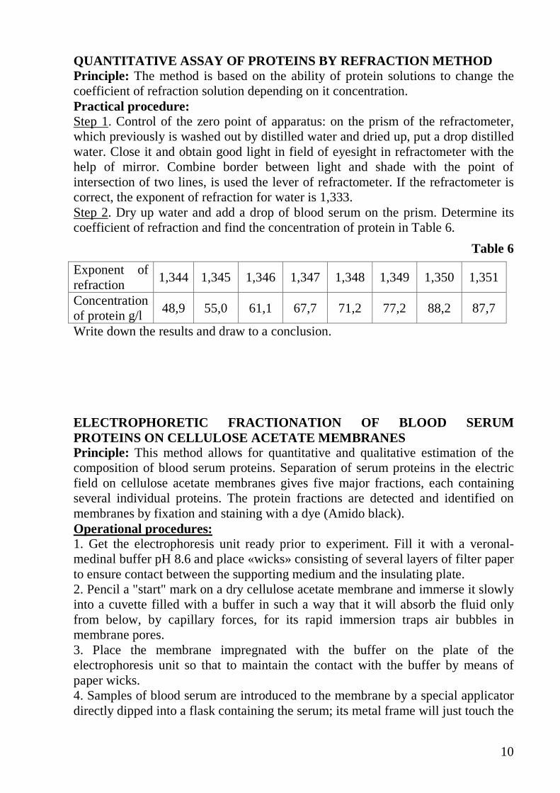
QUANTITATIVE ASSAY OF PROTEINS BY REFRACTION METHOD Principle: The method is based on the ability of protein solutions to change the coefficient of refraction solution depending on it concentration. Practical procedure: Step 1. Control of the zero point of apparatus: on the prism of the refractometer, which previously is washed out by distilled water and dried up, put a drop distilled water. Close it and obtain good light in field of eyesight in refractometer with the help of mirror. Combine border between light and shade with the point of intersection of two lines, is used the lever of refractometer. If the refractometer is correct, the exponent of refraction for water is 1,333. Step 2. Dry up water and add a drop of blood serum on the prism. Determine its coefficient of refraction and find the concentration of protein in Table 6.
Table 6
Exponent of refraction
1,344 1,345 1,346 1,347 1,348 1,349 1,350 1,351
Concentration of protein g/l
48,9 55,0 61,1 67,7 71,2 77,2 88,2 87,7
Write down the results and draw to a conclusion. ELECTROPHORETIC FRACTIONATION OF BLOOD SERUM PROTEINS ON CELLULOSE ACETATE MEMBRANES Principle: This method allows for quantitative and qualitative estimation of the composition of blood serum proteins. Separation of serum proteins in the electric field on cellulose acetate membranes gives five major fractions, each containing several individual proteins. The protein fractions are detected and identified on membranes by fixation and staining with a dye (Amido black). Operational procedures: 1. Get the electrophoresis unit ready prior to experiment. Fill it with a veronal-medinal buffer pH 8.6 and place «wicks» consisting of several layers of filter paper to ensure contact between the supporting medium and the insulating plate. 2. Pencil a "start" mark on a dry cellulose acetate membrane and immerse it slowly into a cuvette filled with a buffer in such a way that it will absorb the fluid only from below, by capillary forces, for its rapid immersion traps air bubbles in membrane pores. 3. Place the membrane impregnated with the buffer on the plate of the electrophoresis unit so that to maintain the contact with the buffer by means of paper wicks. 4. Samples of blood serum are introduced to the membrane by a special applicator directly dipped into a flask containing the serum; its metal frame will just touch the

11
fluid surface. Aliquots of the serum are carefully applied onto the start line of the cellulose acetate membrane. 5. Close the lid of the electrophoresis unit hermetically just after sample application and switch on the power at the required voltage. The electrophoresis will be run after 20 minutes. 6. When electrophoresis is complete, the power must be switched off, the electrophoresis unit is opened and the membrane is transferred to a solution of a suitable dye (in this case Amido black) poured into a cuvette. 7. After 7 minutes, the dye is poured over back into the flask, and the membrane is washed twice for 5 minutes with 2-7% acetic acid to remove dye excess. Stained protein bands remain on the membrane.
8. The membrane is dried between several layers of filter paper, and the results of electrophoresis are compared with a standard electrophoregram of blood serum from a normal individual. Homework: Study lesson 2 1.Study- Protein structure p.79-85 2.Study- Physico-chemical properties of proteins. Isoelectric point (pI). Molecular mass, shape and charge of molecules 3. Study - the factors determining the solubility of proteins (p.87, 88,fig.8.17). 4. Study - Denaturation and renaturation of proteins p.87 5. Study - the relationship between protein structure and function (p. 89). 6. To prepare for test in written form :1) the structural formulae of 20 amino acids, 2) Write the formula of the peptide

12
LESSON 2 STRUCTURE, PHYSICO-CHEMICAL PROPERTIES AND FUNCTIONS OF PROTEINS Test in written form: 1) The structural formulae of 20 amino acids
2) Write the formula of the peptide Main questions.
• Protein structure: primary (properties of peptide bonds), secondary, supersecondary, tertiary structures.
o Types of interactions between side chains of amino acids residues that form tertiary structure.
• Physico-chemical properties of proteins. Isoelectric point (pI). Molecular mass, shape and charge of molecules.
o The factors determining the solubility of proteins; sedimentation reversible and irreversible.
o Denaturation and renaturation of proteins. • Fractional sedimentation of proteins from a sample of blood plasma. • Precipitation of proteins using organic acids, alcohol and acetone.
PROTEIN STRUCTURE. FUNCTIONS OF PROTEINS Study the main characteristics of the proteins structure. 1. Memorize: proteins have four different levels of structure (primary, secondary, tertiary and quaternary). See p.79, fig. 8.1. The primary structure of a protein is the sequence of amino acids in the polypeptide chain. Differences in the sequence of amino acids along the protein chains result in different three dimensional structures and different functions. 2. Give a molecular interpretation of sickle cell anemia (p.87,fig.8.16 and a clinical note). Note: the Hb function is to deliver oxygen to tissues. HbA is normal hemoglobin in adults. HbS is found in patients with sickle cell anemia. HbS is poorly soluble in venous blood (at low partial pressure of 02), therefore HbS molecules form poorly soluble complexes. HbS-containing erythrocytes have an irregular shape and rapidly are decomposed in the spleen, resulting in anemia. 3. Compare the amino acid sequence of the N-terminal region of HbA (normal hemoglobin) and HbS (atypical hemoglobin) below. HbA 1 2 3 4 5 6 7 8 Val-His-Leu-Thr-Pro-Glu-Glu-Lys…… HbS 1 2 3 4 5 6 7 8 Val-His-Leu-Thr-Pro-Val-Glu-Lys…… 4. Write the formulae of amino acids, which are located in 6th position. Compare their structures and properties. 1.Study the main characteristics of the secondary structure of proteins.

13
Look at fig.8.7, 8.9, 8.10, p.82, 83 and memorize: secondary structure is a regular conformation that is stabilized by hydrogen bonds between the peptide-bond carbonyl oxygen and amide hydrogen in polypeptide backbone. It includes �-helix and �-sheets. 2. Note that some globular proteins are constructed by combining secondary structural elements, forming supersecondary structure (p.84, fig.8.11). Protein tertiary structure. 1.Study the main characteristics of tertiary structure of proteins. Note that tertiary structure is the unique three-dimensional structure, forming by interactions between side chain radicals of polypeptide chain. 2.Look at fig.8.12, p.85 and remember the types of interactions between the side chains of amino acid residues in proteins, forming the tertiary structure. 3.Select the appropriate characteristics for protein structure. A. Secondary
structure 1. The order of sequence of amino acids in the
polypeptide chain. B. Tertiary structure 2. The spatial structure of protein. C. Both 3. The conformation which is stabilized by
interactions between amino acid radicals. D. None 4. The conformation of a polypeptide chain as �-helix
or �-sheets 4.Choose one incorrect answer. The spatial structure of a protein is formed by: A. The bonds between the �-amino and �-carboxyl groups ofamino acids. B. Hydrogen bonds between the amino acid radicals. C. Hydrogen bonds between the atoms of the peptide backbone. D. Hydrophobic interactions between the amino acid radicals. E. Interactions between the carboxyl and amino groups of amino acid radicals. 5. Here is a fragment of the polypeptide chain NHAla-Cys-Lys-Phe-Ser-Asp
O
NH2
CH3O
NH
SH
O
NH
NH2
O
NH
ONH
OH
OH
OH
NH
O
O
a) Mark off the site of hydrogen bond made during the formation of an α-helix by a dotted line. b) What types of bonds may be formed between the amino acid radicals of this peptide?

14
PHYSICO-CHEMICAL PROPERTIES OF PROTEINS. Study the effect of pH on the protein charge. 1.Learn the following: a) Proteins are ampholites. The charge of protein molecule depends on the amount and charge of ionogenic groups in amino acid residues. The degree of ionization of cationic and anionic groups of a protein depends on pH. b) Isoelectric state is the equal amount of positively and negatively charged groups in the protein molecule. c) Isoelectric point (pI) is the pH at which the protein is in the isoelectric state. 2. Study the effect of pH on the protein charge using the following scheme:
2 Match properties to p��tides. ���tides Properties
A. Val-Lys-Ala-Gly B. His-Pro-Gln-Gly C. Phe-Leu-Arg-His 1.At pH =7.0 remains at the start in an electric field. D. Gln-Gly-Asp-Aln 2.The isoelectric point lies around pH < 7.0. E. Ala-Asp-Tyr-Lys 3.At pH 7.0 it moves to the anode in an electric field.
THE FACTORS DETERMINING THE SOLUBILITY OF PROTEINS. Study the factors determining the solubility of proteins (p.87, 88, fig.8.17 1.Memorize: the solubility of proteins depends on: a) The properties of a protein molecule (molecular mass, shape and charge of molecules, number of hydrophobic groups); b) Environmental factors (pH, salt composition of the medium, temperature). c) Solutions of proteins have a duality: in essence they are true molecular solutions, as particles of proteins separate molecules, but at the same time they are colloid solutions as the sizes of particles make from 1 up 100nm.The factors of stability are: a charge and an hydrate surface. The hydrate surface is formed due to a charge, and also for the account hydrophilic groups of amino acids (-OH,-COOH, e.t.�.), located on a surface of proteins. They are capable to sedimentation and coagulation at loss of factors of stability. d) Sedimentation may be reversible and irreversible. Irreversible sedimentation is accompanied by denaturation. 2.Choose the best answer. Proteins are effective buffers because they contain: A. A large number of amino acids.

15
B. Amino acid residues with different pKs. C. N-terminal and C-terminal residues that donate and accept protons. D. Peptide bond that is readily hydrolyzed, consuming hydrogen and hydroxyl ions. E. Large number of hydrogen bonds in �–helix. 3.Peptides. A. Tyr-Phe-Glu-Ala-Asp 1. It is soluble at pH=7. B. Arg-Thr-Val-Lys-Try 2. It is less soluble at pH=3. C. Both 3. At pH=7 it can interact with Ca2+. D. None 4. Its isoelectric point is at pH=7. 4.Look at fig 8.17 and note the main conditions for denaturation and renaturation of proteins LABORATORY MANUAL 1.Fractional sedimentation of proteins from a sample of blood plasma Principle Salting-out: This a reverse sedimentation of proteins, by adding divalent salts, such as sodium chloride and ammonium sulfate (NH4)2 SO4 to a sample of blood plasma. This method is used to separate and purify proteins and medicines containing globulins for treatment. Practical procedure Step 1. Add 1ml of proteins sample into 1ml of saturated ammonium sulfate (test tube 1). 50% saturation is reaches- globulins are as a precipitate. Step 2. Filter the sample in 10 min. Step 3. Wash the filter paper with distilled water (4-5ml) and put it in a separate clean test-tube (No 2) . Step 4. Add ammonium sulfate powder to the filtrate while it reaches full saturation. The precipitant is formed by albumens. Step 5. Filter the sample in 10 min. Step 6. Wash the filted paper with distilled water (4-5ml) and put it in a separate clean test-tube (No 3) . Preserve the solution without proteins for further uses (test-tube N 4). Step 7. Than take test tubes N 2, 3, 4 with its contents and carry out the biuret test. The positive reaction must be in the test-tubes N 2, 3 and the negative reaction must be in test tube N4. Write down the results and draw to a conclusion. 2.Precipitation of proteins by organic acids Practical procedure: Add 10 drops of proteins solution into each of 2 test-tubes. Add 4-5 drops of solution of trichloracetic acid into the first and add 4-5 drops of solution of salicylsulphonic acid into the second of the test-tubes. These reactions are used in practical for detecting and separating proteins from solutions. The white precipitate indicates the presence of protein.
16
3.Precipitation of proteins by alcohol and acetone Dehydrating agents such as alcohols and acetone precipitates the proteins. Practical procedure: Add several drops of acetone or alcohol to 10 drops of protein solution into a test-tube. A white opalescent appears. You will notice the precipitation of proteins. If distilled water is added into the test tube, opalescent disappears. Write down the results and draw to a conclusion. Home work : Study lesson 3 1. Study Classification of proteins: of function, of composition 2. Study the relationship between protein structure and function (p. 89). 3. Learn the main characteristics of the quaternary structure and the properties of the oligomeric proteins (p.85, 86, fig. 8.15). 4. Study the peculiarities of functioning of oligomeric proteins with hemoglobin as an example (p.89, 90, fig.8.21). 5.Study the agents that affect O2 binding by hemoglobin (p.91, fig.8.22, 23,24,25). 6. Study structure and function of immunoglobulins, collagen, hexokinase (p.92-96) Essay for lesson 3: 1. Structure and function of immunoglobulins
2. Structure and function of hemoglobin

17
LESSON 3. THE CLASSIFICATION OF PROTEINS. PROTEIN-L IGAND INTERACTION. PROTEIN STRUCTURE-FUNCTION RELATIONSHI PS Main questions • Classification of proteins:
o of function o of composition
• The protein-ligand interaction • The relationship between protein structure and function
o Domain structure and polymorphysm of proteins. o Structure and function of immunoglobulins. o Structure and function of collagen, hexokinase
• Quaternary structure of proteins. Functioning of oligomeric proteins (cooperative interaction between protomers).
• Hemoglobin – structure and function. THE CLASSIFICATION OF PROTEINS Functions of proteins Proteins have many diverse functions: 1. As catalysts, i.e. enzymes. 2. As structural elements (Collagen, Elastin). 3. As mode of transport (Albumin, Globulin, Hemoglobin) 4. As hormones (Insulin, Growth hormones). 5. As protective agents (Antibodies) 6. As contractive elements (Actin, Myosin) Proteins are classified on the basis of their composition. 1. Simple proteins Simple proteins are made up of amino acids only and on hydrolysis yield amino acid mixture only. Fibrous proteins – These are animal proteins which proteins are highly resistant to digestion by proteolytic enzymes. They are water insoluble.
1. Fibrous proteins a. Collagens
It contains high proportion of hydroxyproline and hydroxylysine. It is a major protein of connective tissues. On boiling with water it forms gelatin.
b. Elastins It is present in tendons and arteries. �. Keratins It contains large amount of sulphur as cystine. It is present
in hair, wool, nails etc.
18
2. Globular Proteins
a. Albumins
Serum albumin and ovalbumin of egg white. It is water-soluble. It is precipitated from solution by full saturation of ammonium sulphate. It is coagulated by heat.
b. Globulins Serum globulins, fibrinogens and muscle myosin. It is soluble in dilute salt solutions. It is, precipitated from solution by half saturation of ammonium sulphate. It is coagulated by heat.
c. Glutelins
Cereal proteins such as glutelins of wheat, oxyzenin from rice and zein of maize. It is soluble in weak acids or bases but insoluble in neutral aqueous solutions.
d. Gliadins (Prolamines)
Gliadin from wheat and zein from corn. It is water insoluble but soluble in ethanol.
e. Protamines Salmine from salmon sperm contains high proportion of arginine.
f. Histones Globulin in hemoglobin. It contains proportion of basic amino acid. It is soluble.
2. Conjugated Proteins They are proteins that contain non-protein group (also prosthetic group) attached to the protein part. On give non-protein component and amino acid mixture.
Conjugated Protein = Protein part + Prosthetic group. Conjugated proteins are classified according to the nature of the non-protein group attached to the protein part (Table 7). Some Proteins Contain Chemical Groups Other Than Amino Acids Many proteins, such as the enzymes ribonuclease and chymotrypsinogen, contain only amino acids and no other chemical groups; these are considered simple proteins. However, some proteins contain chemical components in addition to amino acids; these are called conjugated proteins. The non-amino acid part of a conjugated protein is usually called its prosthetic group. Conjugated proteins are classified on the basis of the chemical nature of their prosthetic groups (Table 7); for example, lipoproteins contain lipids, glycoproteins contain sugar groups, and metalloproteins contain a specific metal A number of proteins contain more than one prosthetic group. Usually the prosthetic group plays an important role in the protein's biological function.
Table 7
Conjugated proteins Class Prosthetic group Example Lipoproteins Glycoproteins Phosphoproteins
Lipids Carbohydrates Phosphate groups
β-Lipoprotein of blood Immunoglobulin G Casein of milk

19
Class Prosthetic group Example Hemoproteins Flavoproteins Metalloproteins
Heme (iron porphyrin) Flavin nucleotides Iron Zinc Calcium Molybdenum Copper
Hemoglobin Succinate dehydrogenase Ferritin Alcohol dehydrogenase Calmodulin Dinitrogenase Plastocyanin
THE PROTEIN-LIGAND INTERACTION. Learn that the protein-ligand interaction is the main mechanism of protein function. 1. Memorize: a) protein molecules have special regions, or binding sites (active centers) where they associate with other compounds (ligands); b) protein binding sites are formed by specific arrangement of amino acids that are approximated during the formation of secondary and tertiary structures; c) the bonds between a protein and a ligand may be covalent or non-covalent; d) proteins manifest high specificity (selectivity) when they bind with ligands at special sites; e) high specificity of protein-ligand interactions is due to complementary (chemical and spatial correspondence) of the protein binding site structure to the ligand structure. 2. The Fig. 1 below shows the structure of a protein and different ligands (A,B,C).
Fig. 1 Interaction of an oligomeric protein with different ligands. a) What ligand will interact with this protein? b) Write the formulae of the amino acids which are located in the active site and the amino acids of the appropriate ligand. c) What does the term “active site” mean?
THE QUATERNARY STRUCTURE OF PROTEINS. Learn the main characteristics of the quaternary structure and the properties of the oligomeric proteins. Memorize: quaternary structure is three-dimensional structure of a protein composed of multiple subunits (p.85, 86, fig.8.15). Note that complex oligomeric proteins can interact with several ligands at sites that are widely separated from one another (allosteric sites). 1. Choose the correct answer. Quaternary structure is defined as:
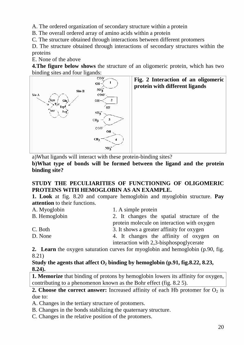
20
A. The ordered organization of secondary structure within a protein B. The overall ordered array of amino acids within a protein C. The structure obtained through interactions between different protomers D. The structure obtained through interactions of secondary structures within the proteins E. None of the above 4.The figure below shows the structure of an oligomeric protein, which has two binding sites and four ligands:
Fig. 2 Interaction of an oligomeric protein with different ligands
a)What ligands will interact with these protein-binding sites? b)What type of bonds will be formed between the ligand and the protein binding site? STUDY THE PECULIARITIES OF FUNCTIONING OF OLIGOMERI C PROTEINS WITH HEMOGLOBIN AS AN EXAMPLE. 1. Look at fig. 8.20 and compare hemoglobin and myoglobin structure. Pay attention to their functions. A. Myoglobin 1. A simple protein B. Hemoglobin 2. It changes the spatial structure of the
protein molecule on interaction with oxygen C. Both 3. It shows a greater affinity for oxygen D. None 4. It changes the affinity of oxygen on
interaction with 2,3-bisphospoglycerate 2. Learn the oxygen saturation curves for myoglobin and hemoglobin (p.90, fig. 8.21) Study the agents that affect O2 binding by hemoglobin (p.91, fig.8.22, 8.23, 8.24). 1. Memorize that binding of protons by hemoglobin lowers its affinity for oxygen, contributing to a phenomenon known as the Bohr effect (fig. 8.2 5). 2. Choose the correct answer: Increased affinity of each Hb protomer for O2 is due to: A. Changes in the tertiary structure of protomers. B. Changes in the bonds stabilizing the quaternary structure. C. Changes in the relative position of the protomers.

21
D. Cooperative changes in the conformation of the protomers. E. Changes in the localization of an iron atom in the heme. 3.Choose the correct answer. In what direction does this reaction proceed? HbO2 + CO2 + H2O HbH+ HCO3
- + + O2
1
2
HCO3- H+
H2CO3 H2CO3
CO2 H2O
a) in lung capillaries b) in tissue capillaries 4. 2,3-bisphosphoglycerate regulates the function of Hb because: A. It interacts with Hb in the active center. B. It interacts with Hb at a site which is widely separated from the heme. C. It induces dissociation of the protomers. D. It changes the conformation of all the four protomers. 5. In what direction does this reaction proceed: 4O2 + Hb-BPG � Hb(O2)4 + BPG
a) in tissue capillaries b) in lung capillaries
6. Where is the concentration of the Hb-BPG increased: a) in resting muscle capillaries b) in contracting muscle capillaries
7. How is the affinity of Hb for O2 changed during adaptation of a man to high mountain conditions if the concentration of BPG in erythrocytes increases? Essay for lesson 3. 1. Structure and function of immunoglobulins
2. Structure and function of hemoglobin Homework: Study lesson 4 1. Study the properties of enzymes as catalysts (p.99-101, p. 108, fig. 9.12), likeness and distinction of enzymes and non-organic catalysts. 2. Remember the classification and nomenclature of enzymes (p. 101, table 9.1). 3. Study the structure of an active site of enzyme (p.101, 102, fig. 9.5, table 9.3,
p.109). 5. Study the specificity of enzymes; remember the difference between "lock and
key" and “induced fit models” for substrate binding, p. 102, 103 - 107, fig. 9.6, 9.7, 8.34.
6. Study the cofactors of enzymes (metal ions and coenzymes); remember the formulae of coenzymes NAD+, NADP+, FAD, pyridoxal phosphate, biotin,

22
tetrahydrofolate (p. 109-113, fig. 9.14A, 9.15, 9.16, 9.17, p.297, fig. 19.9. p.284, p.615)
7. Fill in the table in your copy book. �oenzyme Structural formula
Full name of coenzyme
Names of vitamins Structural formulae
Class of enzyme
Biochemical functions
FH4
NAD+
FAD
TPP
PLP
Biotin
CoASH
8. Remember the units of enzyme activity (lecture, hand outs) 9. Remember the factors that influence the enzyme reaction rate (substrate
concentration, pH, temperature, p. 115, fig. 9.2 l,p.l 16-117, fig.9.22). 10. Study the information about V max and Km of enzymes (p.116-118, fig. 9.22,
9.24). 11. Learn characteristics of isoenzymes: hexokinase I and glucokinase (p.l 18.
fig. 9.24), LDH. 12. To prepare for test in written form: the structural formulae and biological
role of NAD+, NADP+, FAD, TPP, PLP, Biotin, FH4
23
LESSON 4. PROPERTIES OF ENZYMES, COENZYMES AND CATALYTIC EFFICIENCY OF ENZYMES. Test in written form: the structural formulae of NAD+, NADP+, FAD, TPP, PLP, Biotin, FH4
Main questions
• Enzyme, apoenzyme, coenzyme, holoenzyme, substrate, product of the enzyme reaction, inhibitor, activator; definition
• The properties of enzymes as catalysts, likeness and distinction of enzymes and non-organic catalysts
• The classification and nomenclature of enzymes • The structure of an active site of enzyme • Mechanism of enzyme action, “lock and key” and “induced fit models“ for
substrate binding, catalytic efficiency of enzymes. • Cofactors of enzymes (metal ions and coenzymes); • Coenzyme functions of vitamins (Biotin, Folate, Pantothenic acid, PP, B1, B2,
B6)
PROPERTIES OF ENZYMES, COENZYMES AND CATALYTIC EFFICIENCY OF ENZYMES. Study the structure of enzymes, catalytic properties and specificity of enzymes. 1. Note that enzyme as any catalyst enormously increases the rate of reaction by decreasing the energy barrier. 2. Remember that an enzyme has an active site that is a cavity or a pocket on the surface of enzyme formed by side chains of amino acid residues arranged in the very particular three dimentional shape for each enzyme p. 102, fig.9.5. Active site binds only the ligand which fits to it. Therefore the enzymes can select the special ligands from environment. This property is called specificity. 3. Note the difference between the Lock -and-Key and Induced fit models for substrate binding. 4. Answer the questions: a) What does the absolute specificity mean? b) What does the wide specificity mean? 5. Choose the correct answer: The amino acid at an enzyme active site that can be involved in hydrophobic bond formation is: A. Arginine B. Asparagine C. Glutamine D. Leucine E. Lysine
6.Read about cofactors in catalysis, p. 109.

24
Note that cofactor is located in active site of enzyme and involved in reaction. An enzyme containing its cofactor is called holoenzyme, the protein portion without the cofactor is called apoenzyme or apoprotein. Only holoenzyme is active and can catalyze a reaction:
Apoenzyme + cofactor = holoenzyme Inactive active 7. Look at fig. 9.5, p. 102 , fig.9.18, p. 112. and choose the correct statement: Some enzymes are conjugated proteins, that means that an enzyme which is conjugated protein: A. consists of two subunits. B. requires the coenzyme for catalytic activity. C. has a metal ion in an active site. D. has an allosteric site. E. consists only of amino acids, Note that many coenzymes are formed from vitamines in a body. 8.Choose the correct couples coenzyme - vitamine: 1. NAD+ A. Pantothenic acid 2.FMN B. Pyridoxal 3-CoA C. Nicotinamide 4.TPP D. Riboflavin 5.PLP E. Thiamin 9. Write the formulae of nicotinamide in NAD+ and NADH. STUDY THE MEASURES OF CATALYTIC EFFICIENCY. 1. Remember that to compare normal activity of enzyme in plasma with activity in any disorder it is required to represent it numerically. Different measures are used. The unit of enzyme activity is the amount of enzyme causing transformation of 1 Mol of substrate per minite under optimal conditions of measurements:
minmMol=S
The specific activity of enzyme is the number of units of enzyme activity per milligram of enzyme protein:
mgS
×=
minmMol
2. Choose the correct statement. The specific activity of an enzyme is: A. The amount of enzyme that produces 1 mol of product per second under standard conditions B. The activity of an enzyme in relation to a standard preparation of the enzyme C. The number of enzyme units per milligram of enzyme protein D. The amount of enzyme causing transformation of l , mol of substrate per minute under standard conditions E. The activity of an enzyme in the presence of its preferred substrate

25
CLASSIFICATION OF ENZYMES Study the principles of enzymes classification. 1. Remember that enzymes are grouped into six functional classes by the I.U.B. due to the type of reaction they catalyze. In this classification system each enzyme is designated a four-digit number. The first number defines the type of reaction, which the enzyme catalyzes. 2. Put the listed classes of enzymes in true order. A. Lyases B. Transferases C. Oxidoreductases D. Ligases E. Hydrolases F. Isomerases 3. Which type of reaction catalyzes the enzyme with code number 2.4.1.1.? 4. Which of the reactions numbered below are catalyzed by the listed enzymes: 1.
O
OO
OH
OHNH2
O
OH
O
O
OH
NH2
OO
OH
OH++
2.
NH2
O
OH
NH2
OO
OH
OH
CO2+
3. Sucrose + H2O → glucose + fructose 4. CH3-CO-COOH + CO2 + ATP + H2O→ COOH-CH2-CO-COOH + ADP + Pi 5. -CH2-CH2- → -CH=CH- + 2H A. Lyase B. Ligase C. Transferase D. Oxydoreductase E. Hydrolase Home work: lesson 5 1. Remember the factors that influence the enzyme reaction rate (substrate concentration, pH, temperature, p. 115, fig. 9.2 l,p.l 16-117, fig.9.22). 2. Study the information about V max and Km of enzymes (p.116-118, fig. 9.22, 9.24). 3. Learn characteristics of isoenzymes: hexokinase I and glucokinase (p.l 18. fig. 9.24).
26
4. Study the use of tissue-specific enzymes and isoenzymes as diagnostic tools (p. 123-124, fig. 9.33) Essay for lesson 5: 1. Diagnostic value of plasma enzymes. The using of tissue-specific enzymes and isoenzymes as analytical tools in laboratory diagnostic. Enzymes as drugs. 2. Isoenzymes – the origin, the methods of division and biological importance. The isoenzymic forms of lactate dehydrogenase.

27
LESSON 5. ENZYME KINETICS. FACTORS AFFECTING REACTI ON VELOCITY. ENZYMES IN MEDICINE. Main questions
• The factors that influence the enzyme reaction rate (substrate concentration, pH, temperature, enzyme concentration). V max and Km of enzymes.
• Isoenzymes – origin and clinical significance. The isoenzymic forms of lactate dehydrogenase
• Diagnostic value of plasma enzymes. • The using of tissue-specific enzymes and isoenzymes in laboratory
diagnostic. • Enzymes as drugs. • Principles of qualitative and quantitative estimation of enzyme activity.
o Assay of specificity of urease activity o Assay of termolability of salivary amylase. o Assay of influence of pH on activity of amylase of saliva o Estimation of amylase activity in urine o Inhibition of trypsin
FACTORS AFFECTING REACTION VELOCITY Remember the mechanisms of action of different factors on enzyme activity 1. Pay attention to the figures p. 116 (9.21), p. 117(9.22)
Fig. 3. Effect of substrate concentration on reaction velocity
2. Explain the shape of the curve in Fig. 3 answering the questions: a) How many of active sites of enzyme are bound to molecules of substrates at the points designated as A and B? b) Which of the points - A or B corresponds to the meaning of Km? c) Which part of the plot may be used for measuring the rate of enzyme catalyzed reaction? 3. Draw the plot using the following information of reaction rate vs substrate concentration S mM/I V �M/min 0,8 12 1,4 16 3,3 23 5,0 24 What are the Km and Vmax values of the reaction?

28
4. Study Fig. 4 below
Fig. 4 Effect of substrate concentration on reaction velocities for two enzymes, enzyme 1 with a small Km1, enzyme 2 with a large Km2.
Remember that small Km reflects a high affinity of enzyme for the substrate. Answer the question: Which enzyme – 1 or 2 has higher affinity for substrate? 5. Study fig.9.24, p.118. Answer questions 9.3 and 9.4. Read book notes, p. 118. and say which enzyme glucokinase or hexokinase has higher affinity for glucose. 6. Study Fig. 5. Write down the pH optimum values for given enzymes. 7. Explain the form of the curves. Why does the activity of enzyme decrease if pH of solution differs from optimum value?
Fig. 5. Effect of pH on enzyme-catalyzed reactions.
8. Note that pepsin is the enzyme that hydrolyzes proteins in stomach. If the patient suffers from hypoacidic gastritis the digestion of proteins in stomach is damaged. Give your explanation. 9. The effects of pH on enzyme-catalyzed reactions include which of the following? A. The direction of the reaction may be influenced by the [H+] B. The ionization state of dissociating groups on the enzyme may be modified C. The ionization state of the substrate may be modified D. The protein may be denatured at certain pH values E. All the above Remember that the enzymes undergo the conformational changes during their action. This ability is referred to as “conformational lability”. 10. Select correct completion: The conformation of enzyme active site can change as a result of: A. Binding of ligand at allosteric site. B. Changing of pH from 7,4 to 6,4. C. Increasing of temperature by 10
� D. Binding of substrate in active site. E. Dissociation of regulatory and catalytic subunits of enzyme.

29
11. Select correct completion or answer a) The velocity of an enzyme-catalyzed reaction:
A. Decreases as the substrate concentration increases B. Is the lowest when the enzyme is saturated with substrate C. Increases as the enzyme concentration increases D. Does not depend on the pH of the solution E. Increases with the temperature increase until a maximum is reached, then
the velocity decreases due to denaturation of the enzyme. b) Which of the following statements are true?
A. Enzymes decrease the energy barriers of the reaction B. The rate of the reaction is always directly proportional to the substrate
concentration C. Reaction rate depends on the magnitude of the energy barrier D. The initial rate of an enzyme-catalyzed reaction is directly proportional to the
concentration of enzyme. E. pH affects enzyme activity by changing the charge on the enzyme.
TISSUE-SPECIFIC ENZYMES AND ISOENZYMES AS DIAGNOSTI C TOOLS. Learn that enzymes are used for diagnosis of myocardial infarction and many other diseases. 1. Remember that tissue-specific enzymes and isoenzymes are useful tools for diagnosis of many diseases. Look at p. 124 and note the information given below. 2. Study the information about isoenzymes, (p. 123-124). 3. Note that isoenzymes are the enzymes, catalyzing the same reactions, are usually located in different tissues and are differ in their properties. After cell membranes damage they leak to blood and can be revealed in blood serum. Different organs frequently contain characteristic proportions of different isoenzymes. The pattern of isoenzymes found in the plasma may therefore serves as a means of identifying the site of tissue damage. For example, the plasma level of creatine kinase (CK), lactatedehydogenase (LDH) are commonly used for diagnosis of myocardial infarction. Isoenzymes may contain different numbers of charged amino acids and may be separated from each other by electrophoresis (Fig. 6). 4. Note that creatinekinase contains two subunits. MM type is located in skeletal muscle, BB - in brain and BM - in heart. 5. Look at Fig. 6 and answer the question: What type of creatinekinase isoenzyme will increase after:
a) severe exercising b) myocardial infarction

30
Fig. 6. Subunit structure and electrophoretic mobility of creatine kinase isoenzymes.
6. Look at Fig. 7. and answer the question: a) What enzyme should be determined in the plasma 3 hours after myocardial infarction? b) What enzyme should be determined in the plasma two days after myocardial infarction?
Fig. 7. Appearance of creatine kinase and lactate dehydrogenase in plasma after a myocardial infarction.
Lactate dehydrogenase (LDH): This enzyme catalyses the dehydrogenation of lactate to pyruvate. This occurs in five dufferent isoenzymes. This enzyme is tetramer having two types of units, L and M units. Depending upon the various combination, five isoenzymes are known.
LDH- 1 HHHH LDH- 2 HHHM LDH- 3 HHMM LDH- 4 HMMM LDH- 5 MMMM
LDH-1 is the predominant form in heart and LDH-5 in muscles. LABORATORY MANUAL : 1. Assay of specificity of urease activity The enzyme urease (CE 3.5.1.5.) catalyzes the reaction:
O
NH2NH2
OH2 NH3 CO2+ +2
Urea

31
Ammonia - the product of the reaction can be detected by indicator – phenolphthalein. Two types of molecules have to be tested as substrates for urease in this experiment: urea and thiourea. The structural formula of thiourea:
S
NH2 NH2 Practical procedures: urease solution – extract from watermelon’s seeds
Pipette Test -tube N1 Test -tube N2 Urea solution 1,0 ml - Thiourea solution - 1,0 ml Urease solution 1,0 ml 1,0 ml Phenolphthalein 3 drops 3 drops
Stir the content of the test - tubes thoroughly and incubate them for 5 min at room temperature to let the color develop. Analyze the experimental data and put down your conclusion. 2. Assay of termolability of salivary amylase Principle: Amylase (CE 3.2.1.1.) is normally secreted by salivary glands and pancreas. It digests starch into maltose through amyl dextrin, erytrodextrin and achrodextrin (C6H10O5)n dextrins C12H22O11 (maltose) Practical procedures: Prepare two test-tubes with identical contents: Pipette Test -tube N1 Test -tube N2
Saliva 1,0 ml 1,0 ml At room temperature Boil for 5 min 1% Starch solution 1,0 ml 1,0 ml
10 min of incubation Iodine solution 2 drops 2 drops
Write down the results and conclusion. 3.Assay of influence of pH on activity of amylase. Practical procedures: Prepare 3 test-tubes as shown in the table below: Pipette Tube N1 Tube N2 Tube N3 Buffer solution (2,0 ml) pH =1,0 pH =7,0 pH =10,0 Dilute Saliva (1:10) 1,0 ml 1,0 ml 1,0 ml Starch solution 1% 1,0 ml 1,0 ml 1,0 ml
Stir and after 10 min of incubation add 2 drops of iodine solution. The test–tube which show blue or purple color still contains starch or amylodextrin. But the

32
tubes, which show reddish color, contain erytrodextrin. Write down the results and conclusion. 4. ESTIMATION OF AMYLASE (DIASTASE) IN URINE. Normally, very small amylase activity is present in urine. But its concentration increases highly in acute pancreatitis and parotitis. Principle: The Karavey’s method is based on the ability of strach solution to change it colour into blue on interaction with iodine solution. The intensity of colouring (640 nm)is proportional to the strach concentration in solution. Amylase activity is determined by decrease the intensity of colouring. Practical procesures: Prepare 2 test-tubes as shown in the table below: Control solution, ml Sample, ml Substrate-buffer solution 0,5 0,5 Put into water thermostat for 5 min and add others reagents (all in water thermostat) urine - 0,1 Stir and stand for exactly 7,5 min at 370 C Solution HCl 0,1 n 4,0 4,0 urine 0,1 - iodine solution 0,01 n 0,5 0,5 The solutions are placed in cuvettes (the layer thickness = 1 cm) and analyzed on a photoelectric colorimeter supplied with a wavelength = 640 nm against distilled water Calculation The activity of amylase in urine is defined as the number of mg starch digested by l of sample in 1 s at 370 C.
Amylase activity can be calculated from the following equation:
K4,44*Dc
Ds-Dcl) mg/(s. ,activity Amylase =
Dc - optical density of test-tube 1 (Control solution) Ds - optical density of test-tube 2 (sample)
K-coefficient of dilution of sample. Reference values : in urine < 44 mg/(s. l) in blood serum 3,3-8,9 mg/(s. l)
Write down the results and draw to a conclusion. 5. Inhibition of trypsin Principle: The method is based on estimation of the amount of peptides which react with the biuret reagent. The proteins have been previously precipitated. The activity of inhibitor is identified as the difference in the amount of formed products with presence inhibitor (sample) and its absence. Practical procedures:
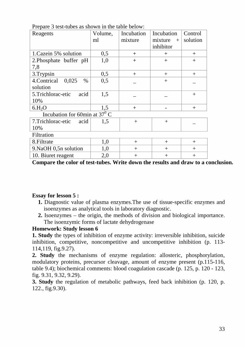
33
Prepare 3 test-tubes as shown in the table below: Reagents Volume,
ml Incubation mixture
Incubation mixture + inhibitor
Control solution
1.Cazein 5% solution 0,5 + + + 2.Phosphate buffer pH 7,8
1,0 + + +
3.Trypsin 0,5 + + + 4.Contrical 0,025 % solution
0,5 _ + _
5.Trichlorac-etic acid 10%
1,5 _ _ +
6.H2O 1,5 + - + Incubation for 60min at 370 C 7.Trichlorac-etic acid 10%
1,5 + + _
Filtration 8.Filtrate 1,0 + + + 9.NaOH 0,5n solution 1,0 + + + 10. Biuret reagent 2,0 + + + Compare the color of test-tubes. Write down the results and draw to a conclusion. Essay for lesson 5 :
1. Diagnostic value of plasma enzymes.The use of tissue-specific enzymes and isoenzymes as analytical tools in laboratory diagnostic.
2. Isoenzymes – the origin, the methods of division and biological importance. The isoenzymic forms of lactate dehydrogenase
Homework: Study lesson 6 1. Study the types of inhibition of enzyme activity: irreversible inhibition, suicide inhibition, competitive, noncompetitive and uncompetitive inhibition (p. 113-114,119, fig.9.27). 2. Study the mechanisms of enzyme regulation: allosteric, phosphorylation, modulatory proteins, precursor cleavage, amount of enzyme present (p.115-116, table 9.4); biochemical comments: blood coagulation cascade (p. 125, p. 120 - 123, fig. 9.31, 9.32, 9.29). 3. Study the regulation of metabolic pathways, feed back inhibition (p. 120, p. 122., fig.9.30).
34
LESSON 6. REGULATION OF ENZYMES. ENZYME INHIBITION. Main questions • Inhibition of enzyme activity, reversible and irreversible inhibition, suicide
inhibition, competitive, noncompetitive and uncompetitive inhibition • Regulation of enzyme activity:
o allosteric, o regulation of metabolic pathways: feed back inhibition. o phosphorylation - dephosphorylation o association - dissociation o precursor cleavage, o amount of enzyme present.
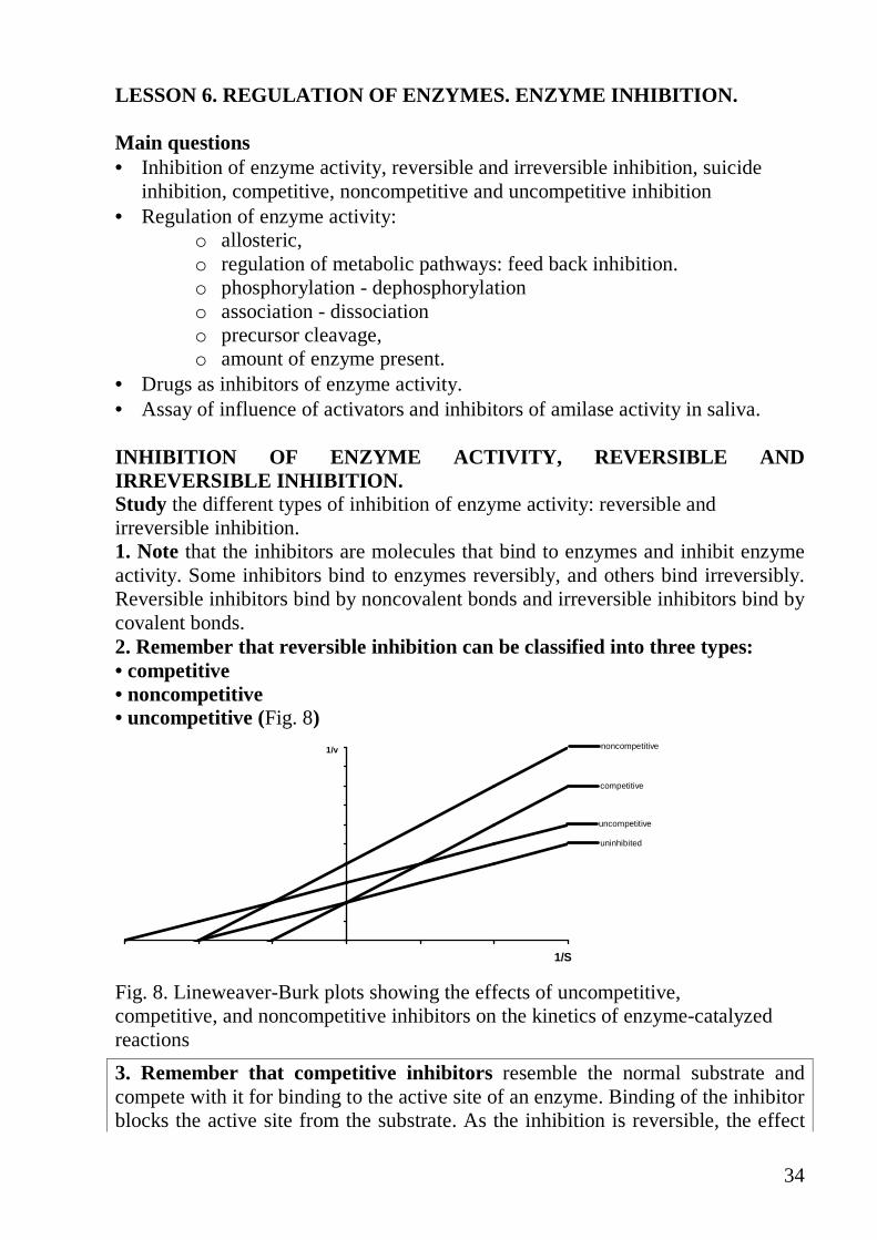
• Drugs as inhibitors of enzyme activity. • Assay of influence of activators and inhibitors of amilase activity in saliva. INHIBITION OF ENZYME ACTIVITY, REVERSIBLE AND IRREVERSIBLE INHIBITION. Study the different types of inhibition of enzyme activity: reversible and irreversible inhibition. 1. Note that the inhibitors are molecules that bind to enzymes and inhibit enzyme activity. Some inhibitors bind to enzymes reversibly, and others bind irreversibly. Reversible inhibitors bind by noncovalent bonds and irreversible inhibitors bind by covalent bonds. 2. Remember that reversible inhibition can be classified into three types: • competitive • noncompetitive • uncompetitive (Fig. 8)
uninhibited
competitive
noncompetitive
uncompetitive
1/S
1/v
Fig. 8. Lineweaver-Burk plots showing the effects of uncompetitive, competitive, and noncompetitive inhibitors on the kinetics of enzyme-catalyzed reactions
3. Remember that competitive inhibitors resemble the normal substrate and compete with it for binding to the active site of an enzyme. Binding of the inhibitor blocks the active site from the substrate. As the inhibition is reversible, the effect

35
of competitive inhibition can be overcome by an increase in substrate concentration. These inhibitors increase the Km of the enzyme, but not the Vmax (Fig. 9and Fig. 10 below) Look at Fig. 9 and say: why many medicines are structural analogs of substrates of enzymes?
Fig. 9. Enzyme-inhibitor interaction. A - The binding of normal substrate to the active site. B - The binding of competitive inhibitor to the active site. C - The binding of noncompetitive inhibitor to a site other than the active site.
4. Study Fig. 10 representing the influence of competitive inhibitor on Vmax and Km of enzyme. Explain why Vmax is the same in the presence of competitive inhibitor.
Fig. 10. Influence of competitive inhibitor on Vmax and Km
Curve 1 - without the competitive inhibitor Curve 2 - in the presence the competitive inhibitor Km is increased and Vmax is the same in the presence of a competitive inhibitor
5. Answer the question. The effects of competitive inhibitor on the kinetics of an enzyme reaction include which of the following? A. The Vmax is not changed. B. Increased concentrations of substrate reverse the inhibition. C. The Km is increased. D. The inhibitor binds to a site on the enzyme other than the catalytic site. E. The inhibitor binds to a catalytic site on the enzyme. 6. Look at fig.6.2C and remember that noncompetitive inhibitors bind to a part of an enzyme other than the active site (allosteric sites). Inhibitor binding changes the shape of the active site so that it cannot bind substrate. Such Inhibitors may decrease the Km and always decrease the Vmax. 7. Remember that uncompetitive inhibitors bind only to the substrate-enzyme complex, they decrease the Km and Vmax of enzyme. E + S ↔ ES + I↔ESI

36
8. Remember that irreversible inhibitors form covalent or extremely tight bonds with functional groups in the active site. The activity of the enzyme in the cell can only be recovered as new molecules of enzymes are synthesized. Study the medicines acting as inhibitors of enzyme activity. 1. Memorize that many medicines act as inhibitors of enzyme activity. For example, such medicines as acetylcholine, aspirin and penicillin act as different types of inhibitors of enzyme activity. 2. Acetylcholine is a neurotransmitter. Extra amount of acetylcholine which does not bind to receptors must be hydrolyzed by enzyme acetylcholinesterase: Acetylcholine + H2O → acetate + choline
CH3
N+
CH2 CH2 O CCH3
CH3O
CH3
Acetylcholine
CH3
N+
CH3
CH3
O C
O
NCH3
CH3
Prozerine 3. Aspirin inhibits cyclooxygenase - the enzyme catalyzing the synthesis of prostaglandins, which take part in the inflammatory process and blood coagulation. Thus aspirin ceases the inflammation and prevents blood coagulation.
NH2 CH3 C O
COOH
O
NH C CH3
O
OH
COOH
E + E +
Active enzyme Inactive enzyme Answer the questions: A. How does aspirin inhibit cyclooxygenase? B. What is the mechanism of inhibition of the enzyme? C. Why do the cells begin to produce prostaglandins some hours later after taking up the medicine? 4. Use the information about penicillin (p. 114) and answer: why penicillin is the "suicide inhibitor" for bacterial enzyme glycopeptidyl transferase?
REGULATION OF ENZYME ACTIVITY.
Study the different modes of regulation of enzymes activity. 1. Remember the main mechanisms of regulation of enzyme activity in the body a) The partial cleavage of enzyme precursor (proteolytic cleavage, p. 123):
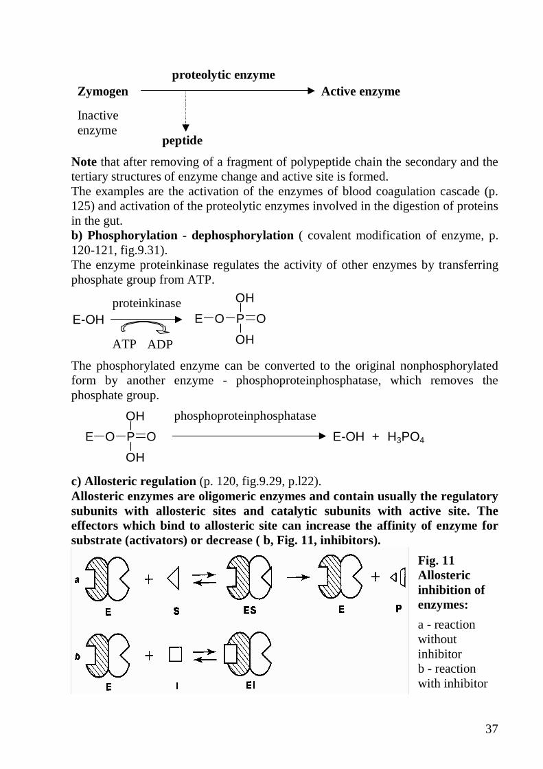
37
peptide
Inactive enzyme
proteolytic enzyme Active enzyme Zymogen
Note that after removing of a fragment of polypeptide chain the secondary and the tertiary structures of enzyme change and active site is formed. The examples are the activation of the enzymes of blood coagulation cascade (p. 125) and activation of the proteolytic enzymes involved in the digestion of proteins in the gut. b) Phosphorylation - dephosphorylation ( covalent modification of enzyme, p. 120-121, fig.9.31). The enzyme proteinkinase regulates the activity of other enzymes by transferring phosphate group from ATP.
E O P O
OH
OHADP ATP
proteinkinase E-OH
The phosphorylated enzyme can be converted to the original nonphosphorylated form by another enzyme - phosphoproteinphosphatase, which removes the phosphate group. phosphoproteinphosphatase
E-OH + H3PO4 E O P O
OH
OH c) Allosteric regulation (p. 120, fig.9.29, p.l22). Allosteric enzymes are oligomeric enzymes and contain usually the regulatory subunits with allosteric sites and catalytic subunits with active site. The effectors which bind to allosteric site can increase the affinity of enzyme for substrate (activators) or decrease ( b, Fig. 11, inhibitors).
Fig. 11 Allosteric inhibition of enzymes:
a - reaction without inhibitor b - reaction with inhibitor

38
d) Dissociation/association of regulatory protein (modulatory protein binding, p. 121) Example:
Proteinkinase A inactive →→→→ Proteinkinase A active
Inactive proteinkinase consists of four subunits: two regulatory ® and two catalytic �. Molecules of cyclic AMP are synthesized in cell when hormone (for example, epinephrine) acts through the receptor on cell membrane. After binding of cAMP to regulatory subunits, catalytic subunits dissociate from regulatory and become active:
R2 C2 + 4 cAMP →→→→ 2(Rx2cAMP) + 2C
Active proteinkinase phosphorylates other enzymes and thus changes their activity. Look at fig.24.14, p.384, the fragment of the scheme, which refers to protein kinase A. 2. Note that the velocity of enzyme catalyzed reactions in the cell depends not only on enzyme activity but also on many other factors (p. l16, table 9.4.). 3. Answer the question. Which of the following mechanisms of enzyme regulation in the cell is the slowest? A. Covalent modification B. Allosteric activation or inhibition C. Modulator protein binding D. Substrate concentration E. Changes in amount of enzyme 4. Pepsin is produced in the chief cells of the stomach as inactive form and then is activated to pepsin in the lumen:
M.w. 35000 peptide
M.w. 42000 pepsin Pepsinogen
What is the main cause of the pepsinogen activation? A. Phosphorylation B. Dissociation of regulatory subunit C. Changing of the primary structure D. Changing of the quaternary structure E. Changing of the secondary structure

39
5. Which of the following regulatory actions involves a reversible covalent modification of an enzyme? A. Allosteric modulation B. Competitive inhibition C. Convertion of zymogen to active enzyme D. Association of apoenzyme with a cofactor E. Phosphorylation of a serine hydroxyl on the enzyme 6.An allosteric modulator influences enzyme activity by: A. Competing for the catalytic site with the substrate B. Binding to a site on the enzyme molecule distinct from catalytic site C. Changing the nature of the product formed D. Changing the specificity of the enzyme to its substrate E. None of the above 7. Allosteric enzymes have the following properties: A. They are oligomeric proteins. B. They contain one binding site. C. They contain two and more binding sites. D. Usually they are the regulatory enzymes in the metabolic pathway. E. They catalyze the reaction at the end of the pathway.
REGULATION OF METABOLIC PATHWAYS. FEEDBACK INHIBITI ON
Remember the characteristics of the regulatory enzymes and feedback inhibition. 1. Note that the regulatory enzymes regulate the velocity of metabolic pathways. 2. Remember the properties of regulatory enzymes. a) They catalyze the reactions at early steps in metabolic pathways or at the metabolic branchpoint. b) Usually they catalyze irreversible reactions or the slowest reactions of the metabolic pathway. c) They are the allosteric enzymes and bind activators and inhibitors at allosteric sites - sites that are separated from the active site 3. Note that feedback inhibition refers to metabolic pathways in which the endoproduct controls its own rate of synthesis.

40
4. Answer the questions: a) how many sites of binding has enzyme E2? b) name these sites c) what compounds of this metabolic pathway can bind to these sites? LABORATORY MANUAL Assay of influence of activators and inhibitors of amilase activity in saliva Practical procesures: Prepare 3 test tubes as shown in the table below Pipette Test tube N1 Test tube N2 Test tube N3 H2O 0,1 ml — — Solution NaCl — 0,1 ml — solution CuSO4 — — 0,1 ml Saliva (dilute 1:10) 1,0 ml 1,0 ml 1,0 ml Starch solution 1% 0,1 ml 0,1 ml 0,1 ml Stir and after 5 min of incubation add 2 drops of iodine solution. Compare the color of test tubes. Write down the results and draw to a conclusion. Home work: repeat the themes «Proteins», «Enzymes», Prepare for colloquim.

41
LESSON 7. COLLOQUIM: PROTEINS. ENZYMES. Main theoretical questions:
1. Structures of 20 amino acids. Classification of amino acids according to radical structure.
2. Biochemical methods of research. Methods of isolation and purification of individual proteins .Electrophoresis.
3. Physico-chemical properties of proteins. Molecular mass, shape and charge of molecules
4. Isoelectric point (pI).The factors determining the solubility of proteins 5. Denaturation and renaturation of proteins 6. Classification of proteins of function, of composition 7. Protein structure: primary (properties of peptide bonds). 8. Secondary, supersecondary structure of proteins. 9. Tertiary structure of proteins. Types of interactions between side chains of
amino acids residues that form tertiary structure. 10. Domain structure and polymorphysm of proteins 11. The protein-ligand interaction 12. The relationship between protein structure and function 13. Quaternary structure of proteins. Functioning of oligomeric proteins . 14. Structure and function of immunoglobulins. 15. Structure and function of collagen, hexokinase. 16. Hemoglobin: structure and function, cooperative interaction between
protomers. 17. Enzyme, apoenzyme, coenzyme, holoenzyme, substrate, product of the
enzyme reaction, inhibitor, activator: definition 18. The properties of enzymes as catalysts, likeness and distinction of enzymes
and non-organic catalysts. 19. The classification and nomenclature of enzymes 20. The structure of an active site of enzyme. 21. Mechanism of enzyme action, “Lock and key”and “Induced fit models” for
substrate binding, catalytic efficiency of enzymes. 22. Cofactors of enzymes :metal ions and coenzymes NAD+, NADP+, FAD,
TPP, PLP, Biotin, CoA-SH, FH4. 23. Coenzyme functions of vitamines ( PP, B1, B2, B6, Pantothenic acid, Folate,
Biotin) 24. Factors affacting reaction velocity ( pH, temperature). 25. Factors affacting reaction velocity (substrate concentration, enzyme
concentration). Vmax and Km of enzymes. 26. Isoenzymes – origin and clinical significance. The isoenzymic forms of
lactate dehydrogenase. 27. Diagnostic value of plasma enzymes.The using of tissue-specific enzymes
and isoenzymes as analytical tools in laboratory diagnostic. Enzymes as drugs.
28. Principles of qualitative and quantitative estimation of enzyme activity.
42
29. Inhibition of enzyme activity, reversible and irreversible inhibition, suicide inhibition, competitive, noncompetitive and uncompetitive inhibition.
30. Kinds of regulation of enzyme activity. Biological significance. 31. Allosteric regulation of enzyme activity. 32. Regulation of enzyme activity: covalent modification of enzyme,
phosphorylation – dephosphorylation. 33. Regulation of enzyme activity: association – dissociation (example –
proteinkinase A). 34. Drugs as inhibitors of enzyme activity.
It is necessary to know laboratory manual: Quantitative assay of proteins by the biuret method. Quantitative assay of proteins by method of refraction. Fractional sedimentation of proteins from a sample of blood plasma. Precipitation of proteins by organic acids, alcohol and acetone. Assay of specificity of urease activity Assay of termolability of amilase Assay of influence of pH on activity of amilase saliva Estimation of amilase activity in urine Inhibition of trypsin Assay of influence of activators and inhibitors of amilase activity in saliva. Home work. 1. Study the main steps of the ATP generation and utilization of the high energy phosphate bonds of ATP. ATP-ADP cycle (p.271-272). 2. Learn the main steps of the energy transformation in mitochondrial fuel metabolism (p.282-284, fig. 18.9, table 18.4). 3. Study the structural organization of the electron transport chain (ETC) in mitochondria (p.311, fig.20.1, p.315-318, table 20.1). 4. Learn the formulae of prosthetic groups of the electron carriers (NAD, FMN, FAD, Q) in oxidized and reduced form (p.283,fig.l8.10, p.284,fig.l8.11, p.297,fig.l9.9, p.317, fig.20.9,20.10). 5. Study the mechanism of okidative phosphorylation (p.312-315). 6. Study the role of coupling of electron transport and ATP synthesis in regulation of energetic metabolism (p.319-325). 7. Memorize some uncoupler's action (p.319-321). Essay for lesson 12: 1. Active form of oxygen. Textbook: "Basic Medical Biochemistry", D. B. Marks et al. Lecture.

43
LESSON 8. BIOENERGETICS OF THE CELL. ATP FORMATION. MITOCHONDRIAL ELECTRON TRANSPORT CHAIN. Main questions:
• Generation of ATP from metabolic fuels o Endergonic and exergonic reactions o Biologi�al oxidation by dehydrogenation o Kinds of phosphorylation (oxidative, substrative and
photophosphorylation) • Electron transport chain. • Oxidative phosphorylation. • Regulation of the electron transport �hain and ATP synthesis. • Active form of oxygen.
GENERATION OF ATP FROM METABOLIC FUELS. Study the main steps of ATP generation. 1. Note: all of the living cells receive chemical energy, which is released when the organic molecules are broken down in catabolic pathways. Some of this energy is lost as heat .The rest energy is used to synthesize ATP. When ATP is hydrolyzed the energy is utilized for energy requiring processes. 2. Look at fig. 18.2 (p.272), 18.4 (p.275) and answer the following questions:
1. In what form is the energy of respiration produced? 2. What is the ATP-ADP cycle?
3. Choose the most correct answer. What reactions does the respiration include? A. Decomposition of organic molecules into their simpler components. B. The utilization of O2 to derive ATP from oxidizing fuels to CO2 and H2O. C. The complete oxidation of the acetyl group to CO2 in the TCA cycle. D. ATP generation from oxidative phosphorylation. E. The electron transport chain.
4. In what processes in the body is the high energy of ATP utilized? A. Mechanical work B. Transport work C. Biosynthetic work D. Activated intermediates formation E. All of the above.
ELECTRON TRANSPORT CHAIN. Learn the main steps of the energy transformation in mitochondrial fuel metabolism. 1.Look at fig.18.9 (p.283) and the scheme below (Fig. 12). Memorize: the electron transport chain is the final common pathway in aerobic cells by which electrons derived from various substrates are transported to oxygen. It is a series of highly organized oxidation-reduction enzymatic reactions. The electrons are first transferred from the fuel molecules to specialized electron carriers NAD+ and FAD. The electrons from the carriers reach molecular oxygen via the mitochondrial electron transport system. In this system electrons are generally transferred from the compounds with a lower reduction potential to
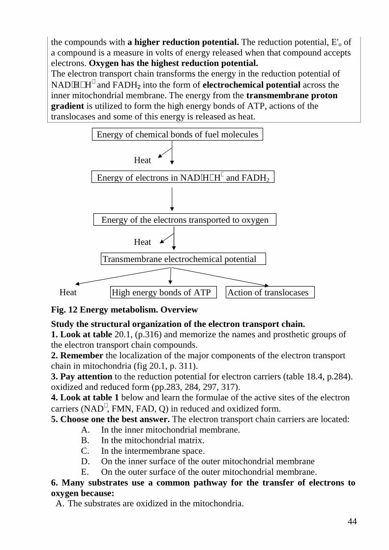
44
the compounds with a higher reduction potential. The reduction potential, E'o of a compound is a measure in volts of energy released when that compound accepts electrons. Oxygen has the highest reduction potential. The electron transport chain transforms the energy in the reduction potential of NAD⋅H+H+ and FADH2 into the form of electrochemical potential across the inner mitochondrial membrane. The energy from the transmembrane proton gradient is utilized to form the high energy bonds of ATP, actions of the translocases and some of this energy is released as heat.
Energy of chemical bonds of fuel molecules
Energy of electrons in NAD⋅H+H+ and FADH2
Energy of the electrons transported to oxygen
Transmembrane electrochemical potential
High energy bonds of ATP Action of translocases
Heat
Heat
Heat
Fig. 12 Energy metabolism. Overview
Study the structural organization of the electron transport chain. 1. Look at table 20.1, (p.316) and memorize the names and prosthetic groups of the electron transport chain compounds. 2. Remember the localization of the major components of the electron transport chain in mitochondria (fig 20.1, p. 311). 3. Pay attention to the reduction potential for electron carriers (table 18.4, p.284). oxidized and reduced form (pp.283, 284, 297, 317). 4. Look at table 1 below and learn the formulae of the active sites of the electron carriers (NAD+, FMN, FAD, Q) in reduced and oxidized form. 5. Choose one the best answer. The electron transport chain carriers are located:
A. In the inner mitochondrial membrane. B. In the mitochondrial matrix. C. In the intermembrane space. D. On the inner surface of the outer mitochondrial membrane E. On the outer surface of the outer mitochondrial membrane.
6. Many substrates use a common pathway for the transfer of electrons to oxygen because: A. The substrates are oxidized in the mitochondria.

45
B. Many of the substrates are oxidized by enzymes linked to NAD+ and FAD. C. All substrates are oxidized by the same enzymes. D. The electrons from all substrates are transferred to ATP. E. Protons from all substrates are used to form water.
7. All the following electron carriers are components of the mitochondrial ETC, EXCEPT:
A. NAD+ B. NADP+ C. FMN D. FAD E. Coenzyme Q
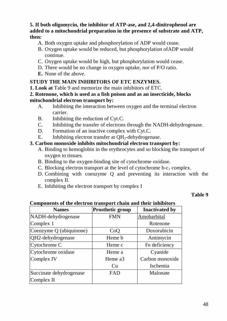
Table 8
Components of the electron transport chain
8. Match the figures with the letters
A. Only NADH-dehydrogenase 1 Accepts and donates two hydrogen atoms.

46
B. Only Q 2.Has FMN as coenzyme
C. Both 3.Accepts the electrons donated by NADH+H+
D. None 4. Transfers electrons to O2 9. Match the correct couple Redox potential Carriers A. 0,06 1.Cyt.a B. 0,816 2.Coenzyme QH2/Q C. -0,32 3.NADH/NAD+ D. 0,29 4. H2O/ O2 OXIDATIVE PHOSPHORYLATION. Study the mechanism of oxidative phosphorylation. 1. Note: the production of ATP using the energy from oxidation-reduction reactions of the ETC is called oxidative phosphorylation. The energy is released when electrons flow down the chain until they are finally accepted by 0 2. The chemiosmotic theory explains how the energy of electrons is transformed into the high-energy bonds of ATP. P/0 ratio is a measure of how many moles of ATP are formed from ADP by phosphorylation per gram atom of oxygen reduced to water. 2. Look at fig 20.3 (p.313) and remember the tenets of the chemiosmotic theory. 3. Learn the three major stages of electron transfer where the energy is produced (Fig. 13).
Fig. 13 The electron transport chain and oxidative phosphorylation. Fe-S- iron-sulfur centers of proteins. nH+indicates that an undetermined number of protons are pumped from the matrix to the cytosolic side. The return of protons to the matrix through the ATP-synthase pore drives ATP synthesis. 4. Which of the following statements about the chemiosmotic theory are true:
A. The function of mitochondrial electron transport is to translocate protons across the inner membrane into the mitochondrial matrix.
B. The free energy released by ETC is stored in an electrochemical gradient.
47
C. F1-ATP-ase catalyzes the in vivo synthesis of ATP. D. The major carriers are organized into three complexes which have a
vectorial arrangement within the membrane. E. Protons can cross the membrane from intermembrane space to matrix only
by passin through the ATP-ase. 5. How many moles of ATP can be formed per a pair of electrons transferred from NAD⋅H+H+ to oxygen?
A. 0 B. 1 C. 2 D. 3 E. 4
6. At which sites in the mitochondrial ETC is more than 7 kkal/mole of energy released?
A. NAD⋅H+H+ → Q B. Cyt.b,c1 → Cyt.c C. Succinate → Q D. Cyt c → Cytochrome oxidase E. Cyt.a3 → oxygen
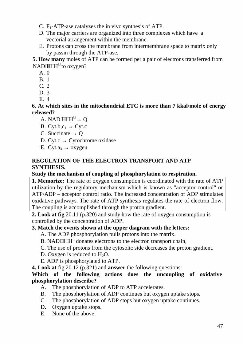
REGULATION OF THE ELECTRON TRANSPORT AND ATP SYNTHESIS. Study the mechanism of coupling of phosphorylation to respiration. 1. Memorize: The rate of oxygen consumption is coordinated with the rate of ATP utilization by the regulatory mechanism which is known as "acceptor control" or ATP/ADP – acceptor control ratio. The increased concentration of ADP stimulates oxidative pathways. The rate of ATP synthesis regulates the rate of electron flow. The coupling is accomplished through the proton gradient. 2. Look at fig 20.11 (p.320) and study how the rate of oxygen consumption is controlled by the concentration of ADP. 3. Match the events shown at the upper diagram with the letters:
A. The ADP phosphorylation pulls protons into the matrix. B. NAD⋅H+H+ donates electrons to the electron transport chain, C. The use of protons from the cytosolic side decreases the proton gradient. D. Oxygen is reduced to H2O. E. ADP is phosphorylated to ATP.
4. Look at fig.20.12 (p.321) and answer the following questions: Which of the following actions does the uncoupling of oxidative phosphorylation describe?
A. The phosphorylation of ADP to ATP accelerates. B. The phosphorylation of ADP continues but oxygen uptake stops. C. The phosphorylation of ADP stops but oxygen uptake continues. D. Oxygen uptake stops. E. None of the above.
48
5. If both oligomycin, the inhibitor of ATP-ase, and 2,4-dinitrophenol are added to a mitochondrial preparation in the presence of substrate and ATP, then:
A. Both oxygen uptake and phosphorylation of ADP would cease. B. Oxygen uptake would be reduced, but phosphorylation ofADP would
continue. C. Oxygen uptake would be high, but phosphorylation would cease. D. There would be no change in oxygen uptake, nor of P/O ratio. E. None of the above.
STUDY THE MAIN INHIBITORS OF ETC ENZYMES. 1. Look at Table 9 and memorize the main inhibitors of ETC. 2. Rotenone, which is used as a fish poison and as an insecticide, blocks mitochondrial electron transport by:
A. Inhibiting the interaction between oxygen and the terminal electron carrier.
B. Inhibiting the reduction of Cyt.C. C. Inhibiting the transfer of electrons through the NADH-dehydrogenase. D. Formation of an inactive complex with Cyt.C. E. Inhibiting electron transfer at QH2-dehydrogenase.
3. Carbon monoxide inhibits mitochondrial electron transport by: A. Binding to hemoglobin in the erythrocytes and so blocking the transport of
oxygen to tissues. B. Binding to the oxygen-binding site of cytochrome oxidase. C. Blocking electron transport at the level of cytochrome b-c1 complex. D. Combining with coenzyme Q and preventing its interaction with the
complex II. E. Inhibiting the electron transport by complex I
Table 9
Components of the electron transport chain and their inhibitors Names Prosthetic group Inactivated by
NADH-dehydrogenase FMN Amobarbital Complex 1 Rotenone Coenzyme Q (ubiquinone) CoQ Doxorubicin QH2-dehydrogenase Heme b Antimycin Cytochrome C Heme c Fe deficiency Cytochrome oxidase Heme a Cyanide Complex IV Heme a3 Carbon monoxide Cu Ischemia Succinate dehydrogenase FAD Malonate Complex II

49
Home work. Lesson 9 1. Study the acetyl CoA formation from pyruvate (p.291, fig.19.1). 2. Study the reactions of the TCA cycle (p.293, fig.19.3, p.294-298). 3. Study the amphibolic role of the TCA (p.300-303, fig. 19.3, 19.4, table 2). 4. Study the regulation of the TCA cycle (p.301-306, fig.19.16, table 19.2).

50
LESSON 9. BIOENERGETICS OF THE CELL. COMMON CATABOL IC PATHWAY. TRICARBOXYLIC ACID CYCLE. Main questions: • Common and specific catabolic pathways • The TCA cycle as a common pathway for the final oxidation of all metabolic
fuels. • The acetyl CoA formation from pyruvate; components and mechanism of the
pyruvate dehydrogenase complex. • Reactions of the TCA cycle; enzymes and coenzymes of the cycle. • Energetics of TCA cycle. • The TCA cycle as an amphibolic pathway. • Regulation of TCA cycle. THE TCA CYCLE AS A COMMON PATHWAY FOR THE FINAL OXIDATION OF METABOLIC FUELS. Study the acetyl-CoA formation from pyruvate. 1. Look at fig 19.1 (p.291) and memorize: In most of pathways of fuel oxidation glucose, fatty acids and amino acids are degraded to the acetyl-CoA. In the TCA cycle the acetyl fragment of acetyl-CoA is oxidized to 2 CO2. Pay attention to acetyl-CoA formation from pyruvate. 2. Memorize that although pyruvate convertion to acetyl-CoA is not part of the TCA cycle, it is a major source of acetyl-CoA. Oxidative decarboxylation of pyruvate and TCA donate electrons to ETC and are coordinated by the same regulatory mechanizms. 3. Learn the summary equation of the oxidative decarboxylation of pyruvate:
OOH
OCH3
+ NAD+ + CoA SH
O
CH3
S CoA+ C
O
O
+ NADH + H+
4. Remember that this reaction is catalyzed by the pyruvate dehydrogenase complex (PDH). PDH is a multimolecular aggregate of 3 enzymes and 5 coenzymes. It is similar to the �-ketoglutorate dehydrogenase complex (see fig. 19.6, p.295). PDH complex is composed of a thiamine-containing pyruvate dehydrogenase, transacylase and lipoamide dehydrogenase. PDH complex is located in mitochondrial matrix. 5.Learn the main steps of the oxydative decarboxylation of pyruvate, using the scheme of the oxidative decarboxylation of pyruvate (Fig. 14). 6.Answer the following question: Which of the following compounds does the PDH complex contain?
A. Biotin B. Thiamine pyrophosphate C. Pyridoxal phosphate D. NAD+ E. FAD

51
Fig. 14 Reactions of the pyruvate dehydrogenase complex. E1-pyruvate dehydrogenase; E2-dihydrolipoyl transacetylase; E3- dehydrolipoyl dehydrogenase. 7. The convertion of pyruvate to acetyl CoA and CO2
A. Is catalyzed by multimolecular aggregate. B. Involves the participation of lipoic acid. C. Occurs in the cytosol D. Depends on CoA. E. Donates electrons to ETC.
8. Put the listed steps of the PDH complex action in true order: A. TPP forms a covalent bond with the �-carbon atom of pyruvate. B. The acetyl group is transferred from lipoic acid to CoA. C. The electrons are transferred to NAD+ D. CO2 is released. E. The electrons are transferred from Lip (SH)2 to lipoamide dehydrogenase.
Study the mechanisms of the PDH complex regulation. 1. Look at fig 19.9 (p.306)and answer the following questions: 2. Each of the following statements concerning PDH complex is true, EXCEPT:
A. It is an example of multienzyme complex. B. PDH complex produces oxaloacetate from pyruvate. C. It is inhibited when NADH and acetyl CoA levels are increased. D. Has two regulatory subunits. E. It is converted to an active form by phosphorylation.
3. Pyruvate dehydrogenase activity is regulated by : A. Covalent modification B. Acceptor control C. Product inhibition D. NADH E. All of the above.

52
Fig. 15 TCA cycle and ETC chain
Fatty acids Glycerin Glucose Aminoacids
Pyruvate
Acetyl CoA
Oxaloacetate
Malate
Fumarate
Succinate
CO2
NADH
FMN NADH-dehydrogenase
Q
ADP+P
ATP
cyt B → C1 ADP+P
ATP cyt C
cyt A → A3 ADP+P
ATP
2e- + 2H+ + ½O2 → H2O
QH2-dehydrogenase
Cytochrome oxidase
ATP ADP
2H
2H
2H
Citrate
Isocitrate
α-Ketoglutarate
Succinyl CoA
CO2
CO2

53
REACTIONS OF THE TCA CYCLE. Study the reactions of the TCA cycle. 1. Look at fig. 19.3 (p.293) and Fig. 15 Learn the reactions in formulas. Remember the names of the enzymes and coenzymes. Pay attention to the relationship of the TCA cycle with the ETC chain. 2. Write down the oxidation-reduction steps of the TCA cycle catalyzed by NAD+ dependent enzymes. 3. Write down the appropriate compounds instead of the letters in the equation of the TCA cycle below. Acetyl-CoA + 3NAD+ + FAD+GDP+ Pi+2H2O → 2CO2 +A+B+C+D+E. 4. Choose the most correct answer. The principle function of the TCA cycle is to:
A. Generate CO2 B. Transfer electrons from acetyl-CoA to NAD and FAD C. Generate heat from oxidation of the acetyl D. Oxidize the acetyl portion of acetyl-CoA to oxaloacetate E. Dispose the excess of the pyruvate and fatty acids
5. Choose the correct answer. The reactions of TCA cycle oxidizing succinate to oxaloacetate:
A. Require CoA B. Include an isomerization reaction C. Produce one high- energy phosphate bond D. Require both NAD+ and FAD E. Produce one mole GTP from GDP and Pi
6. Match the correct couples: How many moles of ATP are produced per a mole of pyruvate in steps listed below?
A. Pyruvate Acetyl CoA 1 - 3 B. Acetyl CoA CO2 + H2O 2 - 9 C. Pyruvate CO2 + H2O 3 - 10 D. Pyruvate succinate 4 - 12 E. Acetyl CoA malate 5 - 15
TCA CYCLE AS AN AMPHIBOLIC PATHWAY. Study the reactions which replenish TCA cycle intermediates (anaplerotic reactions). 1. Look at fig. l9.3 (p.300) and 19.4 (p.301) and memorize: Metabolites of the TCA cycle participates in a number of important synthetic reactions. Pay attention to the major anaplerotic pathways of the TCA cycle. Anaplerotic reactions can increase of the concentration of the TCA intermediates, allowing an increase of the rate of two carbon units oxidation. The same reactions may run in reverse, draining of TCA cycle intermediates for biosynthetic purposes. In this case the reactions are called amphibolic. One of the

54
anaplerotic reactions is the carboxylation of pyruvate to oxaloacetate by pyruvate carboxylase.
OOH
OCH3
+ C
O
O
Pyruvate
O
OH
OOH
OCH2
Oxaloacetate
ATP ATP+Pi
PyruvatecarboxylaseBiotin
2. Choose the correct answer. The enzyme, that catalyzes an anaplerotic reaction in the TCA cycle is:
A. Succinate dehydrogenase B. Citrate lyase C. Citrate synthetase D. Pyruvate dehydrogenase E. Pyruvate carboxylase.
REGULATION OF THE TCA CYCLE. Study the main mechanisms of the TCA cycle regulation. l. Look at fig 19.16 (p.303) and remember the major regulatory reactions in the TCA cycle. Memorize: the TCA cycle is controlled by regulation of several enzyme activities. The most important of these enzymes are citrate synthase, isocitrate dehydrogenase and �-ketoglutarate dehydrogenase complex. Energy consumption due to muscular contraction and biosynthetic reactions results in the hydrolysis of ATP to ADP. The resulting increase in the concentration of ADP accelerates the rate of oxidative phosphorylation and decreases the NADH/NAD+ ratio. ATP and ADP levels and NADH/NAD+ ratio feed information on the rate of ATP utilization back to the TCA cycle. 2. Look at table 19.2 (p.301) and remember the generalization on the regulation of metabolic pathways. 3. Answer the following question: Which one of the following conditions decreases the oxidation of acetyl- CoA by the citric acid cycle?
A. A low ATP/ADP ratio B. Low NADH due to rapid oxidation to NAD+ through ETC C. A low NAD+ / NADH ratio D. High concentration of AMP E. Low GTP/GDP ratio.
4. Choose the correct answers. The isocitrate dehydrogenase is A. Inhibited by increased levels of ATP B. Inhibited by decreased levels of NADH C. Stimulated by a high-energy charge D. Stimulated by increased levels of ADP

55
E. None of the above. 5. Match each item below with appropriate enzyme or enzymes Isocitrate dehydrogenase 1 .Is regulated allosterically by ADP Malate dehydrogenase 2.Liberates CO2 Both 3.Depends on NAD+ D. None 4.Utilizes FAD as cofactor Homework: Lesson 10 1. Study the structures of monosaccharides, disaccharides, starch (p.52-57, 394, 396, 397, 399). 2. Study the main steps of digestion of carbohydrates in the digestive tract, complexes of enzymes, products of digestion (p. 394-399). 3. Read the clinical case of Nona Melos and clinical comments and pay attention to the problems associated with abnormal degradation and absorption of carbohydrates in digestive tract (p. 393, 400, 401, 403). 4. Study absorption of monosaccharides from the lumen of intestine to the enterocytes and transport of monosaccharides from the blood to the tissues, (p. 400-403). 5. Compare the functions of hexokinase and glucokinase (p. 117, 342) Note the role of glucose 6-phosphase for various metabolic pathways (fig.22.2, p.342; 389). 6. Study the glycogen structure, linkages between units in glycogen, function of glycogen, major sites for the storage ofglycogen (p. 57, 408-409). 7. Study the scheme of glucogen synthesis, enzymes and main reactions of the process. Pay attention to precursor for glycogen synthesis. Memoize the key regulatory enzyme for glycogen synthesis (p. 410-411). 8. Study the scheme of glycogenolysis in skeletal muscle and liver. Learn enzymes catalyzing this process. Pay attention to the role of glucose-6-phosphatase and the final products of glycogenolysis in skeletal muscle and liver (p. 410-411). 9. Study the regulation of glycogen synthesis and degradation in liver. Memorize the role of hormones and allosteric modulators (review hormone signal transduction) (p. 412-418). 10. Study the regulation ofglycogen synthesis and degradation in muscle (p. 418, fig.26.9). 11. Study glycogen storage diseases (p. 412; clinical comments p.419-420).

56
LESSON 10. CARBOHYDRATE METABOLISM. DIGESTION OF DIETARY CARBOHYDRATES. GLYCOGEN METABOLISM. REGULATION OF GLYCOGEN METABOLISM. Test in written form. It is necessary to know: • Monosaccharides (glyceraldehyde, dihydroxyacetone, erythrose, ribose,
deoxyribose, xylulose, fructose, galactose, mannose). • Monosaccharide derivatives (phosphosugar, aminosugar, uronic acids,
neuraminic acid, NDP sugars). • Disaccharides (sucrose, maltose, lactose). • Homopolysaccharides (starch, glycogen, cellulose). • Heteropolysaccharides (hyaluronic acid, heparin). Main questions: 1. The composition and the structure of carbohydrates. Classification of carbohydrates. 2. The biological functions of carbohydrates. 3. The carbohydrate moieties of glycoproteins. Peculiarity of structure and
synthesis. The role of carbohydrate moieties in the structure of receptors and of signal molecules.
4. Digestion and absoorption of carbohydrates in digestive tract. 5. Structure and function of glycogen. 6. Glycogen synthesis in skeletal muscle and liver. 7. Glycogen degradation. 8. Regulation of glycogen metabolism. 9. Glycogen storage diseases. DIETARY CARBOHYDRATES. MAIN STEPS OF DIGESTION OF CARBOHYDRATES Study the structures of major dietary carbohydrates (p.52-57, 394, 396, 397, 399). 1. Note:The major dietary carbohydrates are starch, sucrose and lactose. Starch (storage form of carbohydrate in plants) contains amylose (unbranched chains with glucose units linked by �-l,4 bonds) and amylopectin (α-l,4-linked chains with α-1,6- linked branches). Sucrose (a component of table sugar and fruit) contains glucose linked α -1,2 to fructose. Lactose (milk sugar) contains galactose linked β-1,4 to glucose (p.52-57). Starch and sucrose is predominant in the adults diet, lactose is the main carbohydrate in child diet. 2. Using formulas draw the structures of starch fragment (containing 3 monomers connected by α-1,4 bond and by α-1,6 bond), sucrose, lactose and mark out all linkages between units. Study the main steps of digestion of carbohydrates in the digestive tract (p. 394-399).

57
1. Note: Salivary glands; pancreas and intestinal brush-border epithelium synthesize glycosidases which catalyze hydrolytic cleavage of glycosidic bonds of carbohydrates producing monosaccharides. (p.394-397, fig. 25.10, 25. II): A. In the mouth salivary αααα-amylase cleaves starch by breaking some α-1,4
linkages. Dextrins are the major products that enter the stomach. B. In the lumen of small intestine pancreatic a-amylase is the major enzyme
which cleaves α-1,4 linkages between glucose residues and converts starch and dextrins into disaccharides (maltose and isomaltose). Besides maltose and isomaltose the other products of starch digestion (trisaccharides and small oligosaccharides containing α-1,4 and α-1,6 linkages) are present in the lumen.
C. In the brush border membrane of absorptive cells in the intestinal villi complexes of enzymes convert disaccharides, trisaccharides and small oligosaccharides to monosaccharides:
• The sucrase-isomaltase complex hydrolyzes maltose, isomaltose and sucrose. The complex is composed of two enzyme subunits. Both enzyme subunits have maltase activity and hydrolyze α–1,4 glycosyl bonds of maltose releasing two glucose residues. The sucrase subunit converts sucrose to glucose and fructose The isomaltase subunit cleaves α-1,6-linkages of isomaltose releasing glucose residues.
• Glucoamylase complex containing two enzyme subunits cleaves the α-1,4 bond between glycosyl units in oligosaccharides and maltoses and releases glucose residues.
• ββββ-glycosidase complex (lactase) converts lactose to glucose and galactose. • Trehalase hydrolyzes the α-1,1 glycosidic bonds in trehalose, a sugar found in
insects, algae, mushrooms (fig.25.18, p.398). • The major product of digestion of carbohydrate is glucose, but some
galactose and fructose are also produced. Dietary fiber (cellulose, hemicellulose, pectins, mucilages, gums, lignins) are not digested because enzymes produced by human cells can not cleave the β-1,4 bonds.
1. Match the number and the letter: A. Salivary α–amilase 1. Cleaves α1,6-bonds. B. Pancreatic α–amilase 2. Exhibits the most activity at pH 8.0. C. Both 3. The major products of hydrolysis are
disaccharides D. None 4. Is hydrolase. 3. Choose the correct statements about sucrase-isomaltase complex
A. Is synthesized by intestinal brush-border epithelium B. Hydrolyzes α-1,4bonds C. Catalyses glucose formation D. Catalyses fructose formation E. Optimal pH 5,0
4. Choose the enzymes that hydrolyze bonds between the units 1. Maltase A. Glucose (α1-4) glucose 2. Isomaltase B. Galactose (β1-4) glucose

58
3. Lactase C. Glucose (α1-6) glucose 4. Sucrase D. Glucose (α 1-2) fructose 5. Match the number and the letter: A. Glucose 1. The product of sucrose digestion B. Galactose 2. Absorption occurs by simple diffusion C. Both 3. The product of lactose digestion D. None 4. Moves across the membrane by using concentration
gradient in Na
6. Match the number and the letter: A. Fructose 1. The monomer of starch B. Galactose 2. Moves across membrane by transporter proteins C. Both 3. Moves across membrane by active transport D. None 4. The product of digestion of sucrose
DISORDERS OF DIGESTION OF CARBOHYDRATES. Study some problems associated with abnormal degradation and absorption of carbohydrates in digestive tract (p. 393, 400, 401, 403). 1. Note that pathology of digestion of carbohydrates is the result of 1) defects of specific enzymes that take part in the hydrolysis of carbohydrates in intestine. More than half of the world's adults are lactose intolerant. This is particularity manifested in certain race (up to 90% of Asians and Africans may be lactase deficient as adults). Lactose intolerance can be either the result of a primary deficiency of lactase production in the small bowel (read clinical case of Deria Voider, p.393) or it can be secondary to an injury to the intestinal mucosa where lactase is normally produced. 2) defects of transport of monosaccharides through the absorptive cells of the intestine. In both cases osmotic diarrhea occurs and nonhydrolyzed carbohydrates are metabolized by bacteria in lower parts of the intestine. Test dose of definite carbohydrates is used for diagnosis of various disorders. Normally after carbohydrate load the level of glucose in the blood increases approximately up to 150 mg/dl, under pathology increase of glucose concentration is insignificant. 2. Why does lactase deficiency cause diarrhea after a milk meal and why does not after sour clotted milk meal? 3. Read the clinical case of Nona Melos (p.393, 400, 401) and solve the following problems: A. Child is a 2-month-old baby girl. She was thriving bad, has weight loss. After a meal diarrhea often occurs. Elimination of cow's milk from her diet and transfer to glucose feeding didn't relieve her symptoms. Physical examination showed the following results: - glucose load influences insignificantly the sugar concentration in the blood; - diarrhea is still present;
59
- sucrose load causes small increase of sugar level in the blood; - fructose load leads to rapid elevation of sugar concentration in the blood,
fructose is tolerated well; - jejunal biopsy showed normal activity ofsucrase, isomaltase and maltase; - glucose and galactose were detected in stool sample. Suggest the cause of the disease. B. Child is 1,5 year old girl. She is thriving well and has adequate weight. Mixed feeding leads to severe diarrhea, vomiting, abdominal pains that occur immediately after a meal. Elimination of cow's milk from her diet didn't relieve her symptoms. Physical examination showed insignificant elevation of sugar level in the blood. What additional tests are necessary to diagnose the disease? C. Child is 2-year-old girl. She had infectious enteritis. After a meal vomiting, diarrhea, abdominal pains occur. Elimination of cow's milk from her diet relieve her symptoms. Suggest the cause of the disease. 4. A young black man suffered from bloating and diarrhea. His eyes were sunken and the physician noted additional signs of dehydration. The patient's temperature was normal. He explained that a few hours before he had had some milk. The patient reported prior episodes of a similar nature following ingestion of a significant amount of dairy products. This clinical picture is most probably due to a deficiency in:
A. Salivary α-amylase B. Isomaltase C. Pancreatic α-amylase D. Sucrase E. Lactase
GLUCOSE TRANSPORT INTO THE TISSUES Study glucose transport into the tissues (p.400-403). 1. NOTE: Two types of glucose transport proteins are required for glucose transport into tissues: the Na dependent glucose transporters and the facilitative glucose transporters which exist in different cells as a family of similar proteins designated GLUT 1 to GLUT 5 (Table 25.2, p. 402). Glucose enters the intestinal epithelial cells by two mechanisms: 1) facilitated transport that is mediated by facilitative glucose transporters which are located on the lumenal side of the absorptive cells - GLUT 5 (fig.25.21, p.401, Table 25.2, p. 402). In this case glucose moves from a high glucose concentration in the lumen to a lower glucose concentration within the cell. 2) Na+ dependent facilitated transport (cotransport) that is an energy requring process. Na+,K+ATPase pumps Na+ out of the cell into the blood and maintains a low intacellular concentration of Na+ . Glucose moves from low glucose concentration in the lumen to higher concentration within the cell and at the same time Na+ is transported down a concentration radient from the lumen to the cell (fig.25.21, p.401).

60
Glucose enters the blood through the serosal side of intestinal epithelium also by facilitated transport. Galactose is absorbed via the same mechanism as glucose. Fructose both enters and leaves absorptive epithelial cells by facilitated diffusion (p.401, fig. 25.21). From the blood glucose usually travels across the cell membrane on a transport protein. Insulin stimulates glucose transport into muscle and adipose cells causing glucose transport proteins (GLUT4) within cells to move to the cell membrane (fig.25.22, p.402; table 25.2, p.402). Insulin is not required for the transport of glucose into tissues such as liver, brain, and red blood cells. Glucose is phosphorylated immediately upon entering the cell. Hexokinase is the enzyme catalyzing the phosphorylation of glucose at the expense of ATP. The reaction is irreversible, and glucose is efficiently trapped inside the cell, since cells lack transport system for phosphorylated sugar. Several isoenzymes of hexokinase exhibit different Michaelis constant (Km) values for glucose. Most hexokinases have a low Km for glucose and readily take up glucose from the blood when blood glucose concentration is low (in the fasting state). The hexokinase isoenzyme in the brain has a particularly low Km for glucose. In contrast, the liver is the unique because its major enzyme for phosphorylating glucose is glucokinase. This enzyme is specific for glucose and has a high Km. It means that this enzyme is saturated only at very high concentration of substrate. This feature of hepatic glucokinase allows the liver to take up glucose from the blood after a carbohydrate-rich meal (in the fed state) that is to say glucokinase maintains blood glucose level in the absorptive state. Glucose-6-Phosphate is an intermediate of pivotal importance: it is not only an intermediate of glycolytic pathway but also serves as a precursor for several other metabolic pathways (fig. 22.2, p.342, 25.2, 25.9 p. 389). 2. Match the correct couples: A.Glucose transport into muscle and adipose cells
1. Is independent on insulin
B. Glucose transport into brain and liver cells
2. Requires transport proteins
C. Both 3. Requires Na+,K+ ATPase pump
D. None 4. The mechanism involves the recruitment of glucose transporters from intracellular vesicles into the plasma membrane.
STRUCTURE AND FUNCTION OF GLYCOGEN Study the structure and function of glycogen 1. Note: Glycogen is highly branched, very large polymer of glucose molecules linked along by α -1,4-glycosidic linkages; at branch points the linkage is α -1,6-glycosidic bonds at about tenth residue (p. 57, 408-409). The glycogen molecule branches have many nonreducing ends. One glucose unit, located at the reducing

61
end of each glycogen molecule, is attached to the protein glycogenin (glycogenin serves as an acceptor of lucose residues). Glycogen is the major storage form of glucose in animals. It is located in the cytosol as granules. Its role is similar to that of starch in plant cells. Muscle and liver are the major sites for the storage of glycogen. Glycogen concentration in the liver is higher, but the much greater mass of skeletal muscle stores a greater total amount of glycogen. Liver glycogen is the source of glucose for the maintenance of blood glucose levels during fasting or during extreme need. Muscles use its glycogen for its own energy needs. 2. Glycogen is stored in the cells as
A. A component of endoplasmic reticulum membranes, B. Granules, which also contain the enzymes that catalyze their formation and
degradation. C. A component of the Golgi apparatus, where it is formed. D. Free glycogen in solution in the cvtosol.
3. The greatest amount of body glycogen can be found in which of the following human tissues?
A. Liver B. Kidney C. Skeletal muscle D. Cardiac muscle E. Adipose tissue
GLYCOGEN SYNTHESIS ( GLYCOGENESIS) Study glycogen synthesis in muscle and liver (p. 410-411). 1. Note: UDP-glucose is the precursor for glycogen synthesis. Glucose enters cells and is phosphorylated to glucose-6-phosphate by hexokinase in muscle and by glucokinase in the liver. Then glucose 6-phosphate is converted to glucose 1-phosphate by phosphoglncomutase. UDP-glucose formation is catalysed by UDP-glucose pyrophosphorylase (fig.26.3, 26.4, p.410). Glycogen synthase transfers glucosyl residues from UDP-glucose to the nonreducin ends of a glycogen primer (fig. 26.5, p.411). Thus this enzyme cannot initiate synthesis, it can only elongate already existing chain of glucose. It is the key regulatory enzyme for glycogen synthesis. Glycosyl4:6 transferase (the branching enzyme) breaks an α-1,4 bond (the enzymecleaves 6-8 residue piece when chain reaches 11 residues in length) and forms an α -1,6 bond (the enzyme reattaches a 6-8-residue piece to a glucosyl unit). Glycogen is synthesized within 1-2 hours after a carbohydrate meal. 2. Which of the following enzymes are used both in glycogenesis and glycolysis?
A. Glucokinase B. Hexokinase C.Phosphofructokinase 1

62
D. Aldolase E. Phosphoglucomutase
3. The conversion of blood glucose to glycogen A. Involves the net consumption of 2 high energy phosphate bonds. B. Requires UDP-glucose as a precursor. C. Requires the formation of both α-1,4 bonds and α-1,6 bonds. D. Requires glycogen primer for initiation of glycogen synthesis. E. Occurs in cytosol.
4. Glycogen synthase is characterized by all of the following statements except A. Uridine diphosphate glucose is a substrate. B. Requires a primer strand of glycogen. C. Catalyses the regulatory step. D. Attaches the glucosyl residues in 1,6-bonds. E. Can not initiate synthesis.
GLYCOGEN DEGRADATION IN SKELETAL MUSCLE AND LIVER (GLYCOGENOLIS) Study the sheme of glycogenolysis in skeletal muscle and liver. Learn the enzymes catalyzing this process (p. 410-411). 1 Note: In humans a substantial proportion of liver glycogen is degraded within the few hours after eating, (look at table 26.3, p.413 and compare the length of fast and liver glycogen content). Glycogen phosphorylase is the key regulatory enzyme for glycogen degradation. It cleaves �-l,4-glycosidic bonds and removes glucose residues from nonreducing ends producing glucose 1-phosphate (fig.26.3, 26.6, p.410-411). Debranching enzyme has both 4,4-transferase and α-1,6-glucosidase activity: 4,4-transferase removes three of four glucose residues remaining at the branch point and attaches them to the nonreducing end of another chain, α-1,6-glucosidase cleaves the last glucose unit at the branch point forming free glucose. Liver, kidney and intestine contain glucose-6-phosphatase which catalyzes the production of free glucose. Liver glycogen maintains blood glucose level during fasting. Muscle, brain, erythrocytes as well as other tissues lack glucose-6-phosphatase and use glucose 6-phosphate as a source of energy.
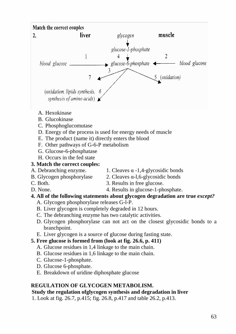
63
A. Hexokinase B. Glucokinase C. Phosphoglucomutase D. Energy of the process is used for energy needs of muscle E. The product (name it) directly enters the blood F. Other pathways of G-6-P metabolism G. Glucose-6-phosphatase H. Occurs in the fed state
3. Match the correct couples: A. Debranching enzyme. 1. Cleaves � -1,4-glycosidic bonds B. Glycogen phosphorylase 2. Cleaves �-l,6-glycosidic bonds C. Both. 3. Results in free glucose. D. None. 4. Results in glucose-1-phosphate. 4. All of the following statements about glycogen degradation are true except?
A. Glycogen phosphorylase releases G-l-P. B. Liver glycogen is completely degraded in 12 hours. C. The debranching enzyme has two catalytic activities. D. Glycogen phosphorylase can not act on the closest glycosidic bonds to a
branchpoint. E. Liver glycogen is a source of glucose during fasting state.
5. Free glucose is formed from (look at fig. 26.6, p. 411) A. Glucose residues in 1,4 linkage to the main chain. B. Glucose residues in 1,6 linkage to the main chain. C. Glucose-1-phosphate. D. Glucose 6-phosphate. E. Breakdown of uridine diphosphate glucose
REGULATION OF GLYCOGEN METABOLISM. Study the regulation ofglycogen synthesis and degradation in liver 1. Look at fig. 26.7, p.415; fig. 26.8, p.417 and table 26.2, p.413.

64
Note: Key regulatory enzymes of glycogen synthesis and degradation can be both hormonally regulated and allosterically regulated. In liver glycogen is synthesized during a carbohydrate meal when blood glucose level is elevated and as blood glucose level decreases glycogen is degraded. Thus the degradative and biosynthetic pathways are regulated by changes in the insulin/ glucagon ratio and by blood glucose levels; glycogen degradation is regulated also by epinephrine (under stress situation, exercise, hypoglycemia). Insulin through its own phosphorylation cascade initiated at the insulin receptor tyrosine kinase (fig.24.17, p. 386) activates hepatic protein phosphatase-1 (fig. 26.7) and thus inhibits glycogen degradation and stimulates glycogen synthesis. Insulin may also activate the phosphodiesterase which converts cAMP to AMP, thereby decreasing cAMP levels. Regulation of liver glycogen degradation and synthesis by epinephrine, acting at �-receptor, and glucagon is similar (through intracellular second messenger cAMP and protein kinase A), (fig. 26.7 and fig.6.4 "Cascade regulation"). Epinephrine, acting at α-receptor in liver also regulates glycogen metabolism by PIP- Ca2+ signal transduction system (fig. 26.8). (Release of Ca2+ is a result of epinephrine uptake). 2. Which of the following statements regarding the regulation of glycogen metabolism in liver are correct?
A. Liver glycogen is synthesized when blood glucose level is elevated. B. The fall of the insulin/glucagon ratio results in activation of the glycogen
synthesis. C. Liver glycogen is degraded to free glucose. D. Protein kinase A and protein kinase C inactivate glycogen synthase. E. Glucogen phosphorylase becomes active due to phosphorylation.
3. Glycogen synthesis in liver is stimulated by A. Increased concentration of glucose. B. Phosphorylation of glycogen synthase. C. Dephosphorylation of phosphorylase kinase D. Epinephrine. E. Phosphodiesterase.
4. When a normal individual in the basal metabolic state ingests a high-carbohydrate meal, there is A. Enhanced glycogen synthase activity in liver. B. An increased ratio of phosphorylase a to phosphorylase b in liver. C. An increased insulin/glucagon ratio. D. Phosphorylation ofglycogen synthase in liver. E. Dephosphorylation ofglycogen synthase.

65
Fig. 16 Regulation of glycogen synthesis Study the regulation of glycogen synthesis and degradation in muscle. 1. Look at fig. 26.9, p. 419; and table 26.2, p.413 and Fig. 16 1. Note: In muscle insulin stimulates the transport of glucose into the muscle cells, providin increased substrates for glycogen synthesis and stimulates synthesis by mechanisms similar to those in the liver. Glucagon has no effect on muscle and thus glycogen degradation do not vary with the fasting/feeding state. In skeletal muscles glycogen degradation is initiated by muscle contraction, neural impulses and epinephrine. Glycogen degradation in muscle is regulated by 1) AMP, allosteric activator of muscle glycogen phosphorylase b, produced from
the degradation of ATP during muscle contraction (fig.26.4, p.419, fig.9.31, p. 123); 2) the effects of Ca2+ that is released from sarcoplasmic reticulum after neural
stimulation (fig. 26.4, p. 419); 3) the effects of epinephrine-stimulated phosphorylation by protein kinase A
during exercise and other stress situations (are similar to those occurring in liver (fig. 26.7, p. 417 in the Textbook and Fig. 16 "Cascade regulation").
2. Which of the following phosphorylated enzymes are active?
A. Phosphorylase kinase. B. Phosphorylase A. C. Glycogen synthase . D. Pyruvate kinase. E. Pyruvate dehydrogenase.
3. Which of the following statements regarding the regulation of glycogenolysis is correct?
66
A. cAMP enhances glycogenolysis by adenylation of glycogen phosphorylase. B. Phosphorylase b is activated by phosphorylation. C. Phosphorylase kinase is inactivated by phosphorylation by cAMP. D. Muscle phosphorylase kinase is inactivated by Ca2+ E. Glycogen synthase is phosphorylated at three sites.
4. An adolescent patient with deficiency of muscle phosphorylase was examined while exercising her forearm by squeezing a rubber ball. Compared to a normal person performing the same exercise, this patient:
A. Could exercise for a longer period of time without fatigue. B. Had increased glucose level in blood drawn from her forearm. C. Had decreased lactate level in blood drawn from her forearm. D. Had lower level of glycogen on biopsies of her forearm muscle.
5. Match the correct couples A. Epinephrine 1. Acts in liver and muscle B. Glucagon 2. Acts in liver only C. Both 3. Stimulates glycogen synthesis D. None 4. Transmits a signal through G protein
6. Regulation of enzymes by a phosphorylation mechanism: A. Always results in increased activity of the phosphorylated enzyme. B. Always results in decreased activity of the phosphorylated enzyme. C. Is only a cAMP-mediated process. D. Involves formation of high energy bond between phosphate and
enzyme E. Involves a phosphoprotein phosphatase.
GLYCOGEN STORAGE DISEASES. Study glycogen storage diseases 1. Note: An inability to degrade glycogen can cause cells to become pathologically engorged; it can also lead to the functional loss of glycogen as a source of cell energy and as blood glucose buffer. A number of these genetic diseases have been linked to mutations in enzymes associated with glycogen processing. The debilitating effect of many glycogen storage diseases depends on the severity of the mutation causing the deficiency. Look at table 26.1 (p. 412) and table below and answer the following questions (p. 412; clinical comments p.419-420): Name of disease
Deficiency Organs involved
Effect of deficiency
von Gierke's disease
glucose-6- phosphatase activity
liver, kidney liver, kidney, intestinal cells engorged with glycogen; exercise hypoglycemia
Pompe's disease
lysosomal 1-4 or 1-6 glucosidase activity
all organs often fatal accumulation of lysosomal glycogen deposits

67
Andersen's disease
branchin enzyme liver, muscle kidney, leukocytes
glycogen with few branches, often fatal childhood
Forbe's/Cori's disease
debranchin enzyme liver, muscle heart, leukocytes
highly branched glycogen with limited availability of glucose monomer
McArdle's disease
muscle phosphorylase
skeletal muscle
limited availability to sustain muscle activity, excess muscle glycogen, limited blood lactate after exercise
Hers' disease liver phosphorylase
liver tendency to hypoglycemia and excess accumulation of liver glycogen
2. Write down the enzymes causing disorders of glycogen degradation. 3. Write down the enzymes causing disorders of glycogen synthesis. 4. Which of the inactivated phosphorylases (liver phosphorylase or muscle phosphorylase) does not cause low blood glucose levels? Why? 5. Choose the correct answer: Hereditary glucose-6-phosphatase deficiency can lead to: A. glycogen synthesis after a meal B. glycogen is degraded and glucose enters the blood during fasting 6. A liver biopsy from an infant with a large liver and kidney, dwarfism, hypoglycemia, acidosis revealed a massive buildup of glycogen with a normal structure. A possible diagnosis would be
A. Pompe's disease B. Andersen's disease C. Cori's disease D. von Gierke's disease E. Hers' disease
What enzyme deficiency causes this disease? LABORATORY MANUAL: 1. Qualitative assay of lactose in milk (reaction of Fehling) Principle of method: The method is based on property of lactose to reduce �opper from Fehling’s solution. Practical procedure: Place tested sample of milk into the test tube, add 5 drops of trichloracetic acid, filter after appearense of precipitate.Transfer 10 drops of solution to test tube, add 10 drops of distillated water, add 10 drops of solution of sodium hydroxide and 5 drops of Fehling’s solution. Heat the upper part of mixture till boiling (carefully). Boil 1 min. Write down the results and conclusion.

68
2. Qualitative assay of starch in bread. Principle of method: The method is based on property of starch to give the color reaction with solution of iod. Practical proctdure: Place 1 drop of solution of iod on the bread. Write down the results and conclusion. 3. Quantitative assay of glucose in blood serum by enzymatic method. Principle of method: Glucooxidase cleaves glucose and releases gluconate and hydrogen peroxide. Produced H2O2 is detected by oxidative condensation, a reaction catalyzed by peroxidase. As a result coloured product is formed. Concentration of coloured product is measured on photoelectric colorimeter. Practical procedure: 1. Prepare 3 test-tubes as shown in table below:
Test-tube � 1 Test-tube � 2 Test-tube � 3 Blood serum 0.1 ml Glucose solution 0.1 ml Distillated water 0.1 ml Glucose reagent 2.0 ml 2.0 ml 2.0 ml Glucose reagent contents glucoseoxidase, peroxidase, 4-Cl-3-methylphenol, 4-aminophenasol. 2. Mix the contents of test-tubes. Heat the test-tubes at 37°C for 30 minutes. 3. Put the contents of the test-tubes in cuvettes and analyse on photoelectric colorimeter. The content of test-tube .No 3 is used as a control solution for colorimetric measurements. Measurements is to be performed for 5 minutes because the color of solution is changed. 4. Calculate glucose concentration using the following equation: [glucose] mMol/l ==== 10 Optical density of test-tube No1/ Optical density of test-tube No2
The blood glucose level is 3.3 mMol/l - 5.5 mMol/l or 60 -100 mg/dl 5. Write down the results and draw a conclusion. Homework: Lesson 11 1. Compare the functions of hexokinase and glucokinase (p. 117, 342) Note the role of glucose 6-phosphase for various metabolic pathways (fig.22.2, p.342; 389). 2. Study aerobic glycolysis, anaerobic glycolysis and complete oxidation of glucose, (study reactions using formulas, learn enzymes) (p. 342-345, 348-349). 3. Study shuttle systems: glycerol 3-phosphate shuttle and malate-aspartate shuttle (p. 347-348). 4. Study the energy yield from aerobic glycolysis, anaerobic glycolysis and full oxidation of glucose (p. 343,345-346).

69
LESSON 11. CATABOLISM OF GLUCOSE Main questions:
• aerobic glycolysis • anaerobic glycolysis • energetic of glycolysis • regulation of glycolysis • complete oxidation of glucose • stages of complete oxidation of glucose • the shuttle systems: glycerol phosphate shuttle and malate aspartate shuttle • energetic of complete oxidation of glucose • regulation of complete oxidation of glucose • alcoholic fermentation
Study aerobic glycolysis, anaerobic glycolysis and complete oxidation of glucose (p. 342-345, 348-349). 1. Note: Glycolysis is an oxidative specific pathway by which one mole of glucose is enzymatically split into two moles of pyruvate. It occurs in cytosol of all cells of the body. The principle function of glycolysis is the generation of ATP. Glycolysis also provides precursors for fatty acids biosynthesis, for the synthesis of amino acids and pentoses. Anaerobic glycolysis is a process that functions in the absence of oxygen, the final product of anaerobic glycolysis is lactate (Fig. 17). In some cell types anaerobic glycolysis generates all of the cell's ATP requirements (in RBC which lack mitochondria) or at least a portion all of the cell's ATP requirements (in skeletal muscles at the onset of exercise and during intensive exercise and in tissues like lymphocytes, white blood cells, the kidney medulla). Aerobic glycolysis is a process that functions when oxygen is available, the final product of aerobic glycolysis is pyruvate (Fig. 18, (A)). Glycolysis can be divided into two stages: 1) the conversion of glucose into 2 triose phosphates: dihydroxyacetone phosphate and glyceraldehyde 3-phosphate (look at Fig.1, steps -1,2,3,4). This stage involves a series of reactions that requires two molecules of ATP for each molecule of glucose that is split. Dihydroxyacetone phosphate is reversibly converted into glyceraldehyde-3- phosphate (Fig. 17, (5)). Thus in this stage one mole of glucose is split into two moles of glyceraldehyde 3-phosphate. 2) the conversion of 2 moles of glyceraldehyde 3-phosphate into 2 moles of pyruvate (Fig. 1, (steps: 6 - 10)). Pay attention that the number of moles of all metabolites in these steps (6 - 10) is multiplied by coefficient 2. During this stage ATP, NADH, pyruvate are produced. Most steps of glycolysis are reversible except three reactions (Fig. 17, (1,3 and 10).

70
Fig. 17 Anaerobic glycolysis
Enzymes 1 – hexokinese / glucokinese (in liver) 2 – phosphoglucose isomerase 3 – phosphofructokinese 4 - aldolase 5 – triose phosphate isomerase 6 - glyceraldehyde 3-phosphate
dehydrogenase 7 – phosphoglycerate kinase 8 - phosphoglyceromutase 9 – enolase 11 – lactate dehydrogenase
10 –pyruvate kinase
Phosphoenolpyruvate
Glucose
Glucose – 6 phosphate
Fructose – 6 – Phosphate
Fructose –1,6 – Phosphate
Glycerolalehyde 3 phosphate
Dihydroxyacetone phosphate
3 – Phosphoglicerate
2 – Phosphoglicerate
Pyruvate
ATP
ATP
NAD+
ATP
Lactate
NADH
ATP
It is multiplie
d by 2
1,3 – Bisphosphoglicerate
H3PO4
NADH+H
NAD
1
2
3
4
5
4
6
7
8
9
10
11

71
Fig. 18 A - Aerobic glycolysis; B - Complete oxidation of glucose. Enzymes: 1 – hexokinase /glucokinase(in liver) 2 - phosphoglucose isomerase 3 – phosphopructokinase 1 4 - aldolase 5 - triose phosphate isomerase 6 -glyceraldehyde 3-phosphate
dehydrogenase 7 – phosphoglycerate kinase 8 – phosphoglyceromutase 9 – enolase 10 – pyruvate kinase
Glucose
Glucose – 6 phosphate
Fructose – 6 – Phosphate
ATP
ATP 3
2
1
B
9
Fructose –1,6 – Phosphate
1,3 – Bisphosphoglicerate
3 – Phosphoglicerate
2 – Phosphoglicerate
Phosphoenolpyruvate
Pyruvate
ATP
ATP
Acetyl -CoA
TCA cycle CO2, H2O,
10
8
7
A
6
Glyceraldehyde 3 phosphate
H3PO4
NADH + H+
NAD+
ATP ETC
5 4
4 Dihydroxyacetone phosphate
It is multiplie
d by 2

72
The energy yield from anaerobic glycolysis is 2 moles of ATP per mole of glucose. Under anaerobic conditions (cells are limited by mitochondrial capacity or oxygen availability) 4 moles of ATP are produced by reactions catalyzed by pyruvate kinase (Fig. 17, (10) and phosphoglycerate kinase (Fig. 17, (7) (substrate level phosphorylation only):
2x(1+1) – 2 = 2 moles of ATP
Fig.16, (7) and (10), substrate level
Fig. 16, (1) and (3), these steps consume two molecules of ATP
Pyruvate is reduced in the cytosol by NADH, forming lactate (convertion of pyruvate to lactate is catalyzed by lactate dehydrogenase) and regenerating NAD+
Aerobic glycolysis provides 6(8) moles of ATP per mole of glucose:
2x(2+2(3)) – 2 = 6(8) moles of ATP
Fig.17, (7) and (10), substrate level phosphorylation
Fig. 17, (6), oxidative phosphorylation also point below about shuttle systems
If cells have sufficiently high oxidative capacity ATP can be produced by reactions catalyzed by glyceraldehyde-3-phosphate dehydrogenase (oxidative phosphorylation), pyruvate kinase and phosphoglycerate kinase (substrate level phosphorylation). When glucose is oxidized completely to CO2 and H2O (Fig. 18 (B)) 36 or 38 moles of ATP are generated. In this case pyruvate may enter mitochondria and be converted to acetyl-CoA, which is oxidized by the TCA cycle, generating additional ATP: 2×The energy yield from aerobic glycolysis + 2×The energy yield from TCA cycle
= 6 (8) +30=36 (38) moles of ATP NADH produced in the cytosol by glycolysis cannot directly cross the mitochondrial membrane. The electrons are passed to the mitochondrial ETC by two shuttle systems: glycerol phosphate shuttle (2x2 moles of ATP are produced by this shuttle) and malate aspartate shuttle (2x3 moles of ATP are produced by this shuttle) (p. 342-345, 348-349, Fig.22.8, 22.9). During intensive muscle exercise the need for ATP exceeds the capacity of mitochondria for oxidative phosphorylation. Under these conditions the contribution of anaerobic glycolysis to ATP synthesis increases and lactate level in muscle cells is elevated. Lactate concentration in the blood is higher (up to 20 mMol/L) than that under normal conditions (1 -2 mMol/L). 2. What step of aerobic glycolysis involves the coupling of ATP synthesis to electron transport chain? A. Glucose Fructose-6-phosphate B. Phosphoenolpyruvate Pyruvate

73
C. Fructose-1,6-bisphosphate Glyceroaldehyde-3 -phosphate D. Glyceroaldehyde-3-phosphate 1,3-bisphosphoglycerate E. 3-phosphoglycerate Phosphoenolpyruvate 3. Write the reaction chosen in previous test by formulas and draw a scheme of hydrogen transfer from cytosol to mitochondria (shuttle system) and the scheme of hydrogen transfer along the electron transport chain. 4. How many moles of pyruvate are formed if 1 mole of glucose is oxidized in aerobic glycolysis? How many moles of ATP are generated in aerobic glycolysis? What energy yield is if 1 mole of glucose is oxidized to lactate without oxygen? 5. Choose the correct statements about glucose catabolism providing energy for erythrocytes metabolism:
A. Occurs in cytosol B. Involves 3 irreversible steps C. Energy yield is 8 moles of ATP per mole of glucose D. Involves steps conjugated to ATP synthesis without ETC E. Involves 2 oxidative steps
6.Match the number and the letter: A. Phosphofructokinase.
1. Requires ATP.
B. Pyruvatekinase.
2. Phosphorylates ADP. C. Both.
3. Catalyzes irreversible step. D. None.
4. Catalyzes the formation of end product.
7. Match the number and the letter: A. Complete oxidation of glucose.
1. Pyruvate is the end product.
B.Anaerobic glycolysis. 2. Pyruvate is an acceptor of electron from NADH
C. Both 3. Requires continuous regeneration of NADH.
D. None 4. Involves 6 steps of oxidative phosphorylation.
8. Match the number and the letter: A. Anaerobic glycolysis
1. Is the most important source of energy for brain
B. Complete oxidation of glucose
2. Provides energy for intensive muscle exercise for a long time
C. Both
3. Is the most important at onset of intensive muscle exercise.
D. None
4. Provides energy for cancer cells.
9. Note that in red blood cells "side reaction" of glycolytic pathway occurs. 3-bisphosphoglycerate is converted to 2,3-bisphosphoglycerate, a compound that decreases the affinity of hemoglobin for oxygen:

74
10. Read p.344 (left) and answer the question: How does 2,3-bisphosphoglycerate reenter the glycolysis?
LABORATORY MANUAL:
1. Alcoholic fermentation. In yeast alcoholic fermentation is two- step process. Pyruvate is decarboxylated to acetaldehyde by pyruvatedecarboxylase in an essentially irreversible reaction. Thiamine pyrophosphate is a required cofactor for this enzyme. The second step, the reduction of acetaldehyde to ethanol by NADH, is catalyzed by alcohol dehydrogenase. The end products of alcoholic fermentation are thus ethanol and carbon dioxide. Practical procedures: Place 1g of yeasts and 20 ml of glucose solution into the apparatus for alcoholic fermentation. Stay the contens of apparatus for 1 hour at 370. a) Determination of carbon dioxide. Add some millilitres of 10% sodium hydroxid solution into the apparatus. Mix. The carbon dioxide is uptook by the sodium hydroxid. Vacuum is created. b) Determination of ethanol. Place 2 ml of solution from apparatus into the test-tube, add 2 drops of 10% iodine. Heat. Write down the results and conclusion. Homework: Lesson 12 1. Study the sources and functions of gluconeogenesis and remember what tissues
this process occurs in (p. 426-427). 2. Study gluconeogenesis from different precursors (pyruvate, lactate, glycerol,
amino acids).Memorize reactions by formulas, learn enzymes, pay attention to irreversible steps (p. 427-430).
3. Study energetics of gluconeogenesis (p. 434). 4. Study glucose-alanine cycle and cycle Cori (349-350). 5. Study the regulatory enzymes, activators and inhibitors of glycolysis and
gluconeogenesis, hormonal regulation of glycolysis and gluconeogenesis (p. 430-434, 349-352).
6. Learn nonproductive substrate cycles (futile cycles) (p.432). 7. Study pentose phosphate pathway as alternative pathway of carbohydrate metabolism. 8. Learn reactions of oxidative phase by formulas, memorize enzymes. Learn scheme of nonoxidative phase. Pay attention to the role of products of pentose phosphate pathway (p. 437-444).
75
LESSON 12. GLUCONEOGENESIS. REGULATION OF GLYCOLYSI S AND GLUCONEOGENESIS. PENTOSE PHOSPHATE PATHWAY. Main questions: • Functions of gluconeosenesis. • Gluconeogenesis from pyruvate, lactate, glycerol, amino acids. • Energetic of gluconeogenic pathway. • Regulation of glycolysis and gluconeogenesis. • Pentose phosphate pathway. • Reactions of pentose phosphate pathway. • Role of products of pentose phosphate pathway. GLUCONEOGENESIS. Study the sources and functions of gluconeogenesis (p. 426-427). 1. Note:
Gluconeogenesis is the synthesis of glucose from noncarbohydrate precursors. It occurs under fasting conditions and maintains normal blood glucose level. (Remember that glucose is universal fuel for human cells and if blood glucose decreases tissues that depend on glucose would suffer from a lack of energy). Carbon sources for gluconeogenesis depend on physiological states in humans. During fasting the breakdown of adipose triacylglycerol releases glycerol that serves as source of carbon in gluconeogenesis. During starvation the major precursors for glucose formation are amino acids obtained by degradation of muscle protein and glycerol. Lactate produced by exercising muscle and red blood cells serves as a source of carbon in gluconeogenesis during exercise, Liver, kidney and intestine contain glucose-6-phosphatase which catalyzes the production of free glucose. The liver is responsible for 85% - 95% of the glucose production, the kidney cortex and epithelial cells of the small intestine also contributes glucose formation. The normal fasting glucose range is 80- 100 mg/dL ( 3.5 -5.5 mM). 2. The functions of gluconeogenesis are described by all of the following statements EXCEPT:
A. Maintains blood sugar levels during fasting B. Useful during strenuous exercises C. Allows the use of acetyl-CoA for glucose production D. Allows the use of amino acids for glucose production E. Maintains blood glucose level during period of limited carbohydrate
intake 3. Gluconeogenesis occurs in tissues:
A. Liver B. Kidney C. Brain D. Epithelial cell of the small intestine

76
E. Muscle 4. Choose the correct answers about gluconeogenesis:
A. All steps are simply reversable of those of glycolysis B. All steps of gluconeogenesis from pyruvate occur in cytosol C. Irreversible steps are catalyzed by regulatory enzymes D. Process involves oxidative steps E. Requires high energy phosphate bond of ATP only
5. Gluconeogenesis is the synthesis of glucose from noncarbohydrate precursors:
A. Lactate. B. Malate. C. Alanine. D. Fatty acids. E. Glycerol. F. Acetyl-CoA. G. Pyruvate. H. Amino acids.
Which of them are key substrates? Study Gluconeogenesis from pyruvate, lactate, glycerol and amino acids. 1. Note: Gluconeogenesis and glycolysis differ at only three points (look at fig.3, «Regulation of glucose metabolism in liver»). They involve the conversion of pyruvate to phosphoenolpyruvate (occurs in two steps and is catalyzed by two enzymes instead of the single enzyme used for glycolysis), removing phosphate from fructose 1,6-bisphosphate to form fructose 6-phosphate and removing phosphate from glucose 6-phosphate to form glucose. Thus glucose is not generated by reactions which are simply reversals of glycolysis (p. 427-430): Pyruvate (produced from lactate, alanine and other aminoacids) is first converted to oxaloacetate (fig.27.11) by pyruvate carboxylase, a mitochondrial enzyme that requires biotin and ATP. Oxaloacetate is converted to malate or aspartate, which travels to the cytosol and is reconverted to oxaloacetate (fig 27.10, 27.12, 27.13). The conversion of oxaloacetate to phosphoenolpyruvate is catalyzed by phosphoenolpyruvate carboxykinase, a cytosolic enzyme that requires GTP. Fructose-6-bisphosphatase converts fructose 1,6-bisphosphate to fructose-6-phosphate releasing inorganic phosphate. Glucose-6-phosphatase releases inorganic phosphate converting glucose 6-phosphate to free glucose, which enters the blood. 2. Using formulas draw the scheme of gluconeogenesis starting with pyruvate. Mark out in red colour irreversible steps, write down their enzymes. 3. Write down in words reactions requiring energy (ATP, GTP). Mark the appropriate enzymes. 4. Write down the conversion of lactate to pyruvate. In what direction will this process occur under low [NAD ]/[NADH] ratio? 5. Excessive alcohol ingestion and reduction in nutrient intake leads to blood glucose level to fall. Read p. 429 of Textbook (right) and answer the question:
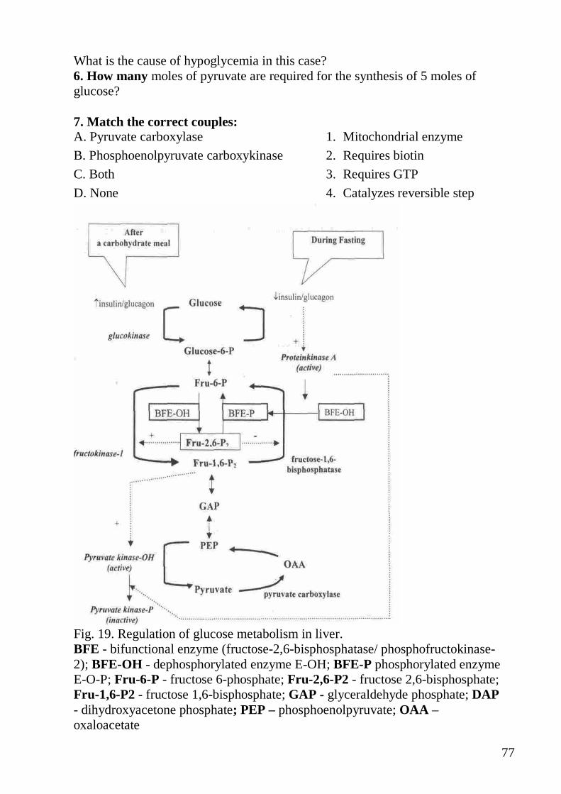
77
What is the cause of hypoglycemia in this case? 6. How many moles of pyruvate are required for the synthesis of 5 moles of glucose? 7. Match the correct couples: A. Pyruvate carboxylase
1. Mitochondrial enzyme B. Phosphoenolpyruvate carboxykinase 2. Requires biotin C. Both
3. Requires GTP D. None
4. Catalyzes reversible step
Fig. 19. Regulation of glucose metabolism in liver. BFE - bifunctional enzyme (fructose-2,6-bisphosphatase/ phosphofructokinase-2); BFE-OH - dephosphorylated enzyme E-OH; BFE-P phosphorylated enzyme E-O-P; Fru-6-P - fructose 6-phosphate; Fru-2,6-P2 - fructose 2,6-bisphosphate; Fru-1,6-P2 - fructose 1,6-bisphosphate; GAP - glyceraldehyde phosphate; DAP - dihydroxyacetone phosphate; PEP – phosphoenolpyruvate; OAA – oxaloacetate

78
8. Match the correct couples: A. Gluconeogenesis from lactate 1. Requires dehydrogenase B. Oxidation of lactate to CO2 and H2O 2. Releases ATP C. Both 3. Requires ATP D. None 4. All enzymes are located in cytosol 9. Choose the correct statements about the glycerol as a substrate of gluconeogenesis:
A. Glycerol is released from adipose stores of triacylglycerol. B. The liver takes up the glycerol and phosphorylates it. C. The conversion is decreased by high level of NAD+ D. In the presence of ethanol the rate of glucose synthesis is decreased. E. Conversion of glycerol to glycerol-3 -phosphate is catalyzed by glycerol
kinase. 10. Gluconeogenesis from glycerol was investigated in liver cells. In glycerol molecule atom C14 in β-position was replaced by C15. In what position will be C15 in metabolites given below? Name these compounds. A.
O
P
O
OHO
HO
OH
B.
O
(R)
HO
(S)
OH
(R)
HO
(R)
OH
P
O
O
OH
OH
Study energetics of gluconeogenesis. 1. Note: During gluconeogenesis three enzymes require high energy phosphate bonds: pyruvate carboxylase (1 mole of ATP), phosphoenolpyruvate carboxykinase (1 mole of GTP) and phosphoglycerate kinase (1 mole of ATP). As 2 moles of pyruvate are required for the synthesis of 1 mole of glucose, 6 high energy phosphate bonds are cleaved (4 moles of ATP and 2 moles of GTP) (p. 434). 2. How many moles of high energy phosphate bonds are required for synthesis of 2 moles of glucose from lactate? GLUCOSE-ALANINE CYCLE AND CORI CYCLE. Study the scheme of glucose-alanine-cycle and Cori cycle (p. 349-350). 1. Note: Glutamine and alanine are the major amino acids that form key substrates for luconeogenesis. Pyruvate formed during glycolysis in muscle can convert to alanine (fig.27.8, p.428). Alanine is delivered from muscle to the liver where it converts to pyruvate with concomitant glucose synthesis. This process is called glucose-alanine cycle. The flow of glucose and lactate between the liver and lactate-producing tissues (erythrocytes, muscle) is known as the Cori cycle (p. 349 and fig. 22.13).
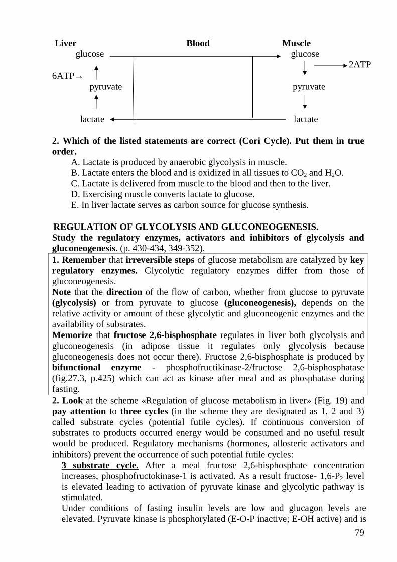
79
Liver Blood Muscle glucose glucose 2ATP 6ATP→ pyruvate pyruvate
lactate lactate 2. Which of the listed statements are correct (Cori Cycle). Put them in true order.
A. Lactate is produced by anaerobic glycolysis in muscle. B. Lactate enters the blood and is oxidized in all tissues to CO2 and H2O. C. Lactate is delivered from muscle to the blood and then to the liver. D. Exercising muscle converts lactate to glucose. E. In liver lactate serves as carbon source for glucose synthesis.
REGULATION OF GLYCOLYSIS AND GLUCONEOGENESIS. Study the regulatory enzymes, activators and inhibitors of glycolysis and gluconeogenesis. (p. 430-434, 349-352). 1. Remember that irreversible steps of glucose metabolism are catalyzed by key regulatory enzymes. Glycolytic regulatory enzymes differ from those of gluconeogenesis. Note that the direction of the flow of carbon, whether from glucose to pyruvate (glycolysis) or from pyruvate to glucose (gluconeogenesis), depends on the relative activity or amount of these glycolytic and gluconeogenic enzymes and the availability of substrates. Memorize that fructose 2,6-bisphosphate regulates in liver both glycolysis and gluconeogenesis (in adipose tissue it regulates only glycolysis because gluconeogenesis does not occur there). Fructose 2,6-bisphosphate is produced by bifunctional enzyme - phosphofructikinase-2/fructose 2,6-bisphosphatase (fig.27.3, p.425) which can act as kinase after meal and as phosphatase during fasting. 2. Look at the scheme «Regulation of glucose metabolism in liver» (Fig. 19) and pay attention to three cycles (in the scheme they are designated as 1, 2 and 3) called substrate cycles (potential futile cycles). If continuous conversion of substrates to products occurred energy would be consumed and no useful result would be produced. Regulatory mechanisms (hormones, allosteric activators and inhibitors) prevent the occurrence of such potential futile cycles:
3 substrate cycle. After a meal fructose 2,6-bisphosphate concentration increases, phosphofructokinase-1 is activated. As a result fructose- 1,6-P2 level is elevated leading to activation of pyruvate kinase and glycolytic pathway is stimulated. Under conditions of fasting insulin levels are low and glucagon levels are elevated. Pyruvate kinase is phosphorylated (E-O-P inactive; E-OH active) and is
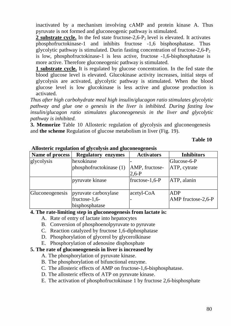
80
inactivated by a mechanism involving cAMP and protein kinase A. Thus pyruvate is not formed and gluconeogenic pathway is stimulated. 2 substrate cycle. In the fed state fructose-2,6-P2 level is elevated. It activates phosphofructokinase-1 and inhibits fructose -1,6 bisphosphatase. Thus glycolytic pathway is stimulated. Durin fasting concentration of fructose-2,6-P2 is low, phosphofructokinase-1 is less active, fructose -1,6-bisphosphatase is more active. Therefore gluconeogenic pathway is stimulated. 1 substrate cycle. It is regulated by glucose concentration. In the fed state the blood glucose level is elevated. Glucokinase activity increases, initial steps of glycolysis are activated, glycolytic pathway is stimulated. When the blood glucose level is low glucokinase is less active and glucose production is activated.
Thus after high carbohydrate meal high insulin/glucagon ratio stimulates glycolytic pathway and glue one o genesis in the liver is inhibited. During fasting low insulin/glucagon ratio stimulates gluconeogenesis in the liver and glycolytic pathway is inhibited. 3. Memorize Table 10 Allosteric regulation of glycolysis and gluconeogenesis and the scheme Regulation of glucose metabolism in liver (Fig. 19).
Table 10
Allosteric regulation of glycolysis and gluconeogenesis Name of process Regulatory enzymes Activators Inhibitors glycolysis hexokinase - Glucose-6-P phosphofructokinase (1) AMP, fructose-
2,6-P ATP, cytrate
pyruvate kinase fructose-1,6-P ATP, alanin Gluconeogenesis pyruvate carboxylase acetyl-CoA ADP fructose-1,6-
bisphosphatase - AMP fructose-2,6-P
4. The rate-limiting step in gluconeogenesis from lactate is: A. Rate of entry of lactate into hepatocytes B. Conversion of phosphoenolpyruvate to pyruvate C. Reaction catalyzed by fructose 1,6-diphosphatase D. Phosphorylation of glycerol by glycerolkinase E. Phosphorylation of adenosine disphosphate
5. The rate of gluconeogenesis in liver is increased by A. The phosphorylation of pyruvate kinase. B. The phosphorylation of bifunctional enzyme. C. The allosteric effects of AMP on fructose-1,6-bisphosphatase. D. The allosteric effects of ATP on pyruvate kinase. E. The activation of phosphofructokinase 1 by fructose 2,6-bisphosphate

81
6. Match the correct couples A. Glycolysis 1. Is regulated by fmctose-2,6-bisphosphate B. Gluconeogenesis 2. Is activated by high fructose-1,6-bisphosphate level C. Both 3. Is activated by high NADPH concentration D. None 4. Is stimulated in the fed state 7. Which of the following compounds is a positive allosteric regulator of the enzyme pyruvate carboxylase?
A. Adenosine triphosphate B. Acetyl coenzyme A C. Biotin D. Phosphoenolpyruvate E. Fructose-1,6-bisphosphate
8. Gluconeogenesis is stimulated under the following conditions A. Fasting B. Low insulin level C. Low dietary intake of carbohydrates D. High glucagon level E. Stress conditions F. Fed state G. Muscle exercises
9. In a fasting individual, each of the following stimulates the production of the blood glucose by gluconeogenesis except
A. An increased supply of substrates B. Induction ofphosphoenolpyruvate carboxylase C. A decrease of the portal blood glucose level below the Km ofglucokinase D. A cAMP-mediated activation of pyruvate kinase E. Induction ofglucose-6-phosphatase.
10. Which of the following compounds decreases the rate of glucose synthesis from alanine? Specify the mechanism of their action.
A. Acetyl-CoA. B. ATP. C. AMP. D. Rotenone. E. lonophores.
11. Read the clinical case of Emma Whezzer (p. 423, 427, 423). The treatment by glucocorticoids and synthetic glucocorticoid dexamethasone stimulating the degradation of muscle protein leads to hyperglycemia. Answer the question: Why are the levels of blood lucose elevated? 12. Match the correct couples (p. 344) A. Fructose-2,6-bisposphate 1. Coenzyme of phosphoglycerate mutase B. 2,3-bisphosphoglycerate 2. Allosteric inhibitor of gluconeogenesis C. Both 3. Allosteric inhibitor of oxygen binding to heme D. None 4. Present in low concentration in most tissues
except RBCs
82
THE PENTOSE PHOSPHATE PATHWAY. Study the main steps of the pentose phosphate pathway (p. 437-444). 1. Note: The pentose phosphate pathway (PPP) is an alternative pathway of carbohydrate metabolism. PPP oxidizes glucose-6-phosphate to intermediates of glycolytic pathway (fructose 6-phosphate and glyceraldehyde 3-phosphate), generating NADPH and ribose 5- phosphate. The NADPH is utilized for reductive pathways, such as fatty acid biosynthesis, detoxification of drugs by monoaminooxidases and the glutathione defense system against injury by reactive oxygen species (pp. 441-442 and fig. 28.7). Ribose 5-phosphate is required for nucleotides synthesis and synthesis of nucleic acids. PPP has two phases. Initially there is oxidative phase that requires 3 enzymes, 2 of which NADP^specific dehydrogenases. Glucose-6-phosphate dehydrogenase catalyzes the rate limiting reaction of the pathway. The cellular concentration of NADP+ is the major controlling factor; its availability regulates the rate limiting reaction. The reactions of nonoxidative phase are reversible. They can serve for hexoses generation from pentose. Pentose phosphate pathway is the most active in the liver, the mammary glands, adipose tissue and the adrenal cortex. 2. Choose the correct statements concernine functions of the PPP:
A. A source of NADPH for reductive pathways B. A source of pentoses for nucleic acids synthesis C. A route for the use ofpentoses and for their conversion to hexoses and trioses D. A source of energy for reductive pathways E. A source of NADH for reductive pathways
3. NADPH is utilized for A. Fatty acid biosynthesis B. Detoxification of drugs by monooxygenases C. Glutathion defense system against injury by reactive oxygen species D. Cholesterol synthesis E. Cytochrome P450
4. Match the correct couples: A. Glycolysis 1. The enzymes are located in the cytosol B. Pentose phosphate
pathway 2. Generates reduced equivalents for reductive
synthesis C. Both 3. Requires dehydrogenase D. None 4. Reduced equivalents donate the electrons
directly to ETC 5. The transketolase requires the following for maximal activity A. A.TPP. B. Biotin. C. CoA. D. Dihydroxyacetone phosphate E. Acetyl-CoA. Home work: Study lesson 13

83
LESSON 13 CHEMISTRY OF LIPIDS. DIGESTION AND ABSORPTION OF LIPIDS. LIPOPROTEINS. Test in written form: the structural formulae and biological role of:
1) saturated fatty acids (C4-C24), unsaturated fatty acids (oleic, linoleic, linolenic, arachidonic acid) Point out with the help of digital symbols position and amount of double bonds in unsaturated fatty acids;
2) mono-, di-, triacylglycerols; 3) phosphoacylglycerols (phosphatidylcholine, phosphatidylserine,
phosphatidylethanolamine, phosphatidylinositol); 4) ceramide and sphingolipids (sphingomyelin, galactocerebroside,
ganglioside); 5) cholesterol and cholesterol esters; 6) bile acids and taurine-, glycoconjugates of bile salts.
Main questions.
• Features of structure and biological role of fatty acids in the human • Triacylglycerols and phosphoacylglycerols: features of a structure and
properties. • Physiological norms of daily lipids intake. The essential components of
dietary lipids for the human organism. • Digestion of dietary fats as hydrolysis of fats under lipases action,
necessary conditions. - Digestion of phosphoacylglycerols. - Structure and biological role of bile acids and their taurine-, and
glycoconjugates. - Infringements of digestion and absorption of lipids. Steatorrhoea.
• Triacylglycerol resynthesis in intestinal epithelial cells. • Synthesis of chylomicrons and transport of fats. • Classification of lipoproteins by density, electrophoretic mobility, their
functions. - Structure of blood lipoprotein - The place of lipoprotein formation, features of lipid structure various LP;
apolipoproteins, their functions. - Dislipoproteinemia. Hyperchylomicronemia. Hypercholesterolemia.
• Lecithin hydrolysis and analysis of hydrolysis products. • Qualitative reaction for cholesterol (Liebermann-Burchard test).
Memorize definition: Lipids are a heterogeneous group of compounds, insoluble in water but soluble in organic solvents such as ether, chloroform. Lipids are classified according to the structure (see lecture). Lipids functions: - an efficient source of energy ( fatty acids, triacylglycerols), - the best reserve food material in the human body (fats), - an insulator for the loss of body heat (fats),

84
- protective coating on the surface of many organs such as kidney, against injury (fats),
- structural components of cell membranes (phosphoacylglycerols, sphingolipids, cholesterol),
- biological active compounds (steroid hormones, eicosanoids), - they facilitate the absorption of the fat soluble vitamins A, D, E, K (bile salts). FEATURES OF STRUCTURE AND BIOLOGICAL ROLE OF FATTY ACIDS IN THE HUMAN. 1. Study fatty acids (p.58-60, fig.6.22, 6.23, 6.24, 6.25, table 6.1) 2.Choose the correct answer: Arachidonic acid contains the number of double bonds
A. 2 B. 3 C. 4 D. 5
3. Choose the correct answer: The prostaglandins are synthesized from A. Arachidonic acid B. Oleic acid C. Linoleic acid D. Linolenic acid
4. All of following statements about features of structure of fatty acids in the human are true EXCEPT:
A. Fatty acids are hydrocarbon derivatives B. Their chain is unbranched C. The double bonds of polyunsaturated fatty acids are almost never
conjugated, but are separated by a methylene group D. The double bonds are in cis configuration E. The double bonds are in trans configuration F. The nonpolar hydrocarbon chain accounts for the poor solubility of fatty
acids in water G. Fatty acids contain even number of carbon atoms
TRIACYLGLYCEROLS AND PHOSPHOACYLGLYCEROLS: FEATURES OF STRUCTURE AND PROPERTIES 1. Study the major phosphoacylglycerols (p.60 fig.6.27, p.61 fig.6.28, 6.29, 6.30, 6.31). 2. Note the effect of lung surfactant p.517 3. Match a line in A with a line in B:
A. B. Neutral lipids Phosphoacylglycerols Storage lipids Triacylglycerols Polar lipids
Membrane lipids
85
4. Name next phosphoacylglycerols which contain such alchohol as ethanolamine, choline, inositol, serine.
5. Lecithin contains nitrogenous base named A. Ethanolamine B. Choline C. Inositol D. All of the above
6. Lecithins contain an unsaturated fatty acid at position A. 1- B. 1- and 2- C. 2- D. None of the above
7. Fill up the blanks of the followings : 1. The lipids are a ____________group of compounds. 2. Phosphatidylinositols are more _________than the other phospholipids. 3. Aminolipids are _______ and ________
PHYSIOLOGICAL NORMS OF DAILY LIPIDS INTAKE. THE ESSENTIAL COMPONENTS OF DIETARY LIPIDS FOR HUMAN ORGANISM.
1. Answer the questions : A. What is the difference between the definition of “lipid” and definitions of
other types of biomolecules that we have considered, such as amino acids, nucleic acids, and proteins?
B. Fat is chemical notion or biological? C. Some fats used in cooking, such as olive oil, spoil rapidly upon exposure to
air at room temperature, whereas others, such as solid shortening, remain unchanged. Why?
2. Name the lipids essential in humans. Define the term essential ( read p.510). 3. Choose the correct answer: The essential fatty acids retard
A. Atherosclerosis B. Diabetes Mellitus C. Nephritis D. Bronchitis
4. Fill up the blanks of the followings : 1. The polyunsaturated fatty acids which are not synthesized in the
body but are taken from natural sources are called _______. 2. The polyunsaturated fatty acids can __________serum cholesterol level.
DIGESTION AND ABSORPTION OF DIETARY LIPIDS. 1. Study the digestion and absorption of dietary lipids (p.492-494, fig.32.8, 32.9);
bonds cleaved by phospholipases (p.519, fig 33.32).

86
2. Choose the correct answer:Lipase present in the stomach cannot hydrolyze fats owing to:
A. Alkalinity. B. Acidity. C. High acidity. D. Neutrality
3. Learn the table “Digestion of dietary lipids ”
Enzyme Pancreatic lipase Intestinal lipase
Phospholipase A1, A2,C, D
Cholesterol esterase
Localization Pancreatic juice
Intestinal juice
Pancreatic juice
Pancreatic juice
Activator of reaction
Bile, Ca2+
colipase - Bile, Ca2+ Bile, Ca2+
Products of reaction
2-monogly-cerides; 1,2- and 2,3-diglycerides, fatty acids
Glycerol, fatty acids
Glycerol, fatty acids, phosphoric acid, nitrogen base (alcohol)
Cholesterol, fatty acid
4. Before the action of lipase the fat is emulsified by A. Lipoproteins B. Phospholipids C. Bile salts D. Ergosterols
5. Choose the correct answer (see p.519, fig.33.32): Phospholipase A1 attacks the ester bond of phospholipids in position
A. 1 B. 2 C. 3 D. All of the above
6. Fill in the table Absorption of products of lipid digestion Products of lipid
digestion Absorption directly into intestinal epithelial cells
Absorption in form of micelles
Glycerol Fatty acids (C4 to C12) Nitrogen base (choline)
Fatty acids 2-monoacylglycerols Cholesterol 7. Choose the correct answer: Phospholipase C release, 1,2-diacylglycerol and
phosphoryl base attack the ester bond in position A. 1 B. 2 C. 3 D. 4

87
8. Pay attention to the question and answer about steatorrhea p.494,495; 543,544.
STRUCTURE AND BIOLOGICAL ROLE OF BILE ACIDS AND THE IR TAURINE- AND GLYCOCONJUGATES 1. Study the action of bile salts (p.492, fig.32.7,p.494, fig.32.10), conjugation
(p.540,fig34.19), structure (p.542,fig.34.21) 2. Discuss the question and answer 6.4 on p. 62-63 3. Memorize bile salts functions: o Bile salts act as emulsifying agents and emulsify fats increasing surface area
and fats miscible with water. o They activate pancreatic lipase and cholesterol esterase. o They combine with free fatty acids and monoglycerides, to form particles called
micelles and help in their absorption, and absorption the fatty-soluble vitamins in the intestines.
o They stimulate intestinal peristalsis. o They stimulate bile production in the liver. o Cholesterol remains soluble in the gall-bladder bile by bile salts.
4. Choose the correct answer: Bile acids are synthesized from cholesterol in the
A. Duodenum B. Intestine C. Gall-bladder D. Liver
5. Choose the correct answer: In the human bile, sodium glycocholate concentration is greater than sodium taurocholate in
A. Two times B. Three times C. Four times D. Five times
6. Choose the correct answer: Bile salts activate
A. Pancreatic lipase B. Cholesterol esterase C. Both D. None
7. Answer the questions (see p.494): What critical concentration of bile salts is optimal for lipid absorption in intestine? 8. All of following statements about biological role of bile salts in the human
are true EXCEPT: 1) Bile salts emulsify fats increasing area surface. 2) Bile salts activate pancreatic lipase.

88
3) They form micelles and help in their absorption in the intestines. 4) Bile salts are the bile pigments. 5) Bile salts stimulate intestinal peristalsis.
9. Pay attention to clinical and biochemical comments (p.498) TRIACYLGLYCEROL RESYNTHESIS IN INTESTINAL EPITHELIA L CELLS . SYNTHESIS OF CHYLOMICRONS AND TRANSPORT OF FATS. 1. Study the reactions on p.495, fig. 32.11and name enzymes. 2. Study the information about synthesis of chylomicrons and transport of fats(
p.494-497, fig.32.13, 32.14, 32.15,32.16). 3. Pay attention to triacylglycerol metabolism in the fed state( p.488, fig 32.4) and
during fasting (p.489, fig.32.5) 4. Choose the correct answer: The great majority of absorbed fat appears in
form of A. HDL B. Chylomicrons C. VLDL D. LDL
CLASSIFICATION OF LIPOPROTEINS. STRUCTURE OF BLOOD LIPOPROTEIN. 1. Memorize definition:
Lipoproteins are a hydrofilic lipoprotein complexes consisting of peripheral and integral apoproteins and polar lipids (phospholipids, cholesterols) on the surface and hydrophobic core of nonpolar molecules (triacylglycerols, cholesterol esters). Lipoproteins may be separated on the basis of their electrophoretic properties and by ultracentrifugation. The density of lipoproteins increases as the protein content rises and the lipid content falls and size of particle becomes smaller: chylomicrons, very low density lipoproteins (VLDL or pre- ß - lipoproteins), low density lipoproteins (LDL or ß - lipoproteins), high density lipoproteins (HDL or � - lipoproteins).
2. Study blood lipoproteins (p.488, table 32.1,p.526, table 34.1, 34.2, 34.3 34.4 p.537, table 34.7).
3. Study the structure of blood lipoprotein (p.495, fig.32.12) and composition of chylomicrons (p.495, fig.32.13), VLDL (p.513, fig33.21), LDL(p.532, fig. 34.9),HDL (p.536, fig34.14)
4. Choose the correct answer: Chylomicrons and VLDL both are released from the intestine or hepatic cell by reverse A. Pinocytosis B. Diffusion C. Osmosis D. Passive diffusion
5. Name lipoproteins on this figure:
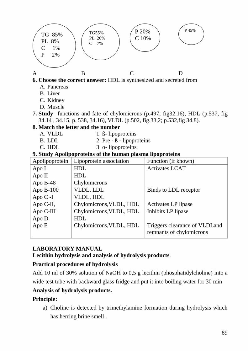
89
A B C D 6. Choose the correct answer: HDL is synthesized and secreted from
A. Pancreas B. Liver C. Kidney D. Muscle
7. Study functions and fate of chylomicrons (p.497, fig32.16), HDL (p.537, fig 34.14 , 34.15, p. 538, 34.16), VLDL (p.502, fig.33,2; p.532,fig 34.8).
8. Match the letter and the number A. VLDL 1. ß- lipoproteins B. LDL 2. Pre - ß - lipoproteins C. HDL 3. �- lipoproteins
9. Study Apolipoproteins of the human plasma lipoproteins Apolipoprotein Lipoprotein association Function (if known) Apo I HDL Activates LCAT Apo II HDL Apo B-48 Chylomicrons Apo B-100 VLDL, LDL Binds to LDL receptor Apo C -I VLDL, HDL Apo C-II, Chylomicrons,VLDL, HDL Activates LP lipase Apo C-III Chylomicrons,VLDL, HDL Inhibits LP lipase Apo D HDL Apo E Chylomicrons,VLDL, HDL Triggers clearance of VLDLand
remnants of chylomicrons
LABORATORY MANUAL Lecithin hydrolysis and analysis of hydrolysis products.
Practical procedures of hydrolysis
Add 10 ml of 30% solution of NaOH to 0,5 g lecithin (phosphatidylcholine) into a
wide test tube with backward glass fridge and put it into boiling water for 30 min
Analysis of hydrolysis products.
Principle:
a) Choline is detected by trimethylamine formation during hydrolysis which
has herring brine smell .
TG55% PL 20% C 7%
TG 85% PL 8% C 1% P 2%
P 20% C 10%
P 45%

90
b) Phosphoric acid finds out reaction with ammonium molybdate solution
c) Unsaturated fatty acids find out reaction with iodine solution
d) Glycerol finds out reaction with Cu(OH)2 solution (test tube 3 –sample, test
tube 4-control). Compare solutions colour in sample and control test tubes.
Practical procedures: Prepare 4 test-tubes as shown in the table below:
Pipette test-tube 1 test-tube 2 test-tube 3 test-tube 4 Hydrolysate 0,5 ml 0,5 ml 0,5 ml - Ammonium molybdate solution
1 ml Boil it
- - -
Iodine solution
- 10 drops - -
Solution of CuSO4
- - 0,5 ml 0,5 ml
30% N� � solution
- - - 0,5 ml
If the reaction is positive we can see:
- yellow precipitate of ammonium phosphomolybdate in the test tube 1,
- discolouration of Iodine solution in the test tube 2,
- dark blue colour of copper glycerate in the test tube 3 against
- clear blue colour in the test tube 4 (control).
Write down the scheme of lecithin hydrolysis.
Write down the results and your conclusion
Qualitative reaction for cholesterol (Liebermann-Burchard test).
The nervous tissue is especially rich in cholesterol,where the cholesterol concentration is 20 to30 g/kg; the highest concentration (40-55g/kg) of cholesterol is found in the white substance of brain and spinal marrow. Principle : The method is based on the dehydration of cholesterol followed by a coupling of two dehydrated cholesterol molecules to yield bicholestadiene. Bicholestadiene, in presence of acetic anhydride and sulphonic acid, gives sulphonated derivatives coloured green. Practical procedures: Prepare 2 test-tubes as shown in the table below:
Pipette Test tube 1 Test tube 2 Chloroformic extract of brain
1 ml -
Solution of cholesterol in chloroform
- 1 ml
91
Acetic anhydride 10 drops 10 drops H2SO4 concent . 2 drops 2 drops
Mix well. Red rose colour is formed which quickly changes through blue to green. Write down the results and your conclusion. Home work: Study lesson 14 1. Study Fatty acids as fuels (p.358-359,,fig23.1,23.2) 2. Study Activation of fatty acids and their transport into mitochondria (p.359-
360, fig 23.4,.23.1, 23.3) 3. Study Oxidation of fatty acids (p.49, 357,361-364, fig. 23.5, 23.6, 23.7, 23.8,
23.9, 23.10). 4. Remember Calculate the amount of ATP produced when one molecule of
fatty acids is oxidizid to CO2 and H 2 O (p.464-365, 23.1q-a) 5. Study the regulation of fatty acid oxidation (p.370) 6. Study Oxidation of glycerol and calculate the amount of ATP produced in it
(lecture) 7. Study Degradation of triacylglycerol and its energetic yeild (p.471, lecture). 8. Study The Synthesis of fatty acids, fatty acid synthase complex (501-
509,fig.33.6, 33.7,33.8,33.9, 33.10, 33.11, 33.12, 33.13, 33.14.,33.15, 33.16,33.17) and their regulation (p.561)
9. Study The Synthesis of triacylglycerols (p.510-511,fig33.19) 10. Study Metabolism of glycerophospholipids and sphingolipids. Synthesis of
phospholipids containing glycerol (p.514-521, fig 33.28 –33.34). 11. Study Mechanisms that affect lipolysis in adipose tissue (p.564). 12. Study The Assay of total lipid level in serum. Hyperlipidemia.
92
LESSON 14 LIPID METABOLISM I Main questions: • Fatty acids catabolism by ß-oxidation :
o Activation of fatty acid, o Formation of fatty acylcarnitine and it transport into mitochondria, o Steps of ß- oxidation, o Oxidation of odd chain fatty acids, o Oxidation of unsaturated fatty acids, o The energy yield from ß-oxidation of saturated and unsaturated fatty acids, o Regulation of ß-oxidation.
• Oxidation of glycerol and its energy yield. • The energy yield from triacylglycerol degradation. • Acetyl-CoA metabolism in liver. • Fatty acid synthesis : Steps of process,
o Fatty acid synthase complex, o Regulation of fatty acids synthesis.
• Synthesis and mobilization of adipose and liver triacylglycerols. Hormonal regulation
• Metabolism of glycerophospholipids and sphingolipids • Estimation of total blood serum lipids by sulfovanilline reagent FATTY ACIDS CATABOLISM BY ß-OXIDATION 1. Study ß- oxidation of fatty acids (p.49, 357;Activation, transport into mitochondria, reactions, substrates, products, enzymes, 360-364) 2. Note: Peripheral tissues receive fatty acids from blood by three pathways: a) free fatty acids from adipose tissue are transported by serum blood albumin, b) Lipoprotein lipase releases free fatty acids from chylomicrons, c) Lipoprotein lipase releases free fatty acids from VLDL 3. Origin of fatty acids as substrates for ß- oxidation:
A. Fatty acids are derived from the diet B. Fatty acids are synthesized in the liver from glucose C. Fatty acids are products of mobilization of adipose triacylglycerols D. All of the above
4. Match the letter and the number: Fatty acids may be activated in
A. Shot chain fatty acids 1.Endoplasmatic reticulum B. Medium chain fatty acids 2. Cytosol or mitochondria C. Long chain fatty acids 3. Mitochondrial matrix
5. Choose the correct answer: The enzymes of ß-oxidation are located in:
A. Cytosol B.Mitochondrial inner membrane C.Mitochondrial outer membrane D.Mitochondrial matrix

93
6. Match the letter and the number:. A. Fatty acyl CoA synthetase 1.Transport into mitochondria B. Carnitineacyltransferase 2.Hydration step C. Acyl CoAdehydrogenase 3.Activation D. Enoyl CoA hydrotase 4.Dehydrogenation step E. ß-ketothiolase 5.Cleavage step
7. ß- oxidation occurs in all tissues EXCEPT: A. Liver B. Brain C. Kidneys D. Erythrocyte E. Muscle
8. ß- oxidation is regulated by: A. The mechanism that control the reoxidation of FADH2 B. The mechanism that control the reoxidation of NADH C. Both D. None of the above
9. Read about the energy yield from ß-oxidation of saturated (p.362-363) and unsaturated fatty acids (p.364, q.23.1; p.365, A.23.1). 10. Memorize: energetic of fatty acid oxidation: Fatty acid (n carbons) undergoes n/2-1 times ß-oxidation and produces n/2 molecules of acetyl-CoA. Each time ß-oxidation produces 5ATP (from NADH and FADH2 in ETC). A) Total number of ATP formed by ß-oxidation = ( n/2-1)∗∗∗∗5 B) Total number of ATP formed on oxidation of acetyl-CoA through TCA cycle =
(n/2) ∗∗∗∗12 C) 2 ATP utilized for initial activation of fatty acid
Net total yield = [[[[ ( n/2-1)∗∗∗∗5]]]] +[[[[ (n/2) ∗∗∗∗12]]]] – 2 11.Write down the reactions of ß- oxidation of stearic acid; calculate the amount of ATP produced when one molecule of stearic acid is oxidized to CO2 and H2O. 12. State how many molecules of ATP are formed during the process of ß-oxidation of oleic acid.
OXIDATION OF GLYCEROL AND ITS ENERGY YIELD 1. Study the convention of glycerol to dihydroxyaceton phosphate (p.428, fig.27.9). 2. Write down the scheme of glycerol oxidation: convention of glycerol to dihydroxyaceton phosphate, reactions of aerobic glycolysis, convention of pyruvate to acetyl CoA, reactions of TCA cycle.
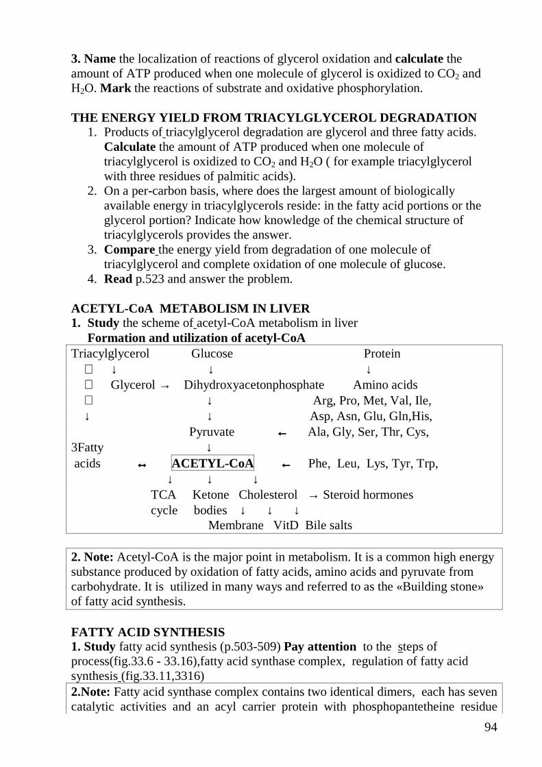
94
3. Name the localization of reactions of glycerol oxidation and calculate the amount of ATP produced when one molecule of glycerol is oxidized to CO2 and H2O. Mark the reactions of substrate and oxidative phosphorylation. THE ENERGY YIELD FROM TRIACYLGLYCEROL DEGRADATION
1. Products of triacylglycerol degradation are glycerol and three fatty acids. Calculate the amount of ATP produced when one molecule of triacylglycerol is oxidized to CO2 and H2O ( for example triacylglycerol with three residues of palmitic acids).
2. On a per-carbon basis, where does the largest amount of biologically available energy in triacylglycerols reside: in the fatty acid portions or the glycerol portion? Indicate how knowledge of the chemical structure of triacylglycerols provides the answer.
3. Compare the energy yield from degradation of one molecule of triacylglycerol and complete oxidation of one molecule of glucose.
4. Read p.523 and answer the problem. ACETYL-CoA METABOLISM IN LIVER 1. Study the scheme of acetyl-CoA metabolism in liver
Formation and utilization of acetyl-CoA Triacylglycerol Glucose Protein ↓ ↓ ↓ Glycerol → Dihydroxyacetonphosphate Amino acids ↓ Arg, Pro, Met, Val, Ile, ↓ ↓ Asp, Asn, Glu, Gln,His, Pyruvate ←←←← Ala, Gly, Ser, Thr, Cys, 3Fatty ↓ acids ↔↔↔↔ ACETYL-CoA ←←←← Phe, Leu, Lys, Tyr, Trp, ↓ ↓ ↓ TCA Ketone Cholesterol → Steroid hormones cycle bodies ↓ ↓ ↓ Membrane VitD Bile salts 2. Note: Acetyl-CoA is the major point in metabolism. It is a common high energy substance produced by oxidation of fatty acids, amino acids and pyruvate from carbohydrate. It is utilized in many ways and referred to as the «Building stone» of fatty acid synthesis. FATTY ACID SYNTHESIS 1. Study fatty acid synthesis (p.503-509) Pay attention to the steps of process(fig.33.6 - 33.16),fatty acid synthase complex, regulation of fatty acid synthesis (fig.33.11,3316) 2.Note: Fatty acid synthase complex contains two identical dimers, each has seven catalytic activities and an acyl carrier protein with phosphopantetheine residue

95
into eukaryotes. Reactions of fatty acid synthesis are catalyzed by different enzymes in prokaryotes (E.coli) . Where is the process of fatty acid synthesis slowly? 3. Specify the correct order of stages of fatty acid extramitochondrial synthesis
A. Transfer of malonyl group to phosphopantetheinyl residue B. Convention of acetyl-CoA to malonyl CoA C. Transfer of fatty acyl group to phosphopantetheinyl residue D. Reduction of ß-ketoacyl group E. Condensation of malonyl and fatty acid groups F. Dehydration G. Deacylation H. Reduction of double bound.
4. Match the letter in No 3 and the number: 1. Acetyl-CoA carboxylase 2. Acetyl transferase 3. Malonyl transferase 4. ß-ketoacyl ACP synthetase 5. ß-ketoacyl ACP reductase 6. ß-hydroxyacyl dehydratase 7. Unsaturated ACP reductase 8. Deacylase
5. Chose the correct answers : Fatty acid synthesis occurs in tissues:
A. Liver B. Kidneys C. Muscle D. Adipose tissue E. Mammary gland
6. Compare ß- oxidation and fatty acid extramitochondrial synthesis and fill in the table: Processes ß- oxidation fatty acid synthesis Localization in cell Substrate carrier through mitochondrial membrane
Coenzymes of oxido –reduction enzymes
Regulatory enzymes Regulatory factors: Activator Inhibitor
Product of process

96
7.The rate-limiting enzyme of fatty acid synthesis is: A. Malonyl transferase B. ß-ketoacyl ACP synthetase C. ß-ketoacyl ACP reductase D. Acetyl-CoA carboxylase.
8. Allosteric activator of acetyl-CoA carboxylase is A. Biotin B. Citrate C. ATP D. All of the above
9. After a person has consumed large amounts of sucrose; the glucose and fructose that exceed caloric requirements are transformed to fatty acid for triacylglycerol synthesis. This fatty acid consumes acetyl-CoA, ATP, and NADPH. How are these substances produced from glucose? 10. In the condensation reaction catalyzed by ß-ketoacyl ACP synthetase, a four-carbon unit is synthesized by combination of a two-carbon unit and a three-carbon unit, with the release of CO2. What is the thermodynamic predominant of this process over one that simply combines two two- carbon units? SYNTHESIS AND MOBILIZATION OF ADIPOSE AND LIVER TRIACYLGLYCEROLS AND ITS REGULATION BY HORMONES. 1. 1.Study Synthesis (p.510,-52,fig.33.20) and mobilization of adipose and liver
triacylglycerols . 2. Study regulation mobilization of adipose triacylglycerol by hormones (p.512,
514, fig.33.26), the scheme below. Memorize: Influence of hormones on adipose tissue
Insulin: 1. It inhibits the release of free fatty acids from adipose tissue, enhances lipogenesis and the synthesis of acylglycerol and increases the oxidation of glucose to CO 2 via HMP-shunt. 2. It inhibits hormone-sensitive lipase activity and reduces the release of free fatty acids as well as glycerol. 3. Insulin, nicotinic acid and prostaglandin E1 inhibit the synthesis of cAMP depressing adenylate cyclase or stimulating phosphodiesterase. Prostaglandins E1 in low concentration causes the release of catecholamines resulting in the increase of free fatty acid mobilization. ACTH (adrenocorticotropic hormone), melanocyte-stimulating hormone, thyrotropic hormone, growth hormone, vasopressin, epinephrine, norepinephrine and glucagon : 1. These hormones accelerate the release of free fatty acids from adipose tissue by increasing the rate of lipolysis of the triacylglycerol stores. 2.Many of them activate hormone-sensitive lipase and increase glucose utilization. 3.Glucocorticoids and thyroid hormones do not increase lipolysis but the presence of these hormones in the lipolytic processes is essential. Thyroid hormone inhibits phosphodiesterase activity. The lipolytic effect of growth hormone in presence of

97
glucocorticoids is slow. 4.These hormones stimulate adenylate cyclase for cAMP formation from ATP and this cAMP stimulates the protein kinase that converts inactive hormone-sensitive triacylglycerol lipase into active lipase. In addition to the hormone-sensitive triacylglycerol lipase, adipose tissue contains diasylgly-cerol and monoacyl-glycerol lipases which are more active than hormone-sensitive triacylglycerol lipase and catalyze the rate limiting step in lipolysis. Lipolysis is controlled by the amount of cAMP present in tissues. cAMP is degraded to 5'-AMP by the enzyme cyclic 3', 5'-nucleotide phosphodiesterase. This enzyme is inhibited by methyl xanthines such as caffeine and theopbylline. Therefore, drinking coffee or caffeine administration causes high elevation of plasma free fatty acids in humans. The sympathetic nervous system, through liberation of norepinephrine, plays a central role in the mobilization of free fatty acids by exerting a tonic influence. 3. The increased levels of plasma free acids is the result of mobilization of fat
from A. Muscle B. Adipose tissue C. Kidney D. Intestine
4.Compose the scheme of process in which activity increases in adipose tissue after intensive manual labour. 1. Protein kinase inactive 2. Protein kinase active 3. TAG lipase dephosphorylated 4. TAG lipase phosphorylated 5. Adenylate cyclase inactive

98
6. Adenylate cyclase active 7. CAMP 8. ATP 9. Epinephrine METABOLISM OF GLYCEROPHOSPHOLIPIDS AND SPHINGOLIPID S 1. Study Metabolism of glycerophospholipids and sphingolipids ( p.514-520, fig 33.28, 33.29, 33.30, 33.31, 33.34, 33.33). 2. Compare glycerophospholipids and triacylglycerols synthesis and mark the identical reactions. 3. In the biosynthesis of complex lipids, components are assembled by transfer of the appropriate group from an activated donor. For example, the activated donor of acetyl group is acetyl-CoA. For each of the following groups, give the form of the activated donor:
A. Phosphate B. D-glucosyl C. Phosphoethanolamine D. Fatty acyl E. Methyl F. The two-carbon group in fatty acid biosynthesis
4. Match the letter in and the number: A. Triacylglycerols synthesis 1.Phosphatidic acid B. Glycerophospholipids synthesis 2. Diacylglycerol C. Both 3. Phosphatidyl ethanolamine
4. CTP 5. CDP ethanolamine 6. S-adenosylmethionine 7. Monoacylglycerol
5. Compose the scheme of phosphatidyl choline synthesis process. Use following components for it:
A. Ethanolamine B. CDP ethanolamine C. Phosphoethanolamine D. Phosphatidylethanolamine E. Phosphatidyl choline
LABORATORY MANUAL Estimation of total lipids by sulfovanilline reagent Total blood serum lipids contain triacylglycerols, phospholipids, cholesterol and its esters, glycolipids and free fatty acids. Principle. Unsaturated lipids and fatty acids, phosphatides and cholesterol react, after their hydrolysis with sulfuric acid, with phosphovanillin reagent to form a red coloration. Reagents: 1.Standart solution (Total lipids Cst =8g/l) 2. Vanilline reagent solution (vanilline and H3PO4)

99
E. H2SO4 concent Procedure Prepare 3 test-tubes as shown in the table below Pipette (ml) test-tube 1
Sample test-tube 2 Standard
test-tube 3 Control solution
Serum blood 0,02 - - Standart solution - 0,02 - H2SO4 concent 1,50 1,50 1,50 Mix. Put test-tubes for 15 min into boiling water bath. Cool using cold tap water and into another test-tubes pipette : Hydrolysate 0,1 0,1 0,1 Reagent �2 1,5 1,5 1,5 The content of the test-tubes is stirred thoroughly and wait for 50 min at room temperature to let the colour develop. After 50 min read the absorbances of sample (A1 ) and standard ( A2 ) against control solution . Wavelenght (500-530) nm Cuvette 1 cm Calculation Total lipids[g/l] == Cst∗ A1/ A2
Reference values : 4-8 g/l Write down the results and your conclusion Notes For the determination of total lipids use the specimen taken off after fasting for at least 14 hours. Home work: Study lesson 15 1.Study Metabolism of ketone bodies (p.366-370, fig23.13, 23.15, 23.16, 23.17, 23.18, 23.19). 2.Study Metabolism of cholesterol (p.529-535), hypercholesterolemia (read clinical and biochemical comments p.540-542, problem1 p.543) 3.Study Dislipoproteinemia and atherosclerosis (p.534-535) 4.Study Synthesis of bile salts (p.538-540, fig.34.17, 34.18, 34.19, 34.20,34.21) and problem2 p.543. 5.Study Metabolism of eicosaoids (p.545-556) 6.Estimation of total blood serum cholesterol by enzimatic method. Qualitative tests for ketone bodies in urine.

100
LESSON 15 LIPID METABOLISM II Main questions: • Metabolism of ketone bodies :
- Synthesis of ketone bodies - Oxidation of ketone bodies - Regulation of ketone bodies utilization - Ketonemia, ketonuria, ketosis
• Metabolism of cholesterol : - Steps of synthesis - Transport of cholesterol by blood lipoproteins - Hypercholesterolemia, dislipoproteinemia, atherosclerosis
• Synthesis of bile salts • Metabolism of eicosanoids • Estimation of total blood serum cholesterol by enzymatic method. • Qualitative tests for ketone bodies in urine. METABOLISM OF KETONE BODIES 1. Study Metabolism of ketone bodies (p.366-370, fig 23.15, 23.16, 23.17, 23.18,
23.19). 2. Note: In liver ketone bodies are formed by two ways: HMG-CoA pathway ( main) and acetoacetyl CoA pathway (minor). Acetoacetyl CoA pathway is simple deacylation catalyzed by enzyme acetoacetyl-CoA deacylase how is shown below:
Acetyl- CoA+ Acetyl- CoA ↓ thiolase Acetoacetyl CoA + CoA-SH ↓ deacylase Acetoacetate + CoA-SH Activity of deacylase is low and this pathway is not significant for us. 3. All of the following statements about ketone bodies are true EXCEPT:
A. Ketone bodies are formed in liver but utilized in the extrahepatic tissue. B. Ketone bodies are formed and utilized in liver. C. Acetoacetate is reduced to β-hydroxybutyrate. D. Acetoacetate is decarboxylated to acetone. E. Two reactions occur in extrahepatic tissue for the activation of acetoacetate to
acetoacetyl CoA 4. Name the biological role of ketone bodies in organism . 5. State how many molecules of ATP are formed during the process of oxidation
of acetoacetate in the extrahepatic tissue (use both ways of activation).

101
6. Which of the following compounds are precursors for ketone bodies? A. Amino acids ( Ala, Asp, Glu, His) B. Glucose C. Fatty acids 7. Match the letter and the number: A. Synthesis of ketone bodies 1. D-β-hydroxybutyrate dehydrogenase B. Oxidation of ketone bodies 2. HMG-CoA synthetase C. Both 3. Enoyl CoA hydratase
D. None of the above 4. Succinyl CoA: acetoacetate CoA
tran-sferase. 8. Give an explanation of the overproduction of ketone bodies in uncontrolled
diabetes or severe starvation. 9. Repeat the energy yield from degradation of one molecule of triacylglycerol
and complete oxidation of one molecule of glucose (lesson 20) and compare amount of acetyl CoA molecules forming in these prosesses .
10. Acetyl CoA is utilized by forming of ketone bodies in diabetes mellitus or severe starvation. Why cannot it be utilize by other ways?
11. Match the letter and the number: A. TCA cycle 1. Deficiency of NADPH B. Synthesis of fatty acids 2. Deficiency of oxaloacetate C. Synthesis of cholesterol
12. Memorize: The overproduction of ketone bodies in uncontrolled diabetes or severe starvation can lead to ketosis. Affects of ketosis: 1. Both acetoacetate and β-hydroxybutyrate are moderately strong acids. They
neutralize bicarbonates resulting in depletion of alkali of body and produce metabolic acidosis. In case of severe ketosis, death may be the result of acidosis
2. The excretion of ketone bodies in the urine involves the loss of Na+ in particular leading to total electrolyte and Na+ deficiency.
3. The severe diabetic patient excrets large quantities of both ketone bodies and glucose in the urine with a large quantities of water developing dehydration. In diabetic acidosis, there is severe alteration in cation-anion balance in plasma.
METABOLISM OF CHOLESTEROL 1. Study Metabolism of cholesterol (p.529-535). 2. Choose the correct answers:
Biological role of cholesterol : A. It is structural component of membrane B. It is an energy source for cells. C. It is a precursor for bile salts D. It is a precursor for steroid hormones

102
E. It is assists in formation of vitamin D3 in skin F. All of the above
3. Specify the correct order of stages of cholesterol synthesis A. Condensation of six isoprene units to give C30 terpene (squalene) B. Cyclization of squalene to lanosterol C. Formation of mevalonate
4. Match the letter in No 3 and the number: 1. HMG-CoA reductase 2. Mevalonate kinase 3. Cyclase
5. Choose the correct answers: A. HMG-CoA reductase is inactivated by B. Dephosphorylation C. Phosphorylation D. Cholesterol E. Bile salts F. Insulin G. Glucagon
6. The enzymes of cholesterol synthesis are located in: A. The cytosol B. Mitochondrial inner membrane C. Mitochondrial outer membrane D. Mitochondrial matrix
7. Study Transport of cholesterol by blood lipoproteins (p.531-538) , 8. The highest contents of cholesterol is found in:
A. VLDL B. LDL C. HDL D. Chylomicrons
9. Match the letter and the number A. VLDL is converted to IDL 1. Lipoprotein lipase B. Recognize apoE, apoB-100 2. Lecithin:cholesterol acyltransferase C. Esterify cholesterol 3. ”Scavenger” receptor D. Recognize oxidized LDL 4. LDL receptor 10. Cholesterol synthesis “for export” occurs in tissues:
A. Muscle B. Intestine C. Nervous tissue D. Liver E. Adipose tissue
11. Compare the formation, transport and metabolism of exogenes and endogenes cholesterol :
A. Exogenes cholesterol 1. Joins in lipoproteins forming in intestinal
epithelial cells 2. Joins in VLDL forming in liver

103
B. Endogenes cholesterol 3. Is secreted into the lymph in composition
of chylomicrons 4. Is secreted into the blood directly 12. Specify the correct order of stages of cholesterol transport:
A. From intestine to liver 1. Formation of LDL 2. Transport by blood 3. Packing into VLDL(organ in which it is
main formation ) 4. Lipoprotein lipase action 5. Cholesterol and its ester synthesis B. From liver to peripheral tissues
6. Lipoproteins are taken up by receptors of tissue
7. Cholesterol enters different tissues 8. Hydrolysis of dietary cholesterol esters 9. Formation of mixed micelles 10. Absorption 11. Remnants are taken up by liver 12. Formation of chylomicron remnants 13. Formation of chylomicrons 13. Compare the properties of lipases. Match the letter and the number:
1. Class Hydrolases A. Pancreatic lipase 2. Class Lyases B.TG lipase 3. Digests dietary fats C.LPL lipase 4. Digests TG into chylomicrons,VLDL D. All of the above 5. It is activated by glucagon,
epinephrine E. None of the above
6. It is activated by bile salts 7. It is located in capillary walls 8. It is located in adipose tissue
14. Study Dislipoproteinemia and atherosclerosis (p.534-536,fig34.13), hypercholesterolemia (read clinical and biochemical comments p.540-542, problem 1 p.543)
SYNTHESIS OF BILE SALTS 1. Study Synthesis of bile salts (p.538-540, fig.34.17, 34.18, 34.19, 34.20,34.21)
and read problem 2 p.543. 2. Repeat bile salts functions (lesson 17). 3. Answer the question. How does conjugation of bile salts with taurine or
glycine change their amphiphilic properties? 4. Memorize: Clinical importance of bile salts 1) Bile salts in the blood are greatly increased in clinical obstructive jaundice.

104
2) After prolonged obstruction, the concentration of bile salts in blood may diminish due to diminished synthesis of these substances as a result of progressive hepatocellular damage.
3) In the absence of bile salts, gall-stones are formed. 5. Remember:
The enterohepatic circulation (see p.494, fig.32.10) A portion of the bile acids in the intestine undergoes changes by the activity of the intestinal bacteria. The deconjugation and 7�-hydroxylation produce the secondary bile acids, deoxycholic acid from cholic acid and lithocholic acid from chenodeoxycholic acid. The conjugated and unconjugated bile salts are absorbed almost in the ileum. As fecal bile acids are present as the products of bacterial metabolism, therefore, it is assumed that metabolism within the intestinal lumen with reabsorption by passive diffusion is a component of the enterohepatic circulation. This mechanism helps to return 90% of the bile acids secreted into the intestine by the liver each day. But lithocholic acid is not reabsorbed to any significant extent due to its insolubility. 500 mg of bile salts per day are not absorbed and is eliminated in the feces. The enterohepatic circulation of bile salts is so efficient that a small amount of bile acids is cycled through the intestine 6-10 times a day with the loss of a small amount in the feces. 6. Remember:
GALL-STONES In the gall-bladder, cholesterol is solubilized by being held in micelles together with conjugated bile salts and phospholipids. The solubility depends on the ratio of cholesterol to bile salts plus phospholipids. The secretion of phospholipids into the bile depends on the availability of bile salts. If bile salt content is decreased, the phospholipid content is also diminished and hence, the solubility of cholesterol is decreased causing crystallization. These crystals grow to form stones. Gall-stones are formed due to defects in the enterohepatic circulation and with the diseases of the terminal ileum as well as in patients with cirrhosis. In these cases, there is reduction in the bile salt pool. Infection of bile causes the deconmgation of bile acids with a decrease in their solubility. This also results in the production of phospholipase which converts lecithin to lysolecithin. This decreases the stability of the micelles holding cholesterol in solution. Infection can give rise to calcium-bilirubinate stones which were frequent in Japan. Chenodeoxycholic acid decreases the rate of secretion of cholesterol into the bile. Bile then becomes unsaturated with respect to cholesterol and thus the cholesterol stone can be redissolved. Unfortunately bacterial action in the intestine converts chenodeoxy acids to lithocholic acid which is very hepatotoxic in Rhesus monkeys producing proliferation of bile ducts. METABOLISM OF EICOSANOIDS 1. Study Metabolism of eicosanoids (p.545-556, fig.35.1-35.13).

105
2. Pay attention to pathways and steps of synthesis, structure, nomenclature, mechanism of action, biological role, inhibitors of synthesis.
3. Eicosanoids are produced from fatty acids released from membrane phospholipids by: A. Phospholipase A1 B. Phospholipase A2 C. Phospholipase C D. Phospholipase D
4. Pathways for eicosanoid synthesis. Match the letter and the number: A. Cyclooxygenase pathway 1. Leikotrienes B. Lipooxygenase pathway 2. Thromboxanes C. Cytochrome P450 pathway 3. Prostaglandins D. Both B and C 4. Prostacyclin
5. HPETE 6. Lipoxins
5. Read clinical and biochemical comments. Discuss mechanism of action of the eicosanoids in the case. p.555 and problems p.556. What kind of eicosanoids act on thermoregulatory centers of the brain?.
6. All of the following statements about eicosanoids are true EXCEPT: �.They are derived from polyunsaturated fatty acids containing C20 atoms B.Many of them have very short half-lives in the range of minutes or less C.Many of them have half-lives in the range of minutes or houres. D.They have a variety of extremely potent hormonelike actions. E.They are not stored in cells F.They have different target cells and biological actions. G.They interact with a specific receptor on the plasma membrane of a target cell.
LABORATORY MANUAL Estimation of total blood serum cholesterol by enzymatic method Cholesterol is transported in the blood as a component of VLDL and LDL, 60-70% of its in a form of cholesterol esters and 30-40% - free cholesterol. Total blood serum cholesterol contains cholesterol and its esters. Cholesterol level increases during the life. Reference values: for baby 1 year ago 50 ± 10mg/dl
for adult 200 ± 40mg/dl or 5,2 ± 1,3mmol/l Ideal cholesterol level in blood serum ∠ 5,2 mmol/l
Principle. Cholesterol is product of cholesterol esters hydrolysis by cholesterol esterase. It is oxidized by air oxygen due to cholesterol oxidase. Hydrogen peroxide is product of this reaction resulting in chromogeneus substrate oxidation catalyzed by peroxidase with pink coloured compounds appearance. The intensity of colouring is proportional to the cholesterol concentration in plasma.
106
1) Cholesterol esters + H2O 2) Cholesterol + O2 ↓↓↓↓ Cholesterol esterase ↓↓↓↓ cholesterol oxidase Cholesterol + fatty acids 4-cholestenone + H2O2 3) 2 H2 O2 + chromogeneus substrate ↓↓↓↓ peroxidase coloured compound+ 2 H2O Reagents. No 1 Liofilisate of enzymes (cholesterol esterase, oxidase, peroxidase), chromogeneus substrate), Phosphate buffer No 2 Standard cholesterol solution –5,17 mmol/l (200mg/100ml) Procedure: Pipette (ml) Sample Standard Control Blood serum 0,1 - - H2 O - - 0,1 Reagent 1 2,0 2,0 2,0 Standard solution - 0,1 -
The content of the test-tubes is stirred thoroughly and wait 5 min at room temperature to let the colour develop. After 5 min read the absorbances of sample (A1) and standard (A2) against control solution. Wavelenght 490 nm. Cuvette 0,5 cm
Calculation : Total cholesterol (mmol/l) == 5,17∗ A1/ A2
or Total cholesterol (mg/100ml) == 200∗ A1/ A2
Write down the results and your conclusion. Remember: Hypercholesterolemia has been observed in uncontrolled diabetes mellitus, impairment of liver, obstructive jaundice, glomeruliunephritis, hypothyroidism and nephrosis. Hypocholesterolemia has been found in anemia, hyperthyroidism, hepatic diseases, infection, carcinoma, and acute pancreatites. Determination of ketone bodies in urine. Compositionally, the urine includes water, organic and inorganic salts – in total, about 150 components. The ketone bodies in the normal urine are not detected by these tests. 1. Iodoform test for acetone (Liben’s test). Procedure: To 5 drops of urine add 1 drop of 10% NaOH solution. Add 3drops of iodine solution (10%). The iodine solution should be added till no decolouration occurs. 2. Legal’s test for acetone and acetoacetic acid.

107
The method is based on the tendency of acetone and acetoacetic acid to form, when allowed to react with sodium nitroprusside in an alkaline medium, complexes coloured orange-red which turn to cherry-red products in an acidified medium. Procedure: To 5 drops of urine add 1 drop 10% sodium nitroprusside solution and add 2 drops of 10% NaOH solution. The orange-red colour appears. 3. Gerhardt’s test for urinary acetoacetic acid . The method employs the tendency of iron (III) to react with the enolic form of acetoacetic acid to yield a red-violet complex. Procedure: To 5 drops of urine add 1 drop of 1% FeCl3 solution. Note a red-violet coloration to develop. 4.Rapid test for ketone bodies in urine. The method is based on the ability of ketone bodies to enter into reaction with sodium nitroprusside and to change the colour of test tablet into violet. Procedure: Place the test tablet from the diagnostic kit on filter paper and apply 2 drops of urine on it. Wait two minutes Compare the colour developed in the test tablet with the reference colour scale appied to the diagnostic kit. Write down the results and your conclusion. Home work repeat the previous themes, Prepare for colloquim

108
LESSON 16. COLLOQUIM: BIOENERGETICS OF THE CELL. COMMON CATABOLIC PATHWAY. CARBOHYDRATE AND LIPID METABOLISM.
Main questions: 1. Endergonic and exergonic reactions in the living cells. High energy
phosphate bonds connections: definition, examples. 2. Biological oxidation. Biological functions of biological oxidation in a cell.
Dehydrogenation of substrate and reduction of oxygen as an energy source for synthesis ��P.
3. Kinds of phosphorylation as reactions of generation ��P: oxidative, substrate phosphorylation.
4. Oxidative phosphorylation : essence of the process, the generalized scheme, substrates, factor �/ . Mitochondrial structure and localization of components of oxidative phosphorylation system in it.
5. Respiratory chain is a key component of the mitochondrial oxidative phosphorylation system. The structural organization of the respiratory chain. The mitochondrial electron transport chain as a part of respiratory system of all organisms.
6. Dehydrogenases dependent on nicotinamide coenzemes. Dehydrogenases dependent on riboflavin prosthetic groups. NADH dehydrogenase. Cytochrome C reductase. Cytochrome C oxidase. Features of structure, functions. Coenzemes components of the mitochondrial electron transport chain.
7. Transmembrane electrochemical potential as the intermediate form of energy at oxidative phosphorylation. � +-��P-synthetase: biological role, localization, structure, mechanism of ��P synthesis.
8. Regulation of functioning of oxidative phosphorylation system. The respiratory control. Infringements of energetic metabolism. Hypoenergetic conditions as result of hypoxia, hypovitaminosis and other reasons.
9. Uncoupling of ATP synthesis from electron transport. Brown adipose tissue and thermogenesis.
10. Catabolism of carbohydrates, fats, amino acids and proteins. Concept about specific and common pathways of catabolism. The common pathway of catabolism: oxidation pyruvate and acetyl – C��. Biological value, localization in cell.
11. Specific ways of food substances catabolism. Pyruvate formation from carbohydrates and the majority of amino acids. Acetyl - C�� formation from fatty acids and some amino acids.
12. The oxidative decarboxylation of pyruvate: biological value, sequence of reactions. Mechanisms of the PDH complex regulation.
13. The pyruvate dehydrogenase complex of animals. Structure, coenzemes of active centers.

109
14. Tricarboxylic acid cycle: biological role, sequence of reactions, characteristics of enzymes.
15. Key reactions of the tricarboxylic acid cycle. Mechanisms of the TCA cycle regulation.
16. Anaplerotic reactions of the TCA cycle (equation of reactions, enzymes, biological role). Anabolic value of the TCA cycle.
17. TCA cycle as amphibolic pathway. Relationship between TCA cycle and ETC chain. Anabolic functions of the TCA cycle.
18. Formation of reactive oxygen species during biological oxidation in mitochondria. Physiological and toxic effects of reactive oxygen species.
19. Carbohydrates: definition. Classification of carbohydrates, examples. 20. Carbohydrates distribution to nature. Their biological role. 21. Monosaccharides. Structure and biological role. 22. Monosaccharide derivatives – phospho sugars, amino sugars, uronic acids,
neuraminic and sialic acids, UDF-glucose. 23. Disaccharides. Structure and biological role. 24. Polysaccharides. Starch. Glycogen -structure and biological role. 25. Oligosaccharides. Their role in receptors formation. 26. Digestion of carbohydrates. 27. Absorption of carbohydrates. Simple diffusion, transport facility, active
transport as a penetration mechanisms of glucose in cells 28. Liver glycogen formative function. Glycogen synthesis. 29. Liver glycogen formative function and mobilization of glucose. 30. Regulation of synthesis and glycogen degradation in liver. Role of hormones
and proteinkinases in regulation. 31. Anaerobic glycolysis: scheme, energy yield. Regulation of glycolysis. 32. Pyruvate conversion in muscles under anaerobic conditions. 33. Aerobic glucose catabolism: scheme, energy yield. 34. Regulation of aerobic glucose catabolism. 35. Gluconeogenesis. Alternative ways of irreversible reactions of glycolysis.
Biological role. Hormonal regulation. 36. Lactate formation.Cori cycle. Lactate dehydrogenase. 37. Regulation of glycolysis. Pasteur effect. Aerobic glycolysis. 38. Pentose phosphate pathway: scheme. Biological value. 39. Carbohydrate metabolism regulation in organism. Blood glucose
concentration as homeostasis parameter internal environment of organism. 40. Infringements of carbohydrate metabolism. Hypo- and hyperglycemia.
Insulin and carbohydrate metabolism. Diabetes mellitus. 41. Hereditary infringements of monosaccharide and disaccharide. 42. Quantitative assay of blood glucose concentration by glucose oxidase test. 43. Ethanol fermentation. Detection of ethanol fermentation products. 44. Glucose tolerance test. Its significance for clinic. 45. Lipids. Physical properties, features of structure and biological role. 46. Classification of lipids. Features of a structure and properties of different
groups.

110
47. Saturated and unsaturated fatty acids. Features of structure fatty acids of animal origin. Method of designation of the number of carbon atoms, number and position of double bonds. Biological role. Essential and nonessential fatty acids.
48. Triacylglycerols (triglycerides). Simple and combined triglycerides. Physical and chemical properties of fats. Biological role.
49. Phosphoglycerides. Classification of phosphoglycerides. Biological role. 50. Sphingolipids. Structure and biological role. 51. Glycosphingolipids. Structure and biological role. 52. Free and esterified cholesterol. Structure and biological role. 53. Digestion of dietary fats and phosphoglycerides in gastrointestinal tract.
Ensymes. 54. Structure and biological role of bile acids and their taurine-, and
glycoconjugates. Role of bile acids in digestion and absorption of lipids. Steatorrhoea.
55. Triacylglycerol resynthesis in intestinal epithelial cells. Formation of chylomicrons and transport of fats. Role of apolipoprotein in chylomicrons. Lipoprotein lipase. Hyperlipoproteinemias.
56. Plasma lipoproteins. Classification of lipoproteins by density, electrophoretic mobility, their functions. Features of lipid structure and composition of lipoproteins. Mains apolipoproteins. Biological role.
57. Functions of plasma lipoproteins. Sites of formation and metabolism of lipoproteins.
58. Dislipoproteinemia. Classification. Hyperchylomicronemia. Hypercholesterolemia. Hypolipoproteinemia. Clinical value.
59. Catabolism of fatty acids. Activation of fatty acids. Formation of acylcarnitine and it’s transport into mitochondria. Steps of ß-oxidation. Regulation of ß-oxidation.
60. The energy yield from ß-oxidation of saturated and unsaturated fatty acids. The comporison of energy yield from ß-oxidation of fatty acids and glucose.
61. Oxidation of glycerol, the energy yield from oxidation of glycerol and triglycerides.
62. Acetyl-CoA metabolism in liver. Sources of acetyl-CoA and NADPH2 for synthesis of fatty acid.
63. Synthesis of malonyl CoA. Acetyl CoA carboxylase. Features of structure and regulation of enzyme activity.
64. Structure of fatty acids synthase enzyme complex. Localization. Acyl carrier protein (ACP). Role of phosphopantetheine prosthetic group.
65. Reactions catalyzed by fatty acids synthase enzyme complex. Synthesis of butyryl ACP. Formation of palmitic acid. Elongation of fatty acids to C18, C20. Desaturation of fatty acids.
66. Regulation of metabolism of fatty acids (ß- oxidation.and synthesis). 67. Synthesis of triacylglycerol. Activation of the fatty acids. Properties of
triacylglycerol synthesis in the liver and adipose tissue cells. Hormonal control of biosynthesis.
111
68. Lipolysis of triacylglycerol in adipocytes. Triacylglycerol lipase, diacylglycerol lipase monoacylglycerol lipase. Hormonal control of lipolysis in adipocytes.
69. Metabolism of phosphoglycerides. Degradation of phosphoglycerides. The specificity of phospholipases A, A2, C and D. Biological role.
70. Biosynthesis of phosphatidylcholine, phosphatidylserine, phosphatidylethanolamine, phosphatidylinositol and sphingomyelin.
71. Ketone bodies metabolism. Synthesis of ketone bodies. Oxidation of ketone bodies. Ketonemia, ketonuria, ketosis.
72. Metabolism of cholesterol. Sources, utilization and excretion of cholesterol. Level of cholesterol in blood serum. Estimation of total blood serum cholesterol. Clinical value.
73. Biosynthesis of cholesterol. Steps. Regulation. 74. Synthesis of bile salts. Their role for maintaining of homeostasis of
cholesterol in organism. Gall-stones disease. 75. LDL and HDL as transport form of cholesterol in blood. Their role in
metabolism of cholesterol. Atherogenic and antiatherogenic lipoproteins. 76. Hypercholesterolemia as a factor of risk of atherosclerosis. Biochemical basis
of treatment and prophylaxis of hypercholesterolemia. 77. Eicosanoids. Biosynthesis, structure, nomenclature and biological role.
Inhibitores of eicosanoids biosynthesis. Home work: Study lesson 17
1. Study the structure and functions of different cell membranes (p. 129-139). 2. Repeat the formulae of the main phospholipids: phosphatidylcholine,
phosphatidylinositolbisphosphate, sphingomyelin fig. 6.28, 6.30, 6.31, p.61 and fatty acids (p. 58 -59).
3. Study the mechanisms of transport across the membranes (p. 139-143). 4. Study the mechanisms of signal transduction by hormones that bind to
plasma membrane receptors and by hormones that bind to receptors within the cells (p.381-386, p.679 - 687). Memorize the schemes which are represented in fig. 24.13, p.383. 24.14, p.384, 24.16, p.385. structure of insulin receptor, fig.24.17. p.386, fig.43.14. p. 682.
5. Study Reactive oxygen species and cellular damage (p.271-272, 327-331,fig.21.1-21.7, table 21.1, 21.2). Pay attention to lipid peroxidation

112
LESSON 17 STRUCTURE AND FUNCTIONS OF MEMBRANES. ROLE OF MEMBRANES IN SIGNAL TRANSDUCTION. Main questions:
• Main membranes and membrane organelles of cell and their functions . • Structure of cell membranes components.
-Amphiphilic molecules. Their behavior in water and lipid phase. -Main lipids of membranes. Ratio saturated and unsaturated fatty acids and cholesterol. Their influence on fluidity-viscosity and lateral diffusion of biological membranes. Phospho- and sphingolipids, glycolipids, gangliosides, cardiolipin.
-Proteins of membranes (peripheral and integral) and their interaction with lipids (as anchors, hydrophobic interaction).Protein/lipid ratio. Mobility of membrane proteins (lateral diffusion, rotation, flexion, flip-flop). Function of membrane proteins. Receptors as integral proteins. Cytoskeleton and integral proteins. Origin of cell junctions.
-Membrane carbohydrates. • Supramolecular architecture of membranes. The fluid mosaic model and
common features of biological membranes. Fluidity-viscosity, structurally and functionally asymmetry of lipid bilayer and proteins, self-assembling. Structure of red blood cell (RBC) membrane. RBC major proteins.
• Transport of compounds across the membranes. Passive transport
-simple diffusion -facilitated diffusion: glucose permeases (transporters) of erythrocytes mediates passive transport
-membrane channels Active transport -primary -secondary active transport
structure of Na+-K+ ATPase as example Uniport; antiport: symport-e.g. H+-phosphate acid, antiport-e.g.ATP-
ADP translocase Endocytosis, exocytosis.
• Signal transduction by hormones • Reactive oxygen species (ROS) and cellular damage • Cellular defenses against oxygen toxicity
MAIN MEMBRANES AND MEMBRANE ORGANELLES OF CELL AND THEIR FUNCTIONS 1. Note The cell membranes and membrane organelles (p.130-138). Each cell in our body is surrounded by cell (or plasma) membrane. This membrane separates the content

113
of the cell from environment and serves as a selective barrier, permitting only certain compounds to enter the cell and only certain compounds to leave. 2. Note that eukaryotic cells contain internal membrane system which segregate various molecules into organelles that have discrete functions. The membranes of these internal organelles determine which compounds may enter or exit from the region they enclose. 3. Choose nonmembranous cytoplasmic components:
A. Lysosomes B. Peroxysomes C. Cytoskeleton D. Mitochondria E. Nucleus
4. Choose the correct functions of membrane: A. Compartmentalization B. Membrane transport C. Communication. D. Signal transduction by hormones E. Normal turnover of cells and organelles F. ATP synthesis G. Protein synthesis
5. Match the letter in No 4 and the number:
1. Cytoplasmic membrane 2. Mitochondrial membrane 3. Lysosomes 4. All of the above
STRUCTURE OF CELL MEMBRANES COMPONENTS.
Amphiphilic molecules. Their behavior in water and lipid phase 1. Remember: Molecules like phospholipids, carrying both polar and nonpolar group, are called amphiphilic (amphipathic) molecules. The polar group of an amphipathic molecule is hydrophilic (water-attracting) and tends to associate with water molecules in an aqueous medium by hydrogen bonds or ion-dipole interactions. But the nonpolar group of the same molecules hydrophobic (water-repellant) and tends to remain away from water. Amphiphilic molecules can remain dispersed as monomeric particles in an aqueous medium when only few of them occur in a very dilute solution. But whenever present in an aqueous medium above a certain concentration, most amphipathic molecules get arranged or aggregated with their polar groups towards the aqueous medium and their nonpolar groups away from the latter. 2. Give the examples of amphipathic molecules. Outline parts of molecules imparting them amphiphilic properties: continuous line – hydrophobic, datted line – hydrophilic.

114
Surface monolayer
A small spheroidal micelle
3. Study the structure and properties of lipid compounds of membranes. Study the structural formulae of the main phosphoacylglycerols (fig.6.28, p. 61) and sphingomyelin (fig. 6. 31, p. 61) - main component of myelin and of the membranes of brain and other nervous tissue. 4. Compare the structure of phospholipids: �.Sphingomyelin 1. Phospholipid contains glycerol B.Phosphatidylcholine 2. Phospholipid is the amphphilic molecule C. Both 3 Phospholipid contains one residue of fatty acid D. None 4.Phospholipid contains carbohydrate. 5. Look at fig. 10.1, p.130 and remember that membranes are composed of lipid bilayer with embedded proteins. The bilayer is formed primarily by phospholipids, which are arranged with their hydrophilic heads facing the aqueous medium on either side of membrane and their fatty acyl tails forming a hydrophobic membrane
115
core. The hydrophobic portion of cholesterol is also located in hydrophobic membrane core. 6. Remember that hydrophobic region of a lipid bilayer of membrane is mainly formed by fatty acyl tails of phospholipids and hydrophobic portion of cholesterol. This hydrophobic region must be liquid to allow the proteins easily change their conformation during their functioning. As melting temperature of fatty acids depends on the number of double bonds in their radicals, phospholipids of membranes contain big amount of unsaturated fatty acids. One of these acids - arachidonic (fig. 6. 24,p.59) - is usually located in �-position of phosphoacylglycerols and performs two main functions: 1) determines the fluidity of membranes at body temperature (with other unsaturated fatty acids). 2) is the precursor of many molecules (after releasing from phospholipids by phospholipase A2) - the eicosanoids, the potent regulators of cellular function (prostaglandins, thromboxanes and leucotrienes). 7. Write the structural formula of phosphatidylcholine and answer the questions:
A. A.What total charge does this molecule have at pH=7,0 ? B. Outline that part of molecule which in membrane can cooperate with
peripheral proteins. C. Due to what forces this interaction takes place
8. Membrane lipids in tissue samples obtained from different parts of the leg of a reindeer show different fatty acid composition. Membrane lipids from tissue near the hooves contain a larger proportion of unsaturated fatty acids than lipids from tissue in the upper part of the leg. What is the significance of this observation?
Study the properties of membrane proteins. 1. Compare the functions of main components of membranes:
Lipids Proteins
• Form a lipid bilayer embedded by proteins
• Decrease the entry of ions and polar molecules
• Permit the hydrophobic compounds to enter the cells
• Perform a catalytic function • Serve as hormone receptors • Provide elasticity and mechanical strength for membranes • Provide the selective transport across the membranes
2. Give the examples of membrane proteins and their functions (repeat “Electron transport chain”, “Influence of hormones on adipose tissue”) 3. Compare the properties of the integral membrane proteins and peripheral proteins. A. Integral proteins 1. Protein has specific tertiary structure. B. Peripheral proteins 2. Protein spans the cell membrane from one side to the
other. C. Both 3. Protein is embedded in only one side of the
membrane. D. None of the above 4. Protein always has quaternary structure
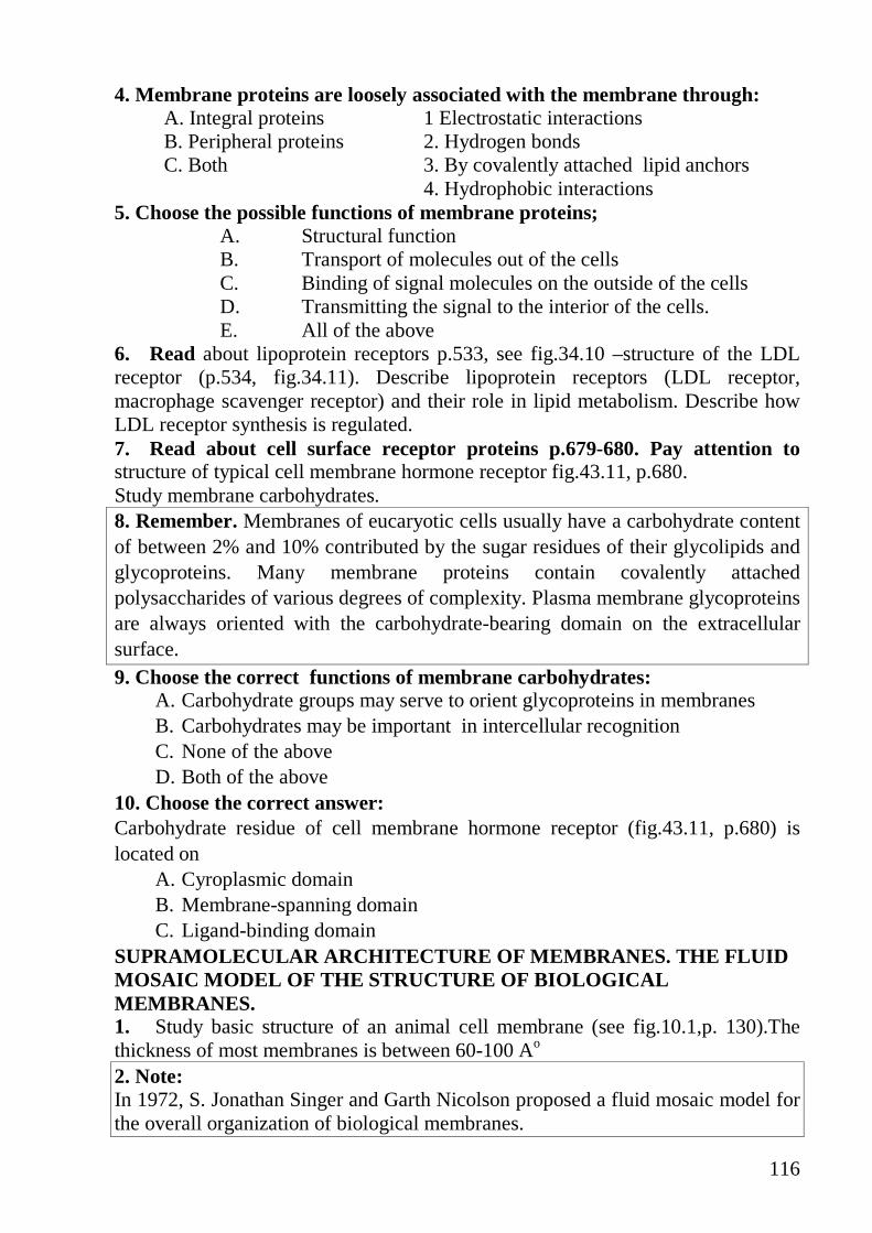
116
4. Membrane proteins are loosely associated with the membrane through: A. Integral proteins 1 Electrostatic interactions B. Peripheral proteins 2. Hydrogen bonds C. Both 3. By covalently attached lipid anchors 4. Hydrophobic interactions
5. Choose the possible functions of membrane proteins; A. Structural function B. Transport of molecules out of the cells C. Binding of signal molecules on the outside of the cells D. Transmitting the signal to the interior of the cells. E. All of the above
6. Read about lipoprotein receptors p.533, see fig.34.10 –structure of the LDL receptor (p.534, fig.34.11). Describe lipoprotein receptors (LDL receptor, macrophage scavenger receptor) and their role in lipid metabolism. Describe how LDL receptor synthesis is regulated. 7. Read about cell surface receptor proteins p.679-680. Pay attention to structure of typical cell membrane hormone receptor fig.43.11, p.680. Study membrane carbohydrates. 8. Remember. Membranes of eucaryotic cells usually have a carbohydrate content of between 2% and 10% contributed by the sugar residues of their glycolipids and glycoproteins. Many membrane proteins contain covalently attached polysaccharides of various degrees of complexity. Plasma membrane glycoproteins are always oriented with the carbohydrate-bearing domain on the extracellular surface. 9. Choose the correct functions of membrane carbohydrates:
A. Carbohydrate groups may serve to orient glycoproteins in membranes B. Carbohydrates may be important in intercellular recognition C. None of the above D. Both of the above
10. Choose the correct answer: Carbohydrate residue of cell membrane hormone receptor (fig.43.11, p.680) is located on
A. Cyroplasmic domain B. Membrane-spanning domain C. Ligand-binding domain
SUPRAMOLECULAR ARCHITECTURE OF MEMBRANES. THE FLUID MOSAIC MODEL OF THE STRUCTURE OF BIOLOGICAL MEMBRANES. 1. Study basic structure of an animal cell membrane (see fig.10.1,p. 130).The thickness of most membranes is between 60-100 Ao 2. Note: In 1972, S. Jonathan Singer and Garth Nicolson proposed a fluid mosaic model for the overall organization of biological membranes.

117
The fluid mosaic model describes certain features common to all biological membranes: 1) lateral diffusion Lipids and most proteins are free to diffuse laterally within the membrane, and the hydrophobic moieties of the lipids undergo rapid thermal motion, making the interior of the bilayer fluid. 2) fluidity is affected by temperature, fatty acid composition, and sterol content. Cells strive to maintain a constant fluidity when external circumstances change. 3)The lipids and proteins of the membrane are inserted into the bilayer with specific sidedness; the membrane is structurally and functionally asymmetric 4)self-assembling is important feature of biological membranes. The constituent protein and lipid molecules are held together by many noncovalent interactions, which are cooperative. 3. Cell membranes are self-sealing – if they are punctured or disrupted mechanically, they quickly and automatically reseal. What properties of membranes are responsible for this important feature? 4. Pay attention to: Each protein has a specific orientation in the bilayer and that protein reorientation by flip-flop diffusion occures seldom. The inner face of human erythrocyte membrane consists predominantly of phosphatidylethanolamine and phosphatidylserine. The outer face consists predominantly of phosphatidylcho-line and sphingomyelin. Although the phospholipid components of the membrane can diffuse in the fluid bilayer, this sidedness is preserved all all times. How? 5. Remember the properties of cell membranes (p.129-131) and select one incorrect answer:
A. Membranes are asymmetric. B. Membrane lipids are amphiphilic. C. Plasma membrane permits any compound to enter a cell. D. Membranes have selective permeability. E. Cholesterol is the compound of only animal cell membranes.
6. Study : Structure of RBC membrane and main proteins into it Erythrocytes lack organelles and thus have only a single membrane, the plasma membrane. The erythrocyte membrane contains two abundant transmembrane proteins. Glycophorin forms a car.bohydrate coat around red cells. The anion channel (band 3 protein) mediates the exchange of bicarbonate and chloride ions. These integral membrane proteins are linked by the band 4.1 protein and ankyrin to a flexible meshwork consisting mainly of spectrin. This membrane skeleton enables erythrocytes to resist strong shearing forces.
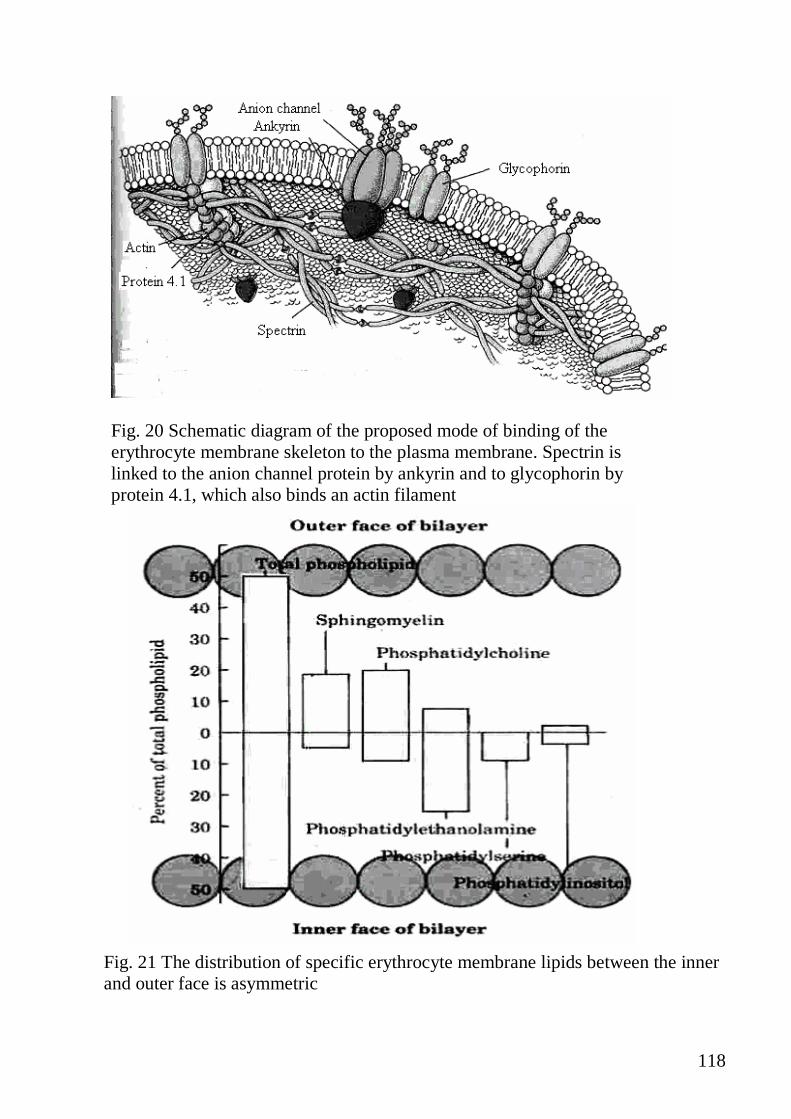
118
Fig. 20 Schematic diagram of the proposed mode of binding of the erythrocyte membrane skeleton to the plasma membrane. Spectrin is linked to the anion channel protein by ankyrin and to glycophorin by protein 4.1, which also binds an actin filament
Fig. 21 The distribution of specific erythrocyte membrane lipids between the inner and outer face is asymmetric

119
7. Poison of some snakes contains phospholipase A2 (see p.519,fig 33.32).If a small amount of poison is added to blood hemolysis occurs rapidly.
A. Write a reaction which will occur under action of this enzyme – component of poison.
B. Explain the reason of hemolysis in this case. C. Whether the structure of sphingomyelin changes under the action of this
enzyme? Explain the answer. TRANSPORT ACROSS THE MEMBRANES 1.Remember The three general classes of transport systems: uniport, symport, antiport differ in the number of solutes (substrates) transported and the direction in which each is transported . Note that this classification tells us nothing about whether these are energy-requiring (active transport) or energy-independent (passive ransport) processes.
2. Look at fig.25.21, p.401 and name type of transport
for each compound on it. A. Glucose 1. Uniport B. Galactose 2. Symport C. Fructose 3. Facilitated transporter
4. Na+- dependent transporter 3. Study different types of transport across the membranes.
Type of transport
Protein carrier
Saturable with
substrate
Produces concentra-
tion gradient
Energy- depen-dent
Energy source (if any)
Examples
Simple diffusion
No
No
No
No
-
H2O, O2, N2, CH4
Passive transport (facilitated diffusion)
Yes
Yes
No
No
-
Glucose permease of erythrocytes. GLUT 2 of

120
Type of transport
Protein carrier
Saturable with
substrate
Produces concentra-
tion gradient
Energy- depen-dent
Energy source (if any)
Examples
Symport
Antiport
hepatocytes. H+, pyruvate
across mitochondria
membrane Cl-, HOC3
- across
erythrocyte membrane
Active transport Primary
Yes Yes Yes Antiport
Yes ATP, light,
Substrate oxidation
Na+K+ ATP ase (animal plasma
membrane)
Secondary Yes Yes Yes Symport
Yes Ion gradient
Amino acids and sugars (Na+-
driven; intestine) Ion channels
Yes No No No* - Na+channel of acetylcholine
receptor (plasma membrane of
neuron) * Although the mechanism of transport via ion channels is not directly energy depent, the direction of ion flow is determined by the transmembrane differences in electrochemical potential. Ions always move down their electrochemical gradient through ion channels. 4. Compare passive and active transport across the membranes (p.40, 141, fig.10.16-20): A. Active transport 1.The substance crosses a membrane down an
electrochemical gradient B. Passive transport 2.The substance pumps against an electrochemical gradient C. Both 3. This type of transport requires proteins D. None 4. Catalyzed by enzymes 5. Draw the graph for the diffusion of steroid hormone across the membrane of target cell and glucose across the plasma membrane of isolated adipose cell. Note that steroid hormones are small lipid soluble molecules and glucose is water soluble. Look at fig.20.15, p.322 and name type of transport for each compound on it. 6. Read about transport of monosaccharides into tissues, glucose transport through the blood-brain barrier and into neurons p. 401-403. Compare glucose transport through the capillary endothelium in neural and nonneural tissues fig.25.23.

121
7. Match the letter and the number: A. Simple diffusion 1. Na + channel of acetylcholine receptor B. Passive transport 2. Glucose permease of erythrocytes C. Active transport primary 3. CH4, O2, N2 D. Active transport secondary 4. Na + K + ATPase (animal plasma membrane) E. Ion channel 5. Amino acids and sugars 8. At pH 7,0 tryptophan crosses a lipid bilayer membrane about 1,000 times more slowly than does the closely related substance indole. Suggest an explanation for this observation. 9. Read about endocytosis (p.138, fig 10.12) Note. There are two types of endocytosis: 1)Phagocytosis occurs only in specialized phagocytic cells present in blood. 2)Pinocytosis leads to the cellular uptake of fluid and fluid contants. The adsorptive pinocytosis is a receptor-mediated, selective process primary responsible for the uptake macromolecules for which there is a finite number of binding sites on the plasma membrane. These high affinity receptors permit pinocytosis to concentrate ligands from the medium and to minimize of fluid or soluble unbound macromolecules. Several hormones, the other macromolecules, are subject to adsorptive pinocytosis and form receptosomes, vesicles which avoid lysosomes and deliver their contents to the Golgi system 10. Pay attention on clinical note :William Hartman and Thomas Appelman p.534-535 SIGNAL TRANSDUCTION BY HORMONES 1. Study the signal transduction by hormones which bind to receptors in the plasma membrane. 2. Look at page 383, fig. 24.13 Note that these hormones are big molecules or hydrophilic molecules so they can't penetrate the membranes. As these hormones bind to the receptor on the plasma membrane the second messenger appears in cytoplasm and activates many enzymes within the cell. Note that hormones that act through the plasma membrane receptors cause the changing of activity of proteins that already exist in the cell. Therefore the result of action of these hormones is very quick (within seconds). The mechanism of action of insulin is much more complicated p.386, fig. 24.17. 3. Write the reaction of hydrolysis of phosphatidylinositol 4,5-bisphosphate (PIP2) by phospholipase C ( fig. 43.15, p. 683). Remember that the products of this reaction - DAG and IP3 are involved in transduction of hormone signal to the cells. 4.Remember: The hormones that act through adenylate cyclase system are - GLUCAGON and EPINEPHRINE ( in case when the last one acts through α2 and β2-adrenergic receptors). You must reproduce fig. 43.13. showing the signal transduction by glucagon or epinephrine. The hormones that act through PIP2 system are angiotensin, bradykinin, vasopressin.

122
5. Note that some toxins of microorganisms disorder the transduction of signal through the membranes and that is the main mechanism of their action. For instance cholera toxin consists of two subunits A and B. A subunit enter the cell membrane and covalently binds the part of the molecule of NAD+ to the α-subunit of G- protein. In this state α--subunit doesn't catalyze the reaction: GTP → GDP cAMP level rises and stimulates the transport of chloride ions into the lumen of the gut, causing the diffusion of water and severe diarrhea that, if untreated, is often fatal.
a) Why does the concentration of cAMP increase in these cells? b) Based on information above, can you suggest how cAMP normally functions in intestinal epithelial cells? c) Suggest a possible treatment for cholera.
REACTIVE OXYGEN SPECIES AND CELLULAR DAMAGE
1.Study Reactive oxygen species and cellular damage (p.271-272, 327-331, fig.21.1-21.7, table 21.1, 21.2). Pay attention on lipid peroxidation 2. Match the letter and the number: A. Lipid peroxidation is initiated by 1. Malondialdehyde B. The free radical chain reaction is propagated by 2. Free radical compound C. Product of lipid peroxidation is 3. The addition of O2 D. The chain reaction can be terminated by 4. Antioxidants 3. Specify the correct order of stages of lipid peroxidation :
A. Propagation B. Degradation C. Initiation D. Termination
4. Match the letter in No 3 and the number (product of each stages):.
1. Lipid radical 2. Lipid peroxy radical 3. Lipid peroxide 4. Malondialdehyde 5. Degraded lipid peroxide 6. Polyansaturated lipid
5. Compare reactive oxygen species origin and characteristics : Find the correct answer for the line in column A from B and C
A B C a) O2 Singlet oxygen With antiparallel spins b) H2O2 Superoxide anion Produced from RH by OH. attack c) OH Hydroxyl radical Produced by ETC d) O2
↑↓ Hydrogen peroxide The most reactive species e) R Organic peroxide radical Occurs during lipid degradation f) RCOO Organic radical Can diffuse into and through cell
membranes

123
6. Based on information p.332, fig21.7, can you suggest possible clinical symptoms due to lipid peroxidation in the mitochondrial membranes? 7. Fill up the blanks of the followings: A. The toxicity of oxygen is related to the production of ______________ B. ROS cause _________________ in membranes C. Lipid peroxidation changes or damages _________________________ D. The free radical damage could be the primary cause of the ___________ E. The free radical damage could enhance_______________of the disease F. The free radical damage could be the_________________of cell damage caused by other agents.
CELLULAR DEFENSES AGAINST OXYGEN TOXICITY
1. Study «Cellular defenses against oxygen toxicity» (p.335-337, fig. 21.12-21.17.). 2. Compare antioxidant enzymes : Find the correct answer for the line in column A from B and C
A (Ensyme) B (Substrate) C (Product) a) Superoxide dismutase H2O2 and LOOH H2O2 b) Catalase O2 GSH c) Glutathione peroxidase H2O2 H2O, O2 d) Glutathione reductase GSSG H2O, GSSG
3. Which of the following compounds have antioxidant properties? A. Tocopherol B. Ascorbate C. Thiamine D. Carotenoid E. Catalase
4. Answer the questions A. Q.21.3, p.336 B. What part does antioxidant system play in bodily functions? C. What is balance of oxidant/antioxidant systems ?
5. Pay attention to problems and answers p.339-340.