Embed Size (px)
Citation preview

PROTEOLYSIS is the breakdown of protein to free amino acids.
Gastrointestinal input
1. Proteins and polypeptides are not absorbed intact but must first be hydrolyzed to free amino acids.
2. Digestion of dietary protein is carried out by proteases (proteolytic enzymes), which are found in gastric and pancreatic juices and on the intestinal cell surface. a. Gastric juice
(1) The hydrochloric acid (pH = 2) in gastric juice kills microorganisms, denatures proteins, and provides an acid environment for the action of pepsin.
(2) The protease pepsin works at acid pH. It is secreted into the stomach as the proenzyme pepsinogen, which is activated by the autocatalytic removal of 44 N-terminal amino acids at low pH.
(3) Peptic hydrolysis of proteins yields peptides and some free amino acids.
b. Pancreatic juice (1) Proteases in pancreatic juice also are secreted as
proenzymes from the pancreatic acinar cells. Activation of proenzymes occurs by the action of enteropeptidase (enterokinase), which is secreted by the duodenal epithelial cells.
(2) Enteropeptidase activates trypsinogen by removing six amino acids, and the activated trypsin in turn activates the proenzymes of chymotrypsin, elas-tase, and carboxypeptidases A and B.
c. Intestinal proteases (1) Aminopeptidases and dipeptidases continue the
digestion of peptides to free amino acids. (2) Di- and tripeptides are usually absorbed as such and
digested to free amino acids within the intestinal epithelial cells.

3. Absorption of free amino acids takes place in the small intestine. a. Five major systems and a few minor systems have been
identified that transport different classes of amino acids from the gut lumen into the intestinal epithelial cell.
b. Disorders associated with defects in amino acid transport lead to increased levels of those amino acids in the urine (amino acidurias). (1) Hartnup's disease is due to a defect in the transport
system for large neutral and aromatic amino acids. • Symptoms similar to pellagra are observed in some
patients. This is due to a deficiency of niacin, which may be synthesized from tryptophan.
• Treatment. These symptoms can be relieved by administration of niacin.
(2) Cystinuria is due to a defect in the transport system for basic amino acids, which also transports cystine, the disulfide-linked dimer of cysteine. • Symptoms. Cystine is relatively insoluble in urine, and
its accumulation leads to deposition of crystals, which may cause urinary tract infections and renal stones.
• Treatment to reduce deposition of cystine crystals includes administration of the drug penicillamine (β,β-dimethylcysteine), which reacts with cystine to form a soluble cysteine-penicillamine adduct.
(3) Glycinuria (iminoglycinuria) is due to a defect in the transport system for glycine and the imino acids, proline and hydroxyproline. There are no clinical abnormalities other than increased urinary excretion of these amino acids.
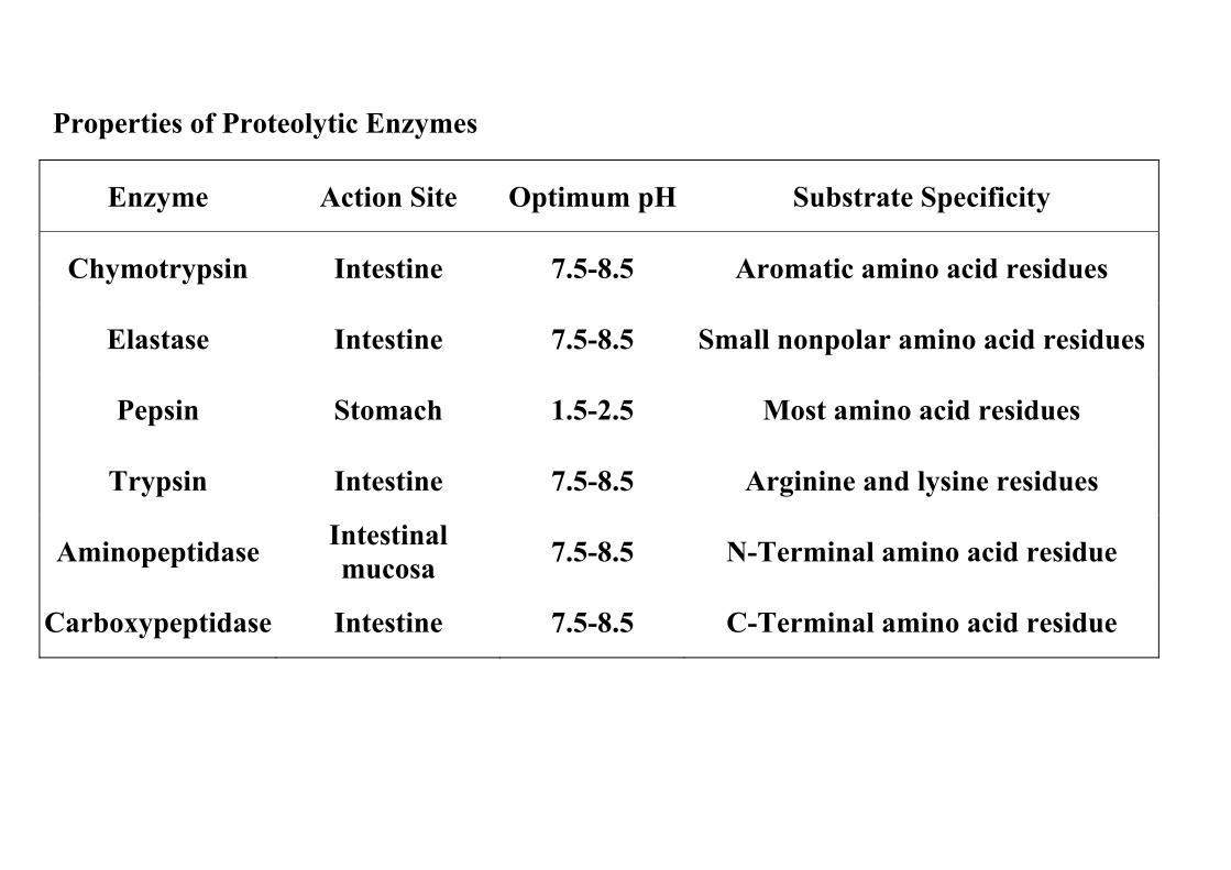
Properties of Proteolytic Enzymes
Enzyme Action Site Optimum pH Substrate Specificity
Chymotrypsin Intestine 7.5-8.5 Aromatic amino acid residues
Elastase Intestine 7.5-8.5 Small nonpolar amino acid residues
Pepsin Stomach 1.5-2.5 Most amino acid residues
Trypsin Intestine 7.5-8.5 Arginine and lysine residues
Aminopeptidase Intestinal mucosa 7.5-8.5 N-Terminal amino acid residue
Carboxypeptidase Intestine 7.5-8.5 C-Terminal amino acid residue
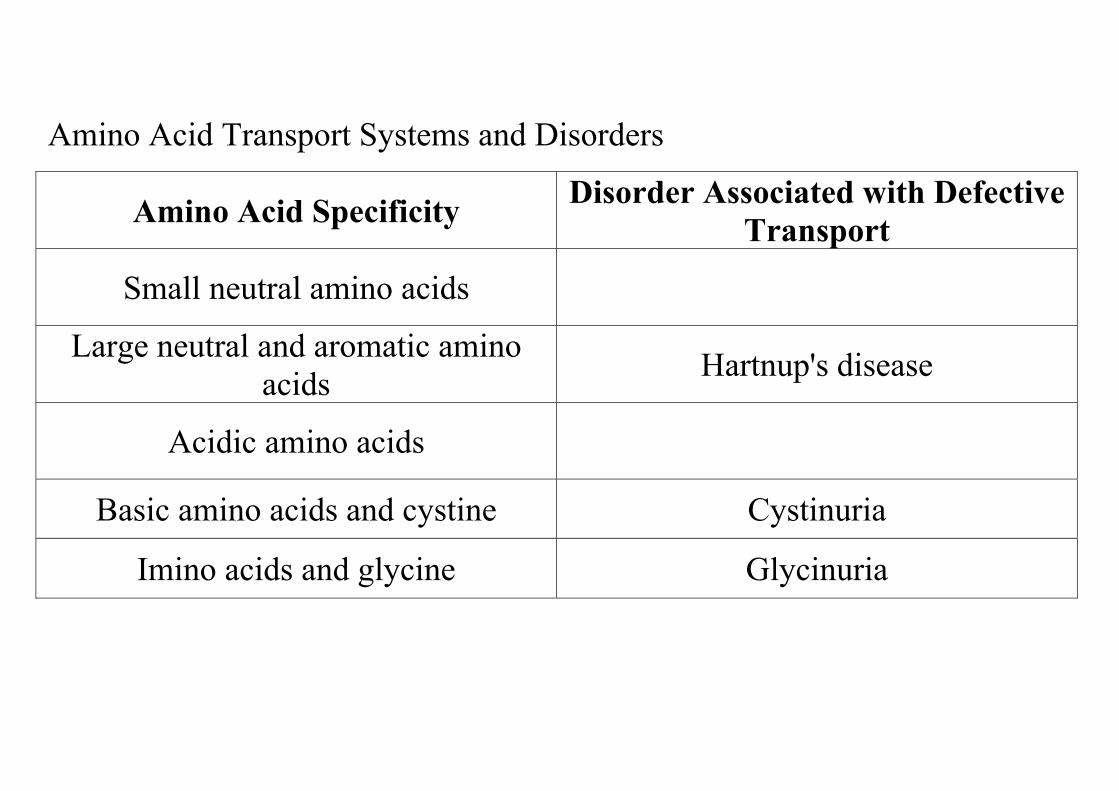
Amino Acid Transport Systems and Disorders
Amino Acid Specificity Disorder Associated with Defective Transport
Small neutral amino acids
Large neutral and aromatic amino acids Hartnup's disease
Acidic amino acids
Basic amino acids and cystine Cystinuria
Imino acids and glycine Glycinuria

Proteolysis of endogenous protein. Body protein is continuously being broken down to free amino acids, and the rate of degradation for individual proteins varies widely. Some liver enzymes have half-lives of only a few hours, whereas hemoglobin and the red blood cells have a total lifetime of 120 days, and some structural proteins (e.g., collagen) have half-lives too long to be measured.
1. Factors affecting the rates of protein degradation a. Denaturation (i.e., loss of its preferred native configuration)
accelerates proteolysis. b. Activation of lysosomes increases the rate of intracellular
proteolysis. c. Glucocorticoids increase protein degradation in muscle tissue. d. Excessive thyroid hormones increase protein turnover. e. Insulin
reduces proteolysis and increases protein synthesis.
2. Purpose a. Abnormal, defective, and damaged proteins must be removed
because they are of no use to the body, and they may inhibit processes that require the functional proteins.
b. Inducible enzymes must be removed when their activities are no longer beneficial.
3. Ubiquitin, an 8.5-kilodalton protein in eukaryotic cells, plays an important role in designating the proteins to be degraded. The C-terminal residue of ubiquitin becomes covalently attached to lysine residues of proteins that are then degraded via an adenosine triphosphate (ATP)-dependent process, a. The protein E1 catalyzes the formation of an intermediate ubiquitin
complex by formation of a thioester bond to a sulfhydryl group of E1. b. The protein E2 receives the ubiquitin molecule from the ubiquitin-E1
complex and transfers it to protein E3. c. The protein E3 couples the ubiquitin molecule to the protein that is to
be degraded. Usually, the protein to be degraded becomes conjugated to several molecules of ubiquitin.

AMINO ACID POOL
Essential amino acids are those that cannot be synthesized by humans and are, therefore, essential dietary factors. Nine of the 20 amino acids are essential. Inputs to the amino acid pool are from dietary protein and proteolysis of endogenous cellular protein. Outputs from the amino acid pool are for amino acid degradation (catabolism), synthesis of special compounds, and protein synthesis. Nitrogen balance. If the total daily nitrogen losses in urine, skin, and feces are equal to the total daily nitrogen intake, the subject is said to be in nitrogen balance, as a healthy, adequately fed adult should be.
The Nine Essential Amino Acids
Histidine Isoleucine Leucine
Lysine Methionine Phenylalanine
Threonine Tryptophan Valine
Note—In growing children, arginine may not be synthesized in amounts adequate to fill the requirements for both protein synthesis and urea formation; under these circumstances, it would be considered an essential amino acid. 1. Positive nitrogen balance. If nitrogen losses are less than intake, the subject is in positive nitrogen balance, as healthy, growing children and convalescing adults should be. 2. Negative nitrogen balance. If nitrogen losses are greater than intake, the subject is in negative nitrogen balance, as occurs in people with diseases involving tissue wasting or in those undergoing starvation. Prolonged periods of negative balance are dangerous and can be fatal. 3. Estimation of muscle protein breakdown. Some of the histidine residues of the muscle protein complex actomyosin are methylated after their incorporation. When actomyosin breaks down, 3-methyl histidine is liberated and excreted into the urine. The urinary levels provide a measure of muscle protein breakdown.

METABOLIC FLOW OF AMINO ACID NITROGEN. The primary means by which amino acid-derived nitrogen is metabolized is by the sequential action of aminotransferases, glutamate dehydrogenase, and the urea cycle.
Transamination involves the transfer of an amino group from an amino acid to an α-keto acid to form a new amino acid and a new α-keto acid.
1. Aminotransferases (transaminases) catalyze these reactions. For example, aspartate aminotransferase interconverts the amino acid aspartate plus the α-keto acid α-keto-glutarate, with the amino acid glutamate and the α-keto acid oxaloacetate.
Aspartate + α-Ketoglutarate ↔ Oxaloacetate + Glutamate
Pyridoxal phosphate is an essential cofactor of all aminotransferases.
2. Oxidative deamination by glutamate dehydrogenase occurs in the mitochondrial matrix by the following reaction:
Glutamate + NAD(P)+ + H2O ↔ α-Ketoglutarate + NAD(P)H + H+ + NH3
3. Glutamate dehydrogenase can use either oxidized nicotinamide adenine dinucleo-tide (NAD+) or oxidized nicotinamide adenine dinucleotide phosphate (NADP+) as a cofactor. a. NADPH-NADP+ ratio. Under normal conditions in the liver, the
ratio of reduced NADPH to oxidized NADP+ is high, and the ratio of reduced NADH to oxidized NAD+ is low. Thus, there is always a pyridine nucleotide coenzyme available in its oxidized or reduced state to participate in the above reaction in either direction.
b. The reaction above is controlled, therefore, by the relative levels of glutamate, α-ketoglutarate, and ammonia (NH3).
4. Allosteric regulators a. Activators include adenosine diphosphate (ADP) and guanosine
diphosphate (GDP). b. Inhibitors include ATP, guanosine triphosphate (GTP), and NADH.

Alternative mechanisms for deaminating amino acids
1. Direct deamination by serine and threonine dehydratase a. Because of the chemistry of the hydroxyl side chain of serine and threonine (i.e., the
hydroxyl group is a good leaving group), their direct deamination is facilitated. b. The reactions are:
Serine → Pyruvate + NH3
Threonine → α-Amino-β-ketobutyrate → Pyruvate + NH3
c. Pyridoxal phosphate is a required cofactor.
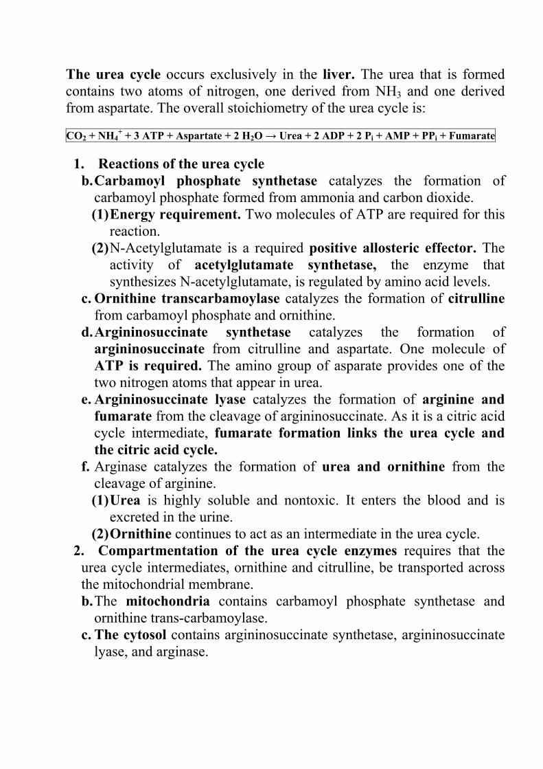
The urea cycle occurs exclusively in the liver. The urea that is formed contains two atoms of nitrogen, one derived from NH3 and one derived from aspartate. The overall stoichiometry of the urea cycle is:
CO2 + NH4+ + 3 ATP + Aspartate + 2 H2O → Urea + 2 ADP + 2 Pi + AMP + PPi + Fumarate
1. Reactions of the urea cycle b. Carbamoyl phosphate synthetase catalyzes the formation of
carbamoyl phosphate formed from ammonia and carbon dioxide. (1) Energy requirement. Two molecules of ATP are required for this
reaction. (2) N-Acetylglutamate is a required positive allosteric effector. The
activity of acetylglutamate synthetase, the enzyme that synthesizes N-acetylglutamate, is regulated by amino acid levels.
c. Ornithine transcarbamoylase catalyzes the formation of citrulline from carbamoyl phosphate and ornithine.
d. Argininosuccinate synthetase catalyzes the formation of argininosuccinate from citrulline and aspartate. One molecule of ATP is required. The amino group of asparate provides one of the two nitrogen atoms that appear in urea.
e. Argininosuccinate lyase catalyzes the formation of arginine and fumarate from the cleavage of argininosuccinate. As it is a citric acid cycle intermediate, fumarate formation links the urea cycle and the citric acid cycle.
f. Arginase catalyzes the formation of urea and ornithine from the cleavage of arginine. (1) Urea is highly soluble and nontoxic. It enters the blood and is
excreted in the urine. (2) Ornithine continues to act as an intermediate in the urea cycle.
2. Compartmentation of the urea cycle enzymes requires that the urea cycle intermediates, ornithine and citrulline, be transported across the mitochondrial membrane. b. The mitochondria contains carbamoyl phosphate synthetase and
ornithine trans-carbamoylase. c. The cytosol contains argininosuccinate synthetase, argininosuccinate
lyase, and arginase.

Genetic defects have been documented for each of the urea cycle enzymes.
1. Diseases a. Type I hyperammonemia is due to a defect in carbamoyl phosphate
synthetase. b. Type II hyperammonemia is due to a defect in ornithine
transcarbamoylase. c. Citrullinuria is due to a defect in argininosuccinate synthetase. d. Argininosuccinic acidemia is due to a defect in argininosuccinate
lyase. e. Hyperargininemia is due to a defect in arginase.
2. Symptoms a. Hyperammonemia. High serum levels of ammonia are quite toxic
and can cause brain damage. b. Episodic encephalopathies, such as convulsions and ataxia, may
occur in children with partial deficiencies of a urea cycle enzyme. 3. Treatment. Infants who totally lack any of the urea cycle enzymes do
not usually survive the neonatal period. Patients with defects that reduce, but do not eliminate, the activity of one of the urea cycle enzymes are more likely to respond to treatment. a. Low-protein diets. Severe consequences of these disorders may be
avoided when protein intake is restricted. However, very early diagnosis is critical in preventing mental retardation. A diet must be provided that provides adequate, but not excessive, amounts of essential amino acids. Arginine is not normally considered an essential amino acid because it may be synthesized by the action of the urea cycle. Therefore, low-protein diets should be supplemented with arginine.
b. Administration of sodium benzoate and sodium phenylacetate is beneficial in reducing the serum ammonia level. These compounds react with glycine and glutamine to form adducts, which are excreted in the urine. Serum ammonia must then be used to synthesize more of these nonessential amino acids, which helps to lower the overall ammonia level.
c. Blood transfusion and hemodialysis may be required to prevent brain damage from hyperammonemia.

CLASSIFICATION OF AMINO ACIDS.
Amino acids are categorized by the final products of their degradation pathways.
Ketogenic amino acids are degraded to either acetyl coenzyme A (acetyl CoA) or acetoacetyl CoA, which can give rise to ketone bodies. Purely ketogenic amino acids are leucine and lysine.
Glucogenic amino acids are degraded to pyruvate or citric acid cycle intermediates, which can give rise to glucose. Purely glucogenic amino acids include alanine, arginine, asparagine, aspartate, cysteine, glutamate, glutamine, glycine, histidine, methionine, proline, serine, and valine.
Ketogenic and glucogenic. Some amino acids can be degraded into multiple intermediates, which classify them as both ketogenic and glucogenic. They are isoleucine, phenylalanine, threonine, tyrosine, and tryptophan.

FATE OF CARBON SKELETONS
Three-carbon amino acids, threonine, and glycine are converted to pyruvate.
1. Alanine is converted by transamination with α-ketoglutarate. 2. Serine is converted by direct deamination by serine
dehydratase. 3. Cysteine may be converted by three different pathways. 4. Threonine (a four-carbon amino acid) is converted via an α-
amino-β-ketobutyrate intermediate. This intermediate in pyruvate formation may alternatively be metabolized to acetyl CoA and glycine.
5. Glycine (a two-carbon ammo acid) may be converted by senne hydroxymethyl-transferase to senne, and it is then converted to pyruvate.
Four-carbon amino acids are converted to oxaloacetate.
1. Aspartate is converted by transammation with α-ketoglutarate 2. Asparagine is converted by asparagmase to aspartate and then
to oxaloacetate
Some five-carbon amino acids are converted to glutamate, a precursor of α-ketoglutarate.
1. Glutamine is converted to glutamate by glutammase 2. Histidine is converted to glutamate by a series of reactions that
include the transfer of its formimmo group to tetrahydrofolate. 3. Arginine is converted by argmase to ornithme, which may be
converted to glutamate semialdehyde and oxidized to glutamate 4. Proline undergoes reactions that involve ring opening and
conversion to glutamate semialdehyde and then to glutamate

Degradation of branched-chain amino acids
1. Valine, leucine, and isoleucine all are converted to their corresponding α-keto acid by the actions of three specific ammotransferases
2. Branched-chain keto acid dehydrogenase is a common enzyme that catalyzes the oxidative decarboxylations of each of the three α-keto acids derived from valine, leucine, and isoleucine. a. Branched-chain keto acid dehydrogenase is very similar to pyruvate
dehydrogenase and also utilizes as cofactors lipoamide, thiamine pyrophosphate (TPP), flavin adenine dinucleotide (FAD), and NAD+.
b. Maple syrup urine disease (branched-chain ketoaciduria) is caused by a genetic defect in branched-chain keto acid dehydrogenase. (1) Diagnosis. Plasma and urine levels of valine, leucine, and
isoleucine and their corresponding α-keto acids are abnormally high. These keto acids give the urine the characteristic odor for which the disease is named.
(2) Treatment includes dietary restriction of valine, leucine, and isoleucine and, for acute episodes, a blood transfusion. In some cases, the defect reduces the enzyme's affinity for TPP. These patients respond to treatment with therapeutic doses of thiamine. Untreated infants do not survive long after birth.
c. Intermittent branched-chain ketoaciduria is a variant of maple syrup urine disease in which the deficiency in enzyme activity is much less severe. Symptoms of this variant may not appear until later in life and may be intermittent. Treatment by dietary restriction of branched-chain amino acids is more successful than in patients with maple syrup urine disease.
3. The three CoA derivatives are then degraded by different pathways to yield different final products. a. Isovaleric acidemia is a disease caused by a deficiency of isovaleryl
CoA dehydrogenase, an enzyme involved in the degradative pathway of leucine.
b. Treatment includes dietary restriction of leucine and administration of glycine, which reacts with isovaleric acid, the toxic intermediate that accumulates in this disorder.

Catabolism of amino acids to succinyl CoA
1. Methionine, isoleucine, and valine are degraded by different pathways to form succinyl CoA via L-methylmalonyl CoA.
2. Methionine and isoleucine are first converted to propionyl CoA, which is converted to D-methylmalonyl CoA by propionyl CoA carboxylase, a biotin-containing enzyme.
3. D-Methylmalonyl CoA undergoes racemization to L-methylmalonyl CoA followed by isomerization to form succinyl CoA. a. Methylmalonyl CoA mutase catalyzes this isomerization. It contains 5'-
deoxyadenosylcobalamin, which is formed from vitamin B2 and ATP. b. Methylmalonic aciduria is caused by a defect in methylmalonyl CoA mutase. Some forms of
this disorder are treatable by administration of high doses of vitamin B12. Patients who do not respond to vitamin B12 must be placed on a diet that restricts the intake of methionine, isoleucine, and valine. This is difficult to manage because these are essential amino acids.
Catabolism of tryptophan yields crotonyl CoA, formate, and alanine. Crotonyl CoA is an intermediate in the (β-oxidation of fatty acids, and thus, tryptophan is ketogenic. It is also glucogenic because alanine is also formed during its degradation.
Catabolism of lysine also yields crotonyl CoA. Thus, lysine is a ketogenic amino acid.

Catabolism of phenylalanine and tyrosine.
1. Phenylalanine hydroxylase catalyzes the hydroxylation of phenylalanine to form tyrosine. a. Tetrahydrobiopterin is a required cofactor. The cofactor is oxidized
to dihydro-biopterin during this reaction and must be regenerated by another enzyme, dihydrobiopteridine reductase, which uses NADPH as a reductant.
b. Phenylketonuria (PKU) is a genetic disorder associated with the inability to convert phenylalanine to tyrosine, with the subsequent accumulation of toxic derivatives of phenylalanine (e.g., phenylpyruvate). (1) Classic PKU is due to a genetic defect in phenylalanine
hydroxylase. This disorder is treated with a phenylalanine-restricted diet. If not diagnosed early, mental retardation occurs in untreated infants.
(2) Atypical PKU is due to a defect in dihydrobiopteridine reductase, which is needed to regenerate the reduced form of the cofactor. Tetrahydrobiopterin is also required for the synthesis of certain neurotransmitters. Thus, this disorder presents neurologic problems, in addition to those seen in classic PKU, that are not abated by restricting phenylalanine.
2. Tyrosine aminotransferase catalyzes the reaction with α-ketoglutarate to form 4-hydroxyphenylpyruvate. Tyrosinemia type II is caused by a genetic defect in this enzyme.
3. 4-Hydroxyphenylpyruvate oxidase catalyzes the conversion of 4-hydroxyphenylpy-ruvate to homogentisate. Neonatal tyrosinemia is due to a defect in this enzyme.
4. Homogentisate oxidase catalyzes the conversion of homogentisate to 4-maleylacet-oacetate. Alkaptonuria is due to a genetic defect in this enzyme. Patients excrete large amounts of homogentisate in the urine, which causes it to darken after exposure to air. This disorder causes no other symptoms in children and usually is not treated. Later in life, patients may develop a form of arthritis, which is treated as such.
5. Maleylacetoacetate isomerase converts 4-maleylacetoacetate to 4-fumarylacet-oacetate.
6. Fumarylacetoacetate hydrolase cleaves 4-fumarylacetoacetate to form fumarate, a glucogenic product, and acetoacetate, a ketogenic product. Tyrosinemia type I (tyrosinosis) is due to a defect in this enzyme.

INHERITED DISORDERS OF AMINO ACID CATABOLISM. Several disorders have been documented that are caused by mutations in genes that code for specific enzymes needed for amino acid degradation.
These disorders usually lead to excessive accumulation in the blood or urine of the intermediate that is a substrate for that enzyme, or derivatives of that compound, and a decrease of subsequent intermediates in that pathway. The severity of the disorder depends on the toxicity of the accumulated metabolites.
Treatment often consists of a low-protein diet, which is supplemented with just enough of the unmetabolized amino acid(s) to allow normal protein synthesis but not so much as to allow catabolism and accumulation of toxic intermediates.
Some Inherited Disorders of Amino Acid Catabolism Disorder
Defective Enzyme Amino Acid Degradation Pathway
Phenylketonuria Phenylalanine hydroxylase
Phe
Alkaptonuria Homogentisate oxidase Phe, tyr Tyrosinemia type I 4-Fumarylacetoacetate
hydrolase Phe, tyr
Tyrosinemia type II Tyrosine transaminase Phe, tyr Neonatal tyrosinemia 4-Hydroxyphenyl-
pyruvate oxidase Phe, tyr
Methylmalonic aciduria Methylmalonyl CoA mutase
Met, val, ile
Maple syrup urine disease Branched-chain α-keto acid dehydrogenase
Ile, leu, val
Isovaleric acidemia Isovaleryl CoA dehydrogenase
Leu
Histidinemia Histidine-ammonia lyase
His
Hyperprolinemia Proline oxidase Pro His = histidme; lie = isoleucine; leu = leurine; met = methiomne, phe = phertylalamne; pro = prolme; tyr = tyrosine; val = valine.

Biosynthesis of Amino Acids and Amino Acid-Derived Compounds
BIOSYNTHESIS OF AMINO ACIDS
The nine essential amino acids must be acquired from the diet
The eleven nonessential amino acids are synthesized by mammals as follows
1. Alanine is formed from pyruvate by transamination.
2. Aspartate is formed from oxaloacetate by transamination.
3. Asparagine is formed by amidation of aspartate.
4. Glutamate is formed by reductive amination of α-ketoglutarate
5. Glutamine is formed by the amidation of glutamate.
6. Arginine is formed in the urea cycle.
7. Proline is formed in two steps from glutamate.
8. Serine is synthesized from 3-phosphoglycerate, a glycolytic intermediate.
FIGURE 25-1. Precursors for the biosynthesis of nonessential amino acids.
9. Glycine is converted to serine by serine hydroxymethyltransferase.
10. Cysteine is synthesized from serine and methionine. Thus, it is nonessential only if methionine is provided in the diet.
11. Tyrosine is formed from phenylalanine. Thus, it is nonessential only if phenylalanine is provided in the diet.
AMINO ACIDS AS MAJOR INPUTS TO THE ONE-CARBON POOL. One carbon groups are frequently transferred during metabolism. These one-carbon fragments exist in a readily available pool. Different carriers are employed for one-carbon groups of different oxidation states.
Biotin is a carrier of carbon dioxide.

Tetrahydrofolate (THF) is derived from folic acid. It carries one-carbon groups of all oxidation states except carbon dioxide. Examples of THF-dependent enzymes include:
FIG. Tetrahydrofolate and its derivatives Only the reactive portions of the substituted tetrahy-drofolate molecules are shown.
1. Serine hydroxymethyltransferase
Glycine + N5,N10-Methylene THF ↔ Serine + THF
2. Homocysteine methyltransferase
Homocysteine + N5-Methyl THF ↔ Methionine + THF
3. Thymidylate synthase
2'-Deoxyuridylate + N5, N10-Methylene THF ↔ 2'-Deoxythymidylate + Dihydrofolate
S-Adenosylmethionine (SAM) is the primary donor of methyl groups to a wide variety of acceptors during biosynthesis. This process occurs by a series of reactions referred to as the activated methyl cycle.
1. Methionine to SAM. In this reaction, the adenosyl group of adenosine triphosphate (ATP) is transferred to the methionine sulfur.

2. Methyl transfer from SAM to an acceptor molecule. The methyl group attached to the sulfur of SAM is transferred to an acceptor molecule to form a methylated product plus S-adenosylhomocysteine. Examples of such methylation reactions include:
a. Norepinephrine to epinephrine
b. Guanidinoacetate to creatine
c. Acetylserotonin to melatonin
d. Phosphatidylethanolamine to phosphatidylcholine
e. Methylation of DNA
3. S-Adenosylhomocysteine is hydrolyzed to adenosine and homocysteine.
4. Regeneration of methionine from homocysteine is catalyzed by homocysteine methyltransferase, which requires two cofactors.
a. The methylcobalamin form of vitamin B12 provides the methyl group needed to form methionine.
b. N5-methyl THF provides a methyl group to regenerate methylcobalamin.
c. N5-Methyl THF must then be regenerated.
(1) Serine hydroxymethyltransferase (see II B 1) catalyzes the conversion of THF to N5,N10-methylene THF.

(2) N5,N10-Methylene THF reductase catalyzes the conversion of N5,N10-methylene THF to N5-Methyl THF.
5. An important side reaction of the activated methyl cycle is the synthesis of cysteine from homocysteine.
FIG. The activated methyl cycle and biosynthetic pathway for cysteme. Enzyme names are in italics. ATP = adenosme triphosphate; B12 = cobalamin; O/;-B,2 = methylcobalamin; P, = inorganic phosphate; PP, = inorganic pyrophosphate; R-CH3 = methylated product; R-H = substrate molecule to be methylated; THF = tetrahydrofolate.
a. Cystathionine synthase catalyzes the condensation of homocysteine and serine to form cystathionine. This enzyme requires pyridoxal phosphate (PLP) as a cofactor.
b. Cystathionase catalyzes the hydrolysis of cystathionine to cysteine and α-ketobutyrate. A deficiency in this enzyme causes cystathioninuria.
6. Homocystinuria is the accumulation of homocystine, the disulfide-linked dimer of homocysteine, which forms when homocysteine is in excess.
a. Causes
(1) A defect in cystathionine synthase
(2) A defect in homocysteine methyltransferase
(3) The unavailability of Ns-methyl THF or methylcobalamin
b. Treatment. Some forms of this disorder may be treated with therapeutic doses of vitamins B6, B12, or folic acid.
c. High serum levels of homocysteine also have been implicated as a contributing factor in heart disease.
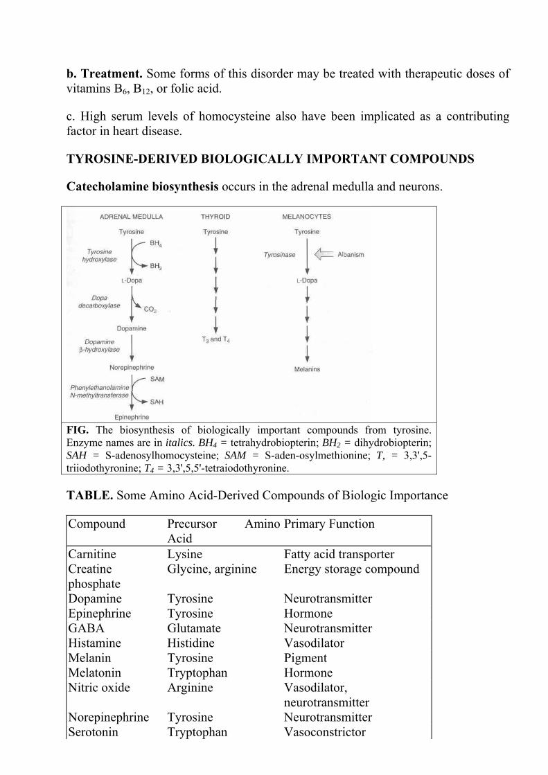
TYROSINE-DERIVED BIOLOGICALLY IMPORTANT COMPOUNDS
Catecholamine biosynthesis occurs in the adrenal medulla and neurons.
FIG. The biosynthesis of biologically important compounds from tyrosine. Enzyme names are in italics. BH4 = tetrahydrobiopterin; BH2 = dihydrobiopterin; SAH = S-adenosylhomocysteine; SAM = S-aden-osylmethionine; T, = 3,3',5-triiodothyronine; T4 = 3,3',5,5'-tetraiodothyronine.
TABLE. Some Amino Acid-Derived Compounds of Biologic Importance
Compound Precursor Amino Acid
Primary Function
Carnitine Lysine Fatty acid transporter Creatine phosphate
Glycine, arginine Energy storage compound
Dopamine Tyrosine Neurotransmitter Epinephrine Tyrosine Hormone GABA Glutamate Neurotransmitter Histamine Histidine Vasodilator Melanin Tyrosine Pigment Melatonin Tryptophan Hormone Nitric oxide Arginine Vasodilator,
neurotransmitter Norepinephrine Tyrosine Neurotransmitter Serotonin Tryptophan Vasoconstrictor

Thyroxine Tyrosine Hormone GABA = -γ-aminobutyric acid.
1. Formation of dopa. Tyrosine is hydroxylated to 3,4-dihydroxyphenylalanine (dopa) by tyrosine hydroxylase, which requires tetrahydrobiopterin (BH4) as a cofactor. This is the rate-limiting step in catecholamine biosynthesis. The enzyme is alloste-ric, with dopamine, norepinephrine, and epinephrine acting as negative effectors.
2. Formation of dopamine is catalyzed by dopa decarboxylase, a PLP-dependent enzyme.
3. Formation of norepinephrine. Dopamine p-hydroxylase hydroxylates the aromatic ring of dopamine to yield norepinephrine. This enzyme requires copper and vitamin C as cofactors. Norepinephrine is the major neural transmitter of the sympathetic nervous system.
4. Formation of epinephrine occurs in the adrenal medulla, where the amino group of norepinephrine is methylated by phenylethanolamine N-methyltransferase, using SAM as the methyl donor. The synthesis of the enzyme is induced by glucocor-ticoids from the surrounding adrenal cortex, and its production is inhibited by epinephrine.
5. Catabolism of catecholamines. Catechol-O-methyl transferase uses SAM to methylate catecholamines. This is followed by oxidation by monoamine oxidase and aldehyde dehydrogenase to give the major excretory product, 3-methoxy-4-hydroxy-mandelic acid (vanillylmandelic acid; VMA). VMA excretion levels are used as an aid in the diagnosis of adrenal pheochromocytomas, which are tumors that produce huge amounts of catecholamines.
Biosynthesis of melanins. Melanins are biologic pigments. Their synthesis occurs only in pigment-producing cells called melanocytes. Melanins are synthesized from tyro-sine by a sequence of several reactions. The first step is the conversion of tyrosine to dopa.
1. Tyrosinase catalyzes the conversion of tyrosine to dopa in melanocytes.
2. Tyrosinase does not require tetrahydrobiopterin, but uses copper as a cofactor.
3. Albinism is due to a genetic defect in tyrosinase, which causes afflicted individuals (i.e., albinos) to lack pigmentation because they cannot synthesize melanins.
Biosynthesis of thyroid hormones

1. 3,3',5,5'-Tetraiodothyronine (thyroxine; T4) and 3,3',5-triiodothyronine (T,) are hormones secreted by the thyroid gland.
2. T4 and T3 are formed in the follicle cells of the thyroid gland by iodination of tyrosine residues, which are in peptide linkage in chains of the protein thyroglobulin.
3. lodinated thyroglobulin is stored in the lumen of the thyroid follicles until it is required. Then T3 and T4 are hydrolyzed off and secreted into the circulation.
TRYPTOPHAN-DERIVED BIOLOGICALLY IMPORTANT COMPOUNDS
Serotonin is a potent vasoconstrictor and stimulator of smooth muscle contraction.
1. Serotonin is synthesized by neurons in the hypothalamus and brain stem, by the pineal gland, and by chromaffin cells of the gastrointestinal tract.
2. The rate-limiting step is catalyzed by tryptophan hydroxylase, which requires tetrahydrobiopterin and forms 5-hydroxytryptophan (5-HTP) from tryptophan.
3. Decarboxylation of 5-HTP yields serotonin (5-hydroxytryptamine; 5-HT).
4. Degradation of serotonin is initiated by oxidative deamination by monoamine oxidase followed either by oxidation by aldehyde dehydrogenase to yield 5-hydroxyindoleacetic acid (5-HIAA) or by alcohol dehydrogenase to yield 5-hydroxytryptophol (5-HTOL). 5-HIAA and 5-HTOL are excreted in the urine.
Melatonin is a hormone produced by the pineal gland, which has effects on the hypothalamic-pituitary system.
1. Serotonin formed in the pineal gland is converted to 5-hydroxy-N-acetyl tryptamine by N-acetyl transferase.
2. This compound is methylated with SAM by an O-methyl transferase to melatonin.
The nicotinamide ring of nicotinamide adenine dinucleotide (NAD+) can be synthesized from intermediates in the catabolism of tryptophan via quinolinate.
GLUTATHIONE is a tripeptide formed from glutamate, cysteine, and glycine. Its structure is unusual in that the peptide bond to glutamate is formed with the γ-carboxyl group on the amino acid side chain rather than the carboxyl on the α-carbon. Glutathione has important functions in the cell.
It serves as a sulfhydryl buffer, regulating the redox state of the cell by maintaining an appropriate equilibrium between its oxidized (dimeric) and reduced (monomeric) forms.

It plays a role in the transport of amino acids across the plasma membrane in certain cells.
It serves as a cofactor for certain enzymes, such as glutathione peroxidase, which uses reduced glutathione to detoxify peroxides.
BIOSYNTHESIS OF OTHER AMINO ACID-DERIVED COMPOUNDS
γ-Aminobutyrate (GABA) is formed from glutamate by glutamate decarboxylase, a PLP-dependent enzyme. GABA appears to be an inhibitory transmitter in the brain and spinal cord.
Histamine is a potent vasodilator and may be a neural transmitter. It is released during allergic reactions.
1. Histidine decarboxylase catalyzes the formation of histamine from histidine.
2. Antihistamine drugs are compounds that bear a structural similarity to histamine and can prevent its action.
Nitric oxide (NO) is an important biomolecule involved in vasodilation and neural transmission. It activates guanylate cyclase to produce cyclic guanidine monophosphate, an important second messenger.
1. NO is derived from one of the guanidino nitrogens of the side chain of arginine. Its formation is catalyzed by nitric oxide synthase.
2. NO is very short-lived and reacts with oxygen to form nitrite, which is then converted to nitrate and excreted in the urine.
Carnitine is required for transport of long-chain fatty acids across the mitochondrial membrane.
1. Carnitine is derived from lysine residues of certain proteins.

2. The side chain amino group of lysine is trimethylated using SAM to form trimethyllysine.
3. This residue is released from the protein by hydrolysis and converted in four steps to carnitine.
Creatine phosphate is a high-energy phosphate-storage compound found in muscle. It is formed from glycine, arginine, and SAM.
1. The guanidinium group of arginine is transferred to glycine to form guanidinoacetate.
2. Guanidinoacetate is methylated by SAM to form creatine.
3. ATP is used to phosphorylate creatine to creatine phosphate.
Carnosine and anserine are formed from histidine and (β-alanine. Carnosine and anserine are dipeptides that occur in muscle in some species, although their function is unclear. Carnosine also is present in the olfactory pathway in the brain.
Polyamines (i.e., putrescine, spermine, and spermidine) are derived from ornithine.
The polyamines are polycations at physiologic pH, and they associate with negatively charged cell components, such as membranes and nucleic acids.
The use of amino acids in nucleotide metabolism is described further.
HEME METABOLISM. Heme possesses a porphyrin ring and is the prosthetic group of several proteins and enzymes, including hemoglobin, cytochrome c, catalase, and certain peroxidases.
FIG. A heme molecule with an atom of iron (Fe) in the center of the porphyrin ring, and side chains abbreviated as M = methyl; V= vinyl; P = propionate.
Biosynthesis. The complex heme molecule is synthesized from two simple precursors, glvcine and succinvl coenzyme A .

FIG. The biosynthesis of heme. The names of key enzymes are indicated in italics. Reactions that are blocked in metabolic disorders of heme synthesis are indicated by a double arrow. CoA = coen-zyme A; CO2 = carbon dioxide.
1. 8-Aminolevulinate (ALA) synthesis from glycine and succinyl CoA is catalyzed by ALA synthase, which utilizes PLP as a cofactor.
a. This is the rate-limiting, committed step in heme biosynthesis,
b. This reaction is regulated by the level of heme.
(1) ALA synthase activity is inhibited by heme.
(2) The transcription of the ALA synthase gene is repressed by heme.
2. Porphobilinogen, which contains a pyrrole ring, is synthesized from two molecules of ALA by ALA dehydratase (porphobilinogen synthase).
3. Uroporphyrinogen, a tetrapyrrole, is synthesized from four molecules of porphobilinogen.
a. Uroporphyrinogen synthase catalyzes both the condensation of the four substrate molecules and the subsequent cyclization to form a tetrapyrrole ring, uro-porphyrinogen I. Acute intermittent porphyria is due to a deficiency of this enzyme.
b. Uroporphyrinogen III cosynthase catalyzes an isomerization reaction to yield Uroporphyrinogen III. Congenital erythropoietic porphyria is due to a deficiency of this enzyme.

4. Protoporphyrin IX is formed from uroporphobilinogen III.
a. Uroporphyrinogen decarboxylase catalyzes the decarboxylation of four of the side chains of uroporphobilinogen III to form coproporphyrinogen III. Porphyria cutanea tarda is due to a deficiency of this enzyme.
b. Coproporphyrinogen oxidase decarboxylates two of the side chains of coproporphyrinogen III to form protoporphyrinogen IX. Coproporphyria is due to a deficiency of this enzyme.
c. Protoporphyrinogen oxidase removes six hydrogen atoms to form protoporphy-rin IX.
5. Protoheme IX (heme) is formed from protoporphyrin IX by the insertion of iron (Fe2+) in a reaction catalyzed by ferrochelatase. Protoporphyria is due to a deficiency of this enzyme.
Disorders of heme biosynthesis.
1. Porphyrias are genetic defects in enzymes of heme biosynthesis.
a. Porphyrias result in excessive accumulation of intermediates in heme biosynthesis that cannot be further metabolized by the body.
b. Symptoms of porphyrias include abdominal pain, dermatologic problems, neurologic abnormalities, and photosensitivity.
2. Lead poisoning affects heme biosynthesis. Two enzymes, ALA dehydratase and ferrochelatase, are inactivated by lead.
Heme degradation
1. Red blood cell destruction usually occurs in the spleen, although some occurs in the liver. When red cell destruction occurs elsewhere (e.g., in hemolytic anemias), two carrier proteins prevent the loss of iron via the kidney that could otherwise occur.
a. Haptoglobin binds methemoglobin dimers. b. Hemopexin binds free heme.
2. Conversion of heme to biliverdin is catalyzed by heme oxygenase. The porphyrin ring is cleaved, and iron is released.
3. Biliverdin is reduced by nicotinamide adenine dinucleotide phosphate (NADPH) to bilirubin. Bilirubin, which is poorly soluble, is transported to the liver bound to serum albumin.
4. In the liver, bilirubin is conjugated to glucuronic acid. The bilirubin diglucuronide that is formed is soluble and is secreted into the bile.

a. Uridine diphosphate (UDP)-glucuronyl transferase catalyzes this reaction.
b. Deficiencies
(1) Crigler-Najjar syndrome is due to a deficiency in this enzyme and results in severe jaundice.
(2) Neonatal jaundice is a temporary condition caused by production of insufficient levels of UDP-glucuronyl transferase by the infant. This is typically treated by phototherapy. The products from the irradiation of bilirubin are more soluble than bilirubin and can be excreted by the liver into the bile without conjugation with glucuronic acid.
c. Classification of bilirubin
(1) Direct bilirubin refers to conjugated bilirubin.
(2) Indirect bilirubin refers to free, unconjugated bilirubin.
5. Bilirubin diglucuronide is hydrolyzed to free bilirubin in the bowel, where it is converted to urobilinogens and stercobilinogens, which are excreted in the urine and feces.
6. Jaundice is a condition in which the blood contains excessive amounts of bilirubin and related compounds. Bilirubin and related compounds are deposited in the skin and mucous membranes, which gives affected patients a yellowish hue.
a. Prehepatic jaundice is caused by diseases or intoxications that cause abnormally high levels of red blood cell destruction and excessive release of hemoglobin, which overwhelms the body's capacity to degrade heme.
b. Hepatic jaundice is caused by disorders of the liver (e.g., hepatitis, cirrhosis) that prevent uptake of bilirubin or the conjugation of bilirubin to glucuronate.
c. Posthepatic jaundice is caused by conditions or physical obstructions that prevent the bile from reaching the intestinal tract.
Transport and storage of iron (Fe3+)
1. Transport. Fe3+ is transported in the blood bound to a protein synthesized in the liver, transferrin.
2. Storage. Fe3+ is stored in the cells in combination with the protein ferritin.