Embed Size (px)
Citation preview

The front of the thigh
Dr.A
mja
d sh
ata
rat

Femoral triangle (Scarpa’s triangle)
Is a triangular
depressed
area located
in the upper
part of the
medial aspect
of the thigh
immediately
below the
inguinal
ligament.
Dr.A
mja
d sh
ata
rat

Superiorly:
The inguinal
ligament
(the base of the
triangle)
Laterally:
The medial
border of
sartorius muscle
Medially:
The medial
border of
adductor longus
muscle
The apex:
directed
downwards and
is formed by the
meeting point of
Sartorius and
adductor longus
musclesFloor: gutter
shaped
from lateral to
medial is made by
The iliopsoas muscle
The pectineus muscle
The adductor longus
B o u n d a r i e s

Roof :Formed by
1- skin
2- superficial fascia
which contains:A-superficial inguinal lymph
nodes
B-femoral branch of the
genitofemoral nerve
C- branches of ilioinguinal nerve
D-superficial branches of the
femoral artery and
corresponding veins
E- terminal part of the great
saphenous vien
3- deep fascia containing
the Saphenous opining
Dr.A
mja
d sh
ata
rat

C o n t e n t s o f t h e
f e m o r a l t r i a n g l e
1-Terminal part of the femoral
nerve and its branches.
2- The femoral sheath!!!
3- The femoral artery and its
branches.
4- The femoral vein and its
tributaries.
5- Deep inguinal lymph nodes
6- femoral branch of
genitofemoral nerve
7- lateral cutaneous nerve of the
thigh
Dr.A
mja
d sh
ata
rat

Dr.A
mja
d sh
ata
rat
Dr.A
mja
d sh
ata
rat
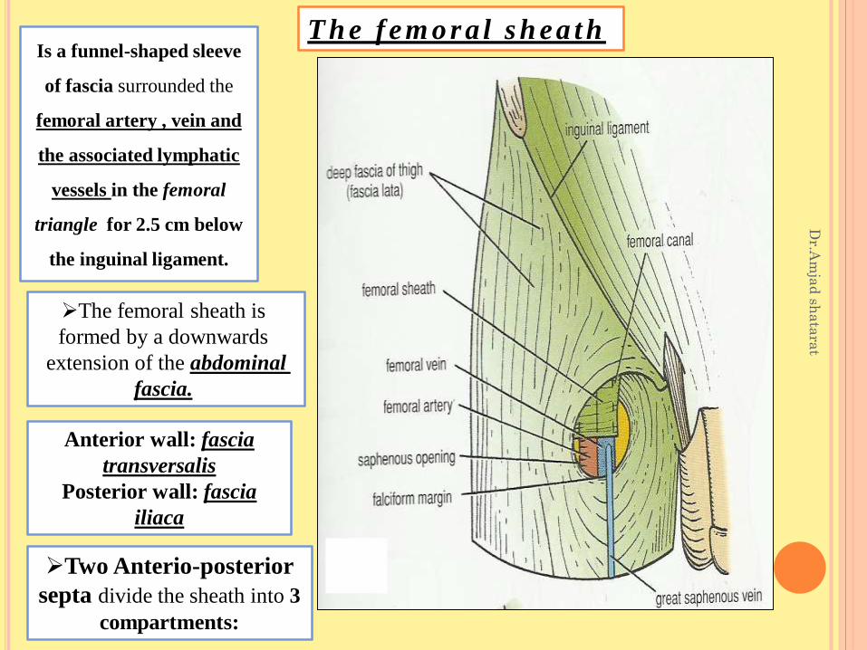
T h e f em o r a l s h ea t hIs a funnel-shaped sleeve
of fascia surrounded the
femoral artery , vein and
the associated lymphatic
vessels in the femoral
triangle for 2.5 cm below
the inguinal ligament.
The femoral sheath is
formed by a downwards
extension of the abdominal
fascia.
Anterior wall: fascia
transversalis
Posterior wall: fascia
iliaca
Two Anterio-posterior
septa divide the sheath into 3
compartments:

2-Intermediate compartment (venous)
Dr.A
mja
dsh
ata
rat
3-Medial compartment (lymphatic)
occupied by the lymph vessels
?
1-Lateral compartment (arterial)
occupied by the femoral vein
occupied by the femoral
artery and femoral branch
of the genitofemoral nerve
(also Called
f e m o r a l c a n a l

Is the small medial
compartment for the
lymph vessels. 1.3 cm In
length. just admits the tip
of the little finger.
Note: the femoral ring is
wider in femals because of
their wider pelvis and
therefore, femoral hernia
is commoner in femals
than in males
The femoral septum
(is a condensation of
extraperitoneal tissue),
closes the ring.
Its upper opening is
called the femoral ring.
Dr.A
mja
d sh
ata
rat
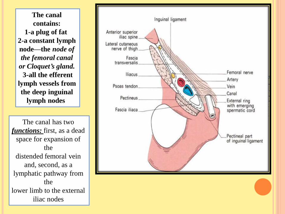
F e m o r a l c a n a l
Dr.A
mja
d sh
ata
rat
The canal
contains:
1-a plug of fat
2-a constant lymph
node—the node of
the femoral canal
or Cloquet’s gland.
3-all the efferent
lymph vessels from
the deep inguinal
lymph nodes
The canal has two
functions: first, as a dead
space for expansion of
the
distended femoral vein
and, second, as a
lymphatic pathway from
the
lower limb to the external
iliac nodes
Dr.A
mja
d sh
ata
rat
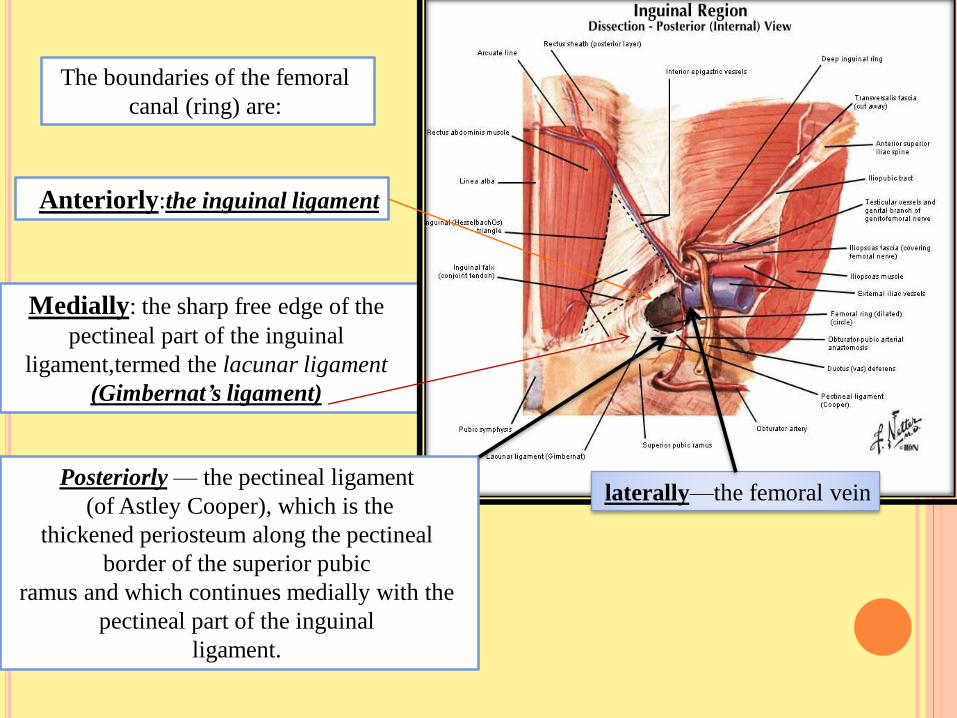
Anteriorly:the inguinal ligament
Medially: the sharp free edge of the
pectineal part of the inguinal
ligament,termed the lacunar ligament
(Gimbernat’s ligament)
laterally—the femoral veinPosteriorly — the pectineal ligament
(of Astley Cooper), which is the
thickened periosteum along the pectineal
border of the superior pubic
ramus and which continues medially with the
pectineal part of the inguinal
ligament.
The boundaries of the femoral
canal (ring) are:

Dr.A
mja
d sh
ata
rat
lacunar ligament (Gimbernat’s ligament)

The part of the femoral sheath that
forms the femoral canal is not adherent
to the walls of the small lymph vessels; it
is this site that forms
a potentially weak area in the abdomen.
The lower end of the canal is
normally closed by the adherence of
its medial wall to the tunica
adventitia of the femoral vein.
A protrusion of peritoneum could be
forced down the femoral canal, pushing
the femoral septum. Such a condition is
known as a femoral hernia.
Dr.A
mja
d sh
ata
rat

Dr.A
mja
d sh
ata
rat

A protrusion of abdominal parietal
peritoneum down
through the
femoral canal to form hernial sac
The neck of the hernial sac is located
below and lateral
to the pubic tubercle
In femoral hernia
While in the inguinal hernia
The neck of the hernial sac
is located
above and medial
to the pubic tubercle
Femoral herniaD
r.Am
jad
sha
tara
t

Dr.A
mja
d sh
ata
rat
NECK
OF HERNIAL
SAC, CAN YOU
SEE THE
DIFFERENCE
BETWEEN THE
TWO?POSITION,
SHAPE

Dr.A
mja
d sh
ata
rat
As the hernial sac enlarges, it
emerges through the saphenous
opening
then turns upwards along the
pathway presented by the
superficial epigastric
and superficial circumflex iliac
vessels so that it may come to
project
above the inguinal ligament.
There should not, however, be
any difficulty
in differentiating between an
irreducible femoral and
inguinal hernia; the
neck of the former must
always lie below and lateral to
the pubic tubercle
whereas the sac of the latter
extends above and medial to
this landmark

The neck of the femoral canal is narrow and bears a particular sharp
medial border; for this reason, irreducibility and strangulation occur
more commonly at this site than at any other. In order to enlarge the
opening of the canal at operation on a strangulated case, this sharp edge
of Gimbernat’s lacunar ligament may require incision;
there is a slight risk
of damage to the abnormal obturator artery in this
manoeuvre and it is
safer to enlarge the opening by making several small nicks into the ligament.
The safe alternative is to divide the inguinal ligament, which can then
be repaired.

Dr.A
mja
d sh
ata
rat
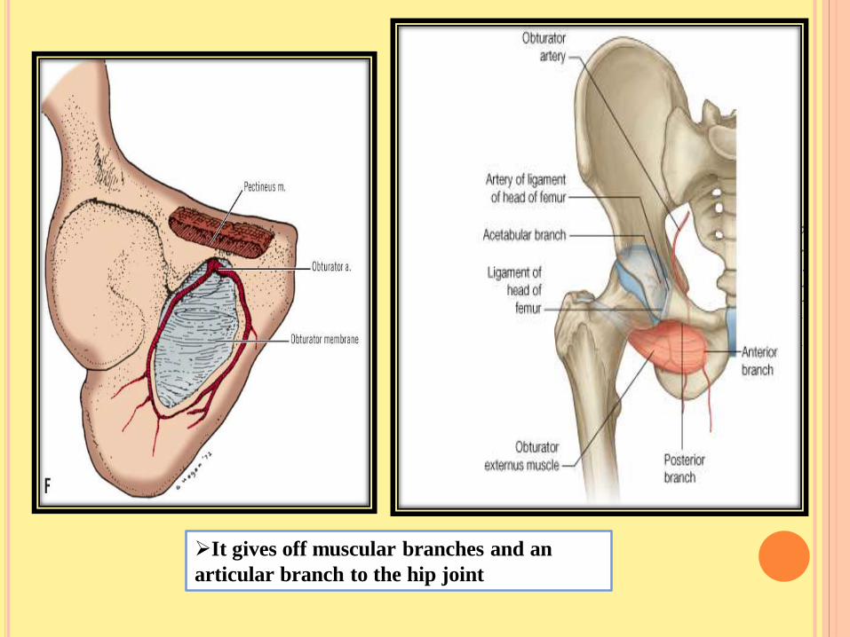
Note.
the obturator artery.
Obturator Artery
The obturator
artery is a branch
of the internal
iliac artery
It passes
forward on the
lateral wall of the
pelvis and
accompanies the
obturator nerve
Dr.A
mja
d sh
ata
rat
It gives off muscular branches and an
articular branch to the hip joint

Dr.A
mja
d sh
ata
rat
Note.
Normally there is
an anastomosis
between the pubic
branch of the
inferior epigastric
artery and the
pubic branch of
the obturator
artery.
A view from inside the abdomen

Dr.A
mja
d sh
ata
rat
Occasionally
the obturator artery is entirely replaced by this branch from the
inferior epigastric—the abnormal obturator artery.;
This aberrant vessel usually passes
laterally to the femoral canal and is out
of harm’s way
rarely, it passes behind Gimbernat’s ligament
and it is then in surgical danger.