Embed Size (px)
Citation preview

Muscle Origin Insertion Nerve Supply Action
Sartorius Anterior superior iliac spine Upper medial surface of shaft of
tibia (SGS)
Femoral nerve Flexes, abducts, laterally rotates thigh at
hip joint; flexes and medially rotates leg
at knee joint
Iliacus Iliac fossa of hip bone With psoas into lesser trochanter
of femur
Femoral nerve Flexes thigh on trunk
Psoas Major Transverse processes, bodies,
and intervertebral discs of
the 12th thoracic and five
lumbar vertebrae
With iliacus into lesser
trochanter of femur
L1.2.3 Flexes thigh on trunk; if thigh is fixed, it
flexes the trunk on thigh as in sitting up
from lying down
Pectineus Superior ramus of pubis Upper end of lineaaspera of
shaft of femur
Femoral nerve Flexes and adducts thigh at hip joint
Quadriceps femoris
Rectus femoris Straight head: anterior inferior
iliac spine
Reflected head: ilium above
acetabulum
Quadriceps tendon into
patella, then via
ligamentum patellae into
tubercle of tibia
Femoral nerve Extension of leg at knee joint; flexes thigh
at hip joint
Vastuslateralis Upper end and shaft of femur Femoral nerve Extension of leg at knee joint; stabilizes
patellaVastusmedialis Upper end and shaft of femur Femoral nerve
Vastusintermed
ius
Anterior and lateral surfaces of
shaft of femur
Femoral nerve Extension of leg at knee joint; articularis
genu retracts synovial membrane
Front of the thigh
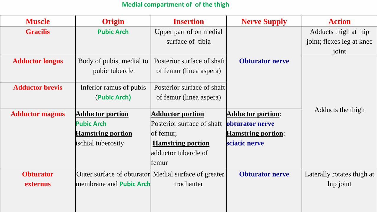
Muscle Origin Insertion Nerve Supply Action
Gracilis Pubic Arch Upper part of on medial
surface of tibia
Obturator nerve
Adducts thigh at hip
joint; flexes leg at knee
joint
Adductor longus Body of pubis, medial to
pubic tubercle
Posterior surface of shaft
of femur (linea aspera)
Adducts the thigh
Adductor brevis Inferior ramus of pubis
(Pubic Arch)
Posterior surface of shaft
of femur (linea aspera)
Adductor magnus Adductor portion
Pubic Arch
Hamstring portion
ischial tuberosity
Adductor portion
Posterior surface of shaft
of femur,
Hamstring portion
adductor tubercle of
femur
Adductor portion:
obturator nerve
Hamstring portion:
sciatic nerve
Obturator
externus
Outer surface of obturator
membrane and Pubic Arch
Medial surface of greater
trochanter
Obturator nerve Laterally rotates thigh at
hip joint
Medial compartment of of the thigh
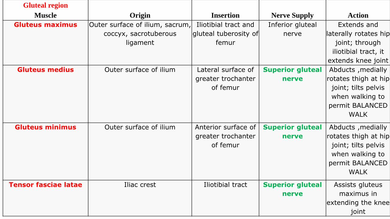
Gluteal region
Muscle Origin Insertion Nerve Supply Action
Gluteus maximus Outer surface of ilium, sacrum,
coccyx, sacrotuberous
ligament
Iliotibial tract and
gluteal tuberosity of
femur
Inferior gluteal
nerve
Extends and
laterally rotates hip
joint; through
iliotibial tract, it
extends knee joint
Gluteus medius Outer surface of ilium Lateral surface of
greater trochanter
of femur
Superior gluteal
nerve
Abducts ,medially
rotates thigh at hip
joint; tilts pelvis
when walking to
permit BALANCED
WALK
Gluteus minimus Outer surface of ilium Anterior surface of
greater trochanter
of femur
Superior gluteal
nerve
Abducts ,medially
rotates thigh at hip
joint; tilts pelvis
when walking to
permit BALANCED
WALK
Tensor fasciae latae Iliac crest Iliotibial tract Superior gluteal
nerve
Assists gluteus
maximus in
extending the knee
joint
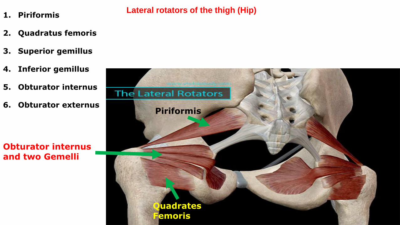
Piriformis
Quadrates Femoris
Obturator internus and two Gemelli
Lateral rotators of the thigh (Hip)1. Piriformis
2. Quadratus femoris
3. Superior gemillus
4. Inferior gemillus
5. Obturator internus
6. Obturator externus
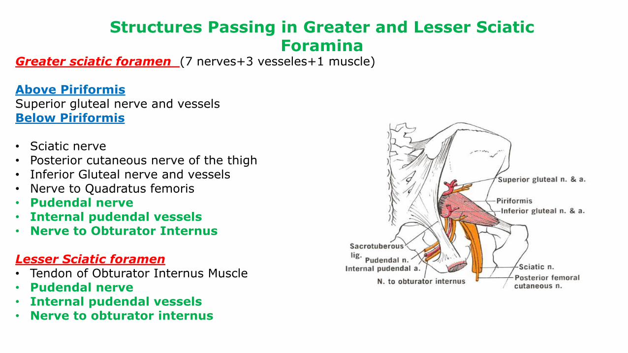
Structures Passing in Greater and Lesser Sciatic Foramina
Greater sciatic foramen (7 nerves+3 vesseles+1 muscle)
Above PiriformisSuperior gluteal nerve and vesselsBelow Piriformis
• Sciatic nerve• Posterior cutaneous nerve of the thigh• Inferior Gluteal nerve and vessels• Nerve to Quadratus femoris• Pudendal nerve• Internal pudendal vessels• Nerve to Obturator Internus
Lesser Sciatic foramen• Tendon of Obturator Internus Muscle• Pudendal nerve• Internal pudendal vessels• Nerve to obturator internus
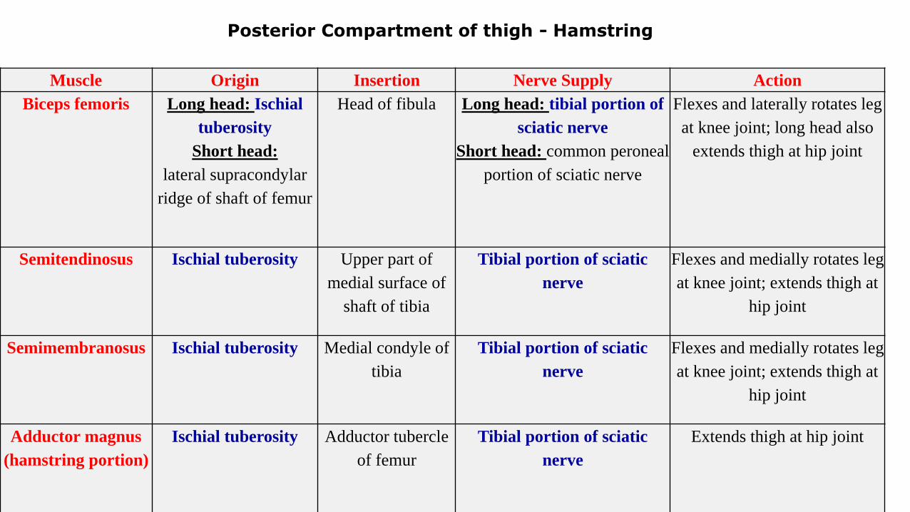
Muscle Origin Insertion Nerve Supply Action
Biceps femoris Long head: Ischial
tuberosity
Short head:
lateral supracondylar
ridge of shaft of femur
Head of fibula Long head: tibial portion of
sciatic nerve
Short head: common peroneal
portion of sciatic nerve
Flexes and laterally rotates leg
at knee joint; long head also
extends thigh at hip joint
Semitendinosus Ischial tuberosity Upper part of
medial surface of
shaft of tibia
Tibial portion of sciatic
nerve
Flexes and medially rotates leg
at knee joint; extends thigh at
hip joint
Semimembranosus Ischial tuberosity Medial condyle of
tibia
Tibial portion of sciatic
nerve
Flexes and medially rotates leg
at knee joint; extends thigh at
hip joint
Adductor magnus
(hamstring portion)
Ischial tuberosity Adductor tubercle
of femur
Tibial portion of sciatic
nerve
Extends thigh at hip joint
Posterior Compartment of thigh - Hamstring
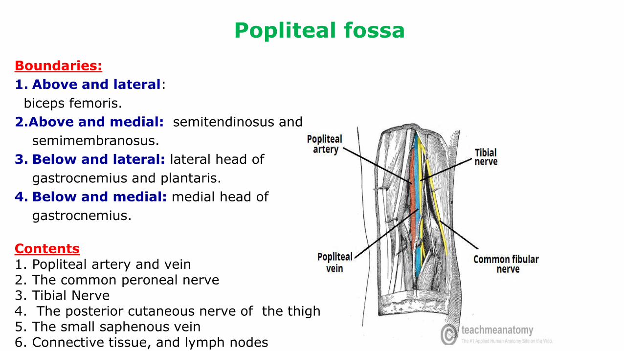
Boundaries:
1. Above and lateral:
biceps femoris.
2.Above and medial: semitendinosus and
semimembranosus.
3. Below and lateral: lateral head of
gastrocnemius and plantaris.
4. Below and medial: medial head of
gastrocnemius.
Contents1. Popliteal artery and vein2. The common peroneal nerve 3. Tibial Nerve4. The posterior cutaneous nerve of the thigh5. The small saphenous vein6. Connective tissue, and lymph nodes
Popliteal fossa
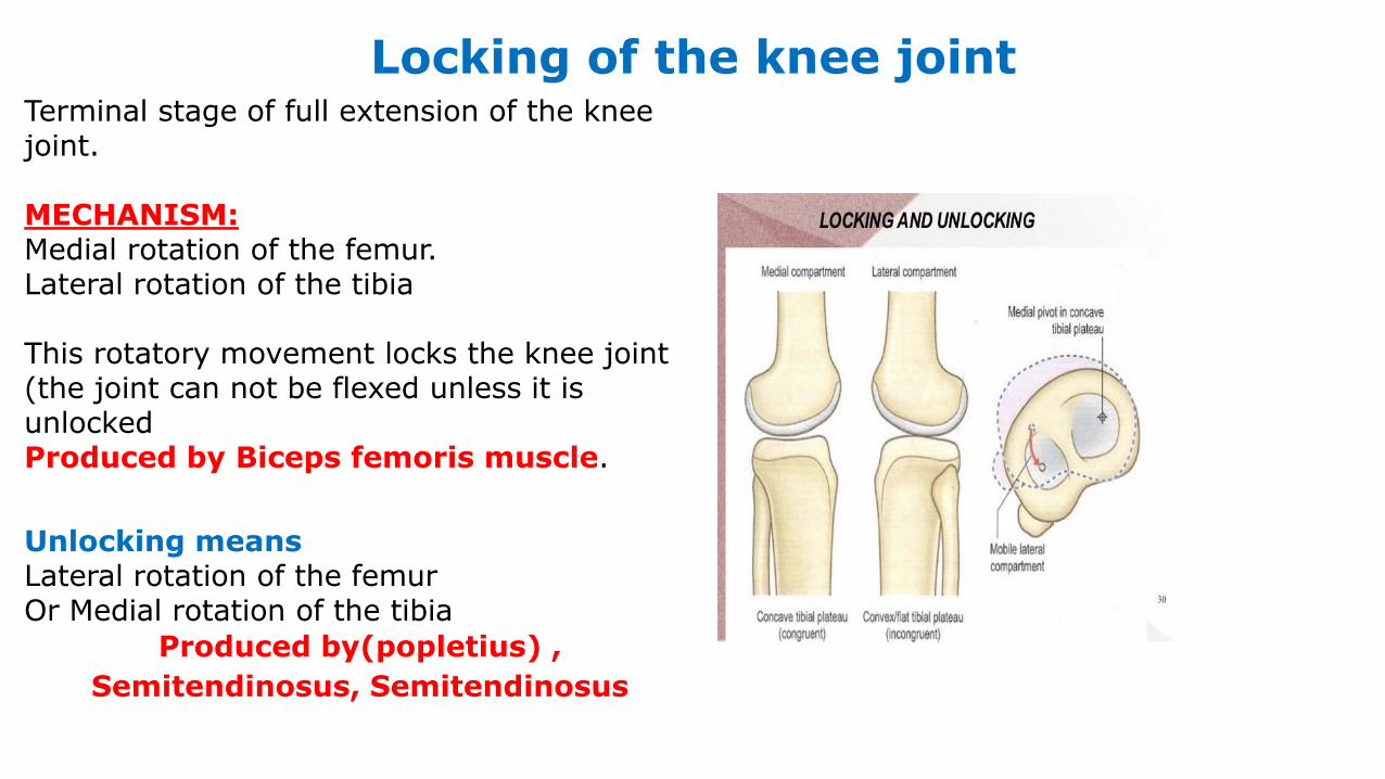
Locking of the knee jointTerminal stage of full extension of the knee joint.
MECHANISM:Medial rotation of the femur. Lateral rotation of the tibia
This rotatory movement locks the knee joint (the joint can not be flexed unless it is unlocked Produced by Biceps femoris muscle.
Unlocking meansLateral rotation of the femurOr Medial rotation of the tibia
Produced by(popletius) ,
Semitendinosus, Semitendinosus
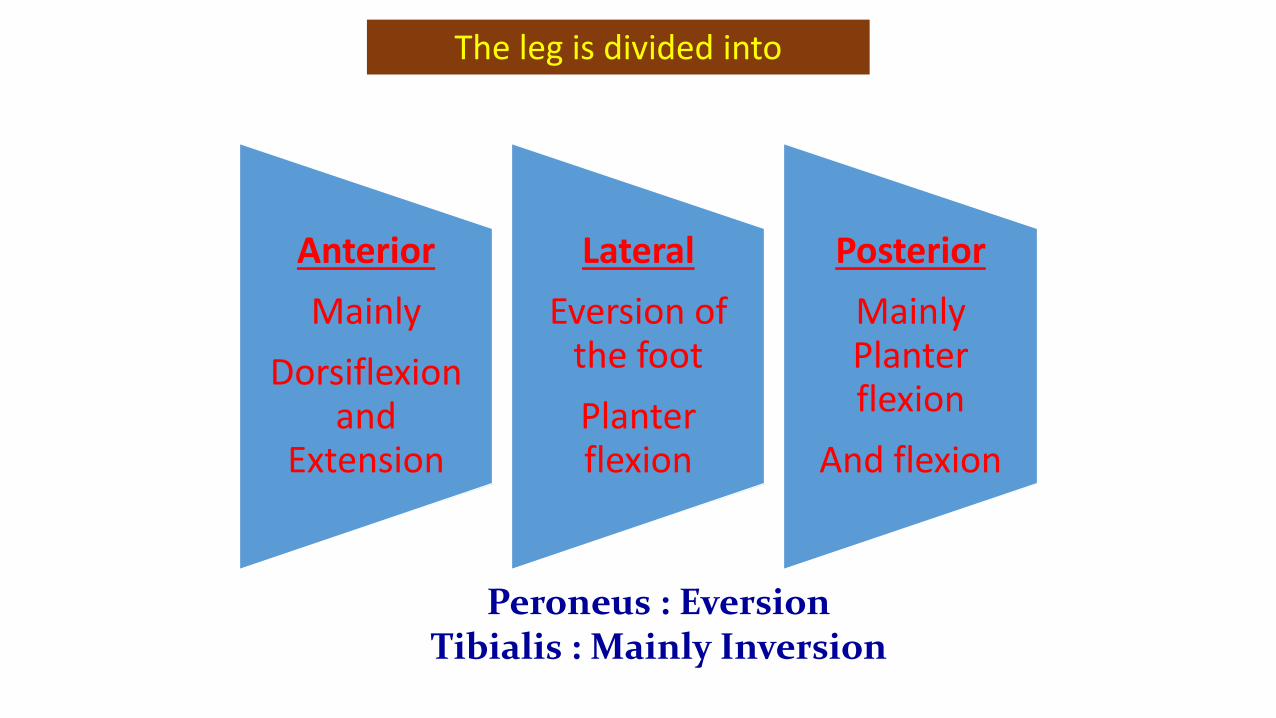
The leg is divided into
Anterior
Mainly
Dorsiflexion and
Extension
Lateral
Eversion of the foot
Planter flexion
Posterior
Mainly Planter flexion
And flexion
Peroneus : EversionTibialis : Mainly Inversion
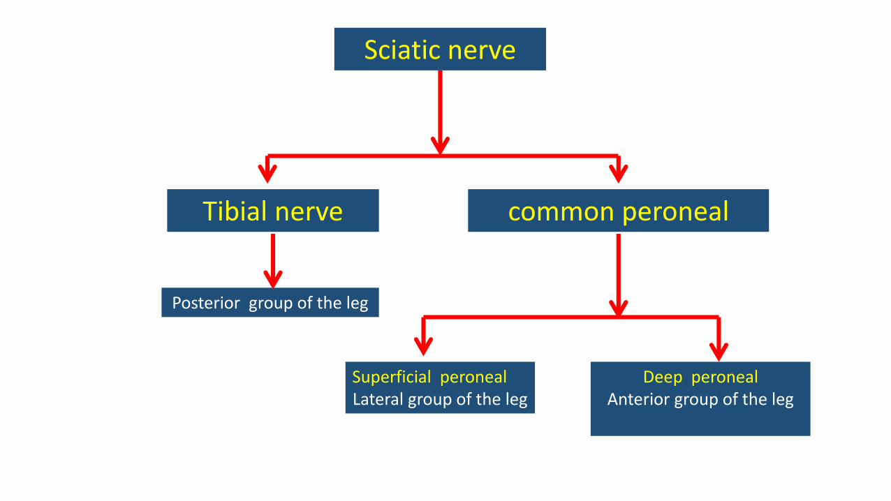
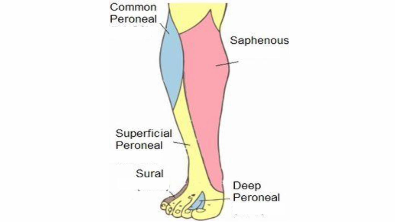
Sciatic nerve
common peronealTibial nerve
Superficial peronealLateral group of the leg
Deep peronealAnterior group of the leg
Posterior group of the leg
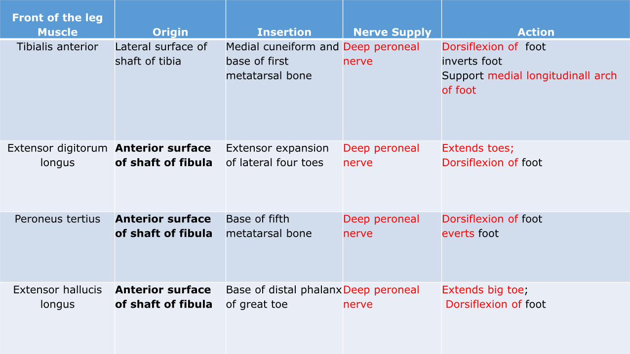
Front of the leg
Muscle Origin Insertion Nerve Supply Action
Tibialis anterior Lateral surface of
shaft of tibia
Medial cuneiform and
base of first
metatarsal bone
Deep peroneal
nerve
Dorsiflexion of foot
inverts foot
Support medial longitudinall arch
of foot
Extensor digitorum
longus
Anterior surface
of shaft of fibula
Extensor expansion
of lateral four toes
Deep peroneal
nerve
Extends toes;
Dorsiflexion of foot
Peroneus tertius Anterior surface
of shaft of fibula
Base of fifth
metatarsal bone
Deep peroneal
nerve
Dorsiflexion of foot
everts foot
Extensor hallucis
longus
Anterior surface
of shaft of fibula
Base of distal phalanx
of great toe
Deep peroneal
nerve
Extends big toe;
Dorsiflexion of foot
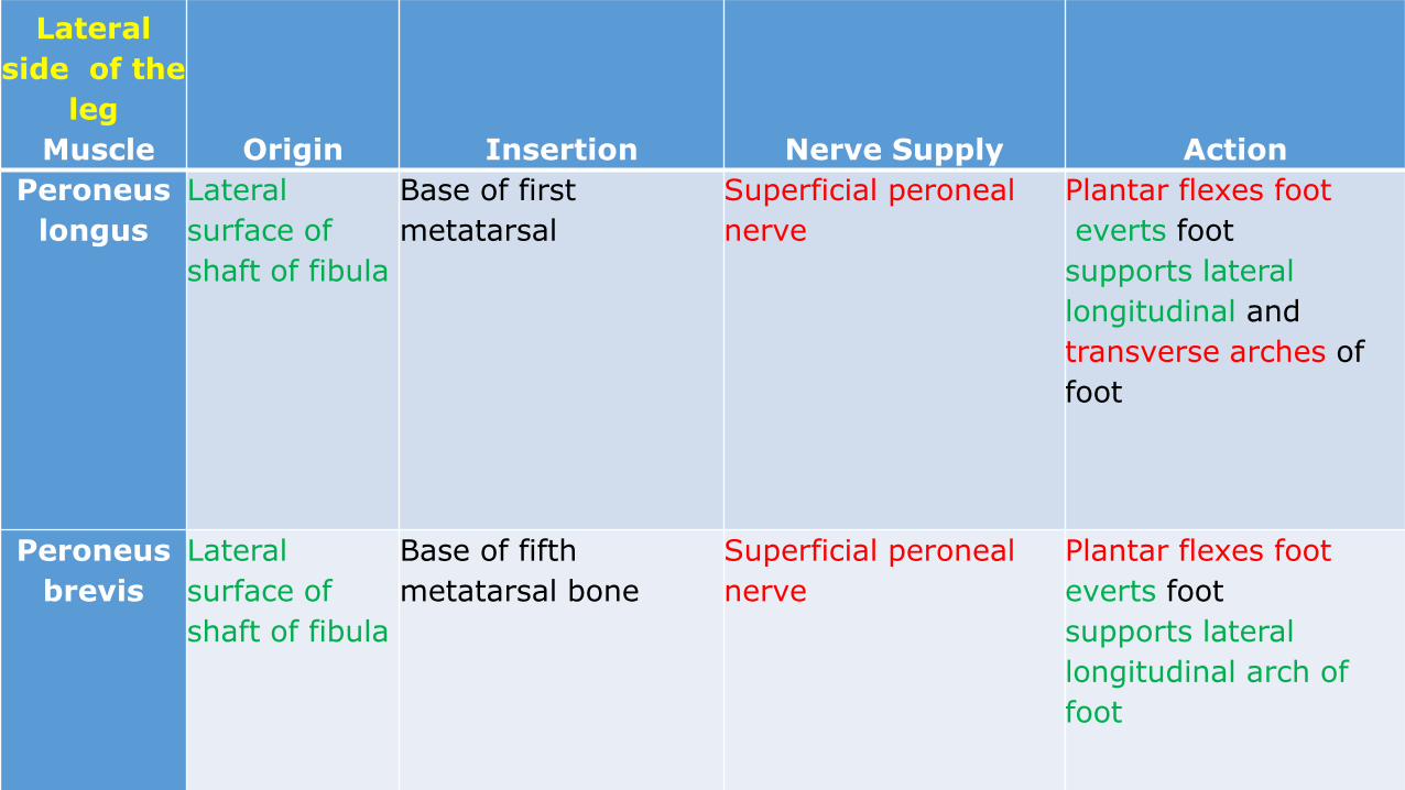
Lateral
side of the
leg
Muscle Origin Insertion Nerve Supply Action
Peroneus
longus
Lateral
surface of
shaft of fibula
Base of first
metatarsal
Superficial peroneal
nerve
Plantar flexes foot
everts foot
supports lateral
longitudinal and
transverse arches of
foot
Peroneus
brevis
Lateral
surface of
shaft of fibula
Base of fifth
metatarsal bone
Superficial peroneal
nerve
Plantar flexes foot
everts foot
supports lateral
longitudinal arch of
foot
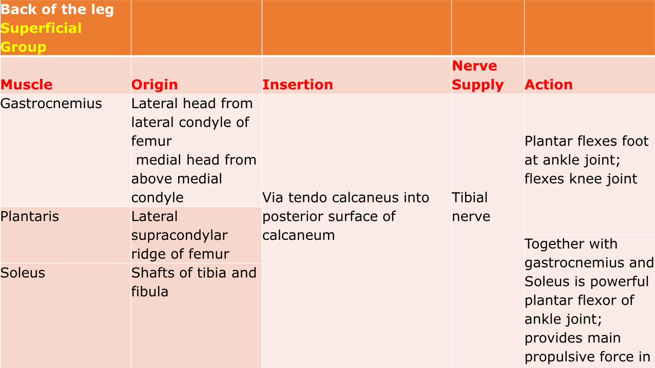
Back of the leg
Superficial
Group
Muscle Origin Insertion
Nerve
Supply Action
Gastrocnemius Lateral head from
lateral condyle of
femur
medial head from
above medial
condyle Via tendo calcaneus into
posterior surface of
calcaneum
Tibial
nerve
Plantar flexes foot
at ankle joint;
flexes knee joint
Plantaris Lateral
supracondylar
ridge of femurTogether with
gastrocnemius and
Soleus is powerful
plantar flexor of
ankle joint;
provides main
propulsive force in
walking and
Soleus Shafts of tibia and
fibula
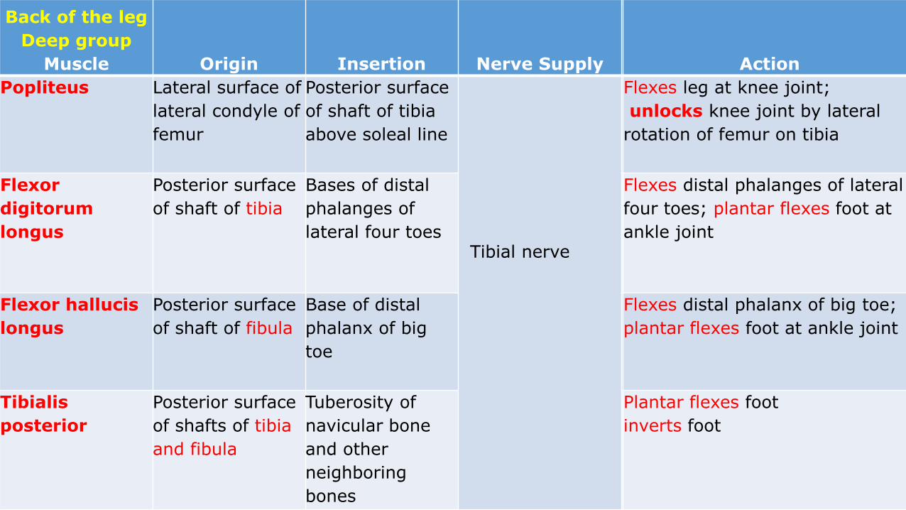
Back of the leg
Deep group
Muscle Origin Insertion Nerve Supply Action
Popliteus Lateral surface of
lateral condyle of
femur
Posterior surface
of shaft of tibia
above soleal line
Tibial nerve
Flexes leg at knee joint;
unlocks knee joint by lateral
rotation of femur on tibia
Flexor
digitorum
longus
Posterior surface
of shaft of tibia
Bases of distal
phalanges of
lateral four toes
Flexes distal phalanges of lateral
four toes; plantar flexes foot at
ankle joint
Flexor hallucis
longus
Posterior surface
of shaft of fibula
Base of distal
phalanx of big
toe
Flexes distal phalanx of big toe;
plantar flexes foot at ankle joint
Tibialis
posterior
Posterior surface
of shafts of tibia
and fibula
Tuberosity of
navicular bone
and other
neighboring
bones
Plantar flexes foot
inverts foot
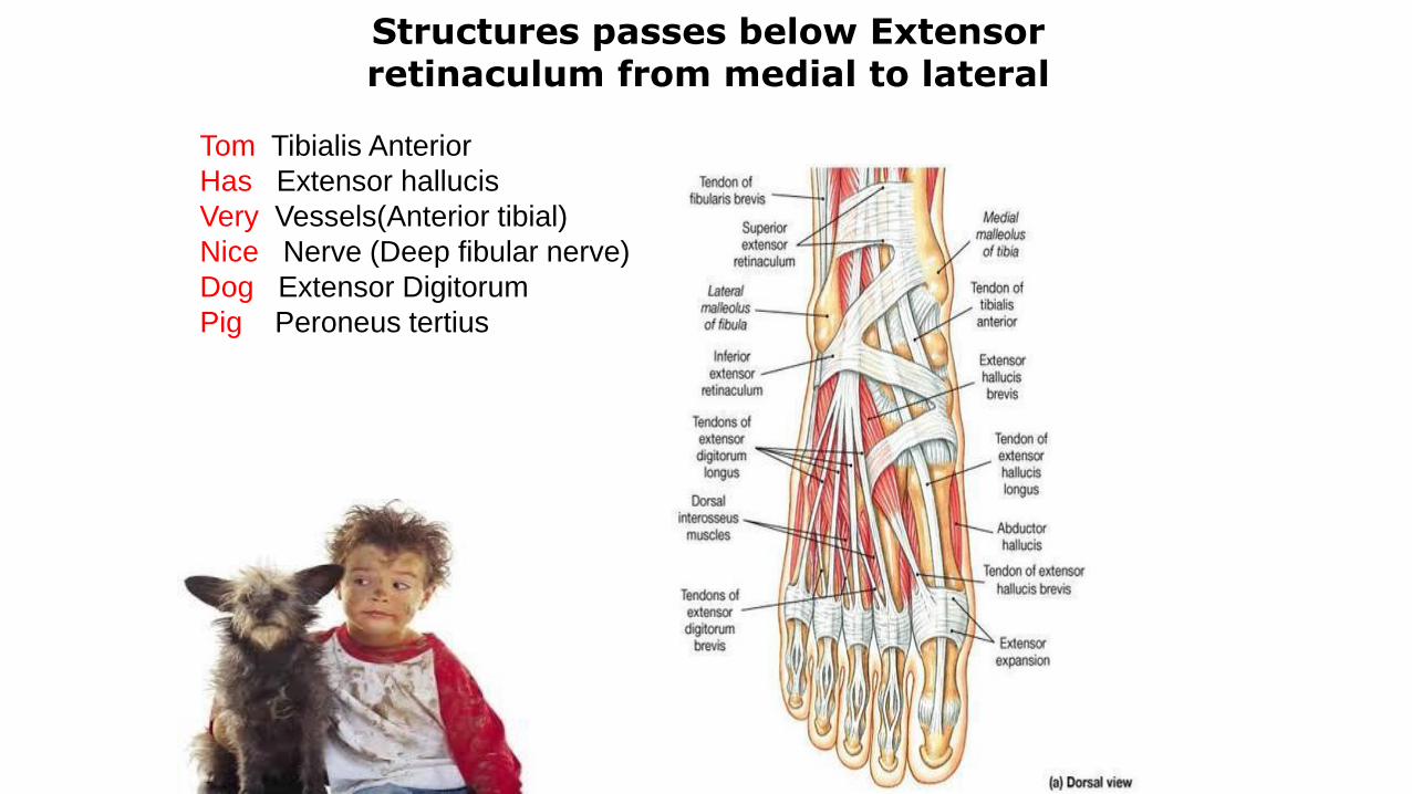
Tom Tibialis Anterior
Has Extensor hallucis
Very Vessels(Anterior tibial)
Nice Nerve (Deep fibular nerve)
Dog Extensor Digitorum
Pig Peroneus tertius
Structures passes below Extensor retinaculum from medial to lateral
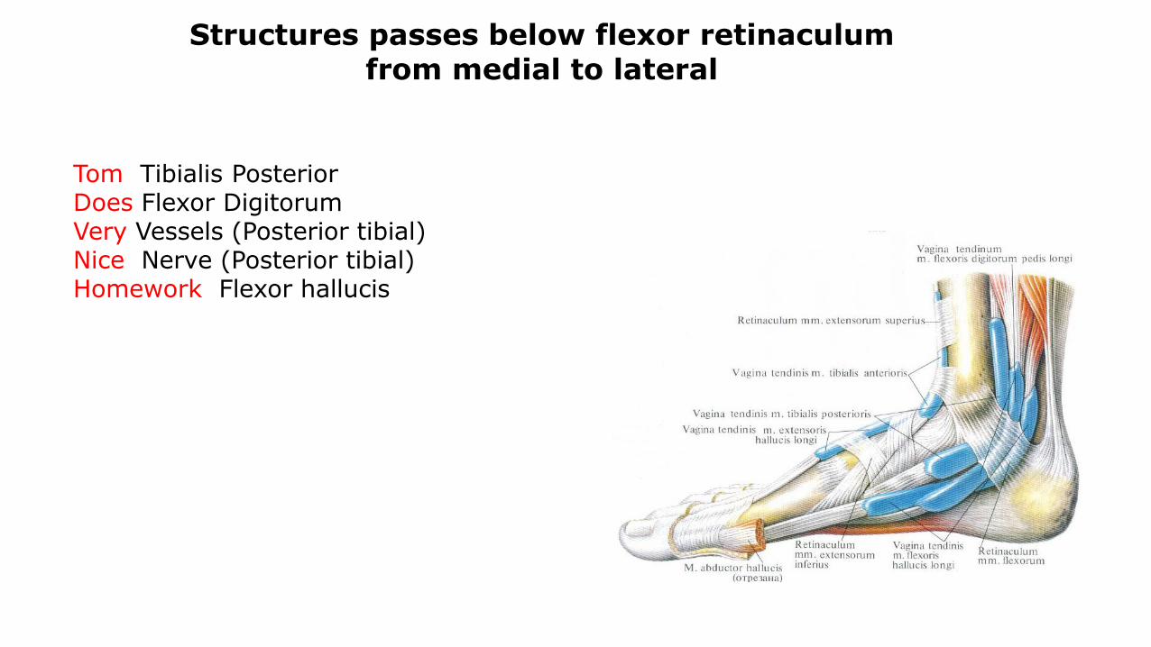
Structures passes below flexor retinaculum from medial to lateral
Tom Tibialis Posterior Does Flexor DigitorumVery Vessels (Posterior tibial)Nice Nerve (Posterior tibial)Homework Flexor hallucis
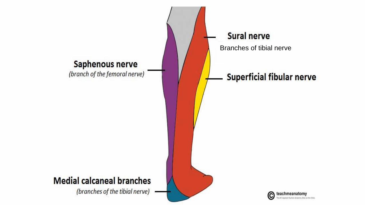
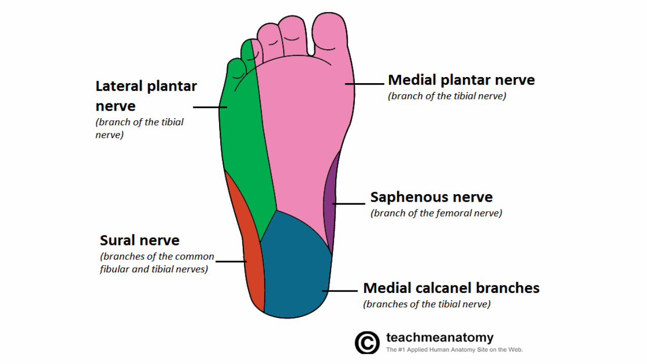
Branches of tibial nerve
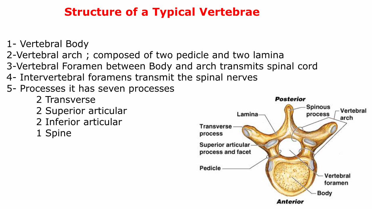
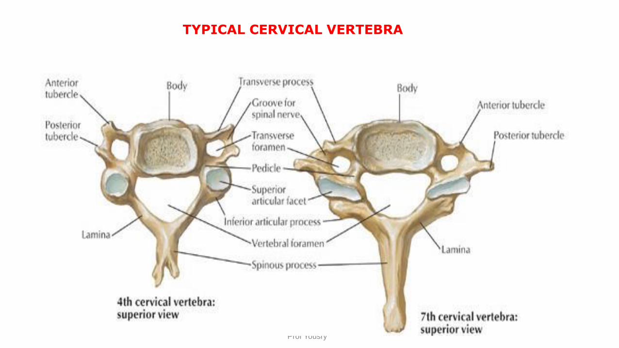
Structure of a Typical Vertebrae
1- Vertebral Body2-Vertebral arch ; composed of two pedicle and two lamina3-Vertebral Foramen between Body and arch transmits spinal cord4- Intervertebral foramens transmit the spinal nerves 5- Processes it has seven processes
2 Transverse2 Superior articular2 Inferior articular1 Spine
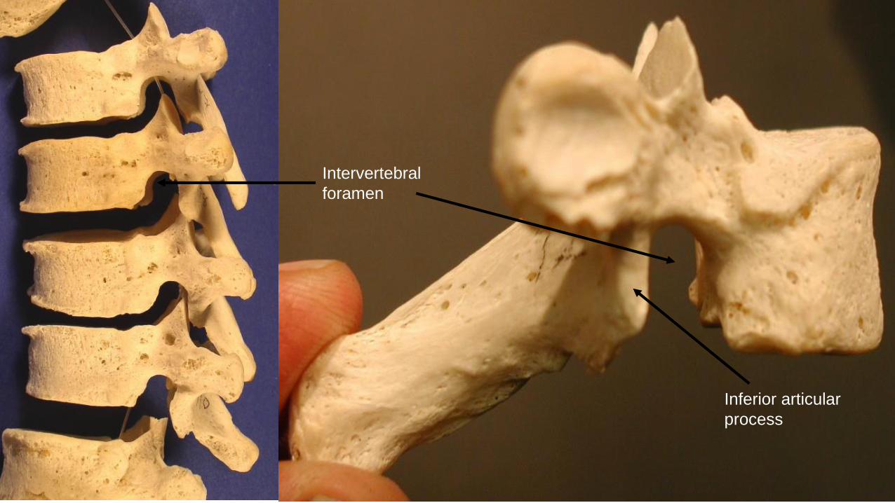
Intervertebral
foramen
Inferior articular
process
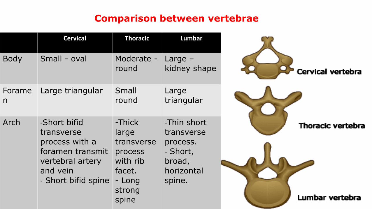
Cervical Thoracic Lumbar
Body Small - oval Moderate -
round
Large –
kidney shape
Forame
n
Large triangular Small
round
Large
triangular
Arch -Short bifid
transverse
process with a
foramen transmit
vertebral artery
and vein
- Short bifid spine
-Thick
large
transverse
process
with rib
facet.
- Long
strong
spine
-Thin short
transverse
process.
- Short,
broad,
horizontal
spine.
Comparison between vertebrae
Prof Yousry
TYPICAL CERVICAL VERTEBRA
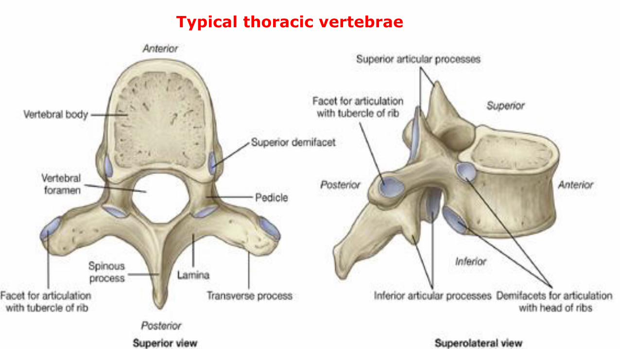
Typical thoracic vertebrae
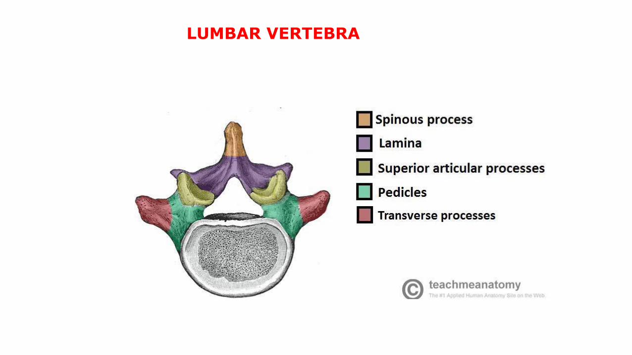
LUMBAR VERTEBRA
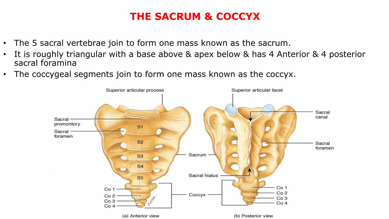
THE SACRUM & COCCYX
• The 5 sacral vertebrae join to form one mass known as the sacrum.
• It is roughly triangular with a base above & apex below & has 4 Anterior & 4 posterior sacral foramina
• The coccygeal segments join to form one mass known as the coccyx.
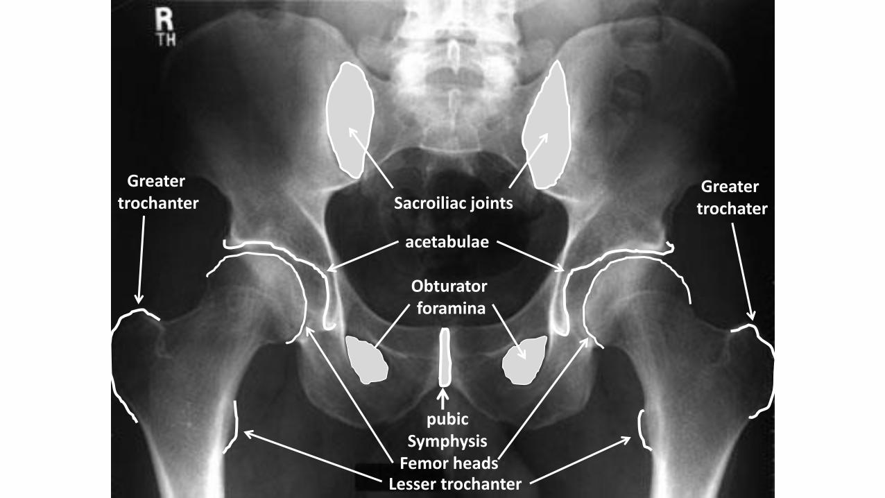
Sacroiliac joints
pubicSymphysis
acetabulae
Obturatorforamina
Lesser trochanterFemor heads
Greater trochanter
Greater trochater
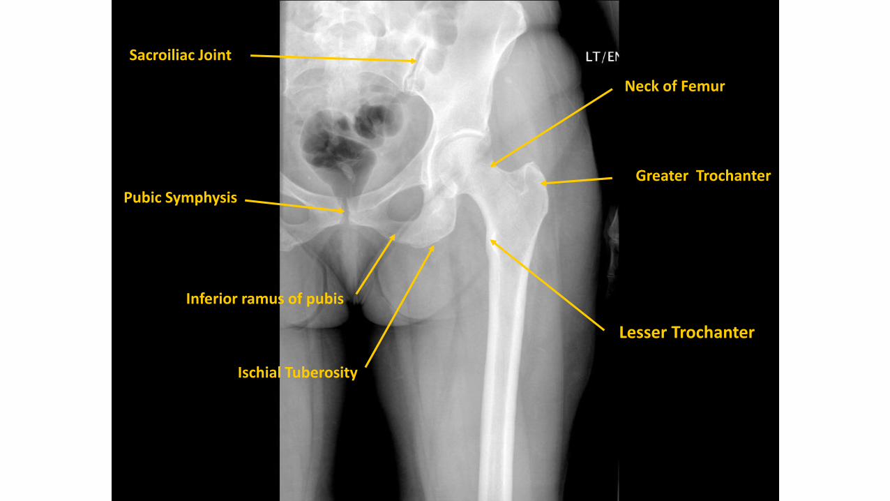
Greater Trochanter
Neck of Femur
Pubic Symphysis
Inferior ramus of pubis
Ischial Tuberosity
Lesser Trochanter
Sacroiliac Joint
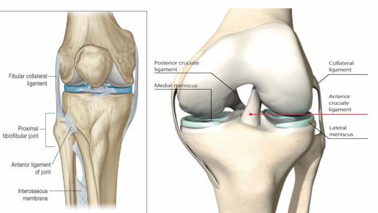
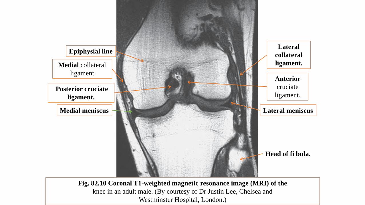
Fig. 82.10 Coronal T1-weighted magnetic resonance image (MRI) of the
knee in an adult male. (By courtesy of Dr Justin Lee, Chelsea and
Westminster Hospital, London.)
Posterior cruciate
ligament.
Medial collateral
ligament
Medial meniscus
Epiphysial line
Anterior
cruciate
ligament.
Lateral
collateral
ligament.
Lateral meniscus
Head of fi bula.
The following joints : know type , ligaments and their movements1-Hip2-Knee3-Superior and inferior tibiofibular joint4-Ankle5-Subtalar6-Mid tarsal joints