Embed Size (px)
Citation preview

AVTBOR'S AEWBACT OF THIS PMER ISSUSD BY THI BIBLIOQWEIC SERVICE. JULY 19
THE DEVELOPMENT OF THE HYPOPHYSIS CEREBRI OF THE RABBIT (LEPUS CUNICULUS L.)
WAYNE; J. ATWELL
From the Department of Anatomy, University of Michigan
THIRTY-NINE FIQURES
CONTENTS 1. Introduction.. . . . . . . . . . . . . . . . . . . . . . . . . . . . . . . . . . . . . . . . . . . . . . . . . . . . . . . . . . 271 2. Review of literature ..........................................
A. Special questions concerned with the development of the h 1. Entodermal origin.. . . . . . . . . . . . . . . . . . . . . . . . . . . . . . . 2 . The relation of the notochord to the hypophysis. 3. The lobes of the hypophysis.. . .
a) General ....................... b) The anterior process.. . . . . . . . . . . . . . . . . . . . . . . . . . . . . . . . . . . . 277
d) The relation of the lateral lobes to the 'pars tuberalis'. ... 281
. . . . . . . . . . . . . . . . . . . 276
. . . . . . . . . . . . . . . . . . . 276
....................... 280
4. The structure of the intermediate part.. ...................... 283 . . . . . . . . . . 285
c) The lateral lobes.. . . . . . . . . . . . . .
5. The development of the neural lobe.. . . . .
3. Material and methods., . . . . . . . . . . . . . . . . . . . . . . . . . . . . . . . .
5. Discussion of observations.. .......................... . . . . . . . . . . 318
1. The hypophysial stalk . . . . . . . . . . . . . . . . . . . . . . . . . . . . . . 320 2. The residual iumen.. . . . . . . . . . . . . . . . . . . . . . . . . . . . . . . . . 321
D. The development of the neural lobe.. ............................. 325 E. The neuro-epithelial contacts and their significance. . . . . . . . . . . . . . . . 327 F. Terminology and phylogeny of the lobes of the hypophysis.. ....... 329
....................... 334 6. Summary and conclusions.. ............................................ 331 7. Literature cited.. ................. ,. .........
1. INTRODUCTION
Notwithstanding the numerous studies to which the hypoph- ysis has been subjected, many of its deeper problems .remain unsolved. Few investigators have confined themselves to the
TEI AYBRICAX JOURNAL OF INATOMY. VOL. 24, NO. 3 SEPTEMBER, 1918
271
272 WAYNE J. ATWELL
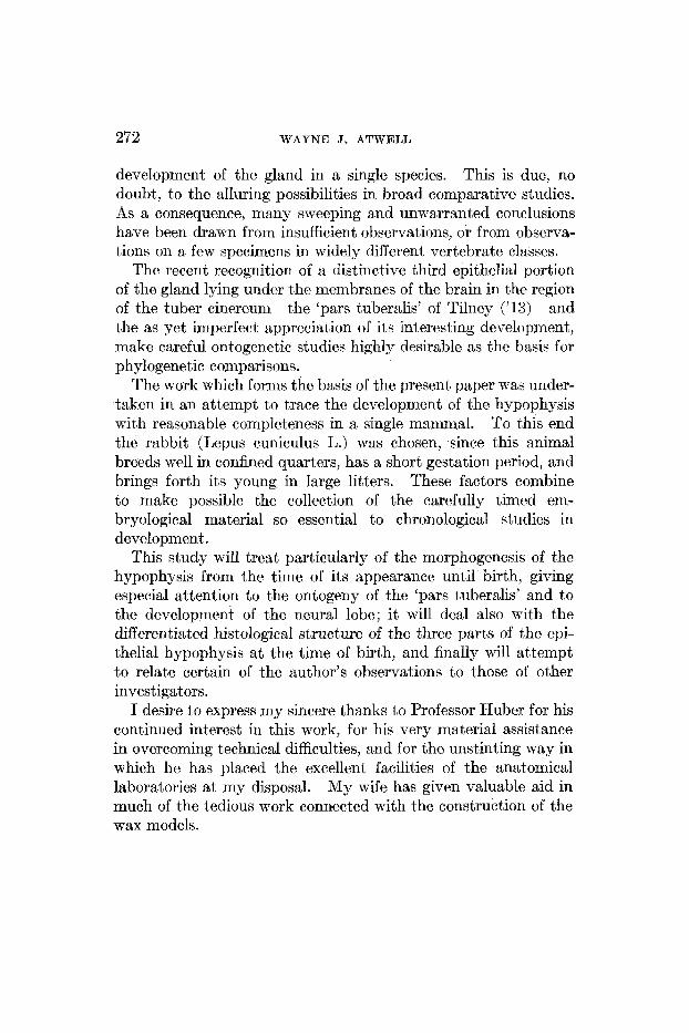
development of the gland in a single species. This is due, no doubt, to the alluring possibilities in broad comparative studies. As a consequence, many sweeping and unwarranted conclusions have been drawn from insufficient observations, or from observa- tions on a few specimens in widely different vertebrate classcs.
The recent recognition of a distinctive third epithelial portion of the gland lying under the membranes of the brain in the region of the tuber cinereurn-the ‘pars tuberalis’ of Tilney (’13)-and the as yet imperfect, appreciation of its interesting development, make careful ontogenctic studies highly desirable as the basis for phylogenetic comparisons.
The work which forms the basis of the present paper was under- taken in an attempt to trace the development of the hypophysis with reasonable completeness in a single mammal. To this end the rabbit (Lepus cuniculus L.) was chosen, since this animal breeds well in confined quarters, has a short gestation period, and brings forth its young in large litters. These factors combine to make possible the collection of the carefully timed em- bryological material so essential to chronological studies in development.
This study will treat particularly of the inorphogenesis of the hypophysis from the time of its appearance until birtrh, giving especial attention t o the ontogeny of the ‘pars tuberalis’ and to the development of the neural lobe; it will deal also with the differentiated histological structure of the three parts of the epi- thelial hypophysis at the time of birth, and finally ~7ill attempt to relate certain of the author’s observations to those of other investigators.
I desire to express my sincere thanks to Professor Huber for his continued interest in this work, for his very material assistance in overcoming technical difficulties, and for the unstinting way in which he has placed the excellent facilities of the anatomical laboratories at my disposal. My wife has given valuable aid in much of the tedious work connected with the construction of the wax models.

DEVELOPMENT OF HYPOPHYSIS O F RABBIT 2’73
2. REVIEW OF LITERATURE
A . Special questions concerned wish the development of the hypo ph ysis
1. Entodermal origin. An endless chain of discussion has been evoked by the question as to whether the epithelial hypophysis is derived from the entoderm or from the ectoderm, or whether it is compounded of elements derived from both of these germ layers. Since the time of Goett,e (’74), Balfour (’74), and Mi- halkovics (’75), only a few authors have maintained that this portion of the gland is developed from entoderm alone. There have been many, however, to state that, the primary ectodermal anlage is later augmented by a larger or smaller contribution from the entoderm.
Hoffman (’86) and Ostroumoff (’88) believe the hypophysis in certain reptiles to be entodermal in origin. Kupffer (’94) de- scribes a growth from the cephalic end of the foregut to meet the dorsal ~ v a l l of Rat>hke’s pocket. This he interprets as an at- tempted recurrence of a primitive pre-oral mouth or ‘paleostoma.’ This suggestion of Kupffer’s has caused many writers to attach great phylogenetic importance to the hypophysis. Furthermore, according to Kupffer, the entoderrn makes important contri- butions to the epithelial lobe of the hypophysis.
In a previous paper (Atwell, ’15) reference was made to the observations of Saint-Reniy (’95), Valenti (’95 a, ’95 b, and ’97)’ Nixsbaum (’96), and of Bruni (’141, according to all of whom the entoderm of the pouch-like cephalic extremity of the foregut (‘Seessel’s pouch’) fuses with t,he ectodermal hypophysial anlage and possibly contributes a few cells to it.
Orrri (’00) divides the glandular portion of the hypophysis of Gongylus ocellatus into two lobes, one of which is in relation to the infundibular process, while the other lies ventral to thc first. Although be cannot trace a difference histologically in these two lobes, he believes it very probable that the dorsal lobe, which lies close to the irifundibular process is entodermal and that the ventral lobe is ectoderrnal.

274 WAYNE J . ATWELL
A considerable portion of the controversy concerning the ento- dermal origin of the glandular part of the hypophysis has centered about the Ganoid, Amia calva. Dean (’96) holds that in this fish the hypophysis is ectodermal. Reighard (’00) states that the Amian hypophysis is developed entirely from ectoderm, while Prather (’00) states that it is entirely from entoderm. Gregory (’02) cannot agree with Prather and believes that this part of the hypophysis is both ectodermal and entodermal.
Reighard and Mast (’08) have presented the most conclusive observations for this form. They find that the hypophysis of Amia is ectodermal in origin. They show that Prather was in error, 1) from a lack of the early stages which show a connection of the hypophysis anlage with the mother ectoderm, and, 2) be- cause of imperfect fixation which failed to bring out the line of separation between the hypophysis and the entoderm.
P. E. Smith (’14) has reopened the problem and conies to the conclusion that the anlage is ectodermal, but adds that “it can be said with considerable probability that the entodem con- tributes to the composition of the hypophysis.”
Atwell (’15) saw the epithelial connection between Seessel’s and Rathke’s pouches in the chick much as described by Saint Rerny, Valenti and Bruni. He adds the observation that the entodermal bud which forms the connecting strand has constantly in relation to it the cephalic extremity of the notochord, both before and after the ecto-entodermal fusion has formed. The entodermal strand loses its connection with Seessel’s pouch and becomes incorporated into the dorsal wall of Rathke’s pouch. A relationship was noted between the anterior end of the notochord and a small entodermal bud in rabbit embyros, but it could not be shown that any entoderm fuses with the hypophysis anlage in this mammal.
It is to be notred that while the above-mentioned investigators believe that entoderm enters into the formation of the hypoph- ysis, the majority of them freely admit that the main portion of the gland is ectodermal. The only one in recent years to claim that a considerable portion of the definitive mammalian hypophysis is derived from the entoderm is Miller (’16). This

DEVELOPMENT OF HYPOPHYSIS OF RABBIT 275
author, who studied the hypophysis of the pig (Sus scrofa) has noted that “the notochord pulls away from the pharynx carrying with it a mass of cells (entoderm),” which later becomes fused with the ectodermal anlage. This is much as described by Atwell for the chick. However, Miller further maintains that this entodermal component ‘rotates anteriorly and superiorly,’ be- comes encapsulated by growth of the ‘lateral cords,’ and form3 the medulla of the anterior lobe. The cells supposed to be de- rived from the entoderm have a different histological appearance from those derived from the ectoderm.
P. E. Smith (’16)’ likewise B. M. Allen (’16, ’171, has removed the glandular ectodermal anlage from young larvae of the frog. In successfully operated animals, the anterior lobe was entirely lacking. Smith concludes that this apparently demonstrates conclusively that the entoderm has not the intrinsic power to form a hypophysis. If it enters into the formation of the gland at all, it must be considered as a tissue inclusion which be- comes changed through its adaptability into glandular parenchyma, a conclusion previously drawn by the writer, Smith (’14).
The close proximity of the cephalic extremity of the chorda to the hypo- physial anlage has been taken by many to be significant of some influence the chorda may exert on the developing gland.
Koelliker (’79) has noted in a rabbit embryo of eleven days a close relation between the anterior end of the notochord and an outgrowth from the inferior part, of the dorsal wall of the hypophysis.
In the Normentafel of the rabbit’s dcvelopment Minot and Taylor (’05) note a “distinct connection between notochord and hypophysis” in both ten-and-one-half-day and eleven-day embryos. (Nos. 11 and 12). This connection apparently had disappeared entirely by eleven and one-half days of development.
Woerdeman (’131, observing embryos of Sus scrofa corre- sponding in age to Nos. 71 and 78 in Keibel’s Kormentafel, has seen a true contact between the chorda and the dorsal wall of Rathke’s pocket. He bclieves that he is justified in calling it a true contact because at the place of union there is no inembrana
2. The relation qf the notochord to the hypophysis.
276 WAYNE J. ATWELL
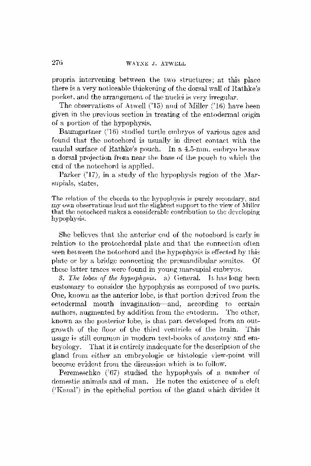
propria intervening between the two structures ; at this place there is a very noticeable thickening of the dorsal mall of Rathke's pocket, and the arrangement of thc nuclci is very irregular.
The observations of Atwell ('15) and of Miller ('16) have heen given in the previous section in treating of the entodermal origin of a portion of the hypophysis.
Raurrigartner ('16) studied turtle embryos of various ages and found that the notochord is usually in direct contact with the caudal surface of Rathke's pouch. In a 4.5-mm. embryo he saw a dorsal projection from near the base of the pouch to which the end of the notochord is applied.
Parker ('17), in a study of the hypophysis region of the Mar- supials, states,
The relation of the chorda to thc hypophysis is purely secondary, and r r i y own observations lend not the slightest support to thc view of Miller that the notochord makes a considerable contribution to the dcvcloping hypophysis.
She believes that the anterior end of the notochord is early in relation to the protochordal platc and that the connection often seen between the notochord and the hypophysis is effected by thiy plate or by a bridge connecting the preniandibuler sornitcs. Of these latter traces were found in young rriarsupisl crnbryus.
It has long been customary to consider the hypophysis as composed of two parts. One, known as the anterior lobe, is that portion derived from the ectodermal mouth invagina tion-and, according to ccrtain authors, augmented by addition from the entoderm. Thc other, known as thc postorior lobc, is that part developed frem an out- growth of the floor of the third ventricle of the brain. This usage is still common in modern text-books of anatomy and em- bryology. That it is entirely inadequate for the description of the gland from either an embryologic or histologic vien--point will become evident from the discussion which is to follow.
Peremeschko ('67) studied the hypophysis of a number of domestic animals and of man. He notes the existence of a cleft ('Kanal') in the epithelial portion of the gland which divides it
3. The Zobes of the hypophysis. a) General.

DEVELOPMENT OF HYPOPHYSIS O F lZhBBlT 277
into two unequal part,s-one, the ‘Korkschicht,’ and the other, the ‘Markschicht.’ The former is the main body of the epithelial lobe, while the latter is a thin lamina closely applied to the neiiral lobe. These two portions are also characterized by differences in histological appearance. The cells of the ‘Markschicht ’ are poorer in protoplasm, have clearer nuclei, and are not easily changed by reagents.
Lothringer (’86) notes this intraglandular cleft and agrees with Peremeschko that, it is not the separation between epithelial and brain parts. Tnstend, it, separates the ‘Epithelsaurn’ from the ‘Epithelkorper.’ The former corresponds to Peremeschko’s ‘Markschicht’ and the latter to his ‘Korkschicht.’
Herring (’08b) speaks of the epithelial investment of the neural lobe as the ‘pars intermedia’ obviously because of its position betn een the neural lobe and the.rcnmindcr of‘ the epithe- lial lobe, ‘thc atntcrior lobc proper.’ Pars intermedia and anterior lobe proper arc scparated by the residual lumen of Rathke’s pocket.
Stendell (’13) calls these two divisions of the anterior lobe thz ‘Zwischenlappen’ and the ‘Hauptlappen.’ rcspectively. The residual lumen he knows as the ‘Hypophysenhohle.’
A11 of these writers take pains to emphasize the fact that the ‘pars intermedia’ i:, inseparably bound to the neural lobe. This is particularly evident when an a pt has heen made to mechan- ically separate the so-called anterior and posterior lobes. Almost invariably a thin epithelial layer, the ‘pars intermedia,’ is found to have remained adherent to the neural lobe.
W. Rlliller (’73) describes for 16- and 18-cm. human, sheep, and pig embryos an anterior process of the hypophysis. His description reads : “Erstrecktc sich cin schmaler, conisch sich TTerjungender Fortsatz langs der vorderen FISiche des Processus infundibuli nach oben und vorn gegcn dss Chiasmn hin. ”
lClihalkovics (’76) observed the formation of an anterior proc- ess during the derelopment of the hypophysis in thc rabbit. In an embryo 2 cm. in length the epithelium of the inferior part of the hypophysial sac, at the place where the stalk is attache2
b) The anterior process.
278 WAYNE J. ATWELL
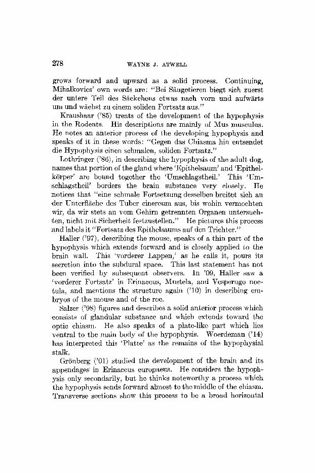
grows forward and upward as a solid process. Continuing, Mihalkovics’ own words are: “Bei Saugetieren biegt sich zuerst der untere Teil des Sackchens etwas nach vorn und aufwarts urn und wachst zu einern soliden Fortsate aus.’,
Kraushaar (’85) treats of the development of the hypophysis in the Rodents. His descriptions are mainly of Mus musculus. He notes an anterior process of hhe developing hypophysis and speaks of it in these words: “Gegen das Chiasma hin entsendet die Hypophysis einen schmalen, soliden Fo~tsatz.”
Lothringer (’86), in describing the hypophysis of the adult dog, names that portion of the gland where ‘Epithelsaum’ and ‘Epithel- korper’ are bound together the ‘Umschlagstheil.’ This ‘Um- schlagstheil’ borders the brain substance very closely, He notices that “eine schmale Fortsetzung desselben breitet sich an eder UnterflSiche des Tuber cinereum aus, bis wohin vermochten wir, da wir stets an vom Gehirn getrennten Organen untersuch- .ten, nicht mit Sicherheit festeustellen.” He pictures this process and labels it “Fortsate des Epithelmums auf den Trichter.”
Haller (’97)’ describing the mouse, speaks of a thin part of the hypophysis which extends forward and is closely applied to the brain wall. This ‘vorderer Lappen,’ as he calls it, pours its secretion into the subdural space. This last statement has not been verified by subsequent observers. In ’09, Haller saw a ‘vorderer Fortsatz’ in Erinaceus, Mustela, and Vesperugo noc- tula, and mentions the structure again (,lo) in describing em- bryos of the mouse and of the roe.
Salzer (’98) figures and describes a solid anterior process which consists of glandular substance and which extends toward the optic chiasm. He also speaks of a plate-like part which lies ventral to the main body of the hypophysis. Woerdeman (’14) has interpreted this ‘Platte’ as the remains of the hypophysial stalk.
Gronberg (’01) studied the development of the brain and its appendages in Erinaceus europaeus. He considers the hypoph- ysis only secondarily, but he thinks noteworthy a process which the hypophysis sends forward almost to the middle of the chiasm. Transverse sections show this process to be a broad horizontal

DEVELOPMENT O F HYPOPHYSIS OF RABBIT 279
plate. It grows forward from the place of attachment of the hypophysial stalk.
Joris (’07) saw in the meninges of the brain a mass of glandular cells which is attached at the anterior end of the hypophysis. This cell mass extends from theoptic chiasm to the base of the infundibulum and divides into two diverging branches. These make an angle, open posteriorly, embracing the neck of the infun- dibulum. To this cell mass Joris gave the name of ‘lobule de la tige.’ He believed that it becomes united with the hypophysis secondarily.
Staderini (’08) describes somewhat similar relations and speaks of a ‘lobus chiasmaticus’ which extends forward and of a ‘lobus praemammillaris’ the cells of which are within the brain coverings and surround the infundibular neck.
Herring (’08a) describes and figures a lobe which he names the “tongue-like process of the pars intermedia.” It extends for- ward and is closely applied to the brain wall. He notes that this part, is more vascular than the ‘pars intermedia.’
Bolk (’10) speaks of a ‘lobulus bifurcatus’ in primate embryos. The two arms of the lobe embrace the infundibulum near its attachment to the brain. Bolk believes that this ‘lobulus bifur- catus’ becomes detached to form the cell masses found embedded in the meninges.
Tilney (’13) differentiates histologically three portions of the glandular hypophysis in birds and mammal. His ‘pars distalis’ and ‘pars infundibularis’ correspond t o the ‘anterior lobe proper’ and the ‘pars intermedia’ of Herring, respectively. The ‘pars tuberalis’ is closely applied to the tuber cinereum and extends foward toward the optic chiasm. Tilney believed that he was presenting the histological structure of a “hitherto undescribed portion of the hypophysis.” Woerdeman (’14) point,s out that the ‘lobule de la tige’ of Joris, Standerini’s ‘lobus praemammilaris’ and ‘lobus chiasmaticus,’ and Balk's ‘lobulus bifurcatus’ are without doubt trhe same structure as Tilney’s ‘pars tuberalis.’ Baumgartner (’16) expresses himself as in accord with this view of Woerdeman’s. Tilney’s account of the development of the ‘pars tuberalis’ will be referred to later.

280 WAYNE J. ATWELL
c ) The lateral lobes. Gaupp (’93) states that the hypophysis of the lizard has a three-fold anlage-a large round ‘Mittelknospe’ and two long ‘Lateralknospen’ which bud out from the mouth epithelium. These parts are separated by a venous ring. Later there appears a fourth part anterior to the ‘Mittelknospe,’ the ‘Vordcre Knospe.’ The two lateral buds first unite with the gland arid later separate as solid bodies. They attach themselves closely to the brain floor. I t seems that they arc present in the adult animal, but further observation is required to establish this point.
Chiarugi (’94) notes t\w epithelial strand:, in C‘avia cobaya located at the place wherc the hypophysis is constricted off from the epithelium of the mouth. He believes that these are homol- ogous to the ‘Lateralknospen’ of Gaupp.
Weber (’98) saw, in Chiroptera, a tripartite hypophysis funda- ment. He does not hold, as does Gaupp, that t8hese three parts are separate a t the beginning. Rather, :z single anlage early differentiates into two ‘bourrelets Istteraux’ and a ‘crete mediane.’
Rossi (’96) describes a median part arid two latcral parts for the hypophysis of the chick.
Nusbaum (’9s) (referred to by Woerdeinan, ’14) states that the hypophysis develops from two sources, namely, froin an unpaired out-pouching of mouth cpithelium and from a pair of epithelial thickenings derived from the primitive gut.
Economo (’99) speaks of two ‘Seitensprossen’ to be seen during the development of the hypophysis in doves and chicks. In dove embryos the buds, or sprouts, appear between the fourth and seventh days. They are said to be arranged on each side of the infundibular process. During a part of their development they possess lumina which communicate with the hypophysis cavit y. He notes it similarity to Gaupp’s observations on the Reptiles.
Sltariderini (’03) traces the developing hypophysis in reptiles. The anlage is simple, but later the gland consists of a median part and two lateral parts.
Bolk (’I 0) describes an hypophysial adage of three divisions in young embryos of Macacus cynomolgus.

DEVELOPMENT O F HYPOPHYSIS O F RhBUlT 28 1
‘I’ilney (’11) observed in Aspidonectes two accessory pouches which arise from the main oral wagination. Hc believed that the importance of these and similar accessory pouches has been exaggerated, “since it is a coninmi tendency in many forms for the anlage of the gland to present multiple diverticula.”
Bruni (’13) noted t8hat in the Sauropsida Rathke’s pouch is early differentiated into a single ’lobo nzedio’ and two ‘lohi laterali.’ In xrirtrrimals the lateral lobes appear much later.
d) The relation of the lateral lobes to the ‘pars tubcralis.’ Gisi (’07)’ in a dissertation treating of the brain of Hatteria punctata, speaks of a thin anterior process of the hypophysis which is termed the ‘pars terminalis.’ Of this part it, is stated: “Wahrscheinlich ist diese Pars terminalis der Hypophyse das Endproduct der seiten Knospen an den frkheren Embryorral- st adien . ’ ’
Herring (’08 a) speaks only briefly concerning the de.;elopment of his “tongue-like process of the pars intermedia.” He says:
The neck of the sac retains a tubular character for some time, and beconics somewhat convoluted. One of these convolutions (fig. 5, k ) applies itself to the under surface of the brain antl gives rise to the tongue-shaped process which extends forwards from the aritrrior lobe towards the optic chiasma.
T o Tihey (’13) must be given the credit for first clearly show- ing that the ‘pars tubcralis’ has its origin from two lateral buds. Tilney has traced the development of the ‘pars tuberalis’ in the cat and in the chick.
arises as a relatively late structure. It has its origin in in-o secoridary diverticula or sprouts from the body of the pituitary sac. These sprouts, the tuberal processes, ultimately fuse with each other across the iiiedian line, displace the body of the pituitary sac vcntrad and thus secondarily assume their justa-neural position.
Tilney emphasizes both the histological and developmental sepamteness of ‘pars tuberalis’ from ‘pars infundibularis’ (Her- ring’s ‘pars intermedia’).
In his interesting study of the comparative development of the hypophysis, Woerdeinan (’14) has seen the ‘lobuli laterali’ in
The anterior lobe also grows forward antl laterally.
It

282 WAYNE J. ATWELL
several mammals and traces their development into the ‘lobulus bifurcatus’ of Bolk. This lobule divides into cell masses which lie in the meninges of the brain and which, in some cases, lose connection with the main body of the hypophysis. Woerdeman also has traced the early history of the lateral lobes. They arise from two enlargements of the thickened epithelial plate which lies anterior to Rathke’s pocket and which later beconies incorporated into the hypophysial anlage.
Miller (’16), in his study of the hypophysis of Sus scrofa, speaks only briefly of the ‘lateral cords.’ According to his description, these cords ‘grow round and encapsulate’ that mass of cells sup- posedly derived from the entodcrm. They eventually form the cortical layer of the anterior lobe.
Baumgartner (’16) has described the development of the lateral lobes of the reptilian hypophysis. He sees the lateral buds early separated from Rathke’s pouch by furrows which begin on the cranial side. He states:
In the later development of the lateral buds in turtles, the tips grow forward and form a thin layer closely applied to the floor of the brain (the part termed by Tilney ‘pars tuberalis’) and to a thin cortical zone around the middle of the anterior lobe.
In alligators, the lateral buds give rise to the pars tuberalis and two bands encircling the anterior lobe; in lizards, they appear t o persist as isolated masses or to disappear, while in snakes, they completely disappear.
Baurngartner believes that the cortical zone or bands described by him for turtles and alligators have been overlooked in other vertebrates, since Miller (’16) is the only observer who has described a similar structure (Sus scrofa).
Parker (’17) finds that the development of the ‘pars tuberalis’ in the Marsupials begins at an early stage. The portion of Itathke’s pouch lying posterior to the duct becomes sub- divided into two lobes, which are respectively distal and proximal in relation to the hypophysial duct, and are separated from cach other by a horizontal constriction. Whilc the distal lobe thickens and forms the glandular tissue of the pars buccalis (probably meaning Tilney‘s ‘pars distalis’) as well as the pars infundibularis, the proximal lobe remains thin walled.

DEVELOPMENT OF HYPOPHYSIS OF RABBIT 283
It is drawn out laterally and curves up to reach the brain wall on each side. Later the t’wo sides fuse to surround the infundibulum.
4. The structure of the intermediate par&. Besides the glandular cells and the small amount of connective tissue accompanying the few blood-vessels, a number of observers have noted in the pars intermedia certain distinctive cells which have been variously interpreted as nerve cells, sensory cells, or supporting cells.
Lothringer (’86) describes scattered marginal cells in addition to the cylindrical secreting cells. They may either reach the sur- face or be bent back upon themselves.
Pirone (’05) sees in the intermediate portion of the hypophysis, cylindrical cells which’present the structure characteristic to the supporting cells of sensory epithelium. He used Cajal’s method and states that he is able to confirm the findings of Gentes and Gemelli.
Gemelli (’05, ’06) makes mention of nerve fibers entering the pars intermedia from the neural lobe and also of ‘glio-epitheliari’ cells in this part. He considers that the posterior lobe of the hypophysis is sensory in nature.
Reteius (’94) describes and figures structures in the pars inter- media which he has called neuroglia cells. They are shown by the Golgi method. One type consists of long, fine spindle-shaped cells which extend through the entire thickness of the pars inter- media. Others are peculiar, branched forms which touch only one surface of the epithelium or neither. The nuclei for the most part lie near t8he surface bordering the cleft. The end of the spindle-shaped cells towards the neural lobe often widens out into a three-cornered foot.
Herring (’08) finds long, thin nucleated cells in the pars inter- media of the kitten’s hypophysis. These cells, which are brought out by use of Cajal’s silver method, are numerous and take a vertical course through the epithelium. They appear to be of ectodermal origin and to act as supporting cells. Similar cells may be found in the adult, but are better seen in the young animal (p. 139).
Trautmann (’09), in studying the hypophysis of the cat, has seen ‘fadenartige’ cells which extend through hhe entire width of

284 WAYNE J. ATWELL
the intermediate part. These are well brought out by the Golgi method. Another type of cell also was seen which Trautman describes in these words:
Im Epithclsaum der Katze konnte ich ferner durch die Golgische Methode zwischen den obengenannten fadenartigen, Zellgebilden ver- astelte Gebiltle darstellen, die verschiedenartig au den ersten verlaufen, mannigfaltige Gestalten aufweisen und weder Basis noch Peripherie erreichen.
Cajal (’11) figures bi-polar cells in the intermediate lobe of the mouse. These are made visible by the use of Golgi’s method. He has also seen nerve fibers which extend from neural lobe to intermediate part. For these reasons, he considers that the superior (posterior) lobe of the hypophysis has a sensory nature. On this point Cajal says:
Deuxfait semblent indiquer que Ic lobe superieur de l’hypophyse doit etre un organe sensorial; c’est, d’une part, la richesse Tu plexus axile inclus dans le lobe nerveux et l’epithelium adjacent; c’est, d’autre part, l’existence de nombreuses cellules bipolaires epitheliales particulieres, signalees par Retzius et nous dans l’epithelium de la gland.
Miller (’ 16) remarks that the ‘spindle-shaped supporting cells’ constitute one of the interesting structures in the intermediate lobe of the hypophysis of the pig.
Vanderburgh (’17) sees in the pars intermedia of the guinea- pig’s hypophysis, support,ing cells which extend from the cleft towards the pars nervosa. The branched variety appear as “little black triangles which are molded to fit the interspace be- tween the cells.” The unbranched were “much elongated and usually more transparent .” The stains used by Vandenburgh did not produce satisfactory evidence as to the nature of these cells.
Stendell (’14) gives a summary of the observations concerned with these special cells of the pars intermedia. He says that true ectoderriial supporting cells having the nature of the ependyma of the central nervous system have been definitely observed only in the hypophysis of mammals, more specifically in the Carnivora and in a few Rodents. They are best seen in the cat and dog and are most readily demonstrated by silver impregnation methods.

DEVELOPMENT O F HYPOPHYSIS O F RABBIT 285
He considers that without doubt these elements are true support- ing cells. More remarkable and quite foreign to the nature of the pars intermedia are the glia cells which have been described. They have not often been called neuroglia cells, but are described as ‘branched structures’ which are brought out, by the Golgi methods (Trautmann and ot,hers). Of the probable origin of t,hese cells Stendell says :
Da jedoch die ependyrniiren Stutzzelleii als von Hirnlappen her eingewanderte Elemente anzusehen sind, ist auch fur die glibsen, die ja init jciien genetisch ein System susmachen, die Erklarung gegeben. Xie sind entweder mit jenc zusammen eingedrungen oder direct inner- halh dcs Zwischenlappens von der Oberflikhe in profunde Lagen ver- drangte Ependymzellen.
5. The development of the neural lobe. Recent studies on the morphogenesis of the hypophysis have been confined almost entirely t o the epithelial portion of the gland. As a consequence, the literature treating of the development of t’he neural lobe is very scanty.
Miiller (’71) believed that in maminals the specific neural‘ tissue of the lobe is much reduced in amount during the latter half of fetal life and that connective tissue is substituted. Muller calls the fully developed infundibular process st “connective- tissue appendage of the brain.”
Mihalkovics (’75), treating of the development of the neural lobe of the rabbit’s hypophysis, states that its earliest appearance is due to the pressure of the hypophysial pouch against the wall of the forehain, and not to a relation with the anterior extremity of the notoehord. A protrusion of the brain wall results above the apes of the epithelia1 pouch and may be termed the ‘primitive Trichter.’ It represents not only the later ‘Trichter fortsatz,’ but also that portion of the ventricular floor which is to become the tuber cinereurn.
Gronberg (’01) states that from the beginning the processus infundibuli is a hollow sac which can be compared in shape to trhe finger of a glove. In his &age ‘11’ the lumen begins to disappear at the caudal extremity of thc lobe. Further description is omitted, but he makes reference to his figures 33 to 36’ for details as to the disappearance of the cavity of the lobe.

286 WAYNE J. ATWELL
Herring ('OSa) saw great importance in the early and close union between the buccal and cerebral portions of the hypophysis. Like Salzer, he could find no connective tissue between the infundibular process and the hypophysial sac in early stages. He believes that this close connection has some morphological significance, perhaps bespeaking the bucconeural duct observed by Anctriezen in Ammocoetes, Amphioxus, and Balanoglossus. He denies that the neural lobe degenerates into a connective- tissue appendage. He finds that the connective tissue is small in amount. Treatment with Cajal and Golgi methods shows the structures formerly described as connective-tissue cells to be ependymal and neurogliar elements.
Stendell ('14) traces the form and position of the neural lobe from the lowest fishes to the Mammals. In the fishes, the neural portion can be considered as little more than a modified region of the floor of the third ventricle. Tn the Elasmobranchs and the Ganoids, the neural part sends numerous hollow processes into the substances of the intermediate lobe. In the Teleosts, the processes are solid and branched. Stendell considers that first in the Amphibia does one find a true neural lobe, that is, an un- branched, solid thickening of the ventricular floor to which the epithelial part attaches itself and develops (p. 27). This he considers the usual arrangement of parts in all the higher vertebrates .
So far a~ I have been able to discover, no author has confined himself t'o describing the development of the hypophysk in the rabbit alone. Several investigators have included, however, a consideration of this animal in their comparative studies.
Mention has already been made of a connection between noto- chord and dorsal wall of the hypophysis which was observed in an eleven-day rabbit embryo by Koelliker. Likewise it has been noted that peculiar 'neuroglia-like' cells were seen by Retnius in the pars intermedia of a rabbit eight days after birth.
Miiller ('71) used embryos of the pig, the sheep, and the rabbit in his study of the development of the hypophysis. He gives a common description for all embryos of the same length. For
B. Development of the hypophysis in the rabbit.

DEVELOPMENT O F HYPOPHYSIS O F RABBIT 287
example, his description of a 16-mm. stage may be applied equally well to any one of thesc three forms.
The rabbit was the mammalian type chosen by Mihalkovics (’75) in his study of the hypophysis. His descriptions begin with an embryo 5 mm. in length. The oral membrane is still intact. The first appearance of the hypophysis is the ‘Hypophysenwin- kel’ a shallow infolding of t8he oral ectoderm just anterior to the oral plate. In a 6 - m . embryo, the oral membrane has just ruptured. Both upper and lower stumps are still to be seen. In this stage the earliest appearance of the primitive infundib- ulum is to be noted. A 12-mm. embryo shows .a definitely formed ‘Hypophysentasche’ which communicates with the oral cavity by a much-constricted opening. The ‘l’richterfortsatz’ is small and conical. A 16-mm. embryo presents a longer ‘Trich- terfortsatz’ and the hypophysial pocket has been further con- stricted off from the mouth epithelium, so that the connecting ‘Hypophysengang’ contains only a minute lumen. The further cutting-off of the hypophysial pouch, the formation of the anterior process and of the ‘Driisenschliiuche’ are t’raced by a description of 2-, 3-, and 4-em. embryos.
Although not studying the development of the hypophysis, Lothringer (’86)’ Rogowitsch (’89), and Stieda (’90) made impor- tant early observations on the anatomy and histology of the gland in the rabbit.
Minot and Taylor (’05) give definite but necessarily brief statements concerning the development of the hypophysis in their “Normal Plates of the Development of the Rabbit.” The hypophysis of the rabbit is first visible in their embryo No. 9, nine and one-half days of development, as a “very small evagina- tion of ectoderm on the dorsal side of t,he mouth just in front of the oral plate,” which has begun to rupture. In an embryo of ten days no essential difference is to be noted. L4t ten and one- half days the diverticulum of the hypophysis is “well marked, rather four-sided in cross-section, and closely approximated to the wall of the forebrain.” During the next day the pouch becomes longer and more closely applied to the brain wall. In embryo No. 14 ( twelve days) the anlage of the infundibuluin is “present
THE -4YERICLN JOURNAL OF ANATOMY, VOL. 24, NO. 3

288 WAYNE J. ATWELL
as a veyy small evagination of the floor of the forebrain. The upper end of the hypophysis is slightly expanded laterally, slightly concave toward the forebrain, and is joined to the infun- dibulum.” During the next day and a half the infundibular evagination becomes more distinct and the mouth of the hypo- physial pouch more constricted. At fourteen days the hypo- physis is no longer open to the mouth, but is connected with the oral ectoderm by a solid epithelial cord. In embryo No. 19 (fifteen days) the infundibulum is a little longer than in preceding stages and overlies the hypophysis (epithelial portion) more. The hypophysis is bent concave toward the forebrain. By six- teen days there can be seen a beginning of the cords of the hypoph- ysis which first appear as solid outgrowths. These cords are larger and more vascular in a seventeen-day embryo. At eighteen days the connecting cord between hypophysis and oral ectodenn is broken through just above the ectoderm. The Normentafel studies do not extend beyond twenty days of development. At this stage some embryos still show a small connecting strand be- tween hypophysis and oral ectoderm. The infundibulum is open to the third ventricle. The hypophysis is much bent and con- tains a cavity. Two lateral upward prolongations of the hypoph- ysis are to be seen on either side of the infundibulum. The cords of the hypophysis appear as a vasculariaed outgrowth of the anterior wall, irregular in shape, solid except for the contained vessels. The pituitary fossa is well marked at this time.
3. MATERIAL AND METHODS
The rabbit embryos used in this study were obtained from the rabbit colony of the Department of Anatomy of €he University of Michigan. The majority of them were obtained during the course of the study and accurate records have been kept of their ages.
Females which have already born young were selected and the date of birth of the previous litter obtained if recent. It is known that a female will generally submit to coition shortly after par- turition, but such a procedure is undesirable, since the suckling of young probably lengthens the gestation period (compare King

DEVELOPMENT O F HYPOPHYSIS OF RABBIT 289
’13, on the albino rat). It is possible to wean the young rabbits at the end of one month, and the mother will then usually submit to coition within one or two days. Record was made of the time of insemination (Long and Mark, ’11), and all ages referred to in this study are counted from the time of insemination to the time the embryos were obtained.
When a certain stage of development was desired the mother was sacrificed and the uterus was quickly removed and placed in normal salt solution, from which the freed embryos were trans- ferred to the fixing fluid. In the case of all the younger embryos the amnion was carefully removed under the binocular micro- scope and the umbilical cord was tied to retain as much blood as possible in the vessels of the embryo.
For fixation Zenker’s, Carnoy’s and Bouin’s fluids were em- ployed. After sectioning and staining had been begun, it was found that Zenker’s solution did not produce entirely satisfactory results in conjunction with the stains employed, so for the most of the work Carnoy’s or Bouin’s fluid was used. Decalcifica- tion was found necessary beginning with the eighteen-day em- bryos, and was accomplished by the use of a decalcifying fluid, made up after a somewhat empirical formula devised by Professor Huber and used successfully in his laboratory for several years.
The formula is : HNOs . . . . . . . . . . . . . . . . . . . . . . . . . . . . . . . . . . . . . . . . . . . . . . . . . . . . . 100 CC.
. . . . . . . . . . . . . . . . . . 600 cc. NaCl . . . . . . . . . . . . . . . . . . . . . . . . _ . _ . 20 grrtms
Absolute alcohol ...........................
It has the advantage of maintaining the tissues in a fairly high grade of alcohol during the process of decalcification. This is especially to be desired in the treatment of material which has been preserved in Carnoy’s fluid. The tissues were cleared in xylol and embedded in 58” paraffin.
In the case of some of the younger stages series were cut on the rot.ary microtome, but most of the embryos were sectioned wit.h the sliding microtome, by means of Huber’s water-on-the-knife method. Not any series was cut at a thickness greater than 5 microns and several complete series were prepared at 3 microns.

290 WAYNE J. ATWELL
The most useful staining combination was found to be iron- alum hematoxylin and Congo red. The latter solution was pre- pared as suggested by Huber ('15). For stages up to and in- cluding the twentieth day, series were prepared in sagittal, frontal, and transverse planes; later stages were prepared in the sagittal plane only.
To better show the form of the del-eloping gland and to avoid errors in the interpretation of sections, the Born method of wax- plate reconstructions was freely used. A model of the hypoph- ysis was prepared for each day of its development from earliest appearance up to and including the twentieth day. From this t ihe until birth every second day is represented by a model. Additional reconstructions from other ten-, eleven-, twelve-. and thirteen-day embryos, together with an enlargement of the neural lobc and pars intermedia of one of the sixteen-day embryos raises the total number of models constructed to twenty-six. The magnification chosen was comparatively great- x 400 for the younger stages, including the sixteen-day embryo; x 200 for the seventeen- to twenty-two-day stages, inclusively; while for the four oldest stages it was found necessary to reduce the magnifi- cation to 100 diameters. The plan has been to reconstruct the neighboring brain wall, the oral (or nasal) epithelium, and a part of the cartilage of tho hypophysial fossa, when that is present. This has been adhered to in all of the younger stages. The brain wall has been removed from the models of the nineteen- and twenty-eight-day embryos to present a dorsal view of the gland.
Use has also been made of the His method of projective recon- struction, and by this graphic means ventral views of the neural lobe have been obtained from transverse sections for certain st ages.
4. THE DEVELOPMENT OF THE HPPOPHYSIS OF THE RABBIT BY DAYS
10-day stage. The hypophysis is well indicated in embryo A, which possesses sixteen pairs of primitive segments and has a seventeenth partly formed. A wax-plate reconstruction of this embryo, which includes the anterior part of the notochord, the epithelium of the anterior end of the forcgut, the epithelium of the

DEVELOPMENT O F HYPOPHYSIB O F RABBIT 29 1
mouth invagination, and the adjacent brain wall, is shown in figure 1. The hypophysis anlage is present as a shallow pouch much wider from side to side than from front to back. The oral plate is intact. The cephalic extremity of the notochord bends around the foregut and terminates at the dorsal wall of the hy- pophysis fundament. A contact between notochord and hy- pophysis cannot be observed in t{his embryo.
Figure 2 presents a sagittal section of a timed ten-day embryo (27D). It is slightly older than the preceding, but the oral membrane is still unbroken. A comparatively large evagination of the brain wall is seen dorsal to Rathke’s pocket. This eor- responds to the ‘primitive Trichter’ of Mihalkovics. In this
n C.
b. w.
R
Fig. 1. Model of hypophysis region of a sixteen-somite rabbit (embryo A). x 100. The anterior end of the notochord and portions of the epithelium of the foregut, oral pit, and brain wall are shown. nc . , notochord; f.g., foregut; R, Rathka’s pouch, and b.w., brain wall.
Viewed from the left side.
embryo the extremity of the notochord is in close contact with the wall of hypophysis anlage.
Another ten-day embryo (27A) shows the oral membrane in the process of rupture. A sagittal section of this cmbryo is given in figure 3. A noteworthy feature is the presence of a thickened epithelium, continuous with the hypophysial wall, which extends nasalward from Rathke’s pocket for some distance. Its rather abrupt termination is marked by x (fig. 3).
A wax-plate reconstruction prepared from this embryo shows that the hypophysial pouch has deepened, and on each side, a t its nasal border, has developed a ridge-like protuberance. As seen

292 WAYNE J. ATWELL
2
3 Fig. 2 Sagittal section of head end of a ten-day rabbit embryo (27D) showing
oral membrane intact. X 50. Nasal end a t right. p . ins., primitive infundib- ulum; R, Rathke’s pouch; or . p l . , oral plate; S. Seessel’s pouch; nc., notochord.
Fig. 3 Sagittal section of head end of ten-day rabbit embryo (27A) showing oral membrane ruptured. X 50. Nasal end a t right. s t . , stump of oral plate; other abbreviations as in figure 2.

DEVELOPMENT OF HYPOPHYSIS OF RABBIT 293
from the inside, each ridge is indicated by a shallow groove. It appears as if these ridges were developing from the thickened epithelium which lies in front of the early hypophysial pouch.
The rupture of the oral membrane is not complete, as is shown by the presence of two openings through which the foregut com- municates with the exterior. The end of the notochord is drawn out to a point and is in contact with a prominent bud extending from the dorsal wall of Rathke’s pocket.
I
2 n c.
4 5 Fig. 4 Model of hypophysis region of rabbit embryo B (about ten days),
R, Rathke’s pouch; 6.w., brain wall; 1,
Fig. 5 Model of hypophysis region of eleven-day embryo (25A) viewed from LZ., lateral lobe; d. diverticulum from Rathke’s pouch
Other abbreviations as in figures 1 and
viewed from the oral surface. 8 , 3 , 4 , perforations in the oral plate.
the left side. X 100. toward which the notochord is directed. 2.
X 100.
Another embryo (Series B) also shows the breaking of the oral membrane. In figure 4 one views a reconstruction of this em- bryo from the oral side. The membrane has broken through in four places, thus furnishing four separate communications between the foregut and the oral invagination.
By the end of the eleventh day of development the hypophysiai pouch has deepened considerably and has ex- panded laterally near its apex. A constriction near the middle of the pouch serves to separate the apical portion from the more inferior portion bearing the ridge-like protuberances (I. 1. fig. 5). These latter have become more prominent due to their partial
11-day stage.

294 WAYNE J. ATWELL
constriction from the oral epithelium. These prominent eleva- tions I interpret as the homologues of the lateral lobes of lower forms. Later stages of development of the rabbit will show how these early lateral lobes give rise to the pars tuberalis of complete development. ,4 transverse section through the region of the lateral lobes is shown in figure 7. The lobes are constricted from the thickened epithelium just nasal to the early hypophysial pouch.
b. sv.
I. I.
3
Fig. 6 Sagittal section, hypophysis region of eleven-day rabbit embryo (25A). X 100. Nasal end a t right. p . inf., primitive infundibuluin; R, Rathke’s pouch; S , Seessel’s pouch; nc., notochord; d, diverticulum of Rathke’s pouch t o which the notochord is directed.
Transverse scction just nasal to Rathke’s pouch, from rabbit embryo C, about eleven days. 1.1.) lateral lobe in process of being constricted off; b.w., brain wall.
Fig. 7 X 100.
In embryo 25A (figs. 5 and 6) the notochord ends close to a large thickened evagination of the dorsal wall of the hypophysis from which it is separated by a very narrow space. The condi- tion presented by t5his embryo corresponds very closely to that of the eleven-day rabbit embryo described by Koelliker (’79).
In embryo C, which is only slightly more advanced, the noto- chord divides into two parts near its cephalic termination. One branch ends at! the apex of Seessel’s pouch, while the other is directed dorsally, forming almost a right angle with the first part.
DEVELOPMENT OF HPPOPHYSIS 01” ItABBIT 295
The primitive infundibulum is present as a large, shallow evagi- nation of the brain wall.
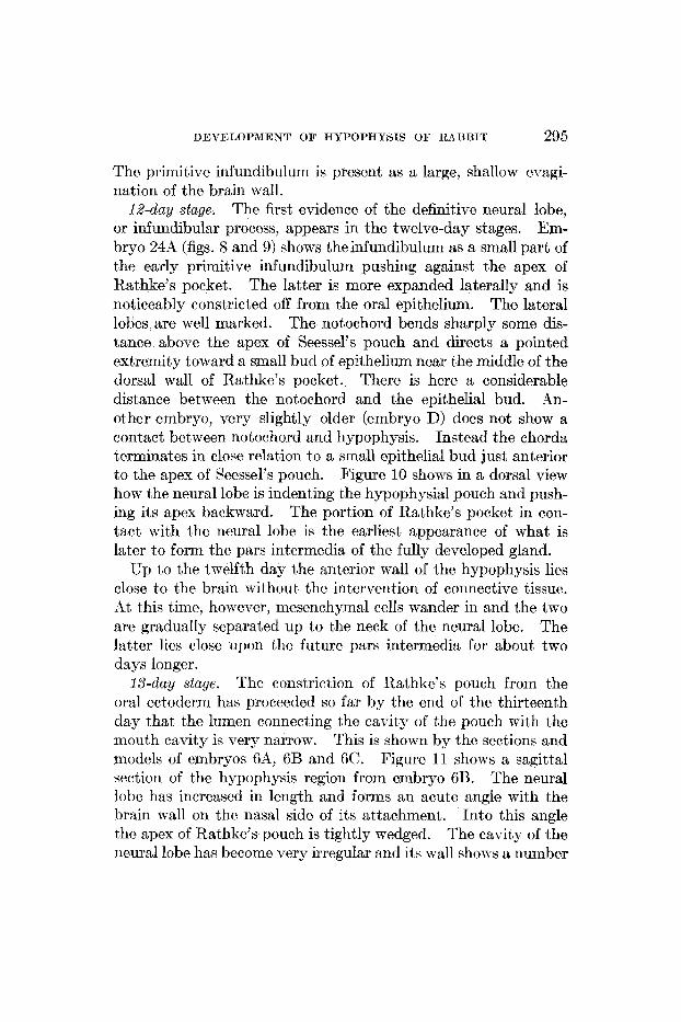
The first evidence of the definitive neural lobe, or infundibular process, appears in the twelve-day stages. Em- bryo 24A (figs. 8 and 9) shows theinfundibulum as a small part of the early primitive infundibulum pushing against the apex of Rathke’s pocket,. The latter is more expanded laterally and is noticeably constricted off from the oral epithelium. The lateral lobes are well marked. The notochord bends sharply some dis- tance above the apex of Seessel’s pouch and directs a pointed extremity toward a small bud of epithelium near the middle of the dorsal wall of Rathke’s pocket. There is here a considerable distance between the notochord and the epithelial bud. L4n- other embryo, very slightly older (embryo D) does not show a contact between notochord and hypophysis. Instead the chorda terminates in close relation t o a small epithelial bud just anterior to the apex of Seessel’s pouch. Figure 10 shows in a dorsal view how the neural lobe is indenting the hypophysial pouch and push- ing its apex backward. The portion of Rathke’s pocket in con- tact with t8he neural lobe is the earliest appearance of what is later to form the pars intermedia of the fully developed gland.
Up to the twelfth day the anterior wall of the hypophysis lies close to the brain without the intervention of connective tissue. At this time, however, mesenchymal cells wander in and the two are gradually separated up to the neck of the neural lobe. The latter lies close upon the future pars intermedia for about two days longer.
The constriction of Rathke’s pouch from the oral ectoderm has proceeded so far by the end of the thirteenth day that the lumen connecting the cavity of the pouch with the mouth cavity is very narrow. This is shown by the sections and models of embryos 6A, 6B and 6C. Figure 11 shows a sagittal section of the hypophysis region from embryo 6B. The neural lobe has increased in length and forms an acute angle with the brain wall on the nasal side of its attachment, Into this angle the apex of Rathke’s pouch is tightly wedged. The cavity of the neural lobe has become very irregular and its mall shows a number
12-day stage.
13-day stage.

296 WAYNE J. ATWXLL
nc
S
a
12 Fig. 8 Model of hypophysis region, twelve-day rabbit embryo, viewed from
left side. X 100. b.w., brain wall; n.Z., neural lobe; n.c., notochord; S, Sees- sel's pouch; 1.E., lateral lobe.

DEVELOPMENT OF HYPOPHYSIS O F RABBIT 297
of angular foldings. This is the beginning of a very complicated series of reduplications and compressions by which the lumen of the lobe is obliterated. In rabbit 6B the anterior termination of %he notochord is near a small epithelial bud projecting from the tip of Seessel’s pouch (fig. 11). In 6A the notochord sends a strand of cells toward a small epithelial bud which projects from the dorsal wall of Rathke’s pocket near its constriction from the oral epithelium.
The cavity of the epithelial pouch of the hypophysis is con- stricted slightly near its middle (fig. 11). The entire pouch has assumed a curved shape with the concavity toward the brain floor.
The lateral lobes, on account of the constriction of the hypo- physial sac from the mouth, have been drawn together and form a transverse ridge across the nasal end of the hypophysis near its attachment to the oral epithelium. The rounded termination of this ridge on each side is the t’ip of the lateral lobe (Ll., fig. 12).
In the fourteen-day embryos the attachment of the hypophysis to the oral epithelium has been reduced to a solid stalk. The original cavity of Rathke’s pouch is present through- out the length of the gland, but does not extend into t,he stalk. The gland has assumed a more concave form and a considerable amount of connective tissue is present between the hypophysis and the brain wall (fig. 13).
The lateral lobes have begun to grow laterally forming definite, bud-likc projections (l.l., fig. 14). Each bud lies close to the main body of the hypophysis, but is clearly separated from it by a deep groove. These buds will be traced into the formation of the pars tuberalis.
14-day stage.
Fig. 9 Sagittal section of hypophysis region, twelve-day embryo (24A) ; nasal end a t right,. X 100. R, Rathke’s pouch; p.int., pars intermedia; n.Z., neural lobe; ne., notochord; 8, Seessel’s pouch.
Fig 10 Model of hypophysis region of rabbit embryo D, about twelve days, viewed dorsally and somewhat from the right. X 100. d., neural lobe; R, Rathke’s pouch; nc., notochord.
Fig. 11 Sagittal section of hypophysis region, thirteen-day embryo (6B), nasal end a t right. X 100. R, Rathke’s pouch; n.L, neural lobe; p . int., pars inter- media; nc., notochord; S, Seessel’s pouch.
Fig. 12 Model of epithelial portion of hypophysis from thirteen-day embryo (6A) viewed from the right. X 100. R , Rathke’s pouch; Z.Z., lateral lobe.

298 WAYNE J . ATWELL
Fig 13 Sagittal section of hypophysis region, fourtecn-day embryo (l0A). X 100 YL I , neural lobe; p . int., pars intermedia; nc., notochord; S , Seessel’s pouch; st., stalk.
Fig 14 Model of hypophysis region, fourteen-day embryo (10A). X 100. Viewed from the right and somewhat dorsally. ant. Z., anterior lobe; b.w , brain mall; 7~ l . , neural lobe; Z.Z., lateral lobc; s t . , stalk; S, Seessel’s pouch; nc , notochord; ”2., constricted portion perhaps equivalent to Woerdeman’s ‘dor- salrr Mittelraum.’
Xasal end a t right.

DEVELOPMENT OF HYPOPHYSIS O F RABBIT 299
Just dorsal to each bud is another, smaller outgrowth, m, fig. 14. It seems to me not improbable that. this cminence can be compared to the dorsal part of the ‘Mittclraurn’ of Woerdeman (’14). A median eminence of the transverse ridge connecting the two lateral buds may corrcspond to the Torraum’ of Woerde- man. This, at least, is the interpretation Woerdernan has placed on Salzer’s (’98) model from a 1.9-cm. pig embryo. I cannot agree with Woerdeman that this single median eminence forms the anterior extension of the pars tuberalis, as will be explained more fully further on.
The neural lobe is irregular in outline and its lumen has berome very tortuous and labyrinth-like. As a result of the corrugations and compressions of its wall, folds of the wall and detached cells are to be found in tho cavity of the lobe and constitute an impor- tant factor in its obliteration. Xeural lobe and pars intcrrriedia have become separated by a small amount of conncctivc tissue. The notochord terminates just anterior to the apex of Seesscl’s pouch.
KO new structures are visible in the fifteen-day stages, but the gland shows a gradual development in all its parts. The stalk is longer and of smaller diameter. It tapers gradwally from the gland to its attachment with the oral epithelium. The neural lobe is proportionately larger and more convoluted and its lumen has bccomo more intricately divided. Besides the cells which are to be found in the cavity of the neural lobe, other cells rnay be seen which have wandercd outward through the basement membrane and are forming a cortex around tho outside of the lobe. The connective tissue separating if from the interrriediate part is somewhat increased in amount. One of the embryos shows some interesting connections (cont., fig. 15) betwccsn neural and intermediate parts. They arc eirrlilar t,o the contacts to be described for the sixteen-day and older embryos and will be treated more fully with those stages.
The lateral lobes (l.l., figs. 16 and 17) are larger than in the previous stage. They are solid, never having shown a lumen. Because of the continued ‘cupping’ of the hypophysis, these lobes have been drawn closer to the brain mall and are directed some-
15-dny stage.

300 WAYNE J. ATWELL
what towards it as well as laterally. The median eminence on the transverse ridge connecting the two lateral lobes, seen in the fourteen-day stage, is here present. The extremity of the noto- chord is in relation with a pointed bud of epithelium from Sees- sel’s pouch (figs. 15 and 16).
At sixteen days the lateral lobes have enlarged and lie close to the brain wall. On each side the lobes are shsrply
16-day stage.
Fig. 15 Sagittal section of hypophysis region, iifteen-day embryo (7A). X C O T Z ~ . , definite contact between neural lobe and pars 100. Nasql end at right.
intermedia; other abbreviations as previously.
constricted from the remainder of the gland. .In the midline they are not well separated from each other. The mass formed by the two lobes will form the pars tuberalis of Tilney. Already it has begun to be vascularized, and in this respect is in advance of the remainder of the gland. This is shown in the model (fig. 18) by its roughened surfaces. Precartilage indicates the future sphenoid bone with a shallow fossa to contain the gland. The hypophysial stalk is attached to the gland just nasal to the con-

.n. I.
. I . 1.
Fig. 16 Model of hypophysis region, fifteen-day embryo, viewed from left
Fig. 17 Same model as shown in figure 16, here viewed dorsally and caudally, aide. X 100. Abbreviations as in figure 14.
with Seessel’s pouch removed. X 100. iibbreviations as in figure 14. 301

302 WAYNE J. ATWELL
1.
-sphen.
19 6
20
DEVELOPMENT O F HYPOPHYSIS O F RABBIT 303
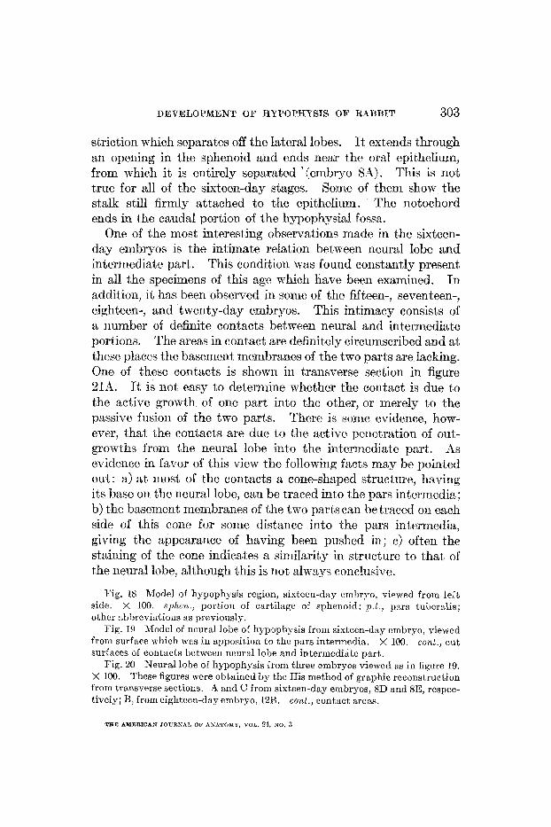
striction which separates off the lateral lobes. It extends through an opening in the sphenoid and ends near the oral epithelium, from which it is entirely separated '(embryo SX). This is not true for all of the sixteen-day stages. Sonic of them show the stalk still firmly attached to the epithelium. The notochord ends in the caudal port8ion of the hypophysial fossa.
One of the most interesting observations made in the sixteen- day embryos is the intimate relation between neural lobe and intermediate part. This condition was found constantly present in all the specimens of this age which have been examined. In addition, it has been observed in somo of the fifteen-, seventeen-, eighteen-, and twenty-day embryos. This intimacy consists of a number of definite contacts between neural and intermediate portions. The areas in contact are definitely circumscribed and at these places the basement membranes of the two parts are lacking. One of these contacts is shown in transverse section in figure 2111. It is not easy to determine whether the contact is due to the active growth of one part into the other, or merely to the passive fusion of the two parts. Therc is some evidence, how- ever, that the contacts are due to the active peneti-ation of out- growths from the neural lobe into the intermediate part. As evidcncc in favor of this view the following facts may be pointed out: a) at most of the contacts a cone-shaped structure, having its base on the neural lobe, can be traced into the pars intermedia; b) the basement membranes of the two parkcan be traced on each side of this cone for some distance into the pars intermedia, giving the appearance of having been pushed in; c) often the staining of the cone indicates a similarity in structure to that of the neural lobe, although this is not always conchisive.
Fig. 18 Model of hypophysis region, sixtccn-day cmbryo, viewed from left side. x 100. sphen , portion of cartilage of sphenoid; p 1 , psrs tuheralis; othcr abbrcvi:ii ions as previously.
Fig. 19 Model of ncural lobe of hypophysis from sixteen-day crnbryo, viewed from surface nhich wus in apposition to the pars intermedia. X 100. cont., cut surfaces of contacts bctwecn neural lobe and intermediate part.
Fig. 20 Neural lobe of hypophysis from three embryos viewed as in figure 19. X 100. These figures were obtained by the His method of graphic reconstruction from transverse sections A and C from sixteen-day embryos, 8D and SE, respec- tivcly; B, from eighteen-day embryo, 12B. cont., contact areas.
THE AMERICAN JOURNAL O F ANbTOhtY, VOL. 24, NO. 3

304 WAYNE J. ATWELL
These contacts vary in number from one to five. To show their relative sizes and position, the neural lobe and pars intermedia of embryo 8A were reconstructed in wax a t a magnification of five hundred diameters. The two were then separated by cutting the contacts. The model of the neural lobe is shown in figure 19 viewed from the surface which was in apposition t o the pars intermedia. Four distinct areas of contact may be seen. They are arranged in t,wo pairs, one of which is near the caudal or free end of the lobe, while the other lies near its middle. They are unequal in size and the caudal pair is the larger. Similar views
Fig. 21 Transverse sections showing contacts between neural lobe and pars intermedia. A , from sixteen-day embryo (8D); R f rom eighteen-day embryo (12B); C, from twenty-day embryo (1313). n.Z., neural lobe; p int., pars intermedia; cant., contact; r 1 , residual lumen; ant.Z . aiitcrior lobe
of the neural lobe in othcr sixteen-day embryos have been ob- tained by the His method of graphic reconstruction. One, showing three contacts (from embryo 8D), is shown in figure 20A. Another showing one contact (embryo 8E) is given in figure 20C. In general the contacts are near the caudal end of the neural lobe. This is always true when only a single contact is present.
On the surface of the intermediate part facing the residual lumen of the hypoph- ysis is found a slight indentation t<o correspond to the center of each contact (fig. 21).
X 100.
A further observation should be recorded here.

DEVELOPMENT OF HPPOPHYSIS O F RARRIT 305
17-day stage. In the model of embryo 2 8 h (fig. 22) the pars tuberalis is seen to have a larger surface in contact with the brain wall than in the previous stage. The lateral lobes which com- pose it are coming to lie between the anterior lobe proper and the brain wall. The two lateral lobes are now well separated in thc midline. That, portion of the lateral lobe between the brain wall and the hypophysial stalk is deeply constricted off from the re- mainder of the gland and forms a cortex for the nasal end of the anterior lobe proper (cort., fig. 22). In this embryo the stalk is attached to the oraI epithelium. Embryo 28A shows one of the contacts mentioned in the previous stage as seen connecting neural and intermediate parts. Near its apex the hypophysis is sending up two processes ip . , fig. 22) to surround more closely the neck of the neui-al lobe. A study of sections shows that the residual lumen extends into these processes.
The foIdings of the neural lobe have resulted in the formation of two layers in its wall. The innermost consists of cells radially arranged about the central lumen, which is partially filled by cells irregularly disposed. An outer, or cortical, layer consists of cells very irregularly placed. Many remnants of the original basement membrane are to be seen butmen thew two layers (compare fig. 21B, cightcen days). The neural lobe ncnv makes an angle of about ninety degrees with the brain wall at its attachment.
In this statgc the pars tuberalis is moreextm- sively applied t o thc brain wall and processes havc bzgun to ex- tend both nasally and caudally. The lobes have become more closely compressed between thp anterior lobe proper and the brain. Between the brain wall arid the hypophysis a cup, or fossa, is forrncd. J t contains connective tissue rich in blood-vcssols, many of which may be traced into the anterior lobc. Tho cortex at the nasal end of the anterior lobe is here well rnarked (curt., fig. 23).
The neural lobe is directed caudally (fig. 23) making now an acute angle on the caudal side of its attachment. The lobe has elongated and is constricted at its neck. The processes from the apex of the hypophysis have extended farther backward around
18-day stage.
This is the ‘obcrrie Dell’ of Mihalkovics.

2 2
Fig. 22 Model of hypophysis region, seventeen-day embryo (28A), viewed from right side. X 50. b.w., brain waII; n.Z., neural lobe; p . t . , pars tuberalis; cort., cortical plate; sphen., portion of sphenoid; st., stalk; p . , process surround- ing neck of neural lobe.
Fig. 23 Model of hypophysis region, eighteen-day embryo, viewed from left side. X 50. r., remains of stalk below sphenoid, or ‘pharyngeal hypophysis ’ Other abbreviations as in figure 22.
306

DEVELOPMEKT O F HYPOPHYSIS O F RA4BBIT 307
its neck. Certain embryos show contacts between neural and intcrmediatc parts and others do not. Figure 21. R, a transverse section from embryo 12B, shows a cone-shaped process of the neural lobe cntering the intermediate part. ,4 graphic recon- struction of the neural lobe of this embryo (b., fig. 20) shows that in all there are five contact areas. This is the greatcst number counted in any embryo. Small strands of connective tissue are to bc observed penetrating into the epithelium of trhe pars intermedia.
The stalk in embryo 12A is represented by a sinall knot at- tachcd to the gland and by an entirely isolated mass of epithe- lial cells, situated vcntral to the sphenoid cartilage (T., fig. 23) . Such an epithelial mass forms a ‘pharyngal hypophysis.’ The possession of a pharyngeal hypophysis is perhaps the normal condition for certain stages of dcveloprncnt.
By the end of the ninetecrith day a most inter- esting stage of development has been attained by the lateral lobes. They now have a considerable area flattened out against the brain wall. They are united in the midline and each is sending out a pair of blunt nasal horns (11.h.) and a pair of sharper, longer caudal horns jc.h.), as shown in figures 24, 25, and 26. The two nasally directed horns extend toward the optic chiasm, lying close to the brain wall. The caudal horns likewise lie close to the brain wall and are extending back to surround the neck of the neural lobe. This part which lies close to t,hc brain wall is the pars tuberalis. It is rapidly assuming its final position. Ven- tral to the pars tuberalis on each side i s a comparatively large solid process (m., fig. 24). The processes growing up to surround the neck of the neural lobc are prominent ( p p . , figs 24,25, and26)
The stalk (which has been cut short in the model) extends through the sphenoid cartilage and is attached to the nasal epithelium. The attachment of the stalk to the gland is nearer the nasal end than formerly.
At twenty days (figs. 27 and 28) the pars tuber- alis has extended farther caudally and its caudal horns are insinu- ating themselves between the brain wall and the anterior lobe proper (c.h., fig. 27). The entire surface of the epithelial portion
19-day stage.
20-day stage.

308 WATRE J. ATWELL
of the gland is much roughened by the presonco of blood-vessels. The anterior lobe and the pars tuberalis arc well vascularized at this time. The pars intermcdia is non-vascular. As shown in figure 2’7, the stalk extends through the sphcrroid cartilage and cnds near the nasal epithelium, but shows 110 connection wilh it.
24
c. h,
n 1.
P
1.
26 25 Figs. 24, 25, and 26 Model of hypophysis from nineteen-day embryo. X 50.
Brain wall, sphenoid cartilage and nasal epithelium not reconstructed. Figure 24, from right side; figure 25, dorsally and from right side; figure 26, dorsally. c.h. and n.h., caudal and nasal horns, of pars tuberalis; m., sccondary eminence (dorsal part of ‘Mittelraum?’); other abbreviations as in figure 22.
A sagittal section of embryo 13A is shown in figure 28. The fossil, f, caused by the bending of the hypophysis and the growth of the lateral lobes toward the brain, is somewhat smaller than previously, having become reduced by the rapid growth of the anterior lobe. A transverse section (embryo 13B, fig. 21C) shows very clearly one of the outgrowths of the neural lobe extending into the pars intermedia. This is the latest stage in which such neuro-epithelial contacts were observed.

QEVELOPMENT O F HYPOPHYSIS O F RA4RBIT 309
W%dccy stuge. The general relations of the fully developed gland have been nearly attained at this time. The nasal horns of the pars tuberalis have extended to the optic chiasm. The horn of the lcft side in embryo 20-1 has outstrippcd its fellow in development and the two meet well to the right of the midlinc. This is not the general rule, as is shown by other stages. Usually the two sides are symmetrical. The caudal horns have entirely displaced the remainder of the hypophysis from contact with the
'n. I. -
st.
b. w.
h.
Y
Fig. 27 Model of hypophysis region from twenty-day embryo viewed from right side. X 60. n.h., nasd horn, c .h . , caudal horn, of IJLL~S tuberalis; other abbreviations as in figurc 22.
floor of the third ventricle. They havc encircled the neck of the neural lobe and lie close together, but arc not yet united (fig. 29). The processes from thc body of the hypophysis (p.p., fig. 29) have extended well around the neck of the neural lobe. It is to be noted that they have the caudal horns of the pars tuberalis be- tween themselves and the brain wall. Two knob-like processes (e.e., fig. 29) extend from the pars intcrmedia to the caudal ex- tremity of the neural lobe and are fused with it. They have the structure of the pars intermedia.

310 WAYNE J. ATWELL
The pars tuberalis lies in the pia mater of the brain. It has begun to show a tubular or alveolar structure and is histologically different from both the pars intermedia and the anterior lobe proper. The third ventricle sends lateral extensions to correspond with the area of contact of the pars tuberalis.
Fig. 28 Sagittal section of hypophysis region from twenty-day embryo (134 . x 75. Nasal end at right. n.Z., neural lobe; p . int., pars intermedia; r.Z., residual lumen; sphen., cartilage of sphenoid; p.t., pars tuberalis; f., fossa containing connective tissue.
The fossa or ‘oberne Dell’ has become greatly reduced and the connective tissue it contains has been compressed. This tissue serves to separate distinctly the pars tuberalis from the pars intermedia.

DEVELOPMENT OF HYPOPHYSIS OF RABBIT 31 1
The stalk in embryo 20A extends through the sphenoid, but is not attached to the nasal epithelium.
W4-day stage. As shown in figure 30, the most notable change exhibited by the gland at this stage is its increase in cephalo- caudal diameter. Up to this time the transverse diameter has been as great or greater than the anteropost,erior. The change is due mainly to rapid growth of the tissue of the antcrior lobe proper Another result of the rapid growth has been a pressing of the
b I"
P
n. I .
e .
29 St
Fig. 29 Model of hypophysis and brain wall from twenty-two-day embryo, viewed dorsocaudally. c.h., caudal horn of pars tuhernlis; b . ~ . , brain wall; p.p . , processes growing around neck of neural lobe; n I , neural lobe; e x . , knob- like processes having structure of pars intermedia.
Fig. 30 Model of hypophysis and brain wall from a twenty-four-day embryo, viewed from left side. X 25. b.w., brain wall; n L , ncural lobe; p.t., pars tuberalis; st., stalk.
neural lobe toward the brain wall so that the angle formed caudal to its attachment is very acute. The attachment of the hypo- physial stalk is relatively much nearcr the nasal extremity of the gland than formerly.
The advances in development shown at this time are a general increase in the.size of the gland, especially of the glandular anterior lobe, a closer compression of the neural lobe and the brain wall, and the further apparent migration nasalward
26-day stage.

312 WAYNE J. ATWELL
n. I
r. I

DEVELOPMENT OF IIYPOPHYSTS OF RABBIT 31 3
of the attachment of the hypophysial st8alk. A sagittal section fmrn einbr3.o 22A is shoM n in figurc 31. A wix-plate reconstruc- tion made from this embryo is vicwed from the left side in figure 32 and from the nasal end in figure 33. Figure 31 is from a sec- tion which is riot' exaclly ccntrsl. This makes it possible to view structures which are riot at this time present in the midline. For exaniple, rinsal and caudal horns of the pars tuhcralis are cut, and likewise one of the processes ( p . ) which grow up to nearly surround the neck of t,he neural lobe. The residual lumen can be traced into these processes and the part lying next to the neural lobe presents the structure of the park intermedia. It is not out of place to emphasize again the distinctness of the caudal horns of the pars tuberalis and these processes of the pars intermedia. It, is evident that Herring ('08) has not distiIiguished between the two, as he labels them (in thc cat) the "Cxtuision of the pars intermedia round neck of gland."
X nasal view of the gland (fig. 33j shows the two nasal horn3 of the pars tuberalis as broad plates with blunt, rounded termina- tions. They have grown close together in the midline but, have not fused. Likewise the caudal horns are close together but unfused.
Ry this time the caudal horns of the pars tu- beralis have fused with each other across the midline, and corn- pletely surround ths neck $ the neural lobe. The nasal horns
28-day slage.
Fig. 31 Sagittal section of hypophgsis region from twcnty-six-day enibryo (2211). X 50. Nasal end a t right. Section is not exactly median so that the process, p . , which with its fellow tends to surround 6hc neck of the neural lobe, is shown. n.Z., neural lohc; r.Z., rcsidusl luirien; p . int., pars intermedia; c.h., caudal horn, and n.h., nasal horn of pars tuberalis; c . L . ? conncctivc tissue in the fossa; st . , attachment of stalk.
Fig. 32 Model of hypophysis and adjacent brain wall from a twenty-six-day embryo, viewed from left side. h . ~ . , lirain wall; n L , neural lobe; p.t., pars tuhcralis; st. , stalk.
Fig. 33 Sarne model shown in figure 32, viewed from nasal end. X 25. n.h., nasal horns of pars tuberulis; ant.Z., anterior lohe; st . , stalk.
Fig. 34 Model of hypophysis from twcnty-cight-day ernliryo. x 25. Brain wall has been removed so as to present a dorsal view of t.he gland. The h - 0 n:isal horns and the two caudal horns of pars tuberalis havc [used. p . t . , pars t,uheralis; p . , process growing up to surround neck of neural lohc; n.Z.? ncural lobe.
X 25.

Fig.
35
Sag
itta
l se
ctio
n of
hy
poph
ysis
reg
ion
from
thi
rty-
day-
embr
yo
(at
term
).
X 5
0.
Nas
al e
nd a
t ri
ght.
n.Z
., n
eura
l lo
be;
p. id., p
ars
inte
rmed
ia;
r.Z.,
resi
dual
lum
en; d
.m.,
dura
m
ater
; p.
, pr
oces
s gr
owin
g up
aro
und
neck
of
neur
al lo
be;
p.t.,
par
s tu
bera
lis;
c.t
., c
onne
ctiv
e ti
ssue
of
foss
a; s
t., s
talk
.

DEVELOPMENT O F HYPOPHPSIS O F RABBIT 315
also have fused, but not so completely. The processes of the pars intermedia have grown up so as almost to surround the neural lobe, not only at its neck, but also for a considerable dis- tance towards its free extremity. This is well shown by a model from which the brain wall has been removed, thereby presenting a dorsal view of the gland (fig. 34). Pars tuberalis and pars inter- media are distinctly separated by the connective tissue which has been imprisoned in the fossa. The neural lobe shows irregular lumina. It has been pushed dorsally to lic almost, parallel with the floor of the third ventricle. The stalk is attached at the nasal end of the gland.
SO-day stage. This stage presents practically the condition of development of the hypophysis of the rabbit at birth. In fact, before the embryos could be removed from the anaesthetized mother at least one was found to have entered the birth canal. The entire gland has increased in size, but especially the ant'erior lobe. The neural lobe is relatively inore constricted at its neck, while the caudal extremity is enlarged and knob-like. The pars intermedia lies in contact with a larger proportion of the surface of the neural lobe, by reason of the processes (p . , fig. 35) which it sends around thc infundibular neck. Outgrowths from the pars intermedia may be seen entering the neural lobe. One of these is plainly shown in figure 35. The pars tuberalis nom7 completely surrounds the infundibulurn and extends nasally to the optic ehiasm. The dura mater caudal to the infundibulum separates the process of the pars intermedia from thc pars tubcralis, while nasal and ventral to the infundibulum the connective tissue (c. t . , fig. 35) imprisoned in the fossa servcs a similar purpose. The pars t'uberalis lies in the pia rnater of the brain floor the arach- noid spaces being located between it and the dura mater. The two nasal horns show a line of fusion in the model of embryo 17A.
The three epithelial parts of the gland, anterior lobe or pars anterior, pars intermedia, and pars tuberalis, are histologically distinct at this time. A somewhat schematized midsagittal sec- tion of a wax-plate reconstruction from a thirty-day embryo is shown in figure 37. Cut surfaces are distinguished as follows: brain wall and ncural lobe, lined; pars anterior, fine stipple; pars

31 6 WAYNE J. ATWELL
intermedia, solid black, and pars tuberalis, coarse stipple. En- larged drawings for histologic detail were made from the three regions squared (A, B, and C, fig. 37). These are reproduced in A, B, and C, respectively, of figure 38.
p. t. h., W
36
37 Pig. 36 Model of hypophysis and adjacent brain w d l from thirty-day embryo,
v i e w d from left side. b . ~ . , brain wall; p.t., pars tuberalis; st. , stalk; ant.l., anterior lobe; n 1.. neural lobe.
Diagrammatic midsagittal section of model shown in figure 36. X 25. Brain wall and neural lobe, lined; pars intermedia, solid black; pars tuberalis, coarse stipplc; anterior lobe proper, fine stipple. A, B, and C indicate the regions en1:trged and shown in A, B, and C, respectively, of figure 38.
X 25.
Fig. 37 K'asal end at right.
The pars anterior (A, fig. 38) is made up of an irregular network of cell-cords between which lie large, irregular shaped blood- vessels. A small amount of connective tissue intervenes between the wall of the blood-space and the glanduIar parenchyma.

The pars intermedia (13, fig. 38) is a relatively thin epithe1i:~l btrip, very poorly vascularizccl, Fthich lies dose to the neural I o b ~ , or infundihular prc)cc\~. 1 thin layt’r of connective tiizue con- t:tining blow-vessels is to be seen bet\\ een the two. btrands o f connective tissue penetrate from this layer into the pars inter- iriedia. Behides the epit Mia1 cells of the part, spindle-shaped ( ~ 4 s which dain clarkly \vit!h thc iron hmiatoxylin are to be swn, extending mainly in a direction perpmdicular tc, the c.pithpliul surface. On account of the thinness of the sections employed ( & I not many can be traccd
A few show evidences of branching.
I .
h w
.int.
. I.
i l l l l l . A c
from on(’ surface t o the other. ‘I’kese are doubtless the siipportiiig cells observed b j Jietzius, Cajal. ‘I’rautmanii, Herring :md others.
It exhibits a distinct tubular or alveolar structure. Tbcl lumen of the tubule i b small. The n-dl off the tubrrle, nhen this is c u t syuart4y, is seen to be made up of a single lager of cells which are cuhicd or low columnar. Figure 39 presents another v icv of the pars t uberalis arid shows the fairly large and numerous blood- vessels, many of which can bc wen brtwem the g1andul:ir lamin2 and thc brain floor.
Thc pars tubcralis is a thin la^^ lying in t,he pia mater.

318 WAYNE J. ATWELL
5 . DISCUSSlON O F OBSERVATIONS
A. The relation of the entoderm and the notochord to the hypophysis
I can find no evidence for asserting that the entoderm con- tributes to the formation of the hypophysis of the rabbit. bl- though the anterior extremity of the notochord is sometimes attached to the dorsal wall of Rathke’s pocket in young embryos, it has not been possible to observe that it brings a bud of ento- derrn from the wall of the foregut to become fused with the hy- pophysis, after the manner described for the pig (Miller, ’16).
Fig. 39 Another detail drawing of the pars tuberalis. X 400. h . ~ . , brain wall; p. t . , pars tuberalis. Several bloodvessels are shown in the pia mater.
Miller states that the notochord pulls away from thc pharynx, carrying with it a mass of entoderrnnl cells which come to lie in relation with Rathke’s pouch. It seems to me not unlikcly that what he has seen is comparable to what has been described for bird embryos (Atwell, ’15). It is also probable that his ‘mass of entodermal cclls’ is the same as the cell cord obscrvcd by Nus- bauni (’96) in dog embryos, although Xusbaum docs not speak of a relation of the notochord to these cells.
But to assert, as Miller does, that the medulla of the anterior lobe-a very considerable portion of the gland-is developed
DEVELOPMENT OF HYPOPHYSIS O F RABBIT 31 9
from the entodermal cell mass seems to me unwarranted. Cer- tainly, the fact that thc medulla of the anterior lobe has a dif- ferent histological structure from that of the cortex is not suf- ficient proof that the two are derived froni diff went germ layers. To be convincing, it would be necessary to trace the development of the medullary part from the entodermal mass, maintaining the identity of the latter during the various stages.
Since the publication of a previous study (Atwell, ’15), a number of other ten-, eleven-, and twelve-day embryos have been prepared, and their study has revealed some new instances of connection betn-een the notochord and Rsthke’s pocket. It is ta be emphasized, however, that this connection is not constant. Its adventitious nature argues that it is the slowly disappearing remains of‘ some ancient structure. Further work is needed to elucidate this point, and for the present it must be considered highly problematical. The recent suggestion made by Parker (’17) that the connection between notochord and hypophysis may be nothing more than the remnant of the canal uniting the pre- mandibular head cavities is worthy of consideration. It is of interest to note that a somewhat similar idea was expressed by Mrs. Gage (’06).
Since Kupffer’s suggestion in 1894 that a fusion of the foregut with Rathke’s pocket may be taken as indicative of an earlier communication between their cavities-the pre-oral mouth-not a few observers have attached great morphological significance to the hypophysis. Miller (’16) does not maintain that the ento- dermal mass observed by him t o fuse witrh Rathke’s pouch is the relic of a pre-oral mouth. Rather, he attaches such a signifi- cance to a perforation in the superior free stump of the oral mem- brane, If it can bf? demonstrated that the oral membrane frequently ruptures in a number of places, as is definitely shown by two reconstructions froin the rabbit, then perforations in its fragments cannot be considered particularly significant, especially since my obser- vations do not indicate that there is any regularity in their arrangenient .
This, again, seems to me questionable.
TEE AMERICAN JOURNAI~ OF AKATOXY, VOL. 24, KO. 3

320 WAYNE J. ATWELL
5. Xowbe general features of the development of the hypophysis
1. The hypophysial stalk. Rathke’s pouch is widely open to the mouth invagination at the beginning of its growth. At the twelfth day constriction of the opening has begun. By the end of the thirteenth day, a short stalk has been formed (figs. 11 and 12) which contains a narrow lumen, joining the cavity of the pouch with that of the mouth. At fourteen days, the stalk is solid and is much decreased in diameter (figs. 13 and 14). At fifteen days (fig. 16) it has lengthened and tapers from the gland to the epithelium.
The time at which the stalk becomes separated froin the oral, or nasal, epithelium is subject to great variation. Thus, it has been seen that the stalk is separated from the oral ectoderm in one sixteen-day embryo (8L4, fig. 18), while it is still attached as late as nineteen days (embryo 11B). The stslk may have lost its connection with the gland, and the sphenoid have become solid as early as the eighteenth day (embryo 12A, fig. 23), or the con- nection may persist, and the sphenoid contain a foramen, as late as the twenty-fourth day (embryo 23A). Attention has been cslled by Ckonberg (’01) to a similar variation in the development of the hypophysis of Erinaceus europaeus.
It is a rule that the stalk separates from the epithelium earlier than from the gland. This is amply shown by a consideration of the several stages just mentioned. Epithelial remains are common between the sphenoid and the epithelium of the naso- pharynx. Their persistence gives rise to a structure of irregular shape, which has been termed the pharyngeal hypophysis (T . ,
The place of attachment of the stalk to the gland may be identi- fied in all the stages up to and including the thirty-day embryo (fig. 35). In the latter of these stages, it shows itself as a short knob of glandular tissue having the structure of the anterior lobe proper. Mention has already been made of the apparent migra- tion of the attachment of the stalk from the ventral side to the nasal end of the anterior lobe. This is due to the bending of the gland to form the fossa, to the approximation of the lateral lobes
fig. 23).

DEVELOPMENT O F HYPOPHYSIS O F RA4BBIT 321
t o the brain wall in the formation of the pars tuberalis, and, in the later stages, to a rapid growth of the glandular anterior lobe proper. The identification of this point is of use in tracing the fate of the temporary cortex which is formed at the nasal end of the gland during thc development of the lateral lobes, and nien- tion will be made of it in ?hat connection.
After the hypophysial stalk has be- come a solid structure, a t fourteen days of development, the remains of thc original cavity of Rathke's pocket is known as the residual lumen, or epithelial cleft. It serves to separate the pars intermedia from the ant,erior lobe proper. Its true significance was first recognized by Peremeschko ('67) and later by Loth- ringer ('86).
As shown by figure 13, the residual lumen a t first extends from end to end of the gland and conforms to its general shape. Very soon, however, as the nasal end of the gland bends closer to the brain wall, the lumen does not extend to this extremity. As the bending continues and the neural lobe presses more against the dorsal wall of the epithelial portion, the lunien becomes Eshaped, in a sagittal section with one limb extending ventrally into the anterior lobe. As the neural lobe continues to increase in length, an additional limb of the lumen is formed, and thus the L-shape is converted into a Y- or T-shape. This process is shown a t its initial stage in a nineteen-day embryo and at a later one in a twenty-day embryo (fig. 28). The limb of the Y which extends forward into the anterior lobe hecomes obliterated. Approxi- mation of its walls is seen in a twenty-six day embryo. The residual lumen may also be traced laterally in relation with the processes of the pars intermedia which surround the neck of the neural lobe.
2 . T h e residuaE lumen.
C. T h e pars tuberalis and i t s development
Pars tuberalis is the name givcn by Tilney ('13) to a portion of the hypophysis lying in close relation with tphe tuber cinereum of the diencephalic floor. Doubtless, Bolk ('10) and Woerdeman ('14) refer to the same structure when they speak of the 'lobulus bifurcatus.'

322 WAYNE J. STWELL
For the rabbit it has been possible to trace the anlagen of the pars huberalis from an earlier stage than that given by Tilney ('13) for the chick and the cat or by Miller ('16) for the pig. On this point my observations are essentially in accord with those of Woerderrmi ('14) for the rat. From the thickened epithelial plate which early lies in front of Rathlte's pocket (fig. 3) two thick- ened ridges are soon developed. These ridge-like protuberances or lateral lobes may be sccn in sections of rabbit embryos taken during the tenth day of devclopment. They are well shown in eleven- and twelve-day embryos (figs. 5 and 8). As the hypo- physial pouch is coxlstricted from the mouth cavity the lateral lobes form the lateral knob-like extremities of a transverse ridge or thickening lying at the nasal end of the hypophysis (fig. 12). At fourteen days (fig. 14) the lateral lobes have begun to grow out laterally and present rather sharper extremities than formerly. This, I take it, is the stage at which the lateral lobes were first recognized by Tilney and by Miller. From the observations here recorded I would reverse the chronology given by Tilney for the appearance of the pars tuberalis and the pars intermedia. Tilney states that the anlagen of the pars tuberalis appear relatively late, while I find that they may be recognized in the rabbit at ten days of development, and that a definite pars intermedia is not present until the definitive neural lobe is formed (twelfth day). At fifteen days the lobes are somewhat larger (figs. 16 and 17) and by sixteen days they form a large mass, deeply constricted off from the remainder of the gland, and having its tip in contact with the brain wall (fig. 18). Reconstructions of the seventeen- and eighteen-day stages (figs. 22 and 23) show the development of a cortical layer or plate overlying the nasal end of the anterior lobe. 'It has been formed from that part of the lateral lobes which lies between the stalk and the tips touching the brain wall. Baumgartner ('16) has traced the development of cortical zones and bands from the lateral lobes, which persist in the adult of certain reptiles. He suggests that these have been overlooked in mammals, since Miller ('16) is the only observer to record the fonnat,ion of a cortex from the lateral cords (for the pig).

DEVELOPMENT O F HYPOPHYSIS O F RABBIT 323
In the case of the rabbit, a cortex is not formed from the lateral lobes for the entire anterior lobe, and the cortical plate at the nasal end of the gland, as seen in the seventeen- and eighteen-day models, does not persist long in this position. This is conclusir-ely shown by the shifting of the attachment of t,he stalk. When the cortex is present, it lies between the stalk and the root of the form- ing pars tuberalis. In the later stages of development the attach- ment of the stalk is a t the extrcine nasal end of the anterior lobe and conies very close to the attachment of the pars tuberalis. A layer extending between the two would cover only a very incon- siderable portion of the gland.
Salzer (’98) describes an epithelial ‘Platte’ to be seen a t the anterior end of a reconstruction of the hypophysis from a 19-em. pig embryo. Woerdeman (’14) believes that Salzer’s epithelial plate is nothing else than a remdins of the hypophysial stalk, but this would seem inipossible, since Salzw states that the ‘Plntte’ is about one-third as wide as the entire gland when viewed from in front. It is not unlikely that this ‘Platte’ is the same structure as the cortical layer, or plate, here described for seventeen- arid eight een-day rabbit embryos .
At eighteen days the lateral lobes have just begun to flattcn out on the sufacc of the brain and thus to form the definite pars tuberalis. One day later a considerable portion lies on the brain wall. Each half of the pars tuberalis possesses a blunt, nasal horn and a longer, sharpor caudal horn (figs. 25 and 26). These latter constitute the ‘cornua’ of Bolk’s ‘lobulus bifurcatus,’ and the stmng central fusion of the two halves corresponds to his ‘corpus lobuli bifurcati.’ It would seem to me that the term ‘lobulus bifurcatus’ is applicable only to a passing stage of de- velopment of the pars tuberalis, such, perhaps, as here shown in figures 25 and 26. As we have seen, the pars tuberalis is not forked when development has been completed, but entirely sur- rounds the neck of the neural lobe, the two prorigs of the fork (caudal I-iorns) having fured at their extremities. Huch a ter- minology must, of course, disregard tlhe presence of a pair of nasal horns, and neither Bollr nor Woerdenran rimitions having distinguished thcse.

324 WAYNE J. ,4TWELL
Later development consists mainly of a spreading out of the pars tuberalis on the surface of the brain. The ceudal horns extend backward and surround the neck of the neural lobe. The two horns havc met in the midline by the end of the twenty-eighth day. The two riasal horns extend forward to the optic chiasni. Their fusion with each other is accomplished almost to completion during the last thrcc days of intra-uterine development,.
At birth the pars tuberalis lies in the pia mater of the flow of the brain except for a strong connection vihich unites it with the anterior lobe propcr, this passing through the dura mater. The structure of this part is diffcrent from either that of the pars intermedia or that of the anterior lobe. It is well vasculnriaed, and is thus in strong contrast to the pars intermedia. However, its vessels are not so prominent as those of the anterior lobe. But the most striking feature noticed in the pars tubcralis is the tubu- lar arrangement of its cells. In general, tho tubules seem to run nasocaudally. The tubular structure of the part may be ob- served as early as the twenty-second day.
The connective tissue imprisoned in the fossa (c.t., figs. 28, 31, and 35) is of the greatest importance, since it serves to demark sharply the pars tuberalis from the pars intermedia. Thus it is dearly shown that the ‘Fortsatz’ (anterior part of the pars tuber- alis) of Lothringer is not properly to be spoken of as the ‘Fortsatz des Epithelsaums.’ The same objection must be made to Hcr- ring’s use of the term ‘tongue-like process of the pars intermedia,’ as Tilney (’13) has pointed out.
Haller’s statement (’97)’ that a thin layer of the hypophysis lying close to the brain-doubtless the pars tuberalis-pours its secretion into the subdurd space, has not received confirmation from other observers. If it shall be shown that the pars tuber- alis possesses a secretory function different from that of the other two parts of the gland, then the manner of absorption of its prod- uct will be of interest.
From the richness of its blood-supply, it is generally believed that the secretion of the anterior lobe enters the vascular system directly. On histological grounds, Herring (‘08 b) believed that the pars intermedia sends its secretion into the neural lobe, and
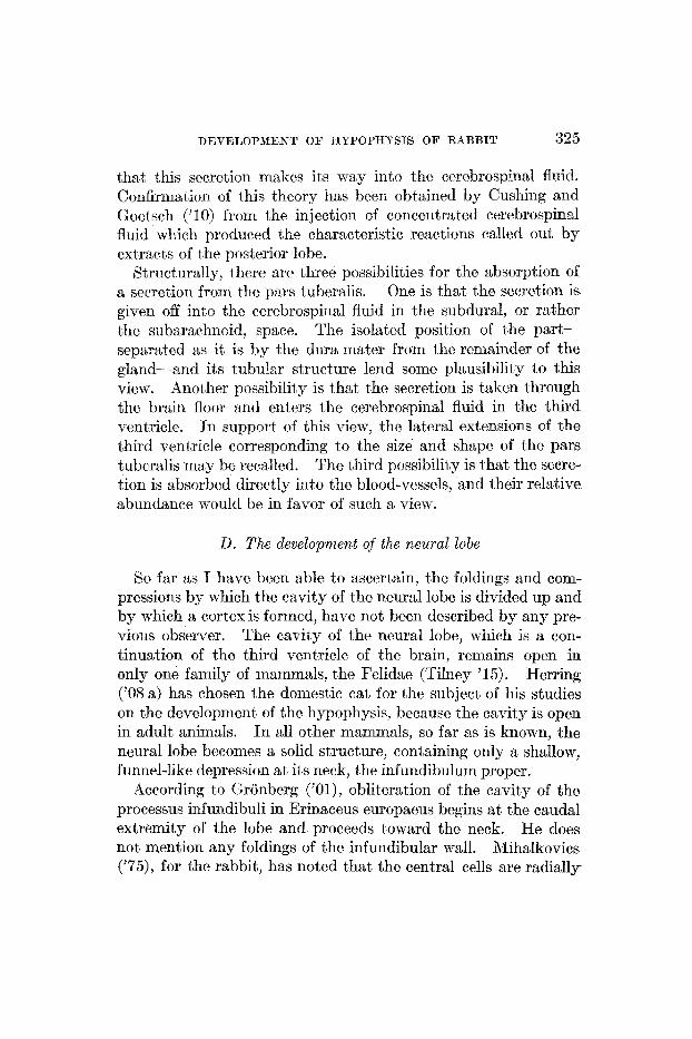
DEVELOPMENT O F HYPOPHYSIS OF RABBIT 325
that this secretion makes its way into the cerebrospinal fluid. Confirmation of this theory has been obtained by Cushing arid Goctsch ('1 0) from the injection of concentratcd cerebrospinal fluid which produced the characteristic reactions called out by extracts of the posterior lobe.
Structurally, there arc three possibilities for the absorption of a secretion from the pars tuberalis. One is that the secretion is given off into t8he cercbrospinal fluid in the subdural, or rather the subnrachnoid, space. The isolated position of the part- separated as it is by the dura inater from the remainder of the gland-and its tubular structure lend some plausibility to this view. Another possibility is that the secretion is taken through the brain floor and enters the cerebrospinal fluid in the third ventricle. In support of this view, the lateral extensions of the third ventricle corresponding to the size and shape of the pars tuberalis may be recalled. The third possibility is that the sccre- tion is absorbed directly into the blood-vessels, and their relative abundance would be in favor of such a vicw.
D. The development of the neural lobe
So far as I have bccn able to ascertain, the foldings and corn- pressions by which the cavity of the neural lobe is divided up and by which a cortcx is formed, have not been described by any pre- vious observer. The cavity of the neural lobe, which is a con- tinuation of the third ventricle of the brain, remains open in only one family of mammals, the Felidae (Tilney '15). Herring ('08 a) has chosen the domestic cat for the subject of his studies on the development of the hypophysis, because the cavity is open in adult animals. In all other mammals, so far as is hop-n, the neural lobe becomes a solid structure, containing only a shallow, funnel-like depression at its neck, the infundibulum proper.
According to Gronberg ( 'Ol ) , obliteration of the cavity of the processus infundibuli in Erinaceus europaeus begins at the caudal extremity of the lobe and proceeds toward the neck. He does not mention any foldings of the infundibular wall. iVihalkovics ('75), for the rabbit, has noted that the central cells are radially

326 WhYh-E J. ATWELL
arranged, while the outer cells are smaller and irregularly dis- posed. He states merely that the cavity of the lobc is obliterated by a growth of the infundibular .v\-alls.
Almost from its earliest appearance, the walls of the neural lobe of the rabbit’s hypophysis show a number of foldings (figs. 11 and 13). These rapidly become more complicated, and during the fourteenth day, cells from the wall have entered the lumen of the lobe so that its cavity is partially obliterated and very ir- regular in shape. Sometimes septa, or partitions, are formcd which divide the communication with the third ventricle into two or more openings. This is scen in models of fifteen- and seventeen- day ern bryos.
Around the outside of the lobe, a cortical zone is formed by much thc same process as tlhat by which cells obliterate its cavity. Apparently the basement meinbrane is broken through to permit masses of cells to escape to the exterior of the lobe. At any rate, the cell inasqes often do not seem to be definitely surrounded by a membrane and portions of the original basement membrane are to be seen between the medulla and the cortical zone. AS shown by figure 21, the medullary layer has its cells radially arranged, while the cells of the cortical zone are very irregularly disposed. The cortical layer becomes fibrillar in structure in later stages, and the cell bodies become more widely separated. This is well shown in a tnenty-day embryo (fig. 21, C).
Miiller (’71) and Mihalkovics (’75) have maintained that the infundibulnr procesi, or neural lobe, becomes converted into a 4 l connective-tissue appendage of the brain” through the gradual replacement of its nervous elements by connective-tissue cells. Herring (’08 a), by employing the methods of Golgi arid Cajal, has shown that thc neural lobe does not become a conn npperi dage, but that it is composed of a somewhat modified epen- dynial and iieurogliar tissue. I have not made use of these special stains for nervous tissue, but, by carefully following the devel- oprnent of the neural lobe, I am not able t o find that any considerable amount of connective tissue becomes incorporated into it. The blood-vessels are very few in the lobe prior to the twentieth day of development. Even after this time, they are

DEVELOPMENT O F HYPOPIIYSIS O F RABBIT 327
never very numerous, and the connective tissue accompanying them is small in amount.
The angle formed by the attachment of the neural lobe to the brain wall undergoes an intcrcsling series of changes. At thir- teen days (fig. 11) the lobe makes an acute angle with the brain wall in the nasal side of its attachment. It is inio this angle that the apex of Rathke’s pocket is seen to he fitted. By serenteen days (fig. 22) , the angle formed by the neural lobe and the brain wall is approxirnately ninety deguees. After this time, the anglc. is acute on the caudal side of the attachment of the lobe. The acuteness of the angle gradually increases, until at thirty days the neural lobe lies almost, parallel to the floor of the third ven- tricle (fig. 35).
E. The newo-epitheliul contacts and their aign@ican,ce
Contacts between the neural lobe and the intermediate part, such as are here recorded for certain fifteen- and sixteen-day and older embryos, have not been described by any other obserrcr. It is true that Herring(’08 b) based his theory, that the secretion of the pars interniedia is absorbed through the brain, on the fact that the neural lobe is apparently invaded by cell Inasses and sc- cretion globules from the intermediate part. But this is quite different from what I have observed in the sixteen-day and related stages of the rabbit’s development. Reasons have already been enumerated why these contacts appear to me to be due to an active groi5 th of processes of the neural lobe into the intermediate part. Xttentioir has been called to the presence of a small dc- pression, corresponding to the center of each contact which may be seen on that, surface of the p r s intermedia which faces the residual lunzen. I t has been noted that thc contacts vary in num- bers from one to five, and that they \.i.e~c fourid constantly present in all of the sixteen-day embryos cxaniined and in certain fifteen- seven teen- eighteen -, and tmen t y-da y embryos.
Wit’h these facts in mind, a survey may be made of the explana- tions which are to be found for the possible significance of these contacts or ingrowths.

328 WAYNE S. ATTVELL
1. Herring ('08 a) has seen, in a singl: cat embryo near term, an opening uniting the cavity of the neural lobe with the residual lumen. He interprets this as a recurrence of some ancestral bucconeural communication. It might be argued that the con- tacts observed for the rabbit artre an atIteimpt to form such a com- munication, notwithstanding the fact that, no lumen is to be found in them. If there were constantly only one of these con- tacts to be seen, this hypothesis might demand serious attention, but since they vary in num.ber, and may be as many as five, it seems to me very improbable that this is the correct explanation.
2. Another possibility is that thesc contacts are an imperfect, temporary recurrence of the condition normal for the fishes, in which forms nunierous processes from the brain floor enter the pars intermedia. Such structures are pictured by Stendell ('14) in his figures 19 to 24, pages 20 to 26. If this interpretition be corrcct, similar structures should sometjlnes be found in the verte- brates between the mainmals and the fishes, and one would expect them to become larger and more constant ax the vert'ebrate scale is descended.
3. The most likely possibility, it seems to me, of the significance of these contacts is that they are a means for supplying the epen- dymal and neurogliar supporting tissue to the pars intermedia. That such tissue is really to be found in the intermediate part is indicated by the observations of Pirone, Geinelli, Retzius, Herring, Trautmann, Cajal, and others. They have been de- scribed by Retzius for the rabbit, eight days after birth. Stendell believes that these tissue elements have wandered in from the neural lobe, but he quotes no observations to substantiate such a view. The other possibility is, of course, that the spindle-shaped elements which have been notcd are peripheral supporting cells developed in situ in the epithelium of the pars intermedia after a manner similar to the supporting cells of the olfactory epithe- lium. This view would receive some support from the close relation of the hypophysis to the olfactory organ in the Monor- rhina. However, the fact that, the pars intermedia of the hypoph- ysis in the highest order of vertebrates shows cells which may

DEVELOPMENT O F I-IYPOPHYRIH O F RABBIT :329
be tenned neurogliar and others which may be called ependyrnal (Stendell) gives the transfer theory considerable significance, al- t,hough for the present it must remain an hypot,hesis mcrely.
F. Terminology and phylogeng of the lobes of the hypophysis
The terminology, still to be found in text-books of anetomy and embryology, which speaks of that portion of t8he hypophysis de- rived froin the brain as the ‘posterior lobe’ and that derivcd from t8he oral invagination as the ‘anterior lobe,’ must be considered entirely inadequate. Physiologists recognize two very different internal secretions as being produced by thc hypophysis. One is obtained from the ‘posterior lobe’ and causes a rnarkcd rise of blood-pressure, increased secretion of urine and of milk, a general contraction of involuntary muscle, especially of the uterus, etc. The other is obtained from the ’anterior lobe’ and does not mark- edly influence blood-pressure on introduction into the circulation, but is thought to influence body growth, particularly of the bones. It, is held that, the secretion attributed to thc ‘posl’erior lobe’ is in reality not formed by the iiervous portion of the hypophysis, but by its epithelial covering, the ‘pars intermedia.’ ‘Posterior lobe’ to the physiologist means neural portion together with the epi- thelial layer which is closcly adherent, to it. Or, more succinctly stated, ‘posterior lobe’ nieans neural lobe plus pars intermedia, and ‘anterior lobe,’ as understood by the physiolagist until recently, means buccal portion minus pars intermedia.
The recently recognized pars tuberalis must be considered a distinct portion of the gland, both embryologically and histo- logically, as urged by the present study and by the investigations of Tihey, Baumgartner, and Parker. ‘Antcrior lobe,’ then, should mean buccal portion minus pars intermedia and pars tuberalis. Thus any adequate consideration of the lobes of the mammalian hypophysis must recognize four parts-the neural lobe, and three divisions of the epithelial lobe. These three parts arc pars anterior propria, pars intermedia and pars tuberalis.
It is well known that in the Elasmobranchs there is to be found a pair of hypophysial lobes which lie inferior to the main body of

the gland, and arc known as the ‘inferior sacs’ or ‘inferior lobes.’ Baunigartncr (’15) has studied the development of the hypo- physis in Squalus scanthias and figures several >Tax reconstruc- tions of the embryonic organ. He speaks of the epithelial part, of the hypophysis in Squalus as consisting of four lobes: an ‘anterior lobe,’ a ‘superior lobe,’ and a pair of ‘inferior lobe L’ In another study Baumgartner (’16) has treated very fully the de- velopment of the hypophysis in the Reptiles. He speaks of Rathke’s pouch, two lateral buds, and an antcrior bud. Woerdernan (’14) feels justified in hoinologizing the lateral lobes of rnarniritlls with similar structures in the Reptiles and also with the inferior lobes or ventral sacs, of the Selachians. The ‘lobulus bifurcatus’ of mammals in reality represents a much-reduced complex of Selachiaii Torraum,’ ‘Mittelraum,’ and ‘lobuli later- ali.’ Woerdeman believes that “wahrscheinlich der Lobulus bi- furcatus ein rudimentares Organ darstellt, und dass der vordere Teil der Hypophysen anlage sich sehr menig entnickclt bei den meisten, ja fast vollig unentwickelt bleibt bei anderen S:iuge- t ieren. ”
If we accept the hoinologics drawn by Woerdeman, we xilay briefly summarize the history of the lateral lobes as follows: In the Elasmobranchs the hypophysis possesses a pair of lobes which lie vcntral t o the main body of t8he hypophysis and thus are B
considerable distance removed from the brain wall. These inferior lobes or ventral sacs remain separate from one anothccr, but are connected by ducts to the cavity of the body of thc hypa- physis. In the higher vertebrates, two homologous lobes (lobuli lat,erali) do not retain a position ventral to the main body of the gland, but grow toward the brain. They may be said to insinu- ate themselves between the main body of the gland and the dien- caphalic floor. They fuse with each other and forin a lobe which extends forward to the optic chiasm and posteriorly to embrace the infundibular stalk. Thus a new ‘juxta-neural’ (Tilncy) por- tion of the gland is formed. The anterior portion of this lobe has been called ‘Fortsatz des Epithelsaums’ (Lothringcr) ; ‘rordere Lappcn,’ ‘vordere Fortsatz’ (Haller) ; ‘lobus chittsmaticus’ (Sta- derini) ; ‘pars terminalis’ (Gisi), and ‘ tongue-like process of the

DEVELOPMENT O F HYPOPHYSIS OF RA4BBIT 331
pars intermedia’ (Herring). The posterior part of the lobe Staderini kas called the ‘lobus praemammilaris.’ Ontogeny shows that anterior and post,erior partions are a unity, so they may bc named as one lobe. Joris has called the lobe the ‘lobule de la tige,’ Bolk terms it the ‘lobulus bifurcatus,’ and Tilney narncs it the ‘pars tuberalis.’
From their very similar development and fate it n;ouId seem that the lateral lobes of the Mammals may safely be considered homologous to the lateral lobes of the Reptiles.
It is to be noted that no adequate description exists concerning the development of lateral lobes in either the Teleosts or the Arnphibia. For this reason, it must be somewhat precarious at the present time to homologize the lateral lobes of embryos of the Amniota with the ventral sacs of the Elasmobranchs.
I cannot agree wit8h Woerdeman w h n he homologizes the ail- terior part of the mammalian ‘lobulus bifurcatus’ (pa,rs tuberalis) with the median unpaired T o ~ ~ a u m ’ of the reptilian and selachian hypophysis (compare his f ig . 30, G and H). According to my observations on the rabbit, the anterior portion of the pars tuberalis has a paired origin, being formed by the fusion of the blunt nasal horns of the lateral lobes. If a homologue of the ‘V0rrau;m7 be present during the development of the hypophysis of tho rabbit, it is best recognized at the fourteenth and fifteenth days. After this time it must be very much reduced, even more so than stated by Wocrdernan.
6 . SUMMARY AND CONCLUSIONS
1. The hypophysis evagination is plairily visible in a rabbit embryo having sixteen primitive segments. In age this corre- sponds to the earlier part of the tenth day after insemination. The definite neural lobe first appears during the twelfth day.
2. The cephalic extremity of the notochord is directed toward a bud-like outgrowth of the dorsal wall of Rathke’s pouch in cer- tain ten-, eleven-, and twelve-day embryos, but tchis relation is not constant. No evidence can be found for stating that the entoderm contributes to the formation of the hypophysis of the rabbit.

332 WSYNE J. ATWELL
3. The stalk connectihg the hypophysis with the oral epithelium becomes solid during t,he fourteenth day. I t loses its connection wit’h the epithelium at a variable time-as early as the sixteenth- day or as late as the twenty-fourth. The place of attachment of the stalk to the gland is indicated’even until the time of birth; it shows an apparent migration to the nasal end of the anterior lobe proper. 4. The residual lumen of Rathke’s pouch at first extends from
end to end of thc epithelial port,ion; later it does not reach t o the nasal extremity. The bending of the gland gives the lumen an L-shape in sagittal sections, with one limb extending into the anterior lobe proper. The L-shape is changed into a T- or Y- shape by the addition of another limb which is in relation with the portion of the pars intemiedia extending aromd the caudal end of the neural lobe. The limb extending into the anterior lobe becomes oblitcratcd. The remainder of the lumen is present at birth and separates the interrncdiate part from the anterior lobe proper.
5. From the thickened epitheliuni just nasal to the early Rathkc’s pscket two ridge-like elevations are developed. These havc been called the lateral lobes. As thc hypophysial evagi- nation is constricted from the oral cavity the lateral lobes con- stitute the lateral terminations of a transverse ridge lying at the nasal end of the gland. At fourteen days the lobes have begun to grow out laterally, and are more sharply constricted from the body of the hypophysis.
The lateral lobes are the anlagen of the pars tuberalis and of a temporary cortical plate at the nasal extremity of the gland. They begin to be in relation with the brain wall at sixteen days and by nineteen days a considerable portion lies spread out under the floor of the third ventricle. There are present at this stage two blunt nasal horns extending toward the optic chiasrn and two sharpcr caudal horns extending backward to surround the neck of the neural lobe. The caudal horns have completely surrounded the infundibulum and have met in the midline by the end of the twenty-eighth day. The nasal horns fuse with each other during the last two or three days of development.

DEVELOPMENT O F HYPOPHYSIS OF RABBIT 333
Since the pars intermedia is not definitely present unt,il the definitive neural lobe has begun to form (twelfth day), the anlagen of the pars tuberalis precede it in appearance by two days (in the rabbit). This reverses the chronology of their appearance as given *by Tilney (’ 13) .
The pars tuberalis comes to lie in the pia niater of the dien- cephalic floor. In the rabbit it maintains its connection with the parent gland, at least as late as the time of birth, by a strong stalk which penetrates the dura mater of the diphragma sellae. It shows distinctly a tubular structure with the tubules extending for the most part in a nasocaudal direction.
The pars tufseralis constitutes a p6rtion embryologically and histologically different from the remainder of the gland. It is sharply denlarked from the pars intermedia, even until the time of birth, by the dura inater and by coniiective tissue which has become imprisoned in the fossa. Thus Lothringer’s name, ‘Fortsatz des Epithelsaums’ and Herring’s term, ‘tongue-like process of the pars intermedia,’ as used to designate the anterior portion of the pars tuberalis, are not descriptive of the develop- ment of the part. The same objection may be urged in refer- ence to Herring’s confusion of the caudal part of the pars tuberalis with the “extension of pars intermedia round neck of gland.”
6. The neural lobe undergoes a series of complex foldings and compressions which results in the dividing up and partial oblitera- tion of the cavity of the lobe and in the formation of a medullary layer arid a cortical layer.
7 . Definite contacts between the neural lobe and the inter- mediate part are to be seen in sixteen-day and related stages of development. Reasons are given for the belief that these con- tacts are due to the growth of processes from the neural lobe into the intermediate part. The suggestion is offered that the con- tacts may constitute a mechanism for supplying the ependymal and neurogliar elements which have been observed in the inter- mediate part by Rctzius, Cajal, Trautmann, Herring, and others.
8. From a similarity in development and fate the lateral lobes of the bIammals and of the Reptiles may be considered homolo- gous. It is somewhat precarious, for the present, to homologize

334 WAYNE J. ATWELL
the lateral lobes of the Reptiles and +Mammals with the inferior lobes of the Elasmobranchs.
In the rabbit the anterior part of the pars tuberalis cannot be homologized witrh the ‘Vorraum’ of the lower vertebrates, since j t is seen to be developed from the paired blunt nasal horns of the lateral lobes.
7 . LITERATURE CITED
ALLEN, B. M. 1916 The results of extirpation of thc anterior lobe of the hypo- physis and of the thyroid of Rana pipiens larvae. Scicnce, hr. s., vol
1917 Extirpation of the hypophysis and thyroid glands of Rana pipiens.
ATWELL, Tiv. J. 1915 The relation of the chords dorsalis t o the entodermal conr- ponent of the hypophysis. Anat. Rec., 1701. 10, p. 19.
BALFOUR, F. M. 1874 A prcliniinury account of thc development of the elasrno- branch fishes. Quar. Jour. Micros. Sc., N. S., vol. 14, p. 324.
BAUMUARTNER, E. A. 1915 The development of the hypophysis in Squalus acanthias. Jour. Morph., vol. 26. 1916 The development of the hypophysis in reptiles. Johr.Morph., vol. 28.
BOLK, L. 1,dlO Over de ontwickkeling der Hypophyse van de Primaten in het byzonder by Tarsius en den Mensch. Verslag d. Kon. Akad. v. We- tehsch. Amsterdam.
BRUNI, -4. C. 1914 Sullo svilluppo del lob0 ghiandolare dell’ipofisi negli Am- nioti. Internat. Monatschr. f . Anat. u. Physiol., Bd. 31, Heft 4/6.
CAJAL, S. R. 1911 Hisbologie du Systhme Nerveux de 1’Homme et des Verte- brEs. T. 11, Hypophyse ou Glande Pituitaire, p. 487.
CHIARUGI, G. 1894 Sull’ esistenza di m e gemma bilaterale nell’ abbozzo della ipofisi dei Mammiferi. Monit. zool. ital., Anno. V, No. 8.
CUSHING, HARVEY, AND GOETSCR, EmL 1910 Concerning the secretion of the infundibular lobe of the pituitary body and its presence in the cere- brospinal fluid. Amer. Jour. Physiol., vol. 27, p. 60.
DEAN, B. 1896 On the larval development of Amia cailva. 2001. Jahrb., Bd. 9. ECONOMO, C. J. 1899 Zur Entwicklung der Vogelhypophyse. Sitzungsber.
d. math.-naturwiss. K1. d. K. Alwd. d. Wiss. Wien. Bd. 107, Abt. 3. GAGE, SUSAPU’KA PHELPS. 1906 The notochord of thc head in human embryos of
the third to the twelfth week and comparisons with other vertebrates. Science, N.S., vol. 24, p. 295.
GAUP~, E. 1893 tfber die Anlagen der Hypophyse bei Saurien. $rch. f . mikr. Anat., Bd. 42, S. 569.
GEMELLI, A. 1905 Sur la structure de la r6gion infundibulaire des poissons. Journ. de l’Anat., An. 42. 1906 Ulteriori osservazioni siilla strutturs dell’ ipofisi. Anat,. Anz., Bd. 28, S. 613.
44, p. 755.
Anat. R,ec., vol. 11, p. 486.
\

DETE2LOPMENT O F HYPOPHYSIS O F RABBIT 335
GISI, JULIA 1907 Das Gehirn von Hatteria punctata. Diss. Basel. GOETTE, A. 1874 Kurae Mittheilungen aus der Entwickelungsgeschichte der
Unke. Archiv. f . mikr. Anat., Bd. 9, p. 396. GREGORY, E. H. 1902 Beitrage zur Entwickelungsgeschichte der Knochenfische.
Anat. Hefte, Bd. 20, Abt. 1. GRBNBERG, G. 1901 Die Ontogenese eines niederen Siiugergehirns nach Un-
tersuchungen a n Erinaceus europaeim. Zool. Jahrb., Anat. Abt., Bd. 15, S. 261.
HALLER, B. I897 Untersuchungen iiber die Hypophyse und die Infundibularor- gane. Morph. Jahrb., Bd. 25. 1909 tfber dfe Hypophyse niederer Placentalier und den Saccus vas- culosus der urodelen Amphibien. 1910 Ober die Ontogenese des Saccus vascuIosus und der Hypophyse der Saugetiere.
The development of the mammalian pituitary and its morphological significance. 1908 b. The histological appearances of the mammalian pituitary body. Ibid., p. 121.
HOFFMAN, C. K. 1886 Weitere Untersuchungen zur Enlwicklungsgeschichte der Reptilien. Rlorph. k h r b . , Bd. 11.
HUBER, G. ( 3 . 4 ~ ~ 1915 The development of the albino fat , Mus norvegicus albinus. Memoirs of The Wistar Institute of Anatomy and Biology, no. 5.
JORIS, H. 1907 Contribution a l'etude de l'hypophyse, M h i . cour. sutr . publ. Acsd. Roy. Med. Bclgique, Tome XIX, fasc. 6.
KING, H. D. 1913 Some anomalies in the gestation of the albino ra t (Mus nor- vegicus albinus).
KOELLIKER, A. 1879 Embryologische Mittheilungen: 1. cber das vordcre Ende der Chordx dorsalis bei Ihninchen embryonen. Festchr. z. Feier d. Hundertjahr. Bestch. d . Naturforsch. Gesell. in IIalle, 8. 115.
KRAUSHAAR, R. 1885 Entwicklung dcr Ilypophysis und Epiphysis bei Nage- thieren.
KUPFFER, C . v. 1892 Entwicklungsgeschichte des Kopfes. Ergeh. d. Anat. u. Entw., Bd. 2, S. 501. 1894 Die Deutung des Hirnanhanges. Sitz. Ber. d. Gesellsch. f . Morph. u. Physiol. zu Miinchen, S. 59.
LONG, J. A,, AND MARK, E. 1,. The maturation of the egg in the mouse. Carnegie Inslitute of Washington, Publication No. 142.
LOTHRINCER, S. 1886 Untersuchungen an dcr Hypophyse einiger Siugethiere und des Menschen. Arch. f . mikr. Rnst., Rd. 28, S. 257.
LIJSCHKA 1860 Der Hirnanhang und die Steissdriise des Menschen. v. MIIHALKOVIES VICTOR 1875 Wirbelsaite und Hirnanhang. Arch. f. mikr.
Anat., Bd. 11, S. 389. MILLER, M. Ril. 1916 A study of the hypophysis of the pig. S n a t . Rec., vol.
10, p. 226. MINOT, C . 8., A V D TAYLOR, E. Normal plates of the dcvclopinent of the
rabbit (Lepus cuniculus L.) , Heft 5 in Keihel's Kormentafeln zur Entwicklungsgeschichte der Wirbelthiere.
Arch. f . mikr. Anat., Bd. 74, S. 812.
Anat. Anz., Bd. 37, 8. 242. HERRING, P. T. 1908 a.
Quar. Jour. Ex, Physiol., vol. 1, p. 161.
Biol. Bull., vol. 24.
Zeitschr. f . wiss. Zool., Bd. 41, S. 79.
s
1911
1905
TAE AMERICAN JOUILNAL OF ANATOMY, YOL. 24, NO. 3

336 WAYNE J. ATWELL
MULLER, W. 1871 cber Entwicklung und Bau tier Hypophysis und des Pro- c e s s ~ ~ infundibuli. Jenaische Zeitschr. f . Med. u. Naturwiss., Bd. 6, s. 354.
NUSBAUIM, J. 1896 Einge ncuc Thatsachen zur Entwickelungsgeschichte der Hypophysis cerehri bie Saugetieren. 1898 Przyczynek do historyi rozmoju hypofyz (Hypophysis ccrebri) u Zwierzat ss3cych. I. Taf., Kosmos, Roczuik 2“. (Cit,ed by Woerde- man, 1914).
O R R ~ , E. 1900 Sullo sviluppo dell’Ipofisi. Internat. Monatschr. f . Anat.,
OSTROUMOFF, A. 1S88 Zur Entwicklungsgeschichte der Eidcchsen. Arb. d. naturf. Gesellsch. zu Kazan.; Bd. 19. Abstract. in Zool. Anz., Bd. 2.
PARKER, KATHERIKE M. 1917 The development of the hypophysis cercbri, pre- oral gut, and related structures in the Marsupialia. Jour. Anat., vol. 51, part 3.
PEREMESCRKO 1867 fiber den Bau des Hirnanhanges. Archiv. f . path. Anat. u. Physiol. (Virchow), Bd. 38, S. 329.
PIRONE, RAFFAE’LE Sulla Finn Struttura e Sui Fenomeni di Secrezione dell’ Ipofisi. A4rch di. Fisiol., vol. 2? p. 60.
PRITHER, J . M. 1900 The early stages in the development of the hypophysis of Amia Calva. I3iol. Bull., vol. 1.
REIGHARD, J. 1900 The development of the adhesive organ and hypophysis in Amia. Science, N. S., vol. 11: p . 251.
RXICHARD, J., AND Mawr, 8. 0. 1908 Studies on Ganoid fishes: 11. The de- velopment of the hypophysis of Amia.
RETZIUS, GUSTAF 1894 Die Neuroglia lia dcr Neuro-Hypophysc der Sau- gethiere.
ROGOWITSCH, N. 1889 Die Veranderung der Hypophyse nach Entfernung der Schilddruse.
Rosur, N. 1896 Sui lobi laterali della Ipofisi. Monit. zool. ital., 7. ~ A L , Z S R , HANS 1898 Zur Entwicklung der Hypophysc bei SSiugern. Archiv. f .
mikr. Anat., Bd. 51, S. 55. SEESSEL, A. 1877 Zur Entwicklungsgeschichte des Vorderdarms. Arch. f .
Anat. u. Physiol., Anat Abt., 8.449. S m m , 1’. E. 1914 The development, of the hypophysis of Amia calva. Anat.
Rec., vol. 8, p. 499. 1916 a Experimental ablation of the hypophysis in the frog embryo. Science, N. S., vol. 44, p. 280. 1916b The effect of hypophysectomy in the early embryo upon the growth and development of the frog.
STADERINI, R. 1903 Lo sviluppo dei lobi dell’ ipofisi nel’ Gongylus ocellatus. Arch. I ta l . Anat. Embriol., vol. 2. 1908 D i un prolungamento ghiandolare dell’ ipofisi accolto in uno speciale recess0 premammillare nel cervelIo del gatto ztdulto. Anat. ilnz., Bd. 33> S. 271.
STENDELL, W. 1913 Zur vergleichenden Anatomie und Histologic der Hypo- physis cerebri. 1914 Die Hypophysis Cerebri, vol. 8, in Oppel’s Lehrbuch der ver- gleichenden niilcroscopischen Anatomic der Wirbeltiere. Fischer, Jena.
Anat. Anz., Bd. 12, S. 161.
Bd. 17, S. 424.
1905
.Jour. Morph., vol. 19.
Biol. Untersuch., N. F., Bd. 6, S. 21.
Beitrage zur path. Anat. und allg. Path., Bd. 4, S. 453.
Anat. Rec., vol. 11, p. 57.
Arch. f . mikr. Anat., Bd. 82.

DEVELOPMENT OF HYPOPHYSIS O F RABBIT 337
STIEDA, HERMANN 1890 c b e r das Verhalten der Hypophyse des Kaninchens nach Entfernung der Schilddr~se. Beitrsige z. path. Anat,. u. allg. Path., Bd. 7 , S. 537.
Sur la signification morphologique de la poche pharyngienne de Seessel.
Contribution to the study of the Hypophysis cerebri with especial reference t o its comparative histology. Memoir KO. 2, Wistar Inst'itutc of Anatomy, Philadelphia. 1913 An analysis of the juxtaneural epithelial portion of t,he hypophy- sis cerebri, with an embryological and histological account of an hither- to undescribed part, of the organ. Internat. Monzhschr. f . Anat. u. Physiol., Hd. 30. 191.5 The morphoiogy of the dienccphalic floor. J w r . Comp. Ncur., vol. 2.5.
TRAUTXASN, ALFRED 1901) rin&omie und Histologie der Hypophysis cerebri einiger Sauger. Brch. f . mikr. Anat., Bd. 74, S . 311.
'SIALENTI, G . 189.5a sullo sviluppo dell' ipofisi. h a t . , Anz., Bd. 10, 8. 538. 189513 bled.-Chir. di Perugia. No. 7. 1897 dorsale e dell' intestino.
vol. 12. p. 95.
ST. REYY, G. 1595
TILNEY, FREDERICK C. R. de la Soci6tC de Biologie, p. 423.
1911
Sullo orginie e sul significato dell' ipofisi. htt.i dell' h a d .
Sopra i primitivi rapporti delle estremita cefalichc della corda
VANDERBTTRGH, C. M. 1917 The hypophysis of t.he guinea-pig. Anat. Rec.,
WEBER, A. 1898 Observations sur les premieres phases du dheloppement de I'hypophyse chez les Ch6iroptBres. Bibliogr. Anat.. fasc. 3 .
WOERDEMAN, M. W. 1913 t h e r einen Zusammenhdng der Chorda dorsalis mit der Hypophysenanlage. Anat. Anz., Rd. 43. 1914 Vergleichenden Ontogenie der Hypophysis. Archiv. f . mikr. Anat., Bd. 86.
Atti Soc. tosc. Sc. nat. Pisa.


![Abses Cerebri[1]](https://img.dokumen.tips/doc/110x75/55cf94a8550346f57ba38426/abses-cerebri1-5652e84c18f54.jpg)