Embed Size (px)
Citation preview
TERSON’S SYNDROME
Z. Jamaleddine, S. El Haddad, A. El Quessar
Service de Radiologie, Hopital Cheikh ZaidRabat - Morocco

Introduction
Terson’s syndrome is the association of vitreous or retinal haemorrhage with subarachnoid haemorrhage (SAH).
The diagnosis is typically made fundoscopically.
Objectives
To describe, the clinical, radiological characteristics of this syndrome.
To describe the therapeutic indications.
Materials and methods
Three cases hospitalised for subarachnoid haemorrhage (SAH) and secondary complicated by intravitreous haemorrhage.
Imaging exploration based on:
Computer tomography 16 bars,
MRI 1.5 Tesla,
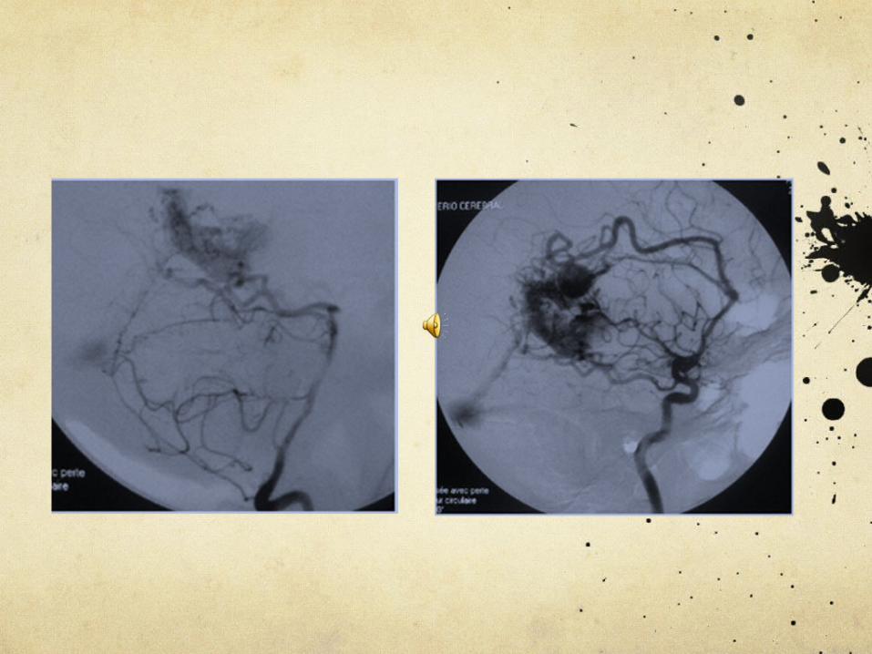
Cerebral angiography.
Ocular ultrasonography intravitreous haemorrhage.
All patients were treated by embolisation for the etiology of SAH : two aneurysms and one AVM.
SAH

37 years old woman,
Left hemiplegia.
CASE 1
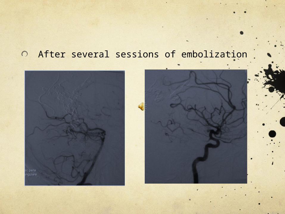
After several sessions of embolization

Three months later: loss of vision in a right eye

Diagnosis of Terson syndrome
Treatment: Vitrectomy after 5 months
Visual acuity improved immediately
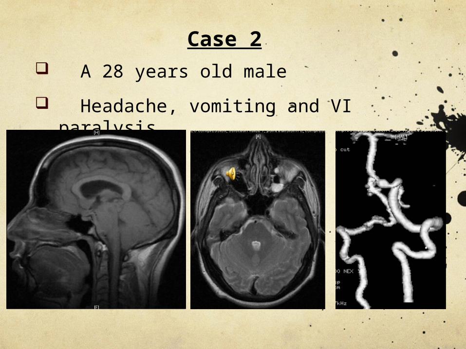
A 28 years old male
Headache, vomiting and VI paralysis
t
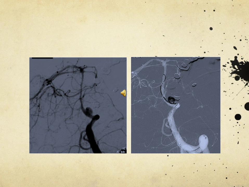
Case 2
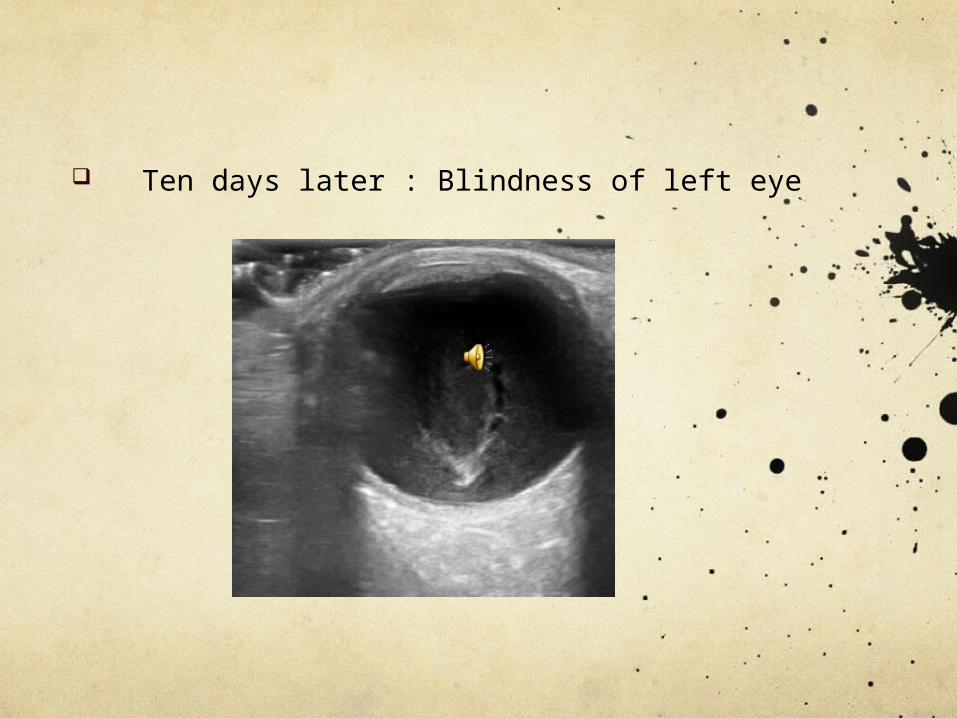
Ten days later : Blindness of left eye

Diagnosis of Terson syndrome
Treatment: Vitrectomy after 2 months
Recovery of visual acuity

Woman of 51 years
Case 3

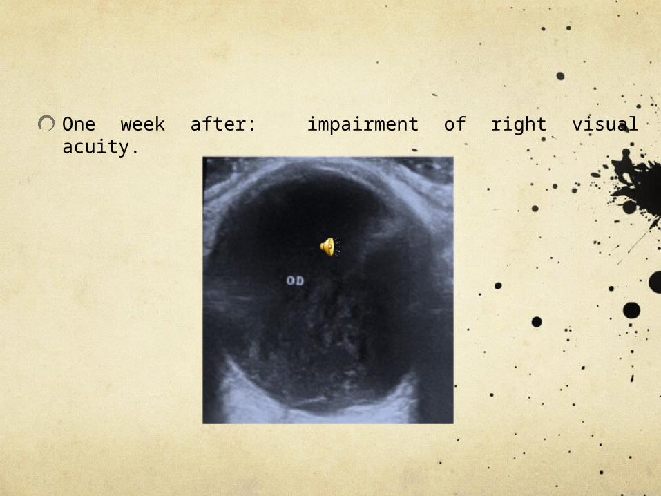
One week after: impairment of right visual acuity.
Diagnosis of Terson syndrome
Vitrectomy is programmed, despite a partial improvement of vision

Discussion
Terson syndrome mentioned for the first time in 1900.
Vitreous haemorrhage: <10% of ruptured intracranial aneurysm.
Bilateral: 14% - 60% of cases.
Intraocular haemorrhage
Adult Children
The rate in % 18 - 41 70
Discussion
Etiology:
Ruptured aneurysm
AVM
Traumatic cause = very rare.
Clinical manifestation: the significant decrease in visual acuity is the most common symptom.
The most commoncause
Ultrasound search:
Characteristic of intravitreous haemorrhage
Dense,
Mobile: feature of Terson syndrome
Abundant,
Posterior vitreous detachment usually total.
Retinal detachment

Vitrectomy has been shown to be extremely effective in clearing the vitreous haemorrhage
Indications:
Patients with intraocular bilateral haemorrhage
There is not signs of spontaneous resorption after 1 to 3 month.
Conclusion
It appears necessary to examine visual acuity in case of subarachnoid haemorrhage for an early diagnosis and the better treatment of this rare syndrome.