Embed Size (px)
Citation preview

210 J Cerebrovasc Endovasc Neurosurg
Subarachnoid Hemorrhage with Negative Baseline Digital Subtraction Angiography: Is Repeat Digital Subtraction Angiography Necessary?
Dong-Woo Yu, MD, Young-Jin Jung, MD, Byung-Yon Choi, MD, Chul-Hoon Chang, MDDepartments of Neurosurgery, College of Medicine, Yeungnam University, Daegu, Korea
Introduction : Patients with negative initial digital subtraction angiography (DSA) are at significant risk for re-bleeding, which can lead to severe dis-ability and death. The purpose of this study was to evaluate the neces-sity of repeat DSA in subgroups of patients with subarachnoid hemor-rhage (SAH) with negative initial DSA.
Methods : A total of 904 spontaneous SAH patients were admitted to our department between May 2005 and May 2012. Twenty eight pa-tients were selected for inclusion in this study because repeated DSA per-formed due to the etiology of the SAH could not be demonstrated on the initial DSA. According to the SAH pattern on initial computed tomog-raphy scans, patients were divided into perimesencephalic nonaneurysmal SAH (PN-SAH) and non PN-SAH (NPN-SAH) groups. Repeat DSA was per-formed in all patients, and two of these patients underwent a third DSA.
Results : Of the 904 patients, 28 patients (3.1%) had no vascular abnormal-ity on initial DSA. Sixteen PN-SAH patients underwent a repeat DSA; how-ever, no aneurysms were found. In contrast, 12 patients with NPN-SAH underwent repeat DSA, with detection of two cerebral aneurysms. Overall, the false-negative rate of the initial DSA was 7.1% (2/28 patients). No significant differences in false-negative results on initial DSA were ob-served between the PN-SAH and NPN-SAH groups.
Conclusion : In the line with the results of the current study, we should be highly suspicious of patients with a nonaneurysmal SAH, especially those with a NPN-SAH pattern. In order to reduce the morbidity and mortality resulting from a misdiagnosis, repeat DSA is necessary, and ex-clusion of an aneurysm is important.
J Cerebrovasc Endovasc Neurosurg. 2012 September;14(3):210~215Received : 24 August 2012Revised : 7 September 2012Accepted : 10 September 2012
Correspondence to Chul-Hoon Chang, MDDepartment of Neurosurgery, Yeungnam University Medical Center, Yeungnam University School of Medicine, 317-1 Daemyeung-5-dong, Namgu, Daegu 705-717, Korea
Tel : (001) 82-53-620-3790, 3795Fax : (001) 82-53-620-3770E-mail : [email protected]
Disclosure : This work was supported by a grant from the Chunma medical research foundation, Korea, 2011.
This is an Open Access article distributed under the terms of the Creative Commons Attribution Non- Commercial License (http://creativecommons.org/li-censes/by-nc/3.0) which permits unrestricted non- commercial use, distribution, and reproduction in any medium, provided the original work is properly cited.Keywords Subarachnoid hemorrhage, Digital subtraction angiography, Aneurysm
Journal of Cerebrovascular and Endovascular NeurosurgeryISSN 2234-8565, EISSN 2287-3139, http://dx.doi.org/10.7461/jcen.2012.14.3.210 Original Article
INTRODUCTION
The incidence of initial digital subtraction angiog-
raphy (DSA) negative subarachnoid hemorrhage (SAH)
is approximately 15% of all patients.4) Perimesencephalic
nonaneurysmal SAH (PN-SAH) was first described by
Van Gijn et al.13) in 1985 as a computed tomography
(CT) scan pattern of hemorrhage restricted to the peri-
mesencephalic or prepontine cisterns, no aneurysmal
structure in DSA, and low rates of re-bleeding and
vasospasm with good recovery.8)9) However, in pa-
tients with non-diagnostic initial DSA, a significant
risk for re-bleeding persists (e.g, from vascular mal-
formation or undetected aneurysm), which can result

DONG-WOO YU ET AL
Volume 14 · Number 3 · September 2012 211
in considerable morbidity and death.9) It is important
to determine whether a cerebral aneurysm exists but
cannot be found, or whether the hemorrhage is the
result of other unknown causes. For this reason, re-
peat DSA has been performed after a negative initial
DSA. False-negative rates have shown a decrease fol-
lowing improvements in diagnostic imaging capa-
bilities, however, the need for repeat DSA in sponta-
neous SAH patients with a negative initial DSA is still
being debated. While DSA has high sensitivity and
specificity for detection of cerebral aneurysms, this in-
vasive procedure is associated with some complica-
tions (SAH-specific mortality in 0.17% of patients,
neurological morbidity in 3.2% of patients, with per-
manent disability in 0.04%).5) Given the variety of
available diagnostic tests and differences in their asso-
ciated risks and costs, it is important to determine the
diagnostic yield of each test and to compare the yield
between SAH subgroups in which initial DSA was
negative.11) The purpose of this study was to evaluate
the necessity of repeating DSA in SAH patients with
negative initial DSA.
MATRIALS AND METHODS
A total of 904 spontaneous SAH patients were ad-
mitted to our department of neurological surgery be-
tween May 2005 and May 2012. DSA was performed
for initial evaluation on 458 patients. Criteria for pa-
tients of this study included the following: SAH was
easily confirmed by initial CT scan, and patients with
an SAH etiology that could not be verified on initial
DSA, and on whom at least one more repeated DSA
was performed. A monoplane neuro X-ray system
(Allura Xper FD20; Philips, Best, the Netherlands)
was used in performance of DSA examinations. Of
the 28 patients who underwent a repeat DSA, two of
these patients underwent even a third DSA.
According to the SAH pattern on initial CT scans, pa-
tients were divided into two groups. PN-SAH com-
prises (1) a center hemorrhage located immediately in
front of the midbrain or within perimesencephalic,
prepontine, or medullary cisterns; (2) absence of intra-
parenchymal bleeding; (3) extension of blood into the
sylvian fissure with no more than a minute amount of
blood in the lateral sylvian fissure; (4) extension of
blood without complete filling of the anterior inter-
hemispheric fissure; (5) absence of a frank intra-
ventricular hemorrhage (sedimentation of a small
amount of intraventricular blood is allowed).10)
Hemorrhages that did not meet all of these criteria
were classified as non PN-SAH (NPN-SAH). None of
the patients was diagnosed with SAH by lumbar
puncture after a negative CT scan. All patients under-
went CT angiography, which was negative for struc-
tural pathology and subsequently underwent DSA.
Clinical grade was based on the Glasgow Coma Scale
(GCS) and Hunt-Hess (H-H) grading system at the
time of admission. Functional outcome was assessed
according to the Glasgow Outcome Scale (GOS). All
patients received conservative treatment according to
the standard SAH protocols, which included adequate
hydration, calcium channel blockers (Nimodipine),
and anticonvulsants during the acute stage. The SPSS
program (version 12.0; SPSS Inc., Chicago, IL) was
used in performance of chi-square tests and t-tests.
RESULTS
Of the 904 patients with spontaneous SAH, 28 pa-
tients (3.1%) had no vascular abnormality on initial
DSA. Repeated DSA was performed within 5-13
(mean 8.2) days for patients in whom the initial DSA
did not reveal any aneurysmal structures or vascular
malformation. Fourteen male and 14 female patients
(M:F = 1:1) were included and the mean age was 60.3
(range 37-77). Initial CT scans showed PN-SAH pat-
terns in 16 patients (57.1%) and NPN-SAH patterns in
12 patients (42.9%). Patients’ demographic data were
presented with their clinical grades on admission
(Table 1). The PN-SAH group had relatively lower
Fisher grade distributions when compared to the

NEGATIVE BASELINE DSA IN SAH PATIENTS
212 J Cerebrovasc Endovasc Neurosurg
Demographics PN-SAH (n = 16) NPN-SAH (n = 12)
AgeSex (M:F)GCS score (on admission)H-H grade
1234
Fisher grade1234
59.4 (± 9.7) 7:9 14.3
7 6 3 0
0 016 0
61.5 (± 11.5) 7:5 13.8
4 6 1 1
0 1 011
PN-SAH = perimesencephalic nonaneurysmal subarachnoid hemorrhage; NPN-SAH = nonperimesencephalic nonaneurysmal subarachnoid hemorrhage; n = number; GCS = Glasgow Coma Scale; M = male; F = female; H-H = Hunter-Hess
Table 1. Demographic summary and patient characteristicsby hemorrhagic pattern
PN-SAH (%) (n = 16) NPN-SAH (%) (n = 12)
GOS*
12345
0000
16 (100)
2 (16.7)01 (8.3)09 (75)
False negative DSA† 0 2 (16.7)
*p = 0.096, P †= 0.175GOS = Glasgow Outcome Scale; DSA = digital subtraction angiography
Table 2. Clinical outcomes and false negative rate of initial DSA
A B
C D
Fig. 1. Initial noncontrast brain computed tomography (CT) and digital subtraction angiography (DSA). Diffuse sub-arachnoid hemorrhage (SAH) in the region of both the sylvian fissure and interhemispheric fissure (A). Initial three-dimensional rotational angiography image reveals no aneurysmal structure (B). Repeat DSA was performed on the eighth day of admission.Three-dimensional rotational angiography images clearly show a distal ICA aneurysm (C, D).
NPN-SAH group (p < 0.001). The GOS was used for
analysis of clinical outcomes. Every patient in the
PN-SAH group and 75% of patients in the NPN-SAH
group showed recovery to grade 5 (Table 2).
Regardless of their initial SAH patterns, most of the
patients had good clinical outcomes upon discharge.
No statistically significant difference was observed be-
tween the PN-SAH group and NPN-SAH group (p =
0.096). Sixteen PN-SAH patients underwent a repeat
DSA, however, no aneurysm was found. In contrast,
12 NPN-SAH patients underwent repeat DSA, with
detection of cerebral aneurysms in two patients. One
patient had re-bleeding of the supraclinoid dorsal
wall aneurysm, and a supraclinoid dorsal wall aneur-
ysm was detected in the other patient following a re-
peated DSA. The overall false-negative rate of the ini-
tial DSA was 7.1% (2/28 patients).
Illustrative cases
Case 1
A 67-year-old female patient complaining of head-
ache, dizziness, and nausea visited our emergency
department. Neurologic examination revealed no focal
neurological deficits, except for neck stiffness. Findings
on brain noncontrast CT and CT angiography re-
vealed moderate SAH, predominantly in the right syl-
vian fissure, however, no aneurysm was detected.
DSA was performed, but it did not show any aneur-
ismal structure or vascular malformation. The patient
received conservative treatment in the intensive care
unit and recovered without any complications. Repeat
DSA on the eighth day after SAH revealed a dorsal
wall aneurysm measuring 3.0 × 1.3 mm on the right
distal internal carotid artery (ICA) (Fig. 1).
Case 2
A 37-year-old male patient was transferred to our
DONG-WOO YU ET AL
Volume 14 · Number 3 · September 2012 213
A B
C D
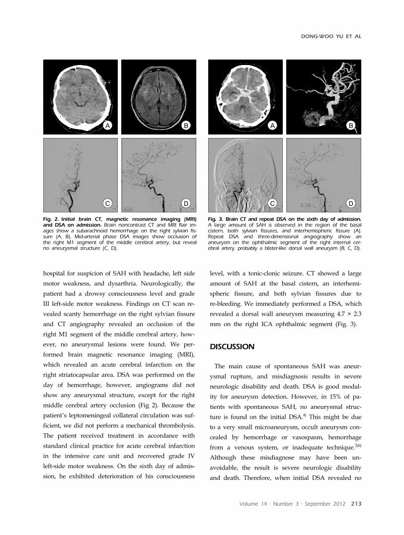
Fig. 2. Initial brain CT, magnetic resonance imaging (MRI) and DSA on admission. Brain noncontrast CT and MRI flair im-ages show a subarachnoid hemorrhage on the right sylvian fis-sure (A, B), Mid-arterial phase DSA images show occlusion of the right M1 segment of the middle cerebral artery, but reveal no aneurysmal structure (C, D).
A B
C D
Fig. 3. Brain CT and repeat DSA on the sixth day of admission.A large amount of SAH is observed in the region of the basal cistern, both sylvian fissures, and interhemispheric fissure (A). Repeat DSA and three-dimensional angiography show an aneurysm on the ophthalmic segment of the right internal cer-ebral artery, probably a blister-like dorsal wall aneurysm (B, C, D).
hospital for suspicion of SAH with headache, left side
motor weakness, and dysarthria. Neurologically, the
patient had a drowsy consciousness level and grade
III left-side motor weakness. Findings on CT scan re-
vealed scanty hemorrhage on the right sylvian fissure
and CT angiography revealed an occlusion of the
right M1 segment of the middle cerebral artery, how-
ever, no aneurysmal lesions were found. We per-
formed brain magnetic resonance imaging (MRI),
which revealed an acute cerebral infarction on the
right striatocapsular area. DSA was performed on the
day of hemorrhage, however, angiograms did not
show any aneurysmal structure, except for the right
middle cerebral artery occlusion (Fig 2). Because the
patient’s leptomeningeal collateral circulation was suf-
ficient, we did not perform a mechanical thrombolysis.
The patient received treatment in accordance with
standard clinical practice for acute cerebral infarction
in the intensive care unit and recovered grade IV
left-side motor weakness. On the sixth day of admis-
sion, he exhibited deterioration of his consciousness
level, with a tonic-clonic seizure. CT showed a large
amount of SAH at the basal cistern, an interhemi-
spheric fissure, and both sylvian fissures due to
re-bleeding. We immediately performed a DSA, which
revealed a dorsal wall aneurysm measuring 4.7 × 2.3
mm on the right ICA ophthalmic segment (Fig. 3).
DISCUSSION
The main cause of spontaneous SAH was aneur-
ysmal rupture, and misdiagnosis results in severe
neurologic disability and death. DSA is good modal-
ity for aneurysm detection. However, in 15% of pa-
tients with spontaneous SAH, no aneurysmal struc-
ture is found on the initial DSA.4) This might be due
to a very small microaneurysm, occult aneurysm con-
cealed by hemorrhage or vasospasm, hemorrhage
from a venous system, or inadequate technique.2)6)
Although these misdiagnose may have been un-
avoidable, the result is severe neurologic disability
and death. Therefore, when initial DSA revealed no

NEGATIVE BASELINE DSA IN SAH PATIENTS
214 J Cerebrovasc Endovasc Neurosurg
negative positive
SAH
Negative baseline study
(CTA, DSA with 3-DRA)
PN-SAH NPN-SAH
Repeat DSA with 3-DRA
Conservative
treatment
Surgery or
Endovascular treatment
Fig. 4. Flow-sheet for diagnosis of patients with subarachnoidhemorrhage.
aneurysm, repeat DSA is required in order to reduce
the incidence of morbidity and mortality due to
misdiagnosis.
In the current study, all patients who had negative
findings on the initial DSA underwent repeat DSA; 28
of the patients had a negative initial DSA, and cere-
bral aneurysms were found in two patients (false-neg-
ative rate 7.1%). The rate of misdiagnosis was re-
duced, compared to other studies, which might be
due to developments in the capacity of diagnostic
DSA. We evaluated the necessity of repeat DSA with
negative initial DSA according to the SAH pattern.
PN-SAH is a distinct characteristic imaging pattern of
nontraumatic, nonaneurysmal SAH, which is asso-
ciated with a good clinical outcomes.13) Compared to
aneurysmal SAH, it usually presents only with head-
aches and no mental deterioration and with a good
clinical grade.9)13) On admission, clinical grades corre-
sponding to GCS scores and the GOS score at dis-
charge was higher in the PN-SAH group than in the
NPN-SAH group; however, these results showed no
statistical significance. Most of these patients had
good clinical outcomes, however, one patient in the
NPN-SAH group died due to re-bleeding and con-
sequential vasospasm.
Two patients had an aneurysm on repeat DSA and
the size and shape of the aneurysms were changed
between the initial and repeat DSA. These two aneur-
ysms were located on the dorsal wall of the ICA. Two
of the false-negative initial DSA patients showed an
NPN-SAH pattern on the initial CT scan. When com-
pared to the PN-SAH group, the NPN-SAH group
had a high false-negative rate (16.7%) (0%, p = 0.175).
Based on the results of our study, because structural
abnormalities, such as aneurysms, can be obscured in
the first angiogram, repeated DSA is always indicated
in patients with NPN-SAH patterns upon CT scan
and negative initial DSA. In particular, configura-
tional changes have been observed in serial cerebral
angiography of supraclinoid dorsal wall aneurysms.
Repeated DSA should result in reduced false-negative
rates and the incidence of re-bleeding. PN-SAH pat-
terns have a very high predictive value for normal
angiogram.10) Due to improvements in diagnostic
imaging capabilities, such as three-dimensional rota-
tional angiography (3-DRA), the recent incidence of
DSA-negative SAH has shown a remarkable reduction.
This technique is better for resolving complicated
anatomy, allowing the investigator to detect aneur-
ysms that are otherwise not visible when using con-
ventional DSA. Ishihara et al.4) reported that the in-
cidence of DSA-negative SAH was 8.6% in the DSA
group and 4.2% in the 3-DRA group. Although DSA
with 3-DRA is the gold standard for detection of
aneurysms, it is invasive and may be associated with
neurological complications. In our series of 16 PN-SAH
patients, the cause of bleeding was not detected
through repeated DSA. Thus, provided that the initial
DSA was technically adequate and revealed no vaso-
spasm, a repeat DSA might not be required.11) We ad-
vocate the need for only a single DSA with 3-DRA in
the PN-SAH group. Due to the benign clinical course
of this subgroup, we believe that the risks of DSA are
too high in comparison with CT angiography.12)14) CT
angiography has multiple advantages over DSA, as it
is a noninvasive, widely available technique, which

DONG-WOO YU ET AL
Volume 14 · Number 3 · September 2012 215
requires shorter time, and uses less contrast media
than the DSA performed in patients with negative CT
angiography.3) However, when the hemorrhage shows
an NPN-SAH pattern, repeat DSA is necessary due to
the possibility of aneurysmal SAH, even if the initial
DSA is negative (Fig. 4). Configurational changes
have occasionally been observed in repeat DSA: blis-
ter-like aneurysms have shown changes in config-
uration into a saccular type and have even shown
spontaneous regression.1)7)
CONCLUSION
In the current study, compared to previous studies,
the rate of misdiagnosis was reduced, owing to the
development of diagnostic DSA techniques, including
3-DRA. However, all clinicians should be highly sus-
picious of patients with non-aneurysmal SAH, espe-
cially those with an NPN-SAH pattern, and 3-DRA
should be performed during the initial DSA for pa-
tients with SAH of unknown etiology upon the initial
CT scan and CT angiogram. It is necessary for pa-
tients in the NPN-SAH group to undergo repeat DSA
in combination with 3-DRA in order to exclude
aneurysms, which is important for reducing morbid-
ity and mortality due to misdiagnosis.
REFERENCES
1. Abe M, Tabuchi K, Yokoyama H, Uchino A. Blood blis-terlike aneurysms of the internal carotid artery. J Neurosurg. 1998 Sep;89(3):419-24.
2. Broderick JP, Brott TG, Duldner JE, Tomsick T, Leach A. Initial and recurrent bleeding are the major causes of death following subarachnoid hemorrhage. Stroke. 1994 Jul;25(7):1342-7.
3. Cruz JP, Sarma D, Noel de Tilly L. Perimesencephalic subarachnoid hemorrhage: when to stop imaging? Emerg Radiol. 2011 Jun;18(3):197-202.
4. Ishihara H, Kato S, Akimura T, Suehiro E, Oku T, Suzuki M. Angiogram-negative subarachnoid hemorrhage in the era of three dimensional rotational angiography. J Clin Neurosci. 2007 Mar;14(3):252-5.
5. Kaufmann TJ, Huston J 3rd, Mandrekar JN, Schleck CD, Thielen KR, Kallmes DF. Complications of diagnostic cerebral angiography: evaluation of 19,826 consecutive patients. Radiology. 2007 Jun;243(3):812-9.
6. McMahon J, Dorsch N. Subarachnoid haemorrhage of unknown aetiology: what next? Crit Rev Neurosurg. 1999 May 25;9(3):147-55.
7. Ogawa A, Suzuki M, Ogasawara K. Aneurysms at non-branching sites in the supraclinoid portion of the internal carotid artery: internal carotid artery trunk aneurysms. Neurosurgery. 2000 Sep;47(3):578-83;discussion 83-6.
8. Rinkel GJ, Wijdicks EF, Vermeulen M, Hageman LM, Tans JT, van Gijn J. Outcome in perimesencephalic (nonaneurysmal) supraclinoid hemorrhage: a follow-up study in 37 patients. Neurology. 1990 Jul;40(7):1130-2.
9. Rinkel GJ, Wijdicks EF, Hasan D, Kienstra GE, Franke CL, Hageman LM, et al. Outcome in patients with sub-arachnoid haemorrhage and negative angiography accord-ing to pattern of haemorrhage on computed tomography. Lancet. 1991 Oct;338(8773):964-8.
10. Rinkel GJ, Wijdicks EF, Vermeulen M, Ramos LM, Tanghe HL, Hasan D, et al. Nonaneurysmal perimesencephalic subarachnoid hemorrhage: CT and MR patterns that dif-fer from aneurysmal rupture. AJNR Am J Neuroradiol. 1991 Sep-Oct;12(5):829-34.
11. Topcuoglu MA, Ogilvy CS, Carter BS, Buonanno FS, Koroshetz WJ, Singhal AB. Subarachnoid hemorrhage without evident cause on initial angiography studies: di-agnostic yield of subsequent angiography and other neuroimaging tests. J Neurosurg. 2003 Jun;98(6):1235-40.
12. van Gijn J, Rinkel GJ. Subarachnoid haemorrhage: diag-nosis, causes and management. Brain. 2001 Feb;124(Pt 2): 249-78.
13. van Gijn J, van Dongen KJ, Vermeulen M, Hijdra A. Perimesencephalic hemorrhage: a nonaneurysmal and be-nign form of subarachnoid hemorrhage. Neurology. 1985 Apr;35(4):493-7.
14. Velthuis BK, Rinkel GJ, Ramos LM, Witkamp TD, van Leeuwen MS. Perimesencephalic hemorrhage. Exclusion of vertebrobasilar aneurysms with CT angiography. Stroke. 1999 May;30(5):1103-9.