Embed Size (px)
Citation preview

ANESTH ANALC 715 1985;64:715-30
Review Article
Distribution of Local Anesthetic Solutions within the Subarachnoid Space
Nicholas M. Greene, MD
Uptake of local anesthetics injected into the subarach- noid space determines which neuronal functions are affected during spinal anesthesia. Elimination of local anesthetics from the subarachnoid space determines the duration of these effects. Distribution of local an- esthetics within cerebrospinal fluid (CSF) determines the extent of altered neuronal function. Uptake and elimination have been reviewed previously (1). The present review deals with distribution, the determi- nant of the level of spinal anesthesia.
Studies of drug distribution usually rest upon mea- surements of concentrations of the drug as a function of time after administration. Technical and ethical considerations make it impossible to take multiple samples of CSF at different levels of the subarachnoid space in patients. Reliance must, therefore, be placed upon estimates of distribution of local anesthetics in CSF not by measuring drug concentrations in CSF but, instead, by measuring the extent of neurologic responses to local anesthetics in CSF. The neurologic response that will be relied upon in this review, for estimation of local anesthetic distribution in CSF, is the spinal segmental level of anesthesia. Anesthesia is defined (for the present purposes only) as loss of pinprick sensation. This definition of anesthesia is employed because it is the definition most widely used by clinicians in determining the level to which local anesthetic solutions have spread. Differences be- tween the levels of anesthesia as thus defined and levels of analgesia, somatic motor paralysis, sympa- thetic denervation, and other forms of neuronal im- pairment are not dwelt upon. These other forms of
Received from the Department of Anesthesiology, Yale Uni- versity School of Medicine, New Haven, Connecticut. Accepted for publication January 17, 1985.
Address correspondence to Dr. Greene, Department of Anes- thesiology, Yale University School of Medicine, 333 Cedar Street, New Haven, CT 06510.
neuronal impairment during spinal anesthesia reflect differences in uptake by different neuronal tissues and differences in sensitivity of various nerve tissues to the effects of local anesthetics. They are neuro- physiologically and clinically important, but they are basically irrelevant to the question addressed in this review: the factors that determine distribution of local anesthetic solutions in CSF.
In using anesthesia as defined above as an index of distribution of local anesthetics in CSF, it should be noted that many of the studies on spinal anesthesia that will be cited, especially those from Britain, make a clear and, given the purpose of these studies, im- portant distinction between levels of anesthesia and levels of analgesia. These studies usually define an- algesia as inability to appreciate pinprick, and anes- thesia as the inability to appreciate touch (see Brown et al. (2)). The difference in definition of anesthesia, as used in these citations and as used in this review, should be borne in mind when the present text cites the levels of anesthesia reported in the British studies.
Only the distribution of local anesthetic solutions in CSF will be considered. The distribution of in- trathecally administered opioids in CSF, a different subject, will not be considered. Furthermore, the re- view is limited primarily to anesthetic solutions that not only are approved for spinal anesthesia in the US but also enjoy widespread clinical use.
Finally, distribution is considered in terms of levels of anesthesia after establishment of a pharmacologic steady state. Maximum levels of anesthesia are used as an index of maximum spread in CSF. Time to onset of anesthesia and time required to achieve maximum levels are not considered.
Twenty-five factors have been invoked as deter- minants of the spread of local anesthetic solutions in CSF (Table 1). Some are hypothetical; though often cited, many of the hypothetical factors have not been
0 1985 by the International Anesthesia Research Society
ANESTH ANALG 1985;M: 71 5-30
71 6 GREENE
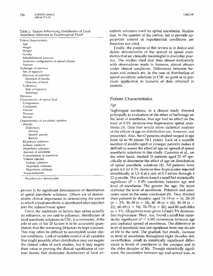
Table 1. Factors Influencing Distribution of Local Anesthetic Solutions in Cerebrospinal Fluid”
Patient characteristics Age Height Weight Gender Intraabdominal pressure Anatomic configuration of spinal column Position
Site of injection Direction of injection
Direction of needle Direction of bevel
Rate of injection Barbotage
Technique of injection
Turbulence
Diffusion Characteristics of spinal fluid
Composition Circulation Volume Pressure Density
Density Characteristics of anesthetic solution
Definitions Density Specific gravity Baricity
Hypobaric solutions lsobaric solutions Hyperbaric solutions Amount of anesthetic Concentration of anesthetic Volume injected
Isobaric solutions Hypobaric solutions Hyperbaric solutions
Vasoconstrictors
UHypothetical or demonstrable.
proven to be significant determinants of distribution of spinal anesthetic solutions. Others are of demon- strable clinical importance in determining the extent to which a local anesthetic is distributed after injection into the subarachnoid space.
Given the multitude of factors that either actually do influence, or are said to influence, distribution of local anesthetic solutions in CSF, it is necessary, if the role of one of the 25 factors is to be accurately quan- titated, that the remaining 24 factors be kept constant. This may often be difficult to accomplish under clin- ical conditions. Lack of suitable control of all variables that might possibly affect distribution may not negate the clinical value of such studies, but it may negate their value in proving the clinical significance of var- ious factors that determine distribution of local an-
esthetic solutions used for spinal anesthesia. Studies that, in the opinion of the author, fail to provide ap- propriate control of experimental conditions are therefore not cited.
Finally, the purpose of this review is to derive and define determinants of the spread of spinal anes- thetics that are clinically meaningful in everyday prac- tice. The studies cited deal thus almost exclusively with observations made in humans, almost always under clinical conditions. Differences between hu- mans and animals are, in the case of distribution of spinal anesthetic solutions in CSF, so great as to pre- clude application to humans of data obtained in animals.
Patient Characteristics
Age Nightingale mentions, in a clinical study directed principally to evaluation of the effect of barbotage on the level of anesthesia, that age had no effect on the level of 0.5% dextrose-free bupivacaine spinal anes- thesia (3). Data that would allow statistical analysis of the effects of age on distribution are, however, not presented. Also, the 67 patients studied ranged in age from 62 to 90 (mean 74.1 years). Lack of a suitable number of middle-aged or younger patients makes it difficult to assess the effect of age on spread of spinal anesthetic solutions in this study. Cameron et al., on the other hand, studied 33 patients aged 37-97 spe- cifically to determine the effect of age on distribution of spinal anesthetic solutions (4). All patients were given 4.0 ml 0.5% dextrose-free bupivacaine injected intrathecally at L3-4 at a rate of 0.5 ml/sec through a 22-g needle. The authors found a small but statistically significant ( P < 0.05) correlation between age and level of anesthesia. The greater the age, the more cephalad the level of anesthesia. Pitkanen and asso- ciates came to the same conclusion (5). They divided their patients by decades: aged 15-19 ( n = 6) , 20-29 (rz = 23), 30-39 ( n = 24), 40-49 ( n = 14), 50-59 (n = 21), 60-69 ( n = 16), 70-79 (71 = lo), and 80 and older (rz = 11). All patients were given 3.0 mlO.5% dextrose- free bupivacaine. They, too, found a small but statis- tically significant ( P < 0.05) correlation between age and cephalad spread of anesthesia. The difference in level of anesthesia was not sigruficant from one decade of life to the next. The gradual, but steady, increase in level of anesthesia throughout eight decades did, nevertheless, result in statistically significant differ- ences in levels of anesthesia in the younger and in the older decades of life. Though statistically signif- icant, the correlation between age and spread was, as
DISTRlBUTlON OF SPINAL SOLUTIONS ANESTH ANALG 71 7 1985;64:715-30
the authors noted, weak, and the age-related differ- ences in spread were so small as to be of marginal clinical significance. The maximum spread of anes- thesia averaged T9 in patients 20-28 years old, T7 in patients aged 70-79 and T6 in patients 80 years old and older. The data of Pitkanen et al. and Cameron et al. suggest that the tendency to an increase in spread with age, also observed by Bengtsson et al. (6), may well be more than just a tendency.
The data of Pitkanen and Cameron and their as- sociates demonstrate that increasing age is associated with a small, but statistically significant, increase in level of anesthesia of perhaps modest clinical signif- icance when an essentially isobaric solution of 0.5% bupivacaine is injected. Comparable well controlled data on the spread of hypo- and hyperbaric spinal anesthetic solutions are not available. Nor can they necessarily be inferred from data derived from studies in which an isobaric solution is used. Such an infer- ence would be unwarranted in the absence of an ex- planation as to why spread of isobaric solutions in CSF is a direct function of age. No data on the effect of age on volume of spinal CSF are available. If, how- ever, age were associated with progressive decrease in CSF volume, this might mean, because volume of CSF is, as amplified below, a determinant of spread of hyperbaric solutions, that the data of Pitkanen et al. and Cameron et al. could also be applied to situ- ations in which hyperbaric solution are used. How- ever, this requires further studies.
Height There have been no controlled, systematic studies of the effect of patient height on distribution of spinal anesthetic solutions. Common sense and clinical ex- perience tell us, nevertheless, that injection of a local anesthetic sohtion at the L3-4 interspace in a short patient is associated with a more cephalad spinal seg- mental level of anesthesia than is injection of the same amount of the same anesthetic injected in the same way in a tall patient. One reason for this is that even if the anesthetic solution were to spread to an equal extent, 20 cm, say, from the site of injection in both the tall and the short patient, a 20-cm spread will reach a higher spinal segmental level in the short patient than it will in the tall patient.
An additional reason for lower segmental levels of anesthesia in taller patients than in shorter patients is that the local anesthetic solution that spreads 20 cm from the site of injection in a short patient may not spread the same distance, 20 cm, in a tall patient. It may not spread as far in the tall patient because of height-related differences in spinal CSF volume. The
volume of CSF below the termination of the cord at L2 is greater in tall than in short subjects because the length of the cauda equina is greater. The initial vol- ume of CSF into which the local anesthetic is injected at L3-4 being greater, there will be greater dilution of the anesthetic solution at the site of injection and therefore less cephalad spread in the taller patient. The volume of CSF above termination of the cord at L2 may also be greater in tall patients than in short patients. The diameter of the spinal cord is greater in taller patients than in shorter patients. So, too, is the depth of the subarachnoid space, i.e., distance from dura to pia mater. Although the ratio between depth of the subarachnoid space and diameter of the cord remains constant regardless of height, there is, how- ever, a slight increase in absolute volume of CSF at any given level of the cord due to the increase in depth of the subarachnoid space. The increase in volume of CSF above L2 in taller patients would further dilute local anesthetic solutions injected at L3-4 and so be a contributory factor to the lower levels of anesthesia observed in taller patients.
Differences in height must be fairly substantial if they are to have clinically significant effects on dis- tribution of spinal anesthetic solutions. Men are, on the average, taller than women. The difference is not great enough, however, to result in sex-related dif- ferences in levels of anesthesia when identical anes- thetic techniques are used in both sexes (2). But dis- tribution is certainly different in patients 210 cm tall than it is in patients 120 cm tall. The role of patient height in determining sensory levels of anesthesia becomes clinically most important when spinal anes- thesia is used in children.
Weight How much a patient weighs has no effect on distri- bution of local anesthetic solutions in CSF. The spread of a spinal anesthetic solution is the same in a 70 kg patient as it is in a patient weighing 100 kg when all other factors determining spread, including height, are constant. The theoretical possibility that obesity may be associated with an accumulation of epidural fat sufficient to reduce the volume of CSF and, there- fore, alter distribution of local anesthetic solutions in the subarachnoid space has not been rigorously stud- ied. Clinical experience indicates that obesity is of little, if any, direct clinical significance in determining spread of local anesthetic solutions in CSF.
Obesity may, however, indirectly affect spread of a spinal anesthetic solution that is normally governed by gravity. A hyperbaric solution may, for example, be associated with unexpectedly great cephalad spread

ANESTH ANALG 1985;h4.71>30
718 GREENE
in a steatopygous patient. Such a patient will be in the slight head-down position even though lying su- pine on an operating table that is horizontal.
Gender The sex of a patient has no direct effect on distribution of local anesthetic solutions in CSF if all other factors involved in determining distribution are constant (2). From a practical point of view it should, however, be borne in mind that in women the width of the hips is usually greater than the width of the shoulders. A woman may thus be slightly head-down when in the lateral position on an operating table that is horizontal with the floor. This will particularly affect distribution of hyperbaric solutions. Men, having shoulders that are usually wider than their hips, will be in the slight head-up position under the same conditions.
lntraabdoininal Pressure An increase in intraabdominal pressure may be, and often is, associated with an increase in spread of local anesthetic solutions in the subarachnoid space. This is because increased intraabdominal pressure causes obstruction of vascular channels that normally ac- count for venous drainage from the abdomen. The result is dilation of collateral venous channels from the abdomen, including venous channels that pass through the lower portions of the epidural space. Epi- dural venodilation in turn reduces the volume of CSF in the lumbar subarachnoid space, and thus the vol- ume of CSF in the lumbar and lower thoracic sub- arachnoid space. The effect of decreases in volume of spinal CSF on distribution of spinal anesthetic solu- tions is dealt with further below (CSF Volume sec- tion), but it should be noted that chronic increases in intraabdominal pressure have more effect on altering distribution of spinal anesthetic solutions than do acute increases in intraabdominal pressure. The effects are clinically most evident in term pregnancies and in patients with ascites or large intraabdominal tumors.
Anatomic Configuration of the Spinal Coluinir Scoliosis has no significant effect on the distribution of spinal anesthetic solutions. Kyphosis, flattening of the lumbar lordotic curvature (as with flexion of the thighs on the abdomen in the supine position), and accentuation of the lordotic curvature (as in term preg- nancy) may aLl significantly affect &stribution of spinal anesthetic solutions the spread of which is governed by gravity. They do so because they either accentuate or eliminate the lower portion of the S-shaped curve
of the subarachnoid space normally present when a patient lies in the supine position. Elimination of the lordotic curve increases the cephalad spread of hy- perbaric solutions (7). Exaggeration of the lordotic curve may decrease the cephalad spread of hyperbaric so- lutions in the supine position by causing pooling of the anesthetic solution in the deepest part of the S- shaped curve. Kyphotic accentuation of the thoracic curvature similarly affects thoracic distribution of local anesthetic solutions the spread of which is deter- mined by gravity. An increase in anterior-posterior thoracic diameter, as seen with emphysema, may re- sult in a kyphotic-like condition such that the upper thoracic spine may be in the slight head-up position when an emphysematous patient lies supine on an operating table in the horizontal position.
Posif io t r Position of the patient and baricity or density of the local anesthetic solution injected as determinants of distribution are so closely related that one cannot be discussed without the other. The roles of both are considered below in the section that deals with the effect of density on distribution.
Technique of Injection Where and how a local anesthetic solution is injected into the subarachnoid space may affect its distribution within the subarachnoid space.
Site of lnjection Injection of a local anesthetic solution into the sub- arachnoid space cephalad to the L2-3 interspace causes, of course, a shift in the cephalad direction of the ep- icenter from which subsequent distribution of the an- esthetic solution takes place. If injected at the T6-7 level, the nerve roots primarily affected will be tho- racic roots instead of lumbar roots, as with more con- ventional spinal anesthetics. But injection above L2 does more than alter the epicenter of spinal anes- thesia. It also alters distribution of the anesthetic so- lution in CSF. It does so because the volume of CSF per spinal cord segment is less above L2 than below L2. It is less because the spinal cord ends (in adults) at L2. Above L2 the spinal cord occupies a substantial portion of the subarachnoid space with consequent reduction in volume of CSF. Accordingly, injection of, say, 2 ml of anesthetic solution at T6-7 is associ- ated with a greater spread than if the same volume of anesthetic solution were injected into the greater volume of CSF at the L3-4 interspace. Denervations

DISTRIBUTION OF SPINAL SOLUTIONS ANESTH ANALG 719 1985;64:715-30
would accordingly be more extensive and. involve more nerve roots in the thoracic area than in the lumbar area.
Spinal anesthesia is, of course, rarely if ever inten- tionally induced by injecting local anesthetic solutions above the L2-3 interspace. The proximity of the cord to the dura above L2, and the resulting shallowness of the subarachnoid space, increase the risk of trauma to the cord by the spinal needle. Spinal anesthetics resulting from injections made above L2 are usually complications associated either with nerve blocks in the cervical or posterior thoracic area, or with thoracic or cervical epidural anesthesia. If the dura is acciden- tally penetrated during such procedures, even though the volume of local anesthetic going into the sub- arachnoid space is little, the resulting area of dener- vation will be unexpectedly great.
Direction of Injection Direction of needle. Though unquantitated, it ap-
pears likely that the direction of the needle at the time the spinal anesthetic solution is injected-that is, the angle between the needle and the longitudinal axis of the subarachnoid space-may influence the direc- tion in which the local anesthetic goes after injection. If the needle is directed in a cephalad direction, the stream of anesthetic solution coming from the needle during injection would be expected to carry the an- esthetic solution farther in a cephalad direction than if the same anesthetic solution were injected through a needle inserted through the dura at a right angle to the long axis of the spinal column. When injected through a needle at a right angle to the spinal column, the initial distribution of anesthetic solution in CSF is essentially equal above and below the site of injection. The initial distribution in CSF of anesthetic injected through a needle pointing in the cephalad direction would be likely to be greater above the site of injection than below it.
Direction of bevel. The direction in which the bevel of a standard lumbar puncture needle faces has no effect on the distribution of local anesthetic solutions in CSF (8). The lumen of a standard lumbar puncture needle lies in the same axis as the lumen of the shaft of the needle. There is no bend or angulation of the terminal lumen at the bevel. A liquid injected through such a needle exits from the needle in the same straight line formed by the lumen of the needle throughout its length. When a solution is injected into air through a standard beveled lumbar puncture needle, the exit stream goes in the same straight line regardless of the direction in which the bevel faces.
There are, however, lumbar puncture needles spe- cifically designed to influence the direction in which an injected solution exits from the needle. One of these is the Whitacre needle. The Whitacre needle has a closed, pencil-point tip with a lateral exit port im- mediately adjacent to the start of the angle forming the pointed tip. Another is the Tuohy needle. The tip of a Tuohy needle has a sharp but closed bevel; the lumen of the needle is curved at the distal end so that the exit port lies on the extreme tip of the shaft at the start of the angle of the bevel. Both these needles determine the angle at which fluids leave the lumen. With the Whitacre needle, the exit stream is essen- tially at a 90" angle to the shaft of the needle. With the Tuohy needle, the exit stream is at a 45" angle. Both significantly affect the direction in which anes- thetic solutions are injected into CSF. Thus both affect distribution of spinal anesthetic solutions.
Turbulence Rate and force of injection. Turbulence inevitably oc-
curs in CSF when solutions are injected into the sub- arachnoid space. If turbulence has a clinically signif- icant effect on spread of spinal anesthetic solutions within the subarachnoid space, then the force used for injection should increase the level of spinal anes- thesia. Neigh et al. found, however, that the level of sensory anesthesia was the same in patients in whom a hyperbaric solution of tetracaine was injected at a rate of 1 ml/sec as it was in patients in whom the same volume of anesthetic solution was injected through the same size needle at a rate of 0.2 ml/sec (8). Sim- ilarly, McClure et al. found that the injection of 4 ml of isobaric tetracaine at a rate of 0.2 mVsec through a 25-g needle resulted in essentially similar levels of anesthesia as did injection of 4 ml of the same solution through the same size needle at a rate of 0.1 mlisec (9).
The studies of Neigh and of McClure and their associates indicate that turbulence created within the subarachnoid space, when the force or rate of injec- tion was altered, has no clinically significant effect on the spread of spinal anesthetic solutions in CSF. Per- haps, however, differences in the amount of turbu- lence created by varying the rate of injection through needles of the same size, as in these clinical studies, were not great enough to produce clinically mean- ingful changes in the spread of the anesthetic solu- tions used.
Barbotage. What happens when even greater de- grees of turbulence are deliberately produced by bar- botage, that is, aspiration of CSF and anesthetic so-

ANESTH ANALC 1985;64:715-30
720 GREENE
lution just injected back into the syringe used for injection immediately upon completion of injection, followed by reinjection into CSF of the contents of the syringe? The volume of fluid aspirated back into the syringe may be less than, equal to, or greater than the volume of the initial injectate. Aspiration and rein- jection may be done one or more times.
IGtahara et al., evaluating the spread of local an- esthetic solutions in patients by measuring the dis- tribution of a small amount of radioactive iodine added to the anesthetic solution (lo), found that barbotage (the details of the barbotage technique were not de- scribed) in an unstated number of patients had no effect on the spread of isobaric solutions of dibucaine or tetracaine. Lanz et al. also found that, in a constant temperature model of the subarachnoid space, vig- orous barbotage had no significant effect on distri- bution of isobaric solutions of bupivacaine, lidocaine, mepivacaine, prilocaine, or tetracaine (11). In a well- controlled clinical study, Levin et al. found that bar- botage (aspiration of the injected volume back into the syringe followed by reinjection 2-4 times with 0.5 ml increases in each aspirated volume) had no sig- nificant effect on the spread of either iso- or hyper- baric solutions of tetracaine (12). Nightingale, in a similar clinical study of isobaric bupivacaine spinal anesthesia with ( n = 31) or without (11 = 36) barbo- tage (half the initial injected volume aspirated and reinjected followed by a second aspiration and rein- jection of one quarter the initial injected volume), also found that barbotage had no effect on the level of anesthesia (3).
Though there are no clinical studies of the effect of barbotage on distribution of hypobaric solutions, the above studies show that turbulence, no matter how produced, has no clinically significant effect on distribution of hyper- and isobaric spinal anesthetic solutions. There is no a priori reason why those find- ings cannot be applied to hypobaric solutions.
How can the above objective data be reconciled with the intuitively reasonable hypothesis that tur- bulence at the site of injection increases the spread of spinal anesthetic solutions? The most likely expla- nation is that the turbulence is not only relatively brief in duration, but is also restricted to the area at, and immediately adjacent to, the site of injection. To alter distribution to any significant degree, turbulence should last for more than moments. It should also extend well beyond the site of injection. Neither seems likely. Transient turbulence localized about the L3-4 interspace will hardly affect spread of a solution of any baricity 20 cm away from the site of injection. On the other hand, local turbulence at the site of injection will thwart attempts to limit the cephalad level of
anesthesia to L2 or L3 as well as attempts to induce totally unilateral low spinal anesthesia.
Diffusion Diffusion, a term often loosely applied to spinal anes- thesia, is of no importance in determining distribution of spinal anesthetic solutions under clinical condi- tions. True physical diffusion consists of the inter- mingling of different types of molecules uninfluenced by turbulence or differences in density. It is a slow process, requiring hours to cover a distance of cen- timeters. Distribution of local anesthetic solutions in CSF is completed in minutes, not hours.
Characteristics of CSF Determinants of the spread of one solution in another solution include the physical characteristics of each of the solutions. The physical characteristics of CSF are thus one of the factors governing distribution of local anesthetic solutions in CSF.
C O l t l p 0 S ~ t i O f Z Of CSF The normal range of values of CSF pH and CSF con- centrations of cells, protein, glucose, and ions is too narrow to have any effect on distribution of spinal anesthetic solutions (13). Values beyond the normal range may affect distribution, but they are associated only with neurologic conditions that contraindicate spinal anesthesia in the first place.
The concentration of protein in CSF increases pro- gressively with descent from the ventricles to the lum- bar subarachnoid space (14). Though unquantitated, this may be associated with an increase in CSF den- sity. If so, then theoretically a hyperbaric solution might become increasingly hyperbaric as it ascends in the subarachnoid space. The possibility that, in the head-down position, spread of a hyperbaric solution might accelerate in the upper thoracic area, because it becomes increasingly hyperbaric, seems remote.
Circtllntiolt of CSF CSF is produced, mainly by the choroid plexus, at a rate of about 0.35 d m i n (500 mVday) in normal adults. CSF is absorbed into the venous circulation through herniations of the arachnoid that protrude into the lumina of veins through gaps in the dura (15). These herniations, the arachnoid villi, are particularly nu- merous in the superior sagittal sinus, but are also present in other intracranial veins. Arachnoid villi are also found adjacent to spinal nerve roots as they emerge

DISTRIBUTION OF SPINAL SOLUTIONS ANESTH ANALG 72 1 1985;64:715-30
through the dura. The quantitative sigruficance of spinal arachnoid villi in the absorption of CSF is unknown, but is minor compared to the role played by intra- cranial arachnoid villi (15). The direction of flow of the 500 ml of CSF produced per day is thus mainly intracranial from the site of production in the choroid plexus to the principal sites of vascular absorption through intracranial arachnoid villi. Downward flow of CSF through the spinal subarachnoid space is min- imal, involving perhaps less than 10% of the 500 ml produced daily. Circulation of CSF in the spinal sub- arachnoid space in either direction is thus too slow (16) to have any significant effect on distribution of spinal anesthetic solutions in the relatively few min- utes during which distribution occurs.
Volume of CSF The volume of CSF in normal adults averages about 150 ml. Approximately 75 ml is intracranial (15). The remaining 75 ml of CSF lies within the spinal sub- arachnoid space. The latter is the volume of CSF within which spinal anesthetics can, at least potentially, be distributed. The volume of spinal CSF within which anesthetic solutions are actually distributed under most clinical conditions is, however, substantially less than 75 ml. The exact volume of CSF below C8, the volume of most concern in spinal anesthesia, has not been quantitated. A considerable portion of this 75 ml must be in the area of the subarachnoid space occupied by the cauda equina distal, that is, caudad to L2.
The volume of CSF in the spinal subarachnoid space is decreased in the presence of chronic increases in volume of the contents of the epidural space. The epidural structures most susceptible to enlargement are the epidural veins. Chronic engorgement of epi- dural veins in the lumbar and lower thoracic areas, due to obstruction of normal venous effluent channels associated with an increase in intraabdominal pres- sure, significantly decreases the volume of CSF in the lumbar and lower thoracic subarachnoid space. This is clinically most evident in full term parturients (see below) and in patients with ascites or large intraab- dominal masses. The decrease in CSF volume in the lumbar and lower thoracic areas in such patients means that the volume of CSF, in which a given volume of spinal anesthetic solution is distributed, is decreased. The result is levels of anesthesia greater than those that would be obtained if the same anesthetic solution were administered in patients in whom there is no increase in intraabdominal pressure.
The effect of term pregnancy on the distribution of spinal anesthetic solutions is graphically demon- strated by the studies of Assali and Prystowsky (17).
Using a continuous spinal technique, these authors found that 5-20 ml of 0.2% procaine was required to produce anesthesia to pinprick at the fourth cervical level (sic) in 10 normal women at term. In the same 10 women, the amount of procaine needed to obtain the same level of anesthesia 36-48 hr after delivery was 3-4 times greater. Similarly, Barclay et al. found that the injection of 4 mg tetracaine through a spinal catheter produced an average sensory level of T8 in 15 pregnant women at term (18). The same amount of tetracaine similarly injected in 20 nonpregnant women of comparable age resulted in an average level of anesthesia of T11.
CSF Pressure Conditions associated with a chronic increase in CSF pressure contraindicate lumbar puncture because of the danger of producing intracranial herniation. The effect of a chronic increase in CSF pressure on dis- tribution of local anesthetic solutions is, therefore, of no clinical importance.
The decrease in lumbar and lower thoracic CSF volume due to epidural venous engorgement asso- ciated with increased intraabdominal pressure is not associated with an increase in CSF pressure. CSF pres- sure is, for example, normal in term pregnancies (19,20).
Sudden, acute increases in CSF pressure associated with labor, a Valsalva maneuver, coughing, or strain- ing do not increase spread of spinal anesthetic solu- tions, clinical impressions to the contrary. In fact, nei- ther uterine contractions nor bearing down during labor increase spinal CSF pressure (19,21,22). Such increases in CSF pressure as do occur during labor are secondary to either skeletal motor activity or tran- sient increases in arterial pressure during labor. Even these transient increases in spinal CSF pressure have no significant effect on spread of spinal anesthetic solutions. Dubelman and Forbes, for example, found that although CSF pressure increased in (nonpreg- nant) patients who gave three vigorous coughs within seconds of the intrathecal injection of 12 mg hyper- baric tetracaine, the level of anesthesia was the same as it was in patients given the same amount of tetra- caine who did not cough after injection (23).
Acute, transient increases in CSF pressure associ- ated with coughing, etc., do not increase spread of local anesthetic solutions in CSF because a brief in- crease in CSF pressure is instantaneously transmitted throughout the entire CSF system, spinal as well as intracranial. It must be. An increase in pressure at one point in closed space filled with a noncompres- sible liquid is instantaneously transmitted throughout the entire system. Because no hydrostatic pressure
ANESTH ANALG 1985;64:715-30
722 GREENE
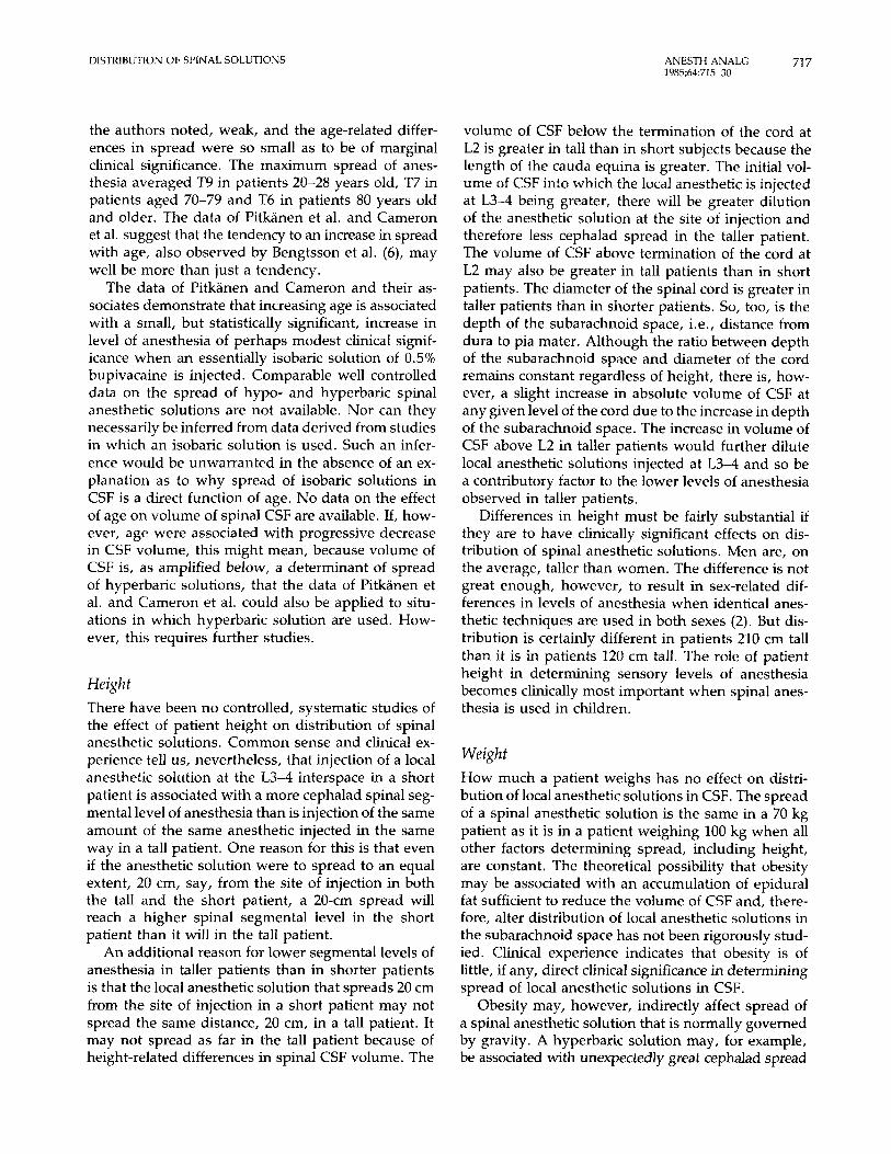
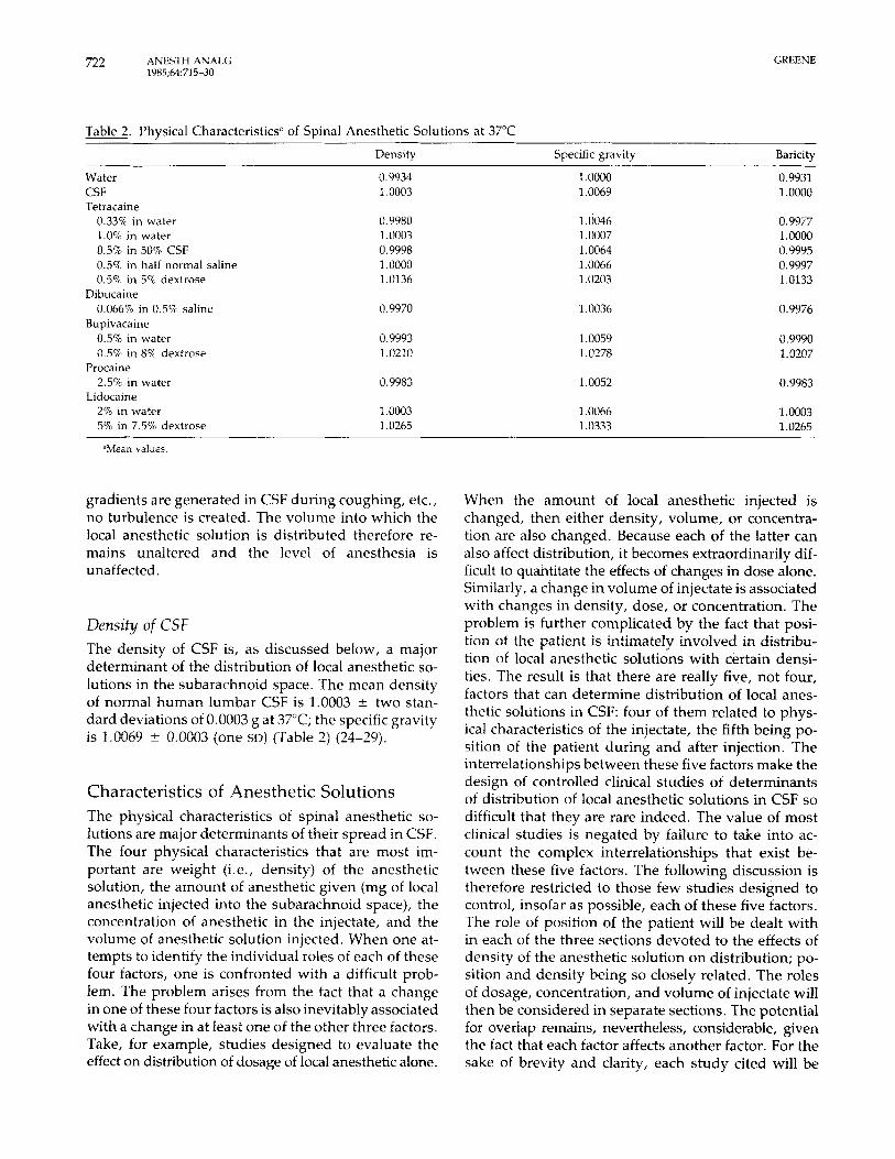
Table 2. Physical Characteristics” of Spinal Anesthetic Solutions at 37°C
Density Specific gravity Baricity
Water CSF Tetracaine
0.33% in water 1.0% in water 0.5% in 50%. CSF 0.5% in half normal saline 0.57~. in 5% dextrose
0.066% in 0.5% saline
0.5% in water 0.5% in 8% dextrose
2.5% in water
2% in water 5% in 7.5% dextrose
Dibucaine
Bupivacaine
Procaine
Lidocaine
“Mean values.
0.9934 1.0003
0.9980 1.0003 0.9998 1 .0000 1.0136
0.9970
0.9993 1.0210
0.9983
1.0003 1.0265
1.0000 1.0069
1.0046 1.0007 1.0064 1.0066 1.0203
1.0036
1.0059 1.0278
1.0052
1.0066 1.0333
0.9931 1.0000
0.9977 1 .oooo 0.9995 0.9997 1.0133
0.9976
0.9990 1.0207
0.9983
1.0003 1.0265
gradients are generated in CSF during coughing, etc., no turbulence is created. The volume into which the local anesthetic solution is distributed therefore re- mains unaltered and the level of anesthesia is unaffected.
Density of CSF The density of CSF is, as discussed below, a major determinant of the distribution of local anesthetic so- lutions in the subarachnoid space. The mean density of normal human lumbar CSF is 1.0003 i two stan- dard deviations of 0.0003 gat 37°C; the specific gravity is 1.0069 i: 0.0003 (one SD) (Table 2) (24-29).
Characteristics of Anesthetic Solutions The physical characteristics of spinal anesthetic so- lutions are major determinants of their spread in CSF. The four physical characteristics that are most im- portant are weight (i.e., density) of the anesthetic solution, the amount of anesthetic given (mg of local anesthetic injected into the subarachnoid space), the concentration of anesthetic in the injectate, and the volume of anesthetic solution injected. When one at- tempts to identify the individual roles of each of these four factors, one is confronted with a difficult prob- lem. The problem arises from the fact that a change in one of these four factors is also inevitably associated with a change in at least one of the other three factors. Take, for example, studies designed to evaluate the effect on distribution of dosage of local anesthetic alone.
When the amount of local anesthetic injected is changed, then either density, volume, or concentra- tion are also changed. Because each of the latter can also affect distribution, it becomes extraordinarily dif- ficult to quantitate the effects of changes in dose alone. Similarly, a change in volume of injectate is associated with changes in density, dose, or concentration. The problem is further complicated by the fact that posi- tion of the patient is intimately involved in distribu- tion of local anesthetic solutions with certain densi- ties. The result is that there are really five, not four, factors that can determine distribution of local anes- thetic solutions in CSF: four of them related to phys- ical characteristics of the injectate, the fifth being po- sition of the patient during and after injection. The interrelationships between these five factors make the design of controlled clinical studies of determinants of distribution of local anesthetic solutions in CSF so difficult that they are rare indeed. The value of most clinical studies is negated by failure to take into ac- count the complex interrelationships that exist be- tween these five factors. The following discussion is therefore restricted to those few studies designed to control, insofar as possible, each of these five factors. The role of position of the patient will be dealt with in each of the three sections devoted to the effects of density of the anesthetic solution on distribution; po- sition and density being so closely related. The roles of dosage, concentration, and volume of injectate will then be considered in separate sections. The potential for overlap remains, nevertheless, considerable, given the fact that each factor affects another factor. For the sake of brevity and clarity, each study cited will be
DISTRIBUTION OF SPINAL SOLUTIONS ANESTH ANALG 723 1985;61:715-30
summarized and discussed only once in one of the four sections.
Demity The density of a solution is the weight in grams of one ml of solution (i.e., g/ml). The specific gravity of a solution is a ratio: the density of the solution divided by that of water. The baricity of a local anesthetic so- lution is also a ratio: the density of the solution di- vided by that of CSF. One way of estimating distri- bution of a local anesthetic solution in CSF is calculation of the ratio between the specific gravity of the local anesthetic solution and the specific gravity of CSF. This involves calculating the ratio between two ratios, each having the density of water as a common de- nominator. A simpler and more direct method is to rely upon baricity, the ratio between the density of the anesthetic solution and the density of CSF. If this ratio is 1.0000, the solution is isobaric. If the ratio is greater than 1.0000, the solution is hyperbaric; if less than 1.0000, it is hypobaric.
Essential to the calculation of either specific grav- ities or baricities of local anesthetic solutions as de- terminants of their distribution in CSF is assurance that densities of all solutions involved (water, CSF, and anesthetic solutions) have been measured at the same temperature. This is necessary because the den- sity of a solution is inversely related to its temperature (24,26,27,29). Because temperatures of local anes- thetic solutions rapidly equilibrate with the temper- ature of CSF, the clinically important densities are those measured at 37°C. The density of water at stan- dard temperature is, by definition, 1.0000. At 37°C the density of water is 0.9934, with, again by defi- nition, a specific gravity of 1.0000 at 37°C (Table 2). The baricity of CSF is, also by definition, 1.0000 at 37°C (see Table 2; see also references 24-29 for details on densities and specific gravities of CSF and various local anesthetic solutions cited in Table 2 and in the following text). Unfortunately, many of the data on densities, specific gravities, and baricities of local an- esthetic solutions cited in the literature are not always entirely reliable. One reason for this is failure to make all measurements at 37°C. Another is use of methods that are not accurate enough to give results repro- ducible to the fourth decimal point, a level of precision desirable if the full potential clinical significance of the data is to be realized. A third reason is quotation of data without citation of the source from whence the figures are derived. The significance of temper- ature is illustrated by the fact that the density of a solution determined at room temperature may indi- cate it is isobaric, but at 37°C it will prove to be hy-
pobaric, not isobaric. Furthermore, the effect of tem- perature on density must be separately measured for each solution. The fact that the density of water de- creases from 1 .OOOO at standard temperature to 0.9934 at 37°C does not mean that the densities of all solu- tions decrease similarly with the same increase in tem- perature. The effect of temperature on density vanes from one solution to another.
The following discussion of the effects of density, specific gravity, and baricity on spread of local an- esthetic solutions in CSF is predicated on the as- sumption that the temperature of CSF is a normal 37°C. Though clinically unquantitated, hypo- and hy- perthermia would be expected to have complex, sub- tle effects on the distribution of local anesthetic so- lutions, the specific gravity and baricity of which have been determined on the basis that CSF temperature is 37°C.
Hypo bar ic Solutions The density of a local anesthetic solution at 37°C must be far enough below the mean density of CSF at 37°C (1.0003) to take into account the small but important normal variation in density of CSF about the figure of 1.0003 if the solution is to be hypobaric in all pa- tients, not just in some patients. Local anesthetic so- lutions with baricities less than 0.9990 are predictably hypobaric in all patients.
Solutions that have been clinically demonstrated to be reliably hypobaric include those containing tetra- caine or dibucaine (Table 2). A 0.33% solution of te- tracaine in water has a baricity of 0.9977, provides anesthesia lasting for up to 2 hr, and is easily prepared using commercially available 1 .O% solutions of tetra- caine for spinal anesthesia and U.S.P. sterile distilled water without preservatives or other additives. Though not as widely used today, the commercially available 0.066% solution of dibucaine in 0.5% saline, with a baricity of 0.9967, is equally effective for hypobaric spinal anesthesia. A 0.5% dextrose-free solution of bupivacaine has a baricity of 0.9990 (Table 2). It is, therefore, slightly hypobaric in most patients, as dem- onstrated in the rigidly controlled studies of Cham- bers et al. (31), Kalso et al. (13), and Tuominen et al. (32). Dextrose-free 0.5% solutions of bupivacaine are, nevertheless, so slightly hypobaric that they are gen- erally regarded, and are clinically used, as if they were isobaric. For this reason, 0.5% bupivacaine so- lution is discussed below in the section in which iso- baric solutions are considered. Strongly hypobaric so- lutions of bupivacaine with baricities less than 0.9980, and yet with concentrations of bupivacaine adequate to provide good anesthesia and muscle relaxation, can
ANESTH ANALC 1985;64:715-30
724 GREENE
be prepared by adding distilled water to 0.75% bu- pivacaine solutions. Experience with such solutions has been too limited, however, to document their efficacy at the present time.
Hypobaric solutions of procaine, lidocaine and other local anesthetics with similar anesthetic potencies, though eminently suitable for diagnostic and thera- peutic spinal anesthesia, are not satisfactory for op- erative hypobaric spinal anesthesia. By the time local anesthetics such as these have been diluted suffi- ciently to create a hypobaric solution, they are, be- cause the anesthetics do not have the potency of te- tracaine, dbucaine, or bupivacaine, approachmg their minimum effective local anesthetic concentrations. A 2.5% solution of procaine in water is hypobaric (Table 2). After the intrathecal injection of 2.5% procaine in water, the solution is, however, further diluted by CSF. It is so diluted that the duration of anesthesia is so brief as to be impractical in most operations. Successful hypobaric spinal anesthesia can be acheved only with highly potent local anesthetics.
The position of the patient during, and for the first minutes after, intrathecal injection of a hypobaric so- lution is the major determinant of its distribution in CSF. If the patient is in the head-up position during and after injection in the lumbar area, the anesthetic solution ascends in a cephalad direction. How high it ascends depends upon how hypobaric the solution is, the degree of the head-up position, and, second- arily, upon factors discussed below (see the Volume Injected section, below). If the patient is in the head- down position during and after injection, the anes- thetic solution is distributed caudad to the site of injection.
Thoracic levels of anesthesia adequate for intraab- dominal operations can be achieved with hypobaric solutions injected with the patient in the head-up po- sition. This was once a widespread technique. It is, however, a potentially dangerous technique. It is dan- gerous because the head-up position in the presence of extensive preganglion sympathetic denervation produced by the anesthesia may lead to severe, even calamitous, decreases in cardiac output and blood pressure (30). Hypobaric spinal anesthetics are rarely used today when levels of anesthesia to T10 or above are required. Hypobaric spinal anesthetics are, on the other hand, both effective and safe for rectal or per- ineal operations, especially those performed in the prone, jack-knife position. In such cases, induction of spinal anesthesia in the same position as that re- quired for the operation assures that anesthesia will be restricted to the sacral and lower lumbar roots and that physiologic responses will be minimal-all with- out having to move the patient after injection. Hy-
pobaric techniques are also useful for low unilateral spinal anesthesia, especially anesthesia of only one lower extremity. Unilateral hypobaric spinal anes- thesia can, however, be produced only if turbulence at the site of injection is minimized.
Isobaric S o h tions Because of the slight but important variation in the density of CSF about the mean figure of 1.0003, one cannot assure that an anesthetic solution with a den- sity of 1.0003 will prove to be isobaric in all patients. Nevertheless, there are solutions that under clinical conditions functionally behave, insofar as distribution in CSF and the resulting levels of anesthesia are con- cerned, as if they have the same density as CSF. Func- tionally isobaric solutions of tetracaine include 1% in water, 0.5% in 50% CSF, and 0.5% in half normal saline (Table 2). As mentioned above, though 0.5% bupivacaine is (barely) hypobaric (Table 2), under clinical conditions its distribution resembles that of isobaric solutions more than that of truly hypobaric solutions.
As with hypobaric solutions, the best and most reliable isobaric solutions are those that use highly potent local anesthetics. Lidocaine 2% in water is functionally isobaric (Table 2), but after dilution by CSF following injection it becomes too weak to pro- vide anesthesia for more than short periods.
The effect of position of the patient, during and after injection of isobaric spinal anesthetic solutions, on subsequent distribution of anesthetic has been evaluated by Wildsmith et al. (33). They found that position of patients during and after injection of iso- baric tetracaine solutions had no effect on the level of anesthesia. In 10 patients, 5 mg (1 ml) isobaric tetra- caine was injected with patients in the seated posi- tion, and left seated for 5 min before being placed in the supine horizontal position. In another 10 patients, 10 mg (2 ml) isobaric tetracaine was injected with patients in the lateral position, and left in the lateral position for 5 min before being placed in the supine horizontal position. There was no significant differ- ence in the levels of anesthesia in the two groups. In a comparable study, Tuominen et al. (32) determined sensory levels of anesthesia after injection of 3 ml of 0.5% bupivacaine in 10 patients in the seated position, the seated position being maintained for 2.5 min be- fore the patients were placed in the supine horizontal position. Maximum cephalad spread averaged T7. In another 10 patients, the same volume of the same anesthetic solution was injected with patients in the lateral position and then immediately placed in the supine horizontal position. Maximum cephalad spread

DISTRIBUTION OF SPINAL SOLUTIONS ANESTH ANALG 725 1985;64:715-30
averaged T8. The difference in spread was not statis- tically significant. The same authors also found that, when 0.75% bupivacaine was injected, the level of anesthesia was the same (T8) as it was when 0.5% bupivacaine was injected in the lateral position with immediate return to the horizontal supine position. However, when 0.75% bupivacaine was injected with patients in the sitting position, and the sitting position maintained for 2.5 min, the level of anesthesia (T4) was statistically significantly higher than when the same concentration was injected in the lateral position (T8). It was also significantly higher with 0.75% in the seated position (T8) than it was with 0.5% in the seated position (T7). The authors attributed these differences to the fact that glucose-free bupivacaine is not truly isobaric but, instead, is slightly hypobaric.
The major clinical virtue of isobaric spinal anes- thetics lies in the fact that position of the patient has no effect on distribution of the anesthetic. Distribu- tion is essentially the same if injection is made in the sitting or in the head-down position. Distribution is also unaffected by movement of the patient after in- jection. These attributes are clinically particularly use- ful when levels of anesthesia to T10 or below are required. Isobaric solutions are rarely used when lev- els of anesthesia in the mid-thoracic area are required; the volumes of anesthetic needed to produce a level of T5 become excessive.
Hyperbaric Solutions For an anesthetic solution to be reliably hyperbaric in all patients, it must have a baricity of at least 1.0015 at 37°C. The easiest and most widely used way to achieve this is by the addition of dextrose to the an- esthetic solution. Because dextrose is neurologically benign, the concentrations of dextrose used are usu- ally far in excess of those required to increase baricity above 1.0015. Although some authors have reported that the concentration of dextrose affects spread (34), the consensus is that once enough dextrose has been added to increase baricity of the solution above 1.0015, concentration of dextrose has little effect on distri- bution. Maximum cephalad spread is the same with a 5% concentration of lidocaine in 5% dextrose, for example, as it is with a 5% concentration of lidocaine in 7.5% dextrose (35), a finding also reported in a study comparing spread of bupivacaine made hyper- baric by either 5% or 8% dextrose (31,36,37). Physical characteristics of solutions made hyperbaric by the addition of dextrose are summarized in Table 2.
The distribution of hyperbaric spinal anesthetic so- lutions in CSF is influenced by position. If injection is made with the patient in the seated position, and
if the patient is left in the seated position for 10 min, the area of anesthesia can be restricted to sacral and lower lumbar roots. Whether only sacral roots are affected, or whether sacral and lower lumbar roots are affected, depends upon the volume of anesthetic solution injected. If hyperbaric solutions are injected with the patient in the horizontal or head-down po- sition, distribution is significantly different than it is when injection is made with the patient seated. This is shown in the study by Wildsmith et al. (33). In one group of 10 patients, 3.0 ml(l5 mg) of 0.5% tetracaine were injected with patients in the seated position, the position being maintained for 2 min before patients were placed in the supine horizontal position; the level of anesthesia averaged T8. The same dose and the same volume injected with patients in the lateral position and then immediately turned to the supine horizontal position resulted in a significantly higher level of anesthesia (T4).
Distribution of hyperbaric solutions injected with patients in the horizontal or head-down position may, however, not be as predictable and reliable as when injections are made with the patient seated. This is demonstrated by the data reported by Sinclair and associates (38). They studied 20 patients, all women, in whom 3.0 ml of 0.5% bupivacaine in 8% dextrose were injected at L3-4 with a 25-g needle while lying in the right lateral position with the table horizontal, all patients being turned immediately into the supine position after injection. In 10 patients, the table was left horizontal. In 10, the table was put in the 15" head- down position for 10 min before being returned to the horizontal position. The mean level of anesthesia was higher in the patients put in the head-down po- sition. However, because of the wide range in levels of anesthesia in the patients placed in the head down position (C4-T4), the difference in levels between these patients and those left in the horizontal position was not statistically significant, the range of spread in the latter patients being T2-6. The authors concluded that the head-down position is not necessary to achieve levels of anesthesia adequate for intraabdominal op- erations, and, furthermore, its use is associated with less control over spread; the head-down position often resulting in unnecessarily high and potentially dan- gerous levels of anesthesia.
Amount of Anesthetic Injected Dose (i.e., mg of local anesthetic injected) was found by Pflug et al. to have no effect on distribution of local anesthetic solution in CSF. In their study, 7.5 and 15.0 mg of 0.75% hyperbaric bupivacaine (i.e., 1-2 ml) resulted in similar levels of anesthesia (39). Nolte

ANESTH ANALG 1985;64:71>30
726 GREENE
Table 3. Concentration and Dose of Dextrose-Free Bupivacaine and Volume of Solution Used in the 6 Groups of 12 Patients each Studied by Shesky et al. (42)
Group Cvncentration (%)
I 0 5 2.0 10 I1 0.5 3.0 15 111 0.5 4.0 20 1v 0.75 1.3 10 V 0.75 2.0 15 VI 0.75 2.7 20
Volume (ml) Dose (mg)
and Stark also found no correlation between dose of 0.5% bupivacaine without dextrose and level of anes- thesia (40). On the other hand, Bengtsson et al. con- cluded that ”dosage (in mg) is more important than either volume or concentration” in determining spread of dextrose-free bupivacaine solutions (41). This con- clusion was, however, based simply on the obser- vation that injection of 4.5 ml 0.5% bupivacaine (22.5 mg) gave a level of anesthesia no different than did injection of 3.0 ml 0.75% bupivacaine (22.5 mg). Whether the experimental design in this study is ad- equate to support the conclusion, purely on an ex- clusionary basis, is debatable. In any case, these, as we11 as many other studies, are plagued by the prob- lem of how to identify the role of one factor in de- termining spread of spinal anesthetic solutions when other important factors (dose, concentration, volume) are simultaneously being changed. The problem of multiple simultaneously operative factors has, how- ever, been addressed in a unique and particularly elegant way by Shesky et al. (42).
What Shesky et al. did was to conduct a double- blind study in which age, height, position, and details of technique were controlled (42). Seventy-two pa- tients were studied. All were injected with dextrose- free bupivacaine in the seated position and, after 2 min, placed in the lithotomy position with the table horizontal. The patients were divided into six groups of 12 patients each with the concentration (70) of bu- pivacaine, the volume (ml) injected, and the amount of bupivacaine given (mg) altered in the six groups as shown in Table 3. The number of groups studied made possible statistical comparisons that could sep- arate out the individual roles of concentration, vol- ume, and dose. The results showed that the levels of anesthesia were significantly higher (T2-4) in patients given 15 or 20 mg bupivacaine (groups 11, 111, V, and VI) than they were in patients given 10 mg (T5-8; groups I and IV). Furthermore, the levels of anes- thesia were similar in patients who were given the same amount of bupivacaine (groups I and IV) even though the concentration of bupivacaine and the vol- ume injected differed. Also, in patients who received
the same volume of anesthetic solution (2 ml), the levels of anesthesia were significantly higher in those patients given 0.75% bupivacaine (group V) than in those given the 0.5% solution (group I). The authors concluded, therefore, that ”total dosage of bupiva- caine is more important than volume or concentration of anesthetic solution” in determining spread of the anesthetic solution in CSF, the same conclusion ar- rived at by Bengtsson et al. (41). The only caveat with regard to these data is that the bupivacaine solutions used are slightly hypobaric. Thus the results may not be entirely applicable to solutions that are more clearly hypobaric, more definitely isobaric, or strongly hy- perbaric. The data are, nevertheless, convincing enough to conclude that dosage has a significant effect on distribution independent of concurrent changes in concentration, volume, and baricity. But the conclu- sion of Shesky et al., that dosage is ”more important than volume or concentration” in determining spread of anesthetic solutions is appropriate. The data of Shesky et al. do not necessarily exclude the possibility that concentration and volume are not also determi- nants, though secondary determinants, of the distri- bution of anesthetic solutions in CSF.
Cotzceiz t rat ioii of Anesthetic There is no compelling evidence that concentration of local anesthetic injected intrathecally has any sig- nificant effect per se on distribution of local anes- thetics in CSF. Nor are there any theoretical reasons to expect that concentration might affect distribution, independently of changes in dosage, density, or vol- ume of injectate, associated with changes in concen- tration of anesthetic in the solution injected into the subarachnoid space,
Volume lizjected The injection of a spinal anesthetic solution into the subarachnoid space causes bulk displacement of CSF away from the site of injection. The displacement of CSF by anesthetic solution at the site of injection causes
DISTRIBUTION OF SPINAL SOLUTIONS ANESTH ANALG 727 1985;M: 71 5-30
changes in neurologic function at the site of injection. The extent of changes in neurological function at the site of injection depends, however, not only upon the volume of anesthetic solution injected, i.e., the vol- ume of CSF displaced, but also upon whether or not the injected solution disperses away from the site of injection so rapidly that neurologic function at the site of injection is altered little or not at all. Whether the anesthetic solution disperses rapidly from the site of injection depends upon the baricity of the solution injected and the position of the patient during and after injection. The effect of volume of anesthetic so- lution injected on subsequent distribution varies, therefore, with the baricity of the solution injected. If the solution is hypo- or hyperbaric, distribution ef- fected by baricity will be additive to distribution ef- fected by the volume injected. The question as to whether volume significantly affects distribution of spinal anesthetic solutions is therefore considered (below) in terms of the baricity of the anesthetic so- lution used.
Hypobaric solutions. The only controlled study of the effect of changes in the volume of injected hy- pobaric spinal anesthetic solutions on distribution of the solution is that of Brown et al. (2). They found that injection in the lateral position of 1.0 or 1.5 ml hypobaric tetracaine, with the patient immediately turned to the supine horizontal position, produced similar levels of anesthesia. This is perhaps not sur- prising because the horizontal position provided no opportunity for gravity to affect spread of the hypo- baric solution. Also, the difference in volumes in- jected, only 0.5 ml, may have been too slight to produce clinically meaningful differences in spread.
Isobaric solutions. The effect of volume injected on subsequent distribution of spinal anesthetic solutions has been said to be most evident when isobaric so- lutions are used. The best studies on the effects of volume on distribution of isobaric solutions are those in which tetracaine has been used to formulate spinal anesthetic solutions as nearly isobaric as possible, given the normal slight variation in density of CSF men- tioned above. Three studies have evaluated the effects of volume of isobaric tetracaine on distribution of te- tracaine in CSF under suitably controlled clinical con- ditions. In one of these, that by Brown et al., 3.0 ml of isobaric 0.5% tetracaine (15 mg) produced the same levels of anesthesia (T8-9) as did 2.0 ml of the same solution, all injections being made in the lateral po- sition with the patients then immediately turned to the supine horizontal position. Levels of anesthesia were the same in this study despite the increase in
volume (and dose) of tetracaine given (2). Similarly, when Wildsmith et al. (33) injected 1 ml of 0.5% iso- baric tetracaine (5 mg) with patients ( n = 10) seated during and for 2 min after injection, the average level of anesthesia (as defined in this review) was T10. When 2.0 ml of 0.5% isobaric tetracaine were injected with the patients (n = 10) in the lateral position dur- ing and for 5 min after injection before being turned to the supine horizontal position, the average level of anesthesia was also T10. The levels of anesthesia were the same even though both dosage of tetracaine and volume of solution injected, as well as position of the patients during and after injection were different in the two groups. Finally, McClure et al. also found that the levels of anesthesia were similar after injec- tion of 10 mg tetracaine added to 1.0, 2.0, or 4.0 ml of normal saline when patients were turned from the lateral position to the supine horizontal position im- mediately after injection (9).
These data demonstrate that position of the patient during and after injection of isobaric tetracaine has no effect on distribution of tetracaine in CSF (33). The data also suggest that dose of isobaric tetracaine has no effect on distribution (2,33), a finding at variance with the finding by Shesky et al. (42) that dose of 0.5% bupivacaine is a major determinant of spread. Most important, these data also suggest that the vol- ume of isobaric tetracaine injected has no effect on distribution: increasing the volume from 1.0 to 2 ml had no effect (2,9,33); even increasing the volume to 4.0 ml had no effect on distribution (9). Failure to see an increase in spread by increasing the volume by 1 .O ml could perhaps be attributed to the fact that a 1.0 ml increase in volume is too modest to produce clin- ically significant effects on distribution. Failure to see an increase in spread when the volume was increased to 4.0 ml may be related to the fact that 10 mg tetra- caine crystals were added to increasing volumes of saline with a resulting decrease in specific gravity of the anesthetic solution from 1.0085 (at 25°C) with 1.0 ml, to 1.0077 with 2.0 ml, and to 1.0065 with 4.0 ml. The changes in specific gravity may have obscured the effects of changes in volume.
That the relationship between volume of isobaric tetracaine and extent of spread in CSF is clinically important is illustrated by the fact that commercially available solutions of 1 .O% tetracaine, while isobaric, are not used for isobaric spinal anesthesia. They are not used because a 10 mg dose of tetracaine given in 1 ml produces profound anesthesia but, because the volume is so low, the extent of anesthesia is too lim- ited for most operations. Increasing the volume to 2 ml increases the extent of anesthesia, but 20 mg of tetracaine is excessive for the limited amount of anes-

ANESTH ANALG 728 1985;64:715-30
GREENE
thesia achieved. More dilute solutions of tetracaine provide satisfactory anesthesia while permitting use of larger volume to assure adequate spread.
Parenthetically, the observation by Brown et al. (2) that 3.0 ml of 0.5% hypobaric tetracaine produced the same levels of anesthesia as did 3 ml of 0.5% isobaric tetracaine does not indicate that the spreads of hy- pobaric and isobaric solutions are similar, because in both instances patients were in the supine, horizontal position. In that position the distribution of a hypo- baric solution would not be materially different than that of an isobaric solution.
The effects on distribution of changes in volume of dextrose-free 0.5% bupivacaine injected have been evaluated in several studies. In general, 3.0 ml of bupivacaine injected into the lumbar subarachnoid space produces anesthesia to the T7-8 level, and it is the consensus that increasing or decreasing the vol- ume injected above or below 3.0 ml produces pro- portionately higher or lower levels of anesthesia (4,5,31,37,40,43,44). Under controlled clinical condi- tions, for example, Axelsson et al. (43) found that decreasing the volume injected to 2.0 mi significantly decreased the level of anesthesia to T10-11, but that a further decrease in volume to 1.5 ml was not as- sociated with a further decrease in level of anesthesia. On the other hand, increasing the volume injected in this study was not associated with an increase in level of anesthesia (43). Interpretation of data on the spread of 0.5% bupivacaine is, however, difficult in terms of defining precisely the relationship between volume injected and spread of isobaric solution because dex- trose-free 0.5% bupivacaine is, as mentioned above, slightly hypobaric at 37°C (13,31,32).
Isobaric solutions of mepivacaine (45,46), procaine (47), and lidocaine (48) have also been used clinically. With the exception of the lidocaine study, no attempt was made, however, to examine systematically the role of volume as a determinant of isobaric solutions of these local anesthetics. In the study of isobaric 270 lidocaine (48), whether 2 or 5 ml of solution were injected had no clinically significant effect on the level of anesthesia. The volume injected, however, was dictated by the operation to be performed. In the ab- sence of details describing precisely how, why, and to what extent the operation altered the volumes in- jected, an unknown factor is introduced that makes difficult the evaluation of exactly why changing the volumes of isobaric lidocaine had no effect on distribution.
Hyperbaric solutions. Distribution of hyperbaric so- lutions away from the site of injection would be ex- pected to be related to both the effects of gravity (po-
sition of the patient) and the volume of anesthetic solution injected. The effects of gravity and position on distribution of hyperbaric solutions have been dis- cussed above.
That baricity of a spinal anesthetic solution is of greater importance than the volume of solution in- jected is demonstrated by the fact that, with patients in the supine, horizontal position, the spread of 3.0 ml hyperbaric 0.5% bupivacaine in 5% or 8% dextrose is significantly greater than is the spread of 3.0 ml of isobaric 0.5% bupivacaine (2,37). That volume is also involved in determining spread of hyperbaric solu- tions has been demonstrated by Axelsson et al. (49). These authors injected 1.5, 2.0, 3.0, or 4.0 ml of hy- perbaric 0.5% bupivacaine in 8% dextrose in 40 pa- tients. Injections were made with patients in the sit- ting position. Two minutes after injection, patients were put in the lithotomy position with the table hor- izontal. The results showed that maximum cephalad spread was directly related to the log volume of the solution injected. The observation by the same group of investigators that the cephalad spread of 0.5% bu- pivacaine without dextrose, observed under similar conditions (43), was a sigruficant 2-2.5 segments higher than it was in this study (49), is an indication of the hypobaricity of 0.5% bupivacaine without dextrose, not evidence that hyperbaric dextrose solutions fail to affect distributions. Wildsmith et al. also found that distribution of hyperbaric tetracaine was volume- related (33). The injection of 3.0 ml of 0.5% hyperbaric tetracaine (15 mg) in patients in the lateral position, and then immediately placed in the supine horizontal, resulted in an average T4 level of anesthesia. Injection of 2.0 ml of the same solution in patients in the lateral position, and left there for 5 min before being placed in the supine horizontal position, produced average levels of anesthesia at T7. Sundnes et al. similarly found volume-related levels of anesthesia with hy- perbaric 0.5% bupivacaine in dextrose (50). Injections of 1.5, 2.0, and 3.0 ml (n = 10 for each) with patients in the lateral position, and then immediately turned to the horizontal supine position, gave levels of anes- thesia at T10, T8, and T7, respectively. The differences in levels with 1.5 and 2.0 ml, and with 1.5 and 3.0 ml, were statistically significant; the difference be- tween 2.0 and 3.0 ml was not.
On the other hand, Bengtsson et al., in comparing 2.0 ml of 0.75% bupivacaine with 3.0 ml of 0.5% bu- pivacaine, both in 8% dextrose, found no difference in maximum level of anesthesia (6). Also, Chambers et al. found that 2.0, 3.0, and 4.0 ml of 0.5% bupiv- acaine in 8% dextrose produced similar sensory levels of anesthesia (51). In the same study, however, 1.3, 2.0, and 3.0 ml of 0.75% bupivacaine in 8% dextrose

DISTRIBUTION OF SPINAL SOLUTIONS ANESTH ANALG 729 1985;M: 71 5-30
produced volume-related increases in levels of anes- thesia; indeed, study of the 3.0 ml volume was pre- maturely abandoned because of the excessively high levels of anesthesia produced. Under the conditions of this study, increasing volumes of 0.75% bupiva- caine were associated with increasing levels of anes- thesia, while increasing volumes of 0.5% bupivacaine were not. The reason for this difference is not clear and cannot be established on the basis of the data presented.
In summary, the preponderance of data from con- trolled clinical studies supports clinical impression and common sense: the effects of volume of hyperbaric spinal anesthetic solutions injected are additive to the effects of gravity, position, and dosage.
Vasoconstrictors Neither epinephrine nor phenylephrine (Neosyne- phrine), added to spinal anesthetic solutions to pro- long the duration of anesthesia, affect the distribution of iso- or hyperbaric solutions of tetracaine, lidocaine, or bupivacaine within the subarachnoid space (46,50-55). The volume of vasoconstrictor solutions added to spinal anesthetic solutions is too small to affect significantly the baricity of the anesthetic solutions.
Conclusions The 25 factors that conceivably could affect distribu- tion of local anesthetic solutions can be divided into two groups. Those that have no clinically demon- strable significant effects include the following: pa- tient weight; patient gender; the direction in which the bevel of a standard lumbar puncture needle is facing when the anesthetic solution is injected; tur- bulence (except under special circumstances) created either by alterations in rate of injection or by barbo- tage; diffusion of local anesthetic in CSF; the com- position of CSF; CSF circulation; CSF pressure; con- centration of local anesthetic in the solution injected; and the addition of vasoconstrictors to the local an- esthetic solution. Factors that demonstrably affect dis- tribution of local anesthetic solutions in CSF, though of widely varying clinical significance, include the fol- lowing: patient age; patient height; anatomic config- uration of the spinal column; the site of injection; the direction of the needle during injection; the volume of CSF; density of CSF; density and baricity of the anesthetic solution injected; the position of the patient (with hypo- or hyperbaric solutions); the dosage of local anesthetic; and the volume of anesthetic solution injected.
References 1. Greene NM. Uptake and elimination of local anesthetics during
spinal anesthesia. Anesth Analg 1983;62:1013-24, 2. Brown DT, Wildsmith JAW, Covino BG, Scott DB. Effect of
baricity on spinal anaesthesia with amethocaine. Br J Anaesth 1980;52:589-95.
3. Nightingale PI. Barbotage and spinal anaesthesia: the effect of barbotage on the spread of analgesia during isobaric spinal anaesthesia. Anaesthesia 1953;38:7-9.
4. Cameron AE. Arnold RW. Ghoris MW. lamieson V. Suinal
5.
6
7
8.
9
10.
11.
12.
13.
14.
15.
16.
17.
18.
19.
20.
21.
22.
23.
analgesia using bupivacaine 0.5% plain: variation in the extent of the block with patient ago. Anaesthesia 1981;36:318-22. Pitkanen M, Haapaniemi L, Tuominen M, Rosenberg PH. In- fluence of age on spinal anaesthesia with isobaric 0.5% bupiv- acaine. Br J Anaesth 1984;56:279-84. Bengtsson M, Edstrom HH, Lofstrom JB. Spinal analgesia with bupivacaine, mepivacaine and tetracaine. Acta Anaesthesiol Scand 1983;27278-83. Smith TC. The lumbar spine and subarachnoid block. Anes- thesiology 1968;29:60-4. Neigh JL, Kane PB, Smith TC. Effects of speed and direction of injection on the level and duration of spinal anesthesia. Anesth Analg 1970;49:912-6. McClure JH, Brown DT, Wildsmith JAW. Effect of injected volume and speed of injection on the spread of spinal anaes- thesia with isobaric amethocaine. Br J Anaesth 1982;54:917-20. Kitahara T, Kuri S, Yoshida J. The spread of drugs used for spinal anesthesia. Anesthesiology 1956;17:205-8. Lanz E, Theiss D, Erdmann K, Becker J. Modelluntersuchun- gen zur Ausbreitung der isobaren Spinalanesthesie. Reg An- aesth 1980;3:4-9. Levin E, Muravchick S , Gold MI. Isobaric tetracaine spinal anesthesia and the lithotomy position. Anesth Analg 1981;60:810-3.
Kalso E, Tuominen M, Rosenberg PH. Effect of posture and some CSF characteristics on spinal anaesthesia with isobaric 0.5% bupivacaine. Br J Anaesth 1982;54:1179-84. Merritt HH, Freemont-Smith F. The cerebrospinal fluid. Phil- adelphia: WB Saunders Co, 1938. Fishmann RA. Cerebrospinal fluid in diseases of the nervous system. Philadelphia: WB Saunders Co, 1980:6-43. Grundy HF. Movement of a dye in the spinal subarachnoid space. J Physiol 1960;153:59P. Assali NS, Prystowsky H. Studies on autonomic blockade. I . Comparison between the effects of tetraethylammonium chlo- ride (TEAC) and high selective spinal anesthesia on the blood pressure of normal and toxemic pregnancy. J Clin Invest 1950;29:1354-66. Barclay DL, Renegar OJ, Nelson EW. The influence of inferior vena cava compression on the level of spinal anesthesia. Am J Obstet Gynecol 1968;101:792-800. Marx GF, Oka Y, Orkin LR. Cerebrospinal fluid pressures dur- ing labor. Am J Obstet Gynecol 1962;&1:213-9. Marx GF, Zemaitis MT, Orkin LR. Cerebrospinal fluid pres- sures during labor and obstetrical anesthesia. Anesthesiology
Franken H. Warum ist die Lumbalanastie beim Kiserschnitt besonders gefahrlich? Zentralbl Gynak 1934;58:2191-6. Hopkins EL, Hendricks CM, Cibils LA. Cerebrospinal fluid pressure in labor. Am J Obstet Gynecol 1965;93:907-16. Dubelman AM, Forbes AR. Does cough increase the spread of subarachnoid anesthesia? Anesth Analg 1979;58:306-8.
1961;26:348-54.
ANESTH ANALG 1985;61:715-30
730 GREENE
24. Levin E, Muravchick S, Gold MI. Density of normal cerebro- spinal fluid and tetracaine solutions. Anesth Analg 1981;60:8147.
25. Davis H. Variability of cerebrospinal fluid density. Anesth An- alg 1982;61:803.
26. Davis H, King WR. Densities of common spinal anesthetic solutions at body temperature. Anesthesiology 1952;13:184-8.
41. Bengtsson M, Malmqvist L-A, Edstrom HH. Spinal analgesia with glucose-free bupivacaine: effects of volume and concen- tration. Acta Anaesthesiol Scand 1984;28:583-6.
42. Shesky MC, Rocco AG, Bizzarri-Schmid M, Francis DM, Ed- strom H, Covino BG. A dose-response study of bupivacaine for spinal anesthesia. Anesth Analg 1983;62:931-5.
27.
28.
29.
30.
31.
32.
33.
34.
35.
, . V I
Davis H, King WR. Densities of cerebrospinal fluid of human beings, Anesthesiology 1954; 15:666-72. Rosenberg H. Density of tetracaine-water mixtures and the effectiveness of 0.33% tetracaine in hypobaric spinal anes- thesia. Anesthesiology 1 Y76;45:682-4. Rosenberg H. Temperature and density of tetracaine. Anes- thesiology 1977;47:479-80. Greene NM. The physiology of spinal anesthesia. 3rd ed. Bal- timore: Williams and Wilkins, 1981;83-146. Chambers WA, Edstrom HH, Scott DB. Effect of baricity on spinal anaesthesia with bupivacaine. Br J Anaesth 1981;53:279-82. Tuominen M, Kalso E, Rosenberg PH. The effects of posture in the spread of spinal anaesthesia with isobaric 0.754 or 0.5% bupivacaine. Br J Anaesth 1982;54:313-8. Wildsmith JAW, McClure JH, Brown DT, Scott DB. Effects of posture on the spread of isobaric and hyperbaric amethocaine. Br J Anaesth 1981;53:273-8. Eckstein KL, Vincente-Eckstein A, Steiner R. Erfahrungen mit hyperbaren Bupivacaine-Losungen in der Spinalanesthesie. Reg Anaesth 1978;1:69-73. Axelsson K, Widman B. Clinical significance of specific gravity of spinal anaesthetic agents: two double-blind studies with hyperbaric 5% lidocaine. Acta Anaesthesiol Scand 1979;23:.127-34.
43.
w.
45.
46.
47.
48.
49.
50.
Axelsson KH, Edstrom HH, Widman GB. Spinal anaesthesia with glucose-free 0.5%- bupivacaine: effects of different vol- umes. Br J Anaesth 1984;56:271-7. Nightingale I'J, Marstrand 7. Subarachnoid anaesthesia with bupivacaine for orthopaedic procedures in the elderly. Br J Anaesth 1981;53:369-71. Winnie AP. Spinal anesthesia for hip pinning given with the patient supine. JAMA 1969;2071663-6. Henschel EO, Remus CJ, Mustata K, Jacoby JJ. Isobaric me- pivacaine in spinal anesthesia. Anesth Analg 1967;46:475-Y. Baldwin RE. Clinical observations on isobaric spinal anesthesia. South Med J 1958;51:147-9. Lawrence VS, Rich CR, Magitsky L, Lee JH. Spinal anesthesia with isobaric lidocaine and the effect of phenylephrine. Reg Anesth 19&1;9:17-21. Axelsson KH, Edstrom HH, Sundberg A, Widman GB. Spinal anaesthesia with hyperbaric 0.5% bupivacaine: effects of vol- ume. Acta Anaesthesiol Scand 1982;26:43Y-45. Sundnes KO, Vaagenes P, Skretting P, Lind B, Edstrom HH. Spinal analgesia with hyperbaric bupivacaine. Effects of vol- ume of solution. Br J Anaesth 1982;54:69-73.
51. Chambers WA, Littlewood DG, Edstrom HH, Scott DB. Spinal anaesthesia with hyperbaric bupivacaine: effects of concentra- tion and volume administered. Br 1 Anaesth 1982:54:75-9.
36. Cummins GC, Bamber DB, Edstrom HH, Rubin AP. Subarach- noid blockade with bupivacaine: A comparison with cincho- caine. Br J Anaesth 1984;56:573-9.
37. Mnller IW, Fernandes A, Edstrom HH. Subarachnoid anaes- thesia with 0.5% bupivacaine: effects of density. Br J Anaesth 1984;56: 1 191 -4.
38. Sinclair CJ, Scott DB, Edstrom HH. Effect of the Trendelenberg (sic) position on spinal anaesthesia with hyperbaric bupiva- caine. Br J Anaesth 1982;54:497-500.
39. Pflug A, Aasheim G, Beck H. Spinal anesthesia: bupivacaine versus tetracaine. Anesth Analg 1976;55:48Y-92.
40. Nolte H, Stark P. Die Dosis-Wirkungsrelation des isobaren Bu- pivacain zur Spinalanaesthesie. Reg Anaesth 1979;2:14.
52, Chambers WA, Littlewood DG, Logan MR, Scott DB. Effect of added epinephrine on spinal anesthesia with lidocaine. Anesth Analg lY81;60:417-20.
53, Armstrong iR, Littlewood DG, Chambers WA, Spinal anes- thesia with tetracaine: effect of added vasoconstrictors. Anesth Analg 1983;62:793-5.
j4, Chambers WA, Littlewood DG, Scott DB, Spinal anesthesia with hvperbaric bupivacaine: effect of added vasoconstrictors. Anesth Analg 1982;61:49-52.
55, Conception M, Maddi R, Francis D, Rocco AG, Murray E, Covino BG. Vasoconstrictors in spinal anesthesia with tetra- caine: a Comparison of epinephrine and phenylephrine. Anesth Analg 1984;63:134-8.


![Laboratory Diagnosis of Bacterial Meningitis - cmr.asm.org · Meningitis occurs in the subarachnoid space (between the arachnoid [including the trabeculae] andthepia mater). The subarachnoid](https://img.dokumen.tips/doc/110x75/5c4a91b493f3c31760718c27/laboratory-diagnosis-of-bacterial-meningitis-cmrasmorg-meningitis-occurs.jpg)