Embed Size (px)
Citation preview

REPORT DOCUMENTATION PAGE Form Approved OMB No. 0704-0188
Public reporting burden for this collection of information is estimated to average 1 hour per response, including the time for reviewing instructions, searching existing data sources, gathering and maintaining the data needed, and completing and reviewing the collection of information. Send comments regarding this burden estimate or any other aspect of this collection of information, including suggestions for reducing the burden, to Department of Defense, Washington Headquarters Services, Directorate for Information Operations and Reports (0704-0188), 1215 Jefferson Davis Highway, Suite 1204, Arlington, VA 22202-4302. Respondents should be aware that notwithstanding any other provision of law, no person shall be subject to any penalty for failing to comply with a collection of information if it does not display a currently valid OMB control number. PLEASE DO NOT RETURN YOUR FORM TO THE ABOVE ADDRESS. 1. REPORT DATE (DD-MM-YYYY)
05-08-2004 2. REPORT TYPE
Final Report 3. DATES COVERED (From – To)
01-Jul-03 - 03-Nov-04
5a. CONTRACT NUMBER ISTC Registration No: 2704
5b. GRANT NUMBER
4. TITLE AND SUBTITLE
Mechanism Of Interaction Of Sleep-Affecting Compounds With Neuronal Receptors
5c. PROGRAM ELEMENT NUMBER
5d. PROJECT NUMBER
5d. TASK NUMBER
6. AUTHOR(S)
Dr. Sergey Bachurin
5e. WORK UNIT NUMBER
7. PERFORMING ORGANIZATION NAME(S) AND ADDRESS(ES) Inst of Physiologically Active Substances, Russian Academy of Sciences 142432 Chernogolovka Moscow Russia
8. PERFORMING ORGANIZATION REPORT NUMBER
N/A
10. SPONSOR/MONITOR’S ACRONYM(S)
9. SPONSORING/MONITORING AGENCY NAME(S) AND ADDRESS(ES)
EOARD PSC 802 BOX 14 FPO 09499-0014
11. SPONSOR/MONITOR’S REPORT NUMBER(S)
ISTC 01-7016
12. DISTRIBUTION/AVAILABILITY STATEMENT Approved for public release; distribution is unlimited. 13. SUPPLEMENTARY NOTES
14. ABSTRACT
This report results from a contract tasking Inst of Physiologically Active Substances, Russian Academy of Sciences as follows: The goal of this project is to study the mechanism of interaction of some endogenous sleep-affecting compounds with the GABA (gamma aminobutyric acid)- and glutamate receptor channel complexes in neuronal cell culture. Electrophysiological and biochemical methods will be used to assess those interactions. Results will provide new insights into those naturally existing compounds that induce and regulate the sleep-wake cycle or affect related fatigue via key neurotransmitter systems.
15. SUBJECT TERMS EOARD, Biological & Medical Sciences, Stress Physiology
16. SECURITY CLASSIFICATION OF: 19a. NAME OF RESPONSIBLE PERSON VALERIE E. MARTINDALE, Lt Col, USAF a. REPORT
UNCLAS b. ABSTRACT
UNCLAS c. THIS PAGE
UNCLAS
17. LIMITATION OF ABSTRACT
UL
18, NUMBER OF PAGES
23 19b. TELEPHONE NUMBER (Include area code)
+44 (0)20 7514 4437
Standard Form 298 (Rev. 8/98) Prescribed by ANSI Std. Z39-18

ISTC 2704р
FINAL
PROJECT TECHNICAL REPORT of ISTC 2704р
The Study of Mechanism of Action of Delta-Sleep Inducing Peptide (DSIP); Uridine; Muramyl Peptides (MDP); Prostaglandins (PGS); Interleukin-1
(IL-1); Desacetyl-alpha-melanocyte-stimulating Hormone; Corticotropin-like Intermediate Lobe Peptide (CLIP); Cholecystikinin (CCK); Insulin; and Somatostatin on GABA and Glutamate Receptors in Mammalian CNS
Neurons (From 1 July 2003 to 30 June 2004 for 12 months)
Vladimir Viktorovich Grigoriev
(Project Manager) Institute of Physiologically Active Compounds, Russian Academy of Sciences
July 2004
_________________________________________________________ This work was supported financially by EOARD and performed under the contract to the International Science and Technology Center (ISTC), Moscow.

ISTC 2704р
The Study of Mechanism of Action of Delta-Sleep Inducing Peptide (DSIP); Uridine; Muramyl Peptides (MDP); Prostaglandins (PGS); Interleukin-1 (IL-1); Desacetyl-alpha-melanocyte-
stimulating Hormone; Corticotropin-like Intermediate Lobe Peptide (CLIP); Cholecystikinin (CCK); Insulin; and Somatostatin on GABA and Glutamate Receptors in Mammalian CNS
Neurons (From 1 July 2003 to 30 June 2004 for 12 months)
Vladimir Viktorovich Grigoriev (Project Manager) Institute of Physiologically Active Compounds, Russian Academy of Sciences*
The goal of the project is to study the mechanisms of interactions of some endogenous, sleep-affecting substances with gamma-aminobutyric acid (GABA) and glutamate receptor-channel
complexes in mammalian neurons. It was revealed that endogenous sleep affecting substances: DSIP, MDP, CLIP and uridine enhance GABA-evoked ion currents in cultural hippocampal neurons. Contrary, insulin blocks the ion currents. Somatostatin has both inhibiting and enhancing effects on GABA-activated ion currents depending on concentration used. DSIP, CLIP, MDP, uridine and somatostatin block partially NMDA-evoked ion currents in hippocampal neurons. Insulin decreases the ion currents in most of tested neurons, but it potentiates response to NMDA in few neurons as well. All the compounds studied affected in a complicated manner response to kainic acid. Enhancement of ion currents elicited by kainic acid as well as blockade of them was observed at different concentrations of the studied substances. All the studied substances altered back uptake of calcium by cortical synaptosomes. This suggests that the substances influence the regulatory presynaptic mechanisms and the return uptake and release of mediators GABA and glutamate in nerve terminals. A series of experiments were performed on neuronal cell cultures from different areas of mammalian brain (rat pups) by electrophysiological and biochemical methods using specific pharmacological tools. The following tasks are planned: Study of the mechanism of action endogenous substances with GABA receptors on neurons from different brain areas and particularly, assess the role of the benzodiazepine site in the mechanism of SAS interaction with GABA receptors. Assess SAS actions on the NMDA-receptor-channel complex. Investigation of SAS effects on AMPA and kainate receptors; in particular, their effects on desensitization processes. Assess SAS effects on glutamate - stimulated calcium cell uptake. Key words: endogenous sleep-affecting substances, GABA receptors, NMDA receptors, AMPA receptors, calcium uptake and mechanisms of action. ______________________________________________________________________ * 142432, Chernogolovka, Moscow region, 1, Severny proezd, Russian Federation. Phone +7 (095) 785-7024; Fax +7 (095) 785-7024.
E-mail: [email protected]

ISTC 2704р List of Contents • Introduction • Method • Results • Conclusion • References

Introduction
The goal of the project is to study the mechanisms of interactions of some endogenous, sleep-affecting compounds with gamma-aminobutyric acid (GABA) and glutamate receptor-channel complexes in mammalian neurons. A series of experiments will be performed on neuronal cell cultures from different areas of mammalian brain (rat pups) by electrophysiological and biochemical methods using specific pharmacological tools. The main goal of the proposed project is to study the interactions of established endogenous SAS with GABA (BZ)- and glutamate-receptors subtypes from different brain regions, in particular, cerebellum, hypothalamus and truncus cerebri of mammalian brain. The general idea of this study is to reveal similarities and differences in the mechanism of action on GABA- and glutamatergic neurotransmitter systems of the established SAS and then to predict the effect of these hormones and peptides at various stress- (and pathological) conditions as well as the action of different exogenous factors that influence the endogenous level these SAS on sleep-awake cycle in human.
The following tasks were planned: 1. Study of the mechanism of action endogenous substances with GABA receptors
on neurons from different brain areas and particularly, assess the role of the benzodiazepine site in the mechanism of SAS interaction with GABA receptors.
2. Assess SAS actions on the NMDA-receptor-channel complex. In particular, identify NMDA receptor subsites responsible for such interactions using NMDA-receptor agonists and antagonists.
3. Investigation of SAS effects on AMPA and kainate receptors; in particular, their effects on desensitization processes.
It is well recognized that acetylcholine, noradrenaline and serotonin are the transmitters active in wakefulness and sleep processes in mammals [1]. In addition, GABA and glutamatergic systems (the main inhibitory and excitatory neurotransmitters systems in the brain) play an important role in the regulation of the sleep-wake state. In particular, it was shown in vitro that the main population of sleep-promoting neurons are GABA-containing cells [2]. The activation of GABA receptors (in particular, the benzodiazepine recognition site, BZ) in some brain areas represents the final step in sleep induction [3]. On other hand, the activation of NMDA1 and АМРА2 neuronal receptors in the nucleus basalis of Meynert reduces slow-wave sleep (SWS), whereas activation of АМРА receptors alone reduces desynchronized sleep (DS) [4]. It is obvious that number of endogenous factors must exist that induce and regulate sleep-wake cycle by interacting with these neurotransmitter systems. These factors may play important roles in protection from fatigue and from the development of some neuropathological conditions. Though attempts to elucidate the structure and mechanisms of action of such endogenous sleep-regulators have long been made, the specific endogenous sleep-affectors and promoters were defined only recently. In particular, it was shown that IL-1 blocks currents induced by GABA receptor activation, leading in turn to the development of feverish, excited states [5-6]. On the contrary, some compounds (e.g., uridine) potentiate GABA receptors, leading to relaxation and behavioral sleep [7]. Some endogenous SAS acts on glutamate receptors. For example it was revealed that delta-sleep-inducing peptide (DSIP) [8], cholecystekinin (CCK) [9] and prostaglandin Е2 [10] specifically block NMDA-receptors, whereas insulin [11-12] and somatostatin [13] can potentiate NMDA-induced currents. However most of the peculiarities of SAS interactions with GABA and glutamate receptor complexes, that could significantly modulate sleep-wake cycle in normal and some ubnormal conditions, remain obscure.
1 N-methyl-D-aspartate. 2 Alpha-amino-3-hydroxy-5-methyl-4-isoxazole propionic acid.

Method
The following scientific and technical approaches were used: 1. Single neurons containing functioning receptors acquired from cell cultures. 2. The assessment of SAS effects on GABA- and glutamate receptors were performed by
patch-clamp method using the EPC-9 (HEKA, Germany) setup, equipped with hardware and software for electrophysiological data analysis. Application of selected agonists permits us to obtain transmembrane currents for the investigation of the functional characteristics of the neuronal receptors. Upon the subsequent application of SAS, we were able to estimate its possible effect on the functional characteristics of these receptors and, thus, on the functional state of the neuron.
3. Different agonists and antagonists of the neuronal receptors were used for the assessment of the mechanisms of action of SAS on different neuronal receptors.
4. All studied SAS were screened for their effects on NMDA -stimulated calcium uptake on synaptosomal preparations from the P2 fraction. To obtain primary cell culture neurons were dissociated from newborn rats of 1-8 days old. Recordings were made from neurons which had been maintained in culture up to 30 days. The whole cell configuration of the patch-clamp method was used to study macroscopic ion currents activated by amino acids. GABA-elicited ion currents were activated by application GABA at 2, 5 or 10 µM. NMDA at concentration 100 and 200 µM was used to elicit NMDA-evoked ion currents. To activate AMPA receptors kainic acid was used. All the studied compounds used were bath applied at different concentrations. After exposition of neurons to the studied compound for certain period responses to the amino acids was tested and ion currents recorded. Repeated amino acid stimuli were applied under given concentration of the studied compound till steady response was reached. Results
Task 1. Investigation of SAC action on GABA receptors of neurons from different brain parts, in particular, cerebellum, hippocampus and truncus cerebri. In particular, study of the role of benzodiazepine site in the mechanism of SAC interaction with GABA receptors. Effect of DSIP, somatostatin, MDP, insulin, uridine and CLIP on the GABA-activated ion currents in rat’s cultural hippocampal and cerebellar neurons was investigated. Comparative study of effect of DSIP on GABA-activated currents in neurons from a cerebellum and hippocampus was carried out. DSIP in a range of concentration 0.01 pМ – 1 nM increases the responses to GABA. Maximal increase of the responses in cerebellum neurons is on average 150%, and occurs at DSIP concentration 0.01 pM. The potentiation of the responses at DSIP concentrations higher than 0.01 pM is less prominent decreasing about monotonically from the maximum to none at 10 nM (Fig. 1). The currents became 5% smaller than in control at DSIP concentration 0.1 µM. On the other hand, a dose-response curve for hippocampal neurons is non-monotonic with prominent peaks of 231% at concentration 1 pM and of 173% at 1 nM (Fig. 2). The data received support our hypothesis about several subtypes of DSIP receptors being present in hippocampal neurons. The subtypes of DSIP receptors differ of each other by their sensitivity to DSIP and by their modulating effect on GABAA receptors, resulting in either potentiation or inhibition of the GABA-elicited currents.

0
50
100
150
200
250
300
350
400
450
0.01 0.1 1 10 100 1000 10000 100000
Concentration, pM
% re
lativ
e to
con
trol
*
*
Fig.1. Effect of DSIP on the GABA-induced currents in cultural rat cerebellar neurons. Asterisk indicates that the value of response amplitude is significantly different (P<0.05) from the control one.
0
50
100
150
200
250
300
350
400
0.01 0.1 1 10 100 1000 10000 100000
Concentration, pM
% re
lativ
e to
con
trol
*
*
*
Fig.2. Effect of DSIP on the GABA-induced currents in cultural rat hippocampal neurons. The values of response amplitude which are significantly different (P<0.05) from the control one marked by asterisk.
In range of concentrations 10-100 pM somatostatin increases the responses to GABA showing maximal effect in 46% at 100 pM. Somatostatin inhibits the responses by 10% at 0.1-1 pM, and larger inhibition by 15% and 35% at concentrations 1 nM and 10 nM respectively (Fig. 3).

0
20
40
60
80
100
120
140
160
180
0.01 0.1 1 10 100 1000 10000
Concentration, pM
% re
lativ
e to
con
trol
Fig.3. Effect of somatostatin on the GABA-induced currents in cultural rat hippocampal neurons.
MDP in the whole range of tested concentrations increased GABA-activated ion currents in hippocampal neurons. The increase was statistically significant at MDP concentrations 0.1-10 nM (Fig. 4). In contrast to MDP insulin in range of concentrations 0.01 pM – 10 nM decreased the GABA-elicited ion currents. Inhibition of the currents was statistically significant at some insulin concentrations (Fig. 5). Uridine increased the GABA-elicited ion currents. Effect of uridine on ion current amplitude was non-monotonic showing a wave-like behavior of dose-response curve with two peaks at 0.1 pM and 10 pM. At these two uridine concentrations increase of the response to GABA was statistically significant (Fig. 6). CLIP in the whole range of tested concentrations increased GABA-activated ion currents. Maximal potentiating effect of CLIP (235% relative to control) was observed at concentration 1 pM. However the significant potentiation occurred in range 0.01 – 10 pM (Fig. 7).
0
50
100
150
200
250
0.01 0.1 1 10 100 1000 10000
Concentration, pМ
% re
lativ
e to
con
trol
Fig. 4. Effect of MDP on the GABA-activated ion currents in cultural rat hippocampal neurons. The values of response amplitude which are significantly different (P<0.05) from the control one marked by asterisk.

0
20
40
60
80
100
120
140
0.01 0.1 1 10 100 1000 10000
Concentration, pМ
% re
lativ
e to
con
trol
Fig. 5. Effect of insulin on the GABA-evoked ion currents in cultural rat hippocampal neurons. Asterisk indicates that the value of response amplitude is significantly different (P<0.05) from the control one.
0
50
100
150
200
250
300
0.01 0.1 1 10 100 1000 10000 100000
Concentration, pМ
% re
lativ
e to
con
trol
Fig. 6. Effect of uridine on the GABA-elicited ion currents in cultural rat hippocampal neurons. The values of response amplitude which are significantly different (P<0.05) from the control one marked by asterisk.

0
50
100
150
200
250
300
350
0.01 0.1 1 10 100 1000 10000
Concentration, pM
% re
lativ
e to
con
trol
Fig. 7. Effect of CLIP on the GABA-activated ion currents in cultural rat hippocampal neurons. Asterisk indicates that the value of response amplitude is significantly different (P<0.05) from the control one.
Thus DSIP, CLIP, MDP and uridine increase definitely the GABA-elicited ion currents in hippocampal neurons (DSIP in cerebellar neurons too) in the whole range of concentrations tested. All of the compounds show non-monotonic effect on the ion currents. A dose (concentration) step up does not result in corresponding enhancement of the potentiating effect on the GABA-evoked ion currents. On the contrary, in some cases, for instance, effect of DSIP on ion currents in cerebellar neurons, the largest potentiation occurs at the lowest dose of substance. Presence of several peaks in the dose-response curve suggests availability in neuron membrane of several receptor types for the compounds. Insulin inhibits the GABA-evoked ion currents in the whole range of concentrations tested. The inhibiting effect of insulin does not depend straightforward upon concentration of the compound, rather inverse dose-response dependency observed, namely, low concentrations of insulin 0.1 pM and 1 pM block the ion currents more effectively than high concentrations of insulin. Somatostatin at concentrations 0.1 pM, 1 pM, 10 nM and 100 nM blocks the ion currents but at concentrations 10 pM and 100 pM potentiates the GABA-evoked ion currents. This suggests a complicated mechanism of action of the substances and their effect on GABA receptors. Task 2. Study of SAC action on NMDA-receptor-channel complex. In particular, identification of NMDA receptors’ subsites responsible for such interaction using NMDA-receptors agonists and antagonists. Effect of DSIP, MDP, somatostatin, insulin, uridine and CLIP on NMDA-elicited ion currents in cultivated hippocampal and cortex neurons from rat was studied. Application of DSIP at range of concentrations 0.1 pM – 10 nM resulted in a dose-dependent decrease of the responses to NMDA in cortex neurons (Fig. 8). The responses to NMDA at DSIP concentrations higher than 10 pM were on average 40% of control. Effect of DSIP in hippocampal neurons differs somewhat from the effect in cortical neurons showing the wave-like dose-response curve and blocking effect of DSIP at concentrations higher than 1 pM (Fig. 9).

0
20
40
60
80
100
120
0.01 0.1 1 10 100 1000 10000
Concentration, pM
% re
lativ
e to
con
trol
Fig. 8. Effect of DSIP on the NMDA-activated ion currents in cultural rat cortical neurons. The values of response amplitude which are significantly different (P<0.05) from the control marked by asterisk.
0
20
40
60
80
100
120
0.01 0.1 1 10 100 1000 10000 100000
Concentration, pM
% re
lativ
e to
con
trol
Fig. 9. Effect of DSIP on the NMDA-activated ion currents in cultural rat hippocampal neurons. The values of response amplitude which are significantly different (P<0.05) from the control marked by asterisk.
MDP at concentration 0.01 pM potentiates by 10% the NMDA-activated ion currents in cultural rat cortical neurons. MDP at doses range 0.1 – 10 pM blocks the ion currents up to an average amount of 40% of the control value (Fig. 10). Effect of MDP on the responses to NMDA in hippocampal neurons shows pretty complicated character (Fig. 11). In the dose-response curve: initial potentiation of the ion currents is missing at dose 0.01 pM, strong blockage of the NMDA-evoked ion currents occurs at concentrations higher than 0.01 pM, but in range of doses 0.1-10 nM the blockage of ion currents is less prominent.

0
20
40
60
80
100
120
140
0.01 0.1 1 10
Concentration, pM
%re
lativ
e to
con
trol
Fig. 10. Effect of MDP on the NMDA-activated ion currents in cultural rat cortical neurons.
0
20
40
60
80
100
120
140
160
0.01 0.1 1 10 100 1000 10000
Concentration, pМ
% re
lativ
e to
con
trol
Fig.11. Effect of MDP on the NMDA-induced currents in cultural rat hippocampal neurons. The values of response amplitude which are significantly different from the control (P<0.05) marked by asterisks.
Dose-response curve of an inhibiting effect of somatostatin on the responses to NMDA in hippocampal neurons has a U-shaped form (Fig. 12). Maximum of the inhibition reached at somatostatin concentration 1 pM. Amplitude of NMDA-response at this dose was 36% of control. On the other hand, inhibition is negligible (amplitude of NMDA-response was 90% of control value) at MDP concentration 10 nM.

0
20
40
60
80
100
120
0.01 0.1 1 10 100 1000 10000
Concentration, pM
% re
lativ
e to
con
trol
Fig.12. Effect of somatostatin on the NMDA-evoked ion currents in cultural rat hippocampal neurons.
Insulin inhibited NMDA-evoked ion currents in 4 of 5 essayed neurons through all tested concentrations from 0.01 pM to 1 nM. The inhibition was statistically significant at all tested concentrations. Maximal inhibition of NMDA-evoked ion currents occurred at 0.01 and 1 pM, having the response amplitudes on average 33% relative to control. At insulin concentration 1 pM the blocked response to NMDA was on average 68% relative to control. Amplitude of the NMDA-evoked ion currents was slightly monotonically decreasing along with increase of insulin concentrations higher than 1 pM (Fig. 13). One has to point out, that insulin at concentration 1pM increased NMDA-evoked ion currents by 20% in the only neuron.
0102030405060708090
100
0.01 .1 1 10 100 1000
Concentration, pМ
% re
lativ
e to
con
trol
Fig.13. Effect of insulin on the NMDA-induced currents in cultural rat hippocampal neurons. Asterisks above the data point indicate that the value of response amplitude is significantly different from the control one (P<0.05).
Uridine inhibits NMDA-evoked ion currents in hippocampal neurons through all tested concentrations from 0.01 pM to 100 pM. The inhibition of the responses to NMDA was insignificant at concentration 0.01 pM, amplitude of response to NMDA was 93% of control value, and substantial at concentration 100 pM, amplitude of the response was 38% of control value (Fig. 14).

0
20
40
60
80
100
120
140
0.01 0.1 1 10 100
Concentration, pМ
% re
lativ
e to
con
trol
Fig.14. Effect of uridine on the NMDA-induced currents in cultural rat hippocampal neurons. Asterisks above the data point indicate that the value of response amplitude is significantly different from the control one (P<0.05).
CLIP inhibits significantly NMDA-evoked ion currents in hippocampal neurons through all tested concentrations. The inhibition was statistically significant at all tested concentrations except a dose 10 nM. The dose-response curve for CLIP has almost U-like shape. Maximal inhibition of NMDA-evoked ion currents was achieved at 10 pM (Fig. 15).
0
10
20
30
40
50
60
70
80
90
100
0.01 .01 1 10 100 1000 10000
Concentration, pМ
% re
lativ
e to
con
trol
Fig. 15. Effect of CLIP on the NMDA-induced currents in cultural rat hippocampal neurons. Asterisks above the data point indicate that the value of response amplitude is significantly different from the control one (P<0.05).
This set of experiments was designed to study an effect of the studied substances on presynaptic NMDA receptors. Effect of DSIP, MDP, uridine, insulin (Table 1), CCK-4, CLIP, somatostatin (Table 2), interleukin-1β, L-L-MDP and prostaglandin D2 (Table 3) on uptake of 45Са+2 by cortical synaptosomes of newborn rat (9 days) under the uptake stimulation by NMDA 200 µM along with glycine 5 µM. It was demonstrated that insulin within the concentration range from 1 to 10 nM inhibits the uptake of 45Са+2 into the synaptosomes by 100% (IC50 = 2.24⋅10-10 М). Uptake of 45Са+2 into the synaptosomes increased by 70% at low insulin concentrations of 1-10 pM (Fig. 16). The dose-response curve of uptake of 45Са+2 by cortical synaptosomes was obtained for uridine ( IC50 = 7.1⋅10-12 М ). Uridine inhibited uptake of 45Са+2 most effectively at concentrations 0.01 – 0.1 µM (Fig. 17). Without NMDA stimuli uridine at concentrations 0.01 –

0.1 µM increases control level of 45Са+2 in synaptosomes by 10-15%. Application of MDP 0.1-10 nM resulted in increase of control level of 45Са+2 in synaptosomes by 12-25%. DSIP and insulin did almost not affect the control level of 45Са+2 in synaptosomes. The dose-response curve of uptake of 45Са+2 by synaptosomes for MDP is shown in Fig. 18 ( IC50 = 5⋅10-12 М). Pretty complicated dose-response curve of uptake of 45Са+2 by cortical synaptosomes was obtained for DSIP (Fig. 19). DSIP at concentrations 0.1 pM and 100 pM inhibited the uptake of 45Са+2 into the synaptosomes by 100%.
Table 1. Effect of DSIP, MDP, uridine and insulin on uptake of 45Са+2 by cortical
synaptosomes under the uptake stimulation by NMDA.
% Ca relative to control (control - 100%) Concentration, М DSIP MDP Uridine Insulin
10-13 0 113,6 ± 6,4 - -
10-12 83,6 ± 0,9 90,9 ± 1,9 65,5 ± 3,1 173 ± 9,9
10-11 39,1 ± 2,2 30,7 ± 2,4 45,3± 2,0 169,3 ± 5,2
10-10 0 0 26,9 ± 0,9 68,7 ± 2,7
10-9 38,6 ± 2,3 21 ± 9 3,1 ± 1,0 0
10-8 27,7 ± 1,8 48,3 ± 2,8 0 0
10-7 22,9 ± 0,7 - 0 -
10-6 98,7 ± 16,9 - - -
Table 2. Effect of CCK - 4, CLIP, somatostatin on uptake of 45Са+2 by cortical synaptosomes under the uptake stimulation by NMDA.
% Ca relative to control (control - 100%) Concentration, М CCK - 4 CLIP Somatostatin
10-14 - 86,6 ± 13 98,3 ± 7,3
10-13 - 57,2 ± 5,5 112,6 ± 25,3
10-12 114,8 ± 12,3 46,5 ± 6,5 119,4 ± 9,1
10-11 109,2 ± 6,4 14,9 ± 4,2 67,4 ± 22,7
10-10 101,9 ± 2,8 101,5 ± 6 23,8 ± 11,9
10-9 7,1 ± 7,1 103,9 ± 9,2 10,3 ± 10,3
10-8 7,0 ± 5,1 92,2 ± 14,3 23,3 ± 5,7
10-7 85,4 ± 20,1 96,1 ± 6,6 35,0 ± 20,8

Table 3. Effect of interleukin-1β , L-L-MDP, prostaglandin D2 on uptake of 45Са+2 by cortical synaptosomes under the uptake stimulation by NMDA.
% Ca relative to control (control - 100%) Concentration, М Interleukin -1β L-L- MDP Prostaglandin – D2
10-14 95,9 ± 4,8 - -
10-13 58,9 ± 4,9 - 104,7 ± 6,1
10-12 43,5 ± 4,7 103,1 ± 6,3 114,8 ± 4,0
10-11 60,4 ± 5,1 98,5 ± 7,7 120,6 ± 8,0
10-10 110,5 ± 9,7 89,3 ± 6,5 132,2 ± 8,3
10-9 95,2 ± 4,8 63,8 ± 3,7 119,5 ± 4,1
10-8 - 47,8 ± 9,0 117,8 ± 11,7
10-7 - 14,2 ± 7,7 102,8 ± 5,68
10-6 - 11,5 ± 11,5 99 ± 5,6
-100
-80
-60
-40
-20
0
20
40
60
80
100
-13 -12 -11 -10 -9 -8 -7 -6lg C, M
% o
f inh
ibiti
on
Fig.16. Effect of insulin on uptake of 45Са+2 by cortical synaptosomes under the uptake stimulation by NMDA.

0102030405060708090
100
-13 -12 -11 -10 -9 -8 -7 -6lg C, M
% o
f inh
ibiti
on
Fig.17. Effect of uridine on uptake of 45Са+2 by cortical synaptosomes under the uptake stimulation by NMDA.
-20
0
20
40
60
80
100
-14 -13 -12 -11 -10 -9 -8 -7 -6lg C, M
% o
f inh
ibiti
on
Fig.18. Effect of MDP on uptake of 45Са+2 by cortical synaptosomes under the uptake stimulation by NMDA.
0
10
20
30
40
50
60
70
80
90
100
-14 -13 -12 -11 -10 -9 -8 -7 -6 -5 -4lg C, M
% o
f inh
ibiti
on
Fig.19. Effect of DSIP on uptake of 45Са+2 by cortical synaptosomes under the uptake stimulation by NMDA.

It was revealed that CCK-4 within the concentration range from 1 pM to 100 pM does not affect the 45Са+2 uptake into rat cortical synaptosomes. CCK-4 within the concentration range from 1 to 100 nM inhibited the uptake of 45Са+2 into the synaptosomes under the uptake stimulation by NMDA (IC50 = 0.398 nM) (Fig. 20). CLIP within the concentration range from 0.01 to 10 pM inhibits the 45Са+2 uptake by synaptosomes (IC50 = 0.56 pM) (Fig. 21). Maximal inhibition of the uptake of 45Са+2 into the synaptosomes observed at CLIP concentration 10 pM. CLIP within the concentration range from 100 pM to 100 nM does not significantly affect the 45Са+2 uptake into synaptosomes. Somatostatin within the concentration range from 10 pM to 100 nM inhibited the 45Са+2 uptake (IC50 = 28 pM) (Fig. 22). Somatostatin at lower concentrations from 0.01 pM to 1 pM does not significantly influence the 45Са+2 uptake by rat cortical synaptosomes comparing to the control value. Neither CCK-4, nor CLIP, nor somatostatin affected the control 45Са+2 uptake into the synaptosomes.
-20
0
20
40
60
80
100
-14 -13 -12 -11 -10 -9 -8 -7Lg C, M
% o
f inh
ibiti
on
Fig. 20. Effect of CCK on uptake of 45Са+2 by cortical synaptosomes under the uptake stimulation by NMDA.
-20
0
20
40
60
80
100
-15 -14 -13 -12 -11 -10 -9 -8 -7 -6
lg C, M
% o
f inh
ibiti
on
Fig. 21. Effect of CLIP on uptake of 45Са+2 by cortical synaptosomes under the uptake stimulation by NMDA.

-40
-20
0
20
40
60
80
100
120
-15 -14 -13 -12 -11 -10 -9 -8 -7 -6lg C, M
% o
f inh
ibiti
on
Fig. 22. Effect of somatostatin on uptake of 45Са+2 by cortical synaptosomes under the uptake stimulation by NMDA.
Prostaglandin D2 in range of concentrations 0.01 pM – 1 µM stimulated an uptake of 45Са+2 by cortical synaptosomes under the uptake stimulation by NMDA (Fig. 23). Contrary, L-L-MDP in range of concentrations 100 pM – 1 µM inhibited an uptake of 45Са+2 by cortical synaptosomes under the uptake stimulation by NMDA. Maximal inhibition of the uptake of 45Са+2 into the synaptosomes observed at L-L-MDP concentrations 0.1-1 µM (IC50 = 6 nМ) (Fig. 24). Interleukin-1β in range of concentrations 0.01 pM – 1 nM inhibited an uptake of 45Са+2 by cortical synaptosomes showing maximal inhibition of the calcium uptake at concentration 1 pM (IC50 = 0.447 pМ) (Fig. 25). Neither interleukin-1β, nor MDP, nor prostaglandin D2 affected the control 45Са+2 uptake into the synaptosomes.
80
90
100
110
120
130
140
150
-14 -13 -12 -11 -10 -9 -8 -7 -6 -5
lg C, M
% C
a of
con
trol
Fig. 23. Effect of prostaglandin D2 on uptake of 45Са+2 by cortical synaptosomes under the uptake stimulation by NMDA.

-20
0
20
40
60
80
100
120
-13 -12 -11 -10 -9 -8 -7 -6 -5lg C, M
% o
f inh
ibiti
on
Fig. 24. Effect of L-L-MDP on uptake of 45Са+2 by cortical synaptosomes under the uptake stimulation by NMDA.
-30
-20
-10
0
10
20
30
40
50
60
70
-15 -14 -13 -12 -11 -10 -9 -8
lg C, M
% o
f inh
ibiti
on
Fig. 25. Effect of interleukin-1β on uptake of 45Са+2 by cortical synaptosomes under the uptake stimulation by NMDA.
Thus the tested SAC affected considerably and in dose-dependent manner the presynaptic NMDA-receptors, thereby regulating uptake of calcium ions by presynaptic terminals and in that way influencing the excitability of the nerve terminals and release of mediator from them. Task 3. Investigation of SAC effects on AMPA and kainate receptors, in particular, their effects on desensitization processes. Effect of DSIP, uridine, somatostatin, MDP and CLIP on kainic acid (KA)-elicited ion currents in hippocampal and cerebellar neurons from rat brain was studied. DSIP decreased responses to KA in hippocampal neurons at concentrations higher than 0.1 pM. At this concentration KA-elicited currents are 48% of control value. Increasing DSIP concentration up to 10 pM resulted in decrease of the blocking effect up to 0. In range of concentrations 100 pM-100 nM dose-response curve showed zigzag-like behavior varying between 80% and 65% at concentrations 100 pM and 10 nM respectively (Fig. 26).
0
20
40
60
80
100
120
0.1 1 10 100 1000 10000 100000
Concentration, pM
% re
lativ
e to
con
trol
Fig.26. Effect of DSIP on the kainic acid-elicited ion currents in cultural rat hippocampal neurons.
Uridine increased responses to KA in hippocampal neurons in 25% at dose 0.1 pM. KA-responses were 88% and 69% at uridine concentration 1 and 10 pM respectively. Uridine at 100 pM did not affect the amplitude of response to KA (Fig. 27).
0
20
40
60
80
100
120
140
0.01 0.1 1 10 100
Concentration, pM
% re
lativ
e to
con
trol
Fig. 27. Effect of uridine on the kainic acid-activated ion currents in cultural rat hippocampal neurons. Asterisk indicates that the value of response amplitude is significantly different (P<0.05) from the control one.
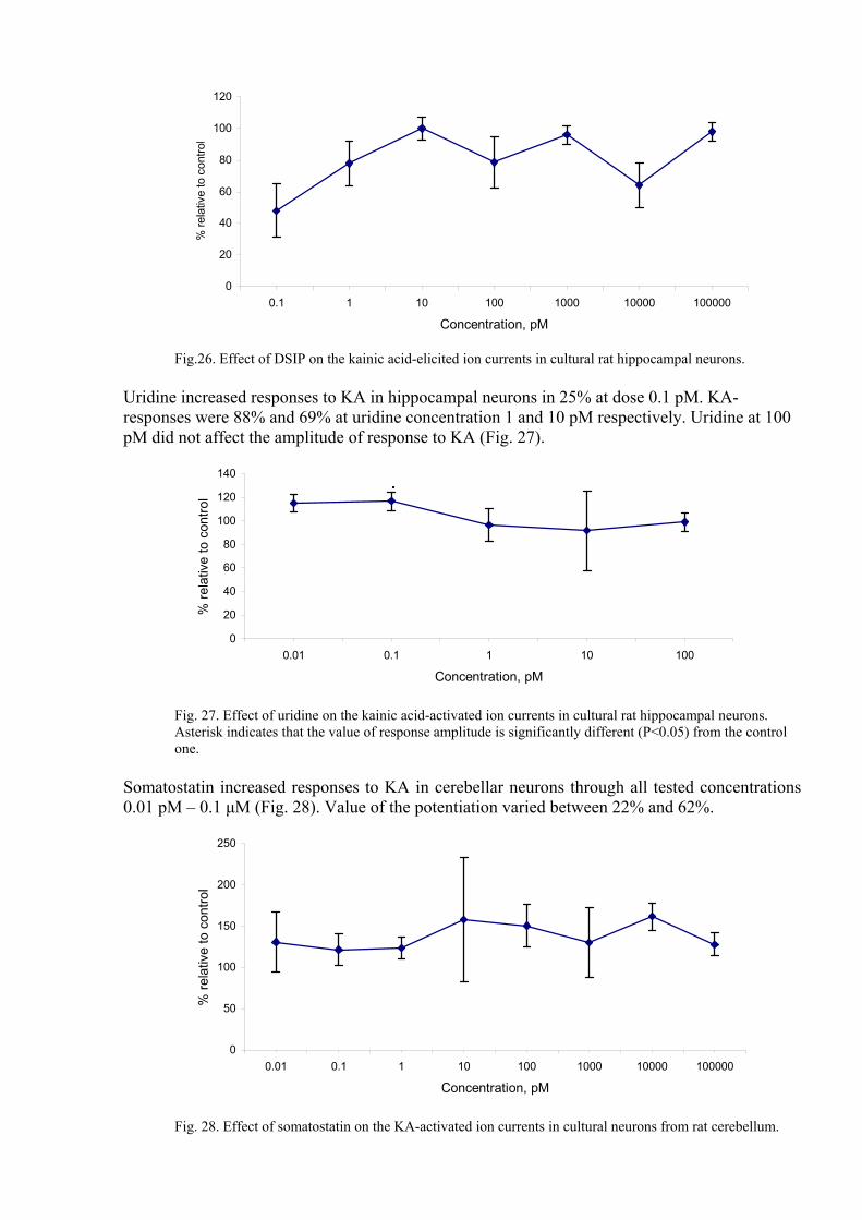
Somatostatin increased responses to KA in cerebellar neurons through all tested concentrations 0.01 pM – 0.1 µM (Fig. 28). Value of the potentiation varied between 22% and 62%.
0
50
100
150
200
250
0.01 0.1 1 10 100 1000 10000 100000
Concentration, pM
% re
lativ
e to
con
trol
Fig. 28. Effect of somatostatin on the KA-activated ion currents in cultural neurons from rat cerebellum.

MDP decreased most efficiently the KA-elicited ion currents in hippocampal neurons at concentration 0.01 pM. On the other hand in range of doses from 0.1 pM to 1 nM it increased the ion currents. At concentration 10 nM MDP again inhibited the KA-elicited ion currents (Fig. 29).
0
50
100
150
200
250
0.01 0.1 1 10 100 1000 10000
Concentration, pМ
% re
lativ
e to
con
trol
Fig. 29. Effect of MDP on the kainic acid-activated ion currents in cultural rat hippocampal neurons. Asterisk indicates that the value of response amplitude is significantly different (P<0.05) from the control one.
CLIP, like MDP, decreased the KA-elicited ion currents in hippocampal neurons only at concentration 0.01 pM. But at other tested doses it was not considerably effective (Fig. 30).
0
20
40
60
80
100
120
140
0.01 0.1 1 10
Concentration, pМ
% re
lativ
e to
con
trol
Fig. 30. Effect of CLIP on the kainic acid-activated ion currents in cultural rat hippocampal neurons. Thus the tested compounds showed complicated effect on kainic acid-elicited ion currents in hippocampal and cerebellar neurons from rat brain.

Conclusion As a result of the research, for the first time we obtained electrophysiological data about effect of number of endogenous sleep affecting compounds on parameters of GABA and glutamate receptors in isolated mammalian neurons. The data embrace facts about effect of DSIP on GABA and AMPA receptors, strict evidence (rather than behavior data) for blockade NMDA receptor by DSIP, effect of muramyl dipeptides (MDP) on GABA and glutamate receptors, influence of corticotropin-like intermediate lobe peptide (CLIP) on the receptors, effect of insulin on GABA receptors, influence of uridine on GABA and glutamate receptors. The results obtained in this work supported data published elsewhere as well as were diverse (even contrast) with them. The latter might be illustrated by effect of insulin on NMDA receptors. . For instance, Lin with coworkers [Lin L., Brown J.C., Webster W.W J. Neuroscince Lett., 1995, v. 192, N. 1, pp. 5-8.] and Liao & Leonard [Liao G.Y., Leonard J.P. J. Neurochem., 1999, v. 73, N. 4, pp. 1510- 1519] showed that short insulin exposition potentiates NMDA-elicited ion currents. We observed such a potentiation of NMDA-evoked ion currents in single neuron only, but in four other neurons insulin inhibited responses to NMDA. It was demonstrated elsewhere that somatostatin increases NMDA-activated ion currents [Pittaluga A., Bonfanti A., Raiteri M. Br. J. Pharmacol., 2000, v. 130, N. 3, pp. 557- 566]. In contrast to that it blocked the responses to NMDA. On the other hand we observed potentiation of kainic acid-evoked ion currents by somatostatin. Data obtained in the current work encourage call for additional research. Moreover, data was for the first time obtained about effect of the endogenous sleep affecting compounds on the NMDA-stimulated calcium uptake by cortical synaptosomes. This indicates to a complex regulation by these compounds the mechanism of release and uptake of GABA and glutamate by presynaptic terminals.

References 1. Mendelson W.B. Neurotransmitters and sleep. J. Clin. Psychiatry, 2001, 62, Suppl.,10,
pp. 5-8.
2. Gallopin T. et al., Identification of sleep-promoting neurons in vitro. Nature, 2000, 404: 992
3. Mendelson WB. Neuropharmacology of sleep induction by benzodiazepines. Crit Rev Neurobiol. 1992;6(4):221-32.
4. Manfridi A., Brambilla D., Mancia M. Stimulation of NMDA and AMPA receptors in the rat nucleus basalis of Meynert affects sleep. Am. J. Physiol., 1999, v.46, R1488- R1492.
5. Pringle A.K., Gardner C.R., Walker R.J. Reduction of cerebellar GABA-A responses by interleukin-1 (IL-1) through an indometacin-insensitive mechanism. Neuropharmacology, 1996, v. 35, N. 2, pp. 147-152.
6. Wang S., Cheng Q., Malik S., Yang J. Interleukin-1 beta inhibits gamma-aminobutyric acid type A (GABAA) receptor current in cultured hippocampal neurons. J. Pharmacol. Exp. Therap., 2000, v. 292, N. 2, pp. 497- 504.
7. Guarneri P.,Guarneri R., La Bella V. Interaction between uridine and GABA- mediated inhibitory transmission: studies in vivo and in vitro. Epilepsia, 1985, v. 26, N. 6, pp. 666- 671.
8. Shandra A.A., Godlevskii L.S., Brusentsov A.T. Effects of delta-sleep-inducing peptide on NMDA-induced convulsive activity in rats. Neurosci. Behav. Physiol., 1998, v, 28, N. 6, pp. 694- 697.
9. Tamura Y., Sato Y., Akaike A. Mechanisms of cholecystekinin-induced protection cultural cortical neurons against N-methyl-D-aspartate receptor-mediated glutamate cytotoxicity.-Brain Res.,1992, v. 592, N. 1-2, pp. 17- 25.
10. Akaike A., Kaneko S., Tamura Y. Prostoglandin E2 protects cultural cortical neurons against N-methyl-D-aspartate receptor-mediated glutamate cytotoxicity. Brain Res., 1994, v. 663, N.2, pp. 237- 243.
11. Lin L., Brown J.C., Webster W.W. Insulin potentiates N-methyl-D-aspartate receptor activity in Xenopus oocytes and rat hippocampus. J. Neuroscince Lett., 1995, v. 192, N. 1, pp. 5-8.
12. Liao G.Y., Leonard J.P. Insulin modulation of cloned mouse NMDA receptor current in Xenopus oocytes. J. Neurochem., 1999, v. 73, N. 4, pp. 1510- 1519.
13. Pittaluga A., Bonfanti A., Raiteri M. Somatostatin potentiates NMDA receptor function via activation of InsP(3) receptors and PKC leading to removal of the Mg(2+) block without depolarization. Br. J. Pharmacol., 2000, v. 130, N. 3, pp. 557- 566.
Director, Institute of Physiologically Active Compounds, Russian Academy of Sciences Academician N.S. Zefirov
Project Manager V.V. Grigoriev