Embed Size (px)
Citation preview

j . Cell Set. 24, 95-108 (1977) 95
Printed in Great Britain
NUCLEAR DNA CONTENT AND CHROMOSOME
NUMBERS THROUGHOUT THE LIFE CYCLE
OF THE COLONIA STRAIN OF THE
MYXOMYCETE, PHYSARUM POLYCEPHALUM
JOYCE MOHBERG*
Genetics Department, University of Leicester LEi yRH, England
SUMMARY
Nuclear DNA content and ploidy have been determined at different stages of the life cycleof the Colonia strain of the myxomycete Physarum polycephalum. Analyses at the plasmodialstage showed that (a) Burton and Feulgen DNA analyses agreed within 15% with strainswhich ranged from 06 to 36 pg of DNA per nucleus; (b) S-phase in Colonia is during theearly part of interphase as in the Wisconsin strain; (c) in heterothallic and heterothallic xColonia crossed strains there are 10-12 pg of DNA and 70 chromosomes per nucleus andin Colonia o-6 pg of DNA and 40 chromosomes.
Germinating spores of all strains contained one population of cells with about 05 pg ofDNA and 40 chromosomes and another of larger cells with up to 2-5 pg of DNA and 200chromosomes. The polyploid nuclei comprised 2-20 % of the total in heterothallic strains,2-65 % in heterothallic x Colonia crosses and 25-75 % m Colonia.
A method was devised for making chromosome spreads of amoebae grown on bacteriallawns. Cells were first exposed to dilute formaldehyde at 26 °C for 30 min, then spread onslides with hot lactic acid and stained. Such spreads of CLd (Colonia) and RSD4 (hetero-thallic) amoebae both contained about 40 chromosomes.
The data are consistent with the view that Colonia is haploid throughout its life cycle andthat chromosome number is neither halved during sporulation nor doubled during plasmoidalformation. However, the possibility exists that an alternance of ploidy occurs by way of thefew diploid nuclei present in the plasmodium.
INTRODUCTION
Although the myxomycete Physarum polycephalum has been in use for morethan a decade for biochemical studies of the cell cycle and of differentiation (seeJockusch, 1975, for the most recent review), very few of the investigations haveemployed genetic analysis. The main reason for this is that only a few geneticallymarked strains are available, owing to the technical difficulties involved in isolatingrecessive mutants from the usual heterothallic strains, where the mutant amoebahas to be mated with one of another mating type (mi) if a plasmodium is to beproduced (Wheals, 1970; Haugli & Dove, 1972). A major breakthrough in solvingthis problem was the discovery of the Colonia strain (Wheals, 1970) because in thisstrain plasmodia develop within amoebal clones and mutants can be isolated directly,
• Present address: Institut fiir Biochemie und Experimentelle Krebsforschung, derUniversitat Innsbruck, Fritz-Pregl-Str. 3/VII, A-6020 Innsbruck, Austria.
96 J. Mohberg
as already demonstrated for several nutritional mutants (Dee, Wheals & Holt, 1973;Cooke & Dee, 19746).
In spite of the obvious usefulness of Colonia strain, however, there are still someaspects of the life cycle which need to be clarified before the genetics of the organismcan be completely understood, and among them is the question of whether there areploidy changes during formation of the plasmodium or during sporulation. Wheals(1970) found recombinants among the progeny of a cross of Colonia and hetero-thallic (mtt) amoebae and concluded that the strain was homothallic (mating type h)and that two mth amoebae fused, underwent karyogamy and developed into aplasmodium in the same way as heterothallic strains. However, when Cooke & Dee(1974 a) did Feulgen analyses of nuclei of Colonia and heterothallic amoebae andof Colonia and Colonia x heterothallic crossed plasmodia, they found the relativeDNA contents to be in the ratio of 1:1:1:2. They interpreted these data as meaningthat karyogamy is involved in the development of a plasmodium from mtb and mtamoebae but not in formation of a plasmodium from mth amoebae alone. Theysuggested that instead the plasmodium must arise either by apogamy, with a singleamoebal cell developing into a plasmodium by repeated nuclear division, or byfusion or coalescence of 2 identical amoebae to give a dikaryotic cell, which thendevelops into a plasmodium. A recent cinematographic study by the same laboratorygroup (Anderson, Cooke & Dee, 1976) has almost completely ruled out the latterpossibility and supported the idea of apogamy.
There are, however, other more trivial explanations for Cooke & Dee's DNAdata. First, there could actually be a change in ploidy during sporulation in Coloniabut involving only the few diploid nuclei which are present in plasmodia. Theymight be able to undergo a normal meiosis to give haploid spores (Laffler & Dove,1975). Two other possibilities were proposed by Yemma & Therrien (1972) to accountfor very similar DNA results with selfing and heterothallic strains of the myxomycete,Didymium iridis. (1) The selfed plasmodium was actually diploid but had its 5-phaselate instead of early in the mitotic cycle, as in the heterothallic strains of D. iridisand P. polycephalum (Nygaard, Guttes & Rusch, i960), so that DNA measurementsmade before late interphase were of diploid Gj- and/or 5-phase, not of haploid G2.(2) DNA content per chromosome doubled immediately before spore cleavage sothat the spore nucleus could go through an essentially normal meiosis.
In order to answer the question of whether Cooke & Dee's DNA analysis actuallyreflected ploidy, chromosome counts and DNA analyses were done at differentstages of the life cycle of Colonia and of crosses of Colonia with heterothallic amoebae.Data presented in this paper show that Colonia strain had no delay in 5-phase inthe plasmodium, no 'diploidization' during spore formation and no net change inploidy during the life cycle.

DNA content and ploidy in Colonia 97
MATERIALS AND METHODS
Culture strains and methods
Plasmodial strains were chosen to include examples of plasmodia arising from mth amoebaeand from matings of mtb with heterothallic amoebae. The mtb strains used were C50 (Wheals,1970). CL, CLd, LU348 and LU640; mtiXm(h crosses were LU624 x CLd, LU647 xLUsooid, LU86o x E65; m(, x mth was CH188 x LU860 and mtt x mth was CH207 x LU860.Three m^xmtt crosses - LU648 x LU688, LU648XLU1 and RSD9 x RSD8 - were alsoincluded, as well as MscVIII, a strain grown from sclerotia which were originally collectedin the field (Daniel & Baldwin, 1964). Except for RSD9 x RSD8 (Haugli, 1971), LUi andM,cVIII, which are Wis 1 derivatives (Dee, 1973), all strains had a Colonia background.Mating type and other characteristics of CH188 and CH207 were given by P. N. Adler,Massachusetts Institute for Technology, in a personal communication to J. Dee. Mating andplasmodial fusion types and nutritional markers (where present) of LU640, LU647 andLU860 have been reported by Cooke (1974) and of other CL and LU strains by Cooke &Dee (19746)-
Methods for maintaining inoculum stocks, growing plasmodia and for inducing sporulationwere as described previously (Mohberg, Babcock, Haugli & Rusch, 1973). To avoid effects ofageing (McCullough et al. 1973) stock cultures were never carried as plasmodia on agar platesand all strains were either analysed within 3 or 4 weeks of mating or were stored as spherules.Genetic markers, such as the temperature sensitivity in E65 (Gingold, Grant, Wheals &Wren, submitted for publication) and the lysine requirement in LU348 should not have beenexpressed, since all cultures were grown at 26 °C in semi-defined medium (Daniel & Baldwin,1964).
Amoebal strains used included two mtb strains with delayed plasmodial formation - CLdand Cso-and three mty strains - LU648, a (Dee, 1966) and RSD4 (Haugli & Dove, 1972).RSD4 was grown axenically in protein-free medium (McCullough & Dee, 1976) and theothers on Escherichia coli lawns on liver infusion agar (Dee & Poulter, 1970). Agar platecultures to be used for DNA analysis were started with 5 x 10* cells per plate and wereharvested at 2 to 3 x 10* per plate to ensure that all cells were actively growing.
Spore viability tests were done as follows: after plasmodia had fruited, dishes were keptat room temperature for 7-14 days. Sporangia were then harvested and broken in a Potter-Elvehjem homogenizer to release spores, which were washed 2-4 times with distilled water,counted in a haemacytometer and plated with E. coli. Plaques were counted after 4 or 5 days.Spores for DNA analysis and chromosome counts (see below) were washed in the same wayand then germinated by incubation in water (1 to 2 x io8 spores in 5 to 8 ml in a 9-cm PyrexPetri dish at 26 CC).
Isolation of nuclei
Plasmodial nuclei were isolated at metaphase for chromosome spreads and at M + 2~5 h forDNA analyses, according to methods already described (Mohberg & Rusch, 1971). Amoebanuclei were isolated, using the same medium and blending time as for plasmodia, i.e. o-i %Triton X-100 and 15-30 s, instead of the 0-4% and 1 min originally specified. Germinatingspores were harvested after 4-5-5 h of incubation, by which time most were flagellated, andwere handled in the same way as amoebae, except that the homogenate was centrifuged in50-ml round-bottomed tubes for 5 min at 50 g to remove the bulk of the spore walls beforeit was passed through milk filter. Centrifugation through 1 M sucrose solution was necessaryonly with the plasmodial metaphase nuclei, which otherwise clumped so that chromosomesdid not spread well.
Preparation of chromosome spreads
Plasmodial chromosome spreads were made by heating metaphase nuclei in lactic acidand staining with aceto-orcein (LaCour, 1941) as described by Mohberg et al. (1973). Theprocedure for making spreads of spore chromosomes was as follows. Washed spores were
7 CEL24

98 J. Mohberg
incubated in water for 75 min before sampling was begun. Then, at 15- or 20-min intervalsfor the next 2 h an o-8-ml aliquot was transferred to a 50-ml conical tube, where it was madeto 10 ml with water and mixed with 05 ml of 37% commercial formalin. After 30 min atroom temperature, the suspension was diluted with 40 ml of ice-cold nuclear isolation medium(NIM) and set aside in cracked ice until the entire 6 or 8 samples had been harvested. Tubeswere then cer.trifuged at 1000 g for 10 min and washed twice with 40-ml of NIM (to preventformation of a brown background residue with aceto-orcein.) Pellets were finally suspendedin about 02 ml of NIM by vortexing for 1 min. Two drops were mixed on a slide with 2 dropsof 66% (v/v) lactic acid and heated over a small flame to spread chromosomes. Chromosomeswere stained with aceto-orcein or were fixed in 3:1 ethanol-acetic acid and stained withGiemsa's (10 min in a 1:10 dilution of stock Giemsa's in 67 mM phosphate buffer at pH 68).Although metaphase was usually found in the 90- to 120-min samples, in different experimentswith the same dish of spores it might occur as early as 75 and as late as 180 min after plating.
Amoebae (early to mid log phase) were washed twice with ice water, then were treatedwith formalin, spread and stained in the same way as the germinating spores.
Analytical methods
DNA analyses were done on isolated nuclei either by the Burton (Burton, 1956) or by theFeulgen method, using the procedure of Darlington & LaCour (1962), as modified by Cooke &Dee (1974a). Chicken erythrocyte nuclei were used as an internal standard (Rasch, Barr &Rasch, 1971). The nuclei were isolated by lysing oxalated chicken blood in o-i % saponin in1 % saline and centrifuging at 1000 g for 10 min to pellet nuclei. The pellet was washed onceor twice with NIM, suspended in NIM, parcelled out in amounts sufficient for 20-30 slidesand stored frozen until needed. DNA content of Physarum nuclei was calculated on theassumption that erythrocyte nuclei contained 2'45 pg of DNA, a rough mean of the valuesgiven by Rasch et al. (1971).
RESULTS
Relation of DNA content to chromosome number in plasmodia
Since data on DNA content of Physarum nuclei have been expressed both inarbitrary Feulgen units (Bovey & Ruch, 1972; Cooke & Dee, 1974a; Adler & Holt,1974; Laane & Haugli, 1976) and in pg DNA by Burton analysis (Mohberg et al.1973), a series of plasmodial strains, ranging from o-6 to 3-6 pg DNA per nucleus,were analysed by both methods to establish a basis of comparison. The 2 DNAassays (Table 1) were in reasonable agreement throughout the range, although theFeulgen values usually were about 15% lower. Chromosome counts were also inrough agreement, for most strains with o-6 pg of DNA per nucleus had 35-40chromosomes and strains with i-o—1-2 pg had about 70 chromosomes, giving anaverage DNA: chromosome ratio of 0-015. Ratios for C50 and RSD5 x RSD2 wereconsiderably higher, presumably because both strains contained a large proportionof polyploids and they tend to give low chromosome counts because they are hardto spread completely.
As expected from the findings of Cooke & Dee (1974a), the mtxy.mtu strainLU624 x CLd, and the mtx x mt2 strain with a Colonia background, LU648 x LU688,both had about the same nuclear DNA content as the Wis 1 heterothallics, M3cVIIIand RSD9 x RSD8, and almost twice the DNA content of the mth strains - CL,LU640 and B u .
5-phase was located in the mitotic cycle of the CL plasmodium by Feulgen analysis.

DNA per nucleus, pgA
Burton*
1-23 ±o-o8 (4)1-04 ±0-07 (6)o-88 ±0-03 (5)3-64 ±0-19 (6)0-71 ±O'O2 (4)o-68 ±o-oi (2)o-73±o-o2 (5)119 ±003 (6)1-15 ±0-05 (4)not done
Feulgen**
I O I
1 1 4
0-72
3-5Oo-6o0 6 3not done1 02I O I
1 09
Mean/"*Vi rnmAOAmp
numberf
68-172-535-0
168037'736-54 0 0
72-87 0 0
68-5
DNA per
Pgtt0-0148001580-02060-02080015900173
—
0-01400014400159
DNA content and ploidy in Colonia 99
At early prophase, approximately 15 min before metaphase, there was 0-63 pg ofDNA per nucleus; at reconstruction, just after separation of the daughter nuclei,0-29 pg; at M+3 h, 0-57 pg and at M+5 h, 0-65 pg. A. Polkinhorne (unpublisheddata) did a similar experiment with C50 and found that there also DNA doublingwas 90 % completed during the first 4 h after mitosis.
Table 1. DNA content and chromosome numbers in Colonia, Colonia x heterothalliccrosses and heterothallic strains of plasmodia
Strain
M3cVIIIRSD9 x RSD8C50RSD5 x RSD2CLLU640B nLU624 x CLdLU648XLU1LU648 x LU688
DNA analyses and chromosome counts were made as described in Materials and methods.Burton analyses of the first 4 strains are from Mohberg et al. (1973).
• DNA content in pg per nucleus ± standard error of the mean; no. of samples analysed inparentheses.
• • Fifty Physarum nuclei and 10 or 20 chicken erythrocyte nuclei were measured for eachsample. Data are given as mean DNA content, including polyploids.
f Mean chromosome number, including polyploids.f t By Feulgen analysis.
DNA content and chromosome numbers in growing amoebae
Since the results of the preceding experiment indicated that the Feulgen analysiscould be used with nuclei having as little as 0-3 pg of DNA, and since this methodrequires much less starting material than the Burton procedure and is less sensitiveto interference from bacteria, whole spores and agar (Mohberg & Rusch, 1971), itwas used for all analyses of amoebae and germinating spores done in this study.Mid log phase CLd and C50 amoebae contained o-6 pg of DNA; LU648, 0-46 pg;strain a, 0-41 pg, and RSD4, 0-34 pg. CLd and C50, like bacterially grown RSD4and RSD5 (Mohberg & Rusch, 1971), appear to be predominantly in haploid G2-phase; whereas the other strains, particularly the axenically grown RSD4, are chieflyin 5-phase.
Countable chromosome spreads were rarely found among exponentially growingamoebae, not only because metaphase, as in the plasmodium, occupies only about5 min of the 8-12 h generation time; but also because the few spreads present wereusually obscured by mitochondria and partially digested bacteria (Fig. ID, F). Whenamoebae were pretreated with formalin (see Materials and methods), the problemwith ingested bacteria was reduced, apparently by causing cells to eject most of their
7-2
IOO J. Mohberg
$?p&{~
10//m 1A B
4 • *JE •" * \
$ \
DNA content and ploidy in Colonia 101
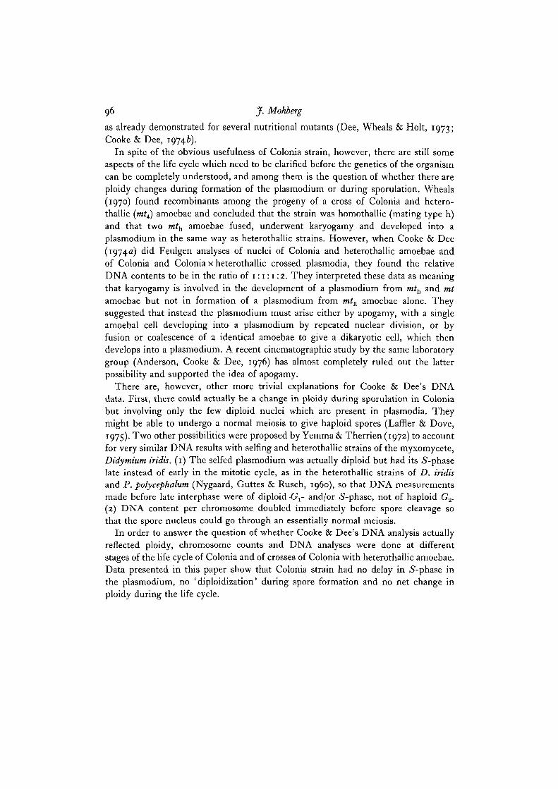
cytoplasmic inclusions and spreads like those in Fig. i E, G could be found. Countsof 8 such spreads of CLd gave a mean chromosome number of 38, with a standarddeviation of + 5-4 and a range of 27-48. RSD4 had a mean of 42 chromosomeswith a standard deviation of ±4-0 and a range of 35-49 in 12 spreads.
In order to make it possible to count a more meaningful number of chromosomespreads, several agents were tested for their ability to block mitosis in amoebaegrowing on bacterial lawns on agar. However, none were successful. Griseofulvin,which blocks mitosis in plasmodia (Gull & Trinci, 1974) and the herbicide, isopropyl3-chlorophenyl carbamate (CIPC), at levels of 50-100 /tg per ml were inhibitorybut did not arrest cells in metaphase. Colchicine (0-16%), actinomycin C (15 fig perml) and vinblastine sulphate (50 fig per ml) did not affect growth.
DNA content and chromosome numbers in germinating spores
Since a metaphase blocker for amoebae could not be found, germinating spores,which undergo a synchronous nuclear division shortly after hatching (Howard, 1931),were tested as a substitute for a synchronous amoeba culture. This was successfulwith mtxmt and mtxmth crosses, where spore germination was between 10%(RSD9XRSD8) and 70% (LU647xLU5ooid, CH207 x LU640 and CHi88xLU640). However, in mtb strains, where hatching was between 0-2% in CL (evenafter 3 passages through the life cycle) and 2% in C50 and LU348, it was notpossible to find enough chromosome spreads for reliable histograms and the proportionof nuclei of different ploidy levels had to be estimated from Feulgen DNA data.
Typical chromosome spreads of germinating spores are shown in Fig. 2. Chromo-somes tended to be shorter and thicker than in the plasmodium and were morefrequently split so that individual chromatids were visible (Fig. 2F, G), but thismay have been caused by the formalin treatment, rather than a real difference inchromosome structure. Spreads in Fig. 2C through F contain from 42 to 50 chromo-somes and are apparently in the 'normal' range for both mt and mth strains. Inaddition, highly polyploid nuclei (Fig. 2G) were found among spores of all strains,with the proportion being lower in the heterothallic than in the mth strains, as below.
The mtxy.mtt cross, LU648 x LU688, contained I-I pg of DNA and about 70chromosomes in the plasmodial nucleus (Table 1) and 0-5 pg of DNA (same as inthe growing LU648 amoeba) and between 40 and 45 chromosomes in the bulk ofthe germinating spore nuclei (Fig. 3). The remainder of the spores seemed to be
Fig. 1. A, B, chromosome spreads of typical Colonia and Colonia x heterothalliccrossed plasmodia. A is from CL, B from LU647 x Sooid. Spreads were photographedthrough a phase-contrast microscope at 1500 x. c—G, spreads of amoebae aftertreatment with dilute formalin and hot lactic acid, c, CLd swarm cells, showingfiagella, flagellar apparatus and nuclei; in the cytoplasm are mitochondria andgranules which are presumably remnants of food vacuoles. D, 'spread' of partiallydigested bacteria from a myxamoeba; structure at lower left is an unspread nucleus.E, RSD4 amoeba chromosomes. F, telophase in CL amoeba, showing chromosomesand mitochondria (at arrows). G, CLd amoeba chromosomes. c-E were stainedwith Giemsa, all others on the plate with aceto-orcein. Photography and magnificationas given for A and B.

102 J. Mohberg
Fig. 2. Chromosome spreads in germinating spores, A, partially spread metaphasein a C50 spore; mitochondrion at arrow, B, telophase in a C50 spore. c-F, chromosomespreads of spores of C50 (c), RSDg x RSD8 (D), and LU648 x LU688 (E, F). G, poly-ploid spread of LU648 x LU688 spore with approximately 150 chromosomes.Staining of D was with Giemsa and of others with aceto-orcein. Photography asin Fig. 1. Magnification of A-E same as for G.
B

DNA content and ploidy in Colonia 103
diploid or triploid. Mean DNA content of the smaller nuclei was 0-51 pg at 1-25 h,0-56 pg at 2-25 h (just prior to nuclear division in this experiment), and 0-53 pg at4-75 h after plating of the spores in water. This indicates that hatching amoebaeare predominantly in haploid G2-phase, and that there is an 5-phase shortly afternuclear division so that cells are again in G2-phase within a few hours after dividing.Germinating spores of RSD9 x RSD8 and M3cVIII gave DNA and chromosomehistograms (not shown) like those of LU648 x LU688, except that in the formerthere were about 20% polyploids and in the latter only 2%.
400 -
30 0 -
20 0 -
100 -
200 -
100 -
0 0
A '
C
-
1
m i
1
1 1
B '
: r
i i
i i
D
- A,-0 0 0-5 1 0 1-5 20 40 80 120
No. of chromosomesDNA/nucleus, pg
Fig. 3. Feulgen DNA analyses and chromosome counts in germinating spores(A, B) and plasmodium (c, D) of the mtl x mt» strain, LU648 x LU688. DNA readingswere made on 50 nuclei of each sample and DNA content calculated by means ofthe chicken erythrocyte nuclei included as internal standards. (See Materials andmethods.) Data were then grouped in intervals of 005 pg and plotted againstfrequency in percent. Chromosome histograms were made from counts of 22 spreadsof germinating spores and 42 of plasmodial nuclei. In this and later histogramsdata for spores are in the upper panels and for plasmodia in the lower.
In both of the m^xm^ crosses, LU647 x LUsooid and LU860XE65, theplasmodial DNA profiles resembled those of the mtx x mt2 strains and had a singlemain peak at about 1 pg of DNA (Fig. 4). Spores, however, had major peaks at0-55 and o-8 pg. Counts of 26 chromosome spreads of LU647 x LU5ooid sporesshowed that 35% of the nuclei were in a peak with a mean of 48 chromosomes;35% were in a second peak at 75 chromosomes, and the remaining spreads rangedfrom 90 to 250 chromosomes. Mating mth with either mt^ (CH188) or mtt (CH207)gave plasmodial DNA profiles like LU648 x LU688 but in spores of CH188 x LU640

104 J. Mohberg
(not shown) there were only 2 % polyploids and in CH207 x LU640 (Fig. 4) therewere 15 %.
In strains arising from mth amoebae only - LU640 (Fig. 4), C50 (Fig. 5), LU348and CL (last two not shown) - 90 % or more of the plasmodial nuclei had a DNAcontent of o-6 pg and 2-10 % had a content of 1-0-1-2 pg. However, in the germinatingspores only 25-50% of the nuclei were in the 0-5 pg peak. The remainder contained1-5-2 times as much DNA. Chromosome counts of C50 (Fig. 5) and 640 (not shown)showed that both diploid and polyploid nuclei were present.
200 -
100 -
00
300 -
200 -
100 .
00
A ' ' '
• m \nl n nnm i
D
n
-
, J 1
B
1
1 1
~\ nn i rE
-
c
" J
"i n
c '
r n-
r
VI '-
1
i 1 m00 0-5 10 15 0 5 10
DNA/nucleus, pg05 10
Fig. 4. Feulgen DNA analyses of matings of mtD with heterothallic amoebae. Strainsused were the mtb strain, LU640, and crosses of mtb with mtu LU647 x LUsooid,and with mtt, CH207 x LU640. A-C, spores: A, LU647 x LUsooid (m^xwjt,,);B, CH207 x LU640 (mtt x mtj; c, LU640 (mtj. D-F, the corresponding plasmodia.Data were obtained and plotted as for Fig. 3.
DISCUSSION
By combining the data of this and several earlier papers (Arescaldino, 1971;Mohberg & Rusch, 1971; Mohberg et al. 1973; Laane & Haugli, 1976) it is possibleto deduce the DNA content and times of synthesis throughout almost the entire lifecycle of the heterothallic, Wis 1-derived strains of P. polycephalum. The G2-phaseDNA content is 1-0-1-2 pg per nucleus (4C) in the growing, starving and sporulatingplasmodium, and it remains at this level until the first meiotic division, where itdrops to half (2C), and the second meiotic division, where it is halved again, giving

DNA content and ploidy in Colonia 105
100 -
50 0
300 -
200 -
100 -
00
A ' ' ' ' '
- C
fin n i n n . n
B
' nD
1
1 1
1
1
1
0 0 0-5 10 1-5 20
DNA/nucleus, pg
2 5 40 80
No. of chromosomes
Fig. 5. Feulgen DNA analyses and chromosome counts in germinating spores (A, B)and plasmodium (c, D) of the mtb strain, C50. Data were obtained and plotted asfor Fig. 3. Chromosome histograms were based on 15 spreads of germinating sporesand 50 of plasmodial nuclei.
iC. At some time between meiosis II and germination, DNA is replicated, since thehatching spore contains 0-5 pg of DNA, close to the 2C amount.
The change in ploidy indicated by the DNA data is supported by chiomosomecounts, which show that such mtx x mt2 crosses as LU648 x LU688 and RSD9 x RSD8have approximately 70 chromosomes per plasmodial nucleus and 40 in the germi-nating spore. This corresponds to a haploid number of 40 instead of the 25 proposedearlier (Mohberg et al. 1973). The earlier counts were almost certainly too lowbecause spores were not treated with formalin before spreading and the unstabilizedchromosomes were destroyed during heating in lactic acid prior to staining.
Several M3c derivatives (Mohberg et al. 1973) contained nuclei with 50—55chromosomes, and what relation they have to the haploid or diploid number isunknown. However, on several occasions M3 cultures have been observed to dropto 40 chromosomes per nucleus after continuous submersed culture for long periodsof time (a year or more). Either a population of microplasmodia with small nucleislowly outgrows those with diploid nuclei, or diploid nuclei lose chromosomes untilthey are haploid and the nuclei with 50-55 chromosomes are an intermediate in thetransition. We are trying to work out a method for doing at least a partial karyotypeso that we can answer this question and also determine whether the range which wesee in chromosome counts within individual strains is owing to aneuploidy or merelyto poor spreading technique.
106 J. Mohberg
We have not established whether the polyploid cells among germinating sporescan develop into amoebae colonies and finally into plasmodia. Conceivably thesecells have hatched before meiosis is completed and later drop to a normal chromosomenumber, as can occur in another species of myxomycete, Fuligo septica (Cathcart &Aldrich, 1972). They may also not be viable, beyond being able to germinate, orthey may grow so slowly that they are diluted out by normal amoebae and lostfrom the population. However, if they do survive, the ploidy of plasmodia fromuncloned heterothallic amoebae probably is even more heterogenous than alreadythought, since a diploid (zn) amoeba might produce three different kinds of plasmodialnuclei when mixed with a normal (in) amoeba - 371 by mating, 2» by selfing andin by inducing the other amoeba to self (Therrien & Collins, 1976). On the otherhand, it should be possible to take advantage of the polyploid amoebae to constructplasmodial strains with a wider range of ploidy than found among the various RSDcrosses (Mohberg et al. 1973) and hopefully also to select strains which spherulatebetter than the RSD strains, many of which have been lost because of poor spheruleviability.
Strains from mtlt mt3 and wif4 x mth matings all resembled heterothallic strainsin that plasmodial nuclei had approximately twice the DNA content and chromosomenumber as spores and amoebae, and differed principally in that polyploid amoebaewere more numerous among germinating spores. In plasmodia arising solely frommth amoebae, however, there were only o-6 pg of DNA and about 40 chromosomesper nucleus and this did not change throughout starvation and sporulation up tothe time of the precleavage mitosis (A. Polkinhorne, unpublished data; Mohberget al. 1973). In germinating spores the average nuclear DNA content was actuallyhigher than in the parent plasmodium, for in all 4 of the strains examined, at least25 % of the nuclei had the diploid DNA content and chromosome number. (Whetherthe germinating spores were also bi- and multinucleate, as reported for CL byLaane, Haugli & Mellem (manuscript submitted) is not known, since our DNAanalyses were done on isolated nuclei and not whole cells.)
The data presented in this paper are in accord with Cooke & Dee's (1974a, b)proposals: that there is no change in ploidy during the life cycle of Colonia; andthat fusion of amoebae and karyogamy occur when a plasmodium arises from themating of heterothallic amoebae with other heterothaUics or with mth amoebae, butnot when the plasmodium develops from mth amoebae alone. Since synaptonemalcomplexes have been found in CL spores by Laane et al. (submitted for publication)and in C50 spores by I. Arescaldino-LaCorre (personal communication), it seemsdefinite that meiosis at least begins, von Stosch, van Zul-Pischinger & Dersch, 1964,claimed that the meiosis was abortive, but the electron-microscope studies ofLaane and co-workers suggest that the first division is completed and the secondsuppressed, so that recombination occurs without reduction in chromosome number.
As with the heterothallic strains, we know very little of either the origin or fateof the polyploid cells which hatch from Colonia spores, and here they are particularlyworrisome, both because of their relative abundance and because of the complicationsthey could cause in genetic experiments. It seems unlikely for two reasons that
DNA content and ploidy in Colonia 107
diploid spores are the source, at least not the sole source, of the diploid nuclei foundin Colonia plasmodia. First, we have found that uncloned CL and C50 spores with50-75% polyploid nuclei gave plasmodia with 10% or less diploid and polyploidnuclei. Secondly, plasmodia of all Colonia strains examined thus far have containedat least 2 % diploid nuclei, in spite of the fact that they were grown from clonedamoebae. It would seem rather that diploid plasmodial nuclei are produced fromin nuclei by some event too rare to have been detected in the cinematographic studyof Anderson et al. (1976), such as endomitosis or fusion of nuclei, either in theparental amoebal plaque or in the new plasmodium. Laffler & Dove (1975) havesuggested that the zn nuclei in the plasmodium go through a normal meiosis togive viable haploid spores. Although E/M observations (Laane et al. submitted forpublication) indicate a pseudomeiosis with a single division, rather than a normaldiploid meiosis with two divisions, it will probably not be possible to settle thisquestion until the zn nuclei can be traced through the entire life cycle.
The author gratefully acknowledges the financial support of Science Research CouncilGrant No. B/RG/49103 to Dr Jennifer Dee. Thanks are also extended to Dr Clare McCulloughfor providing axenic amoeba cultures, to Mr James Mackley for help with photography,and to Miss Anna Polkinhorne for permission to quote from her third-year student projectreport.
REFERENCES
ADLER, P. N. & HOLT, C. E. (1974). Changes in properties of Physaruni polycephaium amoebaeduring extended culture. J. Bad. 120, 532-533.
ANDERSON, R. W., COOKE, D. J. & DEE, J. (1976). Apogamic development of plasmodia inthe myxomycete Physarumpolycephalum: a cinematographic analysis. Protoplasma 89, 29-40.
ARESCALDINO, I. (1971). Evolution de la teneur en ADN des noyaux de Physarum polycephalum(Myxomycetes) au cours de la sporulation. C. r. hebd. Sianc. Acad. Sci., Paris, D 273,398-401.
BOVEY, F. & RUCH, F. (1972). Cytofluorometric determination of DNA and protein in thenuclei and nucleoli of Physarum polycephalum during the intermitotic period. Histochemie32, 153-162.
BURTON, K. (1956). A study of the conditions and mechanism of the diphenylamine reactionfor the colorimetric estimation of deoxyribonucleic acid. Biochem.J. 62, 315-323.
CATHCART, M. E. & ALDRICH, H. C. (1972). A case of interrupted meiosis in Fuligo septica.Am. J. Bot. 59, 665.
COOKE, D. J. (1974). Studies on tlie Colonia Isolate of Physarum polycephalum. Ph.D. Thesis,Genetics, University of Leicester.
COOKE, D. J. & DEE, J. (1974a). Plasmodia formation without change in nuclear DNA contentin Physarum polycephalum. Genet. Res., Camb. 23, 307-317.
COOKE, D. J. & DEE, J. (19746). Methods for the isolation and analysis of plasmodial mutantsin Physarum polycephalum. Genet. Res., Camb. 24, 175-187.
DANIEL, J. W. & BALDWIN, H. H. (1964). Methods of culture for plasmodial myxomycetes.In Methods in Cell Physiology, vol. 1 (ed. D. M. Prescott), pp. 9-41. New York and London:Academic Press.
DARLINGTON, C. D. & LACOUR, L. F. (1962). The Handling of Chromosomes, 4th edn, pp.159—162. London: Allen and Unwin.
DEE, J. (1966). Multiple alleles and other factors affecting plasmodium formation in thetrue slime mould Physaruni polycephalum Schw. J. Protozool. 13, 610-616.
DEE, J. (1973). Aims and techniques of genetic analysis in Physarum polyceplialum. Ber. dt.bot. Ges. 86, 93-121.
DEE, J. & POULTER, R. T. M. (1970). A gene conferring actidione resistance and abnormalmorphology on Physarum polycephalum plasmodia. Genet. Res., Camb. 15, 35-41.

108 J. Mohberg
DEE, J., WHEALS, A. E. & HOLT, C. E. (1973). Inheritance of plasmoidal valine requirementin Physarum polycephalum. Genet. Res., Camb. 21, 87-101.
GINGOLD, E. C, GRANT, W. D., WHEALS, A. E. & WREN, M. Temperature sensitive mutantsof the slime mould Physarum polycephalum. II. Mutants of the plasmodial phase. Submittedfor publication.
GULL, K. & TRINCI, A. P. J. (1974). Ultrastructural effects of griseofulvin on the myxomycete,Physarum polycephalum. Protoplasma 81, 37-48.
HAUGLI, F. B. (1971). Mutagenesis, Selection and Genetic Analysis in Physarum polycephalum.Ph.D. Thesis, Molecular Biology, University of Wisconsin.
HAUGLI, F. B. & DOVE, W. F. (1972). Mutagenesis and mutant selection in Physarum poly-cepfialum. Molec. gen. Genet. 118, 109-124.
HOWARD, F. L. (1931). The life history of Physarum polycephalum. Am. J. Bot. 18, 116-132.JOCKUSCH, B. M. (1975). Neuere Forschungen iiber Zellzyklus und Kernteilung am Schleimpilz
Physarum polycephalum. Naturwissenschaften 62, 283-289.LAANE, M. M. & HAUGLI, F. B. (1976). Nuclear behaviour during meiosis in the myxomycete
Physarum polycephalum. Nona. J. Bot. 23, 7-21.LAANE, M. M., HAUGLI, F. B. & MELLEM, T. R. Nuclear behaviour during sporulation and
germination in the Colonia strain of Physarum polycephalum. Submitted for publication.LACOUR, L. (1941). Acetic-orcein. A new stain-fixative for chromosomes. Stain Technol.
16, 169-174.LAFFLER, T. & DOVE, W. F. (1975). Sporulation in the Colonia isolate of Physarum polycephalum.
Abstracts, Sixth Physarum Conference, Gainesville, Florida.MCCULLOUGH, C. H. R., COOKE, D. J., FOXON, J. L., SUDBERY, P. E. & GRANT, W. D.
(:973)- Nuclear DNA content and senescence in Physarum polycephalum. Nature, NewBiol. 245, 263-265.
MCCULLOUGH, C. H. R. & DEE, J. (1976). Denned and semi-defined media for the growthof amoebae of Physarum polycephalum. J. gen. Microbiol. 95, 151-158.
MOHBERG, J., BABCOCK, K. L., HAUGLI, F. B. & RUSCH, H. P. (1973). Nuclear DNA contentand chromosome numbers in the myxomycete Physarum polycephalum. Devi Biol. 34,228-245.
MOHBERG, J. & RUSCH, H. P. (1971). Isolation and DNA content of nuclei of Physarum poly-ceplialum. Expl Cell Res. 66, 305-316.
NYGAARD, O. F., GUTTES, S. & RUSCH, H. P. (i960). Nucleic acid metabolism in a slime moldwith synchronous mitosis. Biochim. biophys. Ada 38, 298-306.
RASCH, E. M., BARR, H. J. & RASCH, R. W. (1971). DNA content of sperm of Drosophilamelanogaster. Chromosoma 33, 1-18.
THERRIEN, C. D. & COLLINS, O. R. (1976). Apogamic induction of haploid plasmodia in amyxomycete, Didymium iridis. Devi Biol. 49, 283-287.
VON STOSCH, H. A., VAN ZUL-PISCHENGER, M. & DERSCH, G. (1964). Nuclear phase alternancein the myxomycete Physarum polycephalum. Abstr. 10th int. Bot. Congr. pp. 481-482.
WHEALS, A. E. (1970). A homothallic strain of the myxomycete Physarum polycephalum.Genetics, Princeton 66, 623-633.
YEMMA, J. J. & THERRIEN, C. D. (1972). Quantitative microspectrophotometry of nuclearDNA in selfing strains of the myxomycete Didymium iridis. Am. J. Bot. 59, 828-835.
(Received 22 July 1976)


![[Jshopen] Phase2 Final](https://img.dokumen.tips/doc/110x75/577d259b1a28ab4e1e9f3ae3/jshopen-phase2-final.jpg)