Embed Size (px)
Citation preview

Vojnosanit Pregl 2019; 76(2): 219–223. VOJNOSANITETSKI PREGLED Page 219
Correspondence to: Aleksandra Sokic-Milutinovic, Clinical Center of Serbia, Clinic for Gastroenterology and Hepatology, Koste Todorovica 6, 11 000 Belgrade, Serbia. Email: [email protected]
C A S E R E P O R T UDC: 616.36-006-033.2-07
https://doi.org/10.2298/VSP160923073S
Melena as a first sign of metastatic hepatic angiosarcoma: A case report
Melena kao prvi znak metastatskog angiosarkoma jetre
Aleksandra Sokić-Milutinović*†, Ljubiša Tončev†, Tijana Glišić*†, Vera Matović‡, Marjan Micev§, Srdjan Djuranović*†, Miodrag Krstić*†
University of Belgrade, *Faculty of Medicine, Belgrade, Serbia; Clinical Centre of Serbia, †Clinic for Gastroenterology and Hepatology, ‡Emergency Center,
§Department of Pathology, Belgrade, Serbia
Abstract Introduction. Angiosarcomas are malignant tumors of vas-cular endothelium that may arise from different locations. Although primary hepatic angiosarcoma accounts for only 1.8% of primary liver tumors, it is the most common malig-nant mesenchymal tumor of the liver. We report a case of primary hepatic angiosarcoma with melena as an unusual in-itial manifestation of this extremely rare tumor. Case re-port. Forty-four-years old patient with melena was referred to our Clinic because melena was not resolved after re-peated argon plasma coagulation of bleeding lesions during esophagogastroduodenoscopy in the regional hospital. Ab-dominal ultrasound and multislice computed tomography (MSCT) revealed enlarged liver, with focal lesion 6 cm in di-ameter localized in the left lobe with multiple satellite le-sions in both liver lobes, enlarged spleen and extremely di-lated and long umbilical vein. Double-balloon enteroscopy and video capsule endoscopy detected the multiple bleeding vascular lesions in the small bowel. Histopathological ex-amination and immunohistochemistry of the small bowel le-sions revealed malignant mesenchymal proliferation with vascular/endothelium differentiation of neoplastic cells. The patient was diagnosed with metastatic angiosarcoma probably of hepatic origin with metastasis in the small bow-el, that caused melena, and in the lumbar spine, causing back pain. Conclusion. Rare causes of melena include bleeding from primary or metastatic hemangiosarcoma lo-calized in the gastrointestinal tract, especially small bowel. Key words: hemangiosarcoma; liver neoplasms; melena; diagnostic techniques and procedures; diagnosis, differential; palliative care.
Apstrakt Uvod. Angiosarkomi su maligni tumori vaskularnog endo-tela koji mogu nastati na različitim lokacijama. Iako primarni angiosarkomi jetre čine samo 1.8% primarnih tumora jetre to su najčešći maligni mezenhimalni tumori jetre. Prikazu-jemo slučaj primarnog angiosarkoma jetre sa melenom kao neobičnom inicijalnom manifestacijom ovog vrlo retkog tumora. Prikaz slučaja. Bolesnik star 44 godine upućen je na našu Kliniku zbog melene koja je perzistirala nakon po-navljanih argon plazma koagulacija krvarećih lezija tokom ezofagogastroduodenoskopije u regionalnoj bolnici. Ultra-zvuk i multislajsna kompjuterska tomografija (MSCT) ab-domena ukazali su na uvećanu jetru sa fokalnom lezijom le-vog lobusa promera 6 cm i multiplim satelitskim lezijama u oba lobusa, uvećanu slezinu, kao i ekstremno proširenu i dugačku umbilikalnu venu. Double balloon enteroskopijom i endoskopskom video kapsulom uočene su brojne krvareće va-skularne lezije u tankom crevu. Patohistološki pregled i imuno-histohemijska bojenja biopsija lezija iz tankog creva ukazala su na maligni mezenhimski tumor vaskularnog/endotelnog pore-kla. Zaključeno je da se kod bolesnika radi o metastatkom an-giosarkomu sa najverovatnijim primarnim ishodištem u jetri i metastazama u tankom crevu, što je uzrokovalo melenu, i kičmenom stubu, što je uzrokovalo bol u leđima. Zaključak. Retki uzroci melene uključuju krvarenje iz primarnog ili meta-statskog hemangiosarkoma lokalizovanog u gastrointestinal-nom traktu, posebno u tankom crevu. Ključne reči: hemangiosarkom; jetra, neoplazme; melena; dijagnostičke tehnike i procedure; dijagnoza, diferencijalna; lečenje, palijativno.

Page 220 VOJNOSANITETSKI PREGLED Vol. 76, No 2
Sokić-Milutinović A, et al. Vojnosanit Pregl 2019; 76(2): 219–223.
Introduction
Angiosarcomas are malignant tumors of vascular endo-thelium that may arise from different locations. They occur most commonly in the skin and soft tissue. Although primary hepatic angiosarcoma accounts for only 2% of primary liver tumors, it is the most common malignant mesenchymal tu-mor of the liver in adults 1. Early reports of hepatic angiosar-coma focused on its association with environmental chemical carcinogens, such as vinyl chloride, thorium dioxide (Thoro-trast) and arsenic, but exposure to these agents is now rare. Other known risk factors include use of androgenic steroids, oral contraceptives and cyclophosphamide, but most of these tumors nowadays occur in the absence of known risk factors 1.
We report a case of metastatic hepatic angiosarcoma with melena as first and unusual manifestation of this ex-ceedingly rare tumor.
Case report
A 44-year-old man, a professional truck driver, was re-ferred to the Clinic for Gastroenterology and Hepatology, Clinical Center of Serbia, with persistent melena. Prior to admission to our Clinic, the patient was admitted and treated for one month in the regional hospital where esophagogas-troduodenoscopy (EGD) revealed two duodenal bleeding le-sions treated with several argon plasma coagulation (APC) sessions. After eight APC sessions in a regional hospital, melena persisted and patient was referred to our Clinic. On the day of admission, patient complained of exhaustion, nau-sea and melena and reported lower back pain he suffered from for past 4 years. The patient`s past medical history was not significant, except for spine injury in a traffic accident 4 years ago and subsequent operation with osteosynthetic met-al implant in lumbosacral spine. He denied exposure to envi-ronmental toxins. Physical examination revealed enlarged liver 2 cm below right costal margin in the medioclavicular line and enlarged spleen palpable for 1cm below left costal margin in the medioclavicular line. Initial laboratory findings were haemoglobin (Hb) 10.2 g/dL, haematocrit (Hct) 30%, platelet count (Plt) 76 x 109/L, prothrombin time (PT) 15.8 s, albumin 29 g/L, alkaline phosphatase (ALP) 186 IU/L, as-partate transaminase (AST) 40 IU/L, alanine transaminase (ALT) 45 IU, erythrocyte sedimentation rate (ESR) 8 mm/h. Since the patient presented with hepatosplenomegaly of un-known origin immediately upon admission, immunology profiles and serology for viral hepatitis were ordered. Hepati-tis A, B and C and immunology profiles [antinuclear anti-bodies (ANA), anti-smooth muscle antibodies (ASMA), an-timitochondrial antibody (AMA), perinuclear antineutrophil cytoplasmatic antibodies (pANCA)] were negative. Further investigation, after ultrasound, revealed focal lesions of the liver, and all tested tumor markers including: alpha-fetoprotein (AFP), carcinoembryonic antigen (CEA), car-cinogen 19-9 (CA 19-9) and 125 (CA 125), total prostatic specific antigen (tPSA), free prostatic specific antigen (fPSA), beta human chorionic gonadotropin (βHCG), were within normal range. The chest radiography was normal. Ul-
trasound of the abdomen showed enlarged liver with a large 6-cm mass in the left liver lobe (Figure 1), multiple up to 2 cm small focal lesions in both lobes of the liver, extremely dilated (20 mm in diameter) and long umbilical vein with turbulent flow, enlarged spleen, but no ascites. EGD was per-formed upon admission and revealed three mucosal non-bleeding lesions resembling stigmata of previous APC in se-cond duodenal portion. Colonoscopy and barium follow-through were normal. Multislice computed tomography (MSCT) angiography of the abdomen identified in the seg-ment IV and VII of the liver two large focal lesions with the CT characteristics resembling cavernous haemangioma (Fig-ure 2) together with numerous intraabdominal collaterals but no thrombosis of v. portae, v. lienalis or v. mesenterica that would explain recanalised umbilical vein.
Fig. 1 – Large mass in left lobe of the liver.
Fig. 2 – Multislice computed tomography shows two large
focal lesions in the IV and VII segment of the liver.
Peroral double balloon enteroscopy was performed and revealed few larger non-bleeding pseudopolypoid lesions with hyperemic edge and central umbilication that were con-sidered to be of a vascular origin, thus contraindicated for biopsy. The patient was treated with sandostatin and beta-blockers that reduced frequency of melena and need for blood transfusion. Due to the metal implant in this patient, we could not order magnetic resonance imaging (MRI), therefore liver scintigraphy and blood pool were performed

Vol. 76, No 2 VOJNOSANITETSKI PREGLED Page 221
Sokić-Milutinović A, et al. Vojnosanit Pregl 2019; 76(2): 219–223.
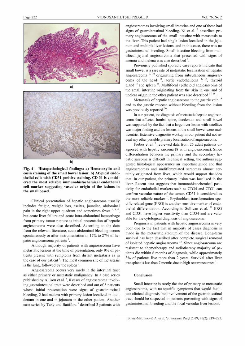
in order to clarify characteristics of the liver lesions and the-se results were suggesting the vascular lesions in both liver lobes. Since melena occurred again, M2A video capsule was performed and showed hyperemic zone without bleeding in duodenum (Figure 3a), active bleeding in distal duodenum without visible lesion (Figure 3b), one prominent bleeding lesion in distal jejunum that could correspond to varix or tu-mor (Figure 3c) and another large lesion in proximal ileum (Figure 3d). Percutaneous liver biopsy was not performed because of the high bleeding risk due to the vascularity of the lesions and patient's impaired coagulation. The patient was discharged in a stable condition with the diagnosis of prob-able liver cavernous hemangioma and enteropathy due to portal hypertension with advised supportive therapy includ-ing proton pump inhibitors (PPI), Lanreotide and β-blockers. One month after discharge, the patient was readmitted to our hospital with persistent melena, back pain and weight loss. Laboratory data at readmission were: Hb 64g/L, Hct 19%, platelet 66 × 109, albumin 25 g/L, alkaline phosphatase 250 IU, D-dimer > 4000 mg/L, vWF (von Willebrand factor) > 150%. Upon readmission, the abdominal ultrasound and MSCT scan showed enlargement of previously diagnosed liver focal lesions and massive osteolytic defects in the lum-bal spine, and therefore, the initial diagnosis was challenged. EGD was repeated and showed the focal pseudopolypoid le-sions with umbilication and scattered bleeding, especially in third portion of duodenum. At this point it was decided that biopsies from the small bowel lesions are lower risk than liv-er biopsy. The histological examination of the small bowel lesions revealed malignant mesenchymal proliferation with vascular/endothelium differentiation of neoplastic cells. The tumor cells stained positive for vimentin and CD31.
In the light of histological appearance (Figure 4a), posi-tive immunohistochemical staining for CD 31 (Figure 4b) and evident progression of the disease, we concluded that pa-tient suffered from primary hepatic angiosarcoma with me-tastatic lesions affecting small bowel that caused melena and lumbal spine that caused back pain. The patient was referred to his regional Oncology Center where he was given only symptomatic treatment due to deteriorated general condition. The patient died two months after the diagnosis was made.
Discussion
Angiosarcomas are rare malignant tumors arising from vascular endothelium that account for less than 1% of all soft tissue sarcomas. The development of the tumor is in 25% of all cases related to previous exposure to environmental tox-ins such as thorium dioxide (Thorotrast), polyvinyl chloride monomers and arsenic-containing insecticides, while in the majority of patients no underlying risk factor is identified 1. Primary hepatic angiosarcoma shows a male predominance of 3 : 1 and majority of patients are diagnosed between the ages of 50 and 59 1, 2.
a)
b)
c)
d)
Fig. 3 – M2A video capsule shows: a) Hyperemic zone without bleeding in duodenum; b) Fresh bleeding in distal duodenum without visible lesion; c) Bleeding lesion in distal jejunum; d) Metastatic lesion in proximal ileum.
Page 222 VOJNOSANITETSKI PREGLED Vol. 76, No 2
Sokić-Milutinović A, et al. Vojnosanit Pregl 2019; 76(2): 219–223.
a)
b)
Fig. 4 – Histopathological findings: a) Hematoxylin and eosin staining of the small bowel lesion; b) Atypical endo-thelial cells with CD31 positive staining. CD 31 is consid-ered the most reliable immunohistochemical endothelial cell marker suggesting vascular origin of the lesions in the small bowel.
Clinical presentation of hepatic angiosarcoma usually
includes fatigue, weight loss, ascites, jaundice, abdominal pain in the right upper quadrant and sometimes fever 1, 3, 4, but acute liver failure and acute intra-abdominal hemorrhage from primary tumor rupture as initial presentation of hepatic angiosarcoma were also described. According to the data from the relevant literature, acute abdominal bleeding occurs spontaneously or after instrumentation in 17% to 27% of he-patic angiosarcoma patients 1.
Although majority of patients with angiosarcoma have metastatic lesions at the time of presentation, only 9% of pa-tients present with symptoms from distant metastasis as in the case of our patient 1. The most common site of metastasis is the lung, followed by the spleen 1.
Angiosarcoma occurs very rarely in the intestinal tract as either primary or metastatic malignancy. In a case series published by Allison et al. 5, 8 cases of angiosarcoma involv-ing gastrointestinal tract were described and out of 5 patients whose initial presentation were signs of gastrointestinal bleeding, 2 had melena with primary lesion localized in duo-denum in one and in jejunum in the other patient. Another case series by Taxy and Battifora 6 described 3 patients with
angiosarcomas involving small intestine and one of these had signs of gastrointestinal bleeding. Ni et al. 7 described pri-mary angiosarcoma of the small intestine with metastasis to the liver. This patient had single lesion localized in the jeju-num and multiple liver lesions, and in this case, there was no gastrointestinal bleeding. Small intestine bleeding from mul-tifocal jejunal angiosarcoma that presented with signs of anemia and melena was also described 8.
Previously published sporadic case reports indicate that small bowel is a rare site of metastatic localization of hepatic angiosarcoma 9, 10 originating from subcutaneous angiosar-coma of the head 11, aortic endothelioma 12–14, thyroid gland 15 and spleen 16. Multifocal epitheloid angiosarcoma of the small intestine originating from the skin in one and of unclear origin in the other patient was also described 17, 18.
Metastasis of hepatic angiosarcoma to the gastric vein 19 and to the gastric mucosa without bleeding from the lesion was previously reported 20.
In our patient, the diagnosis of metastatic hepatic angiosar-coma that affected lumbal spine, duodenum and small bowel was supported by the fact that a large liver lesion with satellites was major finding and the lesions in the small bowel were mul-ticentric. Extensive diagnostic workup in our patient did not re-veal any other possible primary localization of angiosarcoma.
Forbes et al. 3 reviewed data from 25 adult patients di-agnosed with hepatic sarcoma (8 with angiosarcoma). Since differentiation between the primary and the secondary he-patic sarcoma is difficult in clinical setting, the authors sug-gested histological appearance an important guide and that angiosarcomas and undifferentiated sarcomas almost cer-tainly originated from liver, which would support the idea that, in our patient, the primary lesion was localized in the liver. Recent data suggests that immunohistochemical posi-tivity for endothelial markers such as CD34 and CD31 can confirm vascular nature of the tumor. CD31 is considered as the most reliable marker 1. Erythroblast transformation spe-cific related gene (ERG) is another sensitive marker of endo-thelial differentiation. According to Sullivan et al. 21 ERG and CD31 have higher sensitivity than CD34 and are valu-able for the cytological diagnosis of angiosarcoma.
Prognosis in patients with hepatic angiosarcoma is very poor due to the fact that in majority of cases diagnosis is made in the metastatic stadium of the disease. Long-term survival has been described after complete surgical removal of isolated hepatic angiosarcoma 22. Since angiosarcoma are resistant to chemotherapy and radiotherapy majority of pa-tients die within 6 months of diagnosis, while approximately 3% of patients live more than 2 years. Survival after liver transplant is less than 7 months due to high recurrence rates 1.
Conclusion
Small intestine is rarely the site of primary or metastatic angiosarcoma, with no specific symptoms that would facili-tate clinical diagnosis, but involvement of the gastrointestinal tract should be suspected in patients presenting with signs of gastrointestinal bleeding and the focal vascular liver lesions.

Vol. 76, No 2 VOJNOSANITETSKI PREGLED Page 223
Sokić-Milutinović A, et al. Vojnosanit Pregl 2019; 76(2): 219–223.
R E F E R E N C E S
1. Chaudhary P, Bhadana U, Singh RA, Ahuja A. Primary hepatic angiosarcoma. Eur J Surg Oncol 2015; 41(9): 1137 43.
2. Baxter PJ. The British hepatic angiosarcoma register. Environ Health Perspect 1981; 41: 115 6.
3. Forbes A, Portmann B, Johnson P, Williams R. Hepatic sarcomas in adults: A review of 25 cases. Gut 1987; 28(6): 668 74.
4. Molina E, Hernandez A. Clinical manifestations of primary he-patic angiosarcoma. Dig Dis Sci 2003; 48(4): 677 82.
5. Allison KH, Yoder BJ, Bronner MP, Goldblum JR, Rubin BP. An-giosarcoma involving the gastrointestinal tract: A series of pri-mary and metastatic cases. Am J Surg Pathol 2004; 28(3): 298 307.
6. Taxy JB, Battifora H. Angiosarcoma of the gastrointestinal tract. A report of three cases. Cancer 1988; 62(1): 210 6.
7. Ni Q, Shang D, Peng H, Roy M, Liang G, Bi W, et al. Primary angiosarcoma of the small intestine with metastasis to the liver: a case report and review of the literature. World J Surg Oncol 2013; 11: 242.
8. Zacarias Föhrding L, Macher A, Braunstein S, Knoefel WT, Topp SA. Small intestine bleeding due to multifocal angiosarcoma. World J Gastroenterol 2012; 18(44): 6494 500.
9. Ahmad Z, Nisa A, Idrees R, Minhas K, Pervez S, Mumtaz K. He-patic angiosarcoma with metastasis to small intestine. J Coll Physicians Surg Pak 2008; 18(1): 50 2.
10. De Francesco V, Bellesia A, Corsi F, Pennella A, Ridola L, Zullo A. Multifocal Gastrointestinal Angiosarcoma: a Challenging Diagnosis? J Gastrointestin Liver Dis 2015; 24(4): 519 22.
11. Ruffolo C, Angriman I, Montesco MC, Scarpa M, Polese L, Barollo M, et al. Unusual cause of small bowel perforation: metastasis of a subcutaneous angiosarcoma of the head. Int J Colorectal Dis 2005; 20(6): 551 2.
12. Winkelmann RK, van Heerden JA, Bernatz PE. Malignant vascular endothelial tumor with distal embolization. A new entity. Am J Med 1971; 51(5): 692 7.
13. Schmid E, Port SJ, Carroll RM, Friedman NB. Primary metastasiz-ing aortic endothelioma. Cancer 1984; 54(7): 1407 11.
14. Talard P, Lemmens B, Duval JL, Dubayle P, Bouchiat C, Carloz E. Angiosarcoma of the aorta disclosed by intestinal metastasis. Arch Mal Coeur Vaiss 1992; 85(4): 453 6. (French)
15. Santonja C, Martín-Hita AM, Dotor A, Costa-Subias J. Intimal an-giosarcoma of the aorta with tumour embolisation causing mesenteric ischaemia. Report of a case diagnosed using CD31 immunohistochemistry in an intestinal resection specimen. Virchows Arch 2001; 438(4): 404 7.
16. Hsu J, Chen H, Lin C, Yeh C, Hwang T, Jan Y, et al. Primary an-giosarcoma of the spleen. J Surg Oncol 2005; 92(4): 312 6.
17. Al Ali J, Ko HH, Owen D, Steinbrecher UP. Epithelioid angiosar-coma of the small bowel. Gastrointest Endosc 2006; 64(6): 1018 21.
18. Grewal JS, Daniel AR, Carson EJ, Catanzaro AT, Shehab TM, Tworek JA. Rapidly progressive metastatic multicentric epi-thelioid angiosarcoma of the small bowel: A case report and a review of literature. Int J Colorectal Dis 2008; 23(8): 745 56.
19. Nakayama H, Masuda H, Fukuzawa M, Takayama T, Hemmi A. Metastasis of hepatic angiosarcoma to the gastric vein. J Gas-troenterol 2004; 39(2): 193 4.
20. Kim T, Kim G, Heo J, Kang D, Song G, Cho M. Metastasis of he-patic angiosarcoma to the stomach. J Gastroenterol 2005; 40(10): 1003 4.
21. Sullivan HC, Edgar MA, Cohen C, Kovach CK, HooKim K, Reid MD. The utility of ERG, CD31 and CD34 in the cytological diagnosis of angiosarcoma: An analysis of 25 cases. J Clin Pathol 2015; 68(1): 44 50.
22. Timaran CH, Grandas OH, Bell JL. Hepatic angiosarcoma: long-term survival after complete surgical removal. Am Surg 2000; 66(12): 1153 7.
Received on September 13, 2016. Revised on January 21, 2017.
Accepted on May 9, 2017. Online First May, 2017.