Embed Size (px)
Citation preview

RESEARCH Open Access
Medulloblastoma therapy generates risk ofa poorly-prognostic H3 wild-type subgroupof diffuse intrinsic pontine glioma: a reportfrom the International DIPG RegistryHunter C. Gits2, Maia Anderson2, Stefanie Stallard2, Drew Pratt1, Becky Zon2, Christopher Howell3,Chandan Kumar-Sinha1,4, Pankaj Vats1,4, Katayoon Kasaian5, Daniel Polan6, Martha Matuszak6, Daniel E. Spratt6,Marcia Leonard2, Tingting Qin7, Lili Zhao8, James Leach9, Brooklyn Chaney10, Nancy Yanez Escorza10,Jacob Hendershot11, Blaise Jones9, Christine Fuller12, Sarah Leary13, Ute Bartels14, Eric Bouffet14, Torunn I. Yock15,Patricia Robertson16, Rajen Mody2, Sriram Venneti1, Arul M. Chinnaiyan1,4, Maryam Fouladi17,Nicholas G. Gottardo4,18,19 and Carl Koschmann2*
Abstract
With improved survivorship in medulloblastoma, there has been an increasing incidence of late complications. Todate, no studies have specifically addressed the risk of radiation-associated diffuse intrinsic pontine glioma (DIPG) inmedulloblastoma survivors. Query of the International DIPG Registry identified six cases of DIPG with a historyof medulloblastoma treated with radiotherapy. All patients underwent central radiologic review that confirmeda diagnosis of DIPG. Six additional cases were identified in reports from recent cooperative group medulloblastomatrials (total n = 12; ages 7 to 21 years). From these cases, molecular subgrouping of primary medulloblastomas withavailable tissue (n = 5) revealed only non-WNT, non-SHH subgroups (group 3 or 4). The estimated cumulative incidenceof DIPG after post-treatment medulloblastoma ranged from 0.3–3.9%. Posterior fossa radiation exposure (includingbrainstem) was greater than 53.0 Gy in all cases with available details. Tumor/germline exome sequencing of threeradiation-associated DIPGs revealed an H3 wild-type status and mutational signature distinct from primary DIPG withevidence of radiation-induced DNA damage. Mutations identified in the radiation-associated DIPGs had significantmolecular overlap with recurrent drivers of adult glioblastoma (e.g. NRAS, EGFR, and PTEN), as opposed to epigeneticdysregulation in H3-driven primary DIPGs. Patients with radiation-associated DIPG had a significantly worse medianoverall survival (median 8 months; range 4–17 months) compared to patients with primary DIPG. Here, it isdemonstrated that DIPG occurs as a not infrequent complication of radiation therapy in survivors of pediatricmedulloblastoma and that radiation-associated DIPGs may present as a poorly-prognostic distinct molecularsubgroup of H3 wild-type DIPG. Given the abysmal survival of these cases, these findings provide a compelling argumentfor efforts to reduce exposure of the brainstem in the treatment of medulloblastoma. Additionally, patients with radiation-associated DIPG may benefit from future therapies targeted to the molecular features of adult glioblastoma rather thanprimary DIPG.
Keywords: Secondary malignant neoplasm, Diffuse intrinsic pontine glioma, Medulloblastoma, Cranial irradiation, Brainstem
* Correspondence: [email protected] of Pediatrics, Division of Pediatric Hematology-Oncology;Michigan Medicine, Ann Arbor, MI 48109, USAFull list of author information is available at the end of the article
© The Author(s). 2018 Open Access This article is distributed under the terms of the Creative Commons Attribution 4.0International License (http://creativecommons.org/licenses/by/4.0/), which permits unrestricted use, distribution, andreproduction in any medium, provided you give appropriate credit to the original author(s) and the source, provide a link tothe Creative Commons license, and indicate if changes were made. The Creative Commons Public Domain Dedication waiver(http://creativecommons.org/publicdomain/zero/1.0/) applies to the data made available in this article, unless otherwise stated.
Gits et al. Acta Neuropathologica Communications (2018) 6:67 https://doi.org/10.1186/s40478-018-0570-9

IntroductionMedulloblastoma is the most common malignant pediatricbrain tumor, and standard treatment includes surgical re-section followed by adjuvant external beam radiation ther-apy (EBRT) and systemic chemotherapy [14]. As theprognosis of medulloblastoma has improved, late complica-tions such as secondary malignant neoplasms (SMNs) haveincreased in frequency [15, 39]. While 10-year survival ratesof medulloblastoma are now near 80%, the 20-year cumula-tive incidence of SMNs is reported to be as high as 20%,comprising 11.8% of late mortality [15, 29, 31, 39].The elevated risk of SMNs in medulloblastoma survi-
vors may be due to high doses of EBRT. The risks of gli-oma, the most common SMN reported after primarymedulloblastoma, increase linearly with radiation dose[9, 19, 31, 42]. Radiation dosing for medulloblastomavaries based on clinical and molecular risk stratification,and standard treatment involves craniospinal irradiation(CSI) with a posterior fossa boost. No previous studies haveassessed the risk of the development of radiation-associatedDIPG in medulloblastoma survivors, which could impactthe future dose and modality of radiation therapy in futureclinical trials.Diffuse intrinsic pontine glioma (DIPG) is a rare infiltrative
brainstem tumor, and patients rarely survive longer than2 years after diagnosis. DIPG is diagnosed primarily by radio-graphic features showing an intrinsic lesion that encom-passes at least 50% of the pons. When available, histologyfrequently shows features consistent with an infiltratinghigh-grade glioma (HGG). Approximately 80% of these tu-mors harbor a point mutation in the histone H3 (H3.3 andH3.1), which now defines the new histomolecular entity Dif-fuse Midline Glioma, H3K27M-mutant, and is associatedwith epigenetic dysregulation of neuro-developmental path-ways and a worse prognosis than H3 wild-type DIPG [8]. Re-cent studies suggest that H3 mutants are distinct biologicalentities, and that H3.3 mutants alone may display a worseprognosis relative to H3 wild-type [10, 20].There is a paucity of data specifically addressing the
risk and molecular characteristics of radiation-associatedDIPG among medulloblastoma survivors. A recent re-port performed genomic analysis of recurrent tumors ofseventeen pediatric medulloblastoma patients [33]. Thereport revealed some of the tumors as secondary glio-blastomas with known driver mutations and identifiedPDGFRA as a potential molecular target for therapy. Al-though this work addressed radiation-associated cancersfollowing treatment for pediatric medulloblastoma, therewere no pontine tumors and there remains no publishedincidence data for radiation-associated DIPG. Here, thisreport describes a poorly-prognostic molecular subgroupof H3 wild-type DIPG that occurs as a not infrequentcomplication of radiation therapy in survivors ofpediatric medulloblastoma.
Materials and methodsCase acquisitionThe International DIPG Registry (IDIPGR) was queriedfor cases of DIPG diagnosed after radiation treatmentfor primary medulloblastoma. Details of the registry’sstructure and recruitment are described elsewhere [12].Diagnosis of DIPG was confirmed by central radiologyreview by two primary neuroradiologists (BJ, JL). AMedline/PubMed and Google Scholar search was per-formed to identify any additional published cases. Vari-ous combinations of keywords were used including:medulloblastoma, diffuse intrinsic pontine glioma, brain-stem glioma, pontine glioma, secondary malignant neo-plasm. Articles dated from 1999 to 2017 were obtainedand demographic, treatment, and survival data extractedas available. All patients diagnosed with primary medul-loblastoma from age 0–21 years who were subsequentlydiagnosed with brainstem glioma were included. Whileit was not possible to review imaging for all cases ob-tained from primary literature, care was taken to excludepatients with focal (non-diffuse) brainstem tumors.
Methylation analyses of primary medulloblastomaThe medulloblastoma methylation-derived subgroupingwas performed using the Infinium Assay with the IlluminaMethylationEPIC BeadChip platform. DNA was extractedand isolated according to standard protocols, and bisulfiteconversion was performed using the Zymo EZ DNAmethylation kit. A support vector machine was trained ona cohort of medulloblastoma samples to develop amethylation-derived sub-classification prediction algo-rithm. The MethylationEPIC BeadChip 46 CpG dinucleo-tide signature algorithm and R statistical program (version3.0.0) were used to classify the medulloblastoma tumorinto one of four subgroups: Sonic hedgehog pathway acti-vated (SHH), Wnt-pathway activated (WNT), Group 3, orGroup 4. Quality control parameters were assessed usingIllumina Genome Studio V2011.1 (Methylation Module,version 1.9.1000).
Karyotyping and immunohistochemistry of DIPGFresh tumor was disaggregated mechanically and enzy-matically using collagenase V (Sigma-Aldrich, St. Louis,MO). The suspension cultures were incubated overnightor 24 h before harvest, and in-situ cultures were har-vested after 3–12 days in culture. Karyotype was inter-preted according to the International System for HumanCytogenetic Nomenclature (ISCN 2013). Immunohisto-chemical studies were performed as previously publishedusing the Discovery XT processor (Ventana Medical Sys-tems) [40, 41]. In brief, immunostaining was performedusing the rabbit polyclonal anti-H3K27me3 (07–449,Millipore, Billercia, MA; 1 μg/mL) or rabbit polyclonalanti-H3.3 K27 M (ABE419, Millipore, Billercia, MA; 0.5
Gits et al. Acta Neuropathologica Communications (2018) 6:67 Page 2 of 12

μg/mL) antibodies. Streptavidin- HRP and DAB detec-tion kit (Ventana Medical Systems) were used accordingto the manufacturer instructions.
Exome and transcriptome profiling of DIPGFor cases 1 and 3, frozen DIPG tumor and normal braintissue from autopsy were submitted for whole exome(paired tumor and germline DNA) and transcriptome(tumor RNA) sequencing. Clinically-integrated sequencingwas performed according to previously published method-ology [26]. Nucleic acid preparation, high-throughput se-quencing, and computational analysis were performed bythe Michigan Center for Translational Pathology sequen-cing laboratory using standard protocols in adherence tothe Clinical Laboratory Improvement Amendments.For case 6, only frozen DIPG tissue (no germline DNA
sample) was submitted for whole exome sequencing(WES). This sample was processed through the GATK 3.6variant analysis pipeline as a germline sample. After vari-ants were called, variant annotation using SnpEff wascompleted to assign mutation information. This annotatedVCF file was filtered using bcftools on a known set of spe-cific genes/histones of interest to examine the mutationallandscape. These gene specific variants then were exam-ined further based on mutation effect (high, moderate) asto elucidate the presence of any non-synonymous variants,processed to remove common variants with ≥5% allele fre-quencies in 1000 Genomes Project (2015) [18], NHLBIExome Sequencing Project 6500 (ESP6500) [16], ExomeAggregation Consortium dataset (http://exac.broadinstitute.org), and Genome Aggregation Database (http://gnomad.broadinstitute.org), and removed of non-recurrentsomatic variants using annotations in the COSMIC v70database (http://cancer.sanger.ac.uk/cosmic).
Mutational signatureThe somatic mutations in each of the DIPG samples fromcases 1 and 3 processed through MiOncoseq sequencingplatform [26] were categorized into one of the 96 possiblecategories: 6 classes of base substitution (C > A, C > G,C > T, T > A, T > C and T >G) × 16 combinations of basesimmediately 5′ and 3′ to each mutation base (context in-formation), and the frequency of each mutation categoryper sample was computed [2, 3]. The previously defined30 mutational signatures were downloaded from COSMIC(http://cancer.sanger.ac.uk/cosmic/signatures). Assumingthe mutational distribution of a single sample is a linearcombination of the known 30 signatures, an iterativemethod was used that was implemented in a R packagedeconstructSigs [35] to decompose the mutational signa-tures (a 96 × 30 matrix) for the observed mutational distri-bution of each DIPG sample (a 96 × 1 vector). Thecontributions of each known mutational signature in cases1 and 3, the radiation-associated DIPG samples, were
compared to all other primary cases, which were takenfrom both diagnosis and autopsy (n = 9).
Statistical analysisTo estimate cumulative incidence of DIPG in survivorsof pediatric medulloblastoma, the number of observedcases of DIPG during each observation period was di-vided by the total number of patients who underwenttreatment of medulloblastoma [37]. For estimates of cu-mulative incidence from single institutions, data was ob-tained for the time period of January 2000 to December2015. Survival data were extracted from the IDIPGR.Survival functions were estimated using Kaplan-Meiermethods (GraphPad Prism version 7.00). For survival byhistone status in primary DIPGs, analysis was limited tothe subset of tumors for which the OS and sequencinginformation were available. Cox proportional hazards re-gression model was used to investigate the associationbetween radiation exposure or histone status and sur-vival after controlling for potential prognostic factors in-cluding age and sex (PROC PHREG in SAS 9.4). MannWhitney test was performed using GraphPad Prism (ver-sion 7.00) to compare mutation and fusion frequency inDIPG in radiated and non-radiated setting.
ResultsTwelve patients who developed DIPG after radiationtreatment for primary pediatric medulloblastoma wereidentified. Six of these cases were acquired from theIDIPGR, and six were extracted from literature review,reported primarily in results from medulloblastomacooperative group trials: COG A9961 (n = 2), HIT’91(n = 1), HIT-SIOP-PNET4 (n = 1), and CCG 9892 (n = 1)[12, 30, 31, 36, 42, 46]. Within the limits of incompletefollow-up timing and records, the estimated cumulative in-cidence of DIPG after medulloblastoma ranged from 0.3–3.9% among the involved institutions and reported studies(Table 1). The cumulative incidence of radiation-associatedDIPG survivors of the reported trials ranged from 0.3–1.5%with median follow-up of 4.7–10 years, while the estimatedcumulative incidences at single institutions ranged from0.7–3.9%.
Primary MedulloblastomaPatient characteristics and treatments are described inTable 2. Of patients with known sex and age information(n = 7), six were male, and ages at diagnosis of primarymedulloblastoma ranged from 2 to 9 years. For risk strati-fication of medulloblastoma based on clinical criteria,seven cases were average-risk, three were high-risk, andtwo were unreported. All cases with known histology (n =6) showed classic histology (cases 1–6) with subgroupclassification into either Group 3 (cases 3 and 4) or Group4 (cases 1, 5, and 6). Cytogenetics was not performed on
Gits et al. Acta Neuropathologica Communications (2018) 6:67 Page 3 of 12

case 2, which was classified as Group 3/4 by immunohis-tochemistry (IHC).Treatment details are described in Additional file 1:
Table S1. All cases underwent surgical resection. Sevencases achieved gross total resection and three cases hadpartial resections; surgical details were not reported fortwo cases. All patients received CSI (dose range 18.0–36.0 Gy) with a posterior fossa boost (dose range 19.8–36.0 Gy) for total posterior fossa exposure 53.4–60.0 Gy.Ten cases received chemotherapy, which included a var-iety of established regimens including cytotoxic and/ortargeted therapies, one case declined chemotherapy, andone case lacked chemotherapy details.
Radiation-associated DIPGDiagnostic and outcome information on DIPG for allcases are described in Table 2. Median time to diagnosisof DIPG after completion of primary medulloblastomatherapy was 7 years (range 2–11 years). DIPG histologicdiagnoses were reported for 7 patients and includedHGG (n = 3), glioma, grade-unspecified (n = 1), anaplas-tic astrocytoma (n = 2), and pilocytic astrocytoma (n = 1;case was not reviewed centrally by trial). For cases withtissue (cases 1, 3 and 6), IHC revealed the tumors to benegative for H3K27 M staining and GFAP positive(Fig. 1a, positive and negative control tumor staining inAdditional file 1: Figure S1). Additional staining in case3 revealed retention of H3K27me3 (Fig. 1a), consistentwith previous analyses of H3 wild-type DIPG, as tri-methylis lost in H3K27M-mutant glioma [6]. Treatment for DIPG
varied widely. Three patients received focal EBRT, two ofwhom received oral treatment with histone deacetylase in-hibitors (panobinostat, vorinostat) and one of whom re-ceived everolimus. Three additional patients receivedchemotherapeutic agents, with temozolomide being themost common choice. One patient declined treatment,and treatment details were not available for five patients.For cases with complete outcome data (n = 8),
seven patients died at a median of 8 months afterDIPG diagnosis (range 4–17 months), and one patientremains living 5 months after DIPG diagnosis. OSwas shorter for radiation-associated DIPG as compared toradiographically-confirmed primary DIPG cases fromIDIPGR (n = 428, p = 0.046; Fig. 1b). On multivariate ana-lysis, radiation exposure (hazard ratio 2.87; p = 0.014) andage (hazard ratio 1.00; p = 0.019) were associated signifi-cantly with overall survival (Additional file 1: Table S2).Further, the radiation-associated DIPG cohort showed theshortest OS compared to two subgroups of primary DIPGwith sequencing information (n = 38), separated by H3status (vs. H3.3 K27 M mutant, p = 0.038; vs. H3.1 K27 Mmutant, p = 0.024; Fig. 1c). On multivariate analysis, radi-ation exposure (hazard ratio 4.51; p = 0.005) and sex (haz-ard ratio 2.51; p = 0.016) were associated significantly withoverall survival (Additional file 1: Table S2).For illustration, case 1 presented with standard-risk
medulloblastoma at age 8 (Fig. 2a), and underwent grosstotal resection (Fig. 3a), followed by 23.4 Gy CSI with a32.4 Gy boost to primary tumor site (Fig. 2b) with con-current vincristine. At 21 years of age (12 years after
Table 1 Observed cumulative incidence of radiation-associated malignancies in survivors of pediatric medulloblastoma
Source Population Size ofcohort
Number of secondarymalignant neoplasms
Number ofgliomas
Number of DIPG(cumulativeincidence %)
Median follow-up (years)
COG A9961 (Packer et al. 2013)[31]
December 1996–2000, age 3–21years, average-risk only
397 15 7 2 (0.5) 9.7
HIT’91 (Von Hoff et al. 2009) [42] August 1991–December 1997,age 3–18 years
280 12 4 1 (0.4) 10
HIT-SIOP-PNET4 (Sabel et al. 2016)[36]
2001–2006, age 4–21 years,average risk only
338 3 2 1 (0.3) 7.8
CCG 9892 (Packer et al. 1999) [30] January 1990–December 1994,age 3–10 years, average risk only
65 1 1 1 (1.5) 4.7
Single institution (MichiganMedicine)
Pediatric patients diagnosed withmedulloblastoma and treatedbetween 2000 and 2015
77 Not reported Notreported
3 (3.9) Not reported
Single institution (Seattle Children’sHospital)
Pediatric patients diagnosed withmedulloblastoma and treatedbetween 2000 and 2015
91 Not reported Notreported
1 (1.0) Not reported
Single institution (Hospital forSick Children)
Pediatric patients diagnosed withmedulloblastoma and treatedbetween 2000 and 2015
140 Not reported Notreported
1 (0.7) Not reported
Single institution (PrincessMargaret Hospital for Children)
Pediatric patients diagnosed withmedulloblastoma and treatedbetween 2000 and 2015
41 Not reported Notreported
1 (2.4) Not reported
Gits et al. Acta Neuropathologica Communications (2018) 6:67 Page 4 of 12

Table
2Characteristicsof
survivorsof
pediatric
med
ulloblastomawho
develope
dradiation-associated
DIPG
Prim
aryMed
ulloblastoma
Radiation-associated
DIPG
Casenu
mbe
r-locatio
nAge
atdiagno
sis
(years)
Gen
der
Risk
stratification
Histology,
subg
roup
Timein
remission
(years)
Age
atdiagno
sis
(years)
Treatm
ent
Histology
(seq
uencing)
Outcome(m
onths
afterDIPGdiagno
sis)
1-IDIPGR(M
ichigan
Med
icine)
8F
Average
Classic,G
roup
4(isochrom
osom
e17q)
1221
54Gyfocalradiatio
ntreatm
ent,pano
bino
stat
High-gradeglioma
(TP53loss,PTENloss,
NRA
Smutation)
Diedof
disease(17)
2-IDIPGR(M
ichigan
Med
icine)
9M
Average
Classic,G
roup
3/4
716
Patient
declined
Biop
syde
ferred
Diedof
disease(4)
3-IDIPGR(M
ichigan
Med
icine)
4M
High
Classic,G
roup
34
935
Gyfocalradiatio
ntreatm
ent,everolim
usHigh-gradeglioma
(PIK3CAandEZH2
mutations)
Living
(5)
4-IDIPGR(Hospitalfor
Sick
Children)
2M
High
Classic,G
roup
32
7Etop
oside,temozolom
ide,
mechlorethamine,
cyclop
hosphamide
Not
reported
Diedof
disease(5)
5-IDIPGR(Seattle
Children’sHospital)
6M
Average
Classic,G
roup
410
17Temozolom
ide
Not
biop
sied
;diagn
osis
madeby
imaging
Diedof
disease(10)
6-IDIPGR(Prin
cess
MargaretHospitalfor
Children)
4M
High
Classic,G
roup
411
15Focalradiatio
ntreatm
ent,
vorin
ostat
High-gradeglioma
(PIK3CAmutation)
Diedof
disease(8)
7-Packer
etal.2013
[31]
(COGA9961)
3–21
Not
reported
Average
Not
reported
6.5
Not
reported
Not
reported
Pilocytic
astrocytom
aaDiedof
disease(10)
8-Packer
etal.2013
[31]
(COGA9961)
3–21
Not
reported
Average
Not
reported
9Not
reported
Not
reported
Biop
syde
ferred
Not
reported
9-Vo
nHoffet
al.
2009
[42]
(HIT’91)
3–18
Not
reported
Not
reported
Not
reported
Not
reported
Not
reported
Not
reported
Not
reported
Not
reported
10-Sabe
letal.2016
[36]
(HIT-SIOP-PN
ET4)
4–21
Not
reported
Average
Not
reported
5.1
Not
reported
Not
reported
Anaplastic
astrocytom
aDiedof
disease
(Not
repo
rted
)
11-Packer
etal.1999
[30]
(CCG9892)
3–10
Not
reported
Average
Not
reported
4.8
Not
reported
Not
reported
Glioma,gradeun
specified
Diedof
disease
(Not
repo
rted
)
12-Yo
uet
al.2013
[46]
(Yon
seiH
ospital)
8M
Not
reported
Not
reported
7.8
Not
reported
Temozolom
ide
Anaplastic
astrocytom
aDiedof
disease(6)
a Patho
logy
was
notreview
edcentrally
bytrial
Gits et al. Acta Neuropathologica Communications (2018) 6:67 Page 5 of 12

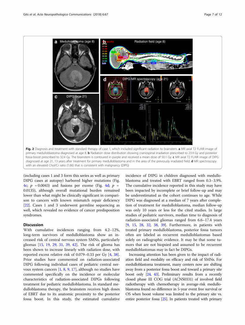
completion of therapy), she presented to her primarycare physician with a one-month history of difficultyswallowing and clumsiness of her right hand. MRI brainrevealed a new infiltrative mass with diffuse pontine T2hyperintensity, consistent radiographically with a DIPG(Fig. 2c). MR spectroscopy revealed markedly elevatedcholine to creatinine peak with depressed NAA peak,consistent with malignancy (Fig. 2d). She underwentre-irradiation and ultimately died of disease 17 monthsafter diagnosis. Histopathology at autopsy revealed adiffusively infiltrating glioma (Fig. 3b). Karyotyping ofher initial medulloblastoma previously had revealed iso-chromosome for the long arm of chromosome 17, a hall-mark feature of Group 4 medulloblastoma (Fig. 3c). Incontrast, copy number analysis of her DIPG revealedcopy number changes frequently seen in gliomas includ-ing homozygous loss of RB1, SETDB2, CDKN2A andCDKN2B with no abnormalities in chromosome 17,distinguishing it from the primary medulloblastoma(Fig. 3d-e). Similar imaging findings, radiation fieldsand MR spectroscopy images were obtained in cases2 and 3 (Additional file 1: Figures S2 and S3).
DIPG sequencingFor cases 1, 3, and 6, DIPG frozen tissue from autopsy(cases 1 and 6) and diagnosis (case 3) were sequenced andmutations were analyzed (Additional file 1: Table S3). Allthree tumors were wild-type for H3F3A and HIST1H3B.Mutational signature analysis of DIPGs showed mutationsconsistent with radiation-induced DNA damage (e.g., in-sertional event in TP53), as well as mutations in otheroncogenic drivers (e.g., PTEN, EGFR, and NRAS), suggest-ive of a distinct mutational process as compared with pri-mary DIPGs. Mutations identified in radiation-associatedDIPGs had molecular overlap with recurrent drivers ofadult GBM, using previously published datasets of adultGBM and primary DIPG (Fig. 4a) [7, 44]. Within a cohortsequenced using the same sequencing methodology (UMMI-ONCOSEQ) (n = 11), COSMIC mutational signatureanalysis demonstrated that radiation-associated DIPGshad the highest predicted somatic mutation counts andwere more likely to harbor Signature 24 than primaryDIPGs, which has not previously been connected to previ-ous malignancy or radiation exposure (Fig. 4b) [3]. Caseswith previous radiation in the MI-ONCOSEQ cohort
cb
a
Fig. 1 Radiation-associated DIPGs define a distinct molecular subtype with poor prognosis. a Immunohistochemistry performed for case 3showed wild-type status for histone H3 (H3K27M) with retention of H3K27me3, as well as diffuse GFAP expression, which was negative in primarymedulloblastoma (not shown). Positive and negative controls are shown in Additional file 1: Figure S1. Insufficient tissue was available for suchanalysis in cases 1 and 6; however, H3 wild-type status was demonstrated by tumor sequencing in these cases. All scale bars are 50 μm. b OSdata for primary DIPG in the IDIPGR (n = 428) was compared via Kaplan-Meier analysis to OS of radiation-associated DIPG cohort (n = 8). OS wassignificantly less for the radiation-associated DIPG group (p = 0.046). c OS data for primary DIPG patients with both genomic and OS informationavailable (n = 38), as categorized by histone mutational status and compared via Kaplan-Meier analysis to OS of radiation-associated DIPG cohort(n = 8). The radiation-associated DIPG cohort showed the shortest OS in comparison to the two subgroups of primary DIPG with significantlyshorter survival compared to H3.3 K27 M mutant (p = 0.038) and H3.1 K27 M mutant (p = 0.024) primary DIPGs
Gits et al. Acta Neuropathologica Communications (2018) 6:67 Page 6 of 12
(including cases 1 and 3 form this series as well as primaryDIPG cases at autopsy) harbored higher mutations (Fig.4c; p = 0.0043) and fusions per exome (Fig. 4d; p =0.0135), although overall mutational burden remainedlower than what might be clinically significant in compari-son to cancers with known mismatch repair deficiency[22]. Cases 1 and 3 underwent germline sequencing aswell, which revealed no evidence of cancer predispositionsyndromes.
DiscussionWith cumulative incidences ranging from 4.2–12%,long-term survivors of medulloblastoma show an in-creased risk of central nervous system SMNs, particularlygliomas [15, 19, 29, 31, 39, 42]. The risk of glioma hasbeen shown to increase linearly with radiation dose, withreported excess relative risk of 0.079–0.33 per Gy [4, 38].Prior studies have commented on radiation-associatedDIPG following individual cases of pediatric central ner-vous system cancers [1, 8, 9, 17], although no studies havecommented specifically on the incidence or molecularcharacteristics of radiation-associated DIPGs followingtreatment for pediatric medulloblastoma. In standard me-dulloblastoma therapy, the brainstem receives high dosesof EBRT due to its anatomic proximity to the posteriorfossa boost. In this study, the estimated cumulative
incidence of DIPG in children diagnosed with medullo-blastoma and treated with EBRT ranged from 0.3–3.9%.The cumulative incidence reported in this study may havebeen impacted by incomplete or brief follow-up and maybe underestimated as the cohort continues to age. WhileDIPG was diagnosed at a median of 7 years after comple-tion of treatment for medulloblastoma, median follow-upwas only 10 years or less for the cited studies. In largestudies of pediatric survivors, median time to diagnosis ofradiation-associated gliomas ranged from 6.6–17.4 years[9, 15, 28, 32, 38, 39]. Furthermore, in patients withtreated primary medulloblastoma, posterior fossa tumorsoften are labeled as recurrent medulloblastomas basedsolely on radiographic evidence. It may be that some tu-mors that are not biopsied and assumed to be recurrentmedulloblastomas may in fact be DIPGs.Increasing attention has been given to the impact of radi-
ation field and modality on efficacy and risk of SMNs. Formedulloblastoma treatment, many centers now are shiftingaway from a posterior fossa boost and toward a primary siteboost only [24, 43]. Preliminary results from a recentlyclosed phase III COG trial (ACNS0331) of involved fieldradiotherapy with chemotherapy in average-risk medullo-blastoma found no difference in 5-year event free survival orOS when boost volume was limited to the primary site vs.entire posterior fossa [25]. In patients treated with primary
Medulloblastoma (age 8) Radiation field (age 8)
DIPG (age 21)
DIPG MR spectroscopy (age 21)
a b
c
d
Fig. 2 Diagnosis and treatment with standard therapy of case 1, which included significant radiation to brainstem. a MR axial T2 FLAIR image ofprimary medulloblastoma diagnosed at age 8. b Radiation dose distribution showing craniospinal irradiation prescribed to 23.4 Gy and posteriorfossa boost prescribed to 32.4 Gy. The brainstem is contoured in purple and received a mean dose of 50.1 Gy. c MR axial T2 FLAIR image of DIPGdiagnosed at age 21, 13 years after treatment for primary medulloblastoma and in the area of the previously irradiated field. d MR spectroscopywith an elevated Chol/Cr ratio (1.66) that is consistent with malignancy (DIPG)
Gits et al. Acta Neuropathologica Communications (2018) 6:67 Page 7 of 12

site boost only, proton radiotherapy may decrease brainstemradiation exposure even further relative to photon therapy[11, 34]. In a multi-institutional cohort study and phase IIsingle center trial, there were no significant differences inrecurrence-free survival or OS between patients treated withphoton vs. proton radiotherapy, and in the phase II trial, noradiation-associated malignancies were reported within amedian follow-up time of 7 years [45]. While longerfollow-up is required to evaluate definitively its impact,smaller boost fields and proton radiotherapy show promisefor reducing the risk of SMNs without sacrificing efficacy oftreatment.Radiation-associated gliomas are molecularly distinct
from their primary counterparts. A previous report ofnon-brainstem radiation-associated pediatric GBM showedoverexpression of a number of genes involved in tumori-genesis as compared to primary pediatric GBM [13]. Add-itionally, prior studies suggest that tumors can bedifferentiated based on these molecular signatures and thatradiation-associated tumors may exhibit distinct patterns
[3, 5]. It has been observed that radiation-associated tumorsexhibit a significantly higher total number of mutations, aswell as balanced inversions, with both small deletions andinversions generating driver mutations [5].In this study, tumor exome sequencing of three
radiation-associated DIPGs demonstrated tumors to beH3-wildtype. This finding is significant in the context ofa recent large cohort of sequenced primary DIPGs, inwhich only 16.8% were found to be H3-wildtype [23].Notably, sequencing confirmed that the tumors were in-deed distinct from their primary malignancies and notlocal recurrences. Moreover, patients did not harborgermline mutations in known cancer predispositiongenes. Although alterations in two of the most fre-quently mutated genes in primary DIPG (H3F3A andACVR1) were not detected, the tumor mutations in thesequenced cases are established tumor drivers in adultGBM (e.g. PTEN, NRAS, and EGFR). Interestingly, case3 was found to have an EZH2 mutation in theradiation-associated DIPG, which has not been identified
d
e
c
Medulloblastoma (age 8) DIPG (age 21)
Copy Number Profile (DIPG)
Loss of Heterozygosity Plot (DIPG)
loss of RB1, SETDB2loss of CDKN2A, CDKN2B
gain of KIT, KDR, PDGFRA
Medulloblastoma
a b
Fig. 3 Histology and molecular results distinguish primary medulloblastoma from radiation-associated DIPG. a Resected medulloblastoma fromcase 1 showing characteristic classic-type features including sheets of cells with primitive hyperchromatic nuclei and scant cytoplasm. b DIPG atautopsy showing an infiltrating glial tumor with small angulated nuclei and abundant amphophilic cytoplasm. c Karyotype analysis of medulloblastomashows near-tetraploid clone with arrow indicating i(17q), most consistent with Group 4. d Copy number analysis of DIPG shows focal and structuralchanges distinct from primary tumor, including focal homozygous loss of RB1, SETDB2, CDKN2A and CDKN2B, focal 1 copy gain of KIT, KDR and PDGFRA,and activation mutations in NRAS and TP53. e Loss of heterozygosity plot showing regions on chromosomes 6 and 18 with copy-neutral loss ofheterozygosity events
Gits et al. Acta Neuropathologica Communications (2018) 6:67 Page 8 of 12

as a recurrent driver in DIPG, but has been established as apotential therapeutic target in pre-clinical DIPG models[27]. Mutational signature analysis of radiation-associatedDIPGs showed mutations consistent with radiation-inducedDNA damage (e.g., insertional event in TP53), at arate similar to cases observed in a recent report ofradiation-associated GBMs, in which two out of five se-quenced GBMs had mutations in TP53 [33]. Unlikeradiation-associated GBMs, however, PDGFRA played asmaller role in the cohort of radiation-associated DIPG. ThePDGFRA amplification in case 1 (Fig. 1d) was the isolatedalteration in the three tumors. While non-silent mutationsin PDGFRA were identified in all five radiation-associatedGBMs, none of radiation-associated DIPGs from this studyhad mutations in PDGFRA.Taken together, these results are suggestive of distinct mu-
tational processes compared with primary DIPGs: primaryDIPGs originate within a particular early developmentaltimespan that is amenable to transformation with H3F3Aand ACVR1 mutation [23], whereas radiation-associatedDIPGs appear to arise as a result of radiation-induced DNAdamage in established oncogenic drivers of primary adult
GBM. Future sequencing of additional cases may eluci-date patterns of distinct biology in radiation-associatedDIPG, which may have implications in terms of clinicalmanagement. These data suggest that patients withradiation-associated DIPG may benefit from futuretherapies targeted to the molecular features of adultGBM rather than primary DIPG.Significantly, the radiation-associated DIPG cohort
demonstrated a shorter OS relative to patients with pri-mary DIPG. The three molecularly sequenced cases ofradiation-associated DIPG cohort additionally are distin-guished as H3 wild-type designation, considered a posi-tive prognostic variable in primary DIPG relative toH3.3 mutant DIPG [10, 20]. Prior studies report a simi-lar decreased survival from radiation-induced HGG ascompared to primary HGG [13, 21]. In conjunction withthe molecular findings, these data suggest thatradiation-associated DIPGs form a distinct molecularsubgroup that has negative implications on survival. Thefindings in this work demonstrate the importance oftumor biopsy or resection at appearance of a secondcancer. Most cases had no or limited histological and
dc
ab
Fig. 4 Radiation-associated DIPGs are molecularly distinct from primary DIPGs. a Plot of recurrent mutations in previously published datasets(adult GBM [n = 500] [7]; primary DIPG [n = 55] [44]) demonstrates that the distribution of driving mutations in radiation-associated DIPG is moresimilar to recurrent alterations in adult GBM than primary DIPG. b Contributions of established COSMIC mutational signatures were determinedfor radiation-associated DIPG samples as compared to all other primary cases sequenced through same sequencing platform (MI-ONCOSEQ). c-dCases with previous radiation in this cohort (including case 1 and 3 and primary DIPG at autopsy) show higher mutations and fusions per exome(p = 0.0043 and p = 0.0135, respectively using Mann Whitney test)
Gits et al. Acta Neuropathologica Communications (2018) 6:67 Page 9 of 12

molecular diagnostic information, which was not clinicallyrelevant until recently. Now, this information is critical forprognostic information, current clinical management, andpotential future therapies [33].
ConclusionsIn conclusion, patients treated for pediatric medulloblas-toma are at increased risk for development ofradiation-associated DIPG, which may represent a dis-tinct molecular subtype with a worse prognosis relativeto other DIPGs. This risk highlights the importance ofradiation volume and modality in the treatment of chil-dren with medulloblastoma and provides a compellingargument for efforts to reduce exposure of the brain-stem. Additionally, the presented molecular data suggestthat patients with radiation-associated DIPG may benefitfrom future therapies targeted to the molecular featuresof adult GBM rather than primary DIPG.
Additional file
Additional file 1: Table S1. Treatment details for primarymedulloblastoma. Table S2. Multivariate analysis of overall survival forprimary and radiation-associated DIPGs. Table S3. Sequencing ofradiation-associated DIPGs. Figure S1. Immunohistochemical stainingfor H3K27 M of positive and negative control pediatric high-gradegliomas. Figure S2. Diagnosis and management of case 2, which includednon-standard treatment for medulloblastoma. Figure S3. Diagnosis andmanagement of case 3, which included standard therapy formedulloblastoma. (DOCX 5366 kb)
AcknowledgementsThe authors gratefully acknowledge all patients and their families who havesupported and contributed to the International DIPG Registry. Supportingfoundations include: The Cure Starts Now Foundation, Hope for CarolineFoundation, Julian Boivin Courage for Cures Foundation, Abbie’s Army,Michael Mosier Defeat DIPG Foundation, Reflections of Grace Foundation,The Cure Starts Now Australia, Brooke Healey Foundation, Soar With GraceFoundation, Jeffrey Thomas Hayden Foundation, Cure Brain CancerFoundation, The Jones Family Foundation, Musella Foundation, Pray, HopeBelieve Foundation, Smiles for Sophie Foundation, Benny’s World, LoveChloe Foundation, Aiden’s Avengers, A Cure from Caleb Society, TheOperation Grace White Foundation, Ryan’s Hope, Wayland Villars DIPGFoundation, American Childhood Cancer Organization, Juliana Rose DonnellyTrust, Sheila Jones & Friends, The Ellie Kavalieros DIPG Research Fund, VoicesAgainst Brain Cancer, The DIPG Collaborative.
FundingCK is supported by NIH/NINDS Grant K08-NS099427–01, Michigan MedicineDepartment of Pediatrics Gorman Scholar Award, The University of MichiganChad Carr Pediatric Brain Tumor Center, the Chad Tough Foundation andHyundai Hope on Wheels. NGG is supported by a Cancer Council of WesternAustralia fellowship.
Availability of data and materialsData is available through the International DIPG Registry or publishedliterature.
Authors’ contributionsAll authors have contributed meaningfully to the manuscript. All authors readand approved the final manuscript.
Ethics approval and consent to participateAll patients have consented to participate or case data were extracted fromliterature review.
Consent for publicationAll authors consent to publication of this work.
Competing interestsThe authors declare that they have no competing interests.
Publisher’s NoteSpringer Nature remains neutral with regard to jurisdictional claims inpublished maps and institutional affiliations.
Author details1Department of Pathology, Michigan Medicine, Ann Arbor, MI 48109, USA.2Department of Pediatrics, Division of Pediatric Hematology-Oncology;Michigan Medicine, Ann Arbor, MI 48109, USA. 3Department of Haematologyand Oncology, Princess Margaret Hospital for Children, Perth, WA 6840,Australia. 4Michigan Center for Translational Pathology, Michigan Medicine,Ann Arbor, MI 48109, USA. 5Ontario Institute for Cancer Research, MG5 0A3,Toronto, ON, Canada. 6Department of Radiation Oncology; MichiganMedicine, Ann Arbor, MI 48109, USA. 7Department of ComputationalMedicine and Bioinformatics; Michigan Medicine, Ann Arbor, MI 48109, USA.8Department of Biostatistics, University of Michigan School of Public Health,Ann Arbor, MI 48109, USA. 9Division of Radiology, Cincinnati Children’sHospital, Cincinnati, OH 45229, USA. 10Division of Oncology, CincinnatiChildren’s Hospital, Cincinnati, OH 45229, USA. 11Division of BiomedicalInformatics, Cincinnati Children’s Hospital, Cincinnati, OH 45229, USA.12Division of Pathology, Cincinnati Children’s Hospital, Cincinnati, OH 45229,USA. 13Department of Pediatrics, Division of Oncology, Seattle Children’sHospital, Seattle, WA 98105, USA. 14Department of Pediatrics, Division ofHaematology/Oncology, Hospital for Sick Children, Toronto, ON M5G 1X8,Canada. 15Department of Radiation Oncology, Massachusetts GeneralHospital, Boston, MA 02114, USA. 16Department of Pediatrics, Division ofNeurology; Michigan Medicine, Ann Arbor, MI 48109, USA. 17Department ofPediatrics, Division of Oncology, Cincinnati Children’s Hospital, Cincinnati, OH45229, USA. 18Kids Cancer Centre, Telethon Kids Institute, Subiaco, WA 6008,Australia. 19Division of Paediatrics, University of Western Australia, Crawley,WA 6009, Australia.
Received: 11 July 2018 Accepted: 12 July 2018
References1. Abboud SE, Wolansky LJ, Manjila SV, Lo SS, Arafah BM, Selman WR, Couce
ME, Rogers LR (2015) Histologically proven radiation-induced brainstemGlioma 93 months after external beam radiotherapy for pituitarymacroadenoma: radiation treatment dose and volume correlation. JNeuroimaging 25:674–676. https://doi.org/10.1111/jon.12181
2. Alexandrov LB, Jones PH, Wedge DC, Sale JE, Campbell PJ, Nik-Zainal S,Stratton MR (2015) Clock-like mutational processes in human somatic cells.Nat Genet 47:1402–1407. https://doi.org/10.1038/ng.3441
3. Alexandrov LB, Nik-Zainal S, Wedge DC, Aparicio SA, Behjati S, Biankin AV,Bignell GR, Bolli N, Borg A, Borresen-Dale AL et al (2013) Signatures ofmutational processes in human cancer. Nature 500:415–421. https://doi.org/10.1038/nature12477
4. Armstrong GT, Stovall M, Robison LL (2010) Long-term effects of radiationexposure among adult survivors of childhood cancer: results from thechildhood cancer survivor study. Radiat Res 174:840–850. https://doi.org/10.1667/RR1903.1
5. Behjati S, Gundem G, Wedge DC, Roberts ND, Tarpey PS, Cooke SL, Van LooP, Alexandrov LB, Ramakrishna M, Davies H, et al (2016) Mutationalsignatures of ionizing radiation in second malignancies. Nat Commun 7:12605 https://doi.org/10.1038/ncomms12605
6. Bender S, Tang Y, Lindroth AM, Hovestadt V, Jones DT, Kool M, Zapatka M,Northcott PA, Sturm D, Wang W et al (2013) Reduced H3K27me3 and DNAhypomethylation are major drivers of Gene Expr in K27M mutant pediatrichigh-grade gliomas. Cancer Cell 24:660–672. https://doi.org/10.1016/j.ccr.2013.10.006
Gits et al. Acta Neuropathologica Communications (2018) 6:67 Page 10 of 12

7. Brennan CW, Verhaak RG, McKenna A, Campos B, Noushmehr H, Salama SR,Zheng S, Chakravarty D, Sanborn JZ, Berman SH et al (2013) The somaticgenomic landscape of glioblastoma. Cell 155:462–477. https://doi.org/10.1016/j.cell.2013.09.034
8. Broniscer A, Baker JN, Baker SJ, Chi SN, Geyer JR, Morris EB, Gajjar A (2010)Prospective collection of tissue samples at autopsy in children with diffuseintrinsic pontine glioma. Cancer 116:4632–4637. https://doi.org/10.1002/cncr.25405
9. Broniscer A, Ke W, Fuller CE, Wu J, Gajjar A, Kun LE (2004) Secondneoplasms in pediatric patients with primary central nervous systemtumors: the St. Jude Children's Research Hospital experience. Cancer 100:2246–2252. https://doi.org/10.1002/cncr.20253
10. Castel D, Philippe C, Calmon R, Le Dret L, Truffaux N, Boddaert N, Pages M,Taylor KR, Saulnier P, Lacroix L et al (2015) Histone H3F3A and HIST1H3BK27M mutations define two subgroups of diffuse intrinsic pontine gliomaswith different prognosis and phenotypes. Acta Neuropathol 130:815–827.https://doi.org/10.1007/s00401-015-1478-0
11. Chung CS, Yock TI, Nelson K, Xu Y, Keating NL, Tarbell NJ (2013) Incidenceof second malignancies among patients treated with proton versus photonradiation. Int J Radiat Oncol Biol Phys 87:46–52. https://doi.org/10.1016/j.ijrobp.2013.04.030
12. Cooney T, Lane A, Bartels U, Bouffet E, Goldman S, Leary SES, Foreman NK,Packer RJ, Broniscer A, Minturn JE et al (2017) Contemporary survivalendpoints: an international diffuse intrinsic Pontine Glioma registry study.Neuro Oncol 19:1279–1280. https://doi.org/10.1093/neuonc/nox107
13. Donson AM, Erwin NS, Kleinschmidt-DeMasters BK, Madden JR, Addo-YoboSO, Foreman NK (2007) Unique molecular characteristics of radiation-induced glioblastoma. J Neuropathol Exp Neurol 66:740–749. https://doi.org/10.1097/nen.0b013e3181257190
14. Fossati P, Ricardi U, Orecchia R (2009) Pediatric medulloblastoma: toxicity ofcurrent treatment and potential role of protontherapy. Cancer Treat Rev 35:79–96. https://doi.org/10.1016/j.ctrv.2008.09.002
15. Friedman DL, Whitton J, Leisenring W, Mertens AC, Hammond S, Stovall M,Donaldson SS, Meadows AT, Robison LL, Neglia JP (2010) Subsequent neoplasmsin 5-year survivors of childhood cancer: the childhood Cancer survivor study. JNatl Cancer Inst 102:1083–1095. https://doi.org/10.1093/jnci/djq238
16. Fu W, O'Connor TD, Jun G, Kang HM, Abecasis G, Leal SM, Gabriel S, RiederMJ, Altshuler D, Shendure J et al (2013) Analysis of 6,515 exomes reveals therecent origin of most human protein-coding variants. Nature 493:216–220.https://doi.org/10.1038/nature11690
17. Fuller GN, Kaba SE, Ginsberg LE, McCutcheon IE, Langford LA (1997) Latesequelae of treated pleomorphic xanthoastrocytoma: malignant brain stemastrocytoma occurring 15 years after radiation therapy. J Neuro-Oncol 32:57–61
18. Genomes Project C, Abecasis GR, Altshuler D, Auton A, Brooks LD, DurbinRM, Gibbs RA, Hurles ME, McVean GA (2010) A map of human genomevariation from population-scale sequencing. Nature 467:1061–1073. https://doi.org/10.1038/nature09534
19. Goldstein AM, Yuen J, Tucker MA (1997) Second cancers after medulloblastoma:population-based results from the United States and Sweden. Cancer CausesControl 8:865–871
20. Hoffman LM, Veldhuijzen van Zanten SEM, Colditz N, Baugh J, Chaney B,Hoffmann M, Lane A, Fuller C, Miles L, Hawkins C et al (2018) Clinical,radiologic, pathologic, and molecular characteristics of long-term survivorsof diffuse intrinsic Pontine Glioma (DIPG): a collaborative report from theinternational and European Society for Pediatric Oncology DIPG registries. JClin Oncol 36:1963–1972. https://doi.org/10.1200/JCO.2017.75.9308
21. Kleinschmidt-Demasters BK, Kang JS, Lillehei KO (2006) The burden ofradiation-induced central nervous system tumors: a single institution sexperience. J Neuropathol Exp Neurol 65:204–216. https://doi.org/10.1097/01.jnen.0000205146.62081.29
22. Le DT, Durham JN, Smith KN, Wang H, Bartlett BR, Aulakh LK, Lu S,Kemberling H, Wilt C, Luber BS et al (2017) Mismatch repair deficiencypredicts response of solid tumors to PD-1 blockade. Sci 357:409–413.https://doi.org/10.1126/science.aan6733
23. Mackay A, Burford A, Carvalho D, Izquierdo E, Fazal-Salom J, Taylor KR,Bjerke L, Clarke M, Vinci M, Nandhabalan M et al (2017) IntegratedMolecular Meta-Analysis of 1,000 Pediatric High-Grade and Diffuse IntrinsicPontine Glioma. Cancer Cell 32:520–537. https://doi.org/10.1016/j.ccell.2017.08.017 e525
24. Merchant TE, Kun LE, Krasin MJ, Wallace D, Chintagumpala MM, Woo SY,Ashley DM, Sexton M, Kellie SJ, Ahern V et al (2008) Multi-institution
prospective trial of reduced-dose craniospinal irradiation (23.4 Gy) followedby conformal posterior fossa (36 Gy) and primary site irradiation (55.8 Gy)and dose-intensive chemotherapy for average-risk medulloblastoma. Int JRadiat Oncol Biol Phys 70:782–787. https://doi.org/10.1016/j.ijrobp.2007.07.2342
25. Michalski JM, Janss A, Vezina G, Gajjar A, Pollack I, Merchant TE, FitzGerald TJ,Booth T, Tarbell NJ, Li Y et al (2016) Results of COG ACNS0331: a phase III trialof involved-field radiotherapy (IFRT) and low dose Craniospinal irradiation (LD-CSI) with chemotherapy in average-risk Medulloblastoma: a report from theChildren's oncology group. Int J Radiat Oncol Biol Phys 96:937–938
26. Mody RJ, Wu YM, Lonigro RJ, Cao X, Roychowdhury S, Vats P, Frank KM,Prensner JR, Asangani I, Palanisamy N et al (2015) Integrative ClinicalSequencing in the Management of Refractory or Relapsed Cancer in Youth.Jama 314:913–925. https://doi.org/10.1001/jama.2015.10080
27. Mohammad F, Weissmann S, Leblanc B, Pandey DP, Hojfeldt JW, Comet I,Zheng C, Johansen JV, Rapin N, Porse BT et al (2017) EZH2 is a potentialtherapeutic target for H3K27M-mutant pediatric gliomas. Nat Med 23:483–492. https://doi.org/10.1038/nm.4293
28. Neglia JP, Robison LL, Stovall M, Liu Y, Packer RJ, Hammond S, Yasui Y,Kasper CE, Mertens AC, Donaldson SS et al (2006) New primary neoplasmsof the central nervous system in survivors of childhood cancer: a reportfrom the childhood Cancer survivor study. J Natl Cancer Inst 98:1528–1537.https://doi.org/10.1093/jnci/djj411
29. Ning MS, Perkins SM, Dewees T, Shinohara ET (2015) Evidence of highmortality in long term survivors of childhood medulloblastoma. J Neuro-Oncol 122:321–327. https://doi.org/10.1007/s11060-014-1712-y
30. Packer RJ, Goldwein J, Nicholson HS, Vezina LG, Allen JC, Ris MD, MuraszkoK, Rorke LB, Wara WM, Cohen BH et al (1999) Treatment of children withmedulloblastomas with reduced-dose craniospinal radiation therapy andadjuvant chemotherapy: A Children's Cancer Group Study. J Clin Oncol 17:2127–2136. https://doi.org/10.1200/JCO.1999.17.7.2127
31. Packer RJ, Zhou T, Holmes E, Vezina G, Gajjar A (2013) Survival andsecondary tumors in children with medulloblastoma receiving radiotherapyand adjuvant chemotherapy: results of Children's oncology group trialA9961. Neuro-Oncology 15:97–103. https://doi.org/10.1093/neuonc/nos267
32. Pettorini BL, Park YS, Caldarelli M, Massimi L, Tamburrini G, Di Rocco C(2008) Radiation-induced brain tumours after central nervous systemirradiation in childhood: a review. Childs Nerv Syst 24:793–805. https://doi.org/10.1007/s00381-008-0631-7
33. Phi JH, Park AK, Lee S, Choi SA, Baek IP, Kim P, Kim EH, Park HC, Kim BC,Bhak J et al (2018) Genomic analysis reveals secondary glioblastoma afterradiotherapy in a subset of recurrent medulloblastomas. ActaNeuropathol 35(6):939–953. https://doi.org/10.1007/s00401-018-1845-8
34. Rechner LA, Howell RM, Zhang R, Newhauser WD (2012) Impact of marginsize on the predicted risk of radiogenic second cancers following protonarc therapy and volumetric modulated arc therapy for prostate cancer. PhysMed Biol 57:N469–N479. https://doi.org/10.1088/0031-9155/57/23/N469
35. Rosenthal R, McGranahan N, Herrero J, Taylor BS, Swanton C (2016)DeconstructSigs: delineating mutational processes in single tumorsdistinguishes DNA repair deficiencies and patterns of carcinoma evolution.Genome Biol 17:31. https://doi.org/10.1186/s13059-016-0893-4
36. Sabel M, Fleischhack G, Tippelt S, Gustafsson G, Doz F, Kortmann R,Massimino M, Navajas A, von Hoff K, Rutkowski S et al (2016) Relapsepatterns and outcome after relapse in standard risk medulloblastoma: areport from the HIT-SIOP-PNET4 study. J Neurooncol 129:515–524. https://doi.org/10.1007/s11060-016-2202-1
37. Stromberg U (1994) A distinction between cumulative incidence and risk asmeasures of disease occurrence. Int J Epidemiol 23:1104–1105
38. Taylor AJ, Little MP, Winter DL, Sugden E, Ellison DW, Stiller CA, Stovall M,Frobisher C, Lancashire ER, Reulen RC et al (2010) Population-based risks ofCNS tumors in survivors of childhood cancer: the British Childhood CancerSurvivor Study. J Clin Oncol 28:5287–5293. https://doi.org/10.1200/JCO.2009.27.0090
39. Tsui K, Gajjar A, Li C, Srivastava D, Broniscer A, Wetmore C, Kun LE, MerchantTE, Ellison DW, Orr BA et al (2015) Subsequent neoplasms in survivors ofchildhood central nervous system tumors: risk after modern multimodaltherapy. Neuro Oncol 17:448–456. https://doi.org/10.1093/neuonc/nou279
40. Venneti S, Garimella MT, Sullivan LM, Martinez D, Huse JT, Heguy A, Santi M,Thompson CB, Judkins AR (2013) Evaluation of histone 3 lysine 27trimethylation (H3K27me3) and enhancer of zest 2 (EZH2) in pediatric glialand glioneuronal tumors shows decreased H3K27me3 in H3F3A K27M mutantglioblastomas. Brain Pathol 23:558–564. https://doi.org/10.1111/bpa.12042
Gits et al. Acta Neuropathologica Communications (2018) 6:67 Page 11 of 12

41. Venneti S, Santi M, Felicella MM, Yarilin D, Phillips JJ, Sullivan LM, MartinezD, Perry A, Lewis PW, Thompson CB et al (2014) A sensitive and specifichistopathologic prognostic marker for H3F3A K27M mutant pediatricglioblastomas. Acta Neuropathol 128:743–753. https://doi.org/10.1007/s00401-014-1338-3
42. von Hoff K, Hinkes B, Gerber NU, Deinlein F, Mittler U, Urban C, Benesch M,Warmuth-Metz M, Soerensen N, Zwiener I et al (2009) Long-term outcomeand clinical prognostic factors in children with medulloblastoma treated inthe prospective randomised multicentre trial HIT'91. Eur J Cancer 45:1209–1217. https://doi.org/10.1016/j.ejca.2009.01.015
43. Wolden SL, Dunkel IJ, Souweidane MM, Happersett L, Khakoo Y, Schupak K,Lyden D, Leibel SA (2003) Patterns of failure using a conformal radiationtherapy tumor bed boost for medulloblastoma. J Clin Oncol 21:3079–3083.https://doi.org/10.1200/JCO.2003.11.140
44. Wu G, Diaz AK, Paugh BS, Rankin SL, Ju B, Li Y, Zhu X, Qu C, Chen X, ZhangJ et al (2014) The genomic landscape of diffuse intrinsic pontine gliomaand pediatric non-brainstem high-grade glioma. Nat Genet 46:444–450.https://doi.org/10.1038/ng.2938
45. Yock TI, Yeap BY, Ebb DH, Weyman E, Eaton BR, Sherry NA, Jones RM,MacDonald SM, Pulsifer MB, Lavally B et al (2016) Long-term toxic effects ofproton radiotherapy for paediatric medulloblastoma: a phase 2 single-armstudy. Lancet Oncol 17:287–298. https://doi.org/10.1016/S1470-2045(15)00167-9
46. You SH, Lyu CJ, Kim DS, Suh CO (2013) Second primary brain tumorsfollowing cranial irradiation for pediatric solid brain tumors. Childs Nerv Syst29:1865–1870. https://doi.org/10.1007/s00381-013-2098-4
Gits et al. Acta Neuropathologica Communications (2018) 6:67 Page 12 of 12


![Medulloblastoma: [Print] - eMedicine Neurology · accounts for approximately 7-8% of all intracranial tumors and 30% of ... Incidence of medulloblastoma is 1.5-2 cases per ... Medulloblastoma:](https://img.dokumen.tips/doc/110x75/5b7fc2317f8b9ae6088caa0e/medulloblastoma-print-emedicine-accounts-for-approximately-7-8-of-all.jpg)

![Medulloblastoma: [Print] - eMedicine Neurology · emedicine.medscape.com eMedicine Specialties > Neurology > Pediatric Neurology Medulloblastoma George I Jallo, MD, Associate Professor](https://img.dokumen.tips/doc/110x75/5d472c3c88c993527c8b60e5/medulloblastoma-print-emedicine-neurology-emedicinemedscapecom-emedicine.jpg)


