Embed Size (px)
Citation preview

Medical thoracoscopy: Rigid thoracoscopy or flexi-rigidpleuroscopy?Yap, K., Phillips, M. J., & Lee, G. (2014). Medical thoracoscopy: Rigid thoracoscopy or flexi-rigidpleuroscopy? Current Opinion in Pulmonary Medicine, 20(4), 358-365. DOI:10.1097/MCP.0000000000000059
Published in:Current Opinion in Pulmonary Medicine
DOI:10.1097/MCP.0000000000000059
Document VersionPeer reviewed version
Link to publication in the UWA Research Repository
Rights statementThis is a non-final version of an article published in final form in Yap, K., Phillips, M. J., & Lee, G. (2014).Medical thoracoscopy: Rigid thoracoscopy or flexi-rigid pleuroscopy?. Current Opinion in PulmonaryMedicine, 20(4), 358-365.
General rightsCopyright owners retain the copyright for their material stored in the UWA Research Repository. The University grants no end-userrights beyond those which are provided by the Australian Copyright Act 1968. Users may make use of the material in the Repositoryproviding due attribution is given and the use is in accordance with the Copyright Act 1968.
Take down policyIf you believe this document infringes copyright, raise a complaint by contacting [email protected]. The document will beimmediately withdrawn from public access while the complaint is being investigated.
Download date: 18. Jul. 2018

1
Medical Thoracoscopy:
Rigid thoracoscopy or flexi-rigid pleuroscopy?
Authors:
Kim Hoong YAP 1,2
MBBS MRCP FAMS FCCP [email protected]
Martin J PHILLIPS 1
MBBS FRACP [email protected]
Y C Gary LEE 1,3
MBChB PhD FRACP FRCP FCCP [email protected]
Affiliations: 1
Department of Respiratory Medicine, Sir Charles Gairdner Hospital, Perth, Australia 2
Department of Respiratory & Critical Care Medicine, Tan Tock Seng Hospital,
Singapore 3
Centre for Asthma, Allergy & Respiratory Research, School of Medicine &
Pharmacology, University of Western Australia, Perth, Australia
Author of correspondence:
Prof. Y C Gary LEE, Harry Perkins Research Building, Rm 533, QE II Medical
Centre, Perth, WA 6009, Australia. Tel: +61 8 61510892
Keywords: pleuroscopy; thoracoscopy; pleural; effusion; pleurodesis
Word Count: 2518

2
ABSTRACT
PURPOSE OF REVIEW:
In managing pleural diseases, medical thoracoscopy is often performed as a diagnostic
and/or therapeutic procedure particularly in undiagnosed pleural effusions. Flexi-rigid
pleuroscopes are now widely available as an alternative to conventional rigid
thoracoscopes. There is ongoing debate on which is the better instrument. This review
analyses the current literature that compared rigid and flexi-rigid approaches, and
outline medical advances that may influence the future role of thoracoscopy.
RECENT FINDINGS:
Both rigid and flexi-rigid thoracoscopies are safe. Although biopsies are smaller with
flexi-rigid biopsy forceps, two small randomized trials reported similar diagnostic
yield using either instrument. No studies have specifically examined patient comfort
or the outcome of talc poudrage using the two devices. New techniques (e.g. IT knife
and cyrobiopsy) have been used as adjuncts with flexi-rigid pleuroscopy to overcome
the difficulties in sampling thickened pleura.
SUMMARY:
The rigid and flex-rigid instruments have different merits and limitations. Both
approaches provide comparable diagnostic yields in the overall patient population
undergoing diagnostic thoracoscopy, though their performances specifically in
patients with fibrotic pleural thickening have not been examined. Operators using
flexi-rigid approach should have alternative strategies for sampling thickened pleura.
Advances in cytopathology and imaging-guided biopsy will likely reduce the need of
medical thoracoscopy in the future.
3
INTRODUCTION
Thoracoscopy employs an optical system to examine the pleural cavity and perform
diagnostic and therapeutic procedures[1]. It has traditionally been divided into
surgical thoracoscopy, better known as Video Assisted Thoracic Surgery (VATS), and
medical thoracoscopy (pleuroscopy).
Historically, both VATS and medical thoracoscopy were performed using rigid
instruments. VATS allow surgeons to replace open thoracotomy in most pulmonary
(e.g. lobectomy) and pleural surgeries (e.g. pleural biopsy and pleurodesis). VATS is
typically performed under general anesthesia and single-lung ventilation. Some
centers however, have performed VATS wedge resection under regional anesthesia
[2, 3]. Pleuroscopy/medical thoracoscopy is usually performed by pulmonologists
under conscious sedation, most commonly in the work-up of undiagnosed pleural
effusions, through visual inspection and biopsy of parietal pleura lesions.
With the introduction of the flexi-rigid pleuroscope in the late 1990s, proceduralists
performing thoracoscopy have the option of using either rigid or flexi-rigid
instruments. Opinions have been polarized as to which is the better device but
comparative studies to address this question are limited. Many experienced
thoracoscopists remain in favor of the rigid scope and its ability to provide sizeable
biopsies whereas advocates of flexi-rigid pleuroscopy embrace the flexibility of the
instrument and its ease of use. It is a common dilemma for pulmonologists setting up
a new pleural service to decide which scope to invest in.

4
This article aims to summarize the current literature and highlight the advantages and
limitations of rigid vs. flexi-rigid approaches and emerging technologies that may
alter the role of pleuroscopy in the foreseeable future.

5
RIGID VS FLEXI-RIGID PLEUROSCOPY: THE INSTRUMENTS
A rigid thoracoscopy set includes a telescope, light source, trocar and forceps. The
conventional stainless steel rigid telescope is 27-31cm in length with a diameter of 7-
12mm; the larger ones (10-12mm) often favored by surgeons. Rigid telescopes have
different angles of vision permitting straight-on (0) or oblique (30 or 50) viewing.
Trocars are made from single-use disposable plastic or stainless steel with a variable
diameter from 5-13mm.[4, 5]
The autoclavable flexi-rigid (semi-rigid) pleuroscope (Olympus LTF 160 or 240) has
a 22cm proximal rigid shaft and 5cm flexible distal tip with an outer diameter of
7mm. The flexible tip allows 2-way angulations (160 up and 130 down). The handle
of the flexi-rigid pleuroscope is similar to that of a flexible bronchoscope complete
with a 2.8mm working channel, lever and suction port (Figure 1). The scope utilizes a
custom-made plastic trocar of 8mm diameter (Figure 2).[6, 7]
The key difference between the two instruments is the flexibility to navigate various
parts of the pleural cavity. The rigid thoracoscope has to travel in a straight line and
has limited maneuverability when examining the posterior and mediastinal aspects of
the thoracic cavity, particularly if the lung remains partially or fully inflated. The
operator inevitably has to angle the scope by levering it against the underlying rib.
The pressure and angling of the rigid instrument over the periosteum is believed to
cause pain. The flexi-rigid pleuroscope, on the other hand, provides more flexibility
[6, 8] and allows the operator to negotiate around a non-deflated lung or dense
adhesions. In very loculated effusions, the ability to retro-flex the pleuroscope to

6
biopsy the parietal pleura adjacent to the insertion site is advantageous (Figure 3).
This is not feasible with rigid thoracoscopy.
For visualization and illumination, the flexi-rigid pleuroscope can be connected to
existing endoscopic processors (Olympus CV-160, CLV-U40) and light sources (CV-
240, EVIS-100 or 140, EVIS EXERA-145 or 160) while the rigid thoracoscope
requires a separate cold light source (xenon) with a camera attached to the eye-piece
of the telescope.[6, 7] The image quality is significantly better with the flexi-rigid
pleuroscope.[9*]
The ‘trade-off’ and key disadvantage of the flexi-rigid scope is the small working
channel which can limit adequate biopsies. The cusps diameter of the flexible biopsy
forceps (FB-55CR-1) used with the flexi-rigid scope is 2.4mm, considerably smaller
than that of the optical rigid biopsy forceps (5mm) used with rigid thoracoscopes
(Figure 4). The flexible forceps also lack the mechanical strength in obtaining
specimens from tough fibrous pleura. Therefore, the sturdier rigid biopsy forceps,
usually used during rigid thoracoscopy, often facilitate bigger and deeper biopsies and
are more efficient in breaking down adhesions.
This difference is probably of less clinical importance if the patient has nodular
pleural abnormalities (often seen with metastatic carcinomas) (Figure 5) which are
easy to capture even with flexible biopsy forceps but is a significant limitation in
patients with densely thickened pleura[8] (Figure 6). The latter can be seen with
patients with mesothelioma (especially the sarcomatoid subtype) and fibrothorax from
any chronic pleuritis. This is noteworthy as mesothelioma is consistently the most

7
common cause of false negative biopsies in patient with pleural malignancies
undergoing medical thoracoscopy in published series even when rigid scopes were
used.[10-12]

8
RIGID VS FLEXI-RIGID PLEUROSCOPY: DIAGNOSTIC YIELD
International guidelines suggest thoracoscopy be considered for the 25% of exudative
pleural effusions that remain undiagnosed after thoracentesis and/or closed pleural
biopsy.[13, 14] In tuberculous pleuritis, the combined yield of histology and culture
for rigid thoracoscopy was nearly 100%.[15, 16]
In malignant pleural diseases, rigid thoracoscopy achieved a high diagnostic yield of
95% in one study.[16] However, in three other series of medical thoracoscopy,
patients with a thoracoscopic biopsy result of ‘non-specific pleuritis’ had a 10-15%
chance of having an underlying pleural malignancy; most commonly
mesothelioma.[10-12]
Published data on flexi-rigid pleuroscopy remain relatively limited. Most studies were
small and did not necessarily contain the necessary variety of pleural malignancies
(e.g. mesothelioma) and the range of diagnostic difficulties.
A systematic review by Mohan et al.[17] included five studies (154 patients) on flexi-
rigid pleuroscopy in the diagnosis of undiagnosed pleural effusions and showed a
pooled sensitivity of 97%, specificity of 100%, a positive likelihood ratio (PLR) of
5.47 and negative likelihood ratio (NLR) 0.08. These studies were also reported in a
more recent meta-analysis by Agarwal et al.[18**] that included 17 studies (755
patients), nine of which prospective and only one was a randomized trial. Flexi-rigid
pleuroscopy showed a good sensitivity (91%) and specificity (100%) in diagnosing
exudative pleural effusions. The PLR and NLR were 4.92 and 0.08 respectively.

9
Although larger specimens are preferred, two randomized trials and one prospective
comparative study showed no significant difference in diagnostic yield between rigid
and flexi-rigid pleuroscopy. Khan et al.[19*] studied 66 patients (42 with malignancy
and 2 with tuberculosis) from two centers in a non-randomized study, and reported
similar diagnostic yields between the flexi-rigid and rigid thoracoscopy (92.3% vs.
96.3%).
Rozman et al.[20**] published the first randomized study with 84 patients comparing
the diagnostic adequacy of biopsy specimens obtained at rigid and flexi-rigid
pleuroscopy. They found similar diagnostic accuracy with rigid (100%) and flexi-
rigid instruments (97.6%) even though specimens obtained through the rigid forceps
were considerably larger (24.7 vs. 11.7mm2). The negative predictive values for rigid
and flexi-rigid biopsies were 100% and 92.3% respectively. In this cohort, 60% of
patients had malignant pleural disease of which two-thirds were mesothelioma.
In another study, Dhooria et al.[9*] randomized 90 patients to undergo rigid or flexi-
rigid pleuroscopy. The diagnostic yield for rigid thoracoscopy was noted to be
superior to flexi-rigid pleuroscopy on an intention-to-treat analysis (97.8% vs. 73.3%)
but was similar (100% vs. 94.3%) after excluding patients in whom pleuroscopy were
not feasible due to extensive adhesions. One major limitation in this study was patient
selection. Seven of the 45 patients from the flexi-rigid thoracoscopy arm crossed over
to the rigid thoracoscopy arm because of the lack of pleural space and only a quarter
of the whole cohort had prior CT-chest or thoracic ultrasound. Unlike the previous
studies, malignancy was the final etiology in only a third of the patients while a
quarter was due to tuberculosis.

10
These studies have obvious limitations but nonetheless there has not been clear
evidence to suggest that smaller biopsies result in inferior diagnostic accuracy. The
size of biopsies from the flexi-rigid scope would be comparable to those from
standard bronchoscopes, which is usually adequate for endobronchial tissue sampling.

11
ADDITIONAL TECHNIQUES TO AID FLEXI-RIGID PLEUROSCOPY
Procuring adequate samples from thickened pleura remain the most important
limitation of flexi-rigid pleuroscopy. Patients with mesothelioma or benign
fibrothorax (e.g. benign asbestos pleural disease, TB pleural fibrosis etc) are often
challenges for users of flexi-rigid pleuroscopes. Cytology has lower yield with
mesothelioma and current guidelines[21] favor histological specimens over pleural
fluid cytology in diagnosing mesothelioma. Therefore, obtaining representative
biopsy samples is crucial in these patients.
To overcome the limitation of small biopsies by flexi-rigid pleuroscope, different
strategies have been explored. Taking repeated “bites” from the same site with the
flexible forceps to obtain tissue of sufficient depth[7] and peeling away the pleura and
removing it together with the pleuroscope through the trocar[20**] are tedious and
time-consuming. Alternatively, swapping flexi-rigid pleuroscopy over to rigid
thoracoscopy during the procedure is an option. However, not all units are equipped
with both types of instruments.
A simpler alternative is to insert a second entry port for the rigid optical biopsy
forceps. The flexi-rigid scope then provides the direct vision to guide biopsies with
the rigid forceps[5]. This method combines the better optics of the flexi-rigid
pleuroscope with the larger biopsies from using rigid forceps.
Several accessories have been designed to improve the biopsy of thickened pleura to
be used via the working channel of the flexi-rigid pleuroscope, without the need of
creating a second entry port. The insulated-tip (IT) knife consists of a conventional

12
diathermic knife with a ceramic ball at the tip to limit the depth of the cut. Sasada et
al.[22] showed, in a study of 20 patients, that the IT knife allowed full-thickness
parietal pleural biopsy with a higher diagnostic yield (85%) when compared to the
standard flexible forceps (60%).
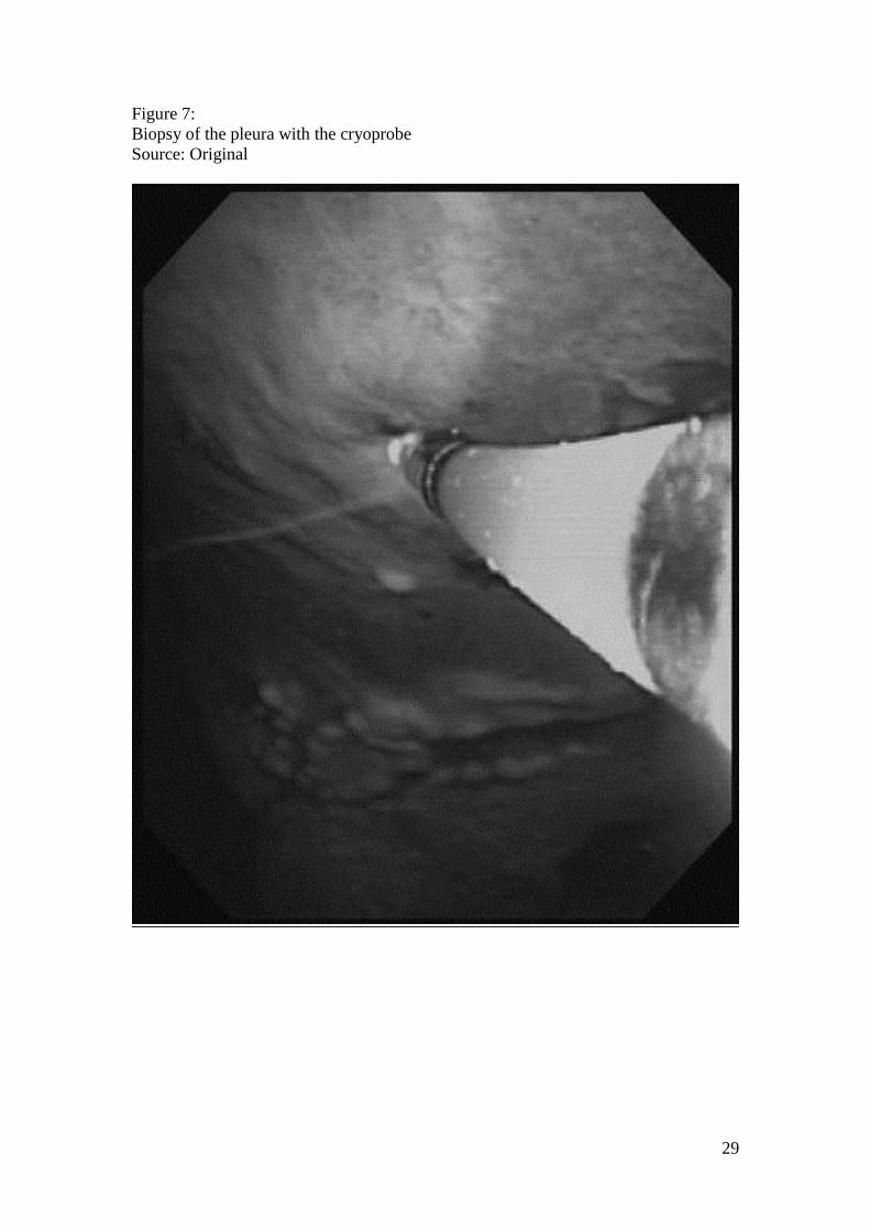
Biopsy via a cryoprobe is another tool being tested. Since its first use in 1968,
cryosurgical technique has mainly been employed in management of obstructive
endobronchial tumors. The equipment consists of a console, cryogen and cryoprobe.
The Joule-Thomson effect states that a compressed gas released at a high flow rapidly
expands and creates a very low temperature.[23] The cryoprobe is compatible with
the working channel of the flexi-rigid pleuroscope and pressed against the targeted
pleural lesion (Figure 7). The tissue is frozen under direct vision, detached with a tug
and removed together with scope. Anecdotally, cryobiopsy can help to obtain larger
size pleural samples even in thickened pleura.
These tools, and others in development, can potentially allow adequate sampling of
thickened pleura, thus offset the key limitation of flexi-rigid pleuroscopy.

13
RIGID VS FLEXI-RIGID PLEUROSCOPY: SAFETY
Medical thoracoscopy, either by rigid or flexi-rigid instruments, is a safe procedure in
experienced hands. The combined mortality rate from 47 studies was 0.34% (95% CI
0.19-0.54%)[14]. In the three recent comparative trials, no procedure-related death
was reported.[9*, 19*, 20**] The pooled rate of major adverse events was 1.8% (95%
CI 1.4-2.2%).[14] These include empyema, severe bleeding, port site metastasis,
persistent air leak and pneumonia. Rozman et al.[20**] found that one (2.6%) patient
developed severe bleeding after pleural biopsy during rigid thoracoscopy while
another (2.4%) developed empyema one-week after flexi-rigid pleuroscopy with
pleurodesis. Both patients recovered with treatment.
Dhooria et al.[9*] found no case of significant bleeding but three cases (3/47, 6.4%)
of empyema or persistent air leak from the rigid thoracoscopy arm and one case (1/35,
2.9%) from the flexi-rigid pleuroscopy arm. They attributed the higher rate of
empyema and persistent air leak to extensive adhesiolysis for complicated
parapneumonic effusions. The pooled incidence of minor complications was 7.3%
(95%CI 6.3-8.4%) including pyrexia, subcutaneous emphysema, skin infection and
minor hemorrhage.[14]
In terms of peri-procedural pain, larger trocars used during rigid thoracoscopy can
cause greater discomfort. The requirement for sedative and analgesics was higher with
rigid thoracoscopy in the study by Dhooria et al.[9*]

14
RIGID VS FLEXI-RIGID PLEUROSCOPY FOR OTHER INDICATIONS
Aside from diagnostic purposes, medical thoracoscopy is often used to perform
thoracoscopic talc poudrage (insufflations of talc as a dry powder) pleurodesis in
patients with recurrent effusions. Noppen et al.[24] also showed that thoracoscopic
talc poudrage was efficacious for recurrent spontaneous pneumothoraces.
No data exist comparing the efficacy of rigid vs. flexi-rigid pleuroscopy in
pleurodesis outcome. It is unlikely that the choice of pleuroscope will influence the
outcome of talc poudrage as the insufflated powder can distribute itself fairly evenly
within the pleural surface regardless of the scopes used. Indeed, no significant
benefits in pleurodesis outcome have been shown with talc delivered as poudrage or
at the bedside as slurry via a chest tube. In a randomized trial of 501 patients with
malignant pleural effusion, successful pleurodesis after 30-days by talc poudrage was
similar to talc slurry (78% vs 71%).[25] Two other randomized trials have also shown
no advantage of talc poudrage over talc slurry.[26, 27] Thoracoscopic talc poudrage
had no benefits over chest tube pleurodesis using iodine in another small randomized
trial.[28] Hence, the outcome difference between talc delivered by rigid or flexi-rigid
routes is likely to be negligible.
Ravaglia et al.[29] showed that medical thoracoscopy was successful in treating
patients with multiloculated empyema (91.7%) but less so in organized ones (50%).
The rigid thoracoscope with its optical forceps are more suited for removing
adhesions. However, the advent of intrapleural therapy with tissue plasminogen
activator and deoxyribonuclease has significantly changed the management of pleural
infection. This combination therapy cured 96% of patients without requiring surgery

15
in a randomized trial.[30] As this therapy is increasingly adopted worldwide, the role
of medical thoracoscopy in empyema is likely to become obsolete.
16
FUTURE DIRECTIONS
Thoracoscopy has played a significant role in the work-up of pleural effusions in the
past 100 years since its first introduction. However, the need for pleuroscopy is likely
to reduce as other less invasive alternatives advance (see our other review [31]). For
example, modern cytological assessment (with better immunohistochemical and
molecular markers) has high sensitivity for most metastatic carcinomas and even for
mesothelioma, reducing the need for tissue biopsy. In a recent study of 815
mesothelioma patients, pleural fluid cytology was positive in 68% and verified as
accurate against post-mortem findings.[32*]
Imaging-guided biopsies are attractive alternatives. Ultrasound-guided bedside pleural
biopsy has a high yield in suitable patients[33], and is growing in popularity. Maskell
et al.[34] showed that CT guided pleural biopsy was useful in over 80% of patients
presented with pleural thickening. Although not formally proven, the sensitivity of
imaging-guided biopsy can be further improved with fluorodeoxyglucose positron
emission tomography (FDG-PET), especially in patients with diffuse pleural
thickening. Targeting FDG-avid areas with imaging-guided biopsy is useful in
selected cases (Figure 8).
Conversely, the usefulness of thoracoscopy as a diagnostic tool has likely reached its
peak; attempts to improve the diagnostic yield with thoracoscopy have been
disappointing. Autofluorescence[35] and narrow band imaging[36, 37] have been
evaluated as an adjunct for diagnostic thoracoscopy but have failed to show
significant additional advantages.

17
CONCLUSION
Rigid thoracoscopy and flexi-rigid pleuroscopy instruments present different
advantages and disadvantages. Recent studies have shown that both instruments
provide high diagnostic yields. The studies to date however are small and it is perhaps
too simplistic to believe that one technique is superior to the other. The ideal pleural
service should be equipped with both to tailor for individual patients’ pleural
conditions. Operators of flexi-rigid pleuroscopy must have alternative techniques
available for patients with thickened pleura to ensure adequate samples are obtained.
The advances in imaging techniques mean that imaging-guided pleural biopsy will be
increasingly used for this group of patients.
KEY POINTS
In the small studies published to date, both flexi-rigid and rigid thoracoscopies
have similar diagnostic accuracy despite differences in biopsy size.
In using flexi-rigid pleuroscopy, special techniques and adjuncts are needed to
overcome difficulties in sampling densely thickened pleura.
Advances in cytopathology and image guided-biopsy will likely reduce the
need for diagnostic thoracoscopy in the future.
Acknowledgements and Funding: Prof Lee is a National Health & Medical Research
Council (NHMRC) Career Development Fellow and receives research project grant
funding from the NHMRC, New South Wales Dust Disease Board, Sir Charles
Gairdner Research Advisory Committee, Westcare and the Cancer Council of
Western Australia.
Conflicts of interest: None declared.
18
FIGURE LEGENDS:
Figure 1:
Flexi-rigid pleuroscope
Source: Original
Figure 2:
Custom-made trocar for the flexi-rigid pleuroscope
Source: Original
Figure 3:
Retro-flexion of the tip of the flexi-rigid pleuroscope
Source: Original
Figure 4:
Rigid optical biopsy forceps and flexible biopsy forceps – showing the smaller size of
the latter
Source: Original
Figure 5:
Malignant pleural nodule: such lesions are easy to biopsy regardless if flexi-rigid or
rigid instruments are used
Source: Original
Figure 6:
Densely thickened pleura: tissue biopsy is easier in this type of lesions using rigid
biopsy forceps
Source: Original
Figure 7:
Biopsy of the pleura with the cryoprobe
Source: Original
Figure 8:
(Left) FDG-avid area demonstrated on PET
(Right) CT-guided pleural biopsy of FDG-avid region
Source: Original

19
REFERENCES AND RECOMMENDED READING:
Papers of particular interest, published within the annual period of review have been
highlighted as:
* of special interest
** of outstanding interest
1. Moisiuc FV, Colt HG. Thoracoscopy: origins revisited. Respiration;
international review of thoracic diseases. 2007;74(3):344-55. PubMed PMID:
17191037.
2. Pompeo E. Awake thoracic surgery--is it worth the trouble? Seminars in
thoracic and cardiovascular surgery. 2012 Summer;24(2):106-14. PubMed PMID:
22920526.
3. Rocco G, Romano V, Accardo R, Tempesta A, La Manna C, La Rocca A, et
al. Awake single-access (uniportal) video-assisted thoracoscopic surgery for
peripheral pulmonary nodules in a complete ambulatory setting. The Annals of
thoracic surgery. 2010 May;89(5):1625-7. PubMed PMID: 20417792.
4. Loddenkemper R. Medical Thoracoscopy/Pleuroscopy. In: Ernst A, Herth F,
editors. Principles and Practice of Interventional Pulmonology. New York,
Heidelberg, Dordrecht, London: Springer; 2013. p. 605-21.
5. Rodriguez-Panadero F, Janssen JP, Astoul P. Thoracoscopy: general overview
and place in the diagnosis and management of pleural effusion. The European
respiratory journal. 2006 Aug;28(2):409-22. PubMed PMID: 16880371.
6. Ernst A, Hersh CP, Herth F, Thurer R, LoCicero J, 3rd, Beamis J, et al. A
novel instrument for the evaluation of the pleural space: an experience in 34 patients.
Chest. 2002 Nov;122(5):1530-4. PubMed PMID: 12426249.
7. Lee P, Hsu A, Lo C, Colt HG. Prospective evaluation of flex-rigid
pleuroscopy for indeterminate pleural effusion: accuracy, safety and outcome.
Respirology. 2007 Nov;12(6):881-6. PubMed PMID: 17986118.
8. McLean AN, Bicknell SR, McAlpine LG, Peacock AJ. Investigation of pleural
effusion: an evaluation of the new Olympus LTF semiflexible thoracofiberscope and
comparison with Abram's needle biopsy. Chest. 1998 Jul;114(1):150-3. PubMed
PMID: 9674462.
*9. Dhooria S, Singh N, Aggarwal AN, Gupta D, Agarwal R. A randomized trial
comparing the diagnostic yield of rigid and semirigid thoracoscopy in undiagnosed
pleural effusions. Respiratory care. 2013 Oct 8. PubMed PMID: 24106326.
90 patients with undetermined exudative pleural effusions were randomized to either
rigid or semi-rigid thoracoscopy. The diagnostic yield of rigid (100%) and semi-rigid
thoracoscopy (94.3%) were similar after excluding non-feasible biopsies.
10. Davies HE, Nicholson JE, Rahman NM, Wilkinson EM, Davies RJ, Lee YC.
Outcome of patients with nonspecific pleuritis/fibrosis on thoracoscopic pleural
biopsies. European journal of cardio-thoracic surgery : official journal of the
European Association for Cardio-thoracic Surgery. 2010 Oct;38(4):472-7. PubMed
PMID: 20219385.
11. Janssen JP, Ramlal S, Mravunac M. The long-term follow up of exudative
pleural effusion after nondiagnostic thoracoscopy. J Bronchol. 2004;11:169-74.
English.
12. Venekamp LN, Velkeniers B, Noppen M. Does 'idiopathic pleuritis' exist?
Natural history of non-specific pleuritis diagnosed after thoracoscopy. Respiration;

20
international review of thoracic diseases. 2005 Jan-Feb;72(1):74-8. PubMed PMID:
15753638.
13. Prakash UB, Reiman HM. Comparison of needle biopsy with cytologic
analysis for the evaluation of pleural effusion: analysis of 414 cases. Mayo Clinic
proceedings. 1985 Mar;60(3):158-64. PubMed PMID: 3974296.
14. Rahman NM, Ali NJ, Brown G, Chapman SJ, Davies RJ, Downer NJ, et al.
Local anaesthetic thoracoscopy: British Thoracic Society Pleural Disease Guideline
2010. Thorax. 2010 Aug;65 Suppl 2:ii54-60. PubMed PMID: 20696694.
15. Diacon AH, Van de Wal BW, Wyser C, Smedema JP, Bezuidenhout J,
Bolliger CT, et al. Diagnostic tools in tuberculous pleurisy: a direct comparative
study. The European respiratory journal. 2003 Oct;22(4):589-91. PubMed PMID:
14582908.
16. Loddenkemper R. Thoracoscopy--state of the art. The European respiratory
journal. 1998 Jan;11(1):213-21. PubMed PMID: 9543295.
17. Mohan A, Chandra S, Agarwal D, Naik S, Munavvar M. Utility of semirigid
thoracoscopy in the diagnosis of pleural effusions: a systematic review. Journal of
bronchology & interventional pulmonology. 2010 Jul;17(3):195-201. PubMed PMID:
23168883.
**18. Agarwal R, Aggarwal AN, Gupta D. Diagnostic Accuracy and Safety of
Semirigid Thoracoscopy in Exudative Pleural Effusions: A Meta-analysis. Chest.
2013 Dec 1;144(6):1857-67. PubMed PMID: 23928984.
A meta-analysis that included 17 studies (755 patients). Semi-rigid thoracoscopy was
shown to be efficacious and safe in diagnosing undetermined exudative pleural
effusions.
*19. Khan MA, Ambalavanan S, Thomson D, Miles J, Munavvar M. A comparison
of the diagnostic yield of rigid and semirigid thoracoscopes. Journal of bronchology
& interventional pulmonology. 2012 Apr;19(2):98-101. PubMed PMID: 23207350.
A comparison between diagnostic yields of thoracoscopic pleural biopsy in unilateral
exudative pleural effusions showed a positive diagnosis of 96.3% (rigid group) and
92.3% (semirigid group).
**20. Rozman A, Camlek L, Marc-Malovrh M, Triller N, Kern I. Rigid versus semi-
rigid thoracoscopy for the diagnosis of pleural disease: a randomized pilot study.
Respirology. 2013 May;18(4):704-10. PubMed PMID: 23418922.
The first randomized prospective study (84 patients) to compare diagnostic accuracy
between rigid and semi-rigid thoracoscopy. Diagnostic yields of both techniques were
comparable despite differences in the size of the biopsies.
21. Husain AN, Colby T, Ordonez N, Krausz T, Attanoos R, Beasley MB, et al.
Guidelines for pathologic diagnosis of malignant mesothelioma: 2012 update of the
consensus statement from the International Mesothelioma Interest Group. Archives of
pathology & laboratory medicine. 2013 May;137(5):647-67. PubMed PMID:
22929121.
22. Sasada S, Kawahara K, Kusunoki Y, Okamoto N, Iwasaki T, Suzuki H, et al.
A new electrocautery pleural biopsy technique using an insulated-tip diathermic knife

21
during semirigid pleuroscopy. Surgical endoscopy. 2009 Aug;23(8):1901-7. PubMed
PMID: 19118434.
23. Babiak A, Hetzel J, Krishna G, Fritz P, Moeller P, Balli T, et al.
Transbronchial cryobiopsy: a new tool for lung biopsies. Respiration; international
review of thoracic diseases. 2009;78(2):203-8. PubMed PMID: 19246874.
24. Noppen M, Meysman M, d'Haese J, Monsieur I, Verhaeghe W, Schlesser M,
et al. Comparison of video-assisted thoracoscopic talcage for recurrent primary versus
persistent secondary spontaneous pneumothorax. The European respiratory journal.
1997 Feb;10(2):412-6. PubMed PMID: 9042642.
25. Dresler CM, Olak J, Herndon JE, 2nd, Richards WG, Scalzetti E, Fleishman
SB, et al. Phase III intergroup study of talc poudrage vs talc slurry sclerosis for
malignant pleural effusion. Chest. 2005 Mar;127(3):909-15. PubMed PMID:
15764775.
26. Terra RM, Junqueira JJ, Teixeira LR, Vargas FS, Pego-Fernandes PM, Jatene
FB. Is full postpleurodesis lung expansion a determinant of a successful outcome after
talc pleurodesis? Chest. 2009 Aug;136(2):361-8. PubMed PMID: 19349389.
27. Yim AP, Chan AT, Lee TW, Wan IY, Ho JK. Thoracoscopic talc insufflation
versus talc slurry for symptomatic malignant pleural effusion. The Annals of thoracic
surgery. 1996 Dec;62(6):1655-8. PubMed PMID: 8957368.
28. Mohsen TA, Zeid AA, Meshref M, Tawfeek N, Redmond K, Ananiadou OG,
et al. Local iodine pleurodesis versus thoracoscopic talc insufflation in recurrent
malignant pleural effusion: a prospective randomized control trial. European journal
of cardio-thoracic surgery : official journal of the European Association for Cardio-
thoracic Surgery. 2011 Aug;40(2):282-6. PubMed PMID: 20961772.
29. Ravaglia C, Gurioli C, Tomassetti S, Casoni GL, Romagnoli M, Gurioli C, et
al. Is medical thoracoscopy efficient in the management of multiloculated and
organized thoracic empyema? Respiration; international review of thoracic diseases.
2012;84(3):219-24. PubMed PMID: 22832393.
30. Rahman NM, Maskell NA, West A, Teoh R, Arnold A, Mackinlay C, et al.
Intrapleural use of tissue plasminogen activator and DNase in pleural infection. The
New England journal of medicine. 2011 Aug 11;365(6):518-26. PubMed PMID:
21830966.
31. Davies HE, Rosenstengel A, Lee YC. The diminishing role of surgery in
pleural disease. Current opinion in pulmonary medicine. 2011 Jul;17(4):247-54.
PubMed PMID: 21537191.
*32. Segal A, Sterrett GF, Frost FA, Shilkin KB, Olsen NJ, Musk AW, et al. A
diagnosis of malignant pleural mesothelioma can be made by effusion cytology:
results of a 20 year audit. Pathology. 2013 Jan;45(1):44-8. PubMed PMID: 23222247.
This is a study of 815 patients with malignant pleural mesothelioma in Western
Australia. The absolute sensitivity of pleural fluid cytology proven by
biopsy/necropsy was 68%. The positive predictive value was 99%.
33. Koegelenberg CF, Diacon AH. Pleural controversy: close needle pleural
biopsy or thoracoscopy-which first? Respirology. 2011 Jul;16(5):738-46. PubMed
PMID: 21435098.
34. Maskell NA, Gleeson FV, Davies RJ. Standard pleural biopsy versus CT-
guided cutting-needle biopsy for diagnosis of malignant disease in pleural effusions: a
randomised controlled trial. Lancet. 2003 Apr 19;361(9366):1326-30. PubMed PMID:
12711467.

22
35. Chrysanthidis MG, Janssen JP. Autofluorescence videothoracoscopy in
exudative pleural effusions: preliminary results. The European respiratory journal.
2005 Dec;26(6):989-92. PubMed PMID: 16319326.
36. Ishida A, Ishikawa F, Nakamura M, Miyazu YM, Mineshita M, Kurimoto N,
et al. Narrow band imaging applied to pleuroscopy for the assessment of vascular
patterns of the pleura. Respiration; international review of thoracic diseases.
2009;78(4):432-9. PubMed PMID: 19844135.
37. Schonfeld N, Schwarz C, Kollmeier J, Blum T, Bauer TT, Ott S. Narrow band
imaging (NBI) during medical thoracoscopy: first impressions. Journal of
occupational medicine and toxicology. 2009;4:24. PubMed PMID: 19709438.
Pubmed Central PMCID: 2748073.

23
Figure 1:
Flexi-rigid pleuroscope
Source: Original

24
Figure 2:
Custom-made trocar for the flexi-rigid pleuroscope
Source: Original

25
Figure 3:
Retro-flexion of the tip of the flexi-rigid pleuroscope
Source: Original

26
Figure 4:
Rigid optical biopsy forceps and flexible biopsy forceps – showing the smaller size of
the latter
Source: Original

27
Figure 5:
Malignant pleural nodule: such lesions are easy to biopsy regardless if flexi-rigid or
rigid instruments are used
Source: Original

28
Figure 6:
Densely thickened pleura: tissue biopsy is easier in this type of lesions using rigid
biopsy forceps
Source: Original
29
Figure 7:
Biopsy of the pleura with the cryoprobe
Source: Original

30
Figure 8:
(Left) FDG-avid area demonstrated on PET
(Right) CT-guided pleural biopsy of FDG-avid region
Source: Original