Embed Size (px)
Citation preview
460
J. Eukaryot. Microbiol., 48(4), 2001 pp. 460–470q 2001 by the Society of Protozoologists
In Vivo and In Vitro Development of the Protist Helicosporidium sp.DRION G. BOUCIAS,a JAMES J. BECNEL,b SUSAN E. WHITEb and MICHEAL BOTT
aDepartment of Entomology and Nematology, University of Florida, Gainesville, Florida, 32611 USA, andbCenter for Medical, Agricultural and Veterinary Entomology, USDA, ARS, Gainesville, Florida 32604, USA
ABSTRACT. We describe the discovery and developmental features of a Helicosporidium sp. isolated from the black fly Simuliumjonesi. Morphologically, the helicosporidia are characterized by a distinct cyst stage that encloses three ovoid cells and a single elongatefilamentous cell. Bioassays have demonstrated that the cysts of this isolate infect various insect species, including the lepidopterans,Helicoverpa zea, Galleria mellonella, and Manduca sexta, and the dipterans, Musca domestica, Aedes taeniorhynchus, Anophelesalbimanus, and An. quadrimaculatus. The cysts attach to the insect peritrophic matrix prior to dehiscence, which releases the filamentouscell and the three ovoid cells. The ovoid cells are short-lived in the insect gut with infection mediated by the penetration of thefilamentous cell into the host. Furthermore, these filamentous cells are covered with projections that anchor them to the midgut lining.Unlike most entomopathogenic protozoa, this Helicosporidium sp. can be propagated in simple nutritional media under defined in vitroconditions, providing a system to conduct detailed analysis of the developmental biology of this poorly known taxon. The morphologyand development of the in vitro produced cells are similar to that reported for the achorophyllic algae belonging to the genus Prototheca.
Key Words. Black fly, entomopathogens, Helicosporidia, Helicosporidium, insect pathogen, pathogenic protist, Simuliidae, Simuliumjonesi.
HELICOSPORIDIUM parasiticum, a pathogenic protist de-tected initially in a ceratopogonid (Diptera), was de-
scribed and named by Keilin (1921) and was subsequentlyplaced in a separate order, Helicosporidia, within the Cnidios-pora (Kudo 1966). Weiser (1970) examined both the type ma-terial and a new isolate from a hepialid larva, and proposed thatthis organism be transferred to the Ascomycetes. Later work byKellen and Lindegren (1974) described the life cycle of a Hel-icosporidium sp. [isolated originally from Carpophilus mutila-tus (Nitidulidae, Coleoptera) Kellen and Lindegren 1973] in alepidopteran host, the navel orangeworm Paramyelois transi-tella, and agreed that this organism belonged with the primitiveascomycete group. Kellen and Lindegren (1974), conductinglab transmission studies, reported that cysts ingested by hostnavel orangeworm released the cyst contents into the gut lumen;both sporoplasms (ovoid cells) and filaments were found in thelumen at 3 h post-infection. Elongate cells 11.5 3 3.5 mm,believed to develop from the released sporoplasms, were de-tected in the hemocoel at 24 h post-challenge. These cells di-vided to form 4 daughter cells within a pellicle. By 72 h, spher-ical cells 3.5 mm in diam. were observed in the hemolymph.Between d 3–6 these cells divided and developed into additionalspherical cells. After this, the daughter cells underwent sporog-ony, secreted a thick spore wall (or pellicle) and differentiatedinto the cassette of a filament and three sporoplasms. Lindegrenand Hoffman (1976) proposed that the developmental stages ofthis organism placed it closer to the Protozoa than to the Fungi.Because of this uncertain taxonomic status, the helicosporidiahave not appeared in classification systems of either the Pro-tozoa or the Fungi and have been unclassified since 1931 (Cav-alier-Smith 1998; Patterson 1999). Fukuda et al. (1976) isolateda Helicosporidium sp. from the mosquito Culex territans anddetermined that larvae were infected per os and that the diseasemay persist though adult eclosion. Additional helicosporidiahave been detected in mites, cladocerans, trematodes, collem-bolans, and pond water samples (Avery and Undeen 1987a;Pekkarinen 1993; Purrini 1984; Sayre and Clark 1978).
Recently, a Helicosporidium sp. was isolated from larvae ofthe black fly Simulium jonesi Stone and Snoddy, collected inFlorida. This identification was based on the presence of a cyststage composed of three ovoid cells and an elongate filamentouscell. Preliminary assays demonstrated that this Helicosporidiumsp. was capable of infecting Helicoverpa zea larvae challenged
Corresponding Author: D. Boucias. Telephone number: 352-392-1901 ext.147; FAX number 352-392-0190; E-mail: [email protected]
per os. The ability to produce quantities of this pathogen in alab insect provided an opportunity to conduct a series of labo-ratory experiments. We present data on the discovery, hostrange, and in vivo development of this Helicosporidium sp. andprovide new evidence on the invasion process. We also dem-onstrate that this pathogen has very simple nutritional require-ments and can be cultivated in vitro on a wide range of mediaand maintain infectivity for insect hosts.
MATERIALS AND METHODS
Insect methods. The test insects, including various dipteranand lepidopteran hosts, were accessed from established lab col-onies and were maintained according to standard protocols aspreviously described (Avery and Undeen 1987a, 1987b).
Field isolation. Black fly larvae were collected from HatchetCreek, Alachua County, Florida (N 298 439 50.499, W 828 14956.699) in the fall of 1998. Larvae from the sample were ex-amined for signs of infection and for species identification. Thetotal number of larvae and the prevalence of infection was es-timated from the samples.
In vivo propagation of the Helicosporidium sp. Cysts ofthe Helicosporidium sp., extracted from infected black flies Si-mulium jonesi, were fed per os to second and third instar cornearworm Helicoverpa zea larva. Eight test larvae, initiallystarved for 24 h prior, were each allowed to imbibe a 10 mldroplet containing 4 3 105 cysts. Treated larvae were placed inindividual cups containing artificial diet and incubated at 26 8Cunder a 16-h light:8-h dark period. At 10–14 d post-challenge,cream-colored hemolymph was harvested from infected larvaeand subjected to several cycles of low-speed centrifugation (600g, 1–2 min). Cysts, located in the pellets, were suspended in 50mM Tris-HCl buffer pH 8.0 and purified on a linear gradientof Ludox (DuPont Chemical, Boston, MA) following Undeenand Vavra (1998). The band containing the cysts was collected,diluted in 10 vols. of water, and subjected to several cycles oflow-speed centrifugation to remove residual gradient material.Cysts were counted using a hemacytometer and measured witha split image micrometer. They were placed either at 4 8C or270 8C for short and long-term storage, respectively.
Insect bioassays. The viability of the purified cysts was as-sessed by per os challenge of early instar H. zea and tobaccohornworm Manduca sexta larvae and by hemocoelic injectioninto late instar waxmoth Galleria mellonella larvae with 105
cysts/insect. Test insects were provided with their respectivediet and incubated at 26 8C.
Bioassays were conducted with several species of Diptera todetermine their susceptibility to per os challenge with purified
461BOUCIAS ET AL.—DEVELOPMENT OF HELICOSPORIDIUM
cysts of the Helicosporidium sp. Mosquito assays were con-ducted with groups of 100 one- to two-day-old (1st instar) col-ony larvae exposed at 27 8C in 3.5 oz plastic cups in 100 mlof water with 2% alfalfa and potbelly pig chow mixture (2:1)for 24 h. Mosquitoes and exposure media were then transferredto enamel pans with 500 ml of water and fed according tostandard protocols. Mosquitoes (Aedes taeniorhynchus, Anoph-eles albimanus, An. quadrimaculatus, and Culex quinquefascia-tus) were exposed to the Helicosporidium sp. at doses rangingfrom 3.5 3 102 to 5.0 3 105 cysts/ml. Because An. quadrima-culatus was highly susceptible, a 30% serial dilution of 5.0 3104 cysts/ml (6 doses, three replicates) was conducted to deter-mine the 50% infective concentration (IC50). Mosquitoes wereexamined for infection as larvae 5 to 6 d post-exposure and asadults 1 to 2 d after emergence. In addition, 100 three-day-old(3rd instar) Musca domestica larvae were continuously exposedto 7.8 3 108 cysts (total) mixed with their diet. Fly larvae wereexamined for infection 6 to 7 d post-exposure for the presenceof cysts and as adults 1 to 2 d after emergence.
In vitro dehiscence of Helicosporidia cysts. The Helicos-poridium sp. used in these experiments was harvested from in-fected H. zea larvae. Initial experiments addressed the abilityof fluids extracted from the midgut lumen to stimulate releaseof the filamentous cell from the cyst stage. The midgut fluidused to stimulate cyst dehiscence was from dissected midgutsof late instar H. zea larva. Midguts were homogenized gentlyand centrifuged at 16,000 g for 15 min. The supernate was thanpassed through an MC centrifugal filter unit (0.45 mm, Milli-pore Corp., Bedford, MA) and frozen at 220 8C. The influenceof midgut extract on the release of the elongate cells was rep-licated twice and monitored over time. Approximately 104
cysts, rinsed in deionized H20, were suspended in either 50 mlof water or in 25 ml of water and 25 ml of midgut fluid. Variousother physical (e.g. exposure to 270 8C freeze-thaw cycle, vac-uum or sonication) and chemical treatments (4 h incubation ineither protease K (1 mg/ml), pH 10 buffer, pH 4.0 buffer, or in50 mM CaCl2) were assessed for the ability to dehisce cystpreparations. Microscopic examination was used to estimatecyst dehiscence in the various treatment and control prepara-tions.
In vitro cultivation of Helicosporidia. Preliminary experi-ments determined if either the cyst or the dehisced cyst prep-arations could develop in insect cell cultures. Dehisced cystpreparations were prepared by placing an aliquot of purifiedcysts between sterile glass slides. The pressure exerted by thetop slide induced cysts to dehisce within minutes releasing theovoid cells and filamentous cells from the pellicle. Releasedcells were collected by rinsing glass surfaces with sterile PBS.Aliquots of the intact cysts and the dehisced cyst preparationswere added to replicate wells containing 1 ml of insect tissueculture medium TC100 1 10% fetal calf serum (FCS) seededwith either vesicular cell lines derived from imaginal discs ofTrichoplusia ni and Spodoptera frugiperda (Lynn et al. 1982)or conventional SF9 insect cells established from pupal ovariantissue of S. frugiperda (Catalog No. CRL1711 ATCC, Rock-ville MD). Inoculated plates were incubated at 26 8C. Growthand development of Helicosporidium sp. in the tissue cultureplates was monitored using Hoffman modulation optics.
A second series of experiments was conducted with cystsharvested after 14 d incubation in the imaginal disc cultures.Aliquots of the cysts were added to TC100 1 10% FCS mediaor media plus the T. ni vesicle cell line at an initial concentra-tion of 8 3 104 and 1 3 104 cysts ml21. The 24-well plate wasincubated at 26 8C. Wells were examined daily using Hoffmanmodulation optics to monitor development. At intervals, cells
were resuspended and aliquots quantitated with a hemacytom-eter.
A final series of assays addressed the ability of Helicospor-idium sp. to develop in conventional lab media including Cza-pek Dox broth (CD), CD 1 2% yeast extract (CDY), Candidaliquid broth, Vogel-Bonner minimal broth, TC100, TC100 110% FCS, and Sabouraud dextrose (SD) broth. These mediawere tested for their ability to support the development of Hel-icosporidium sp. The cells produced in TC100 1 FCS wereinoculated into broth cultures (5 3 103 cells/ml21) and incubatedwithout and with shaking (New Bruswick gyro-rotory shaker,250 rpm) at both 25 8C and 35 8C. At intervals, aliquots wereremoved and cell replication assessed by hemacytometercounts.
Transmission electron microscopy. Gradient-purified cystsproduced in H. zea were prepared for ultrastructural examina-tion by primary fixation in 2.5% glutaraldehyde plus 1% acro-lein at 60 8C for 2 h, post-fixing in 2% osmium tetroxide at RT,dehydrating in an ethanol series, and embedding in Spurr’s res-in. Thin sections, stained in uranyl acetate and lead citrate, wereobserved and photographed at 75 kV.
Scanning electron microscopy (SEM). In vitro and in vivocell preparations purified in Ludox gradients were mounted onslides coated with poly-L-lysine and fixed in buffered 2% glu-taraldehyde, rinsed three times, and fixed in 1% aqueous os-mium tetroxide. Samples were dehydrated in an ethanol seriesand subjected to critical point drying. Samples were coated withgold and viewed with a Hitachi S-4000 FS scanning electronmicroscope operating at 6 kV. Digital images were processedand measured with NIH-image software.
RFLP analysis. The integrity of the in vivo- and in vitro-produced Helicosporidium sp. was assessed by conductingRFLP reactions on the genomic DNA. Two regions of the ITS1-5.8S-ITS2 and the 18S rDNA have been recently PCR-ampli-fied and sequenced in our lab (Tartar, A., pers. commun.). Gra-dient-purified helicosporidial cells produced under both in vivoand in vitro conditions were centrifuged at 8,250 g for 10 min.The pellet, ; 35- to 50-ml, was extracted according to the pro-tocol outlined for Masterpurey Yeast DNA extraction kit (Ep-icentre Technologies, Madison, WI). The final pellet, suspendedin molecular biology-grade water, was frozen at 220 8C. TheITS1-5.8S-ITS2 of the helicosporidial ribosomal DNA was am-plified with a mixture of Taq DNA polymerase (Promega, Mad-ison, WI) and PFU polymerase (Stratagene, La Jolla, CA), us-ing the primers TW81 and AB28 (Curran et al. 1994) producingproducts of approximately 950 bp. Of 844 bases sequenced, thereis an EcoRV recognition site at 435 and a NdeI site at 216. The18S region was amplified using the forward primer 18S-69F (59-CTGCGAATGGCTCATTAAATCAGT-39) and the reverse prim-er 18S-1118R (59-GGTGGTGCCCTTCCGTCAA-39), which re-sulted in a product of approximately 1100 bp. In the sequencedsection (from the forward primer), there is a recognition site forHindIII at 413 (Tartar et al., pers. commun.).
RESULTS
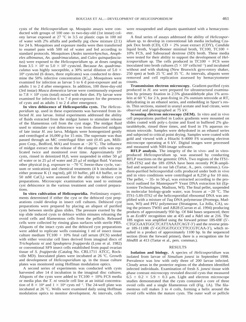
Isolation and biology. A species of Helicosporidium wasisolated from larvae of Simulium jonesi in September 1998.Prevalence was low with only three of 200 larvae infected.Cloudy areas in the posterior regions of the abdomen identifiedinfected individuals. Examination of fresh S. jonesi tissue withphase contrast microscopy revealed discoid cysts that measured6.5 6 0.2 3 5.9 6 0.3 mm. Light and electron microscopestudies demonstrated that the cysts contained a core of threeovoid cells and a single filamentous cell (Fig. 1A). The fila-mentous cell makes 3 to 4 coils, forming a helix around theovoid cells within the mature cysts. The outer wall or pellicle
462 J. EUKARYOT. MICROBIOL., VOL. 48, NO. 4, JULY–AUGUST 2001
Fig. 1. Transmission electron micrographs of the cysts of Helicosporidium sp. from the black fly Simulium jonesi. (A) Mature cyst composedof three central ovoid cells (O) and the peripherally located filamentous cell (F) contained within a multi-layered cyst wall. (B) Sagittal sectionof the filamentous cell within the cyst demonstrating several of the projections (arrows) on the cell wall. Inset. High magnification showing thatthe projections are modifications of the outer two layers of the cell wall.

463BOUCIAS ET AL.—DEVELOPMENT OF HELICOSPORIDIUM
Fig. 2. SEM of mature cysts of Helicosporidium sp. produced in Helicoverpa zea larvae. (A) Preparation of cysts purified by Ludox gradientcentrifugation. (B) Narrow surface of a cyst depicting the coiled filament cell underlying the pellicle. (C) The broad surface of a cyst.
Fig. 3. Light micrographs of purified cysts of Helicosporidium sp. produced in Helicoverpa zea larvae. (A) Mature discoid cysts. (B) Prepa-ration where cysts have opened or dehisced releasing the filamentous cell-ovoid cell complex from the pellicle. The pressure applied to thecoverslip results in a fracturing of the pellicle, a swelling of the central ovoid cells, and an uncoiling of the filamentous cells. In several cases,the three ovoid cells and the filamentous cells can be observed in proximity with the pellicle (arrow).

464 J. EUKARYOT. MICROBIOL., VOL. 48, NO. 4, JULY–AUGUST 2001
Fig. 4. SEM of dehisced helicosporidial cyst. (A) The uncoiled filamentous cell extending away from the ovoid cell complex. (B) The surfaceof a filamentous cell demonstrating the orientation of the projections. (C) The three ovoid cells remain as an aggregate. These have expandedand no longer possess the compressed morphology observed in the cyst stage (cf. Fig 2). Distinct pores can be seen in the previously compressedregion of the central ovoid cells.
is a multilaminate structure, enclosing the peripheral filamen-tous cell within its innermost wall layer. Within intact cysts,three centrally located ovoid cells are compressed in an accor-dion-like fashion. Each of these cells possesses a peripheralnucleus that encloses a cytoplasmic region that contains a va-riety of vacuoles and granules.
Helicosporidium sp. from S. jonesi produced uniform discoidcysts when grown in H. zea larvae (Figs. 2A, 3A). The densityof these helicosporidial cysts, measuring 6.2 6 0.3 3 5.9 6 0.1mm, was estimated to be ; 1.16 g/ml from the Ludox gradient.The cysts, easily partitioned from insect hemolymph compo-nents, were virtually free of other components after beingpassed through the Ludox gradients. SEM revealed that the cyst
possessed a ridged biscuit-shaped structure (Fig. 2A, B). Onone side of the cyst the coil of the filamentous cell was dis-cerned as a coiled ridge in the cyst wall (Fig. 2B) whereas theopposite side was rounded and possessed no coiled ridge (Fig.2C).
When stimulated by pressure, the outer pellicle layer of thecysts (Fig. 3A) splits open or dehisces releasing the filamentouscell-ovoid cell complex from the pellicle (Fig. 3B). Releasefrom the cyst stage produces an expanded ovoid cell aggregateand results in the uncoiling of filamentous cells (Fig. 4A, C).The filamentous cells, measuring 37 mm 6 4.3 mm in lengthby 0.9 6 0.13 mm in diam., were coated with short projections(340 6 60 nm) orientated in the same direction providing po-
465BOUCIAS ET AL.—DEVELOPMENT OF HELICOSPORIDIUM
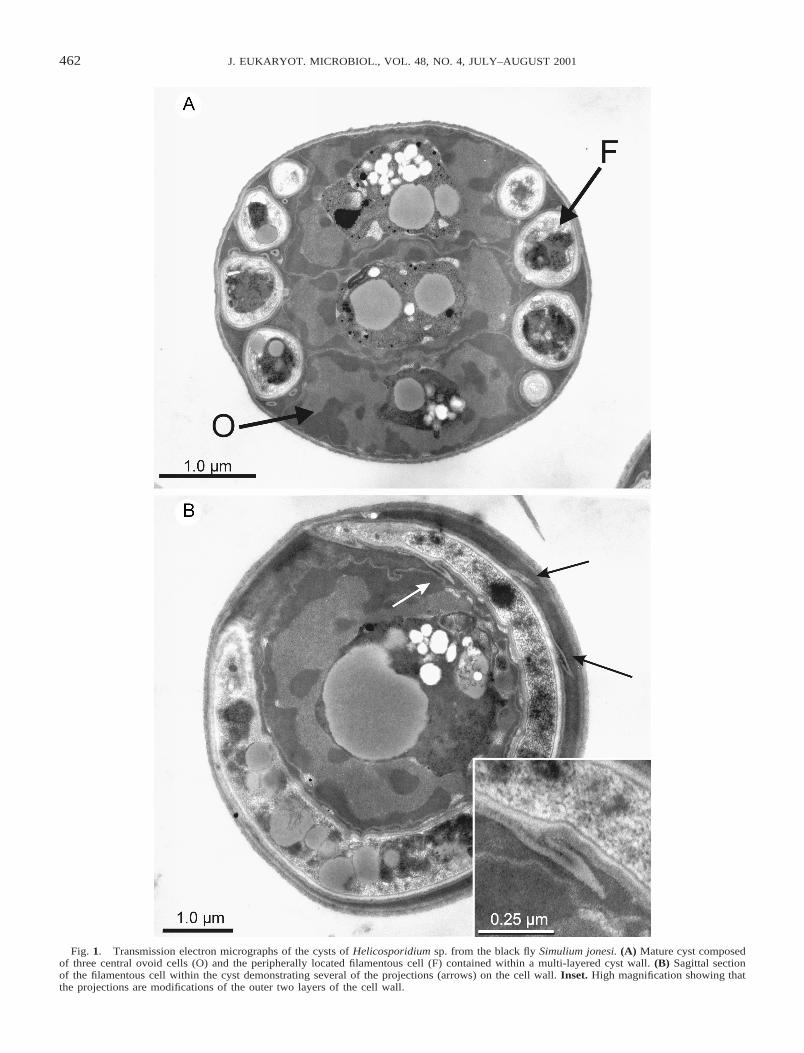
Fig. 5. SEM of the midgut region of Manduca sexta at 4 h post-challenge with Helicosporidium sp. (A) Filamentous cells have penetratedthe microvillar lining of these midgut cells. (B) A filamentous cell embedded in a midgut cell demonstrating the orientation of the surfaceprojections.
Fig. 6. Light micrographs of hemolymph sampled from Manduca sexta larvae infected with Helicosporidium sp. (A, B) Two days post-infection extracellular vegetative helicosporidial cells and normal looking hemocytes. (C) Six days post-infection plasmodial-like hemocytesdisplayed cytopathic effects. (D) Ten days post-infection. Note the number of refractile cysts and vegetative cells as compared to the numbers ofhemocytes.
larity to the filamentous cells (Figs. 1B, 4B). Cyst dehiscencewas triggered readily by the application of gentle pressure tothe coverslip covering a cyst suspension. Alternatively, the in-cubation of purified cysts in midgut fluid extracted from H. zealarvae stimulated the release of the filamentous cells from cystsuspensions. A 20 min exposure to midgut fluids resulted inmore than 50% of the cysts releasing their filamentous cells.Upon activation these cysts increased in volume resulting inpellicle rupture and release of the filamentous cell. Incubatedin the midgut luminal fluid the ovoid cells lysed, whereas thereleased filamentous cell became uncoiled and remained intact.Uncoiled filamentous cells readily clustered with other filamen-tous cells producing rosettes. Whether this clumping was dueto a specific surface adhesion or to simply a result of entangle-ment of the surface barbs is unknown. The component(s) in themidgut fluid that signals dehiscence is not known; exposure ofthe cysts to various physical and chemical agents (see Materialsand Methods) failed to stimulate the release of the filamentouscells.
In vivo host range and infectious process. Susceptibilityvaried among mosquito and fly species challenged with the Hel-icosporidium sp. and doses were adjusted accordingly. The
dose, resulting infection rates and number of replicates for eachspecies were: Anopheles albimanus (5.0 3 104, 58.3%, N eq 1),An. quadrimaculatus (5.0 3 104, 100%, N eq 3), Ae. taenior-hynchus (3.5 3 102, 14.3%, N 5 1), Cx. quinquefascaitus (5.03 105, 0%, N 5 2) and M. domestica (7.8 3 108, 80%, N 51). The IC50 (95% Fiducial Limits) for An. quadrimaculatus was1.0 3 104 (3.1 3 103, 3.6 3 104 cysts/ml). All susceptible spe-cies supported in vivo replication of the Helicosporidium sp.Culex quinquefasciatus larvae had a relatively high mortalitylevel (40.0 6 31.8%) but none of the larval or adult survivorswere infected. Filamentous cells were observed in the gut lu-mens of An. quadrimaculatus and Cx. quinquefasciatus within1 hr post-exposure indicating that barriers to infection in Cx.quinquefasciatus occurred after cysts dehisced. Examination oftissues with phase-contrast microscopy confirmed that cyst pro-duction occurred primarily in the haemocoel of An. quadrima-culatus larvae. Light microscopy of the Helicosporidium sp. inAn. quadrimaculatus revealed that the main multiplicativephase began 3 to 4 d post-challenge and was soon followed bycyst production.
Helicosporidium sp. from S. jonesi is capable of infectingand replicating in a variety of insects other than Diptera. Oral

466 J. EUKARYOT. MICROBIOL., VOL. 48, NO. 4, JULY–AUGUST 2001
Fig. 7. Growth rate of Helicosporidium cultivated at 26 8C. Flaskscontaining TC100 1 FCS media with (v) and without (l) SF-9 insectcells were seeded with 100 (A) or 104 (B) helicosporidial cells. Atintervals the total number of cells in the wells was estimated by he-macytometer counts. Note that after 6-8 d incubation a maximum of107 cells was produced in all four treatments.
challenge of H. zea and M. sexta larvae with cyst preparationswas lethal to tested insects. Examination of dissected alimentarytracts revealed that ingested cysts bound initially to the peri-trophic matrix in larvae of both challenged H. zea and M. sexta.Within 2 h post-ingestion, cysts dehisced releasing filamentouscells from the ovoid-cell pellicle complex. SEM of the midgutsdissected from M. sexta larvae at 4 h post-ingestion revealedthat the released filamentous cells penetrated the peritrophicmatrix and attached to the midgut columnar epithelium (Fig.5A). These filament cells penetrated the midgut with the pro-jections oriented away from the penetration point (Fig. 5B),suggesting that these cell wall extensions may play a role inanchoring the filamentous cell to the gut epithelium. In the caseof M. sexta, vegetative cells were observed in the hemolymphwithin 2 d post-ingestion (Figs. 6A, B). Vegetative cells, con-taining variable numbers of cells within the pellicles, were ob-served to be both associated with circulating hemocytes andpresent as freely circulating cells. By 6 d post-ingestion, infec-tion suppressed the feeding and growth of M. sexta larvae. Atthis time, plasmodial-like hemocytes displayed marked cyto-pathic effects (CPE, Fig. 6C). It is unclear whether the Heli-cosporidium sp. induced a haemocyte fusion, blocked cytoki-nesis, or stimulated hemocyte nuclear division. Within 10–14d, treated larvae contained massive numbers of mature cysts inthe cream-colored hemolymph (Fig. 6D). At this point largenumbers of cysts could be extracted easily using several cyclesof centrifugation followed by high-speed centrifugation througha Ludox gradient.
The injection of purified cysts into G. mellonella larvae re-sulted in a somewhat different developmental pathway. Withinminutes after injection numerous phagocytic hemocytes wereobserved to contain cysts. By 72 h, clusters of melanized he-mocytes were attached to the basement membrane of varioustissues. Dissection of these hemocytic granulomas revealed thepresence of actively developing colonies of helicosporidial veg-etative cells. As time progressed, Helicosporidium sp. contin-ued to multiply, appearing by 10 d post-injection associatedwith the muscles and fat body tissues of these larvae. All larvaeinjected with cysts died at the larval pupal molt, whereas thecontrol larvae pupated and molted to the adult stage.
In vitro growth and development. Helicosporidium sp. iso-lated from S. jonesi, in addition to infecting a range of insects,grew under in vitro conditions. Unlike the in vivo situation,inoculation of imaginal disc cell lines with purified cysts didnot result in an immediate dehiscence; at 48 h post-inoculation(p.i.) only a small percentage of cysts released filamentouscells. In all tested insect cell cultures helicosporidial replicationoccurred in an extracellular fashion. After 10 d, wells inocu-lated with 5 3 103 cells and 4 3 104 cells produced 2.6 3 105
cells and 1.2 3 106 cells respectively, resulting in a 52- and30-fold increase in cyst numbers.
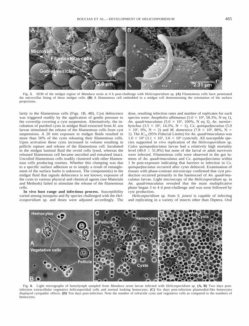
Additional assays conducted with TC100 1 10% fetal calfserum (FCS) medium with and without SF-9 cells demonstratedthat Helicosporidium sp. could develop with or without livingcells (Figs. 7A, B). Daily observations of these cultures dem-onstrated that the helicosporidial cells did not infect the SF9cells. By 48 h, both media stimulated vegetative growth of hel-icosporidial cells (Fig. 8A). Single non-motile cells, detectedduring the vegetative phase, possessed a textured outer surfaceand measured 2.6 6 0.33 mm (N 5 40) in diam. (Fig. 8C). Likethe cyst stage, vegetative cells were enclosed within a pellicle(Fig. 8B), which could contain 1, 2, 4, or 8 vegetative cells.High concentrations of insect cells were observed to restrict thedispersal of progeny cells. Significantly, no lysis of SF9 cellswas observed during this vegetative growth period. In most cas-es, the presence of helicosporidial cells and extracellular ma-
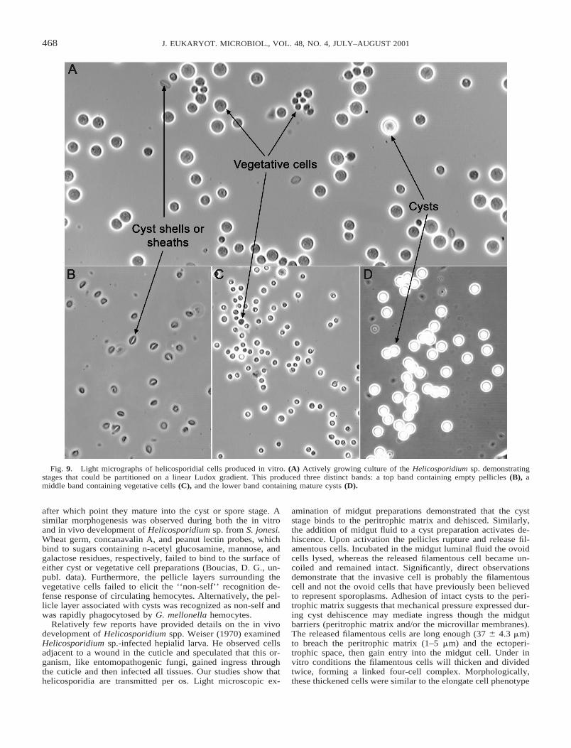
terial formed a biofilm over the surface; suspended cells werenot detected in these cultures. After 72 h, contiguous monolay-ers of helicosporidial cells were observed in wells lacking theSF9 cells. Gradient centrifugation of vegetative growing cul-tures (Fig. 9A) produced three distinct bands; the top band con-tained empty pellicles (Fig. 9B), the middle band contained thevegetative cells (Fig. 9C), and the lower band contained thecyst stage (Fig. 9D).
All of the tested cell-free media, except CD (minimal salts1 glucose media), supported the vegetative growth of this Hel-icosporidium sp. In stationary cultures 12–15 d post-inocula-tion, this organism produced a continous film over the bottomthat coated the surface with mucilaginous cell aggregates thatextended into the media. However, less than 10% of the in vitrocells differentiated into mature cysts. These results suggest thatthe component(s) that signals the in vivo late-stage cyst matu-ration event, is lacking in the in vitro media. Helicosporidium
467BOUCIAS ET AL.—DEVELOPMENT OF HELICOSPORIDIUM
Fig. 8. SEM of helicosporidial cells produced in vitro. (A) Developmental stages of the Helicosporidium sp. after 48 h of growth. Note thepresence of empty pellicles that have released vegetative cells. (B) A pellicle that has split open, like a bivalve mollusc shell, releasing vegetativecells into the medium. (C) Vegetative cells, demonstrating a range of different sizes with a wrinkled surface topography that is distinct from thatobserved with the cyst stage.
sp. did not replicate at 35 8C, and cells exposed to 35 8C for 4d failed to replicate when transferred to the 25 8C permissivetemperature.
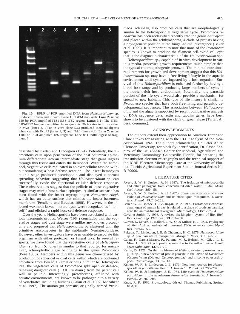
RFLP analysis. The genomic DNA of both the in vitro andin vivo cells was easily extracted using the Masterpurey YeastDNA kit. Microscopic examination revealed the presence ofnumerous, highly refractile cysts before treatment; after incu-bation in the lysis buffer at 50 8C the cells appeared to dehisce.The cyst were split open, releasing the filamentous cells. Nomassive disruption of the ovoid cells or elongate cells was ob-served in these preparations. The DNA preparations, electro-phoresed on an 0.8% agarose gel and stained with ethidiumbromide, produced a single, discrete ; 20-kb band that couldbe directly PCR-amplified with the various pairs of rDNA prim-ers. RFLP analysis using both EcoRI and NdeI to digest theamplified ITS-5.8S region of DNA extracted from both in vivoand in vitro cell preparations produced identical digest patterns(Fig. 10). Similarly the amplified 18S region of both DNA prep-
arations digested with HindIII produced identical fragments ofapproximately 650- and 450 bp.
DISCUSSION
Morphologically, the cyst stage of Helicosporidium sp. iso-lated from S. jonesi is similar to that described from helicos-poridia detected in other invertebrate hosts. Purrini (1984) con-ducted an EM examination of Helicosporidium-infected orbatidmites and described the cysts as spores containing three uni-nucleate germ cells and a single filamentous cell. Enclosing thespore was a dense bilayer exosporium and a more transparentinner endosporium. Pekkarinen (1993) identified a hyperpara-site of a trematode infecting the mussel Mytilus edulis as heli-cosporidia by the presence of discoidal spores containing threedistinct sporoplasms and a coiled filament cell.
Enclosing both the cyst stage and the vegetative stages is aunique pellicle (Lindegren and Hoffman 1976). These cells di-vide within the pellicle until reaching a four or eight cell stage,
468 J. EUKARYOT. MICROBIOL., VOL. 48, NO. 4, JULY–AUGUST 2001
Fig. 9. Light micrographs of helicosporidial cells produced in vitro. (A) Actively growing culture of the Helicosporidium sp. demonstratingstages that could be partitioned on a linear Ludox gradient. This produced three distinct bands: a top band containing empty pellicles (B), amiddle band containing vegetative cells (C), and the lower band containing mature cysts (D).
after which point they mature into the cyst or spore stage. Asimilar morphogenesis was observed during both the in vitroand in vivo development of Helicosporidium sp. from S. jonesi.Wheat germ, concanavalin A, and peanut lectin probes, whichbind to sugars containing n-acetyl glucosamine, mannose, andgalactose residues, respectively, failed to bind to the surface ofeither cyst or vegetative cell preparations (Boucias, D. G., un-publ. data). Furthermore, the pellicle layers surrounding thevegetative cells failed to elicit the ‘‘non-self’’ recognition de-fense response of circulating hemocytes. Alternatively, the pel-licle layer associated with cysts was recognized as non-self andwas rapidly phagocytosed by G. mellonella hemocytes.
Relatively few reports have provided details on the in vivodevelopment of Helicosporidium spp. Weiser (1970) examinedHelicosporidium sp.-infected hepialid larva. He observed cellsadjacent to a wound in the cuticle and speculated that this or-ganism, like entomopathogenic fungi, gained ingress throughthe cuticle and then infected all tissues. Our studies show thathelicosporidia are transmitted per os. Light microscopic ex-
amination of midgut preparations demonstrated that the cyststage binds to the peritrophic matrix and dehisced. Similarly,the addition of midgut fluid to a cyst preparation activates de-hiscence. Upon activation the pellicles rupture and release fil-amentous cells. Incubated in the midgut luminal fluid the ovoidcells lysed, whereas the released filamentous cell became un-coiled and remained intact. Significantly, direct observationsdemonstrate that the invasive cell is probably the filamentouscell and not the ovoid cells that have previously been believedto represent sporoplasms. Adhesion of intact cysts to the peri-trophic matrix suggests that mechanical pressure expressed dur-ing cyst dehiscence may mediate ingress though the midgutbarriers (peritrophic matrix and/or the microvillar membranes).The released filamentous cells are long enough (37 6 4.3 mm)to breach the peritrophic matrix (1–5 mm) and the ectoperi-trophic space, then gain entry into the midgut cell. Under invitro conditions the filamentous cells will thicken and dividedtwice, forming a linked four-cell complex. Morphologically,these thickened cells were similar to the elongate cell phenotype
469BOUCIAS ET AL.—DEVELOPMENT OF HELICOSPORIDIUM
Fig. 10. RFLP of PCR-amplified DNA from Helicosporidium sp.produced in vitro and in vivo. Lane 1: pGEM standards. Lane 2: uncut950 bp PCR-amplified ITS1-5.8S-ITS2 region. Lanes 3-6: The ITS1-5.8S-ITS2 fragment amplified from genomic DNA extracted from eitherin vivo (lanes 3, 4) or in vitro (lane 5,6) produced identical digestswhen cut with EcoRI (lanes 3, 5) and NdeI (lanes 4,6). Lane 7: uncut1100 bp PCR amplified 18S fragment. Lane 8: HindIII digest of frag-ment 7.
described by Kellen and Lindegren (1974). Potentially, the fil-amentous cells upon penetration of the host columnar epithe-lium differentiate into an intermediate stage that gains ingressthrough this tissue and enters the hemocoel. Within the hemo-coel, vegetative cells replicated in an extracellular fashion with-out stimulating a host defense reaction. The insect hemocytesat this stage produced pseudopodia and displayed a normalspreading behavior, suggesting that Helicosporidium sp. cansuccessfully evade the functional cellular defense response.These observations suggest that the pellicle of these vegetativestages may mimic host surface epitopes. A similar scenario hasbeen found with the insect mycopathogen Nomuraea rileyi,which has an outer surface that mimics the insect basementmembrane (Pendland and Boucias 1998). However, in the in-jected waxmoth larvae, mature cysts were recognized as ‘‘non-self’’ and elicited a rapid host-cell defense response.
Over the years, Helicosporidia have been associated with var-ious taxonomic groups. Weiser (1964) concluded that the veg-etative stages and cyst stage were unlike any known protozo-an’s and proposed that Helicosporidium be clustered with theprimitive Ascomycetes in the subfamily Nematosporideae.However, other investigators have been unable to associate thisorganism with either protozoan or fungal taxa. In several re-spects, we have found that the vegetative cycle of Helicospor-idium sp. from S. jonesi is similar to that reported for unicel-lular, achorophyllic algae belonging to the genus Prototheca(Pore 1985). Members within this genus are characterized byproduction of spherical or oval cells within which are containedanywhere from two to 16 smaller cells. Similar to Helicospor-idia, the vegetative cells of Prototheca split open or dehisce,releasing daughter cells (;3.0 mm diam.) from the parent cellwall or pellicle. Interestingly, protothecans, affiliated withaquatic environments, are known to be pathogenic to a varietyof vertebrates including humans (Galan et al. 1997; Mohabeeret al. 1997). The anuran gut parasite, originally named Proto-
theca richardsii, also produces cells that are morphologicallysimilar to the helicosporidial vegetative cycle. Prototheca ri-chardsii has been reclassified recently into the genus Anurofecaand placed within the Ichthyosporea, a clade of protists havinga phylogenetic position at the fungal-animal divergence (Bakeret al. 1999). It is important to note that none of the Protothecaspecies is known to produce the filament cell-ovoid cell cystthat is the diagnostic characteristic of the Helicosporidium spp.
Helicosporidium sp., capable of in vitro development in var-ious media, possesses growth requirements much simpler thanthe typical entomopathogenic protozoa. The minimal nutritionalrequirements for growth and development suggest that this Hel-icosporidium sp. may have a free-living lifestyle in the aquaticenvironment until cysts are ingested by a host organism. Sur-vival of this Helicosporidium is enhanced further by having abroad host range and by producing large numbers of cysts inthe nutrient-rich host environment. Potentially, the parasiticportion of the life cycle would also provide a mechanism forspread to new habitats. This type of life history is similar toPrototheca species that have both free-living and parasitic de-velopmental sequences. The association between Helicospori-dium and the algae is supported by recent comparative analysisof DNA sequence data: actin and tubulin genes have beenshown to be clustered with the clade of green algae (Tartar, A.,pers. commun.).
ACKNOWLEDGMENTS
The authors extend their appreciation to Aurelien Tartar andClare Stokes for assisting with the RFLP analysis of the Heli-cosporidium DNA. The authors acknowledge Dr. Peter Adler,Clemson University, for black fly identification, Dr. Sasha Sha-piro, of the USDA/ARS Center for Medical, Agricultural andVeterinary Entomology, Gainesville, Florida, for preparing thetransmission electron micrographs and the technical support ofthe ICBR Electron Microscopy Core at the University of Flor-ida. Florida Agricultural Experiment Station Journal Series No.R-70900.
LITERATURE CITED
Avery, S. W. & Undeen, A. H. 1987a. The isolation of microsporidiaand other pathogens from concentrated ditch water. J. Am. Mosq.Ctrl. Assoc., 3:54–58.
Avery, S. W. & Undeen, A. H. 1987b. Some characteristics of a newisolate of Helicosporidium and its effect upon mosquitoes. J. Inver-tebr. Pathol., 49:246–251.
Baker, G. C., Beebee, T. J. & Ragan, M. A. 1999. Prototheca richardsii,a pathogen of anuran larvae, is related to a clade of protistan parasitesnear the animal-fungal divergence. Microbiology, 145:1777–84.
Cavalier-Smith, T. 1998. A revised six-kingdom system of life. Biol.Rev. Cambridge Phil. Soc., 73:203–266.
Curran, J., Driver, F., Ballard, J. W. O. & Milner, R. J. 1994. Phylogenyof Metarhizium: analysis of ribosomal DNA sequence data. MycolRes., 98:547–552.
Fukuda, T., Lindegren, J. E. & Chapman, H. C. 1976. Helicosporidiumsp. A new parasite of mosquitoes. Mosquito News, 39:514–517.
Galan, F., Garcia-Martos, P., Paloma, M. J., Beltrun, M., Gil, J. L. &Mira, J. 1997. Onychoprotothecosis due to Prototheca wickerhamii.Mycopathologia, 137:75–77.
Keilin, D. 1921. On the life history of Helicosporidium parasiticum n.g., n. sp., a new species of protist parasite in the larvae of Dashelaeaobscura Winn (Diptera: Ceratopogonidae) and in some other arthro-pods. Parasitology, 13:97–113.
Kellen, W. R. & Lindegren, J. E. 1973. New host records for Helico-sporidium parasiticum Keilin. J. Invertebr. Pathol., 22:296–297.
Kellen, W. R. & Lindegren, J. E. 1974. Life cycle of Helicosporidiumparasiticum in the navelworm Paramyelois transitella. J. Invertebr.Pathol., 23:202–208.
Kudo, R. R. 1966. Protozoology, 6th ed. Thomas Publishing, Spring-field, IL.
470 J. EUKARYOT. MICROBIOL., VOL. 48, NO. 4, JULY–AUGUST 2001
Lindegren, J. E. & Hoffman, D. F. 1976. Ultrastructure of some devel-opmental stages of Helicosporidium sp. in the navel orangewormParamyelois transitella. J. Invertebr. Pathol., 27:105–113.
Lynn, D. E., Miller, S. G. & Oberlander, H. 1982. Establishment of acell line from lepidopteran wing imaginal discs: induction of newlysynthesized proteins by 20-hydroxyecdysone. Proc. Natl. Acad. Sci.USA, 79:2589–2593.
Mohabeer, A. J., Kaplan, P. J., Southern, P. M. & Gander, R. M. 1997.Algaemia due to Prototheca wickerhamii in a patient with Myasthe-nia gravis. J. Clin. Microbiol., 35:3305–3307.
Patterson, D. J. 1999. The diversity of eukaryotes. Amer. Nat., 154(Suppl.):S96–S124.
Pekkarinen, M. 1993. Bucephalid trematode sporocysts in brackish-wa-ter Mytilus edulis, a new host of a Helicosporidium sp. (Protozoa:Helicosporidia). J. Invertebr. Pathol., 61:214–216.
Pendland, J. C. & Boucias, D. G. 1998. Characterization of monoclonalantibodies against cell wall epitopes of the insect pathogenic fungus,Nomuraea rileyi: differential binding to fungal surfaces and cross-
reactivity with host hemocytes and basement membrane components.Eur. J. Cell Biol., 75:118–127.
Pore, R. S. 1985. Prototheca taxonomy. Mycopathologia, 90:129–139.Purrini, K. 1984. Light and electron microscope studies on Helicospor-
idium sp. parasitizing oribatid mites (Oribatei, Acarini) and Collem-bola (Apterygota:Insecta) in forest soils. J. Invertebr. Pathol., 44:18–27.
Sayre, R. M. & Clark, T. B. 1978. Daphnia magna (Cladocera:Chy-doroidea) a new host of a Helicosporidium sp. (Protozoa: Helicos-poridia). J. Invertebr. Pathol., 31:260–261.
Undeen, A. & Vavra, J. 1998. Research methods for entomopathogenicprotozoa. In: Lacey, L. (ed.), Manual of Techniques in Insect Pa-thology. Academic Press, NewYork. p.117-152.
Weiser, J. 1964. The taxonomic position of Helicosporidium parasiti-cum, Keilin 1924. J. Protozool., 11(Suppl.):112.
Weiser, J. 1970. Helicosporidium parasiticum Keilin infection in thecaterpillar of a hepialid moth in Argentina. Protozoology, 17:440–445.
Received: 12/12/00, 03/15/01; accepted 03/15/01