Embed Size (px)
Citation preview

HIGH-CYCLE FATIGUE BEHAVIOR OF TEMPOROMANDIBULAR JOINT
IMPLANT
ZOHREH ARABSHAHI
A thesis submitted in fulfillment
of the requirements for the award of the degree of
Master of Engineering (Biomedical)
Faculty of Biosciences and Medical Engineering
Universiti Teknologi Malaysia
May 2013
ii
My deepest appreciation goes to my beloved father Mohammadali Arabshahi, my
beloved mother KokabTalebi, my beloved partner Jamal, my loved sisters: Zari,
Fereshteh, Fatemeh and Tahereh and my best friends for ever: Miad, Batool and Lili,
for the enormous love, support, encouragement and sacrifice they had given to me.

iii
ACKNOWLEDGEMENTS
Praise is to the God for everything has done to me and bestowing upon me
wisdom, ideasand strength to successfully complete thismaster thesis.
I would like to give my special gratitude toAssoc. Prof. Engr. Dr. Mohammed
Rafiq bin Dato' Abdul Kadir, mysupervisor, and Dr. Abbas Azariand and Dr. Seyyed
Saeed Rahimian Koloor for their effective visions, guidance and supports. Their
intuitions, advices, and enthusiasms were invaluable to the progress and completion
of this thesis.
Most prominently, I would like to extend my warmest gratitude to my
beloved parents and sisters for their precious support, patience and assurance
throughout my education in UniversitiTeknologi Malaysia (UTM).They are always
being my stand all through the period of my life, and I will always be appreciative
for their sacrifice, generosity and love.
My supreme thanks also to all Mediteg's students, my fellow friends, especially
Ahmad Ramli Rasyidi, Mohd Nazri Bin Bajuri, Aisyah Ahmad Shafiand, as well as
supportive lecturer: Dr. Dedy Wicaksono for his priceless guidance and
encouragement.
Last but not least, I offer my regards and blessings to all of those who
supported me in any respect during the completion of the project.
v
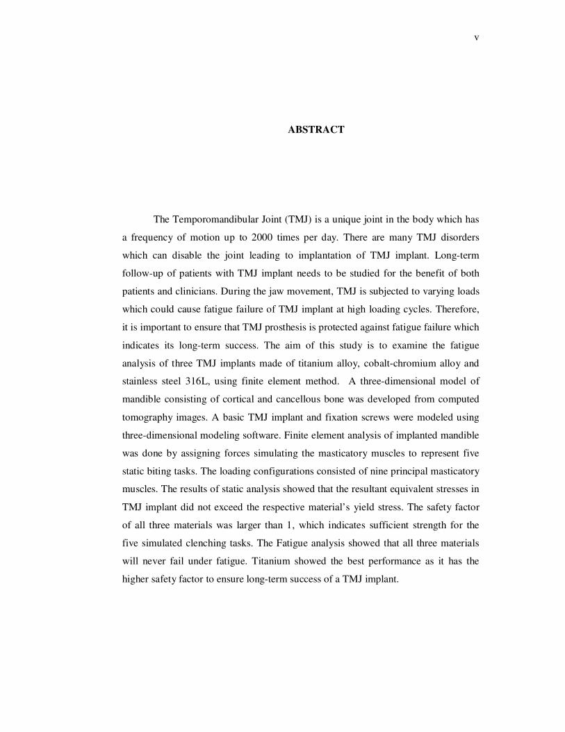
ABSTRACT
The Temporomandibular Joint (TMJ) is a unique joint in the body which has
a frequency of motion up to 2000 times per day. There are many TMJ disorders
which can disable the joint leading to implantation of TMJ implant. Long-term
follow-up of patients with TMJ implant needs to be studied for the benefit of both
patients and clinicians. During the jaw movement, TMJ is subjected to varying loads
which could cause fatigue failure of TMJ implant at high loading cycles. Therefore,
it is important to ensure that TMJ prosthesis is protected against fatigue failure which
indicates its long-term success. The aim of this study is to examine the fatigue
analysis of three TMJ implants made of titanium alloy, cobalt-chromium alloy and
stainless steel 316L, using finite element method. A three-dimensional model of
mandible consisting of cortical and cancellous bone was developed from computed
tomography images. A basic TMJ implant and fixation screws were modeled using
three-dimensional modeling software. Finite element analysis of implanted mandible
was done by assigning forces simulating the masticatory muscles to represent five
static biting tasks. The loading configurations consisted of nine principal masticatory
muscles. The results of static analysis showed that the resultant equivalent stresses in
TMJ implant did not exceed the respective material’s yield stress. The safety factor
of all three materials was larger than 1, which indicates sufficient strength for the
five simulated clenching tasks. The Fatigue analysis showed that all three materials
will never fail under fatigue. Titanium showed the best performance as it has the
higher safety factor to ensure long-term success of a TMJ implant.

vi
ABSTRAK
Sendi temporomandibular (TMJ) adalah sendi unik di dalam badan yang
mempunyai kekerapan pergerakan sehingga 2000 kali sehari. Terdapat banyak
penyakit TMJ yang boleh melumpuhkan sendi dan seterusnya membawa kepada
penggunaan implan TMJ. Tindakan susulan jangka panjang pesakit yang
menggunakan implan TMJ perlu dikaji untuk manfaat pesakit dan doktor. Semasa
pergerakan rahang, TMJ akan dipengaruhi oleh pelbagai beban yang boleh
menyebabkan kegagalan lesu implan TMJ pada kitaran beban yang tinggi. Ia adalah
penting untuk memastikan TMJ prostesis terhindar daripada kegagalan lesu untuk
kejayaan jangka panjang. Tujuan kajian ini adalah untuk mengkaji analisis keletihan
tiga TMJ implan yang diperbuat daripada aloi titanium, aloi cobalt-kromium dan
keluli tahan karat 316L, dengan menggunakan kaedah unsur terhingga. Model tiga
dimensi rahang orang dewasa dibina daripada imej imbasan tomografi yang terdiri
daripada tulang padat dan berongga. Satu implan TMJ dan beberapa skru penetapan
telah dimodelkan menggunakan perisian permodelan tiga dimensi. Analisis unsur
terhingga rahang bawah yang diimplan dijalankan dengan memberi daya kepada otot
mastikasi untuk mewakili lima tugas menggigit. Konfigurasi beban terdiri daripada
sembilan otot utama mastikasi. Keputusan analisis statik menunjukkan paduan
tekanan setara dalam implan TMJ untuk bahan tersebut tidak melebihi tekanan alah
mereka. Faktor keselamatan implan untuk ketiga-tiga bahan adalah lebih tinggi dari 1,
menunjukkan bahawa ianya selamat di gunakan untuk kelima-lima tugas mengetap.
Keputusan analisis keletihan menunjukkan ketiga-tiga implan tidak mengalami
kegagalan di bawah keadaan daya tersebut. Titanium didapati lebih baik kerana ia
menghasilkan faktor keselamatan yang lebih tinggi bagi memastikan kejayaan jangka
panjang implan TMJ.

vii
TABLE OF CONTENTS
CHAPTER TITLE PAGE
DECLARATION ii
DEDICATION iii
ACKNOWLENGEMENTS iv
ABSTRACT v
ABSTRAK vi
TABLE OF CONTENTS vii
LIST OF TABLES xi
LIST OF FIGURES xii
LIST OF ABBREVIATIONS xiv
LIST OF SYMBOLS xv
1 INTRODUCTION 1
1.1 Background 1
1.2 Problem Statement 4
1.3 Research Objectives 5
1.4 Importance of Research 6
1.5 Research Scopes 7
1.6 Structure of Thesis 8
2 LITERATURE REVIEW 9
2.1 Introduction 9
2.2 Temporomandibular Joint 10
2.2.1 TMJ Anatomy 10

viii
2.2.2 TMJ Disorders 15
2.2.3 Total TMJ Replacement System 15
2.2.4 Commercial TMJ systems 16
2.3 Finite Element Method 23
2.3.1 Finite Element Method in
Biomechanics
23
2.3.1.1 Mesh Element 24
2.3.1.2 Von Mises Yield
Criterion
24
2.3.2 Finite Element Method in TMJ 25
2.4 Fatigue Analysis 28
2.4.1 Background Information 28
2.4.2 Stress-Life Diagram (S-N Diagram) 28
2.4.3 Mean Stress Correction 30
2.5 Summary 32
3 MATERIALS & METHODS 33
3.1 Introduction 33
3.2 Mandible Geometry 34
3.2.1 CT scan 34
3.2.2 Image processing 35
3.2.3 Data processing 38
3.2.4 Three dimensional modeling 38
3.3 Modeling of TMJ Implant 39
3.4 Modeling of Implanted Mandible 40
3.5 Finite Element Model 40
3.6 Validation 43
3.7 Material Properties 46
3.8 Loading Condition 48
3.8.1 Static Loading Condition 48
3.8.2 Fatigue Loading Condition 54
3.9 Static Analysis 55

ix
3.10 Fatigue Analysis 55
3.11 Summary 56
4 RESULTS 57
4.1 Introduction 57
4.2 Validation 58
4.3 Static Analysis Results 60
4.3.1 Von Mises Stress (Equivalent
Stress)
60
4.3.2 Safety Factor 64
4.3.3 Mechanical adaptation 66
4.4 Fatigue Analysis Results 68
4.4.1 Failure Analysis 68
4.4.2 Factor of Safety 69
4.5 Summary 71
5 DISCUSSION 72
5.1 Introduction 72
5.2 Validation 73
5.3 Static Analysis Discussion 76
5.3.1 Von Mises Stress 77
5.3.2 Safety Factor 79
5.3.3 Mechanical adaptation 80
5.4 Fatigue Analysis Discussion 81
5.4.1 Failure Analysis 82
5.4.2 Factor of Safety 83
5.5 Summary 84
6 CONCLUSIONS & FUTURE WORKS 85
6.1 Contributions 85
6.2 Limitations and Future works 85
6.3 Conclusions 86

x
REFERENCES 88
List of publications 106

xi
LIST OF TABLES
TABLE NO. TITLE PAGE
2.1 The summarized information about the FDA approved
TMJ implants
22
3.1 Isotropic material properties assigned to the components
in FEA models
47
3.2 Directions of unit vectors (i.e., direction cosines) of
muscular forces and forces assigned to the masticatory
muscles
51
3.3 Weighting. and scaling factors assigned to the masticatory
muscles for live clenching tasks
52
5.1 Mechanical properties of three materials 78

xii
LIST OF FIGURES
FIGURE
NO.
TITLE
PAGE
1.1 The Location of the TMJ in the skull 2
2.1 Anatomy of Temporomandibular Joint 11
2.2 Ligament and capsule attachments in the TMJ 12
2.3 Muscles of mastication: lateral pterygoid (A), the temporalis
muscle (B), masseter muscle (C) and medial pterygoid (D)
14
2.4 Christensen system, its components and application on TMJ 18
2.5 Concepts System, its components and application on TMJ 19
2.6 Lorenz System, its components and application on TMJ 20
2.7 The S-N curve 29
2.8 Fully-reversed loading 30
2.9 The line diagram based on Goodman, Gerber and Soderberg
theories
31
3.1 (a) Before data processing of CT scan images, these images
have been thresholded with HU above 700. There are still
artifacts around the teeth and bone
(b) After data processing of CT scan images. The artifacts
have been removed.
36, 37
3.2 3D model of TMJ implant based on commercially available
TMJ implant
39
3.3 Mesh convergence study curve in TMJ implant and fixation
screws
41
3.4 Finite element model of implanted mandible which has been
meshed with total number of 156,165 elements and 231,724
nodes
42
3.5 The FE model used to simulate the Ramos work 45
3.6 A group of parallel vectors on the right ramus simulating the 50

xiii
masseter muscle loads
3.7 Condylar poles which condyle can rotate around them 53
3.8 S-N curve of all materials used in this study 54
4.1 Micro strain resulted in our FE models for (a) incisive
model, (b) canine model, and (c) molar model
59
4.2 Stress distribution on the TMJ implant under five different
clenching tasks for (a) Ti-6Al-4V, (b) CoCrMo, and (c)
SS316L
61
4.3 Maximum von Mises stress in TMJ implant made of
Titanium Alloy, Cobalt-chromium alloy and SS316L under
five clenching tasks
63
4.4 The safety factor of TMJ implant under five clenching tasks 65
4.5 The elastic regions of the cortical bone, Ti-6Al-4V, CoCrMo
and SS316L
66
4.6 The elastic energies of the cortical bone, Ti-6Al-4V,
CoCrMo and SS316L
67
4.7 Evaluation of implant damage using the Miner’s rule 68
4.8 Minimum safety factor for the TMJ implant made of three
different materials under five clenching tasks
70
5.1 Micro strains resulted in our FE models and experimental
work of Ramos for (a) incisive model, (b) canine model, and
(c) molar model
74

xiv
LIST OF ABBREVIATIONS
TMJ - Temporomandibular Joint
3D - Three Dimensional
TMD - Temporomandibular joint Disorders
FEM - Finite Element Method
CT - Computed Tomography
CAD - Computer Aided Design
MPa - Megapascal
GPa - Gigapascal
FDA - Food and Drug Administration
FEA - Finite Element Analysis
FE - Finite Element
STL - Surface Tessellation Language
N - Newton
S-N curve - Stress versus Number of cycles to failure curve
TIRR - TMJ Implant Registry and Repository
HU - Hounsfield Unit
IGES - Initial Graphics Exchange Specification
ICP - Intercuspal Position
LGF - Left Group Function
LGF+B - Left Group Function with a cross-arch Balancing contact on the
second molar
INC - Incisal Clenching
SOF - Safety Of Factor
D - Damage
SF Safety Factor
E - Elastic modulus
UTM - Universiti Teknologi Malaysia

xv
LIST OF SYMBOLS
o - Degree
% - Percentage
σ - Stress

1
CHAPTER 1
INTRODUCTION
1.1 Background
The temporomandibular joint (TMJ) refers to the area straight in front of the ear
on either side of the head, working in unison. It is one of the most often used joints of
the body [1-3] and connects the upper jaw (maxilla) to the lower jaw (mandible) [1].
There are a hinge and a sliding compartment within the TMJ [2] which allow the lower
jaw to move throughout the functions, particularly in biting and chewing, talking,
swallowing and yawning [1], as shown in Figure 1.1.
There are many diseases such as cancer, congenital malformation, trauma and
osteochondritis [5-6], which can damage TMJ and cause temporomandibular joint
disorders (TMD). The prevalence of TMD symptoms is up to 60 percent, in which the
female represent a greater population rather than male (the ratio is about 3:1). The most
common age of beginning is 20–40 years while the signs are declined with age [7-8].
For treatment of TMD, many traditional approaches have been proposed over the
years [9-17]. However, in some cases, which are not responded to traditional
conservative therapies, a surgical approach to the TMJ is required to treat TMDs [18-

2
23]. But the mechanicistic concepts (on which classic gnathology is based) and the sight
of surgical procedure (as the ultimate treatment option for many supposedly abnormal
TMJ conditions like internal derangements) had been interfaced, directing to an over-use
of surgery for treatment of TMD [24-25]. In result, a number of patients experienced
surgery incorrectly without any indications [26]. This promoted clinical complications
and created a set of anatomically compromised TMJ patients [27]. In this context, early
experiences with alloplastic materials and prosthetic systems for TMJ rehabilitation
were catastrophic [28-34], however, new TMJ prosthetic systems have been come into
existence in recent years and used for treatment of patients who have earlier undergone
multiple failed TMJ (non-surgical and surgical) therapies [5].
Figure 1.1 The location of the TMJ in the skull [35]

3
Since December 30, 1998; the United States food and drug administration (FDA)
have been approved the products of three manufacturers of TMJ implants. The accepted
TMJ prosthetic systems are as follow [4]:
• TMJ Concepts (TMJ Concepts Inc.,Ventura, CA, USA) which the condylar part
of this implant is made of medical grade of titanium alloy;
• Christensen (TMJ Implants Inc., Golden, CO, USA), which the condylar part of
this implant is made of cobalt-Chromium Alloy;
• Biomet/Lorenz (Biomet/Lorenz, Warsaw, IN, USA), which the condylar part of
this implant is made of cobalt-Chromium Alloy.
Despite the current available TMJ prosthesis systems, there is no universally
accepted implant for replacement of the TMJ [36]. Unfortunately, there is still a lack of
data for TMJ prosthesis indications for evaluating about their success and survival rates
[2]. The field of alloplastic TMJ replacement is still demanding, and further research is
needed to characterize the essential design features and biomechanical requirements of
these prostheses [37]. Owing to the nature of the bone structures of this joint, design of
prostheses is somehow complex and materials play a significant role in enhancing the
long-term life of the artificial joint [38-40]. Previous study by Kashi et. al [41]
recommended the concurrent efforts to evaluate currently available and new
biomaterials/implants for their mechanical and biocompatibility properties for TMJ
implants. As previously mentioned, the condylar part of different TMJ prosthesis
systems listed above, was made of medical grade of titanium alloy and cobalt-chromium
alloy [4]. According to the literature there is another biomaterial SS316 which has been
used in other field of orthopedics such as hip [42-43] and knee [44-45]. In this research
we considered two current available biomaterials including medical grade of titanium
alloy and cobalt-Chromium Alloy and also SS316 which is a new material for the field
of TMJ implants.

4
This evaluation gives us an understanding about the characteristics different
materials specially the new material, SS316, for TMJ implant and whether it can be
successful and biocompatible. Consequently it will be easier to draw conclusions about
the indications of TMJ implants material selection.
1.2 Problem Statement
In the United States alone, more than 30 million people may be affected with
TMJ disorders (TMD) [46]. In spite of the fact that a large number of people suffered
from TMD, research on this mysterious joint received relatively little attention [47].
Further research is therefore essential to ensure that the implant design features fully met
the biomechanical requirements of such a complex human joint.
A short-term investigation, follow-up to 1–9 years, on total alloplastic TMJ
reconstructions come out with encouraging results [2]. However, several complications
post-surgery have been reported in recent years related to placement of such implants
[48-51]. On the other hand, the design and material used for the implants significantly
affect the long-term success post-surgery [52]. The TMJ joint has a frequency of motion
up to 2000 times per day with daily movements[2-3]. Forces applied to the implant due
to psychological movements generate stresses which can cause fatigue failure of implant
material after a huge number of load cycles. Therefore fatigue failure is another
potential problem which determines the long-term success of the implants. Hence, a
study related to the biomechanical/fatigue analysis of implant is strongly needed [37].
Fatigue behaviour of implants have been reported for hip arthroplasty [53], knee
arthroplasty [54-55], and dental implants [56-57]. However, there were no reports of
fatigue behaviour of TMJ implants and it is needed to be studied [37]. Therefore, this

5
study investigated the fatigue life of TMJ implant for three different medical grade
materials subjected to various physiological loading conditions via finite element
method (FEM).
1.3 Research Objectives
Temporomandibular joint is one of the most complex human joints and
replacement of the diseased joint requires careful consideration. Long-term follow up
patients with TMJ implant bring many advantages for patients as well as clinicians. The
TMJ implant is subjected to stresses during the daily movement. After a huge number of
cycles of consequent loading, the fatigue failure might be happened. Therefore it is
important to be ensured that TMJ prosthesis is secured against fatigue failure which
demonstrates the long-term success of the prosthesis. Besides that, concurrent attempts
to evaluate the currently available and new implant materials for their mechanical and
biocompatibility properties need to be pursued. Therefore, the overall aim of the
proposed research is then to investigate Fatigue life of TMJ implant made of three
different biomaterials under physiological movements. To do this, the objectives of this
research can be derived as follow:
Objective1: To construct three dimensional model of a human lower jaw and design a
basic TMJ implant.
Objective2: To perform static and fatigue analysis on the implanted lower jaw under
physiological movements via finite element method.

6
1.4 Importance of Research
TMJ is one of the least studied fields, which has not been investigated by the
medical practitioners. Unfortunately, for who are interested in TMJ, there has been no
community where engineers, scientists and clinicians communicate [58]. This project is
significant because it can definitely extend the field of TMJ research and make a
connection between mechanical engineering and medical science.
Reconstructive surgery usually involves replacing or augmenting a prosthetic
implant in the human body. In the case of load-bearing implants, such as orthopaedic or
dental implants, a pre-clinical testing procedure is required to be ensured that implant is
efficacious and safe [59]. Computational modelling method is a useful virtual/non-
invasive engineering tool that provides biomedical engineers a better understanding of
implant performance in vivo. These findings can help practitioners to accomplish high
success rate of various biomedical implants [59-74]. Due to the complexity of TMJ
replacement, several works have utilised FEM to analyse the TMJ joint itself [75-79].
However, there have been fewer studies investigating TMJ implants via FEM [37].
In spite of the large number of patients who are suffered from TMJ disorders
[46], there is still lack of data regarding this mysterious joint [80]. Although previous
studies on total alloplastic TMJ reconstructions revealed satisfying results [2], there
have been reported related complications [48-51]. This study is important because it
gives us a better insight regarding the biomechanics and performance of the TMJ
implant.
One of the design requirements of TMJ implants is expected lifetime up to 20
years [81]. Even though a short-term study (1–9 years follow-up) on total alloplastic

7
TMJ reconstructions reported the satisfying results [2], further research for long-lasting
TMJ implants is needed. Fatigue life is one of the parameters which can indicate the
long-term success of the implants. Fatigue behavior of orthopedic implants, such as hip
and knee, as well as dental implants has been investigated [53-57]. Nevertheless, there is
no research about the fatigue life of TMJ implant [37].
1.5 Research Scopes
This study was performed based on computed tomography (CT) datasets of the
lower jaw of an adult. CT datasets were utilized to reconstruct the tree-dimensional (3D)
model of mandible via image processing software. 3D models were then converted into
a 3D modeling software by means of a data processing software package. In addition, a
3D model of a commercial TMJ implant was developed. This study simulated an
implanted mandible and then the static and fatigue analysis of implanted mandible was
executed to adequately investigate the biomechanical behavior of a TMJ implant. This
computer simulation study was performed using the finite element method (FEM). This
method has been extensively used in other fields of orthopedics such as the hip, knee
and dental implants [53-55, 82-85], and has been accepted by medical researchers as one
of the significant assistive tools in surgical planning and treatment [60-66]. Even though
this method has been used for simulation of different human joints, the number of
studies related to the TMJ is fewer [37].

8
1.6 Structure of Thesis
This thesis consists of six chapters discuss about the introduction, literature
review, methodology, results, discussions, conclusions and recommendations for future
studies. Chapter 1 explains the problem statement, objectives, importance of the study
and the proposed scopes of the research. Chapter 2 presents the literature reviews on the
TMJ anatomy and related issues, finite element analysis and a background on fatigue
analysis. Research methodology and a validation study are described in Chapter 3.
Results obtained were presented in Chapter 4, and the results are discussed in Chapter 5.
Finally, the conclusions, limitations and recommendations for the future work have been
included are presented in Chapter 6.

88
REFERENCES
1. http://www.medicinenet.com/temporomandibular_joint__disorder/article.htm.
[cited 2012 07 June 2012].
2. Guarda-Nardini, L., D. Manfredini, and G. Ferronato, Temporomandibular joint
total replacement prosthesis: current knowledge and considerations for the
future. International Journal of Oral and Maxillofacial Surgery, 2008. 37(2):
103-110.
3. Martina Fricova, Z.H., Svatava Konvickova, Modelling of temporomandibular
joint and FEM analysis Acta of Bioengineering and Biomechanics, 2006. 8.
4. Ingawale, S. and T. Goswami, Temporomandibular joint: disorders, treatments,
and biomechanics. Ann Biomed Eng, 2009. 37(5): 976-96.
5. Mercuri, L., Alloplastic temporomandibular joint reconstruction. Oral Surg Oral
Med Oral Pathol 1998. 85(6): 631-637.
6. Merrill, R.G., Historical perspectives and comparisons of TMJ surgery for
internal disk derangements and arthropathy. Cranio, 1986. 4(1): 74-85.
7. Dworkin, S.F., K.H. Huggins, L. LeResche, M. Von Korff, J. Howard, E.
Truelove, and E. Sommers, Epidemiology of signs and symptoms in
temporomandibular disorders: clinical signs in cases and controls. J Am Dent
Assoc, 1990. 120(3): 273-81.
8. LeResche, L., Epidemiology of temporomandibular disorders: implications for
the investigation of etiologic factors. Crit Rev Oral Biol Med, 1997. 8(3): 291-
305.
9. Dao, T. and G. Lavigne, Oral splints: The crutches for temporomandibular
disorders and bruxism. CRITICAL REVIEWS IN ORAL BIOLOGY &
MEDICINE, 1998. 9(3): 345-361.
10. De Laat, A., K. Stappaerts, and S. Papy, Counseling and physical therapy as
treatment for myofascial pain of the masticatory system. J Orofac Pain, 2003.
17(1): 42-9.

89
11. Dionne, R.A., Pharmacologic treatments for temporomandibular disorders. Oral
Surgery, Oral Medicine, Oral Pathology, Oral Radiology, and Endodontology,
1997. 83(1): 134-142.
12. Dworkin, S.F., J.A. Turner, L. Wilson, D. Massoth, C. Whitney, K.H. Huggins,
J. Burgess, E. Sommers, and E. Truelove, Brief group cognitive-behavioral
intervention for temporomandibular disorders. Pain, 1994. 59(2): 175-187.
13. Le Bell, Y., P.M. Niemi, T. Jamsa, M. Kylmala, and P. Alanen, Subjective
reactions to intervention with artificial interferences in subjects with and without
a history of temporomandibular disorders. Acta Odontol Scand, 2006. 64(1): 59-
63.
14. Manfredini, D., M. Romagnoli, E. Cantini, and M. Bosco, Efficacy of tizanidine
hydrochloride in the treatment of myofascial face pain. Minerva Med, 2004.
95(2): 165-71.
15. Nicolakis, P., B. Erdogmus, A. Kopf, M. Nicolakis, E. Piehslinger, and V.
Fialka-Moser, Effectiveness of exercise therapy in patients with myofascial pain
dysfunction syndrome. J Oral Rehabil, 2002. 29(4): 362-8.
16. Nicolakis, P., C.B. Erdogmus, J. Kollmitzer, K. Kerschan-Schindl, M.
Sengstbratl, M. Nuhr, R. Crevenna, and V. Fialka-Moser, Long-term outcome
after treatment of temporomandibular joint osteoarthritis with exercise and
manual therapy. Cranio, 2002. 20(1): 23-7.
17. Türp, J.C., F. Komine, and A. Hugger, Efficacy of stabilization splints for the
management of patients with masticatory muscle pain: a qualitative systematic
review. Clinical Oral Investigations, 2004. 8(4): 179-195.
18. Dimitroulis, G., The role of surgery in the management of disorders of the
Temporomandibular Joint: a critical review of the literature Part 1. International
Journal of Oral and Maxillofacial Surgery, 2005. 34(2): 107-113.
19. Guarda-Nardini, L., S. Masiero, and G. Marioni, Conservative treatment of
temporomandibular joint osteoarthrosis: intra-articular injection of sodium
hyaluronate. J Oral Rehabil, 2005. 32(10): 729-34.
20. Guarda-Nardini, L., R. Tito, A. Staffieri, and A. Beltrame, Treatment of patients
with arthrosis of the temporomandibular joint by infiltration of sodium

90
hyaluronate: a preliminary study. Eur Arch Otorhinolaryngol, 2002. 259(5): 279-
84.
21. Nitzan, D.W., Rationale and indications for arthrocentesis of the
temporomandibular joint. Alpha Omegan, 2003. 96(2): 57-63.
22. Nitzan, D.W., M. Franklin Dolwick, and M.W. Heft, Arthroscopic lavage and
lysis of the temporomandibular joint: A change in perspective. Journal of Oral
and Maxillofacial Surgery, 1990. 48(8): 798-801.
23. Valentini, V., S. Vetrano, A. Agrillo, A. Torroni, F. Fabiani, and G. Iannetti,
Surgical treatment of TMJ ankylosis: our experience (60 cases). J Craniofac
Surg, 2002. 13(1): 59-67.
24. Dolwick, M.F., The role of temporomandibular joint surgery in the treatment of
patients with internal derangement. Oral Surgery, Oral Medicine, Oral
Pathology, Oral Radiology, and Endodontology, 1997. 83(1): 150-155.
25. Dolwick, M.F. and G. Dimitroulis, Is there a role for temporomandibular joint
surgery? Br J Oral Maxillofac Surg, 1994. 32(5): 307-13.
26. Marciani, R.D., J.V. Haley, P.M. Moody, and G.I. Roth, Identification of
patients at risk for unnecessary or excessive TMJ surgery. Oral Surgery, Oral
Medicine, Oral Pathology, 1987. 64(5): 533-535.
27. Valerand, W.A. and M.F. Dolwick, Complications of TMJ surgery Oral
Maxillofac Surg Clin North Am, 1990. 2: 481–499.
28. Escada, P.A. and J.F. Madeira Da Silva, Inflammatory reaction in the external
auditory canal followed by spontaneous extrusion of alloplastic implant in the
temporomandibular joint. Otolaryngol Head Neck Surg, 2001. 125(5): 559-60.
29. Kearns, G.J., D.H. Perrott, and L.B. Kaban, A protocol for the management of
failed alloplastic temporomandibular joint disc implants. Journal of Oral and
Maxillofacial Surgery, 1995. 53(11): 1240-1247.
30. Raphael, K.G., J.J. Marbach, S.E. Keller, and J.A. Bartlett, Systemic health
consequences of alloplastic implants of the TMJ: a pilot study. J Orofac Pain,
1998. 12(4): 293-9.
31. Raphael, K.G., J.J. Marbach, L.M. Wolford, S.E. Keller, and J.A. Bartlett, Self-
reported systemic, immune-mediated disorders in patients with and without

91
proplast-teflon implants of the temporomandibular joint. Journal of Oral and
Maxillofacial Surgery, 1999. 57(4): 364-370.
32. Trumpy, I.G. and T. Lyberg, In vivo deterioration of Proplast-Teflon
temporomandibular joint interpositional implants: A scanning electron
microscopic and energy-dispersive X-ray analysis. Journal of Oral and
Maxillofacial Surgery, 1993. 51(6): 624-629.
33. Trumpy, I.G. and T. Lyberg, Surgical treatment of internal derangement of the
temporomandibular joint: Long-term evaluation of three techniques. Journal of
Oral and Maxillofacial Surgery, 1995. 53(7): 740-746.
34. Trumpy, I.G., B. Roald, and T. Lyberg, Morphologic and immunohistochemical
observation of explanted Proplast-Teflon temporomandibular joint
interpositional implants. Journal of Oral and Maxillofacial Surgery, 1996. 54(1):
63-68.
35. http://www.aaoms.org/. [cited 2012 08 June 2012].
36. Driemel, O., S. Braun, U.D.A. Müller-Richter, M. Behr, T.E. Reichert, M.
Kunkel, and R. Reich, Historical development of alloplastic temporomandibular
joint replacement after 1945 and state of the art. International Journal of Oral
and Maxillofacial Surgery, 2009. 38(9): 909-920.
37. Kashi, A., A. Roy Chowdhury, and S. Saha, Finite Element Analysis of a TMJ
Implant. Journal of Dental Research, 2010. 89(3): 241-245.
38. Quinn, P.D., Alloplastic reconstruction of the temporomandibular joint. Sel Read
Oral Maxillofac Surg, 2000. 7(1).
39. Speculand, B., R. Hensher, and D. Powell, Total prosthetic replacement of the
TMJ: experience with two systems 1988–1997. British Journal of Oral and
Maxillofacial Surgery, 2000. 38(4): 360-369.
40. Britton C, C.R., Curry JT Use of the Christensen TMJ Fossa-Eminence
Prosthesis System: a retrospective clinical study. Surg Technol Int, 2002. 10:
273-281.
41. Kashi, A. and R.W. Christensen, Temporomandibular Joint Disorders: Artificial
Joint Replacements and Future Research Needs. Journal of Long-Term Effects of
Medical Implants, 2006. 16(6): 459-474.

92
42. Abhijit. R. Shinge, Sandip S. Anasane, Eknath N. Aitavade, Sachinkumar S.
Mahadik, and P.V. Mulik, Finite Element Analysis of Modified Hip Prosthesis.
International Journal of Advanced Biotechnology and Research, 2011. 2(2): pp
278-285.
43. Crowninshield, R.D. and J.R. Tolbert, Cement strain measurement surrounding
loose and well-fixed femoral component stems. Journal of Biomedical Materials
Research, 1983. 17(5): 819-828.
44. SIVARASU, S. and L. MATHEW, STRUCTURAL RESPONSES OF A
NOVEL HIGH FLEXION KNEE (SS316–UHMWPE) USED IN TOTAL
KNEE ARTHROPLASTY USING FINITE ELEMENT ANALYSIS.
Biophysical Reviews and Letters, 2009. 04(03): 289-298.
45. Rajkumar, S.P., S. Sivarasu, and L. Mathew, COMPARATIVE KINEMATIC
ANALYSIS OF THE RANGE OF MOVEMENT OF A NORMAL HUMAN
KNEE JOINT, STANDARD ARTIFICIAL KNEE AND NOVEL ARTIFICIAL
HIGH FLEXION KNEE. Biomedical Engineering: Applications, Basis and
Communications, 2010. 22(01): 41-45.
46. Ramos, A., A. Completo, C. Relvas, M. Mesnard, and J.A. Simões, Straight,
semi-anatomic and anatomic TMJ implants: The influence of condylar geometry
and bone fixation screws. Journal of Cranio-Maxillofacial Surgery, 2011. 39(5):
343-350.
47. Detamore, M., K. Athanasiou, and J. Mao, A Call to Action for Bioengineers and
Dental Professionals: Directives for the Future of TMJ Bioengineering. Annals of
Biomedical Engineering, 2007. 35(8): 1301-1311.
48. Mercuri, L.G. and A. Giobbie-Hurder, Long-term outcomes after total alloplastic
temporomandibular joint reconstruction following exposure to failed materials.
Journal of Oral and Maxillofacial Surgery, 2004. 62(9): 1088-1096.
49. Mercuri, L.G., N.R. Edibam, and A. Giobbie-Hurder, Fourteen-Year Follow-Up
of a Patient-Fitted Total Temporomandibular Joint Reconstruction System.
Journal of oral and maxillofacial surgery : official journal of the American
Association of Oral and Maxillofacial Surgeons, 2007. 65(6): 1140-1148.

93
50. Wolford, L.M., D.J. Dingwerth, R.M. Talwar, and M.C. Pitta, Comparison of 2
temporomandibular joint total joint prosthesis systems. Journal of Oral and
Maxillofacial Surgery, 2003. 61(6): 685-690.
51. Wolford, L.M., M.C. Pitta, O. Reiche-Fischel, and P.F. Franco, TMJ
Concepts/Techmedica custom-made TMJ total joint prosthesis: 5-year follow-up
study. International Journal of Oral and Maxillofacial Surgery, 2003. 32(3):
268-274.
52. Britton, C., R.W. Christensen, and J.T. Curry, Use of the Christensen TMJ
Fossa-Eminence Prosthesis System: a retrospective clinical study. Surg Technol
Int, 2002. 10: 273-81.
53. Senalp, A.Z., O. Kayabasi, and H. Kurtaran, Static, dynamic and fatigue
behavior of newly designed stem shapes for hip prosthesis using finite element
analysis. Materials & Design, 2007. 28(5): 1577-1583.
54. Paganelli, J., H. Skinner, and C.J. Mote, Prediction of fatigue failure of a total
knee replacement tibial plateau using finite element analysis. Orthopedics, 1988.
11(8): 1161-1168.
55. Villa, T., F. Migliavacca, D. Gastaldi, M. Colombo, and R. Pietrabissa, Contact
stresses and fatigue life in a knee prosthesis: comparison between in vitro
measurements and computational simulations. Journal of Biomechanics, 2004.
37(1): 45-53.
56. M. Wierszycki, W.K., T. Łodygowski1, The Screw Loosening and Fatigue
Analyses of three Dimensional Dental Implant Model. ABAQUS Users’
Conferenc, 2006.
57. Chen, L., X. Guo, Y. Li, and T. Li, Finite element analysis for interfacial stress
and fatigue behaviors of biomimetic titanium implant under static and dynamic
loading conditions. Zhong Nan Da Xue Xue Bao Yi Xue Ban, 2010. 35(7): 662-
72.
58. Detamore, M.S. and K.A. Athanasiou, Motivation, characterization, and strategy
for tissue engineering the temporomandibular joint disc. Tissue Eng, 2003. 9(6):
1065-87.

94
59. Prendergast, P.J. and S.A. Maher, Issues in pre-clinical testing of implants.
Journal of Materials Processing Technology, 2001. 118(1): 337-342.
60. Anderson, A.E., B.J. Ellis, S.A. Maas, and J.A. Weiss, Effects of idealized joint
geometry on finite element predictions of cartilage contact stresses in the hip.
Journal of Biomechanics, 2010. 43(7): 1351-1357.
61. Gislason, M.K., D.H. Nash, A. Nicol, A. Kanellopoulos, M. Bransby-Zachary, T.
Hems, B. Condon, and B. Stansfield, A three-dimensional finite element model
of maximal grip loading in the human wrist. Proc Inst Mech Eng H, 2009.
223(7): 849-61.
62. Guo, X., Y. Fan, and Z.-M. Li, Effects of dividing the transverse carpal ligament
on the mechanical behavior of the carpal bones under axial compressive load: A
finite element study. Medical Engineering & Physics, 2009. 31(2): 188-194.
63. Janssen, D., K.A. Mann, and N. Verdonschot, Micro-mechanical modeling of the
cement–bone interface: The effect of friction, morphology and material
properties on the micromechanical response. Journal of Biomechanics, 2008.
41(15): 3158-3163.
64. Kim, S.-H., S.-H. Chang, and H.-J. Jung, The finite element analysis of a
fractured tibia applied by composite bone plates considering contact conditions
and time-varying properties of curing tissues. Composite Structures, 2010. 92(9):
2109-2118.
65. Radev, B.R., J.A. Kase, M.J. Askew, and S.D. Weiner, Potential for thermal
damage to articular cartilage by PMMA reconstruction of a bone cavity
following tumor excision: A finite element study. Journal of Biomechanics,
2009. 42(8): 1120-1126.
66. Varga, P., S. Baumbach, D. Pahr, and P.K. Zysset, Validation of an anatomy
specific finite element model of Colles’ fracture. Journal of Biomechanics, 2009.
42(11): 1726-1731.
67. Lee, C.-C., S.-C. Lin, M.-J. Kang, S.-W. Wu, and P.-Y. Fu, Effects of implant
threads on the contact area and stress distribution of marginal bone. 2010. 5(3):
156-165.

95
68. Huang, S. and C. Tsai, Finite element analysis of a dental implant. Biomed Eng
Appl Basis Comm, 2003. 15: 82-85.
69. Jianhua, A., L. Tao, L. Yanpu, D. Yin, W. Guofeng, H. Kaijin, and K. Liang,
Optimal design of thread height and width on an immediately loaded cylinder
implant: A finite element analysis. Computers in Biology and Medicine, 2010.
40(8): 681-686.
70. Ivanoff, C.J., K. Grondahl, L. Sennerby, C. Bergstrom, and U. Lekholm,
Influence of variations in implant diameters: a 3- to 5-year retrospective clinical
report. Int J Oral Maxillofac Implants, 1999. 14(2): 173-80.
71. Hansson, S. and M. Werke, The implant thread as a retention element in cortical
bone: the effect of thread size and thread profile: a finite element study. Journal
of Biomechanics, 2003. 36(9): 1247-1258.
72. Chun, H.J., S.Y. Cheong, J.H. Han, S.J. Heo, J.P. Chung, I.C. Rhyu, Y.C. Choi,
H.K. Baik, Y. Ku, and M.H. Kim, Evaluation of design parameters of
osseointegrated dental implants using finite element analysis. Journal of Oral
Rehabilitation, 2002. 29(6): 565-574.
73. Asmussen, E., A. Peutzfeldt, and A. Sahafi, Finite element analysis of stresses in
endodontically treated, dowel-restored teeth. The Journal of Prosthetic Dentistry,
2005. 94(4): 321-329.
74. Vidyasagar, L., & Apse, P. , Dental Implant Design and Biological Effects on
Bone-Implant Interface. Stomatologija Baltic Dental and Maxillofacial Journal,
2004. 6(2): 51-54.
75. Beek, M., J.H. Koolstra, L.J. van Ruijven, and T.M. van Eijden, Three-
dimensional finite element analysis of the cartilaginous structures in the human
temporomandibular joint. J Dent Res, 2001. 80(10): 1913-8.
76. Chen, J., U. Akyuz, L. Xu, and R.M.V. Pidaparti, Stress analysis of the human
temporomandibular joint. Medical Engineering & Physics, 1998. 20(8):
565-572.
77. DeVocht, J.W., V.K. Goel, D.L. Zeitler, and D. Lew, Experimental validation of
a finite element model of the temporomandibular joint. Journal of Oral and
Maxillofacial Surgery, 2001. 59(7): 775-778.

96
78. Donzelli, P.S., L.M. Gallo, R.L. Spilker, and S. Palla, Biphasic finite element
simulation of the TMJ disc from in vivo kinematic and geometric measurements.
Journal of Biomechanics, 2004. 37(11): 1787-1791.
79. Beek, M., J.H. Koolstra, L.J. Van Ruijven, and T.M.G.J. Van Eijden, Three
dimensional finite element analysis of the cartilaginous structures in the human
temporomandibular joint. Journal of dental research, 2001. 80: 1913-1918.
80. Fricova, M., Z. Horak, S. Konvickova, and R. Jirman, Modelling of
temporomandibular joint and FEM analysis. Acta of Bioengineering and
Biomechanics, 2006. 8(1): 35-43.
81. van Loon, J.P., L.G.M. de Bont, B. Stegenga, F.K.L. Spijkervet, and G.J.
Verkerke, Groningen temporomandibular joint prosthesis. Development and first
clinical application. International Journal of Oral and Maxillofacial Surgery,
2002. 31(1): 44-52.
82. Kayabaşı, O., E. Yüzbasıoğlu, and F. Erzincanlı, Static, dynamic and fatigue
behaviors of dental implant using finite element method. Advances in
Engineering Software, 2006. 37(10): 649-658.
83. Chen Liangjian, G.X., Li Yiming, Li Ting, Finite element analysis for interfacial
stress and fatigue behavior of biomimitic titanium implant under static and
dynamic loading condition. Journal Sent South Univ (Med Sci), 2010. 35(7): 11.
84. Hyung-Seop, H., Design of new root-form endosseous dental implant and
evaluation of fatigue strength using finite element analysis. 2009, University of
Iowa.
85. Wierszycki, M., W. Kakol, and T. Lodygowski, The Screw Loosening and
Fatigue Analyses of Three Dimensional Dental Implant Model. ABAQUS Users’
Conference, 2006: 15.
86. Harnsberger et al., Diagnostic and surgical imaging anatomy: brain, head and
neck, spine. 2007: Amirsys®.
87. Mark, W., A. Kyriacos, and A. Kyle, Tissue Engineering of the
Temporomandibular Joint, in Tissue Engineering and Artificial Organs. 2006,
CRC Press. p. 52-1-52-22.

97
88. Fei Li , M.Z., Zhong-yi Wang , Hui-ming He, Three-dimensional finite element
analysis on the effects of partial anodontia and its restoration to the stress
distribution in temporomandibular joint. World Journal of Modelling and
Simulation
2005. 1 (2): 123-128.
89. Murad, N. and Ghulam Rasool, Trauma as a most frequent cause of TMJ
ankylosis. Pakistan Oral & Dental Journal, 2011. 31.
90. Saeed, N.R., R. Hensher, N.M.H. McLeod, and J.N. Kent, Reconstruction of the
temporomandibular joint autogenous compared with alloplastic. British Journal
of Oral and Maxillofacial Surgery, 2002. 40(4): 296-299.
91. Gray, R.J.M., S.J. Davies, and A.A. Quayle, Temporomandibular disorders: a
clinical approach. 1995: London : British Dental Association.
92. PAT, H.D.O., Common Disorders of the Temporomandibular Joint (Dental
Practical Handbooks). 1986.
93. Detamore MS, A.K., Motivation, characterization, and strategy for tissue
engineering the temporomandibular joint disc. Tissue Eng, 2003. 9(6): 1065-
1087.
94. Mercuri LG, W.L., Sanders B, White RD, Giobbie-Hurder A Long-term follow-
up of the CAD/CAM patient fitted total temporomandibular joint reconstruction
system. Journal of Oral and Maxillofacial Surgery, 2002. 60(12): 1440-1448
95. Shirish Ingawale, T.G., Temporomandibular Joint: Disorders, Treatments, and
Biomechanics. Annals of Biomedical Engineering, 2009. 37(5): 976-996.
96. Bernard, S., Current status of replacement of the temporomandibular joint in the
United Kingdom. British Journal of Oral and Maxillofacial Surgery, 2009.
47(1): 37-41.
97. Wolford, L.M., Factors to consider in joint prosthesis systems. Proc (Bayl Univ
Med Cent), 2006. 19(3): 232-8.
98. TMJ Concepts, Inc. Description of the Implants. [cited 2011 11 November
2011]; Available from: http://tmjconcepts.com.
99. http://www.biometmicrofixation.com/. [cited 2011 11 February 2011].

98
100. Norman, J.E., Post-traumatic disorders of the jaw joint. Annals of the Royal
College of Surgeons of England, 1982. 64(1): 29-36.
101. Sidebottom, A.J., B. Speculand, and R. Hensher, Foreign body response around
total prosthetic metal-on-metal replacements of the temporomandibular joint in
the UK. British Journal of Oral and Maxillofacial Surgery, 2008. 46(4): 288-
292.
102. Mercuri, L.G., Microbial Biofilms: A Potential Source for Alloplastic Device
Failure. Journal of oral and maxillofacial surgery : official journal of the
American Association of Oral and Maxillofacial Surgeons, 2006. 64(8): 1303-
1309.
103. Donald, B.J.M., Pratical Stress Analysis with Finite Element. Ireland: Glasnevin
P. 2007.
104. Hutton, D.V., Fundamentals of Finite Element Analysis. McGraw Hill: Higher
Education. 2004.
105. Dalstra, M., R. Huiskes, and L. van Erning, Development and validation of a
three-dimensional finite element model of the pelvic bone. J Biomech Eng, 1995.
117(3): 272-8.
106. Garcia, J.M., M. Doblare, B. Seral, F. Seral, D. Palanca, and L. Gracia, Three-
dimensional Finite Element Analysis of Several Internal and External Pelvis
Fixations. J Biomech Eng, 2000. 122(5): 516-22.
107. Manley, M.T., K.L. Ong, and S.M. Kurtz, The potential for bone loss in
acetabular structures following THA. Clin Orthop Relat Res, 2006. 453: 246-53.
108. Bourdin, X., X. Trosseille, P. Petit, and P. Beillas, Comparison of tetrahedral and
hexahedral meshes for finite element modeling: an application to kidney impact.
Paper Number 07-0424.
109. Lo, S.H. and C. Ling, Improvement on the 10-node tetrahedral element for three-
dimensional problems. Computer Methods in Applied Mechanics and
Engineering, 2000. 189(3): 961-974.
110. Yoshimura, S., Y. Wada, and G. Yagawa, Automatic mesh generation of
quadrilateral elements using intelligent local approach. Computer Methods in
Applied Mechanics and Engineering, 1999. 179(1–2): 125-138.

99
111. Brehnan, K., R.L. Boyd, J. Laskin, C.H. Gibbs, and P. Mahan, Direct
measurement of loads at the temporomandibular joint in Macaca arctoides. J
Dent Res, 1981. 60(10): 1820-4.
112. Boyd, R.L., C.H. Gibbs, P.E. Mahan, A.F. Richmond, and J.L. Laskin,
Temporomandibular joint forces measured at the condyle of Macaca arctoides.
American Journal of Orthodontics and Dentofacial Orthopedics, 1990. 97(6):
472-479.
113. Tanaka, E., D.P. Rodrigo, M. Tanaka, A. Kawaguchi, T. Shibazaki, and K.
Tanne, Stress analysis in the TMJ during jaw opening by use of a three-
dimensional finite element model based on magnetic resonance images.
International Journal of Oral and Maxillofacial Surgery, 2001. 30(5): 421-430.
114. Pérez del Palomar, A. and M. Doblaré, Finite element analysis of the
temporomandibular joint during lateral excursions of the mandible. Journal of
Biomechanics, 2006. 39(12): 2153-2163.
115. Tanaka, E., R. del Pozo, M. Tanaka, D. Asai, M. Hirose, T. Iwabe, and K.
Tanne, Three-dimensional finite element analysis of human temporomandibular
joint with and without disc displacement during jaw opening. Medical
Engineering & Physics, 2004. 26(6): 503-511.
116. May, B., S. Saha, and M. Saltzman, A three-dimensional mathematical model of
temporomandibular joint loading. Clinical Biomechanics, 2001. 16(6): 489-495.
117. Hatcher, D.C., M.G. Faulkner, and A. Hay, Development of mechanical and
mathematic models to study temporomandibular joint loading. J Prosthet Dent,
1986. 55(3): 377-84.
118. Chen, J. and L. Xu, A finite element analysis of the human temporomandibular
joint. J Biomech Eng, 1994. 116(4): 401-7.
119. Tanaka, E., K. Tanne, and M. Sakuda, A three-dimensional finite element model
of the mandible including the TMJ and its application to stress analysis in the
TMJ during clenching. Med Eng Phys, 1994. 16(4): 316-22.
120. Korioth, T.W., D.P. Romilly, and A.G. Hannam, Three-dimensional finite
element stress analysis of the dentate human mandible. Am J Phys Anthropol,
1992. 88(1): 69-96.
100
121. Tanaka, E. and J.H. Koolstra, Biomechanics of the Temporomandibular Joint.
Journal of dental research, 2008. 87(11): 989-991.
122. Hsu, J.-T., H.-L. Huang, M.-G. Tu, and L.-J. Fuh, Effect of bone quality on the
artificial temporomandibular joint condylar prosthesis. Oral Surgery, Oral
Medicine, Oral Pathology, Oral Radiology, and Endodontology, 2010. 109(6):
e1-e5.
123. Hsu, J.T., H.L. Huang, M.T. Tsai, L.J. Fuh, and M.G. Tu, Effect of Screw
Fixation on Temporomandibular Joint Condylar Prosthesis. Journal of Oral and
Maxillofacial Surgery, 2011. 69(5): 1320-1328.
124. Ramos, A., M. Mesnard, C. Relvas, A. Completo, A. Ballu, J. Morlier, and J.A.
Simões, The influence of condylar geometry and positions of bone fixation
screws on a TMJ implant. Computer Methods in Biomechanics and Biomedical
Engineering, 2010. 13(sup1): 115-116.
125. Ramos, A., M. Mesnard, C. Relvas, A. Completo, P. Talaia, and J.A. Simões,
The Influence of Screw Positions of Bone Fixation Screws on a TMJ Implant
6th World Congress of Biomechanics (WCB 2010). August 1-6, 2010 Singapore, C.T.
Lim and J.C.H. Goh, Editors. 2010, Springer Berlin Heidelberg. p. 597-600.
126. Stephens, R., A. Fatemi, R. Stephens, and H. Fuchs, Metal Fatigue in
Engineering (Second edition ed.). 2001: John Wiley & Sons, Inc.
127. Teoh, S.H., Fatigue of biomaterials: a review. International Journal of Fatigue,
2000. 22(10): 825-837.
128. Andersen, M.R., M. Biot, B. Bohlmann, A.M. Horn, S. Mahérault-Mougin, J.
Kozak, N. Osawa, Y.S. Jang, H. Remes, J. Ringsberg, and J.v.d. Cammen, ISSC
Committee III.2: Fatigue And Fracture. Vol. 1. 2009, Seoul: 17th International
Ship and Offshore Structures Congress.
129. Bannantine, J.A., J.L. Handrock, and J.J. Comer, Fundamentals of Metal Fatigue
Analysis. 1989.
130. "computed tomography – Definition from the Merriam-Webster Online
Dictionary". 2013 13/004/2013]; Available from: http://www.merriam-
webster.com/dictionary/computed+tomography.
101
131. Smith-Bindman R, L.J.M.R. and et al., RAdiation dose associated with common
computed tomography examinations and the associated lifetime attributable risk
of cancer. Archives of Internal Medicine, 2009. 169(22): 2078-2086.
132. Herman, G.T., Fundamentals of computerized tomography: Image
reconstruction from projection,. 2010, Springer.
133. Scarfe, W.C., A.G. Farman, and P. Sukovic, Clinical applications of cone-beam
computed tomography in dental practice. J Can Dent Assoc, 2006. 72(1): 75-80.
134. Andersson, L., K.E. Kahnberg, and A. Pogrel, Oral and Maxillofacial Surgery.
2012: Wiley.
135. Schwarz, M.S., S.L. Rothman, N. Chafetz, and M. Rhodes, Computed
tomography in dental implantation surgery. Dent Clin North Am, 1989. 33(4):
555-97.
136. Ramos, A., A. Ballu, M. Mesnard, P. Talaia, and J. Simões, Numerical and
Experimental Models of the Mandible. Experimental Mechanics, 2011. 51(7):
1053-1059.
137. Ramos, A., M. Mesnard, C. Relvas, A. Completo, and J.A. Simoes, Implant
fixation of novel and commercial TMJ implants. Comput Methods Biomech
Biomed Engin, 2012. 15 Suppl 1: 324-5.
138. Turkyilmaz, I., O. Ozan, B. Yilmaz, and A.E. Ersoy, Determination of bone
quality of 372 implant recipient sites using Hounsfield unit from computerized
tomography: a clinical study. Clin Implant Dent Relat Res, 2008. 10(4): 238-44.
139. Cheng, A.H.D., M.A. Golberg, E.J. Kansa, and G. Zammito, Exponential
convergence and H-c multiquadric collocation method for partial differential
equations. Numerical Methods for Partial Differential Equations, 2003. 19(5):
571-594.
140. Cahouet, J. and J.P. Chabard, Some fast 3D finite element solvers for the
generalized Stokes problem. International Journal for Numerical Methods in
Fluids, 1988. 8(8): 869-895.
141. Quek, S.S. and G.R. Liu, Finite Element Method: A Practical Course: A
Practical Course. 2003: Elsevier Science.

102
142. Shih, R.H., Introduction to Finite Element Analysis Using Solidworks
Simulation 2012. 2012: SDC Publ.
143. Hart, R.T., V.V. Hennebel, N. Thongpreda, W.C. Van Buskirk, and R.C.
Anderson, Modeling the biomechanics of the mandible: a three-dimensional
finite element study. J Biomech, 1992. 25(3): 261-86.
144. Maurer, P., S. Holweg, and J. Schubert, Finite-element-analysis of different
screw-diameters in the sagittal split osteotomy of the mandible. Journal of
Cranio-Maxillofacial Surgery, 1999. 27(6): 365-372.
145. Schmidt, H., F. Heuer, J. Drumm, Z. Klezl, L. Claes, and H.-J. Wilke,
Application of a calibration method provides more realistic results for a finite
element model of a lumbar spinal segment. Clinical Biomechanics, 2007. 22(4):
377-384.
146. Ramos, A., C. Relvas, M. Mesnard, A. Ballu, and J. Simoes, Study of the
mandible strain pattern for different loadings and mouth apertures, in 1st
International Conference on Biodental Engineering 2009: Porto, Portugal.
147. Mesnard, M., A. Ramos, A. Ballu, J. Morlier, M. Cid, and J.A. Simoes,
Biomechanical Analysis Comparing Natural and Alloplastic Temporomandibular
Joint Replacement Using a Finite Element Model. Journal of Oral and
Maxillofacial Surgery, 2011. 69(4): 1008-1017.
148. Ramos, A., C. Relvas, M. Mesnard, A. Ballu, and J.A. Simões, Numerical study
of the mandible geometry influences using two different TMJ implants. Comput
Methods Biomech Biomed Engin, 2009. 12(1 supp 1): 209-210.
149. Fuxing, T., L. Mingzhe, C. Zhongyi, and L. Xiangji, Formability analysis on the
process of multi-point forming for titanium alloy retiary sheet. The International
Journal of Advanced Manufacturing Technology, 2009. 41(11): 1059-1065.
150. Yıldız, F., A.F. Yetim, A. Alsaran, A. Çelik, and Đ. Kaymaz, Fretting fatigue
properties of plasma nitrided AISI 316 L stainless steel: Experiments and
finite element analysis. Tribology International, 2011. 44(12): 1979-1986.
151. Lula, R.A., Duplex Stainless Steelsl, Ed., American Society for Metals, Metals
Park OH. 1983: Publisher: Metals Park, Ohio : ASM, c1983.

103
152. Kashi, A. and R.W. Christensen, Temporomandibular Joint Disorders: Artificial
Joint Replacements and Future Research Needs. 2006. 16(6): 459-474.
153. Korioth, T.W. and A.G. Hannam, Deformation of the human mandible during
simulated tooth clenching. J Dent Res, 1994. 73(1): 56-66.
154. Pruim, G.J., H.J. de Jongh, and J.J. ten Bosch, Forces acting on the mandible
during bilateral static bite at different bite force levels. Journal of Biomechanics,
1980. 13(9): 755-763.
155. Weijs, W.A. and B. Hillen, Relationship between the physiological cross-section
of the human jaw muscles and their cross-sectional area in computer tomograms.
Acta Anat (Basel), 1984. 118(3): 129-38.
156. Korioth, T.W. and A.G. Hannam, Deformation of the human mandible during
simulated tooth clenching. Journal of dental research, 1994. 73(1): 56-66.
157. Alder, M.E., S.T. Deahl, S.R. Matteson, J.E. Van Sickels, B.D. Tiner, and J.D.
Rugh, Short-term changes of condylar position after sagittal split osteotomy for
mandibular advancement. Oral Surgery, Oral Medicine, Oral Pathology, Oral
Radiology, and Endodontology, 1999. 87(2): 159-165.
158. Miyawaki, S., Y. Tanimoto, Y. Araki, A. Katayama, T. Kuboki, and T. Takano-
Yamamoto, Movement of the lateral and medial poles of the working condyle
during mastication in patients with unilateral posterior crossbite. American
Journal of Orthodontics and Dentofacial Orthopedics, 2004. 126(5): 549-554.
159. Obrant, K., Management of Fractures in Severely Osteoporotic Bone:
Orthopedic and Pharmacologic Strategies. 2000: Springer.
160. Zhang, Q.H., E.C. Teo, H.W. Ng, and V.S. Lee, Finite element analysis of
moment-rotation relationships for human cervical spine. Journal of
Biomechanics, 2006. 39(1): 189-193.
161. Meijer, H.J., F.J. Starmans, W.H. Steen, and F. Bosman, Loading conditions of
endosseous implants in an edentulous human mandible: a three-dimensional,
finite-element study. J Oral Rehabil, 1996. 23(11): 757-63.
162. Bathe, K.J., Finite Element Procedures. 2006: Prentice Hall.

104
163. Cochran, D.L., The scientific basis for and clinical experiences with Straumann
implants including the ITI Dental Implant System: a consensus report. Clin Oral
Implants Res, 2000. 11 Suppl 1: 33-58.
164. van Loon, J.P., G.M. de Bont, and G. Boering, Evaluation of temporomandibular
joint prostheses: review of the literature from 1946 to 1994 and implications for
future prosthesis designs. J Oral Maxillofac Surg, 1995. 53(9): 984-96;
discussion 996-7.
165. Chowdhury, A.R., A. Kashi, and S. Saha, A comparison of stress distributions
for different surgical procedures, screw dimensions and orientations for a
Temporomandibular joint implant. Journal of Biomechanics, 2011. 44(14):
2584-2587.
166. Özkaya, N., M. Nordin, and D. Leger, Fundamentals of Biomechanics:
Equilibrium, Motion, and Deformation. 1999: Springer.
167. Shigley, J., C. Mischke, and T.H. Brown, Standard Handbook of Machine
Design. 2004: McGraw-Hill Education.
168. Needham, C.H.L., Aircraft Design. 1939: Chemical Publishing Company.
169. Sharma, C.S. and K. Purohit, Design Of Machine Elements. 2004: Prentice-Hall
Of India Pvt. Limited.
170. Jorge Alvarado, Ricardo Maldonado, J. Marxuach, and R. Otero, BIiomechaics
of Hip and knee prostheses. Applications of Engineering Mechanics in Medicine,
GED-University of Puerto Rico Mayaguez, 2003: 1-17.
171. Callaghan, J.J., A.G. Rosenberg, and H.E. Rubash, The Adult Hip. 2007:
Lippincott Williams & Wilkins.
172. Bobyn, J.D., E.S. Mortimer, A.H. Glassman, C.A. Engh, J.E. Miller, and C.E.
Brooks, Producing and avoiding stress shielding. Laboratory and clinical
observations of noncemented total hip arthroplasty. Clin Orthop Relat Res, 1992.
274(274): 79-96.
173. Dujovne, A.R., J.D. Bobyn, J.J. Krygier, J.E. Miller, and C.E. Brooks,
Mechanical compatibility of noncemented hip prostheses with the human femur.
J Arthroplasty, 1993. 8(1): 7-22.

105
174. Huiskes, R., H. Weinans, and B. van Rietbergen, The relationship between stress
shielding and bone resorption around total hip stems and the effects of flexible
materials. Clin Orthop Relat Res, 1992. 274(274): 124-34.
175. Chen Liangjian, Guo Xiaoping, Li Yiming, and L. Ting, Finite element analysis
for interfacial stress and fatigue behavior of biomimitic titanium implant under
static and dynamic loading condition. Journal Sent South Univ (Med Sci), 2010.
35(7): 11.
176. Lawrence, J.M., C.A. Engh, G.E. Macalino, and G.R. Lauro, Outcome of
revision hip arthroplasty done without cement. J Bone Joint Surg Am, 1994.
76(7): 965-73.
177. Raut, V.V., P.D. Siney, and B.M. Wroblewski, Cemented revision for aseptic
acetabular loosening. A review of 387 hips. J Bone Joint Surg Br, 1995. 77(3):
357-61.
178. Pruitt, L.A. and A.M. Chakravartula, Mechanics of Biomaterials: Fundamental
Principles for Implant Design. 2011: Cambridge University Press.
179. Chen, L., X. Guo, Y. Li, and T. Li, Finite element analysis for interfacial stress
and fatigue behavior of biomimitic titanium implant under static and dynamic
loading condition. Journal Sent South Univ (Med Sci), 2010. 35(7): 11.

106
List of publications
Journal
1. Arabshahi, Z., M.R. Abdul Kadir, S.S.R. Koloor, and J. Kashani, High cycle
fatigue analysis of TMJ implant under five clenching tasks using finite element
method. Computer Methods in Biomechanics and Biomedical Engineering, 2012.
Manuscript.
2. Arabshahi, Z., J. Kashani, M. Abdul Kadir, and A. Azari, Influence of the TMJ
implant geometry on stress distribution. Advanced Materials Research Journal,
2012. 488-489: p. 991-995.
3. Arabshahi, Z., J. Kashani, M.R.A. Kadir, and A. Azari, Influence of Thickness
and Contact Surface Geometry of Condylar Stem of TMJ Implant on Its Stability.
Physics Procedia, 2011. 22(0): p. 414-419.
4. Arabshahi, Z., J. Kashani, S.S.R. Koloor, M.R. Abdul Kadir, and A. Azari,
Design Analysis of TMJ Implant under Physiological Loading Condition.
Advanced Materials Research Journal 2012. 488: p. 996-1000.
5. Arabshahi, Z., S.S.R. Koloor, J. Kashani, and M.R. Abdul Kadir, Finite element
analysis of TMJ implant under clenching loads. Advanced Science Letters, 2012.
Accepted, In press.