Embed Size (px)
Citation preview

HEREDITY AND THE CRANIOFACIAL COMPLEX”
BERTRAM S. KRAUS, PH.D., WILLIAM J. WISE, D.D.S., AND
RICHARD H. FREI, D.D.S., SEATTLE, WASH.
INTRODUCTION
A S WITH most disciplines, research in orthodontics follows two lines-the applied and the basic. Basic research in cranial morphology is primarily
concerned with two broad fields of inquiry. One is centered about the question : “What are the patterns of eraniofacial growth that characterize the human species and its varieties, and what are the characteristics of human ontogenetic growth that can be associated with adult craniofacial individuality?” The second may be stated: “What is the role of the genetic constitution in the craniofacial complex as presented to the orthodontist?” From the reseasch carried on in growth and heredity, the orthodontist rightfully expects to acquire new and modified concepts that will bear directly upon his methods of diagnosis, treatment, and prognosis.
In this article our concern is with the field of heredity as it applies to orthodontics. We shall restate certain concepts that are fundamental to all re- search in genetics and examine their applicability specifically to the craniofacial complex. After brief mention of typical approaches in genetics, we shall con- centrate upon one in particular, namely, the zygosity method. Since this method has been followed quite frequently by workers in the field of ortho- dontics, the results of their work will be critically examined. Some of the essentials of the zygosity methodology will be set forth. We will then examine some of the analytic methods employed as clinical and classificatory devices in order to determine t,heir compatibility with genetic concepts and to decide whether or not these methods are suitable for defining heritable traits. A method for the study of inheritance in the craniofacial complex will be sug- gested.
PROBLEM
An investigation into the heredity of the craniofacial complex must, on the one hand, follow accepted methodological procedures of genetic research and, on the other hand, make clear-cut decisions as to which aspects of the craniofacial complex are clinical constructs and which are anatomic realities. These two lines of investigation will immediately raise a number of questions
Read before the American Association of Orthodontists, New York City, April 29, 1958. *Winner of the Milo Hellman Research Award for 1968.
172

HEREDITY AND CRANIOFACIAL COMPLEX 173
which must be satisfactorily answered if the major problem is to be resolved. The major problem posed in this article may be stated as follows: ‘(Is the craniofacial complex, either as a whole or in its component parts, under the discernible control of the genetic const,itution?”
In the statement of the problem we use the words “discernible control” since there can be no question that the growth process itself is under genetic contrO1.l”s 4G In the phenotype, however, which is the end result at any given moment of the cumulative effects of interaction between the differential growth processes and changing environments, are there discernible evidences of hereditary influences? In other words, although there is constant interplay between heredity and environment, can the predominant influence of one or the other be demonstrated? Genes do not work in a vacuum; nor does an environment produce a man without a human genotype to begin with! For these reasons, then, any trait that we choose to recognize-be it a blood antigen, the length of the tibia, the deposition of subcutaneous fat, the shape of a bone, the mandibular angle, or the cephalic index-is the ultimate product of genetic and environmental interaction ab ovo.
It is further recognized that these so-called traits are, in a very real sense, arbitrary. There is no gene or group of genes whose primary effect is to achieve a certain length of bone, a certain angle, or a given morphology. In fact, with very few exceptions, primary effects of genes are not known.10s I4 If the “residual pattern” of gene action15 cannot be observed, the differential pattern can bc. The farther we get from primary effects, however, the less apparent is the role of the genes. Our efforts should be directed toward identifying “least common denominators” of the craniofacial complex, keeping in mind that even these elements are far from primary effects. The properties of such elements of the craniofacial complex are the result of chains of biochemical reactions catalized presumably by genes,3 and before any of these genes can reproduce many other genes must have gone into action. As Beadle3 states, the organism is “a complex and highly integrated system in which reactions are systematically related in both time and space,” An importa.nt question is then raised : What components of the craniofacial complex will manifest genie action and control?
We then are faced with the problem of method. What means do we have of recognizing genie control of a phenotypic trait? Since our chief concern is to learn whether a trait is governed predominantly by the genetic constitution rather than by variable environmental factors, the twin method seems most appropriate. The methodologies developed in population genetics and pedigree analysis are directed more toward the discovery of the genetic mechanism in- volved in inheritance. The principal objective of twin studies has been the estimation of heritability, although diovular twins may aid in the detection of simple modes of inheritance.41
The inherent value of the twin method lies in the fact that monovular twins, triplets, or quadruplets possess identical genetic constitutions. Diovular twins or triovular triplets, since each member of the set originates from a separate zygote, are genetically as similar or dissimilar as are siblings of different ages. Pheno- typic differences between monovular persons must be the result of environmental

171 KRAUS, WISE, AND FREI Am. J. Orthodontics March, 1959
influences or of interaction between identical genes and different environmental factors. Nonovular persons are obviously of the same sex. Polyovular persons ma.y he of the same sex, or both sexes may be represented.
In addition to the fact that identical genetic constitutions can be contrasted with dissimilar ones, a further control can be exercised by choosing fraternal sets in which the members are of the same sex. Differences between litter mates due to sex can then be cancelled out. In summary, the use of litter mates (both monovular and polyovular) permits the important variables of age and sex to be disregarded. Differences between litter mates in prenatal environment are re- duced to a minimum, but not eliminated, and presumably phenotypic differ- ences between monovular litter mates then indicate the effect of intrauterine or postnatal differences in environment. However, we are, to quote Price,” “after-the-fact observers of the results, and arc left to guess what environmental factors nature put into the experiments.” It must be emphasized that environ- mental differences do exist for litter mates. Price has called these differential environmental factors primary biases and defines them as “prenatal and natal environmental factors peculiar in kind or degree to twins.” These he classifies as (1) natal factors, which includes placentation, position in utero, crowding, and delivery, (2) lateral inversions, comprising all degrees of asymmetry rc- versa& and (3) mutual circulation, which concerns the after effects of the mutual circulation in monochorial fetuses. In other words, differences observed between monovular persons may be due not to postnatal environmental factors but to prenatal factors peculiar to litter mates and, in some instances, to monochorial litter mates in particular. The effect of these biases is to obscure the heredity factor or to overemphasize the importance of postnatal factors.
Obviously, if conclusions as to heritability are to be drawn from t,win studies, the determination of zygosity is of paramount importance. Examination of the fetal membranes is not conclusive in all cases.36 Only monovular sets have a single chorion, but some may have more than one. Polyovular sets never have a single chorion or amnion. All monovular sets have a single placenta, but many polyovular sets also have a single placenta. Usually by the time twin studies get underway the data concerning the afterbirth either are not available or their reliability cannot be checked. Other methods must be used to deter- mine zygosity. It must be emphasized that monozygosity cannot be diagnosed with absolute certainty with the present available techniques. Polyzygosity can be diagnosed positively in a majority of cases. The probability of monozygosity can be estimated, however, and should be stated for each case wherever litter mates are used for the study of inheritance. Basically, the diagnosis of zygosity depends upon similarity tests. The more reliable of those tests utilize phenotypic traits whose heritability has been demonstrated and whose mode of inheritance is understood. Such traits are the various antigenic groups found in the blood and the ability to taste phenylthiourea. In this article we shall demonstrate the use of certain inherited serologic traits and of the PTC-taster trait in indicating dizygosity and in estimating the probabilities of monozygosity. Of second-order ranking in the diagnosis of zygosity are those traits whose heritability has been
HEREDITY AND CRANIOFACIAL COMPLEX 175
demonstrated but whose precise genetic mechanism is not yet understood. These include certain morphologic structures of the dentition and patterns of ridges on the fingers (dermatoglyphics) . Other traits, which might be termed third-order variables, consist of gross dimensions and general morphologic characters whose heritability is obviously complex and is often masked by environmental factors.
The use of triplets of the same XX presents certain advantages over the use of twins. Triplets may be of three types: monovular, diovular, and triovular. In the case of diovular triplets, the monovular twins may be compared with a litter mate of different genetic constitution. In triovular triplets the probability of underestimating environmental influence is reduced, while in monovular triplets the probability of overestimating heritability is reduced. These distinctions will be pointed out as they occur.
As will be discussed in the following section, the question of heritability of the craniofacial complex and its various components has acquired a prominent place in the orthodontic literature. Both pedigree analyses and twin studies have been utilized in attempts to come up with answers. It is unfortunate that with regard to many of these attempts there is “little or no controlled observa- tion; there is mostly speculation, philosophizing and conjecture.“2F Perhaps an important contributing factor in the failure of many workers to achieve more definitive results lies in the very nature of the phenotypic criteria whose heritabilit,y they are attempting to assess. Again, this brings us back to our original problem. Granted that heredity plays a role in establishing the pheno- type, either in whole or in part, we much nevertheless break down each organic complex into those constituent elements which will reflect the influence of heredity when this influence is predominant over the environment. Genes play a role in the achievement of the individual’s stature and body build. Beyond this generality, we can expect little that is more specific as to the degree of heritability or the genetic mechanism involved. Since each trait is really an expression of all the elements of the organism, we soon find ourselves in a cul- de-sac as far as genetic enlightenment is concerned. In the same way, we must be aware that the craniofacial complex is indeed just that-a complex. Further- more, we may subdivide it into various subcomplexes, such as the dental, the maxillary, the cranial base, etc., each of which in turn consists of many elements. The very nature of genie action, as far as geneticists understand it at the present time, precludes the notion that any such complex has a simple genetic deter- minant or that, indeed, its heritability can be accurately assessed. Why, then, should we continue along this path? Why not reduce the craniofacial complex to its smaller heritable elements, if this be possible, and thereby establish a sounder foundation for our understanding of the role of heredity?
In summary, our major problem is to determine whether the craniofacial complex, either as a whole or in its component parts, is under the discernible control of heredity. The twin method of estimating heritability will be followed in modified form by using triplets. The problem of zygosity diagnosis will be demonstrated and the value of various similarity tests will be indicated. We shall briefly examine some of the contributions of workers in this field and test some of their conclusions with our data. An attempt will be made to distinguish
between aspects of the craniofacial complex which are not amenable t,o genetic investigation and those elements which hold promise for better understanding of the role of heredity.
STATUS OF THE F’ROBLEN
In a recent summary of the research on the hereditary basis of various aspects of the craniofacial complex, Krogman and SassouniZ6 make the following statement:
The really great problem-the central core of the issue-is whether or not we can say (as we observe a specific case of malocclusion) “this is genetics, i.e., this is inherited. ”
Previously they had asked similarly phrased questions, such as : “Is malocclusion inherited?” and “What are the factors of genetic origin that predispose to mal- occlusion in Man?” The authors themselves feel that the latter question is “more properly” put, and with this we agree. There can be no question that the clinical ent’ity that is designated “malocclusion” is the end product of the interplay of biochemical processes, physiologic functions, and growth dynamics with each other and with the changing environment through time. And there can be no question that many of these processes, functions, and dynamics are under strict genetic control. Genetics certainly is involved in predisposing a person to either good occlusion or malocclusion. Occlusion itself, however, is no more a unit of inheritance, such as the pigment melanin or the blood antigen Rh, than is stature, body build, or temperament. Since similarly classified malocclusions may come about in quite different ways, it is misleading to ask, “What are the factors of genetic origin that predispose to malocclusion?” We prefer “What elements of the craniofacial complex involved in occlusion and its variable manifestations are predominantly under gene control?” The answers to this question will not lead us directly into an understanding of all the forces at work in achieving occlusion, good or bad, but they will provide a sound beginning.
When malocclusion is regarded as a unit character or anatomic entity, then we find researchers coming up with misleading interpretations or with the “speculation, philosophizing, and conjecture” which Krogman and Sassouni have noted. As an example, Moorrees, in a study of the Aleut dentition in 1951, indicates that a genetic study by Osborn of the incidence of Class III occlusion in the Eastern Aleut has resulted in the hypothesis that “a single recessive gene suffices to explain the incidence of excessive mandibular prognathism.” In a later monograph on the same subject,, lMoorree5”3 again refers to this genetic study and its interpretation of simple Mendelian inheritance of Class III mal- occlusion. In a more recent summary of his findings on Aleut dentition, Moorrees states :
In the families studied, this particular skeletal disharmony (Class III malocclusion) seems to be determined by a Mendelian recessive gene, while in whites it appears to be determined by a Mendelian dominant.
To our knowledge, the supporting evidence for these statements has not been published.

HEREDITY AND CRANIOFACIAL COMPLEX 177
IwagakP’ made a statistical study of a Japanese population, using more than 2,000 family histories. His interest was centered upon progenia, which includes both mandibular protrusion and edge-to-edge bite. He concluded that both conditions are familial, that is, they show some evidence of heritability. He then tentatively suggested that the inheritance is due to a Mendelian recessive but admitted that his statistics did not support this interpretation!
Rubbrecht”” studied mandibular prognathism and maxillary retrognathism in eight family lines. He concluded that these two conditions are “dependent on heredity, as a rule.” In spite of this admission that there are not consistent manifestations of heritability, Rubbrecht claims that the mode of inheritance is “irregularly dominant.” In addition, he points out that the shape and size of the jaws are “in great measure determined by heredity.”
The work of Hughes and Moorei must be carefully scrutinized, since it has been frequently quoted in the literature and has had a widespread influence on thinking in the field of heredity and orthodontics. Their studies are based upon a group of 554 persons representing 150 families. Their results are stated in terms that are extremely difficult to translate genetically. For example, it is claimed that “hereditary circumstances provide fully 85 per cent of the variation shown in the dentition and in its growth.” It is important, they say, in t,hc examination of a person, to know “whether 50 per cent of a particular type of malocclusion can be ascribed to nurture, 20 per cent to heredity, and 30 per cent to an interaction between the two, or to know that another type of malocclusion has an 85 per cent foundation in heredity.” How shall we interpret the state- ment that, with regard to asymmetries and displacements, “genetic factors ap- pear to have the edge”? In a similar type of generalization drawn from his ex- tensive data, ILuglzesls concludes that his investigation of the dentofacial com- plex has “revealed the extensive operation of heredity factors which contribute to the formation of normal occlusion and malocclusion” Moorea has nothing more definitive to add. IIe states: “Heredity background is unquestionably present in these types of malocclusion.” Hughes does not limit himself to sweep- ing generalities. IIe claims that “palate height has a single gene, while palate width has multiple genes.“19 Hughes’ credo is in the genetic independence of parts, even to the extent that growth complexes within the individual bones are claimed to be independently controlled by heredity! This in spite of the abundant evidence offered by the allometrists :?, 8, 34, X) that a single pattern applies to all bone growth!
Stockard49 and Johnsonzl both made far-reaching statements concerning the genetic independence of the various components of the craniofacial complex. These are ably summarized by Krogman and Sassouni.2F Weidenreich,51 how- ever, in his brilliant monograph on the evolution of the skull, holds that the craniofacial structure is a closeiy knit complex of genetic interrelationships.
A survey of the literature thus far mentioned leads us to a conclusion that may be best stated in the words of Krogman and SassouniZ6 : “It is fair to con- clude, up to this point, that genetics is operative in the dento-facial complex.”
Since t.his is a conclusion that is easily arrived at on the basis of theoretical genetics, we do not feel that much progress has been achieved.
A number of students have utilized twin studies in attempts to assess herit- ability of various components of the craniofacial complex. GoldbergI studied biometrics in fifteen pairs of “identical” twins. Diagnosis of zygosity was made by Newman, who used five criteria : (7 ) a single placenta, which was regarded as “incontrovertible,” (2) an impression of great resemblance or “identity to the visual senses, “I ( 3) anthropologic measuremenfs which should not show “too considerable differences,” (4) configuration of the ears, (5) dermatoglyphics, and (6) eye and hair color. Goldberg confined his attention mainly to the en- vironmental effects upon individual teeth, his observations being metrical rather than “visual.” He felt that environment normally did not exercise a very great role in the determination of arch form.
Snodgrasse4’ studied cephalo-facial growth in a family line which included one pair of twins. He pointed out a number of close resemblances between the two twins and concluded that “this is what one would expect in presumably identical twins.” Bonello4 made a cephalomctrie appraisal of thirty-nine “identical” twin pairs between the ages of 6 and 14 years. Zygosity in each case was “established” either by correspondence with the physician in attendance at, the delivery or by statements by the parents. Bone110 then goes on to “determine if the various measurements used in this study are differentially influenced by heredity and environment.” His argument is apparently based on the assumption that similarity of traits in identical twins proves predominant genetic influence. Without comparable variances in fraternal twins, there can, of course, be no reliable conclusions. For example, he suggests that since the twins arc highly correlated in the degree of malocclusion, then “occlusion or malocclusion is influenced more by genetic variables than by environmental variables.” Since doubt must be cast on the reliability of his twin dia.gnoses and since proper controls, in the form of fraternal twins, are absent, this statement, has little basis.
In a study of craniofacial patterns, Wylies3 used a sample of fifteen families, thirteen of which included a twin pair of like sex. No attempt was made to determine zygosity in these twins. Nevertheless, Wylie expressed surprise that in ten pairs showing great intrapair resemblances in outward appearance, the twelve craniofacial angles studied showed striking differences. In the same paper he predicts that his method will (‘come into sharper focus” when applied to a “group of twins already classified as monozygotic or dizygotic.” Wylie’s con- clusions are significant. He found that :
None of the angles studied bears a relationship to any other angle in the craniofacial complex that is precise enough to be predictable. Furthermore, no definite relationship between any particular angle and any particular side of the polygon can be said to exist; and, finally,
*Goldberg states (P. 375) : “It may be said that the visual sense is more accurate in scrutinizing differences and similarities than any objective laboratory measurements Can be.”

HEREDITY AND CRANIOFACIAL COMPLEX 779
knowing that one particular side is relatively long or relatively short tlocs not. permit one to predict even roughly the length of any other side.
\L’th are not told why this method is “particularly useful in the study of re- scrnblances in a family, and in studies of twins.” Since the degrees of similarit’y 01’ dissimilarity between unrelated persons were not obtained, there is no way to determine whether angular measurements follow family lines. If we recall accurately our plane geometry, there is no correlation between one angle of a pal>-gon and any other angle of that polygon, and, without, knowing some of the angles, varying the length of one side will not allow prediction of the length of any other side.
Brodie and Newman” studied a set of adult male triplets and concluded, on the basis of finger- and pahnprints, handwriting, personality ratings, cephalo- metrics, ha,ndedness, and somatometry, t,hat two of the members were mono- zygotic. Bodic superimposed lateral head and face tracings and also compared fronta. tracings of head outlines. He compared these with similar data obtained from “sibling controls.” A glance at his illustrations suggests that superimposi- tion of total head out,line tracings will provide no information as to the role of heredity. Brodie’s work is definitely a step in the right directon. It is regret- table bhat, ho did not continue his investigations along these lines.
In his study of tooth size and occlusion, Lundstrb;m2” utilized a sample of 100 monozygot,ic and 102 dizygotic pa.irs of twins of like sex. In a review of this work, WalkerJo criticized the m&hod of determining xygosity, since Lundstriim did not use either blood groups or dermatoglyphics. She claimed that. this meant that “there was some doubt concerning the zygosit!- of at least 11 per cent of the monozygotic twins, and, with a possible (‘rror of c\-en less tha.n this, t.he care- ful statistical analysis which follows loses much of its point.” Jn a later work, in which fifty pairs of twins were identical and fifty were fraternal, Lundstriim30 indicated that in fourteen of the pairs “blood grouping was performed for the -4-B-0, M-N, Rh, C-D-E [sic], S, and P factors and in none of these pairs did this analysis contradict the results obtained by the similarity comparison.” Since identity of these blood group factors between twins does not necessarily indicate monozygosity but rather reveals a probability of monozygosity, and since the probabilit,y can be estimated only if the parents’ blood groups are known, we cannot regard the conclusions drawn from this study as any more reliable than those of the earlier work. liundstriim’s conclusions are of a general nature. In fact, he points out that any general conclusions are not warranted with regard to overjet and overbite. His calculations seem to indicate, however, that “genetic factors have a greater influence than nongenetic factors” for most of the craniofacial distances a.nd angles studied. Many of the angles and diameters which Lundstriim employes are identical to those used by Wylie.
LeechZ8 studied a pair of monovular twins, one of whom exhibited a Class II, Division 1 malocclusion while the other had a Class II, Division 2 mal- occlusion. TJsing blood grouping on both twins and parents, Race and Sanger””

determined that the odds ill I’a\~)l* of IIlonoxggosity were 200 to 1. On the jnsti- fiahle assumption t.hat. I hr twins wcr(~ indeed monovnlar, Leech advanrcd the hypothesis that diff’ercnt-ial muscle behavior patterns were responsible for the different, types of malocclusion. Analysis ot’ ~ru~sc~lc Miavior was l)ascd upon electromyography.
From this brief review of some of the research into the problem of heredity and its influence on the craniofacial complex, we must conclude that there is much that is general and obvious and very little t,hat is definitive and supportetl by real evidence. The field is still wide open for inyrstigation.
Six sets of triplets of the same sex provide the data for this study. Thcil identifying set numhcrs, toget,her with ses and age, are as follows :
Set Number I ex 9 Age at Time of Examination
1.03 Male 11 years, 10 months, 27 days 106 Female 11 years, ‘7 months, 13 days 107 Malt 11 years, 9 months, 18 days 108 Male 15 years, 8 months, 1X days 109 Female 14 .vears, 9 months, 19 days Ill Male 9 veal’s, 3 months, 11 days
The following types of data were obtained for each person :
1.. Blood group systems. Three cubic cent,imeters of blood was drawn bJ renipuncture from each subject and sent to the King County Blood Bank, where each specimen was tested for the following blood groupings:
Blood Group System Serum Antibodies T’sed
ABO Anti-A, anti-B, anti-a, MNSs Anti-M, anti-N, ant,i-S, anti-s Rh Anti-C, anti-c, anti-T), anti-R P anti-!? Kell anti-K 1)uff.v anti-l%” . Lewis anti-lie”
The parents of five of the six sets of triplets were also typed. The parents of set No. 106 were not, available.
2. Stature and weight. Stature was recorded with the subject,% shoes rc- moved and with the head oriented in the Frankfort horizontal plane. Weight was recorded with only the shoes removed.
3. Front and lateral head photographs and headfilms. The subject’s head was oriented in t,he Frankfort horizontal plane by means of a mount,ed Western Reserve type of head spanner. Frontal and lateral roentgenograms were taken with the head oriented in a Broadbent-Bolton roentgenographic cephalomcter.
4. Front and lateral group photographs in color.
5. Dental impressions and casts.

6. PingerlJrints. These were taken on a E’aurot fingerprint ink& pad and impressed on Faurot sensitized paper. Prints of all the fingers were made.
7. Taste sensitioity to PTC. A stock solution of 0.13 per cent phenylthio- urea was made up, and seven serial dilutions were prepared according to the specifications given by Harris and Kalmus.“’ An eighth test tube contained plain tap water. Each sub,ject was given a few drops of water first and then started on the PTC solutions, the weaker solutions first. The number of the solution which first cause a definite taste reaction was recorded. If no strong reaction to any of the solutions occurred, the subject was identified as a non- taster. Because of the many possible biases prcscnt, including the uncertainty as to what level should bc recorded in some cases, only- two categories were retained-those of taster and nontastcr.
The members of each triplet set were labeled with the lcttcrs A, 13, and C in order to identify them as individuals.
Zygosity was coded in the following way :
1 = Individuals A and B are monovular. 2 = Individuals B and C are monovular. 3 = Individuals A and C are monovular. 4 = The individuals are triovular. 5 = The individuals are mono\nlar.
The same code was also used to designate phenotypic identity or similarity. Thus :
1 = Individuals A and B are identical or more similar than are pairs B-C or A-C.
y= Individuals B and C are identical or more similar than are pairs A-B or A-C.
3 = Individuals A and C are identical or more similar than are pairs A-B or B-C.
4 = No pair is identical, or the three individuals arc about equally dissimilar.
5 = All three are identical or very similar.
Since the line between qualitative and quantitative traits is arbitrary rather than real, the term “identity” must be defined in each instance. In this article, the term “identity” will be used only in connection wit,11 the blood groups. Two persons who belong to the blood group A, will be called identical with respect to this trait. On the other hand, if they both weigh .58y4 pounds, they will be called similar, due to the inherent error in observing this trait.
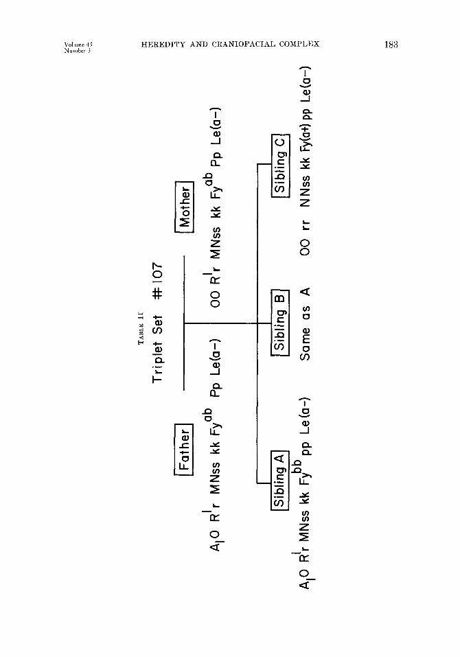
Blood (:?-OUp D&-For the sake of brevit,v, one set of triplets (set No. 107) and their parents will be used to illustrate how the data obtained from blood group analysis may be used in making zygosity determination on the basis of probability. A more detailed account map be found in the book entit,led Human Heredity by Neel and Schull.3”

18%
Table I presents the results of agglutination tests on both parents and triplets of Set No. 107. From these result,s, it is vcr~ often possible to tleterminc the genotypes of some, if not all, of the individuals involved. This is demon- stra,ted in Table II. With regard to t,he ABO system, the mother can have onlp
TABLE I. RESULTS OF AGGLUTIL-ATION TESTS ON FAMILY OF TRIPLET RF,T No.107
.___ __. .~~~~-
SUBJECT I
h~TISF,Rl~M --- -- ABO GROIJP
Father - c/CIotE/n~jN/SIs/K1E'p"j1',
I-- __- ~-
81 i- + + - + + + -- + + - Mother 0 + + + -- + + + - + c Sibling A A* 4. + + - i + + Sibling B A, 4. + + - + + + Sibling C 0 + -- - - + + + - -
the genotype 00, but the father may be either A,O, A,A,, or A,A,. ,411 chil- dren of this union must each have at least one allele derived from the mother. Sibling C, however, is a homozygote with two 0 alleles. Hence, the other 0 must have derived from the father. The father is therefore the genotype A,O. The other two siblings (A and B) have the phenotype A,, but since the A, allele ran hare derived only from the father, each must be a heterozygote, having received an 0 from the mother. For the ABO system, then, we can be certain of the genotypes of all fire persons. Additional explanation of this point. may be found in the work of Lawler and Lawler.”
For the Lewis system, only the anti-J,en serum was used. A positive re- action would indicate the phenotype Le (a+b-) .and the genotype LeaLea, but a negative reaction fails to distinguish between the genotypes LeaLeb and LebLe”. This was the case with all members of family No. 107. Hence, only the pheno- types are indicated-Le (a-).
Both parents gave positive reactions to the Duffy anti-Fy” serum. This means that both have the phenotype Fy (at) which could result from the geno- type Fy”Fy” or the genotype FyaFyb. Since siblings A and B reacted negat,ivel?; and hence have t,he phenotype Fy (a-) and the genotype FybFyb, both parents had to have the allele Fyb. The parents are thus established as heterozygotes with respect to the Duffy locus. On the other hand, sibling C gave a positive reaction and belongs to the phenotype Fy (a+), but it is impossible to determine whether he is a homozygote or a heterozygote. The genotypes for the other blood group loci are similarly debermined where possible.
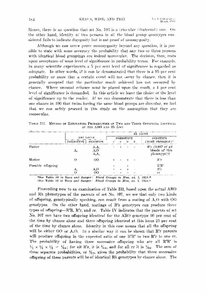
The next step is to estimate the probability of the three offspring of these parents having identical genotypes for all blood group loci tested by chance alone. For simplicity, only two loci arc considered in Table III-the ABO and the Rh loci.
Two statements of explanation must bc presented at this point. First, it is
assumed in these determinations t,hat there is no case of illegitimacy. Certiinly, t,he blood group data gave no evidence of incompatibilit,y. Second, dizygosity is immediately established if a single difference between two persons is found at ali! locus. For cxarnple, in triplet set No. 103, siblings A and B differ at the Rh locus, B and C differ at the ABO locus, and A and CJ differ at the Rh locus.
TABL
E II
=I
Trip
let
Set
# 10
7
I ab
Pp
Le
ta-
I e
AI0
R r
M
Nss
kk
Fy
00
R
r
MN
ss
kk
Fy ab
Pp
Le
ta-
B
5 E
f IS
iblin
g A
l 1 S
iblin
g B1
I
$
Sib
ling
Cl
E
8
A 0
R
’r M
Nss
kk
Fy
bb p
p Le
ta-
I Sa
me
as
A
00
rr N
Nss
kk
Fy(
a+) p
p Le
ta-)
3 E
Ilt~rict~, thtbre is no question that ~1 30. lOi{ is a trio\.ular ( I’raternal 1 ollt~. 011 the other hand, idcntit~- 01’ IWO persons in all thta l~lootl gl’ou]’ gyllotypt’s t*o11-
sidered fails to indicate dizyposit!- but is not p~woi 01’ ttlonozygosit).. L21thougl~ we can ncvtar pro~t’ monozygosii>- beyond any question, it is l)os-
sible to &ate with some accuracy the probability that any two 01’ three persons with identical blood groupings arc indeed ~r~onovula~. The decision, then, rtlsts upon acceptance of some level of significance in probability terms. t”or cxamplr, in many scientific expcrimrnts a 5 per tvnt 1~~4 of significancr is rcgardetl as adequate. In other WOI?~S, it’ it van bc tlcmonstratcd that. thcrt~ is a 95 pt:~’ cent probability or more that a c.cr.tain t~vcnt will not tmw I)?- t~lla~lw, l.lwn it is gt~nrrally accepted that thv particular result achit~vctl has not otvurred by- chance. Jl’herc unusual rt~liancr must bc placed upon the rcsnlt, a 1 per cent Icvel ol significance is tlcnlandcd. hi this article we leave the clioicta of the level of significance up to the rcadrr. II’ wc wn tlcitionstrxtc that tllttw is less t,llarl one chance in 100 that. twins ha\Glg tht> sa1110 blood groups are die\-ular, WC fuel that, we can safely proceed in this study on the assumpt,ion that they arc inonovular.
ABtJ I,Ot:lJS
PHENOTYPE 1 GENOTYPE I’HENOTYI’R GENOTYPE -
t’ ,’ u E i MOST PROBABLE ‘, ’
PatIle &A, + + + - li’r (0.937 of all A, A,0 ldoods of this
A,A, ~JhCllOty~C) t
Motlrcr 0 00 + + i - H’r
Possible oftqxing + - + - K’K’ A. A.0 + + + - FL’1 0' 60 + - - 71
*See Table 40 in Race and Sanger: Blood Croups in &fan, ed. 2, 1954.3” Wee Table 56 in Race and Sanger: Blood (Jrmcps in Man. ed. 2, 1954.aw
Proceeding now to an examination of Table III, based upon the actual ABO and Rh phenotypes of the parents of set No. 107, we see that only two kinds of offspring, genotypically speaking, can result. from a. mating of A,0 with 00 genotypes. On the other hand, matings of R’r genotypes can produce thrctl t,ypes of offspring-R’R, R’r, and rr. Table 1V indicates that the parents of set No. 107 can have two offspring identical for the ABO genotype 50 per cent of the time by chance alone and three offspring identical at, this locus 25 per cent of the time by chance alone. Identity in this case means that all t,hc offspring will be either 00 or A,(). Jn a similar way it can be shown that R’r parents will produce offspring in the expected ratio of one R’R’ to two R’I- to one rr. The probability of having three succc&re offspring who arc all R’R’ is j/i x l/4 x l/4 = z/s4; for all R”r, it is y(;4, and for all rr it is I?&. The sum of these separate probabilities, or y 32, gives the probability that three successive offspring of these parents will be of identical Rh genotypes by chance alone. The
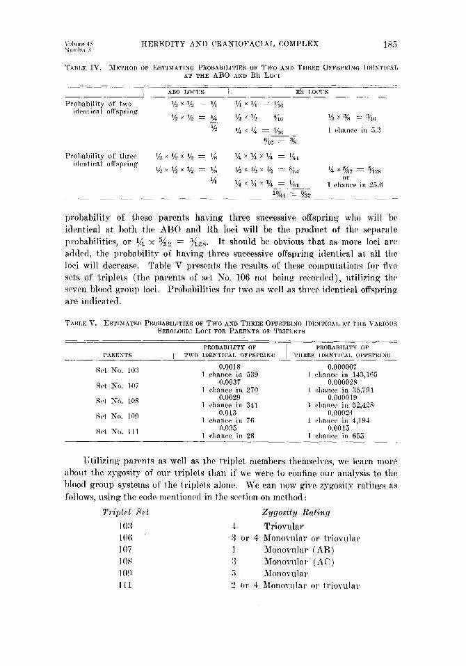
TTEREDITY ANI) CRANIOFACIAL COMPLEX 185
TABLE TV. METHOD OF ESTIMATING PROBABILITIES OF Two AND THREE OFFSPRING LDENTICAL AT THE ABO AND Rh LOCI
probability of these parents ha.ving three successive offspring who will hc identical at both the ABO and Rh loci will be the product of the separate probabilities, or l/4 x Kz = 7&s. Jt should be obvious that as more loci arc added, t,he probability of having three successive offspring identical at all the loci will decrease. Table V presents the results of these computat,ions for five srks of triplets (the parents of set No. 106 not being recorded), ut,ilizing t,he seven blood group loci. Probabilities for two as well as three identical offspring are indicated.
TABLE V. ESTIMATED PROBABILITIES OF Two AND THREE OFFSPRING TDENTICAL AT THE VARIOUS SEROLOGIC I,OCI FOR PARENTS OF TRIPLETS
PROBABILITY OF I'ROBABILITY OF I'ARRNTS TWO II)ENTICAL OFFSPRING THREE IDENTICAL OFFSPRING
Srt No. 103 0.0018 0.000007 1 chance in 539 1 c>hnnee in 143,165
Ret No. 107 0.0037 0.000028 1 chance in 270 1 chance in X.791
’ St+ No. 108 0.0029 0.000019 1 rhnnre in 341 1 chnnw in 52,428
srt No. 109 0.013 rl.00024 1 chance in 76 1 c~hxnce in 4,194
Rrt, No. 111 0.035 0.0015 I chanre in 28 1 chance in 655
TJtilizing parents as well as the triplet members themselves, we learn more about the zygosity of our t,riplet,s than if we were to confine our analysis to the blood group systems of the triplets alone. Wc can now give zygosit,v ratings as follows, using the code mentioned in the s&ion on method :
T1ipld set %ygosity Rating
103 4 Triovular 106 3 or 4 Monovular or triovular 107 1 Monovular ( AB) 10s 3 Monocular (AC) 109 5 Monorular Ill 2 or 4 Monoviilar or triorulal

l’ho YUIBY’ l’csf.--l\‘c tlwt1 ricxt t,o the i.c+zults of tllc taste test. 1’01~ scnsitivit> lo l)llcll?-lca~l)allliilc (I’T(’ j Tlmc MV gi\-cn in Tal)lc VI, togotlwr wit II tI~(i itI- dicat(:d qvgosity hsccl 011 f It<: t;islrr’ test alone ancl t Ilc’ “il('tlUll" qypsit>- 21s (1(x- tcrminrd by combining all tests. The ability to t,astc l~l~enyltl~ioc~~~~l~arrridc is generally thought t,o he inhcrit,ed on the basis of simple Mendclian rlotninancc. LZ homozvgous rcecssivc pcnot,ype will rcndcr the pcrsoii ~mahlc to taste this substance. The correlation bctwcen grnotype and phenotype is not, 100 p(‘r cent, however,‘h so that the test. is more adaptahlc to population gcnctics than to twin studies. As shown in Table VI, the results do not contradict the blood group findings in any instance; on the other hand, they provide no additional information. Those persons whom WC acrrptcd as monovular on the basis of proba,bility arc not discordant, with regard to ability to tastr PTC.
Tams VT. BESVLTS 01” TASTIC TEST FOR HENSITKIT~ To PIIENYLTIIIOCREA (PTC)
TRIPLET SET
A No. 103 H
(1
SEh-SITlVITY
Taste] Taste1 Taste1
A Taster No. 100 J3 Nontaster
c Taster
A so. 107 I3
(‘ /
A No. 108 H
C
Nontaster Nontastel Nontastw
Nontaster Taster
Nontaster
Taster Taster Taster
Taster Taster Tastrl
ZYGGSITY INDICATIOS
.i
:1
3
3
5
! ACTLJAIz ZYGOSITY
Fingerprint’s.-Dermatoglyphics is widely recognized for its great value in identification procedures. It is not as generally known that it possesses great potentialities for biologic and anthropologic research. Fingerprints are com- pletely established, except for size, during the first four or five months of embryonic and fetal development and are not affected by postnatal factors.40 Although not infallible, dermatoglyphics has been used extensively in twin diagnosis. Diagnosis. of monozygosity is made on the basis of high degree of similarity rather than identity. Bonnevie5 and others have indicated that at least one pair of alleles is involved in the determination of the number of ridges. It is generally conceded that the total expression of fingerprints, in pattern and intensity, is dependent upon a large number of genes in several loci. Indeed, this may be a distinct advantage of dermatoglyphics over blood grouping and other techniques in population studies.40 Studies by Ma.cArthu9 have shown that ridge counts have the highest heritability, followed by finger-tip patterns, palmar patterns, and palmar main lines.

HEREDITY AND CKANIOFACIAL COMPLEX 187
TABLE VII. DERMATOGLYPHIC PATTERN TYPES ( GALTON SYSTEM)
TRIpLET SETS -lo:(( IOB 107 108 109 111
FINGER A/BIC/D*(AIBICjD*(AIBICID*(A~BI(:jD”IAlBjCID~~~/h/~C~DD’
Bieht hand ~2 1 L L w 1 L L L 5 I, L w 1 w w L 1 L I, L 5 I, L w 1 2 L L L 5 L L L 3 w ww 5 w w w 5 A L I, 2 1V L L 2 3 LLL5 LLL5 1, L I, 5 LL I, 5 LL L 5 LLW 1 4 TV w w 5 L L w 1 L L TV 1 TV TV m 5 1, L L 5 \vmw 5 3 L w L 3 L L L 5 I, L w 1 L TV L 3 A A L 1 \V 177 w 5
I&f hand 1 L w L 3 L 1, 1, 3 1, L I, 5 \v w 1, 1 1, 1, I, 5 I, 1v J, 3 2 W IV 1, 1 1, A L 3 I, I, \V 1 W \V 1%’ 5 ALL2 WLLZ 3 L L I, 5 1, L I, 5 1, L 1, 5 1, L \v 1 A A A 5 1, L I, 5 4 w 1, w 3 1, L L 3 1, L w 1 w w w 5 r, I, L 5 WLW3 5 L \v TV 2 I, 1, I, 3 1, I, I, 3 \I’ IV iv 6 L I, A 1 I, w w 2
Final diagnosis 4 5 1 1 5 4 Actual diagnosis 4 3 1 :i 5 4
(according to I;lood typ&)
*Diagnosis of zygosity based on similarity of types. Legend is same as for Table VI.
In Table VII the finger-tip pattern for each finger of each subject is re- corded, together with an estimate of zygosit,y based upon over-all resemblance. In no case is there disagreement with the actual zygosit,y diagnosis, although the fingerprint patterns fail to indicate that only siblings A and C are monovular in sets No. 106 and No. 108. When fingerprint patterns are subdivided into numerous types (Table VIII), there seems to bc less power to diagnose ac- curately or decisively.
TABLE VIII. DERMATOGLY~HIC PATTERN TYPES (MA~Rs' CLASSIFICATION MODIFIED BP CUMMlKS AKD l&DLO
FINGER Right hand
1. ?!) B 13 ? 28 28 28 5 28 29 9 ? 21 21 28 1 28 2X 28 5 28 21 13 4 2 28 2828 5 282828 5 13 32 7 5 18 4 27 27 27 5 23624 4 3 28 28 28 5 28 28 28 5 282828 5 282828 5 27 2828 2 2828 5 1 4 ? 8 7 B 28 B BP 28 28 7 1 18 13 18 3 27 28 28 2 18 11 5 4 5 1 8 t a 282828 5 2828 7 1 281328 3 27 27 27 5 855%
Left hand 2 7 ?24 9 24 39 24 3 28 28 28 5 7 2 18 4 27 27 28 1 9 27 15 4 3 24 24 24 5 24 24 24 5 24 24 24 5 242418 1 27 39 39 2 24 18 24 3 4 19 24 2424 88 ? 24 2 ? 18 18 5 1 24 B 24 9 18 27 9 4 5 5: 9 29 2424 I'! B B 9 3 18 B 18 ? B B 89 18 B $7
Final diagnosis 5 3 5 1 5 or 2 4 Actual diagnosis 4 5 1 3 5 4
(accordine to hood typ&)
*Diagnosis of zygosity based on similarity of types. l,egend is same as for Table VT. ;Thumbprints of left hand were not satisfactory.
As predicted by MacArthur, ridge counts seem to be very powerful diagnostic tools. Table IX presents all counts that could be made. Some counts could not be made because of faulty technique in making the prints. It must be pointed

1x IO I (i 3 ":I 16 23 :j !I
0 0 0 3 I ;i 3 !) 4 12
0 4 4 2 II x 71 15
0 2 3 3 (i 4 9 1 13 I,<
0 0 0 5 f 7 7 L'
__- Final diagnosis 4 3 1 3 5 4 Actual diagnosis 4 3 1 3 5 4
Caceordiw to lhood types)
*Diagnosis of zygosity based on similarity of types. Legend is same as for Table VI.
TABLE x. CONCORUANCk: (C) ANIt ~~ISCOBIIANCE (-) IN ELEVEN ;2iI~R1~IIOLOGl~! ~I:AI’l’S OY ‘I’HI’: LOWEK YREMOI,ARS (AFTER litmus ANI) .F~RR)
I TRIPIJCT SETS
‘TRAIT NC).*
1 2 3 4 5 6 7 8 CJ
10
10:3 107 108 I 109 - l/2/3 1 qzj3 / , , 2 , R-~-l--,-2-pz
B s + + - - + + + + + + + + - - + + + + + + + + - - t + + + + +
- + + + i + + + + + + . - + .- + J + +
+ + + + + T + + + + t + + 1. J-
+ - + + + + + i + + -_ + - + - T 1 -c
+ - - + - 4. I - + 11 + - - + - ! + ? + + -
Total number of 3 4 4 10 3 4 4 6 -71 9 11 I1 concordances
Frequency of concordances .60 .4-o .3(i .!I1 .2i .X .4u 54 .9u 1.0 1.0 1.0
Zygosity indication 4 1 3 5
Actual zygosity 4 I 3 5 -______ *Traits deflned as follows:
1 = Number of external lingual 6 z Mesial deuteroconid margin. grooves. 7 = Distal protoconid mar&n.
2 = Sagittal sulcus. X = Central occlusal protoconid ridge. 3 = Position of deuteroconid. Y = Number of occlusal protoconid ridges. 4 = Number of lingual cusps. 10 = Deuteroconid-protoconid relationship. 5 = Me&al protoconid margin. 11 = Size of protoconid ridge.
(See Kraus and Furry for detailed description. )

Yolt~me 4i IIEREDTTT AND CRANIOFACIAL COMPLEX Numhrr i 18!)
out that, zygosity diagnosis is ordinarily made on the basis of total ridge count on all ten fingers. We have made a comparison finger by finger. However, a total count gives exactly the same results. The diagnosis agrees with that obtained by the probability method using blood groupings and goes even farther. 1Vherca.s the blood-grouping technique was not definitive for sets No. 106 and No. 111, indicating that the former could be rated either 3 or 4 and the latter 2 or 4, the ridge counts indicate that in No. 106 siblings A and C are truly monovular while in No. 111 siblings A and B are not monovular. Thus, No. 111 is diagnosed by ridge counts as a triovular set. It is int,eresting to recall that the probability of A and B in set No. 111 being diovular was higher than fol any other t,win pair having identical blood groups.
Dentnl Morphology.-A new technique for diagnosing zygosity has been ad- vanced by Kraus and Fur? and tested population-wise by Kraus.2” It is based upon certain discrete structural characteristics of the lower first premolars. The method is t,herefore limited to persons over 10 years of age and, because of caries and excessive wear, under 25 or 30 years. It has been postulated that these morphologic traits are independently inherited but, the mode of inheritance has not been established. Eleven of these traits were observed and recorded in four triplet sets (Table X). Concordance and discordance were noted between each of the possible pairings (1, 2, and 3) in each set. The concordances were then tot,aled for each pair and the relative frequency of concordances was re- corded. In set No. 103 the concordance frequencies were 0.50, 0.40 and 0.36, indicating that there were as many (or more) discordances bet,ween each pair as there were concordances. The diagnosis is, therefore. triovularity. In set, No. 107 there was 91 per cent concordance for pair A and B, but only 27 per cent and 36 per cent for pairs B and C and A and C, respectively. This is pre- cisely the zygosity indicating by blood grouping and dermatoglyphics. Similarly, in set,s Ko. 108 and No. 109, the zygosity is clearly indicated by the differential concordance frequencies. Unfortunately, the triplets in set No. 111 had not erupted their lower first premolars. It would seem, on basis of this small sample, that the lower first premolar examination ranks with dermatoglyphic ridge count,s for positive diagnosis of monozygosity and with blood grouping for detecting dizygosity. Plates I and II illustrate many of the structures of t,he premolars and permit the reader to see for himself the unusual degree of concordance in monovular twins and triplets.
The well-known Carabelli tuberclc or cusp of the maxillary first molar has been considered by most writers to be inherited according to simple Mendelian dominance.‘*, 24y 42 Dietz” is led to believe t,hat its genetic pattern is a comples one. Family studies have yet, to be carried out to determine the mode of in- heritance. Twin studies, however, indicate that the trait is under rather strict genetic control.‘l~ 23 In diagnosing zygosity, therefore, we must consider the Carabelli trait, like the lower first premolar traits, a second-order variable. Some authors have recognized several different manifestations of the Carabelli trait and have classified them in a varietp of ways.“, u The method employed br Kraus,“” who calls the t,rait “Carabelli’s anomaly,” will be utilized here. Foul aspects aw rwognixccl : (1 ) the prononncwl tnlxwlc, with a palpable apes; (2 j

.I
B.
A.
B.
Plate I.-1LnWr right. first PremOlWS. illustrating inherited rnorphologlc structures. Triplet set No. 103. B, Trwlet set NO. 107.
Plate II.-Lower right first Premolar% illustrating inherited nlorphologic structures, Triplet set NO. 108. B, Triplet set No. 109.

HEREDITY AND CRANIOFACIAI, COMPLEX 191
the slight tubercle, whose apes merges into the side of t,he mesiolingual CLI
(3) one or more pits or grooves at the site of the anomaly; and (4) compl il IS 1 cnre of any manifcst,ation.
l’lak 111.
sp; ete
A.
B
A.
6
108
10;
Plate IV.
Plate III.-Maxillary first molars, illustrating Carabelli’s anomaly. (right molars). R, Triplet set No. 106 (left molars).
A, Triplet set
Plate W.-Maxillary first molars, illustrating Carabelli’s anomaly. (left molars). B, Triplet set No. 108 (left molars).
4, Triplet set
NO.
NO.

Plates III, IV, and 1’ p~senl photographs 01’ t Ilrl cdast 01’ 1 lie maxillary first molar for each triplet set. In stat So. 103 A is difYrrcntiatcd from B arid C ilr having a groove. Tl1t: ot11cr t\ro Ilwlrlhc~i% ol’ the set SllOlV au ahswcc ot’ t11o trait. In set No. 106 both A and (’ tlispla)- ~)ronoanccd tubercles of thrt samt: size, while B shows only a slight, groo\-0. In set No. 107 A and B have slight tubercles of about t,he same size and shape, but (! has a pronounced tuberclc.
Plate V.-AMaxillary flrst molars, illustrating Carabelli’s anomaly. A, Tripkt set So. lo!) (right molars). R, Triplet set No. 111 (left molars).
All three members of set No. 108 look similar, but careful scrutiny of’ the photo- graphs shows that A and C possess a single groove each, while B has a slight tubercle. In set No. 109 the tra,it appears absent. in A and B, with a very slight tube&e apparent on C. It should be pointed out the three teeth are not oriented in exactly the same way, so that a slightly different orientation will reveal that each person has a verv slight bulge at the same site. Ill No. 11 I
the three siblings show clearly differentiated manifestations of the Carabelli anomaly. A has a groove, B has a pronounced tubercle, and C has a slight tubercle with a deep groove. On the basis of the Carabelli anomaly, set No. 111 is diagnosed as triovular. 1.t is interesting to note that it was not possible IO achieve this diagnosis on the basis of blood grouping and probabiliQ*. On the other hand, the diagnosis of diovularity for B and C cannot be made on the basis of the Carabelli anomaly. Otherwise, tho rrsult,s are in accordance with those yielded by blood grouping, dermatoglphics, and the lower first premolars.
Xtature and Weight.-Stature and weight, which we may call third-order variables, have hccn recorded for each person, iIn< an indicator of hods bniltl, the

height-weight index, has been computed. As expected, these observations have no reliability in zygosity diagnosis. In set No. 111, for example, A, B, and (2 are practically identical in weight, stature, and body build, although this set is triovular. The members of the monorular set, Ko. lo!), on the ot,her hand, show dissimilarities in weight and body build. These variables illustrat,e what WC have pointed out earlier, namely, that traits which are really complexes of many elements are inadequate for estimating heritability, even though heredity cer- tainly conkibutes to their formation.
t:.
Photogruphs.-At this point the reader may be curious about the general appearance of the triplets. Having established the zygosity of each set with some assurance, one might wonder if the same results could have been achieved

simply by looking at. the triplt>ts and jndgin, 0 1~~ Iho usual criteria of similarity. , \Vc permit the reader to do this IYor himsel I’ b)- sicrntinizing Plates VI to Xl, urhich provide frontal and lateral head ~icws oricntcd in the Frankfort, horizontal plane.
B.
c.
PIah= VII.-Triplet set h’;o. lO(i.
WC have gone to rather unusLm1 lengths in presenting our diagnosis of zygosity for the six triplet sets. Our purpose has been to underscore the diffi- culties involved in gaining grcatrl* diagno& reliability and to cast suspicion upon the results of-twin studies in which diagnosis has been based upon too hasty or too inadequate examination.

HEREDITY AND CRANIOFACIAL COMPLEX 195
HERITABILITY IN THE CRASIOPACIAL COMPLEX
We have established, with a high degree of probability, the zygosity o-f each of the six triplet sets. The next order of business is to examine the craniofacial complex in order to determine whether it or any of its components can be demonstrated to be predominantly under hereditary control. It should be stated
A.
G.
I’lntc VIII.-Triplrt set No. 1Oi.
that this phase of the research was conduct,ed independently, without previous knowledge of the zygosity of the triplets. In this way, bias was eliminated in making the many tracings.

c.
WE !C choose to recognize, we really are being highly selecti1.e in that we c consciously 01 U nconsciously peel from it, many attributes which make it, a fun&c 3ning part Of a whole living organism. In select,ing from a. lateral hcadfilm a line , an angle, Or a t geometric configuration to follow through in our investigation of the in- flu Lei rice of heredity, we must recognize that we are extra~cting an entit ,J- that has

no biologic individuality and no function. It. is essentially a mental construct. If this be true, and we seem to observe some correlation between this trait and the genetic constitution, WC arc tbrn faced with a serious problem of interprrta-
A.
n.
c.
Plate X.-Triplet set No. 109.
t:ion. Is this trait controlled essentially by heredity! Or does this trait for- t-nitously reflect, one or more biologic entities which are themselves the manifesta- tions of genie action?
For example, the cephalic index is a time-honored ratio which has been employed by antzhropologists in distinguishing the various prehistoric and

198 KRATJS, WISE, AND FILET
modern peoples of the world. It is simply the ratio of maximum breadth of t,hc cranium to maximum lengt,h of t,licl cranium measured from glabclla lo opisthocranion. It ha,s man)- times been obscrvcd that cert,ain pol)ulations arc brachycephalic (round-headctl, with a high cephalic intlcs) au{1 otkrs arc’ dolichocephalic (long-hcadcd, with a low cephalic indcs). Since, in a gcncrwl wag, these populations breed true with respect to values of t,hc intltx, it has
A.
B.
c.
Plate XL-Triplet set No. 111
sometimes been said that this trait (the cephalic index) is inherited. Since length of the cranium is achieved during growth through the operation of man) forces, elements, and their interactions, to say that the index is inherited or that “85 per cent of the trait” is inherited is really to say nothing at all! Cert,ainly, we would predict that any at,tempts to establish the “genetic meehanism of in- heritance” of the index would be doomed to failure or misinformation.

HEREDITY AND CRANIOFACIAL COMPLEX 199
Let us begin with the craniofacial complex as a whole. A midsagittal profile of the cranium and certain structures of the facial skclcton drawn from a lateral hcadfilm is, of course, mcrcly an arbit,rary way of tlcscribing sclccted mauifcsta- tions of the total complex in two dimensions. As with the ccplialic indcs, the lateral profile is the result of tlic operation of many $IYJWth f:KfOl’S, Only SOInC
of which a.re known. Since growth Forces are untlrr gcnctic control, we cau safely say that the lateral head profile is influenced by heredity and environ- ment. Can we learn whether heredity plays the predominant role? Is the lateral head profile a trait which is amenable to this sort of observation1 In Fig. 1 lateral tracings of monovular triplets A and B of t,riplet set Ko. 107 arc supcr- imposed, wit,h registry on R and holding the Bolton planes parallel. The over- all concordance of the two tracings is remarkable and might lead an incautious observer to assert that the lateral profile of the head is under rigid genetic eon- trol. When he then turns to Fig . 2 in which the tracings of diovular triplets A a.nd C of set No. 107 are superimposed, his feelings will he reinforced. These triplets show greater discordance in all areas except the frontal and par&al profiles.
Now we examine similar tracings of another monovular pair, B and C of triplet. set No. 109 (Fig. 3). The degree of discordance found here is greater than that expressed in pair A and B of set No. 107. We further note that there is no consistency in the areas ,of discordance. Jn t,riplcts B and C of set No. 109 t,here is greater discordance in the lambdoidal and frontal areas, as well as in the malar and symphyseal regions. On the other hand, in Fig. 4 (triplets A and C of set Eo. 103) the tracings of diovular triplets show a much higher de- grce of concordance than do those of the monovular pair of set No. 109 and as grea.t a concordance in general as those of monovular pair A and B of set No. 107.
In tracings of pairs taken from the other triplet sets, inconsistencies of the same type were observed. It is quite evident, then, that the craniofacial complex as represented by a lateral head profile is not amenable to heritability study by means of the twin method. We expect, and we observe, an over-all profile similarity in twins. Whether this similarity is meaningful in a hereditary sense can be determined only when it is compared with tracings of unrelated pairs of persons of the same sex, age, and general head size.
Since the lateral profile of the entire craniofacial complex is unsuitable for heritability studies by the twin method, we turn to a component of the complex, namely, a lateral profile of the cranial vault. In Plates XII, XIII, and XIV the lateral vault tracings of the three members of each triplet set are regist,ered on R with Bolton planes drawn parallel. In Plate XII (the triovular set) there are very few points of contact from nasion to Bolton point. In Plate XIV (set
No. 108) there is concordance between the monovular triplets A and C in the bregmatic and nuchal areas, and there is greater similarity throughout the rest of the perimeter as compared with their fra.ternal triplet B. On the other hand, let us examine diovular pair A and B of set No. 103 (Plat,e XII). As high a

Fig. 2.
wlth Fig. 1 .-Superlmporing 0r Irctrral trnrinpn of mnnnrulnr twins cln7 An) rwistdnr on R Bolton planes parallel. FIR. ?.--Nuperimpnnlng 01 I:~tw:~l tr:~~in~.s 0l 4liovul:br t\\ In.s ( Illi .\O b vxktwlnr 011 I:
with Hnlton plonra p:tnrll4!l.

with
with
Fig. 4.
Fig. 3.-Superimposing of lateral tracings of n~~~ovular twins (109 BC) registering on R Bolton planes parallel. Fig. 4.-Superimposing of lateral tracings of diovular twins (103 AC) registering on 1s Bolton planes parallel.
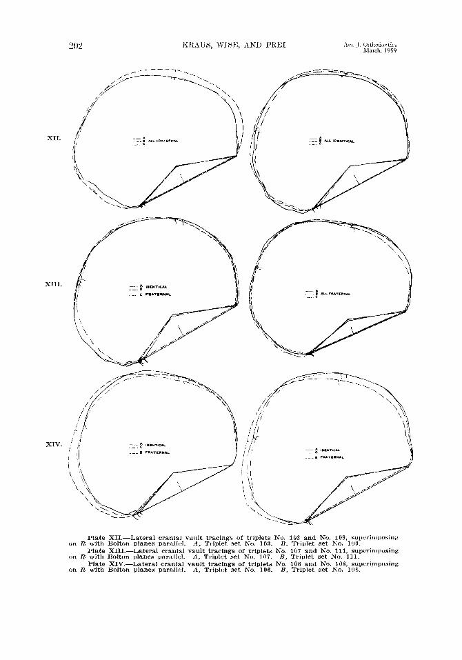
Plate XII.-Lateral cranial vault tracings of triplets No. 103 and No. 109, superimposing on R with Bolton planes parallel. A, Triplet set No. 103. B, Triplet set No. 109.
Plate XIII.-Lateral cranial vault tracings of triplets No. 107 and No. 111, superimposing on R with Bolton planes parallel. A, Triplet set iYo. 107. B, Triplet set No. 111.
Plate XIV.-Lateral cranial vault tracings of triplets No. 106 and No. 108, superimposing on R with Bolton planes parallel. A, Triplet set No. 106. B, Triplet set No. 108.

Volume 45 HEREDITY AND CRANIOFACTAT, COMPLEX 203 Number 3
degree of concordance is found in tlicse tracings as is to be found in anJ mono\-ular pair, and a grcatcr dcgrccl of concordance is evident than is eshiMrt1 t)y the ~nonovular pairings A and C of set No. 108 (Plate XIV), A and c’ or scd
No. 106 (Plate XlV), or A, B, and C, of set No. JO!, (Plate XII). If we were to attempt to judge zygosity on the basis of concordance of lateral cranial vault tracings registered on R, then we would hare to conclude t,hat No. 111 (Plate XIII) was a monovular triplet set, whereas in actuality it is triorular!
Another method of orienting the cranial vault is illustrated in Plates SV to XVII. In these tracings the sella-nasion lines are held parallel, and maxi- mum profile contact is sought. It is readily apparent that triplet set No. 111, a triovular set, shows the greatest degree of concordance. The monovular set, No. 109, reveals considerable discordance in the posterior segment of the profiles. Triplets A and C in set No. 106 are remarkably similar throughout, but it should be noted that this concordance is achieved by a considerable inferior displace- ment of the sella-nasion plane and anterior displacement. of the sella-articularc line. Siblings A and C in set Xo. 108 arc clearly the more similar pair, B show- ing significant differences in the entire profile from nasion to articulare. In set NO. 107 A and B are almost identical except for a small area anberior to bregma. The cranial base lines in this pair are also very similar. In the triovular set, No. 103, the situation can be summed up as gcncrally equal discordance displayed b,v each of the three possible pairs.
Our conclusion at this point is that registration on R throws the cranial vault profiles into positions which tend to obscure the fact that there is a good deal of similarity in triplets in general. Furthermore, it is valueless in pointing out whether or not monovular persons have more similar cranial profiles than diovular or triovular persons. On the other hand, b- attempting to superimpose cranial vault profiles, we see immediately the essential likeness of these triplets and in some instances can note the greater concordance between monovular persons. However, this observation is gained at the expense of superimposing the sella-nasion planes. The latter are displaced in unpredictable ways. The cranial base, then, is not highly correlated, from the point of view of lateral profiles, with the cranial vault outline. This could have been predictecl with a little reflection upon the differential growth processes invo1ved.45
We next test the facial complex itself, utilizing a polygon that is identical or similar to those frequently employed in growth studies and in clinical as- sessment. The method of construction of the polygon is illustrated in Fig. 5, 11. Four additional lines are drawn as indicated. It is perhaps unnecessary to elaborate on the fact that this facial polygon, and all similar ones, are more profound abstractions than is the lateral tracing of the cranial profile. For example, sella is a point in space. The line nasion-pogonion is partly in space, goes partly through teeth, and crosses random portions of discrete bones as well as areas where only soft tissue occurs. If this line is not a. hereditary unit, how much less so is the entire polygon. 1 It must be remembered that the utility of facial polygons for clinical evaluations in trcatmcnt and in growth is not under

T’lntc XVII.
.4 1:
Plate XV.-Lateral crania1 vault tracings of triplets No. 103 and No. 109, superimposing on maximum contact with S-N planes parallel. A, Triplet set No. 103. R. Triplet set No. 109.
Plate XVI.-Lateral cranial vault tracings of triplets No. 107 and No. 111, superimpoa- inr on rnaximutm contact with S-N; planes parallel. A, Triplet set No. 107. R, Triplet set No. 111.
Plate XVII.-Lateral cranial vault tracings of triplets No. 106 and No. 108. superimpos- ing on maximurn rontnct with S-N planes pnraIl~?l. A, Triplet set No. 106. R, Triplet set No. 108.

B
c
Fig. 5, A, B, and C.-Facial polygons. S = Sella turcica. N = Nasion. ANS = An- terior nasal spine. maxillary fissure.
PO = Pogonion. Go = Gonion. Ar = Ar’iculare
Condylar apex. PNR = Posterior nasal spine. Rrc = Rasion. ’ Ro =’ Bolton point.
PTM = Pter;g;-

‘LOti .\ni. I. Orthodontics March, I959
question. Their applicabilit,y to hetwlit,y studies is the point under considera- i ion. ilgain, our method has bwn to c~o~npaw the polygon tracings of each set OH t,riplcts, superimposing on ttic sclla-nasion line with registration on sella. Plates XVIII to XX present the rcsnlts.
If the facial complex as manifest& by a polygon is predominantly con- kolled by hereditary factors, then WC expect identity or close similarity bctw-rcn
Plate XVIII
- $ IDCNTICAL --_
. ..-... c FRATIIINAL
-...__... e... A----
A. h’ Plate XIX.
Plate XVIII.-Facial polygons of triplets No. 103 and No. 109, superimposing on S-N with S registered. A, Triplet set No. 103. B, Triplet set No. 109.
Plate XIX.-Facial polygons of triplets No. 107 and No. 111, superimposing on S-N with S registered, A, Triplet set No. 107. R, Triplet set No. 111.

IIEREUITY AND CHANIOFACIAL COMPLEX 207
polygons of monovular triplets and less similarity between those of diovular triplets. Let us begin with set No. 109 (Plate XVIII). We are struck by the fact that all three sella-articulare lines superimpose; hence the three X-J-s-A angles are identical. From this point on, there is no longer identity in either lines or angles. Since the three S-N lines are of different lengths, as are the N-PO lines, the superimposing of the S-N-PO angles is strictly a matter of chance, or rather of geometric coincidence, and not of heredity. Thus, if lines A-S and S-N are of identical lengths in the triplet set and angle A-S-N is identical as well, but angle S-N-PO differs in each of the three triplets, then the remaining lines and angles will not superimpose. In other words, in a polygon of this nat,ure WC are dealing with a network of highly dependent variables and with a very few degrees of freedom. Vary one element and the interrelationships of many others are altered in, as Wylies3 has observed, unpredictable ways.
--- 0 FCUTORNAL
A. B.
Plate XX.-Facial polygons of triplets No. 106 and No. 108, superimposing on S-N with N registered. A, Triplet set No. 106. B, Triplet set No. 108.
The least amount of discordance is observed, strangely enough, in the triovular set No. 103 (Plate XVIII). In addition to S-N, lines S-A and A-Go superimpose almost 100 per cent. However, line A-Go is of different length in each polygon; hence, angles R-GO-PO, GO-PO-N, and Go-N-S are discordant for the whole triplet set,. The polygon comparison of the other triplet sets yields no reliable or objective data for discriminating monovular triplets. In fact, in triovular set. No. I1 1 the degree of concordance between B and C is greater than that between any monorular pair. It, will hc recalled that thcsc two persons had identical blood groupings and showed great physira.1 resemblance (Plat,c XI).

lip to this point we have ~~saniincd the craniofacial complex as a whole mcl
th di\:idcd it into its two component parts-the cranial vault and the facial complex. We hvc demonstratc~tl that none of tliw tlmc co~i~pleses, iis defined in lateral headfilm tracings, arc amenable to the study of heritability by the twin rnethod of analysis. llet us now turn our attention to smaller units ol’ the facial complex. These are illustrated in Fig. 5, R a.nd C. One may he t.ermod an upper facial quadrilateral defined by points S, N, ANS, and PNS (Fig. 5, ,I ). The other includes denture and mandible and might be labeled t,he nraxillo- mandibular quadrilateral. It is defined bp the points 133, ANS, (:o, and PO. Thr two quadrilaterals together comprise a major portion of the facial polygon.
We shall vary our m&hod of analysis and approach the p~*ohlem from the statistical aspect. If it is assumed that each of the four sides of either quadri- lateral is an independent, variable, that is, independently inherited, then it, is expected that the difference between similar sides should be less in monovular than in diovular. triplets. We hasten to say that there is no gcncbic warranty for this assumption. We mr~rcly exercise a licrnxc which may be justified by the end. It is rthasonablc to suppose that in a few isolated cases chance irr thr form of envir,orrrrrent,srl whimsies might protlucc~ a greater differcncc in monovular than in diovular triplets. In the long run, however, the truth would be apparent, namely, that, monovular are more similar in t,he length of t,he lines of the quadrilaterals than are diovular triplets. Thr same assumptions would apply to the four angles of each quadrilateral.
The statistical procedure was as follows : There were six possible monovular pairings and twelve diovular pairings. In the upper facial quadrilateral the lengths of the four sides were rneasured at random with respect to the eighteen persons. Correction scales1 were used on al I diameter measurements. The differences between corresponding sides of the quadrilat,eral were recorded for each of the eighteen pairs. In order to make each of the four sets of differences comparable to each other and additi\c, they were expressed as proportions of the rnean of the combined lengths, according to the formula a-b . Thr four pro-
(a+b) -)
portional differences were then totaled for each of the eighteen pairs. The rllcans of these sums for the six rnonovular pairs and twelve diovular pairs were computed and the “t,” trst for unpaired data was applied to determine whether or not the difference between the means was significant. The formula
f,-Z, s (Zl--XZ) ’
using pooled variances, was applied. I-ndcr t,he null hypothesis, WC
expect to find no real difference between means; in other words, the two groups would be samples drawn frorn the same universe. The results may be tabulated as follows :
Lines Monovular pails Diovular pairs ~~~
Angles Monovular pairs -- Diovular pairs
0.150 0.144 0.50 ..__-
0.127 0.117 0.333 0.70

HEREDITY AND CR,ANIOFACISL COMPLEX 209
3faxillomandibzclar quadrilateral
Lines
Angles
Monovular pairs 0.179 Diovular pairs 0.211 0.66
Monovular pairs 0.124 Ijiovular pairs 0.169 1.06
0.40
0.30
In none of the four comparisons is the null hypothesis overthrown. The con- clusion is obvious. Neither angles nor diameters of the upper facial or maxillo- mandibular quadrilaterals are able to express the differential influence of
heredity over environment. Angles and diameters, like cranial and facial pro- files, are not units of inheritance but are, rather, constructs which cut across, in random ways, the phenotypic manifestations of complex growth and functional forces operating in the head from prenatal life to moment of observation.
Apparently some of the traditional types of cephalometric concepts and t,echniques are not applicable t,o the study of heritabiliby. Whether we use an approach that embraces the entire craniofacial complex or one that subdivides the complex into smaller components, the results are the same. None is a rea.l biologic entity. None reflects the operation of a single genetically dominated system of development.
Using the techniques of roentgenographic cephalometry, and staying within it,s limitations, can we find smaller units of the craniofacial complex which fit our biologic demands and hence might allow us to learn if, where, and how heredity exercises a major measure of control in the craniofacial complex?
Except in certain cases, the usual lat,eral headfilm permits one to draw with a high degree of reliability the profiles of many skeletal structures. Other
TARLE XI. KEY TO BONE TRAITS
TRAIT NO. 1 DESCRIPTION
From Lateral Headfilms 1 External surface of occipital bone, from Bolton point to lambda, in MSP 2 External surface of calvarium, from lambda to bregma, in MSP 3 Profile of external surface of squamous portion of frontal bone, in MSP 4 External profile of supraorbital portion of frontal bone from nasion to n
point above the supraorbital ridge, in MSP ii Profile of nasal bones in MSP 6 Profile of cerebral face of orbital portion of frontal bone in MSP 7 Profile of cerebral surface of cribriform plates of ethmoid bone in MSP H Profile of dorsum sellae and planum sphenoideum in MSP 9 Profile of sella turcica in MSP
10 Profile of frontal process of zygomatic bone defined by orhital margin and anterior margin of temporal fossa, in MSP
11 Profile of anterior portion of maxillary alveolus and nasal surface of palatine process of maxillary bone, in MSP
12 Profile of oral surface of palatine process of maxillary bone, in MSP 13 Profile of inferior border of mandible, from gonion to menton, in MSP 14 Profile of Dosterior border of ramus of mandible. from apex of eondvle to
16 1 fi
gonion, & MSP Profile of the posterior margin of the mandibular symphysis, in MSP Profile of the anterior margin of the mandibular symphysis, in MSP
17 From Frontal Headfilms
Profile of mandible from a point on the right posterior border of the ramus inters&ed by the mastoid process t,o the equivalent point on the left side

Fig. 6.-Identification of individual bone traits. (For key, see Table XI.)
graphically in Fig. 6. One is traced from a frontal headfilm and the other sixteen from lateral headfilms. It will be observed that each expresses the morphology of one or more aspects of a single bone as observed in two dimensions on a headfilm. These “traits, ” as we call them, can be called constructs only in the sense that any morphologic characteristic which is observed or measured is not descriptive of the entire structure. Each trait describes an aspect of the form of a single structural unit which is the expression of a definable system of
Volume 4; HEKEUITY ANU CRANlOFACIAL COMl’LE:X 211 Number 3
growth. We do not mean to imply that the control of any one of these structures is a simple genetic one or that environment plays no role. This would be naive, as we have stated several times previously. On the other hand, because a single system seems plausible in the development of each trait, we might hope that the genetic influence in this system might. be discerned if predominant.
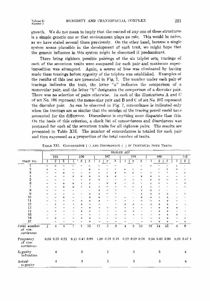
There being eighteen possible pairings of the six triplet sets, tracings of
each of the seventeen traits were compared for each pair and maximum super- imposition was attempted. Again, a source of bias was eliminated by having made these tracings before zygosity of the triplets was est.ablished. Examples of the results of this test are presented in Fig. 7. The number under each pair of tracings indicates the trait, the letter “a” indicates the comparison of a monovular pair, and the letter “b” designates t,he comparison of a diovular pair. There was no selection of pairs otherwise. In each of the illustrations A and C of set No. 106 represent the monovular pair and B and C of set No. 107 represent the diovular pair. As can be observed in Fig. 7, concordance is indicated only when the tracings are so similar that the smudge of the tracing pencil could ha.vc accounted for the difference. Discordance is anything more disparate than this. On the basis of this criterion, a check list of concordances and disordances was prepared for each of the seventeen traits for all eighteen pairs. The results are presented in Table XII. The number of concordances is totaled for each pair and then expressed as a proportion of the total number of traits.
TABLE XII'. CONCORDANCE (t) ANI) I)ISCORWNC~: (-) TN INIWMJAI, )to,us TKAIW
TRAIT NO. / 1 ,I",", 3 1 l ,l& 3 1 1 ,1;7;i‘:"T'2", 3 1 l ,I:, :$ 1 I ,I;,,
1 - t - - - t + - - - + + - - e + 2 t - - - + + - - + + + - + - + 3 - - + - - + + + + + + -1. + - - ._ + 4 - - - + + t + + t - + + + t - - 5 - - - _ - + + - - + + + - + 6 - - + + + + + + + - t - + + + + +
- - t + + f ,. - - - + + + + !3
+ + - - - + - - - + + + + - -
9 _ - - - - + + - - - + + + + - + 10 - t - - + + - - - + t t t - - 11 - - - + + + t - - - + + + t 12 - t - + + t t - - + + t + t + 13 - t t - - + t t f -
- + - + + t + - - + t- ::
+ + t t t - - t t t + -
16 - t - t t t t - - - + t t + - + 17 - + t - - - t + + + - +
rota1 number 1 4 4 7 7 15 17 3 3 4 5 13 16 14 15 5 9 of con- cordances
Frequency 0.06 0.23 0.23 0.41 0.41 0.88 1.00 0.18 0.18 0.23 0.29 0.76 0.94 0.82 0.88 0.29 0.47 ( of eon- cordances
Zvgosity 4 3 1 3 5 4 “indication
Actual 4 3 1 3 5 4 zrpositv

,\I,,. J. Or~hurlontics Mmh. 1959

V&me 45 Number 3
HEREDITY AND CRANIOFACIAL COMPLEX 213
In set No. 103, which is a triovular set, the relative amount of concordances for any possible pairing is less than 25 per cent. In set No. 109, which is a monovular set, the proportion of concordances ranges from 82 per cent to 94 per cent. In set No. 106, in which A and C (the pair designated by 3) is monovular; t,he two diovular pairings have concordance proportions of 41 per cent each, while the monovular pair shows an 88 per cent concordance. The monovular pair A and B of set No. 107 shows concordance in all seventeen traits, but the two diovular pairings of this set show only an 18 per cent concordance. The monovular pair of set No. 108 is similarly distinguishable from the two diovular pairings, with a concordance proportion of 76 per cent as opposed to 23 pcl’ cent, and 29 per cent.
Triplet set No. 111, which is triovular, offers additional rather dramatic evidence of the sensitivity of these bone traits to differential genetic constitu- tions. As has been observed earlier, it was impossible to diagnose dizygosity of B and C on the basis of blood groups and probability estimations based on parental blood groupings. The frequency of concordances in the three pairings of set No. 111-29, 47, and 35 per cent-can bc int,erpreted only as evidence of the triovular origin of this triplet set.
The clear-cut correlations of these comparisons with the known zygosities of the triplets is evidence, we feel, of the predominant role of heredity in the forma- tion of the skeletal units (the individual bones) of the craniofacial complex.
If we may project our results by ascribing to heredit,y the major role in creating the distinctive morphology of each bone, as that morphology is in- terpreted by lateral or frontal tracings, then we are confronted with a most in- teresting problem. It has been established, we believe, that two or three litter- mates having identical genetic constitutions will have practically identical boric morphology. Then why do these same persons develop facial and cranial pro- files which are different in varying degrees and in various ways? The under- lying bone structure may be identical; yet, the total configuration may be quit,e different. An interesting example may be cited at this point. If the lateral tracings of the mandibles of monovular twins are compared by regist,ering on PO and superimposing the inferior border of the corpus, the rami will not necessarily superimpose. If the posterior borders of the rami are first superimposed with registry on the apex of the condyle, then the inferior borders of the corpus will not superimpose. Since bot.h traits (posterior border of ramus and inferior border of corpus) superimpose independently (Pig. 7, 13n and 24~)) the differ- entiating factor must be the gonial angle or, to put it more meaningfully, the way in which ra,mus and corpus have become related to each other. We can onl) speculate as to what forces operate on the mandible to alter this relationship, but if, within a single bony unit, relationships of one portion to another are dependent upon one or more unidentifiable environmental factors, t,hen how may we interpret the craniofacial complex in which many bones somehow achieve an interrelationship that is characteristically human? These are intcrest- ing questions that arise out of the central problem of this a.rticle, but, they will require much thought and perseverence and the application of techniques which are new or borrowed from other fields.

214 ,2m. J. Orthodontics March, 1959
In starching for wa.ys of identifying t,he contribution of heredity to the craniofacial complex, we were forced farther and farther away from a study of t,he czraniofacial complex itself. We wcrc led to the examination of elements of t,hc complex as simple as could bc discc>lncd within t,he limitations of the rocnt- gcnographic cephalometric techniquc~. Only when t,his happened did we strike “pay dirt.” The various contours of individual bones reflect the strong control of genes. This led us to postulate that bones and segments of bones of the cranio- facial complex are, in their morphologic configurations, subject predominantly to the influence of heredit,y. Tt seems apparent, however, that in t,heir various inter- and intrarelat,ionships which together make up a harmonions (or un- harmonious) hcatl and face, t,he cnvironmrrit, is in the driver’s seal;.
Jn a study of six sets of samesex t,riplets, the hypothesis of genetic deter- mination of the craniofacial complex was tested. The major problem was to det,crmine which, if any, traits of the craniofacial complex as defined in lateral and frontal headfilm tracings are amenable to heritability estimations. The use of triplets of the same sex offers distinct advantages over the usual twin method and is the best calculated of t,hc various genetic methodologies for testing the hypothesis of heritability.
The twin study met,hod raises t,he problem of zygosity diagnosis. In this article various techniques were followed to establish zygosity. They included blood grouping, probabiliby estimations based upon parents’ blood groups, lower first premolar structures, Carabelli’s anomaly, fingerprint patterns and ridge counts, the taster test for sensitivity to phenylthiocarbamide, height, weight, and body build. Photographs in color and in black and white with head orientation in the Frankfort horizontal plane were presented in order to provide the reader wit,11 a general impression of physical appearance.
The value of each technique in estimating xygosity was discussed, and the following conclusions were reached : The use of blood grouping, premolar traits, ridge counts, and Carabelli’a trait will together give highly reliable dcter- tninations of zygosity. Nevertheless only dizygosit,y can be indicated with certainty; monozygosity can bc indicated only with a greater or lesser degree of probability, The use of the combined techniques affords the best probabilit) cstimatc. It was pointed out that conclusions as t,o the operation of genetic factors which are based upon ctvwttcous or inadequate diagnosis of zygosity arc n&her reliable nor valid.
Having established zygosity in each of the six triplet, sets beyond a reason- able doubt, we compared the tracings of lateral and frontal headfilms by a rariet,y of methods to determine whether identical and dissimilar genetic consti- tutions in littermates were reflected differentially in the craniofacial complex. A procedure of working from the complex to the simple was followed. The cnt,ire outline of the craniofacial skeleton, as observed in lateral headfilms, was examined as an entity. It, was found that there was no correlation between vari- ability and zygosity; hence, the craniofacial complex was just that---a complex of so many interactions of discrete elements and forces that the role of heredity caould not be discerned, let, alone estimated.

Volume JS HEREDITY AND CRANIOFACIAL COMPLEX 215 Number 3
The craniofacial complex was then divided into its component parts-the cranial vault and the facial complex. By different methods of registry and superimposition, it was revealed that the cranial vault outline is really a composite, consisting of the outline of the calvarium and the outline of the cranial base. The different developmental histories of the calvarial bones and the cranial base give biologic reality t,o this conclusion. When each aspect of the cranial complex was examined singly, however, it was found that neither shows a positive correlation with zygosity; hence, neit,her is able t,o discriminate het~wrcn predominance of heredity or of environment.
The facial complex was studied by means of a conventional type of polygon. With registry on sella, and superimposing S-N, tracings of monovular pairs afforded no greater concordance or discordance than did tracings of diovular pairs. The facial complex was t,hen broken down into two component quadri- laterals-the upper facial and the maxillomandibular. A statistical approach was taken in which the four sides and the four angles of each quadrilateral were measured following the application of correction factors. The mean differences between each of eighteen pairings, six monovular and twelve diovular, were calculated and tests of significance were applied. It was found that there was no greater similarity in lengths of lines or degrees of angles in monovular twins than in diovular twins, in either the upper facial or the maxillomandibular com- plex. The utility of diameters and angles, which are in reality simply mental constructs, for recognizing the inheritance factor is brought into serious question.
The final operation in this study was to define aspects of individual bones, as observed in lateral and frontal headfilms, in terms of profile lines. The pro- file of an individual bone or segment of bone is one expression of the total morphologic configuration of that bone. It possesses the virtue of being closer to reflecting the operation of a single system of growth than any traits previously considered.
Seventeen such bone profiles were defined and examined. (3lomparison was made by attempting to superimpose the single bone profile tracings of monorular pairs and diovular pairs. Almost perfect concordance was the rule in monovulal triplets, while only a low degree of concordance (below 50 per cent) was found in diovular triplets. It was thus demonstrated that t,he simplest type of trait -morphologic aspects of single bones-is t,he best indicator of t,he control of hereditary factors in the craniofacial complex. It was felt that zygosity could be determined in this way with the same .high degree of reliability exhibited by blood groups, premolar traits, and ridge counts, taken singly or together.
From our study of seventeen bone traits defined in terms of lateral and frontal headfilm tracings, we might specula,te that t,he morphology of all the bones of the craniofacid complex are under the rat,her rigid control of hereditary forces. If t,his be so, then how may wvc explain the variability in rraniofacial outline that is as apparent in monovular lit,termatrs as in diovnlar and triovular littermates? It would seem that heredity governs morphology, but environment in its multitudinous facets has much to say about how these bony elements shall combine to achieve what interests us most,--the harmonions (or nnl~armonions) head and face.

“lc, KRAUS, WISE, AND FRET Am. J. Orthodontics March. 1959
This study was supported in part by funds granted the Department of Orthodontics, IJniversity of Washington Dental School, by the Charles H. Tweed Foundation for Ortho- dontic Research. The authors wish also to express their sincere appreciation for the in- valuable and enthusiastic cooperation of the following persons:
Eloise R. Giblett, XI)., Associate Director of the King County Central Blootl Bank, and Clinical Instructor in Medicine, TJniversity of Washington School of Medicine.
Alton W. Moore, D.D.S., Executive Officer of the Department of Orthodontics, University of Washington School of Dentistry.
Stanley M. Gartler, Ph.D., Departments of Medicine and Pediatrics, University of Washington School of Medicine.
(Iliff’ord L. Freehe, Chief Photographer, University of Washington School of Dentistry.
Mrs. Lela J. Gidner, Supervisory Nurse, Depa.rtment of Periodontology, Univer- sity of Washington School of Dentistry.
Mrs. Wallace W. Waak, former president of the King County Triplet Club, &Seattle, Washington.
Errors of fact or interpretation are the responsibility solely of the authors.
REFERENCES
1. Adams, J. W.: Correction of Error in Cephalometric Roentgenograms, Angle Ortho- dontist 10: 3, 1940.
2. Baer, M. J.: Patterns of Growth of the Skull as Revealed by Vital Staining, Human Biol. 26: 80-126, 1954.
3. Beadle, G. W.: Genes and the Chemistry of the Organism, Am. Sci. 84: 31-53, 1946. 4. Bonello, J.: Cephalometric Appraisal of White Identical Twins, Master’s Thesis,
University of Pittsburgh, 1955. 5. Bonnevie, K.: Was lehrt die Embryologie der Papillarmuster iiber Bedeutung als
Rassen- und Familiancharakter41 Ztschr. f. indukt. Abst. ammungs. u. Vererhungsl. 59: l-60, 1931.
6. Brodie, A., and Newman, H.: In Petersen, W. F., Man-Weather-Sun, Springfield, 1947, Charles C Thomas, Chap. I, section A, pp. 3-11.
7. Cummins. H.. and Midlo. C.: Finger Prints, Palms and Soles, Philadelphia, 1943, The
8.
9.
10.
11.
12.
13.
14.
15.
16. 17.
18.
19.
20.
Blakistdn Company.’ _
Deming, J. : Application of the Gompertz Curve to the Observed Pattern of Growth in Length of 48 Individual Boys and Girls During the Adolescent Cycle of Growth, Human Biol. 29: 83-122, 1957.
Dietz, V. H. : A Common Dental Morphotrophic Factor-The Carabelli Cusp, J. Am. Dent. A. 31: 784, 1944.
Dobzhanskv. Th.. and Holz. A.: Manifold Effects of Genes, J. Genetics 28: 295-303, 1943. “’ ’
Euler, H., and Ritter, R.: Die Erbanlagen fiir Gebiss und Zahne. In Bauer, K. H., Hanhart, E., Lange, .T., and Just, cf.: Handbuch der Erbbiologie des Menschen, Berlin, 1939-1940, Julius Springer.
Goldberg, S.: Biometrics of Identical Twins From the Dental Viewpoint, J. D. Res. 9: 553-409, 1929. ’
Goldschmidt, R. B.: Theoretical Genetics, Berkeley, 1955, University of California Press.
Gruneberg, H.: An Analysis of the Pleiotropic Effects of a New Lethal Mutation in the Rat (Mus Norvegious), Proc. Roy. Sot. Biol. 125: 123-144, 1938.
Hadorn, E.: Zur Pleiotropie der Genwirkung. Cited in Caspari, E.: Pleiotropic Gene Action. Evolution 6: l-18. 1952.
Harris, H., and Kalmus, H.: Ann. Eugenics 15: 25-45, 1949. Hughes. B. 0.. and Moore. G. R.: Heredity, Growth, and the Dento-Facial Complex,
a Angle Orthodontist 111 217-222, 1941. - Hughes, B. 0.: Heredity and Variation in the Dento-Facial Complex, AM. J. ORTHO-
DONTICS & ORAL SURG.~O: 543-548,1944. Hughes, B. 0.: Heredity as a Factor in Cranial and Facial Development, AX J. ORTHO-
DONTICS & ORAL Su~(1.28: 357-360,1942. Iwagaki, H.: Hereditary Influence of Malocclusion, AM. J. ORTHODONTICS & ORAL SURQ.
24: 328336, 1938.

Volume 45 Number 3
HEREDITY AND CRANIOFACIAL COMPLEX 217
21.
22.
23.
24.
25 2s:
27.
38.
29. 30.
31.
32.
33.
34.
35.
36. 37.
38.
39.
40.
41. 42.
43.
44.
45.
46.
47.
48.
49.
50.
51.
52. 53.
Johnson, A. L.: The Constitutional Factor in Skull Form and Dental Occlusion, Anr. J. ORTHODONTICS & ORAL SURC. 26: 627-663, 1940.
Korkhaus, G.: Anthropologic and Odontologlc Studies in Twins, INT. J. ORTHODONTIA 16: 640-647, 1930.
Kraus, B. S.: The Genetics of the Human Dentition, J. Forensic Sciences 2: 419-427, 1957.
Kraus, B. S.: Carabelli’s Anomaly of the Maxillary Molar Teeth, Am. J. Human Genet. 3: 348-355, 1951.
Kraus, B. S., and Furr, Montie L.: Lower First Premolars, J. D. Res. 32: 554-564, 1953. Krogman, W. M., and Sassouni, Viken: A Syllabus in Roentgenographic Cephalometry,
Philadelphia, 1957, University of Pennsylvania (private edition). Lawler, S., and Lawler, L.: Human Blood Groups and Inheritance, Cambridge, lM57,
Harvard Universitv Press. Leech, H. L.: Angle’s class II, Division 1, and Class II, Division 2, in Identical Twins,
D. Practitioner 5: 341-345, 1955. LundstrSm, A.: LundstrGm, A.:
Tooth Size and Occlusion in Twins, ed. 2, Basle, 1944, S. Karger. The Significance of Genetic and Nongenetic Factors m the Profile of
the Facial Skeleton, A&I. J. ORTHODONTICS & ORAL SKJR~. 41: 910-916, 1955. MacArthur, J. W.: Reliability of Dermatoglyphics in Twin Diagnosis, Human Biol.
10: 12-35, 1938. Moore, G.: Heredity as a Guide in Dentofaeial Orthopedics, AM. J. ORTIIODONTICS &
ORAL. SURG. 30: 549-554, 1944. Moorrees, Coenraad F. A.: The Aleut Dentition, Tijdschrift voor Tandheelkunds 64:
161-171, 1957. Moss, M. L.: Relative Growth of the Human Fetal Skeleton, Cranial and PO&cranial,
Ann. New York Acad. SC. 63: 528-536, 1955. Neel, J. F., and Schull, W. J.: Human Heredity, Chicago, lQ54, University of Chicago
Press. Patton, B. M.: Price, B.:
Human Embryology, Philadelphia, 1946, The Blakiston Compauy. Primary Biases in Twin Studies; a Review of Prenatal and Natal Difference-
Producing Factors in Monozygotic Pairs, Am. J. Human Genet. 2: 293-352, 1950. Race, R. R., and Sanger, Ruth: Blood Groups in Man, ed. 2, Springfield, Ill., 1954,
Charles C Thomas. Reeve, E. C. R., and Huxley, 5. 8.: Some Problems in the Study of Allometric Growth,
In Clark, W. E. L., and Medawar, P. B. (editors): Essays on Growth and Form presented to D’Arcy Wentworth Thompson, Toronto, 1947, Oxford University Press.
Rife, D. C.: Dermatoglyphics as Ethnic Criteria, Am. J. Human Genet. 6: 319-327, 1954.
Rife, D. C.: Ritter, R. :
Twins and Research, Acta genet. med. et gemel. 1: 14-24, 1952. Uber die Frage der Vererbung von Anomalien der Kiefer und Zahne,
Leipzig, 1937, Herman Meusser. Rubbrecht. 0.: A Studv of the Hereditv of the Anomalies of the Jaws. Ar. J. ORTIIO-
DONTICS & ORAL S&G. 25: 751-779, lG39. Shapiro, M. M. J.: The Anatomy and Morphology of the Tubercle of Carabelli, Die
Offisiele Tvdskrif van die T. V.S.A. 4: 355-362. 1949. Sicher, H. : gkeletal Disharmonies and Malocclusions, A&l. J. ORTHODOXTICR~ 43:
G7R-684 1 Q57. I._ _-- 7 - I - . .
Sinnott, E. W., Dunn, L. C:, and Dobzhansky, Th.: Principles of Genetics, ed. 4, New York, 1950, McGraw-HI11 Book Company.
Snodgrasse, R.: A Family Line Study in Cephalofacial Growth, AM. J. ORTHODONTICS & ORAL SURG. 34: 714, 1948.
Stern, C.: Principles of Human Genetics, San Francisco, 1949, W. H. Freeman Com- pany.
Stockard, C. H.: The Genetic and Endocrinic Basis for Differences in Form and Be- hawor, Philadelphia, 1941, Wistar Institute of Anatomy and Biology.
Walker, N. F.: Review of “Tooth Size and Occlusion in Twins” by A. Lundstriim, Am. J. Human Genet. 3: 77,. 1951.
Weidenreich, F.: The Brain and Its Role in the Phylogenetic Transformation of the Human Skull. Tr. Am. Phil. Sot. 31: 321-442. 1941.
Wiener, A. 8.: Wylie, W. L.:
Rh-Hr Syllabus, Stuttgart, 1954, ‘Georg Thieme. A Quantitative Method for the Comparison of Cranio-Facial Patterns in
Different Individuals: J, Anat. 74: 39-60, 1944,
Its Application to a Study of Parents and Offspring, Am,